Embed Size (px)
Citation preview
1. OVERVIEW
a) Bone scintigraphy is a diagnostic study used to evaluate the distribution of active bone
formation in the body.
b) Whole-body bone scintigraphy produces planar images of the skeleton, including anterior and posterior views of the axial skeleton. Anterior and/or posterior views of the
appendicular skeleton also are obtained. Additional views are obtained as needed.
c) Limited bone scintigraphy records images of only a portion of the skeleton.
d) Bone single-photon emission computed tomography (SPECT) produces a tomographic
image of a portion of the skeleton.
e) Multiphase bone scintigraphy usually includes blood flow images, immediate images, and
delayed images. The blood flow images are a dynamic sequence of planar images of the
area of greatest interest obtained as the tracer is injected.
f) The immediate (blood pool or soft tissue phase) images include 1 or more static planar
images of the areas of interest, obtained immediately after the flow portion of the study and completed within 10 min after injection of the tracer. Delayed images may be limited
to the areas of interest or may include the whole body, may be planar or tomographic, and
are usually acquired 2–5 h after injection. If necessary, additional delayed images may be
obtained up to 24 h after tracer injection.
2. RADIOPHARMACEUTICALS UTILIZED
a) Commercially available kits are available for the preparation of Tc 99m labeled phosphate-based skeletal imaging agents. They are all multi-dose reaction vials which
contain the sterile, non-pyrogenic, non-radioactive ingredients necessary to produce Tc
99m compounds for diagnostic use by intravenous injection.
b) Each 10 mL multi-dose vial contains the phosphate based compound, stannous chloride
(serves as the reducing agent), and often an antioxidant such as ascorbic acid or gentisic acid. The pH is typically adjusted to 5.0-5.5 with sodium hydroxide and/or hydrochloric
acid prior to lyophilization. No bacteriostatic preservative is present and the vial is sealed
under nitrogen.
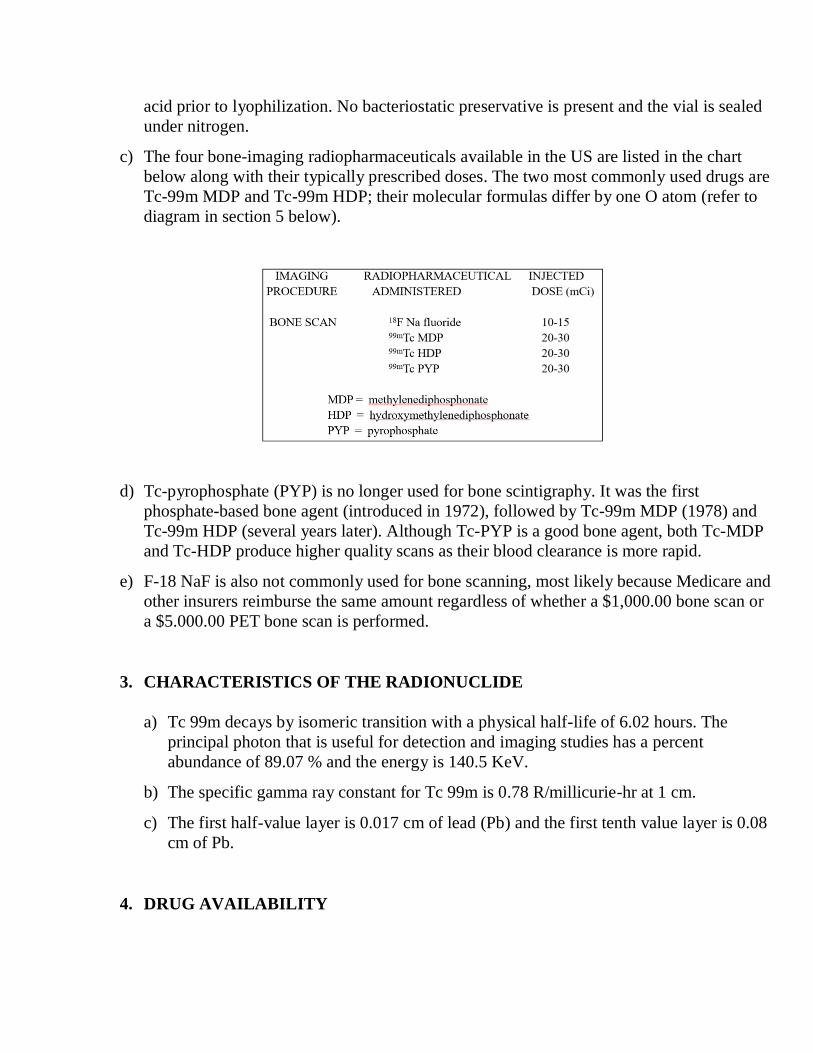
c) The four bone-imaging radiopharmaceuticals available in the US are listed in the chart
below along with their typically prescribed doses. The two most commonly used drugs are
Tc-99m MDP and Tc-99m HDP; their molecular formulas differ by one O atom (refer to
diagram in section 5 below).
d) Tc-pyrophosphate (PYP) is no longer used for bone scintigraphy. It was the first
phosphate-based bone agent (introduced in 1972), followed by Tc-99m MDP (1978) and
Tc-99m HDP (several years later). Although Tc-PYP is a good bone agent, both Tc-MDP
and Tc-HDP produce higher quality scans as their blood clearance is more rapid.
e) F-18 NaF is also not commonly used for bone scanning, most likely because Medicare and
other insurers reimburse the same amount regardless of whether a $1,000.00 bone scan or
a $5.000.00 PET bone scan is performed.
3. CHARACTERISTICS OF THE RADIONUCLIDE
a) Tc 99m decays by isomeric transition with a physical half-life of 6.02 hours. The
principal photon that is useful for detection and imaging studies has a percent
abundance of 89.07 % and the energy is 140.5 KeV.
b) The specific gamma ray constant for Tc 99m is 0.78 R/millicurie-hr at 1 cm.
c) The first half-value layer is 0.017 cm of lead (Pb) and the first tenth value layer is 0.08
cm of Pb.
4. DRUG AVAILABILITY
a) The three phosphate based drugs listed in the chart above are readily available in the
US in the form of unit doses calibrated for a particular patient as well as a “cold kit”
that must be reconstituted with Tc-99m pertechnetate prior to use.
b) F-18 NaF is available on a more limited basis from central pharmacies which make
this chemical form as a precursor to F-18 FDG preparation.
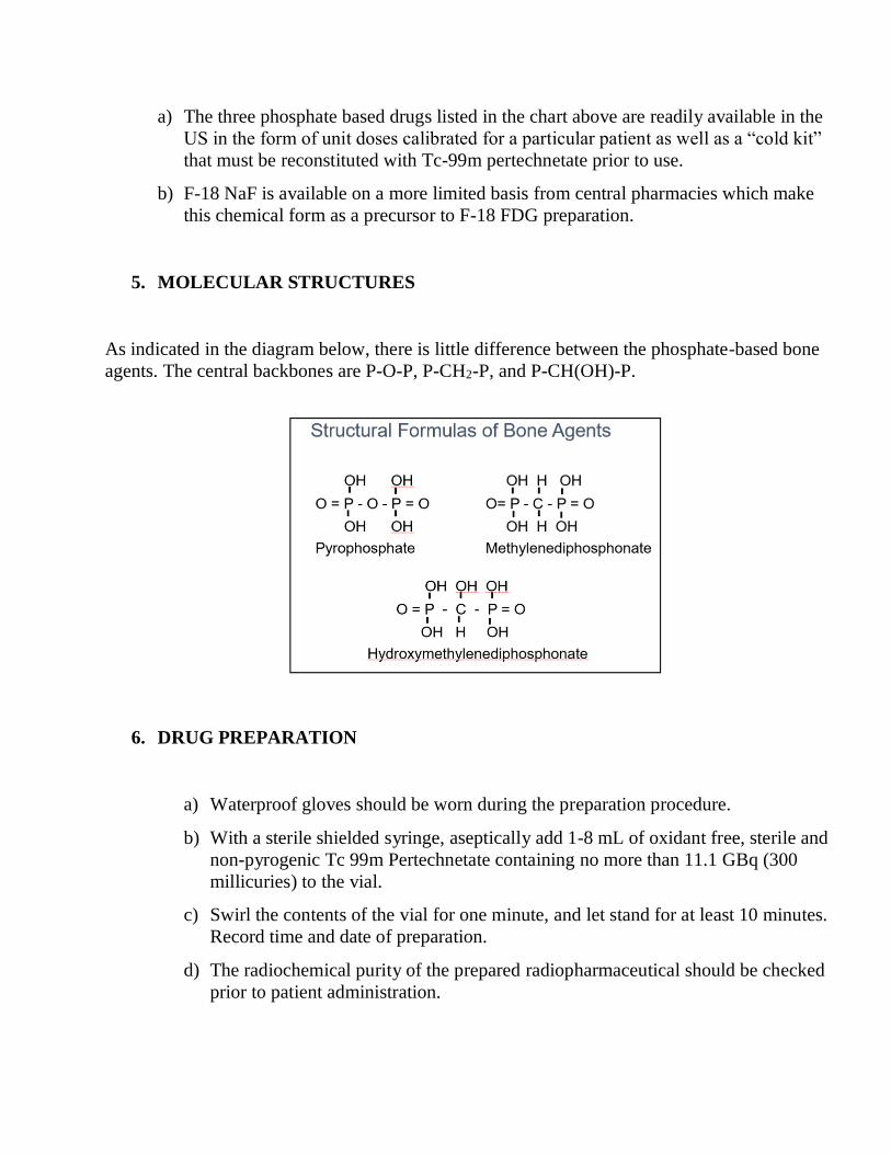
5. MOLECULAR STRUCTURES
As indicated in the diagram below, there is little difference between the phosphate-based bone
agents. The central backbones are P-O-P, P-CH2-P, and P-CH(OH)-P.
6. DRUG PREPARATION
a) Waterproof gloves should be worn during the preparation procedure.
b) With a sterile shielded syringe, aseptically add 1-8 mL of oxidant free, sterile and
non-pyrogenic Tc 99m Pertechnetate containing no more than 11.1 GBq (300
millicuries) to the vial.
c) Swirl the contents of the vial for one minute, and let stand for at least 10 minutes.
Record time and date of preparation.
d) The radiochemical purity of the prepared radiopharmaceutical should be checked
prior to patient administration.
e) Aseptically withdraw material with a sterile shielded syringe for use within six
hours of preparation.
f) The patient dose should be measured in a suitable dose calibration system
immediately prior to administration.
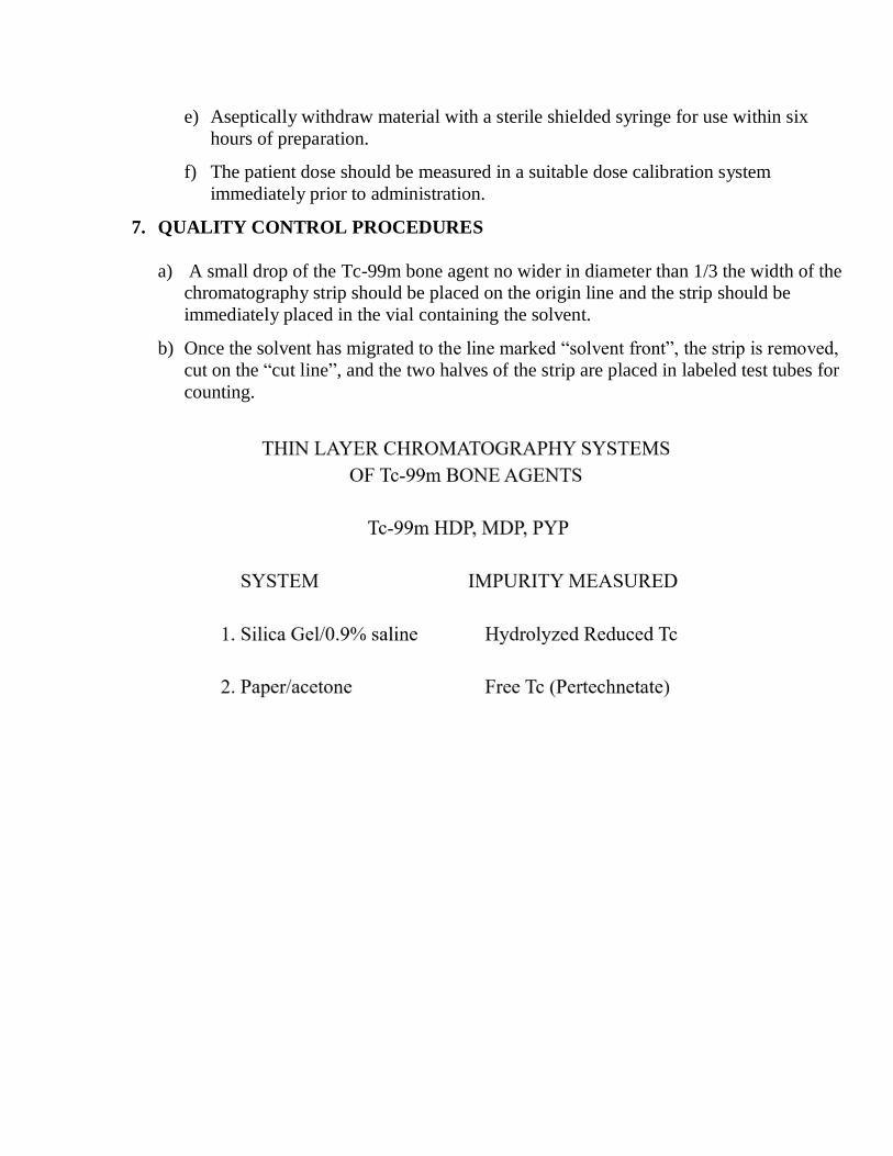
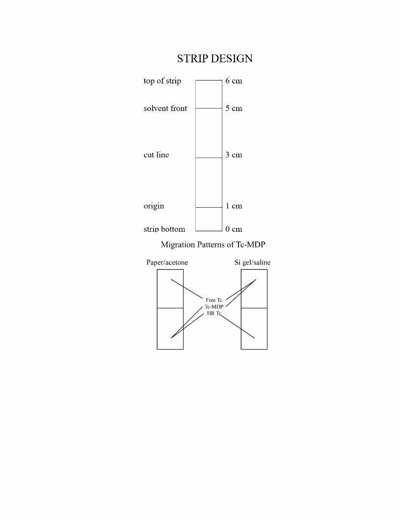
7. QUALITY CONTROL PROCEDURES
a) A small drop of the Tc-99m bone agent no wider in diameter than 1/3 the width of the
chromatography strip should be placed on the origin line and the strip should be
immediately placed in the vial containing the solvent.
b) Once the solvent has migrated to the line marked “solvent front”, the strip is removed,
cut on the “cut line”, and the two halves of the strip are placed in labeled test tubes for
counting.
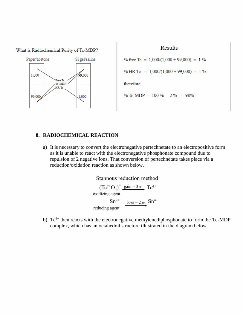
8. RADIOCHEMICAL REACTION
a) It is necessary to convert the electronegative pertechnetate to an electropositive form
as it is unable to react with the electronegative phosphonate compound due to
repulsion of 2 negative ions. That conversion of pertechnetate takes place via a
reduction/oxidation reaction as shown below.
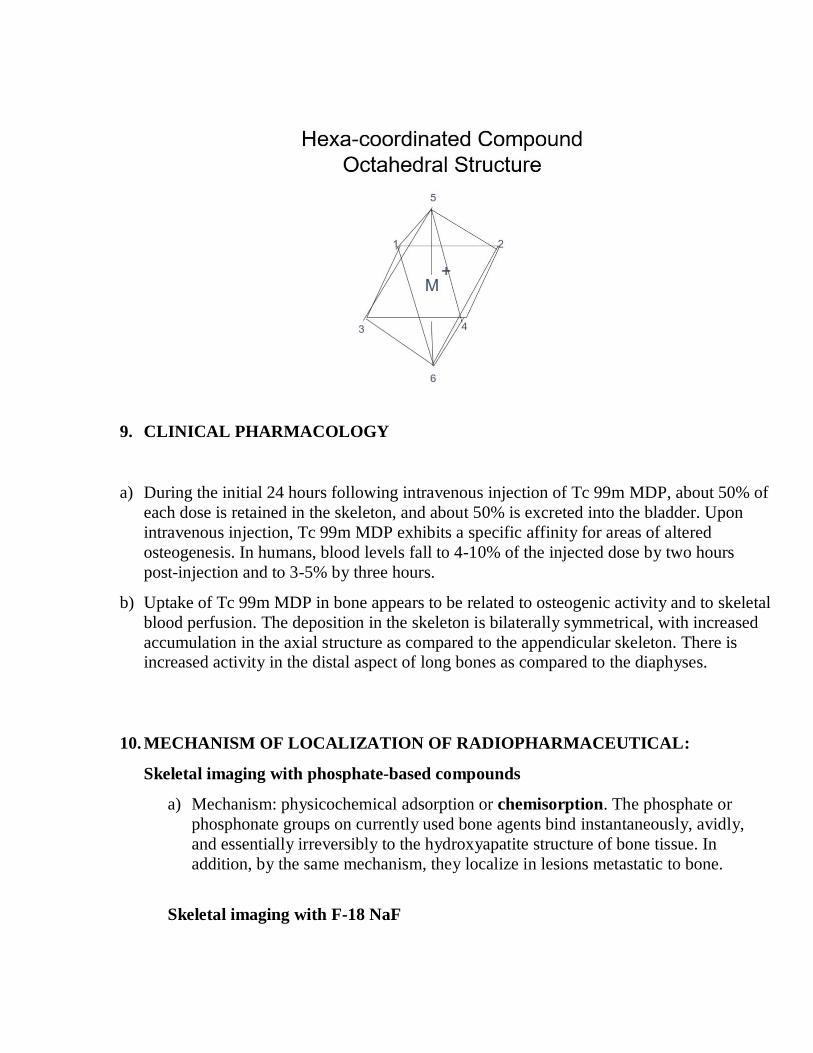
b) Tc4+ then reacts with the electronegative methylenediphosphonate to form the Tc-MDP
complex, which has an octahedral structure illustrated in the diagram below.
9. CLINICAL PHARMACOLOGY
a) During the initial 24 hours following intravenous injection of Tc 99m MDP, about 50% of
each dose is retained in the skeleton, and about 50% is excreted into the bladder. Upon
intravenous injection, Tc 99m MDP exhibits a specific affinity for areas of altered
osteogenesis. In humans, blood levels fall to 4-10% of the injected dose by two hours
post-injection and to 3-5% by three hours.
b) Uptake of Tc 99m MDP in bone appears to be related to osteogenic activity and to skeletal
blood perfusion. The deposition in the skeleton is bilaterally symmetrical, with increased
accumulation in the axial structure as compared to the appendicular skeleton. There is increased activity in the distal aspect of long bones as compared to the diaphyses.
10. MECHANISM OF LOCALIZATION OF RADIOPHARMACEUTICAL:
Skeletal imaging with phosphate-based compounds
a) Mechanism: physicochemical adsorption or chemisorption. The phosphate or
phosphonate groups on currently used bone agents bind instantaneously, avidly,
and essentially irreversibly to the hydroxyapatite structure of bone tissue. In
addition, by the same mechanism, they localize in lesions metastatic to bone.
Skeletal imaging with F-18 NaF
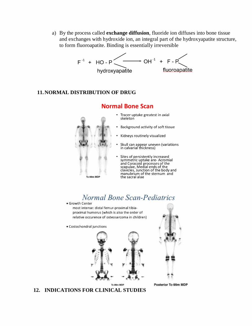
a) By the process called exchange diffusion, fluoride ion diffuses into bone tissue
and exchanges with hydroxide ion, an integral part of the hydroxyapatite structure,
to form fluoroapatite. Binding is essentially irreversible
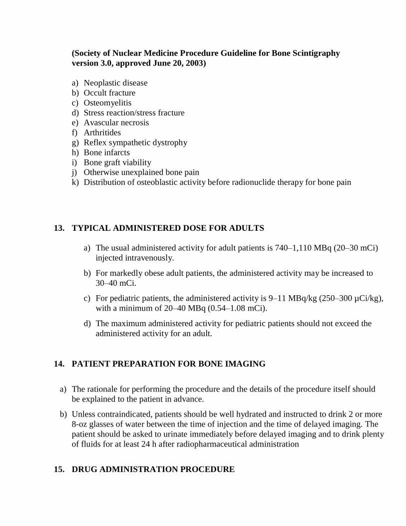
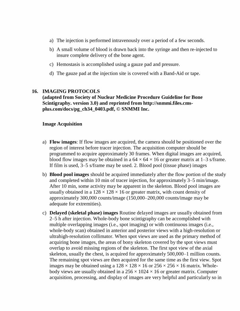
11. NORMAL DISTRIBUTION OF DRUG
12. INDICATIONS FOR CLINICAL STUDIES
(Society of Nuclear Medicine Procedure Guideline for Bone Scintigraphy
version 3.0, approved June 20, 2003)
a) Neoplastic disease
b) Occult fracture
c) Osteomyelitis
d) Stress reaction/stress fracture e) Avascular necrosis
f) Arthritides
g) Reflex sympathetic dystrophy
h) Bone infarcts
i) Bone graft viability j) Otherwise unexplained bone pain
k) Distribution of osteoblastic activity before radionuclide therapy for bone pain
13. TYPICAL ADMINISTERED DOSE FOR ADULTS
a) The usual administered activity for adult patients is 740–1,110 MBq (20–30 mCi)
injected intravenously.
b) For markedly obese adult patients, the administered activity may be increased to
30–40 mCi.
c) For pediatric patients, the administered activity is 9–11 MBq/kg (250–300 µCi/kg),
with a minimum of 20–40 MBq (0.54–1.08 mCi).
d) The maximum administered activity for pediatric patients should not exceed the
administered activity for an adult.
14. PATIENT PREPARATION FOR BONE IMAGING
a) The rationale for performing the procedure and the details of the procedure itself should
be explained to the patient in advance.
b) Unless contraindicated, patients should be well hydrated and instructed to drink 2 or more
8-oz glasses of water between the time of injection and the time of delayed imaging. The
patient should be asked to urinate immediately before delayed imaging and to drink plenty
of fluids for at least 24 h after radiopharmaceutical administration
15. DRUG ADMINISTRATION PROCEDURE
a) The injection is performed intravenously over a period of a few seconds.
b) A small volume of blood is drawn back into the syringe and then re-injected to
insure complete delivery of the bone agent.
c) Hemostasis is accomplished using a gauze pad and pressure.
d) The gauze pad at the injection site is covered with a Band-Aid or tape.
16. IMAGING PROTOCOLS
(adapted from Society of Nuclear Medicine Procedure Guideline for Bone
Scintigraphy. version 3.0) and reprinted from http://snmmi.files.cms-
plus.com/docs/pg_ch34_0403.pdf, © SNMMI Inc.
Image Acquisition
a) Flow images: If flow images are acquired, the camera should be positioned over the
region of interest before tracer injection. The acquisition computer should be
programmed to acquire approximately 30 frames. When digital images are acquired, blood flow images may be obtained in a 64 × 64 × 16 or greater matrix at 1–3 s/frame.
If film is used, 3–5 s/frame may be used. 2. Blood pool (tissue phase) images
b) Blood pool images should be acquired immediately after the flow portion of the study
and completed within 10 min of tracer injection, for approximately 3–5 min/image. After 10 min, some activity may be apparent in the skeleton. Blood pool images are
usually obtained in a 128 × 128 × 16 or greater matrix, with count density of
approximately 300,000 counts/image (150,000–200,000 counts/image may be
adequate for extremities).
c) Delayed (skeletal phase) images Routine delayed images are usually obtained from 2–5 h after injection. Whole-body bone scintigraphy can be accomplished with
multiple overlapping images (i.e., spot imaging) or with continuous images (i.e.,
whole-body scan) obtained in anterior and posterior views with a high-resolution or
ultrahigh-resolution collimator. When spot views are used as the primary method of
acquiring bone images, the areas of bony skeleton covered by the spot views must overlap to avoid missing regions of the skeleton. The first spot view of the axial
skeleton, usually the chest, is acquired for approximately 500,000–1 million counts.
The remaining spot views are then acquired for the same time as the first view. Spot
images may be obtained using a 128 × 128 × 16 or 256 × 256 × 16 matrix. Whole-
body views are usually obtained in a 256 × 1024 × 16 or greater matrix. Computer acquisition, processing, and display of images are very helpful and particularly so in
pediatric populations because of extreme ranges of normal uptake. Films of
scintigrams photographed with different intensities also may be helpful when digital processing and review are not available. When whole-body scanning is used, the count
rate (usually of the anterior chest) should be determined before image acquisition. The
scanning speed should be adjusted so that routine delayed (obtained 2–5 h after
injection) anterior or posterior whole-body images contain >1.5 million counts. If the
scanner electronically joins multiple passes, care must be taken to avoid having the “zipper” superimposed on the spine. When the probability of disseminated disease is
small, a limited study is reasonable. When disseminated disease is more likely, spot
views limited to the area of interest may be a source of error if distant disease is
present.
d) SPECT imaging In some patients, SPECT imaging is helpful to better characterize the presence, location, and extent of disease. SPECT imaging should be performed as
recommended by the camera manufacturer. Typical acquisition and processing
parameters are 360° circular orbit, 60–120 stops, 64 × 64 × 16 or greater matrix, and
10–40 s/stop. An equivalent total number of counts should be acquired if continuous
acquisition is used.
e) Other imaging Additional delayed (6–24-h) images will result in a higher target-to-
background ratio and may permit better evaluation of the pelvis if it was obscured by
bladder activity on the routine delayed images. Six- to twenty-four-h delayed imaging
may be particularly helpful in patients with renal insufficiency or urinary retention. A pinhole collimator may be used if very high-resolution images of a specific area are
necessary. Approximately 75,000–100,000 counts should be obtained for pinhole
collimator views. Zoom magnification or a converging collimator also may be used to
improve resolution, particularly when small structures or pediatric patients are being
imaged. The physician interpreting the image should be notified when collimators that introduce distortion (e.g., a pinhole collimator) are used. Other views (e.g., lateral,
oblique, or tangential) and special views (e.g., frog-leg views of the hips or sitting-on-
detector [caudal] views of the pelvis) may be obtained when necessary.
17. MEDICATIONS THAT AFFECT BONE SCAN QUALITY
The following drugs may interfere with the quality of scintigraphic images:
a) Aluminum: reduced skeletal tracer uptake, diffuse hepatic tracer uptake, increased renal
tracer uptake. Common source: antacids
b) Androgen deprivation therapy for prostate cancer (bicalutamide, estrogens): increased
mammary tracer uptake in case of gynecomastia
c) Bone-modifying agents (including bisphosphonates and denosumab) or agents interfering
with osteoblast function (e.g. cabozantinib): reduced skeletal tracer uptake
d) Corticosteroids: reduced skeletal tracer uptake, reduced tracer uptake at fracture sites
e) Hematopoietic growth factors: increased spinal tracer uptake, possible increased tracer
uptake in the appendicular skeleton
f) Iron: increased renal tracer uptake, increased tracer uptake at site of intramuscular
injection, diffuse hepatic tracer uptake
g) Methotrexate: diffuse hepatic tracer uptake
h) Nephrotoxic chemotherapy: increased renal tracer uptake; reduced skeletal tracer uptake
i) Nifedipine: reduced skeletal tracer uptake
18. ADVERSE REACTIONS FOLLOWING IV INJECTION OF Tc-LABELED
PHOSPHATE BASED BONE AGENTS
Several adverse reactions have been reported. These were usually hypersensitivity
reactions characterized by itching, various skin rashes, hypotension, chills, nausea and
vomiting. They are very rare.
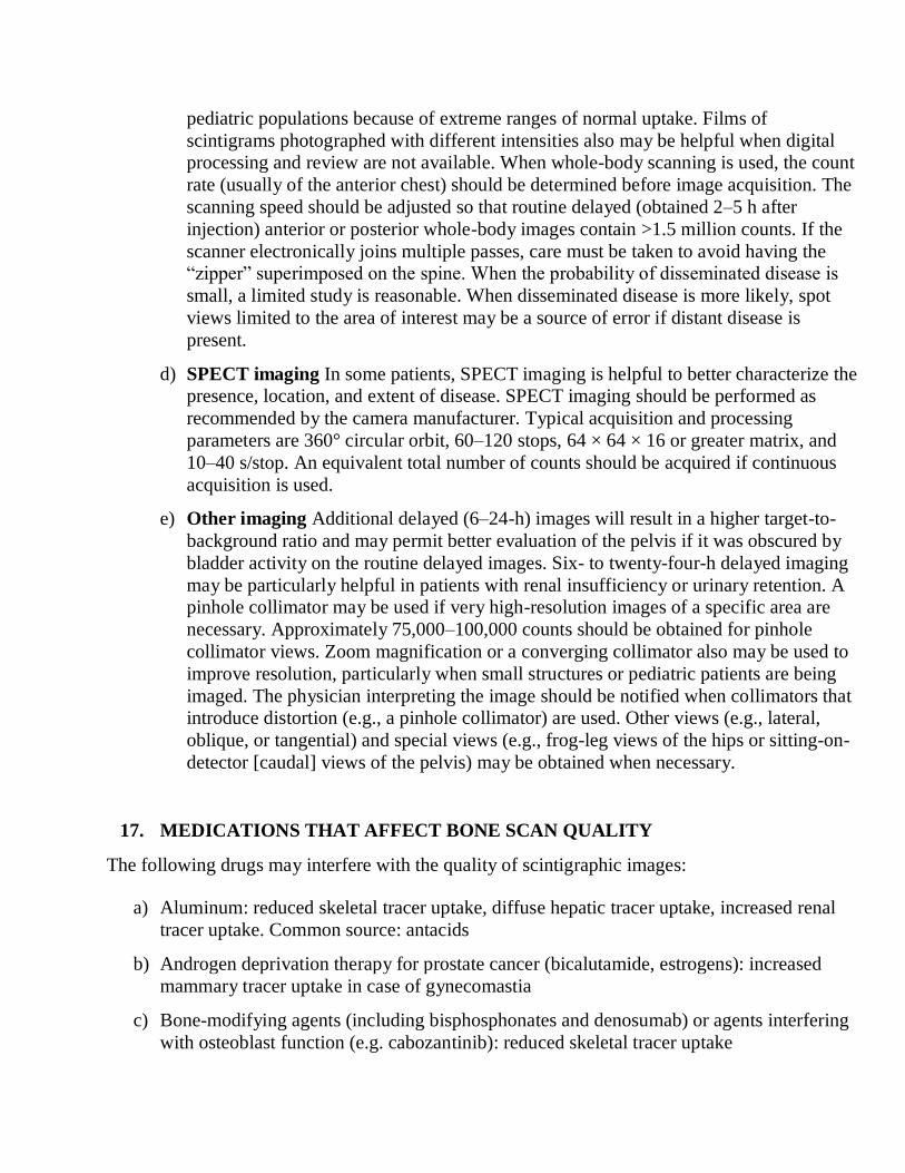
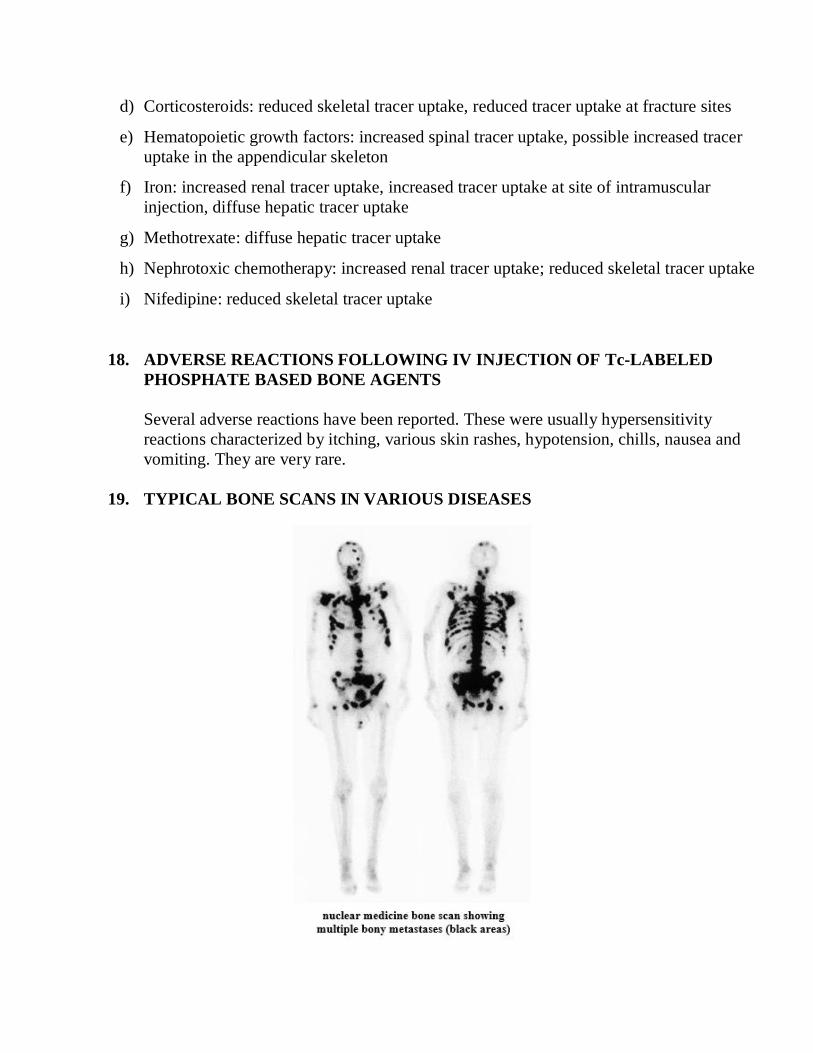
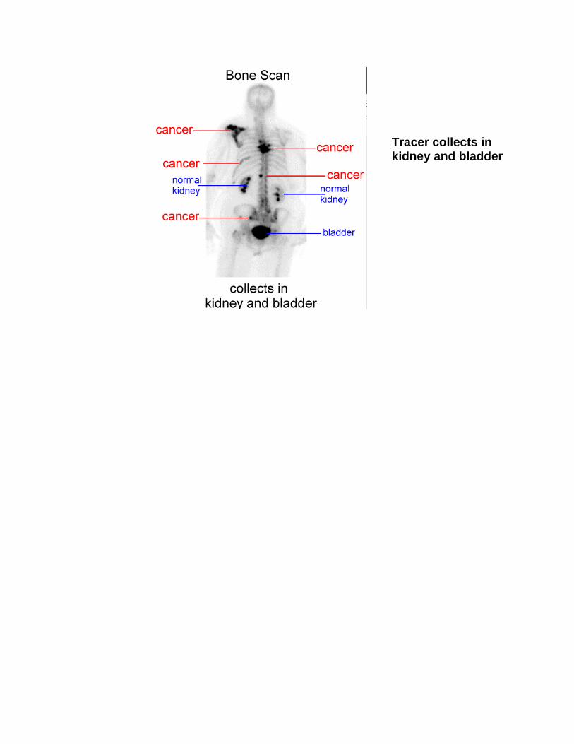
19. TYPICAL BONE SCANS IN VARIOUS DISEASES
Tracer collects in kidney and bladder
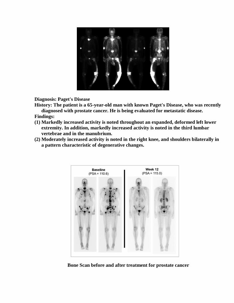
Diagnosis: Paget's Disease
History: The patient is a 65-year-old man with known Paget's Disease, who was recently
diagnosed with prostate cancer. He is being evaluated for metastatic disease.
Findings:
(1) Markedly increased activity is noted throughout an expanded, deformed left lower
extremity. In addition, markedly increased activity is noted in the third lumbar
vertebrae and in the manubrium.
(2) Moderately increased activity is noted in the right knee, and shoulders bilaterally in
a pattern characteristic of degenerative changes.
Bone Scan before and after treatment for prostate cancer
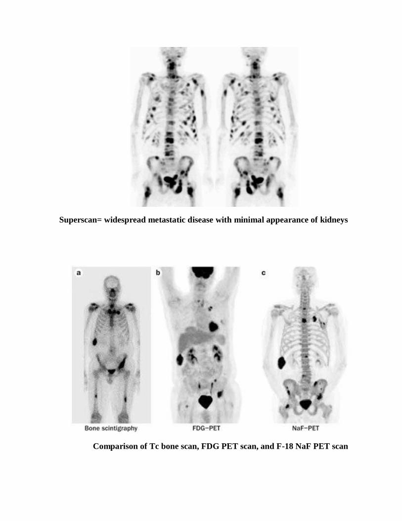
Superscan= widespread metastatic disease with minimal appearance of kidneys
Comparison of Tc bone scan, FDG PET scan, and F-18 NaF PET scan
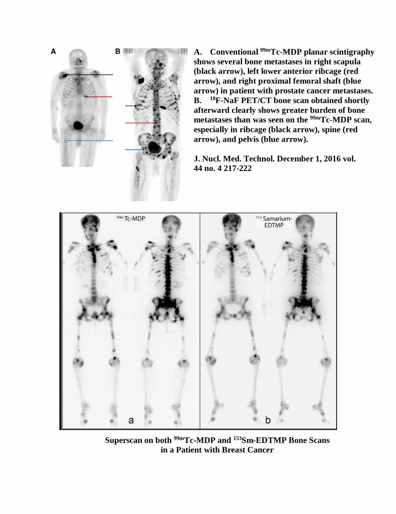
A. Conventional 99mTc-MDP planar scintigraphy
shows several bone metastases in right scapula
(black arrow), left lower anterior ribcage (red
arrow), and right proximal femoral shaft (blue
arrow) in patient with prostate cancer metastases.
B. 18F-NaF PET/CT bone scan obtained shortly
afterward clearly shows greater burden of bone
metastases than was seen on the 99mTc-MDP scan,
especially in ribcage (black arrow), spine (red
arrow), and pelvis (blue arrow).
J. Nucl. Med. Technol. December 1, 2016 vol.
44 no. 4 217-222
Superscan on both 99mTc-MDP and 153Sm-EDTMP Bone Scans
in a Patient with Breast Cancer
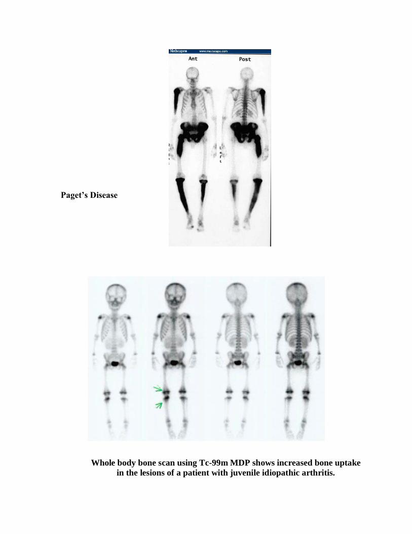
Paget’s Disease
Whole body bone scan using Tc-99m MDP shows increased bone uptake
in the lesions of a patient with juvenile idiopathic arthritis.
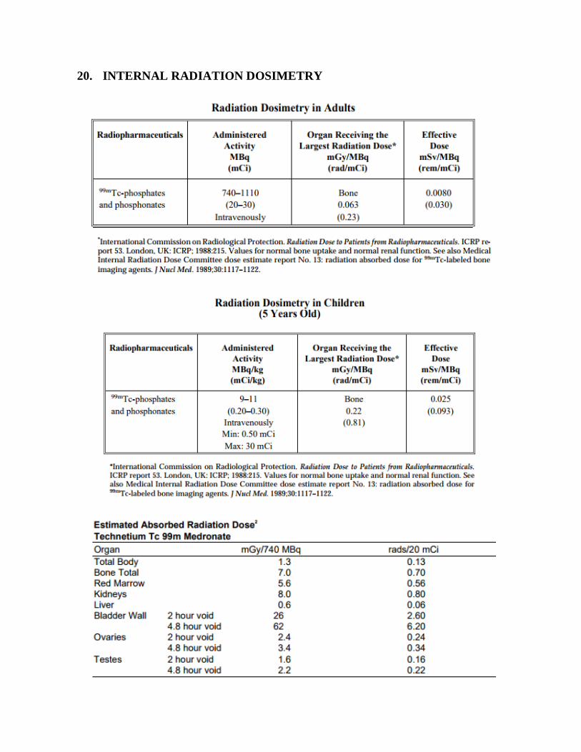
20. INTERNAL RADIATION DOSIMETRY


![Thyroid pathophysiology scintigraphy[1]](https://img.pdfslide.net/doc/110x75/588a7dc81a28abad628b4ebd/thyroid-pathophysiology-scintigraphy1.jpg)