Embed Size (px)
Citation preview
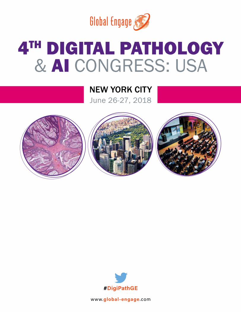
4TH DIGITAL PATHOLOGY & AI CONGRESS: USA
June 26-27, 2018NEW YORK CITY
www.global-engage .com
#DigiPathGE
WARM WELCOME
DIGITAL PATHOLOGY & AI CONGRESS: USA 2018
It is an exciting time for digital pathology in the US, following on the FDA’s 2017 approval of Philips’ IntelliSite Pathology Solution. Attracting experts working in all areas of digital pathology, the conference will examine the latest advancements in digital imaging technology and image analysis techniques like machine learning. There will also be an entire track focused on digital pathology adoption, practicalities and strategies for successful implementation across the workflow.
Highlights of the 2018 meeting include lectures from over 30 experts in the field, as well as several interactive sessions to facilitate collaboration between attendees. Highlights include 9 roundtables, 3 panel discussions and over 7 hours of networking time. To complement the scientific content, there will be a dynamic exhibition room filled with technology providers showcasing their technologies and solutions.
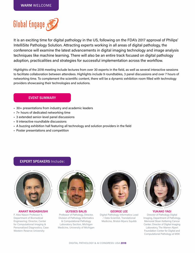
ULYSSES BALISProfessor of Pathology, Director, Division of Pathology Informatics
& Computational Pathology Laboratory Section, Michigan
Medicine, University of Michigan
ANANT MADABHUSHIF. Alex Nason Professor II, Department of Biomedical Engineering; Director, Center for Computational Imaging & Personalized Diagnostics, Case Western Reserve University
EXPERT SPEAKERS Include:
YUKAKO YAGIDirector of Pathology Digital
Imaging, Department of Pathology, Memorial Sloan Kettering Cancer Center; Director of Digital Imaging
Laboratory, The Warren Alpert Foundation Center for Digital and Computational Pathology at MSK
GEORGE LEEDigital Pathology Informatics Lead
/ Data Scientist, Translational Medicine, Bristol-Myers Squibb
• 30+ presentations from industry and academic leaders• 7+ hours of dedicated networking time• 3 extended senior-level panel discussions• 9 interactive roundtable discussions• A buzzing exhibition hall featuring all technology and solution providers in the field• Poster presentations and competition
EVENT SUMMARY
CONFERENCE SYNOPSIS
Roundtable 1: Clinical Utility of AI for Differential Diagnosis
Roundtable 2: LIMS/LIS & PACSRoundtable 3: Use of digital pathology
to support personalized oncologyRoundtable 4: Digital Pathology in Pharma
ROUNDTABLES SESSION 1
COMPUTATIONAL PATHOLOGY & AI
• Overcoming challenges in image analysis• New imaging modalities• User interfaces• Acquisition, processing, archiving & retrieval of WSI• Cloud computing, data storage and management• Integration with LIMS / LIS; PACS• Scoring & annotation tools• Fully-automated image analysis• Deep learning pattern recognition• Case studies & applications in pre-clinical research
and in the clinic• Panel: The potential for digital pathology & AI• Panel: How do we move digital pathology forward?
IMPLEMENTATION & PRACTICALITIES
• Regulation• Barriers to adoption• Making the business case for digital pathology –
developing an RFP• How readers interact with digital pathology systems• Challenge of form factors• Interoperability• Improving WSI workflow efficiency• Telepathology & international consultations• Panel: Biggest challenges in day-to-day use of
digital pathology
Roundtable 1: Integration of Digital Pathology Across the Workflow
Roundtable 2: StandardizationRoundtable 3: Novel Deep Learning Applications
Roundtable 4: Regulation of Digital Pathology
ROUNDTABLES SESSION 2
Roundtables are informal, small-group interactive discussions on key topics in the field. Discussion
leaders will introduce sub-topics/questions for discussion and roundtable attendees are
encouraged to participate actively in the session.
EVENT SPONSORS
DIGITAL PATHOLOGY & AI CONGRESS: USA 2018
Platinum Sponsors
Silver Sponsor
Other Sponsors & Exhibitors
Gold Sponsor
ANIL PARWANIProfessor of Pathology and Biomedical Informatics, Vice-Chair of Anatomic Pathology, Wexner Medical Center, The Ohio State University
THOMAS FUCHSAssociate Professor, Computational Pathology, Memorial Sloan Kettering Cancer Center; Weill Cornell Medicine
DAVID WESTCEO, Proscia
ULYSSES BALISProfessor of Pathology, Director, Division of Pathology Informatics & Computational Pathology Laboratory Section, Michigan Medicine, University of Michigan
DOUGLAS HARTMANAssociate Professor of Pathology & Associate Director, Pathology Informatics, University of Pittsburgh
DRAZEN JUKICConsultant Dermatopathologist, Georgia Dermatology and Skin Cancer Center
JOEL SALTZCherith Professor and Founding Chair, Department of Biomedical Informatics, Stony Brook University
GEORGE LEEDigital Pathology Informatics Lead / Data Scientist, Translational Medicine, Bristol-Myers Squibb
ANANT MADABHUSHIF. Alex Nason Professor II, Department of Biomedical Engineering; Director, Center for Computational Imaging & Personalized Diagnostics, Case Western Reserve University
YAIR RIVENSONPostdoctoral Scholar, Ozcan Nano/Bio Photonics Lab, University of California Los Angeles
JOHN TOMASZEWSKISUNY Distinguished Professor, Peter A. Nickerson Professor and Chair, Department of Pathology and Anatomical Science, University at Buffalo
GUSTAVO KUNDE ROHDEAssociate Professor, Department of Biomedical Engineering, Department of Electrical and Computer Engineering, University of Virginia
MEHRVASH HAGHIGHIAssistant Professor of Pathology & Cell Biology, Columbia University Medical Center
LAURA BARISONIProfessor of Pathology, Division Chief, Renal Pathology Service, Miller School of Medicine, University of Miami
RAJENDRA SINGHAssociate Professor, Pathology, Icahn School of Medicine at Mt. Sinai
ERIC WIRCHChief Technology Officer and Managing Director, Corista
STEVEN HARTAssistant Professor of Biomedical Informatics, Mayo Clinic
SIMON KIRBY (Chair)Assistant Professor, Departments of Laboratory Medicine and Surgery, Faculty of Medicine, Memorial University of Newfoundland, Canada
GERARDO FERNANDEZAssociate Professor, Department of Pathology, Medical Director, Center for Computational and Systems Pathology, Icahn School of Medicine at Mount Sinai & The Mount Sinai Hospital
MICHAEL SURACEScientist, MedImmune
BEATRICE KNUDSENProfessor, Biomedical Sciences and Pathology and Laboratory Medicine, Director, Translational Pathology, Cedars Sinai Medical Center
JASON HIPPLead Pathologist, Google Research
MARTIN STUMPETechnical Lead Pathology, Google Research
JENNIFER GILTNANEPathologist, Genentech
YUKAKO YAGIDirector of Pathology Digital Imaging, Department of Pathology, Memorial Sloan Kettering Cancer Center; Director of Digital Imaging Laboratory, The Warren Alpert Foundation Center for Digital and Computational Pathology at MSK
RICHARD LEVENSONProfessor and Vice Chair for Strategic Technologies, Department of Pathology and Laboratory Medicine, UC Davis Health, Sacramento, CA
ANTHONY MAGLIOCCOChair of Anatomical Pathology, Anatomical Pathology Department, Moffitt Cancer Center
PETER CAIESenior Research Fellow, University of St Andrews, UK
ANNE HELLEBUSTProduct Manager, Life Sciences, Indica Labs
STANLEY COHENEmeritus Chair of Pathology & Founding Director, Center for Biophysical Pathology, Rutgers-NJMS; Adjunct Professor of Pathology, University of Pennsylvania and Jefferson University
CONFIRMED SPEAKERS
DIGITAL PATHOLOGY & AI CONGRESS: USA 2018
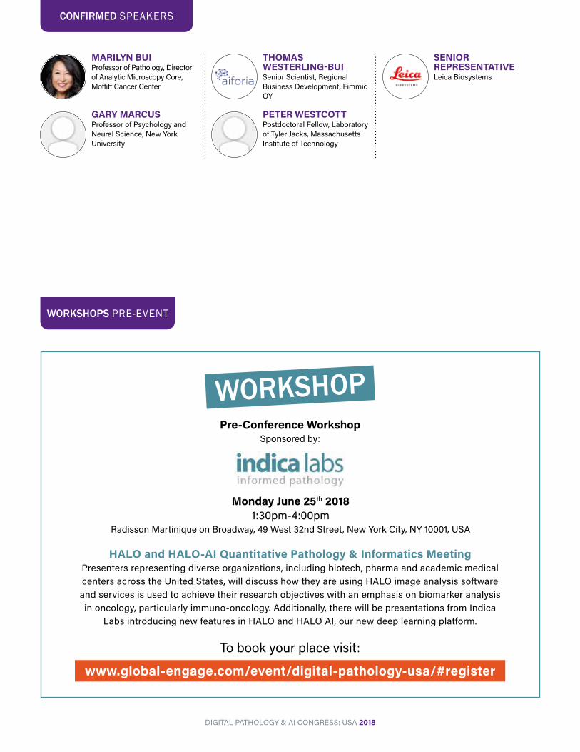
MARILYN BUIProfessor of Pathology, Director of Analytic Microscopy Core, Moffitt Cancer Center
GARY MARCUSProfessor of Psychology and Neural Science, New York University
THOMAS WESTERLING-BUISenior Scientist, Regional Business Development, Fimmic OY
PETER WESTCOTTPostdoctoral Fellow, Laboratory of Tyler Jacks, Massachusetts Institute of Technology
SENIOR REPRESENTATIVELeica Biosystems
CONFIRMED SPEAKERS
WORKSHOPPre-Conference Workshop
Sponsored by:
Monday June 25th 20181:30pm-4:00pm
Radisson Martinique on Broadway, 49 West 32nd Street, New York City, NY 10001, USA
HALO and HALO-AI Quantitative Pathology & Informatics MeetingPresenters representing diverse organizations, including biotech, pharma and academic medical centers across the United States, will discuss how they are using HALO image analysis software and services is used to achieve their research objectives with an emphasis on biomarker analysis in oncology, particularly immuno-oncology. Additionally, there will be presentations from Indica
Labs introducing new features in HALO and HALO AI, our new deep learning platform.
WORKSHOPS PRE-EVENT
DIGITAL PATHOLOGY & AI CONGRESS: USA 2018
To book your place visit:
www.global-engage.com/event/digital-pathology-usa/#register
CONGRESS SCHEDULE
Registration & Refreshments8:00-8:50
8:50-9:00 Global Engage Welcome Address and Morning Chair’s Opening Remarks: Stanley Cohen, Emeritus Chair of Pathology & Founding Director, Center for Biophysical Pathology, Rutgers-NJMS; Adjunct Professor of Pathology, University of Pennsylvania and Jefferson University
DAY 1 TUESDAY JUNE 26TH 2018
KEYNOTE ADDRESS:ANIL PARWANIProfessor of Pathology and Biomedical Informatics, Vice-Chair of Anatomic Pathology, Wexner Medical Center, The Ohio State UniversityBreaking the barriers: Whole Slide Imaging for Primary Diagnosis and Artificial Imaging Applications• Overview of whole slide imaging, applications and challenges• Recognize how artificial intelligence/deep learning is an enabling technology for clinical diagnostics• New imaging tools for cancer diagnostics
9:00-9:3511:50-12:15
JOEL SALTZCherith Professor and Founding Chair, Department of Biomedical Informatics, Stony Brook UniversityWhole Slide Imaging and High-End Computing Come Full CirclePresentation will describe evolution of whole
slide imaging Pathology technology from its 1990s HPC origins to the present. I will describe the HPC origin of the early infrastructure developed by my group at Johns Hopkins Pathology – the first whole slide imaging software system – and go on to describe National Cancer Institute supported infrastructure for visualization, management and analysis of information derived from whole slide Pathology images. Finally, I will describe the role of whole slide Pathology Informatics in Precision medicine giving examples of studies that make use of traditional machine learning and deep learning algorithms.
AI FOR IMAGE ANALYSIS IMPLEMENTATION & PRACTICALITIES
11:50-12:15
KEYNOTE ADDRESS:THOMAS FUCHSAssociate Professor, Computational Pathology, Memorial Sloan Kettering Cancer Center; Weill Cornell MedicineWhat Does It Take to Build a Petabyte Scale AI?• Artificial Intelligence • Computational Pathology • Deep LearningPathology is in the midst of a revolution from a qualitative to a quantitative discipline. This transformation is fundamentally
driven by machine learning in general and computer vision and deep learning in particular. At Memorial Sloan Kettering we are building a computational pathology decision support system based on hundreds of GPUs and one petabyte of image and clinical data. The goal is not only to automate cumbersome and repetitive tasks, but to impact diagnosis and treatment decisions in the clinic.
9:35-10:10
Morning Refreshments / Even Numbered Poster Presentations / One-to-One Meetings10:40-11:50
DIGITAL PATHOLOGY & AI CONGRESS: USA 2018
10:10-10:40
SPONSORED PRESENTATION:ANNE HELLEBUSTProduct Manager, Life Sciences, Indica LabsHALO AI – Collaborative Deep Learning Platform for PathologyIf you’ve attended any digital pathology or medical imaging meetings over the past couple of years, you have heard artificial intelligence or deep learning mentioned at least once. If you are not a computer scientist, algorithm engineer or image analyst,
you might be wondering how these tools differ from all the other image analysis and pattern recognition tools that have been used in digital pathology for years. In this presentation, we will de-mystify this emerging technology with a bit of background, explain its potential applications in pathology and discuss how the HALO-AITM platform is making this powerful technology accessible to the pathology community.
DRAZEN JUKICConsultant Dermatopathologist, Georgia Dermatology and Skin Cancer CenterTopic: Telepathology
Track Chair: Stanley Cohen, Emeritus Chair of Pathology & Founding Director, Center for Biophysical Pathology, Rutgers-NJMS; Adjunct Professor of Pathology, University of Pennsylvania and Jefferson University
Track Chair:
12:15-12:50
PANEL DISCUSSION:The Potential for Artificial Intelligence in Digital Pathology
THOMAS FUCHSAssociate Professor, Computational Pathology, Memorial Sloan Kettering Cancer Center; Weill Cornell Medicine
12:15-12:50
PANEL DISCUSSION:How do we move digital pathology forward?A discussion on strategies for advancing digital pathology for clinical use, covering topics such as regulation, practicalities of implementation, identifying hurdles.
STANLEY COHENEmeritus Chair of Pathology & Founding Director, Center for Biophysical Pathology, Rutgers-NJMS; Adjunct Professor of Pathology, University of Pennsylvania and Jefferson University
JOEL SALTZCherith Professor and Founding Chair, Department of Biomedical Informatics, Stony Brook University
GARY MARCUSProfessor of Psychology and Neural Science, New York University
CONGRESS SCHEDULE DAY 1 TUESDAY JUNE 26TH 2018
DIGITAL PATHOLOGY & AI CONGRESS: USA 2018
2:45-3:10
JASON HIPPLead Pathologist, Google Research
MARTIN STUMPETechnical Lead Pathology, Google ResearchAdvancing Cancer Diagnostics with Deep Learning
2:20-2:45
ULYSSES BALISProfessor of Pathology, Director, Division of Pathology Informatics & Computational Pathology Laboratory Section, Michigan Medicine, University of MichiganTopic: Fully-automated WSI differential
diagnosis generation as made possible by deep learning
2:20-3:10
DOUGLAS HARTMANAssociate Professor of Pathology & Associate Director, Pathology Informatics, University of PittsburghDetermining how to integrate digital pathology into your lab
• Describe various deployment methods• Outline the infrastructure needed for a deployment• Describe the return on investment
3:10-3:35
GERARDO FERNANDEZAssociate Professor, Department of Pathology, Medical Director, Center for Computational and Systems Pathology, Icahn School of Medicine at Mount Sinai & The Mount Sinai HospitalArtificial intelligence methods for grading human cancer
• Large accurate ground truth training is critical in pathology AI applications
• Review spectrum of applications of automated grading algorithms• Normalizing for tissue variability in automated pathology analysis
3:10-3:35
STEVEN HARTAssistant Professor of Biomedical Informatics, Mayo ClinicDigital Pathology at The Intersection of Big Data, AI, and Genomics• Similarities to the recent genomics revolution
• Leveraging lessons learned from genomics• Practical considerations for embracing the digital divide
12:50-1:20
For sponsorship opportunities contact Gavin Hambrook / Nick Best at Email: [email protected]
Tel: +44 (0) 1865 849841
SPONSORED PRESENTATION
12:50-1:20
SPONSORED PRESENTATION:DAVID WESTCEO, ProsciaTitle TBC
Lunch / One-to-One Meetings1:20-2:20
12:15-12:50
12:15-12:50
Continued
3:35-4:00
PETER CAIESenior Research Fellow, University of St Andrews, UKTopic: Automated tumor microenvironment profiling using machine learning
3:35-4:00
MARILYN BUIProfessor of Pathology, Director of Analytic Microscopy Core, Moffitt Cancer CenterMaximizing Patient Care Delivery with Digital Pathology: Resource Guide for Practicing Pathologists• Overview of the resources in digital pathology
• Update of practicing guidelines in whole slide imaging, telepathology and quantitative image analysis
• Organized for busy practicing pathologists
ROUNDTABLE DISCUSSIONS:Table 1: Clinical Utility of AI for Differential DiagnosisULYSSES BALISProfessor of Pathology, Director, Division of Pathology Informatics & Computational Pathology Laboratory Section, Michigan Medicine, University of Michigan
Table 2: LIMS/LIS & PACSMEHRVASH HAGHIGHIAssistant Professor of Pathology & Cell Biology, Columbia University Medical Center• What are different methods of LIS/WSI
implementation? Pros and Cons of each method? • Discuss the feasibility of using PACS in integral model • Why PACS adoption is important?
– Workflow support, even for heavier volume – Integration in healthcare IT environment – Anywhere access because of central storage – Offering stability and performance – Better patient care
• What are lessons learned from Radiology and their journey to full digitalization? – Choose a vendor-neutral solution – Ensure system robustness – Ensure there are clinical references that testify to good support
and vendor relationship – Ensure the internal engagement – Focus on the workflow
• How do we properly prepare for integrating WSI in pathology lab workflow?– LIS operational pre-requisites – Implementing additional quality control measure – Optimizing the pathology workstations: Challenges and solutions – Comprehensive training
Table 3: Use of digital pathology to support personalized oncology ANTHONY MAGLIOCCOChair of Anatomical Pathology, Anatomical Pathology Department, Moffitt Cancer Center
Table 4: Regulation of Digital Pathology
Chair's Closing Remarks / End of Day 16:10
Networking Drinks Reception6:10-7:10
5:45-6:10
YAIR RIVENSONPostdoctoral Scholar, Ozcan Nano/Bio Photonics Lab, University of California Los AngelesDeep Learning Microscopy for Enhanced Digital Pathology• We demonstrate that deep learning can be
used to provide a significant enhancement in imaging throughput of pathology slides, by extending the depth-of-field and resolution of images acquired using a standard brightfield imaging optical microscope, without any hardware modifications.
• The enhanced result image is inferred using a single input image. This proposed deep learning approach is computationally efficient and outputs an image, corresponding to a 40x sample field-of-view in <1 second using a laptop.
• We demonstrate the universal nature of the approach, using a model that was created with a deep neural network which was trained on a specific tissue and stain type, to enhance microscope images that were taken with different tissues, stains and magnifications.
CONGRESS SCHEDULE DAY 1 TUESDAY JUNE 26TH 2018
5:20-6:10
5:20-5:45
RICHARD LEVENSONProfessor and Vice Chair for Strategic Technologies, Department of Pathology and Laboratory Medicine, UC Davis Health, Sacramento, CASlide-free, real-time histology: cooler than frozens
A number of technologies for rapid, ex-vivo, slide-free microscopy are in research-and-development phase and a few have already entered the market as research-use-only instruments. These contenders to replace FFPE-based histology appear promising for such uses as real-time surgical guidance, margin-assessment, rapid on-site evaluation (ROSE), and possibly the generation of final diagnostic reports. This talk will describe a few of these methods, which including 2-photon and confocal microscopy, light-sheet microscopy, optical coherence tomography, and structured illumination, and will focus on microscopy with UV surface excitation (MUSE), a novel approach that demonstrates a favorable combination of speed, simplicity, robustness, and technical quality.
4:50-5:20
For sponsorship opportunities contact Gavin Hambrook / Nick Best at Email: [email protected]
Tel: +44 (0) 1865 849841
SPONSORED PRESENTATION
ROUNDTABLE DISCUSSIONS ADVANCEMENTS IN IMAGING
4:50-5:20
SPONSORED PRESENTATION:THOMAS WESTERLING-BUISenior Scientist, Regional Business Development, Fimmic OY
PETER WESTCOTTPostdoctoral Fellow, Laboratory of Tyler Jacks, Massachusetts Institute of TechnologyTitle TBC
DIGITAL PATHOLOGY & AI CONGRESS: USA 2018
Afternoon Refreshments / Odd Numbered Poster Presentations / One-to-One Meetings4:00-4:50
CONGRESS SCHEDULE DAY 2 WEDNESDAY JUNE 27TH 2018
DIGITAL PATHOLOGY & AI CONGRESS: USA 2018
Refreshments8:20-9:00
8:50-9:00 Global Engage Welcome Address and Morning Chair’s Opening Remarks
KEYNOTE ADDRESS:ANANT MADABHUSHIF. Alex Nason Professor II, Department of Biomedical Engineering; Director, Center for Computational Imaging & Personalized Diagnostics, Case Western Reserve UniversityThe case for hand-crafted features: Not another deep learning in pathology talk• Handcrafted features represent feature engineering approaches which provide interpretability.
• Handcrafted features may offer advantages over deep learning approaches by being more transparent• Handcrafted features can enable computational pathology based companion diagnostics for precision medicine.
9:00-9:3512:25-12:50
JOHN TOMASZEWSKISUNY Distinguished Professor, Peter A. Nickerson Professor and Chair, Department of Pathology and Anatomical Science, University at BuffaloTopic: Traditional AI approaches to computational histopathology
COMPUTATIONAL PATHOLOGY & AI ROUNDTABLE DISCUSSIONS
Morning Refreshments / Odd Numbered Poster Presentations / One-to-One Meetings10:45-11:55
12:25-1:15
ROUNDTABLE DISCUSSIONS:Table 1: Expanding your diagnostic capabilities with digital pathologyDOUGLAS HARTMANAssociate Professor of Pathology & Associate Director, Pathology Informatics, University of Pittsburgh
• Telepathology opportunities • Image analysis opportunities• Better quantification/documentation for diagnostic purposes
Table 2: StandardizationYUKAKO YAGIDirector of Pathology Digital Imaging, Department of Pathology, Memorial Sloan Kettering Cancer Center; Director of Digital Imaging Laboratory, The Warren Alpert
Foundation Center for Digital and Computational Pathology at MSK
12:50-1:15
GUSTAVO KUNDE ROHDEAssociate Professor, Department of Biomedical Engineering, Department of Electrical and Computer Engineering, University of VirginiaInterpretable discriminative modeling of cellular phenotypes for digital pathology
Image-based phenotypic assessment in digital pathology typically rely on parametric numerical features to describe the state of each cellular measurement. In recent years, numerous modeling
10:15-10:45
SPONSORED PRESENTATION:ERIC WIRCHChief Technology Officer and Managing Director, CoristaMigrating algorithms beyond the development sandboxMachine-learning algorithmic development initially occurs in tight conjunction with a single research partner and heavily reflects the protocols of that partner's laboratory. Migrating an algorithm from being effective at a single site to a cross site
environment, with differences in human factors, processing, stains, imaging and calibration, necessitates the application of additional AI & Computer Vision steps and techniques. We will discuss experiences encountered while performing this migration, including initial results and the techniques employed or being tested for overcoming these hurdles.
PANEL DISCUSSION:
YUKAKO YAGIDirector of Pathology Digital Imaging, Department of Pathology, Memorial Sloan Kettering Cancer Center; Director of Digital Imaging Laboratory, The Warren Alpert Foundation Center for Digital and Computational Pathology at MSK
ANIL PARWANIProfessor of Pathology and Biomedical Informatics, Vice-Chair of Anatomic Pathology, Wexner Medical Center, The Ohio State University
BEATRICE KNUDSENProfessor, Biomedical Sciences and Pathology and Laboratory Medicine, Director, Translational Pathology, Cedars Sinai Medical Center
DOUGLAS HARTMANAssociate Professor of Pathology & Associate Director, Pathology Informatics, University of Pittsburgh
Questions to be submitted to [email protected]
9:35-10:15
Challenges & strategies for day-to-day use of digital pathology
11:55-12:25
SPONSORED PRESENTATION:SENIOR REPRESENTATIVELeica BiosystemsTitle TBC
TRACK CHAIR: SIMON KIRBYAssistant Professor, Departments of Laboratory Medicine and Surgery, Faculty of Medicine, Memorial University of Newfoundland, Canada
CONGRESS SCHEDULE DAY 2 WEDNESDAY JUNE 27TH 2018
DIGITAL PATHOLOGY & AI CONGRESS: USA 2018
CASE STUDIES
GEORGE LEEDigital Pathology Informatics Lead / Data Scientist, Translational Medicine, Bristol-Myers SquibbApplications of Artificial Intelligence for Immuno-oncology and Precision MedicineConvolutional neural networks have revolutionized image analysis and digital pathology to uncover insights in medicine which could not previously be performed reliably at scale. At Bristol-Myers Squibb, we are using artificial intelligence to quantify the tumor micro-environment in histopathology images and to inform the best treatment options for patients considering immunotherapies.
2:15-2:40
LAURA BARISONIProfessor of Pathology, Division Chief, Renal Pathology Service, Miller School of Medicine, University of MiamiDigital pathology in nephrology clinical trials, translational research and clinical practiceDuring this presentation, we will discuss a) the application of the recent advancements in digital technology and computational engineering to nephropathology, in the setting of clinical research, trials, and practice; b) the overall benefits and challenges of the new digital ecosystem; c) digital nephropathology in kidney precision and predictive medicine.
2:40-3:05
JENNIFER GILTNANEPathologist, GenentechMore than pretty pictures – Development of quantitative multiplexed tissue assays for clinical trial biomarker supportDigital pathology coupled with multiplex immunofluorescent tissue assays provide spatial distribution information: a key advantage over gene expression approaches to capture tumor biology. . In order for high-order multiplexing technologies to advance out of academic laboratories and into prospective clinical trials, we must offer more utility than other
quantitative assays. It will also be important to overcome current technical challenges and establish a standardized approach to assay development and digital pathology algorithm validation. The current impact, challenges, and future potential benefits to cancer patients of optimizing this strategy will be optimized in this discussion.
3:30-3:553:55-4:20
BEATRICE KNUDSENProfessor, Biomedical Sciences and Pathology and Laboratory Medicine, Director, Translational Pathology, Cedars Sinai Medical CenterNuclear morphometry and cancer biomarker development• Novel tools to analyze nuclei in H&E slides• Multiplex tissue staining for high throughput detailed slide annotation• Data integration and prediction modeling for biomarker discovery
Conference Close4:20
Lunch / One-to-One Meetings1:15-2:15
MICHAEL SURACEScientist, MedImmuneValidation of multiplex immunofluorescence panels using multispectral microscopy for immune-profiling of formalin-fixed and paraffin-embedded human tumor tissues
3:05-3:30
12:25-1:15
There are many different levels and areas of focus with regards to standardization of Digital Pathology. This discussion will cover all aspects of standardization for successful operation of WSI systems in clinical and research areas.• Instrument/Industrial Standard: for interoperability • WSI system standardization to have optimal, consistent quality
and reliable images• Establishing Operational Standard
Table 3: Novel Deep Learning ApplicationsRAJENDRA SINGHAssociate Professor, Pathology, Icahn School of Medicine at Mt. Sinai
Table 4: Digital Pathology in PharmaMICHAEL SURACEScientist, MedImmune
12:50-1:15
frameworks for image-based cell phenotypes. We will describe how non-parametric methods based on the continuity equation can be used to build non-parametric end to end systems that utilize all pixel information directly in statistical modeling. We will show that the modeling framework can not only obtain predictive models of higher accuracies, but due to the invertibility of the modeling framework, allow for biologically interpretable results. Applications in cancer diagnosis using histology and cytology will be shown.
POSTER PRESENTATIONS
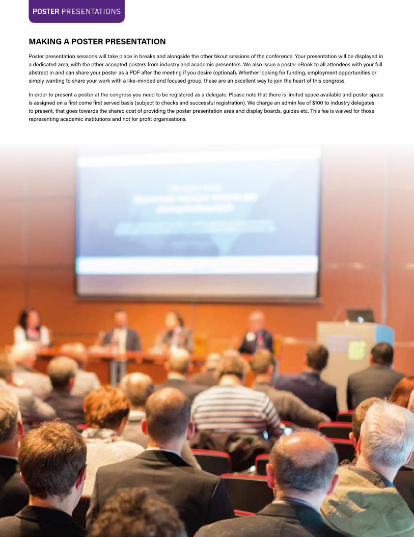
MAKING A POSTER PRESENTATION
Poster presentation sessions will take place in breaks and alongside the other bkout sessions of the conference. Your presentation will be displayed in a dedicated area, with the other accepted posters from industry and academic presenters. We also issue a poster eBook to all attendees with your full abstract in and can share your poster as a PDF after the meeting if you desire (optional). Whether looking for funding, employment opportunities or simply wanting to share your work with a like-minded and focused group, these are an excellent way to join the heart of this congress.
In order to present a poster at the congress you need to be registered as a delegate. Please note that there is limited space available and poster space is assigned on a first come first served basis (subject to checks and successful registration). We charge an admin fee of $100 to industry delegates to present, that goes towards the shared cost of providing the poster presentation area and display boards, guides etc. This fee is waived for those representing academic institutions and not for profit organisations.
DIGITAL PATHOLOGY & AI CONGRESS: USA 2018
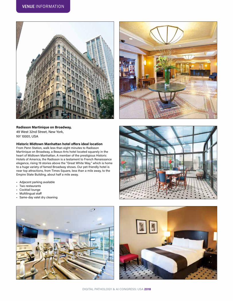
Radisson Martinique on Broadway,49 West 32nd Street, New York,NY 10001, USA
Historic Midtown Manhattan hotel offers ideal locationFrom Penn Station, walk less than eight minutes to Radisson Martinique on Broadway, a Beaux-Arts hotel located squarely in the heart of Midtown Manhattan. A member of the prestigious Historic Hotels of America, the Radisson is a testament to French Renaissance elegance, rising 19 stories above the "Great White Way," which is home to a huge variety of famed Broadway shows. Our pet-friendly hotel is near top attractions, from Times Square, less than a mile away, to the Empire State Building, about half a mile away.
• Adjacent parking available• Two restaurants• Cocktail lounge• Multilingual staff• Same-day valet dry cleaning
VENUE INFORMATION