Embed Size (px)
Citation preview
A Comprehensive Assessment of Lymphatic Filariasis inSri Lanka Six Years after Cessation of Mass DrugAdministrationRamakrishna U. Rao1, Kumara C. Nagodavithana2, Sandhya D. Samarasekera2,
Asha D. Wijegunawardana2, Welmillage D. Y. Premakumara2, Samudrika N. Perera2, Sunil Settinayake2,
J. Phillip Miller3, Gary J. Weil1*
1 Department of Internal Medicine, Infectious Diseases Division, Washington University School of Medicine, St. Louis, Missouri, United States of America, 2 Anti Filariasis
Campaign, Sri Lanka Ministry of Health, Colombo, Sri Lanka, 3 Division of Biostatistics, Washington University School of Medicine, St. Louis, Missouri, United States of
America
Abstract
Background: The Sri Lankan Anti-Filariasis Campaign conducted 5 rounds of mass drug administration (MDA) withdiethycarbamazine plus albendazole between 2002 and 2006. We now report results of a comprehensive surveillanceprogram that assessed the lymphatic filariasis (LF) situation in Sri Lanka 6 years after cessation of MDA.
Methodology and Principal Findings: Transmission assessment surveys (TAS) were performed per WHO guidelines inprimary school children in 11 evaluation units (EUs) in all 8 formerly endemic districts. All EUs easily satisfied WHO criteria forstopping MDA. Comprehensive surveillance was performed in 19 Public Health Inspector (PHI) areas (subdistrict healthadministrative units). The surveillance package included cross-sectional community surveys for microfilaremia (Mf) andcirculating filarial antigenemia (CFA), school surveys for CFA and anti-filarial antibodies, and collection of Culex mosquitoeswith gravid traps for detection of filarial DNA (molecular xenomonitoring, MX). Provisional target rates for interruption of LFtransmission were community CFA ,2%, antibody in school children ,2%, and filarial DNA in mosquitoes ,0.25%.Community Mf and CFA prevalence rates ranged from 0–0.9% and 0–3.4%, respectively. Infection rates were significantlyhigher in males and lower in people who denied prior treatment. Antibody rates in school children exceeded 2% in 10 studysites; the area that had the highest community and school CFA rates also had the highest school antibody rate (6.9%).Filarial DNA rates in mosquitoes exceeded 0.25% in 10 PHI areas.
Conclusions: Comprehensive surveillance is feasible for some national filariasis elimination programs. Low-level persistenceof LF was present in all study sites; several sites failed to meet provisional endpoint criteria for LF elimination, and follow-uptesting will be needed in these areas. TAS was not sensitive for detecting low-level persistence of filariasis in Sri Lanka. Werecommend use of antibody and MX testing as tools to complement TAS for post-MDA surveillance.
Citation: Rao RU, Nagodavithana KC, Samarasekera SD, Wijegunawardana AD, Premakumara WDY, et al. (2014) A Comprehensive Assessment of LymphaticFilariasis in Sri Lanka Six Years after Cessation of Mass Drug Administration. PLoS Negl Trop Dis 8(11): e3281. doi:10.1371/journal.pntd.0003281
Editor: Achim Hoerauf, Institute of Medical Microbiology, Immunology and Parasitology, Germany
Received April 17, 2014; Accepted September 19, 2014; Published November 13, 2014
Copyright: � 2014 Rao et al. This is an open-access article distributed under the terms of the Creative Commons Attribution License, which permits unrestricteduse, distribution, and reproduction in any medium, provided the original author and source are credited.
Data Availability: The authors confirm that, for approved reasons, some access restrictions apply to the data underlying the findings. All relevant data arewithin the paper and its Supporting Information files except for the following: Deidentified individual records from the community surveys. This information isavailable from Becker Library at Washington University School of Medicine (URL http://digitalcommons.wustl.edu/open_access_pubs/3378/).
Funding: This work was supported in part by grants from the U.S. National Institutes of Health (AI065715), www.nih.gov; the Center for Neglected TropicalDiseases, Liverpool School of Tropical Medicine, Liverpool, UK, www.cntd.org; the Barnes-Jewish Hospital Foundation (6794-33), BJHFoundation.org; and the Bill &Melinda Gates Foundation (GH5342), gatesfoundation.org. GJW was the PI for these grants. The funders had no role in study design, data collection and analysis,decision to publish, or preparation of the manuscript.
Competing Interests: The filarial antigen test used in this study uses reagents licensed from Barnes-Jewish Hospital, an affiliation of GJW. All royalties from salesof these tests go to the Barnes Jewish Hospital Foundation, a not for profit charitable organization (http://www.barnesjewish.org/giving/about-us). This does notalter our adherence to all PLOS policies on sharing data and materials.
* Email: [email protected]
Introduction
Lymphatic filariasis (LF, caused by the mosquito borne filarial
nematodes Wuchereria bancrofti, Brugia malayi, and B. timori), is a
major public-health problem in many tropical and subtropical
countries. The latest summary from the World Health Organization
(WHO) reported that 56 of 73 endemic countries have implemented
mass drug administration (MDA) with a combination of two drugs
(albendazole with either ivermectin or diethycarbamazine), and 33countries have completed 5 or more rounds of MDA in someimplementation units [1]. With more than 4.4 billion doses oftreatment distributed between 2000 and 2012, the GlobalProgramme to Eliminate Lymphatic Filariasis (GPELF) is easilythe largest public health intervention to date based on MDA.
Bancroftian filariasis was highly endemic in Sri Lanka in the
past [2–4]. The Sri Lankan Ministry of Health’ Anti Filariasis
PLOS Neglected Tropical Diseases | www.plosntds.org 1 November 2014 | Volume 8 | Issue 11 | e3281
Campaign (AFC) used a variety of methods to reduce filarial
infection rates to low levels by 1999 [5,6]. Sri Lanka was one of the
first countries to initiate a LF elimination program based on
GPELF guidelines [7]. The AFC provided annual MDA with
diethylcarbamazine alone for three years starting in 1999. This
was followed by five annual rounds of MDA with albendazole plus
diethylcarbamazine in all 8 endemic districts (implementation
units, IU) between 2002 and 2006. Various types of surveillance
have been conducted by AFC and other groups since the MDA
program ended in 2006 [8–12]. Post-MDA surveillance results
(based on detection of microfilariae or Mf in human blood by
microscopy) have consistently shown Mf rates much lower than
the target value of 1% in all endemic areas [13]. The AFC also
conducted school-based surveys for filarial antigenemia in 2008
according to WHO guidelines active at that time. Approximately
600 children were tested for circulating filarial antigenemia (CFA)
in 30 schools in each of the 8 endemic districts, and no positive
tests were observed (unpublished data, Sri Lanka Ministry of
Health). WHO guidelines emphasize that LF elimination pro-
grams should provide care for people with acute and chronic
clinical manifestations of filariasis [7], and the AFC has an
excellent network of clinics that is devoted to this activity [13].
The present study represents a significant expansion of earlier
post-MDA surveillance activities in Sri Lanka. Transmission
assessment surveys (TAS) were performed according to current
WHO guidelines [14,15] for sampling primary school children to
detect filarial antigenemia in each district. While TAS results may
be useful for deciding whether MDA can be stopped, TAS cannot
guarantee that LF transmission has been interrupted in evaluation
units (EUs), which are typically districts with populations that may
exceed 1 million. Therefore we conducted more intensive
surveillance activities in smaller areas (Public Health Inspector
‘‘PHI’’ areas) that were considered to be at high risk for persistent
filariasis to complement the TAS program.
Provisional targets have been proposed for documenting the
interruption of filariasis transmission based on studies of the effects
of MDA in Egypt, which also has LF transmitted by Culexmosquitoes [16]. Targets proposed for treated populations after at
least five years of effective MDA were ,2% for filarial
antigenemia in communities (which corresponds to a MF
prevalence rate of ,0.5%), ,2% for antibody to the recombinant
filarial antigen Bm14 in first grade primary school children, and ,
0.25% for parasite DNA rates in mosquitoes as assessed by
molecular xenodiagnosis (MX). The present study provided an
opportunity to gain further experience with these parameters in
the post-MDA setting.
Thus, the first aim of this study was to test the hypothesis that
LF has been eliminated in Sri Lanka some 6 years after the
completion of its national MDA program. The second aim was to
assess the relative value of different methods for detecting low level
persistence of filariasis after MDA.
Methods
Comprehensive surveillance surveys of Public HealthInspector (PHI) areas
Comprehensive surveillance activities in this project used Public
Health Inspector (PHI) areas as sentinel sites. PHIs are sub-district
health administration units that are comprised of smaller Public
Health Midwife (PHM) areas. PHI’s typically have populations in
the range of 10,000–30,000 people, but they are larger in the
country’s capital city of Colombo which does not belong to a
district. Post-MDA comprehensive surveillance studies were
performed in at least two PHIs in each of the 8 LF-endemic
districts in Sri Lanka plus two sites in Colombo town. The mean
area of these PHIs was 6.3 km2 (range 0.6 km2–24.5 km2). Most
PHIs selected for this study were considered to be at increased risk
for persistent filariasis based on high infection rates prior to MDA
or based on results of microfilaremia surveys conducted after 2006.
Field procedures for community surveys and schoolsurveys in Public Health Inspector (PHI) areas
Field teams for collection of demographic information and
blood specimens consisted of a medical officer, a Public Health
Inspector, a phlebotomist, and one or two assistants. Blood
samples were collected during the day. Sterile, single use, contact
activated BD-microtainer lancets (Fisher Scientific, Pittsburgh, PA)
were used for blood collection in community and school surveys.
Approximately 300 to 400 ml of blood was collected by finger prick
from each study subject into an EDTA coated blood collection vial
(Fisher Scientific). Barcode stickers were used to link specimens to
data records. Samples were transported to the AFC headquarters
laboratory in Colombo in coolers. Plasma was separated from
blood samples from school children and stored at 280 C for later
antibody testing.
Community filariasis surveys in PHI areasA pilot study was performed in Peliyagodawatta in Gampaha
district in 2008 as a training exercise and to test the feasibility of
comprehensive LF surveillance in Sri Lanka using methods
pioneered in Egypt. This semi-urban area (with a population of
about 10,560 in an area of 1.59 km2) was resurveyed in 2011. All
other PHIs were only studied once.
The community surveys used a systematic sampling scheme to
sample all areas in each PHM within the PHI being studied. The
AFC obtained census lists with the numbers of houses in each
PHM and PHI along with maps showing the PHMs within PHIs.
The number of houses/households needed for each community
Author Summary
Lymphatic Filariasis (LF, also known as ‘‘elephantiasis’’) is adisabling and deforming disease that is caused by parasiticworms that are transmitted by mosquitoes. The Sri LankanAnti-Filariasis Campaign provided five annual rounds ofmass drug administration (MDA) with diethylcarbamazineand albendazole between 2002 and 2006 in all endemicareas (districts or implementation units), and this reducedinfection rates to very low levels in all sentinel and spotcheck sites. Transmission Assessment Surveys (TAS, surveysfor filarial antigenemia in primary school children) per-formed in 2012–2013 (about 6 years after the last round ofMDA) showed that all 11 evaluation units in formerlyendemic areas easily satisfied a key World Health Organi-zation target for LF elimination programs. More compre-hensive surveillance was performed with other tests toassess LF parameters in 19 study sites in the same eightdistricts. We detected evidence of persistent LF in alldistricts and evidence of ongoing transmission in severalareas. Exposure monitoring (screening for anti-filarialantibodies in primary school children) and molecularxenomonitoring (detecting filarial DNA in mosquito vec-tors) were much more sensitive than TAS for detecting lowlevel persistence of filariasis in Sri Lanka. These methodsare complementary to TAS, and they are feasible for use bysome national filariasis elimination programs. Results fromthis study suggest that TAS alone may not be sufficient forassessing the success of filariasis elimination programs.
Post-MDA Assessment of Lymphatic Filariasis in Sri Lanka
PLOS Neglected Tropical Diseases | www.plosntds.org 2 November 2014 | Volume 8 | Issue 11 | e3281
survey (125) was divided by the number of PHMs in the PHI to get
the number of houses to be sampled in each PHM. That number
was divided by 4 to get the number of houses to be sampled per
quadrant in each PHM. The central house in the quadrant was
sampled, and other houses were selected by moving in the 4
cardinal directions from the central house. The sampling interval
for houses was calculated by dividing the total number of houses in
the PHM quadrant by the number of houses that were to be
sampled in that quadrant. For instance, if there were 60 houses in
a quadrant and 10 houses were to be sampled, the sampling
interval was 6. If a selected house could not be sampled because of
absence or refusal, field teams sampled the next house. Commu-
nity surveys sampled people who were at least 10 years of age, and
a maximum of 4 subjects were enrolled per house.
School-based surveys for antifilarial antibodies and filarialantigenemia
Finger prick blood was collected from children in grades 1 and 2
in primary schools that served children in the study PHIs;
approximately 350 school blood samples were collected per PHI.
Blood was tested for filarial antigenemia by card test, and plasma
was stored for later antibody testing.
Collection of mosquitoes for filarial DNA detectionMosquitoes were collected with gravid traps (Model 1712, John
W. Hock Company, Gainesville, FL) using liquid bait. The liquid
bait was prepared 5–6 days prior to use containing yeast, milk
powder and dry straw in water [17]. In some PHI areas cow dung
was added to the liquid bait to attract mosquitoes.
Gravid traps were placed adjacent to houses for one to four
days; mosquitoes were collected in the morning and traps were
replaced in the evening. Traps were placed in shaded, quiet areas
near natural breeding sites. Traps were placed in all 4 quadrants of
each PHM to ensure sampling from all areas in each PHI.
In the Peliyagodawatta pilot study in 2008, 4835 mosquitoes
were collected from 20 trap sites, and the number of pools
collected from each trap ranged from 1–10 pools of mosquitoes
(range 5–20 mosquitoes per pool). In all subsequent surveys, 4
pools of twenty mosquitoes were collected from each of 50
trapping sites per PHI. Trapped mosquitoes were collected, sorted,
dried at 95uC for 1 hr. and placed in tubes for later testing (20
mosquitoes/pool). The tubes were labeled with barcode stickers
and transferred to the AFC headquarters laboratory for DNA
isolation and qPCR testing.
Laboratory testing of samples from PHI surveysWashington University personnel trained staff in the central
AFC laboratories on standard operating procedures for Mf
detection by microscopy, antibody and antigen testing, DNA
isolation from mosquitoes, and detection of filarial DNA by qPCR.
All samples were tested in AFC laboratories in Colombo.
Blood tests for filarial infection or exposure to filarialparasites
Circulating filarial antigenemia (CFA) was detected with a
simple card test (BinaxNOW Filariasis, Alere Inc., Scarborough,
ME) [16,18].
IgG4 antibodies to recombinant filarial antigen Bm-14 in
human plasma were detected by microplate ELISA (Filariasis
CELISA, Cellabs Pty Ltd, Brookvale, NSW, Australia) as
previously described [19]. Previous studies have shown that this
kit is sensitive and specific for infection and/or heavy exposure to
filarial parasites. Plasma ELISAs were performed with a single well
per sample, and all positive and borderline tests were retested on a
different day. Samples that produced an OD value .0.35 in two
assays performed on different days were considered to be positive
for antibody to Bm14.
Microfilaria (Mf) testing was performed for people with positive
filarial antigen tests (in community household surveys, school
surveys, and TAS) with three-line blood smears (60 ml total volume
of night blood tested).
Detection of filarial DNA in mosquitoesMosquitoes were sorted by experienced technicians. Blood fed,
gravid, and semi-gravid Culex quinquefaciatus mosquitoes were
identified by morphology and sorted into 4 pools of 20 mosquitoes
per collection site. Two hundred and seventy-seven pools of
mosquitoes (mean pool size of 17) were collected and tested from
Peliyagodawatta in the pilot study that was performed in 2008.
Approximately 200 pools were tested from each PHI area in later
surveys. W. bancrofti DNA was detected in mosquito pools by
qPCR as previously described [16,20]. DNA isolation and PCR
analysis for samples from the 2008 pilot study were performed by
AFC personnel together with Washington University technicians
in St. Louis. All subsequent PCR work was conducted by AFC
personnel in the AFC laboratory in Colombo.
Data collection and data managementDemographic information including age, gender, documenta-
tion of informed consent, and a history of compliance with the
previously administered MDA program was collected and entered
into personal digital assistants (PDA) (Dell Axim 651, Dell Inc.
Round Rock, TX or HP iPAQ 211, Hewlett Packard, Palo Alto,
CA) using a preloaded survey questionnaire. Participant data,
specimen ID, and test results were linked using preprinted barcode
labels as described by Gass et al [21]. AFC deployed 2 or 3 teams
for blood collection and 2 or 3 teams for mosquito collection in
each PHI, and teams were comprised of a mixture of personnel
from the district and from AFC headquarters. Data collected by
multiple teams were synchronized at AFC headquarters, and data
were transferred to a laptop computer using LF field office data
manager software designed by the Lymphatic Filariasis Support
Center, Taskforce for Global Health, Decatur, GA. Transferred
files were merged to create a master database, which was backed
up using an external hard drive. Specimens and laboratory test
results were linked to study subject numbers (or to trap site and
pool number for mosquito data) using barcodes. Deidentified,
cleaned data were transferred into Excel files (Microsoft Corp.,
Redmond, WA) for analysis at AFC and at Washington
University.
Spatial analysisGPS coordinates for human and mosquito sampling sites were
plotted using ArcGIS 10.2.1 (ESRI, Redlands, CA) to show the
location of households surveyed and mosquito trapping sites for
each PHI. Waypoints were color coded to show the infection status
of household residents and mosquitoes from these collection sites.
School-based Transmission Assessment Surveys (TAS)TAS were performed in all 8 endemic districts in late 2012 or
early 2013 according to WHO guidelines. The TAS program used
districts as evaluation units (EUs) in 5 cases. However, 3 districts or
areas with large populations (Colombo district plus Colombo
town, Gampaha, and Galle) were each divided into two EUs for
TAS. All EUs met criteria for conducting TAS by having
completed 5 rounds of MDA in 2006 with high MDA compliance
Post-MDA Assessment of Lymphatic Filariasis in Sri Lanka
PLOS Neglected Tropical Diseases | www.plosntds.org 3 November 2014 | Volume 8 | Issue 11 | e3281
rates (.80%). All sentinel and spot check sites in each district had Mf
prevalence rates well below 1% for several years prior to TAS. Since
Sri Lanka has high primary school attendance rates (.95%), TAS
surveys used the cluster method to sample students in 30–35
randomly selected schools per EU[15]. Systematic selection of school
children was performed with Survey Sample Builder software,
SSB.V.2.1 (http://www.ntdsupport.org/resources/transmission-
assessment-survey-sample-builder).
The TAS sampling strategy required filarial antigen testing of
approximately 1500 primary grade children in each EU. Blood
samples were collected with One Touch Ultra Soft lancet holders
with disposable lancets (LifeScan, Inc., Milpitas, CA). Finger prick
blood was collected into capillary tubes provided with the
BinaxNow Filariasis cards, and 100 ml of blood was added directly
to sample application pads of the cards according to the
manufacturer’s instructions. Tests were performed in the school
auditorium, library, or health screening station immediately after
blood collection, and read at 10 minutes. Antigen test results
(positive or negative) were recorded manually using preprinted
data collection forms. Children with positive filarial antigen tests
were tested for microfilaremia with night blood smears as
described above.
Data analysisWe used the software program PASW Statistics 18 (SPSS, now
IBM Corporation, Armonk, NY) and JMP (SAS, Cary, NC). The
Chi-square test was used to assess the significance of differences in
categorical variables such as antigenemia rates. The correlation
between human and mosquito infection parameters was analyzed
by the Spearman rank test. Logistic regression was used to assess
the independence of risk factors for filarial antigenemia. Graphs
were produced with GraphPad Prism V. software (La Jolla, CA).
Filarial DNA rates (maximum likelihood estimates with 95%
confidence intervals) were calculated with PoolScreen 2.02
[22,23]. To sharpen the analysis of risk factors for filarial infection,
we limited the analysis to 14 PHI areas where one or more people
had positive filarial antigen tests. All analyses were performed
assuming simple random sampling for simplicity of exposition. A
generalized linear mixed model was used to estimate design effects
of household-based cluster sampling used in community surveys.
This analysis was performed with data from the two PHIs with the
highest surveyed CFA rates.
Ethical reviewThe study protocol for comprehensive surveillance in PHIs was
reviewed and approved by institutional review boards at Wash-
ington University School of Medicine and at the University of
Kelaniya in Sri Lanka (FWA 00013225). Prior to school surveys
(both PHI surveys and TAS), study personnel held preliminary
meetings with school principals and officials from the Sri Lankan
Ministry of Education about the goals and procedures for the
study. They also met with parents or guardians to discuss the study
design and the significance of the study.
Printed participant information sheets and written consent
forms were provided to participants (or to parents/guardians) in
Sinhalese, Tamil and English. Written consent was obtained from
adults. Participation of minors required written consent from at
least one parent or guardian plus assent by the child/minor.
Consent was also documented electronically into PDAs by study
personnel prior to collection of health information or blood
samples. TAS surveys used preprinted paper forms for parental
consent and other forms for data collection (school name, child
name, age, sex, and CFA result).
Results
Community survey resultsNineteen PHI surveys were conducted in 8 districts and in
Colombo town between March 2011 and July 2013. Demographic
information for survey participants is provided in Table 1, and
results are summarized in Table 2 and Figure 1. Community CFA
rates were ,2% in 17 of 19 PHIs, but upper confidence limits for
CFA were .2% in 5 of 19 PHIs. Microfilaremia rates were ,1%
in all PHI areas studied. Sixteen of 65 CFA-positive subjects (age
range 23–70 yr) were positive for Mf (mean count 14 per 60 ml
range 1–51), and 68% of Mf carriers were males. The Unawatuna
PHI area in Galle district had the highest rates for several filariasis
parameters (Table 2 and Figure 1).
CFA rates were higher in males than females when data from all
community surveys were considered (1.01% vs. 0.42%, P,0.001)
and when localities with no positive CFA tests were excluded from
the analysis (1.39% vs. 0.57%, P,0.001) (Table 3). CFA rates
were also higher in adults than in children, and this was especially
true for people older than 30 years (Table 3). CFA rates were
lower in people who reported having used a bed net the night
before their interview (all localities), but the difference was not
statistically significant (0.57% vs. 0.92%, P = 0.06). However, the
reduced CFA rate in bed net users was significant when localities
with no positive CFA tests were excluded from the analysis (0.76%
vs. 1.29%, P = 0.04). Bed net users also had lower rates of
microfilaremia in these localities (0.17% vs. 0. 52%, P = 0.012).
Reported compliance rates for ingestion of antifilarial medica-
tions during the national MDA program were high in most PHIs
surveyed, but very low rates were reported in PHIs in Galle district
and in Colombo town (Table 2). These results are consistent with
low surveyed compliance rates previously reported for these areas
[10]. CFA rates in community surveys were significantly lower in
people who reported that they had ingested antifilarial medication
during the national MDA program (0.45% vs. 1.15%, P = 0.001).
Logistic regression was used to assess the independence of
different risk factors for CFA for all surveyed communities and for
the subset of communities with one or more subjects positive for
CFA (Table 4). Gender, age, and prior MDA treatment were
significant independent indicators of risk, but reported bed net use
was not.
Intraclass correlations by household in the two locations with
the highest filarial infection rates were 0.16 and 0.08, and these
values correspond to design effects of 1.6 and 1.3.
School survey resultsCFA rates were very low in children tested in school surveys,
and this was consistent with TAS results presented below. Anti-
filarial antibodies were detected in primary school children in 17 of
19 PHIs. Antibody rates exceeded the target rate of 2% in 10 of 19
PHIs; five PHIs had borderline elevated antibody rates, and 5
others had higher rates with upper confidence limits .5%. Only
three of 137 children with positive antibody tests (out of 6198
children tested for antibody from all 19 PHI areas) had positive
CFA tests, and all three of these children were Mf negative.
Antifilarial antibodies in community surveysCommunity antibody testing was performed in a subset of PHIs
that were surveyed in the comprehensive surveillance study (Table
S1). Although CFA and Mf rates in these communities were below
provisional target levels, community antibody rates were high in
all of these PHIs, and this probably reflects high infection rates
that were present in these areas prior to implementation of the
national MDA program.
Post-MDA Assessment of Lymphatic Filariasis in Sri Lanka
PLOS Neglected Tropical Diseases | www.plosntds.org 4 November 2014 | Volume 8 | Issue 11 | e3281
Ta
ble
1.
Bac
kgro
un
din
form
atio
nfo
rP
ub
licH
eal
thIn
spe
cto
r(P
HI)
are
asse
lect
ed
for
com
pre
he
nsi
vefi
lari
asis
surv
eill
ance
and
de
mo
gra
ph
icin
form
atio
nfo
rsu
bje
cts
en
rolle
din
com
mu
nit
yst
ud
ies
con
du
cte
din
the
sear
eas
.
Dis
tric
t(I
U)
Po
pu
lati
on
Siz
eP
HI
PH
Ico
de
Are
a(k
m2
)P
op
ula
tio
nsi
ze
Nu
mb
er
of
PH
Ma
rea
sA
ge
(me
an
)A
ge
(IQ
R)
Pe
rce
nt
Ma
le
Co
lom
bo
2,3
18
,36
6K
atu
kuru
nd
aC
13
31
,28
01
03
41
8–
39
42
.0
Sed
awat
taC
20
.63
5,6
80
63
41
6–
46
44
.0
Mat
takk
uliy
aaa
C3
49
8,0
91
84
02
6–
52
38
.8
Bo
rella
aC
44
.51
37
,42
36
39
25
–5
24
7.5
Gam
pah
a2
,3
25
,67
5K
ela
niy
aG
12
4.5
23
,20
06
37
17
–4
73
9.9
Wat
tala
G2
0.9
32
0,9
31
53
92
0–
57
39
.5
Pe
liyag
od
aW
bG
31
.59
10
,56
0-
35
16
–4
23
9.4
Kal
uta
ra1
,23
7,6
76
Pan
adu
raK
A1
4.5
11
,20
04
42
19
–5
24
1.8
Kal
uta
raN
KA
21
.61
1,7
28
33
91
9–
50
35
.8
Gal
le1
,06
6,9
38
Am
bal
ang
od
aG
L16
.51
3,3
73
53
81
8–
45
44
.8
Un
awat
un
aG
L21
11
6,6
36
73
82
0–
48
44
.3
Mat
ara
81
5,6
25
De
vin
uw
ara
M1
6.2
15
,94
74
34
18
–3
84
0.7
We
ligam
aM
24
.51
0,5
21
33
51
8–
47
40
.8
Pu
ttal
am7
66
,46
9C
hila
tow
nP
16
.42
3,5
54
53
52
1–
47
43
.9
Lun
uw
ilaP
21
32
4,9
77
43
52
1–
50
42
.9
Ku
run
eg
ala
1,6
29
,95
8B
amu
nu
wal
aK
U1
24
.41
6,8
65
43
41
9–
50
42
.5
Nar
amm
ala
KU
23
12
2,2
99
73
72
4–
51
40
.8
Ham
ban
tota
60
7,4
04
HT
tow
nH
19
.21
1,5
21
23
62
2–
52
38
.3
Tan
gal
leH
21
.61
0,9
73
33
62
1–
52
40
.6
aSe
nti
ne
lsi
tes
(PH
I)C
3an
dC
4w
ere
inth
eci
tyo
fC
olo
mb
o.
bSe
nti
ne
lsi
teG
3is
aP
ub
licH
eal
thFi
eld
Off
ice
rar
ea
(PH
FO).
do
i:10
.13
71
/jo
urn
al.p
ntd
.00
03
28
1.t
00
1
Post-MDA Assessment of Lymphatic Filariasis in Sri Lanka
PLOS Neglected Tropical Diseases | www.plosntds.org 5 November 2014 | Volume 8 | Issue 11 | e3281
Ta
ble
2.
Sum
mar
yo
ffi
lari
asis
par
ame
ters
fro
mco
mm
un
ity
(Co
mm
)an
dsc
ho
ol
surv
eys
con
du
cte
din
pu
blic
he
alth
insp
ect
or
(PH
I)ar
eas
.
Dis
tric
tP
HI
PH
Ico
de
%M
DA
aM
fC
om
mb
CF
AC
om
mb
CF
AS
cho
ol
bA
bS
cho
ol
b
Co
lom
bo
Kat
uku
run
da
C1
74
.20
00
2.2
(1.0
–4.
7)
Sed
awat
taC
28
1.2
0.2
(0.0
3–
1.0
)0
.4(0
.1–
1.4
)0
2.1
(0.9
7–4.
5)
Mat
takk
uliy
ac
C3
29
.60
.2(0
.03
–1
.1)
0.8
(0.3
–2
.0)
0.3
(0.0
5–
1.7
)0
Bo
rella
cC
44
5.2
0.2
(0.0
4–
1.1
)0
.8(0
.3–
2.1
)0
1.0
(0.3
–3
.0)
Gam
pah
aK
ela
niy
aG
16
6.2
00
.4(0
.1–
1.5
)0
2.9
(1.6
–5.
3)
Wat
tala
G2
69
.70
00
4.0
(2.4
–6
.6)
Pe
liyag
od
aWG
37
1.0
0.4
(0.1
1–
1.4
)1
.2(0
.5–
2.6
)0
.3(0
.05
–1
.5)
4.3
(2.6
–6
.9)
Kal
uta
raP
anad
ura
KA
17
3.2
01
.0(0
.4–
2.3
)0
3.1
(1.7
–5
.6)
Kal
uta
raN
KA
27
6.4
0.4
(0.1
1–
1.4
)2
.0(1
.1–
3.6
)0
.5(0
.15
–1
.9)
2.9
(1.5
–5.
2)
Gal
leA
mb
alan
go
da
GL1
29
.90
0.4
(0.1
–1
.3)
0.5
(0.1
4–
1.8
)4
.3(2
.6–
6.9
)
Un
awat
un
aG
L22
5.3
0.9
(0.4
0–2.
2)3
.4(2
.1–
5.2
)0.
8(0
.28–
2.4)
6.9
(4.6
–1
0)
Mat
ara
De
vin
uw
ara
M1
80
.50
0.4
(0.1
–1
.4)
01
.2(0
.48
–3
.1)
We
ligam
aM
28
5.5
0.6
(0.2
0–
1.7
)1
.0(0
.4–
2.3
)0
.6(0
.16
–2
.0)
1.8
(0.8
2–
3.8
)
Pu
ttal
amC
hila
tow
nP
18
2.1
0.2
(0.0
4–
1.1
)0
00
.3(0
.1–
1.9
)
Lun
uw
ilaP
27
8.9
00
02.
4(1
.2–
4.8)
Ku
run
eg
ala
Bam
un
uw
ala
KU
18
9.7
00
00
Nar
amm
ala
KU
28
8.3
0.2
(0.0
3–
1.1
)0
.2(0
.03
–1
.1)
00
.3(0
.05
–1
.3)
Ham
ban
tota
HT
tow
nH
17
8.5
00
.2(0
.03
–1
.1)
00
.9(0
.29
–2
.5)
Tan
gal
leH
28
3.4
00
.6(0
.20
–1
.7)
00
.3(0
.05
–1
.5)
aSu
rve
yed
rate
sfo
rin
ge
stio
no
fan
tifi
lari
alm
ed
icat
ion
sd
uri
ng
the
nat
ion
alm
ass
dru
gad
min
istr
atio
n(M
DA
)p
rog
ram
20
02
–0
6.
bP
reva
len
cera
tes
are
me
anva
lue
s(9
5%
CI)
by
PH
I.R
esu
lts
are
sho
wn
asp
ass
(re
gu
lar
fon
t),
bo
rde
rlin
e(i
talic
s)o
rfa
il(b
old
)b
ase
do
np
rovi
sio
nal
en
dp
oin
tcr
ite
ria
de
scri
be
din
the
Intr
od
uct
ion
.cSt
ud
ysi
tes
C3
and
C4
we
rein
the
city
of
Co
lom
bo
.d
oi:1
0.1
37
1/j
ou
rnal
.pn
td.0
00
32
81
.t0
02
Post-MDA Assessment of Lymphatic Filariasis in Sri Lanka
PLOS Neglected Tropical Diseases | www.plosntds.org 6 November 2014 | Volume 8 | Issue 11 | e3281
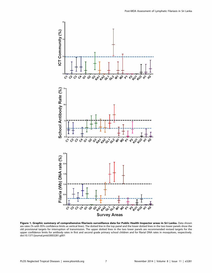
Figure 1. Graphic summary of comprehensive filariasis surveillance data for Public Health Inspector areas in Sri Lanka. Data shownare rates (% with 95% confidence limits as vertical lines). The dotted line in the top panel and the lower dotted lines in the two lower panels show theold provisional targets for interruption of transmission. The upper dotted lines in the two lower panels are recommended revised targets for theupper confidence limits for antibody rates in first and second grade primary school children and for filarial DNA rates in mosquitoes, respectively.doi:10.1371/journal.pntd.0003281.g001
Post-MDA Assessment of Lymphatic Filariasis in Sri Lanka
PLOS Neglected Tropical Diseases | www.plosntds.org 7 November 2014 | Volume 8 | Issue 11 | e3281
Relationships between different human filariasisparameters in community and school surveys
Human filariasis parameters tended to be significantly correlat-
ed with each other [e.g., community Mf rate vs. community CFA
rate (r = 0.63, P = 0.0018), school CFA rate vs. school antibody
rate (r = 0.5, P = 0.0142), and community CFA rate vs. school
CFA rate (r = 0.69; P = 0.0006)].
Transmission assessment survey resultsMore than 17,000 primary grade school children were tested in
TAS in 337 schools located in 11 EUs in 8 districts and in
Colombo town (Table 5). The numbers of positive CFA tests were
well below the TAS threshold level of 18 (critical cut-off value) in
all EUs. Thus all EUs ‘‘passed’’ TAS including the coastal Galle
District EU, where high rates for filariasis markers were noted in
two PHI study areas. None of the 16 children with positive CFA
tests in TAS surveys had microfilaremia. All CFA-positive children
were treated with anti-filarial medications and follow-up surveys
are in progress or planned to further assess people in areas with
positive children.
Filarial DNA rates in mosquitoesAlmost 3,900 pools (20 mosquitoes per pool) of blood fed, gravid
or semi-gravid mosquitoes collected in 19 PHI areas were tested
for filarial DNA by qPCR (Table 6). Filarial DNA rates exceeded
the target of 0.25% in 10 of 19 PHIs. Mosquitoes from both PHIs
surveyed in Galle district and one in Matara district had parasite
DNA rates of more than 1%, and these rates were comparable to
those seen in some filariasis endemic areas in Egypt with continued
filariasis transmission following one or two rounds of MDA [24].
Upper confidence limits for filarial DNA rates were $1% in 5 of
19 PHIs surveyed. On the other hand, three of 19 PHIs surveyed
had no positive mosquito pools. Most of the other filariasis
parameters were also low in these PHIs. Mosquito DNA samples
from Wattala were retested by qPCR at Washington University
and confirmed to be negative.
The percentages of positive mosquito trap sites were highly
variable in different PHIs, and these rates were strongly correlated
with percentages of pools positive for filarial DNA (r = 0.99, P,
0.0001), community CFA rates (r = 0.72, P = 0.0003), and school
CFA rates (r = 0.77; P,0.0001). Percentages of mosquito pools
positive for filarial DNA were highly correlated with community
CFA rates (r = 0.71, P = 0.0001) and school CFA rates (r = 0.79,
P,0.0001). In addition, percentages of houses with at least one
CFA positive resident were highly correlated with percentages of
mosquito trap sites with filarial DNA in mosquitoes (r = 0.75,
P = 0.0001) (Table S2) and with percentages of mosquito pools
that contained filarial DNA (r = 0.73; P = 0.0002).
Spatial analysis of filarial infections in humans andmosquitoes
GPS data for PHI areas with high and low rates of persistent LF
are shown in Figures 2 and S1. These maps show that sampled
households and mosquito collection sites were nicely dispersed to
cover the study areas. Infections in human and parasite DNA in
mosquitoes tended to be dispersed in most study areas.
Longitudinal results from PeliyagodawattaA pilot LF surveillance study was performed in 2008 in
Peliyagodawatta, which is located in Gampaha district just outside
of the city of Colombo. The area was resurveyed in 2011,
approximately 2.5 years after the baseline study. This is a low
Table 3. Filariasis infection parameters by age and gender in Public Health Inspectora areas.
Age Range (Yr) Males CFAb % (CI) Females CFAb % (CI) Total (%, CI)
10–15 1/462 0.22 (0.04–1.22) 1/418 0.24 (0.04–1.34) 2/880 (0.23, 0.06–0.82)
16–20 2/352 0.57 (0.16–2.05) 0/365 0 2/717 (0.28, 0.08–1.01)
21–30 4/447 0.90 (0.35–2.28) 3/692 0.43 (0.15–1.27) 7/1139 (0.62, 0.30–1.26)
31–40 11/490 2.25 (1.26–3.97) 4/838 0.48 (0.19–1.22) 15/1328 1.13, 0.69–1.86)
41–50 12/487 2.46 (1.42–4.26) 3/758 0.40 (0.13–1.16) 15/1245 (1.2, 0.73–1.98)
51–60 8/395 2.03 (1.03–3.95) 4/633 0.63 (0.25–1.61) 12/1028 (1.17, 0.67–2.03)
$61 3/326 0.92 (0.31–2.67) 9/493 1.83 (0.96–3.43) 12/819 (1.47, 0.84–2.54)
aCirculating filarial antigen (CFA) results from 14 public health inspector areas (PHIs) with one or more CFA positives were included in this analysis.bData shown are CFA prevalence rates (95% CI).doi:10.1371/journal.pntd.0003281.t003
Table 4. Multivariable logistic regression of risk factors for filarial antigenemia in community survey data.
All PHI areasa Infected Areasb Only
Factor Odds Ratio (95% CI) P Odds Ratio (95% CI) P
Male gender 2.48 (1.51–4.19) 0.0003 2.54 (1.54–4.29) 0.0002
Denied any prior intake of antifilarial medication 2.55 (1.55–4.22) 0.0002 2.14 (1.30–3.54) 0.003
Denied use of bed net the night before the survey 1.34 (0.80–2.21) 0.25 1.45 (0.87–2.39) 0.15
Age (per decade) 1.32 (1.15–1.52) .0001 1.31 (1.14–1.51) 0.0002
aResults from all 19 public health inspector (PHI) areas that were surveyed.bThis analysis was restricted to results from 14 PHI areas where one or more persons tested had a positive filarial antigen test.doi:10.1371/journal.pntd.0003281.t004
Post-MDA Assessment of Lymphatic Filariasis in Sri Lanka
PLOS Neglected Tropical Diseases | www.plosntds.org 8 November 2014 | Volume 8 | Issue 11 | e3281
income, peri-urban area with high mosquito densities, and no
intervention for LF control was undertaken in this area between
2008 and 2011. Results from the two surveys are summarized in
Table 7. Several filariasis parameters were lower in 2011 than in
2008. While only the reduction in community CFA was
statistically significant, the trend toward reduction was present
for all of these parameters apart from Mf rate, which was already
very low in 2008.
The first survey in Peliyagodawatta identified 37 amicrofilare-
mic subjects with positive filarial antigen tests. These people were
Table 5. Transmission assessment survey (TASa) results from 11 evaluation units (EUs) in 8 districtsb in in Sri Lanka.
Evaluation UnitPopulationsize/EU
Number of primarygrade schools included
Number of primary gradechildren tested
Number of children positive for filarialantigenemiac
Colombo-RDHS 1,761,010 30 1716 2 (0.12, 0.03–0.4)
Colombo-city 557,356 30 1555 2 (0.13, 0.04–0.4)
Gampaha I 898,731 30 1642 1 (0.06, 0.01–0.3)
Gampaha II 1,426,944 30 1462 0 (0)
Kalutara 1,237,676 30 1585 4 (0.3, 0.10–0.6)
Galle I 719,911 31 1557 7 (0.45, 0.22–0.9)
Galle II 347,027 31 1543 0 (0)
Matara 815,625 30 1591 0 (0)
Puttalam 766,469 30 1583 0 (0)
Kurunegala 1,629,958 35 1692 0 (0)
Hambantota 607,404 30 1553 0 (0)
Total 10,768,112 337 17479 16 (0.1, 0.06–0.1)
aThe critical cutoff value for assessing interruption of transmission was 18 in all EUs.bThe 8 endemic districts were MDA implementation units.cBinaxNOW Filariasis tests were used for detection of filarial antigenemia. Data shown are the number of positive tests (% positive and 95% CI).doi:10.1371/journal.pntd.0003281.t005
Table 6. Filarial DNA rates in Sri Lankan Culex quinquefasciatus mosquitoes by Public Health Inspector area.
District PHI areaa PHI codeNumber ofmosquitoes tested
Number of poolstested b
Number (%) ofpositive pools
Filarial DNA rates inmosquitoes c
Colombo Katukurunda C1 4000 200 3 (1.5) 0.07 (0.01–0.22)
Sedawatta C2 4480 224 21 (9) 0.52 (0.31–0.80)
Mattakkuliya C3 4000 200 13 (6.5) 0.34 (0.17–0.59)
Borella C4 4000 200 26 (13) 0.69 (0.43–1.0)
Gampaha Kelaniya G1 4320 216 22 (10) 0.54 (0.32–0.83)
Wattala G2 4000 200 0 (0) 0
PeliyagodaW G3 4080 203 17 (8) 0.43 (0.24–0.71)
Kalutara Panadura KA1 4000 200 9 (4.5) 0.23 (0.10–0.45)
Kalutara N KA2 4080 204 28 (14) 0.74 (0.47–1.09)
Galle Ambalangoda GL1 4000 200 52 (26) 1.49 (1.08–2.01)
Unawatuna GL2 4000 200 54 (27) 1.56 (1.13–2.08)
Matara Devinuwara M1 4160 208 9 (4) 0.22 (0.09–0.43)
Weligama M2 4080 204 51 (25) 1.43 (1.03–1.92)
Puttalam Chila town P1 4000 200 6 (3) 0.15 (0.05–0.34)
Lunuwila P2 4160 208 0 (0) 0
Kurunegala Bamunawala KU1 4160 208 4 (1.9) 0.10 (0.02–0.25)
Narammala KU2 4160 208 11 (5.2) 0.27 (0.13–0.50)
Hambantota HT town H1 4000 200 0 (0) 0
Tanagalle H2 4080 204 2 (1) 0.05 (0.01–0.15)
aSentinel sites (PHIs) C3 and C4 were located in the city of Colombo. Sentinel site G3 is a PHFO area.bEach pool included 20 mosquitoes (blood fed, gravid and semigravid).cFilarial DNA was detected by qPCR. Rates of filarial DNA in mosquitoes (maximum likelihood and 95% CI) were estimated using PoolScreen2. Results are shown as pass(regular font), borderline (italics) or fail (bold) based on provisional endpoint criteria described in the Introduction.doi:10.1371/journal.pntd.0003281.t006
Post-MDA Assessment of Lymphatic Filariasis in Sri Lanka
PLOS Neglected Tropical Diseases | www.plosntds.org 9 November 2014 | Volume 8 | Issue 11 | e3281
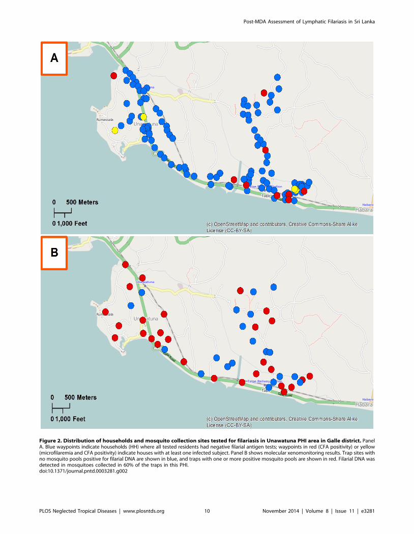
Figure 2. Distribution of households and mosquito collection sites tested for filariasis in Unawatuna PHI area in Galle district. PanelA. Blue waypoints indicate households (HH) where all tested residents had negative filarial antigen tests; waypoints in red (CFA positivity) or yellow(microfilaremia and CFA positivity) indicate houses with at least one infected subject. Panel B shows molecular xenomonitoring results. Trap sites withno mosquito pools positive for filarial DNA are shown in blue, and traps with one or more positive mosquito pools are shown in red. Filarial DNA wasdetected in mosquitoes collected in 60% of the traps in this PHI.doi:10.1371/journal.pntd.0003281.g002
Post-MDA Assessment of Lymphatic Filariasis in Sri Lanka
PLOS Neglected Tropical Diseases | www.plosntds.org 10 November 2014 | Volume 8 | Issue 11 | e3281
not treated for LF at that time. Twenty-five of these people were
retested in 2010, approximately 18 months after the first survey;
others had moved or were otherwise not available for follow-up.
Only 12 of 25 subjects were still CFA-positive (48%), and only 1 of
25 was microfilaremic by 60 ml night blood smear. None of the
subjects reported symptoms or signs of clinical filariasis during the
18 month interval. All subjects with filarial antigenemia were
treated in 2011.
Discussion
This study has provided interesting data on the status of LF in
Sri Lanka approximately 6 years after completion of the country’s
MDA program, and it has important implications for post-MDA
surveillance activities in other LF-endemic countries around the
world. Few countries participating in GPELF have been studied as
thoroughly as Sri Lanka.
Has Sri Lanka successfully eliminated LF?The term ‘‘LF elimination’’ has been interpreted in different
ways, but WHO documents clearly state that one goal of LF
elimination programs is interruption of transmission [15]. WHO is
also responsible for deciding when countries have eliminated LF.
Pending their review, we think it is important to recognize the
achievements of Sri Lanka’s Anti-Filariasis Campaign, which is
one of the finest LF elimination programs in the world. The
program has reduced Mf rates to less than 1% in all sentinel and
spot check sites, all EUs easily passed TAS criteria for stopping
MDA, and the AFC has a network of clinics that provide care to
thousands of lymphedema patients in all endemic districts. By
these criteria, Sri Lanka has achieved several WHO targets and
the country is on track to achieve elimination. If WHO determines
that Sri Lanka has not met criteria for LF elimination, we believe
that the organization should develop criteria and a recognition
program for countries that can document this level of superb
control, because this pre-elimination status is a significant
achievement in public health and an important step on the road
to LF elimination. External recognition of ‘‘superb control’’ or
‘‘near elimination’’ may help national programs obtain political
support and resources needed for the difficult last mile required for
true elimination.
What is the relative value of different approaches andtechnologies for post-MDA surveillance of LF?
While protocols for transmission assessment surveys are based
on solid sampling principles, the sensitivity of TAS for detecting
ongoing transmission of LF has not been adequately tested in field
studies [15]. Our results clearly show that TAS performed
according to WHO guidelines were not sensitive for detecting
ongoing LF transmission in Sri Lanka. There are a number of
reasons for this. First, we believe that EUs of 1 to 2 million are too
much too large, because risk factors that affect LF transmission
often vary widely across such large populations/areas. This
problem could be mitigated by reducing the size of EUs (for
example, to areas with populations of 100,000 or less), but that
would significantly increase the cost of TAS. A second problem
with TAS is that filarial antigenemia rates in young children are
sometimes very low in areas with ongoing LF transmission. Our
study showed that CFA rates in school aged children were much
lower than those in adults. Therefore, the sensitivity of TAS might
be improved by using a similar cluster sampling method to test
adults (for example, those attending primary health clinics) instead
of children in schools. A recent report from Togo described the use
of other types of passive surveillance for assessing LF following
MDA [25].
Since anti-filarial antibody rates are uniformly higher than
antigenemia rates in LF-endemic populations, another potential
solution for the problem of low TAS sensitivity would be to
substitute antibody testing for antigen testing in TAS for samples
of school-aged children. Antibody results from the present study
using a commercially available ELISA kit provide a proof of
principle for this approach. However, ELISA testing may not be
feasible for all LF programs, and available rapid-format antibody
tests have not yet been validated for this purpose.
Results from this study strongly support the use of molecular
xenodiagnosis for post-MDA surveillance in areas where LF is
transmitted by Culex mosquitoes. MX does not require collection
of blood samples or active participation by large numbers of
people in endemic areas. However, MX does require cadres of
skilled personnel, specialized laboratory facilities, and funds for
consumables. While MX was performed by MOH personnel in
this study, this required significant external inputs including
equipment, supplies, training of personnel, and funds for mosquito
collection. Also, additional work is needed to develop and validate
sampling methods for assessment of mosquito DNA rates in areas
larger than PHIs.
To summarize this section of the Discussion, while TAS surveys
may be useful for decisions regarding stopping MDA, they are not
sufficient to show that LF transmission has been interrupted. The
sensitivity of TAS might be improved by reducing the size of EUs
or by sampling adults instead of school-aged children. We
recommend antibody testing of children using TAS sampling
methods and/or MX (especially in areas believed to be at high
risk) to complement antigen-test based TAS, because these
Table 7. Comparison of filarial infection parameters in Peliyagodawattaa in 2008 and 2011.
Filarial infection markers No. tested 2008 Prevalence b 2008 No. tested 2011 Prevalence b 2011 P value c
Mf Community d 944 0.4 (0.16–1.08) 5 0.4 (0.1–1.4) 0.73
CFA Community d 945 3.8 (2.76–5.23) 504 1.2 (0.5–2.4) 0.01
CFA age 6–8 265 1.9 (0.81–4.34) 377 0.3 (0.05–1.49) 0.09
Filarial DNA rate in mosquitoes 277 pools 0.75 (0.52–1.06) 203 pools 0.43 (0.24–0.7) NS
Number (%) of mosquito pools positive for filarial DNA 39/277 (14%) 17/203 (8.3%) 0.07
aPeliyagodawatta is a Public Health Field Officer area in Gampaha district.bResults shown are % positive (95% CI). Filarial DNA rates shown are maximum likelihood estimates (with 95% CI).cP values are based on x2. NS, not significant.dCommunity microfilaremia (Mf) and circulating filarial antigenemia (CFA) rates are for ages $10 years. Mf rates are based on night blood smear results from all subjectsin 2008 and from CFA positives only in 2011.doi:10.1371/journal.pntd.0003281.t007
Post-MDA Assessment of Lymphatic Filariasis in Sri Lanka
PLOS Neglected Tropical Diseases | www.plosntds.org 11 November 2014 | Volume 8 | Issue 11 | e3281
methods appear to be more sensitive than TAS for detecting
ongoing LF transmission.
Revised targets for LF elimination programsThis study has provided new insight regarding provisional
targets for MDA programs that were suggested in 2007 based on
data from Egypt [16]. Since there is uncertainty surrounding all
point estimates, we now recommend using confidence intervals to
express targets as illustrated in Figure 1. The new suggested target
for the antifilarial antibody rate in first and second grade school
children is to have an upper confidence limit of ,5%. The new
target for MX (Culex mosquitoes) is to have an upper confidence
limit of the maximum likelihood estimate of ,1%. The new target
for the community CFA rate (age .9) is to have an upper
confidence limit of ,2%. This target provides a very high level of
confidence that the Mf rate will be less than 0.5% in the
community with a much smaller sample size than what would be
required for Mf testing. Additional studies will be needed to test
the new proposed targets in different regions. We believe that these
targets will be helpful for identifying areas that require continued
surveillance.
Next steps for areas that may have ongoing transmissionfollowing MDA
Existing guidelines do not adequately address this issue. Four
options to consider are resumption of MDA, implementation of
test and treat programs, vector control, and watchful waiting. It
may be difficult to justify resumption of MDA when Mf rates are
well below 1% when one considers that many of those with
persistent infections may have been noncompliant with MDA in
the past. Test and treat campaigns may be more efficient for
finding and treating those with persistent infections than MDA,
and the Sri Lanka AFC has started to do this in Galle district. Our
results suggest that adult males and people who do not recall
having taken MDA in the past should be considered to be high
priority target groups for test and treat programs.
WHO has recommended vector control as a post MDA strategy
[26]. Although vector control can be difficult to implement at the
scale needed for LF elimination, surveillance results may identify
hot spot areas where focused vector control may be feasible. Our
finding that CFA rates were lower in people who reported using
bed nets is interesting, although the logistic regression analysis
suggested that lack of bed net use was not an independent risk
factor for filarial infection. Bed nets are popular in Sri Lanka
because of the mosquito nuisance factor and the risk of dengue.
Beneficial effects of bed nets for LF have been reported from areas
with Anopheles transmission [27,28]. The Sri Lanka government
should consider implementing a health education campaign to
reinforce the popularity of bed nets and increase usage rates in
areas with persistent LF.
The longitudinal data from Peliyagodawatta are intriguing,
because they suggest that some areas with filariasis parameters that
do not meet our provisional criteria for interruption of transmis-
sion may spontaneously improve over time. Thus the strategy of
watching, waiting, and retesting may be the best course of action
for some areas with persistent LF. Other data from Peliyagoda-
watta on the natural history of filarial antigenemia in amicrofi-
laremic individuals in the post-MDA setting are reassuring. These
results suggest that there is no pressing need to actively identify
and treat asymptomatic and amicrofilaremic persons with positive
filarial antigen tests following MDA. This is because the risk of
such people developing microfilaremia is low, and antigenemia
often clears over time without treatment.
We believe that this study has contributed significant new
information regarding post-MDA surveillance and low level
persistence of filariasis following MDA. LF elimination is a dynamic
process [29], and point estimates of persistent infection may be less
important than trends over time. For this reason, we plan to restudy
Peliyagodawatta and several other PHIs with elevated LF param-
eters three years after the evaluations described in this publication.
Supporting Information
Figure S1 Distribution of households and mosquito collection
sites tested for filariasis in Chila Town PHI area in Puttalam
district which has less evidence of persistent filariasis than
Unawatuna PHI (shown in Fig 2). Panel A. Blue waypoints
indicate households (HH) where all tested residents had negative
filarial antigen tests; waypoints in red indicate houses with at least
one infected subject (CFA positive). Panel B shows molecular
xenomonitoring results. Trap sites with no mosquito pools positive
for filarial DNA are shown in blue, and traps with one or more
positive mosquito pools are shown in red. Filarial DNA was
detected in mosquitoes collected in 10% of the traps in this PHI
area.
(TIFF)
Table S1 Community rates for circulating filarial antigenemia
(CFA), microfilaremia (Mf), and IgG4 antibodies to filarial antigen
Bm14 in selected public health inspector.
(DOCX)
Table S2 Filarial infections by household and mosquito trap site
in different Public Health Inspector (PHI) areas in Sri Lanka.
(DOCX)
Checklist S1 STROBE statement. Checklist of items included
in this cross-sectional study Rao et al., A Comprehensive
Assessment of Persistent Lymphatic Filariasis in Sri Lanka Six
Years after Cessation of Mass Drug Administration.
(DOC)
Acknowledgments
We are grateful to the Centre for Neglected Tropical Diseases, CNTD,
Liverpool, UK for donating equipment for the AFC laboratory and for
providing funds for renovating the AFC laboratories. We thank the
Lymphatic Filariasis Support Center (Taskforce for Global Health,
Decatur, GA) for providing PDAs and technical support for the EDGE
data capture system. We thank the Central and Provincial ministries of
Education, Principal, staff members, and participants. We sincerely thank
Dr. Tilaka S. Liyanage and Dr. U. S. B. Ranasinghe for administrative
support for this project at AFC. We are grateful for technical assistance
provided by staff from AFC headquarters in Colombo and for the active
participation of AFC leaders and staff in the eight districts covered in this
study. We would like to thank Dr. Peter U. Fischer, Dr. Sandra J. Laney,
Dr. Ama Pathirage and Dr. Reda M. R. Ramzy who participated in
training activities in 2008 pilot surveys in Peliyagodawatta. We are also
grateful for excellent technical assistance provided by Mr. P.D. Gamini,
Mrs. L. Liyanage, Ms. T. Dassanayaka, and Mrs. M. Surrwandana at
AFC, Colombo, and by Mrs. K. Fischer and Mr. K. Curtis at Washington
University School of Medicine, St. Louis. We would like to thank Mr.
William Winston, GIS services, Washington University for ArcGIS maps.
Finally, we appreciate the assistance of Mr. John E. Lacey and Mr. Robert
White at CDC, Atlanta for providing the barcode stickers used in this
study.
Author Contributions
Conceived and designed the experiments: WDYP SNP SS RUR GJW.
Performed the experiments: WDYP SNP KCN SDS ADW. Analyzed the
data: KCN SDS SNP JPM ADW RUR GJW. Wrote the paper: RUR
GJW.
Post-MDA Assessment of Lymphatic Filariasis in Sri Lanka
PLOS Neglected Tropical Diseases | www.plosntds.org 12 November 2014 | Volume 8 | Issue 11 | e3281
References
1. WHO (2013) Global programme to eliminate lymphatic filariasis: progress
report for 2012. Wkly Epidemiol Rec 88: 389–399.2. Abdulcader MH, Sasa M (1966) Epidemiology and control of bancroftian
filariasis in Ceylon. Jpn J Exp Med 36: 609–646.3. Schweinfurth U (1983) Filarial diseases in Ceylon: a geographic and historical
analysis. Ecol Dis 2: 309–319.
4. Dissanaike AS (1991) Filariasis in Ceylon then (1961) and in Sri Lanka now(1990–30 years on). Ann Trop Med Parasitol 85: 123–129.
5. AntifilariasisCampaign (2013) Annual reports. Ministry of Health, Sri LankaAvailable at http://wwwfilariasiscampaignhealthgovlk/subpgs/03_reportshtml
6. Horton J, Witt C, Ottesen EA, Lazdins JK, Addiss DG, et al. (2000) An analysis
of the safety of the single dose, two drug regimens used in programmes toeliminate lymphatic filariasis. Parasitology 121 Suppl: S147–160.
7. WHO (2000) Preparing and implementing a national plan to eliminatelymphatic filariasis (in countries where onchocerciasis is not co-endemic). World
Health Organization, Geneva, WHO/CDS/CPE/CEE/200016.8. WHO (2011) Halfway towards eliminating lymphatic filariasis: Progress Report
2000–2009 and Strategic Plan 2010–2020 of the Global Programme to
Eliminate Lymphatic Filariasis. WHO, 2011 (WHO/HTM/NTD/PCT/20106) Geneva: World Health Organization.
9. Gunawardena GS, Ismail MM, Bradley MH, Karunaweera ND (2007) Impactof the 2004 mass drug administration for the control of lymphatic filariasis, in
urban and rural areas of the Western province of Sri Lanka. Ann Trop Med
Parasitol 101: 335–341.10. Weerasooriya MV, Yahathugoda CT, Wickramasinghe D, Gunawardena KN,
Dharmadasa RA, et al. (2007) Social mobilisation, drug coverage andcompliance and adverse reactions in a Mass Drug Administration (MDA)
Programme for the Elimination of Lymphatic Filariasis in Sri Lanka. Filaria J 6:11.
11. Yahathugoda TC, Weerasooriya MV, Sunahara T, Kimura E, Samarawick-
rema WA, et al. (2014) Rapid assessment procedures to detect hidden endemicfoci in areas not subjected to mass drug administration in Sri Lanka. Parasitol Int
63: 87–93.12. Yahathugoda TC, Weerasooriya M, Samarawickrema WA (2013) An
independent evaluation of the programme for the elimination of lymphatic
filariasis. Galle Medical Journal 18: 31–43.13. WHO (2012) Expert Mission to Sri Lanka for verification of elimination of
lymphatic filariasis. Report. World Health Organization (SEA-CD-245) NewDelhi, India: 1–37.
14. Chu BK, Deming M, Biritwum NK, Bougma WR, Dorkenoo AM, et al. (2013)Transmission assessment surveys (TAS) to define endpoints for lymphatic
filariasis mass drug administration: a multicenter evaluation. PLoS Negl Trop
Dis 7: e2584.15. WHO (2011) Monitoring and epidemiological assessment of mass drug
administration in the Global Programme to Eliminate Lymphatic Filariasis: A
manual for national elimination programmes. WHO, (WHO/HTM/NTD/
PCT/2011 4) Geneva: World Health Organization.
16. Weil GJ, Ramzy RM (2007) Diagnostic tools for filariasis elimination programs.
Trends Parasitol 23: 78–82.
17. Irish SR, Moore SJ, Derua YA, Bruce J, Cameron MM (2013) Evaluation of
gravid traps for the collection of Culex quinquefasciatus, a vector of lymphatic
filariasis in Tanzania. Trans R Soc Trop Med Hyg 107: 15–22.
18. Weil GJ, Lammie PJ, Weiss N (1997) The ICT Filariasis Test: A rapid-format
antigen test for diagnosis of bancroftian filariasis. Parasitol Today 13: 401–404.
19. Weil GJ, Curtis KC, Fischer PU, Won KY, Lammie PJ, et al. (2011) A
multicenter evaluation of a new antibody test kit for lymphatic filariasis
employing recombinant Brugia malayi antigen Bm-14. Acta Trop 120 Suppl 1:
S19–22.
20. Rao RU, Atkinson LJ, Ramzy RM, Helmy H, Farid HA, et al. (2006) A real-
time PCR-based assay for detection of Wuchereria bancrofti DNA in blood and
mosquitoes. Am J Trop Med Hyg 74: 826–832.
21. Gass K, Beau de Rochars MV, Boakye D, Bradley M, Fischer PU, et al. (2012) A
multicenter evaluation of diagnostic tools to define endpoints for programs to
eliminate bancroftian filariasis. PLoS Negl Trop Dis 6: e1479.
22. Katholi CR, Toe L, Merriweather A, Unnasch TR (1995) Determining the
prevalence of Onchocerca volvulus infection in vector populations by polymerase
chain reaction screening of pools of black flies. J Infect Dis 172: 1414–1417.
23. Katholi CR, Unnasch TR (2006) Important experimental parameters for
determining infection rates in arthropod vectors using pool screening
approaches. Am J Trop Med Hyg 74: 779–785.
24. Ramzy RM, El Setouhy M, Helmy H, Ahmed ES, Abd Elaziz KM, et al. (2006)
Effect of yearly mass drug administration with diethylcarbamazine and
albendazole on bancroftian filariasis in Egypt: a comprehensive assessment.
Lancet 367: 992–999.
25. Budge PJ, Dorkenoo AM, Sodahlon YK, Fasuyi OB, Mathieu E (2014) Ongoing
surveillance for lymphatic filariasis in Togo: assessment of alternatives and
nationwide reassessment of transmission status. Am J Trop Med Hyg 90: 89–95.
26. WHO (2013) Lymphatic Filariasis: Practical Entomology. A Handbook for
National Elimination Programmes. WHO Global Programme to Eliminate
Lymphatic Filariasis: 1–90.
27. Eigege A, Kal A, Miri E, Sallau A, Umaru J, et al. (2013) Long-lasting
insecticidal nets are synergistic with mass drug administration for interruption of
lymphatic filariasis transmission in Nigeria. PLoS Negl Trop Dis 7: e2508.
28. Reimer LJ, Thomsen EK, Tisch DJ, Henry-Halldin CN, Zimmerman PA, et al.
(2013) Insecticidal bed nets and filariasis transmission in Papua New Guinea.
N Engl J Med 369: 745–753.
29. Stolk WA, de Vlas SJ, Habbema JD (2006) Advances and challenges in
predicting the impact of lymphatic filariasis elimination programmes by
mathematical modelling. Filaria J 5: 5.
Post-MDA Assessment of Lymphatic Filariasis in Sri Lanka
PLOS Neglected Tropical Diseases | www.plosntds.org 13 November 2014 | Volume 8 | Issue 11 | e3281