Embed Size (px)
Citation preview

Faculty of Health Sciences
Department of Clinical Dentistry
Ectopic and normal maxillary canine eruption: maxillary
incisor root resorption and interceptive treatment
Sigurd Hadler-Olsen
A dissertation for the degree of Philosophiae Doctor
—
Author’s name and last name
A dissertation for the degree of Philosophiae Doctor – Month Year

2
“When meditating over a disease, I never think of
finding a remedy for it, but, instead, a means of
preventing it”.
Louis Pasteur (1822–1895)

3
1 Contents
1 Contents .............................................................................................................................. 3
2 Acknowledgements ............................................................................................................ 5
3 List of papers ...................................................................................................................... 7
4 Abbreviations and terms ..................................................................................................... 8
5 Summary ............................................................................................................................ 9
6 Introduction ...................................................................................................................... 11
6.1 Normal maxillary canine eruption ............................................................................. 11
6.2 Ectopic maxillary canine eruption ............................................................................. 12
6.2.1 Definition ........................................................................................................... 12
6.2.2 Prevalence .......................................................................................................... 13
6.2.3 Aetiology ............................................................................................................ 14
6.2.4 Root resorption of incisors ................................................................................. 18
6.2.5 Diagnostics ......................................................................................................... 19
6.2.6 Interceptive treatment ......................................................................................... 25
7 Objectives ......................................................................................................................... 29
8 Subjects and methods ....................................................................................................... 30
8.1 Subjects ...................................................................................................................... 30
8.2 Methods ..................................................................................................................... 35
8.3 Reliability of measurements ...................................................................................... 39
8.4 Statistical analysis ...................................................................................................... 40
9 Results .............................................................................................................................. 42
10 Discussion ........................................................................................................................ 45
10.1 Methodological considerations: strengths and weaknesses ................................... 45
10.2 Statistical analysis .................................................................................................. 50
10.3 Ethical considerations ............................................................................................ 51
10.4 Discussion of main findings ................................................................................... 52
11 Conclusions ...................................................................................................................... 57
12 Future perspectives ........................................................................................................... 58

4
13 References ........................................................................................................................ 59
14 Appendix .......................................................................................................................... 69

5
2 Acknowledgements
The research presented here was performed at the Public Dental Health Competence Centre of
Northern Norway, Tromsø, the Department of Clinical Dentistry, Faculty of Health Sciences,
University of Tromsø, and the Medical Research Center Oulu, University Hospital of Oulu,
Finland.
I would like to thank my supervisors. To Professor Heidi Kerosuo, who introduced me to
research and to her collaborators in Finland – I always enjoyed your comments and sharp
scientific eye. To Associate Professor Raija Lähdesmäki, my principal supervisor, who was
bold to take responsibility for a PhD student with limited research experience, and led me
through the research process – Your novel and creative thinking is something that I admire.
Professor Pertti Pirttiniemi, one of my co-supervisors, has always been positive and given
wise comments that usefully guided my work. Co-Supervisor Anders Sjögren, who, luckily
for me, moved to Tromsø during my PhD research, and has been a great help and inspiration –
You tried to fill some of the gaps in my scientific writing and had an open office door for my
many questions. I would also like to thank Napat Bolstad and Soili Kallio-Pulkkinen for being
fine collaborators on radiographic topics, Johanna Julku for collecting material for Paper II,
and Paula Pesonen for statistical advice.
My orthodontist colleges Mari Dubland and Jeanett Steinnes have been very helpful and made
great contributions in collecting material for Paper III. My fantastic secretaries Merete K.
Wold and Sigrunn Hansen have helped me a lot, organizing patients and taking impressions,
radiographs, and photos. Thank you!

6
I would like to thank the Public Dental Health Competence Centre of Northern Norway,
Tromsø, the Department of Clinical Dentistry, Faculty of Health Sciences, University of
Tromsø, and the Medical Research Center Oulu, University Hospital of Oulu for funding, for
scientific study material, and for including me in a great research environment.
I would particularly like to thank my parents for good genes, unconditional love, and always
being supportive no matter what I did. Likewise, I thank my dear brothers and good friends
who have always supported and encouraged me, and my parents in law, who have always
been very helpful and caring. My children, Eirik and Signe – Thank you for disconnecting my
brain from work and for all the sunshine you bring to my life! Elin, my wife and dearest
friend, thank you for love and support every day, and for enduring endless discussions of
statistics and my research.
Thank you all for making my PhD studies a colourful and rich experience that I have
thoroughly enjoyed!

7
3 List of papers
This thesis is based on the following papers, which are referred to by their corresponding
Roman numerals in the text:
I. Root resorptions related to ectopic and normal eruption of maxillary canine
teeth: A 3D study.
Acta Odontologica Scandinavia. 2015;73(8):609–15.
doi: 10.3109/00016357.2015.1020339.
Hadler-Olsen S, Pirttiniemi P, Kerosuo H, Bolstad Limchaichana N, Pesonen P,
Kallio-Pulkkinen S, Lähdesmäki R.
II. Does headgear treatment in young children affect the maxillary canine
eruption path?
European Journal of Orthodontics. 2018;40(6):583–91. doi: 10.1093/ejo/cjy013.
Hadler-Olsen S, Pirttiniemi P, Kerosuo H, Sjögren A, Pesonen P, Julku J,
Lähdesmäki R.
III. Double versus single primary tooth extraction in interceptive treatment of
palatally displaced canines: A randomized controlled trial.
Submitted to European Journal of Orthodontics.
Hadler-Olsen S, Sjögren A, Steinnes J, Dubland M, Limchaichana Bolstad N,
Pirttiniemi P, Kerosuo H, Lähdesmäki R.

8
4 Abbreviations and terms
BDC Buccally displaced canines
CBCT Cone beam computed tomography
CI Confidence interval
CG Control group
DEG Double extraction group
FOV Field of view
ICC Intraclass correlation coefficient
N Number
HG Headgear
HGG Headgear group
P Level of significance
PDC Palatally displaced canine
RCT Randomized controlled trial
SD Standard deviation
SEG Single extraction group

9
5 Summary
Ectopic eruption of maxillary canines is not very common (0.8–5.2 per cent prevalence), but
can create problems if left untreated. Such problems may be the malpositioning and retention
of the ectopic tooth, external root resorption, migration of neighbouring teeth, dentigerous
cyst formation, and referred pain. Treatment is often time consuming and imposes a
substantial cost on the affected patient/family and on society. The scientific evidence
concerning the adverse effects as well as interceptive treatment of ectopic maxillary canine
eruption is scarce. The overall objectives of this study were therefore to provide new
knowledge of the most common adverse effect, i.e., root resorption of maxillary incisors, as
well as new insight into the interceptive treatment of ectopic maxillary canine eruption.
Cone beam computed tomography (CBCT) was used to assess the prevalence and severity of
root resorption of maxillary incisors. Root resorption of maxillary incisors was commonly
found in relation to maxillary canine eruption. Although root resorption was significantly
more frequent in patients with ectopically erupting canines, lateral incisor resorption was
found in association with approximately 1/3 of normally erupting canines. The best predictor
of root resorption was found to be location of the maxillary canine mesial to the lateral incisor
midline.
Headgear (HG) treatment was studied to see whether it affected the maxillary canine eruption
path in young children. We also studied whether space conditions in the maxillary arch
affected the maxillary canine eruption path. The study showed that HG treatment in young
children with Angle Class II occlusion shifts the eruption path of maxillary canines to a more
vertical direction. The change in eruption path seemed to be related to space conditions in the

10
maxillary arch, especially in the intercanine region, with the most significant effect in the HG
group with spaced dental arches rather than crowded dental arches.
The impact of primary canine extraction versus that of primary canine and primary first molar
extraction on the emergence rate of palatally displaced canines (PDCs) was investigated in a
randomized clinical trial. Positional changes of PDCs and factors influencing the emergence
of PDCs after extractions were analysed. The study showed that there was no statistically
significant difference between the two procedures in the emergence rate of PDCs or
emergence in a favourable position in the dental arch. The initial canine angulation and space
conditions in the maxillary arch seemed to be the best predictors of the successful eruption of
PDCs.
In conclusion, the present work showed that root resorption is a common finding for maxillary
incisors in association with normally and ectopically erupting maxillary canines. HG
treatment in children shifts the eruption path of maxillary canines in a more vertical direction
and there was no difference in the emergence rate of PDCs depending on whether the primary
canine or both primary canine and primary first molar were extracted.

11
6 Introduction
The treatment of ectopic maxillary canines can be challenging. The treatment takes longer
than the average orthodontic treatment (1), is technically difficult, and may involve a painful
surgical procedure (2). The treatment involves several dental specialists (i.e., a radiologist,
oral surgeon, paediatric dentist, and orthodontist) and is expensive for the patient as well as
the funding government (3). There are also reported serious side effects of ectopic eruption,
such as root resorption (4).
6.1 Normal maxillary canine eruption
Calcification of the maxillary canine starts at approximately 12 months of age between the
roots of the first deciduous molar at the lower border of the orbit (5). The canine is then left
behind as the deciduous molar erupts, allowing development of the first premolar between the
deciduous molar roots. At this stage, the permanent canine is located immediately above both
the first premolar and the first deciduous molar. As the deciduous teeth erupt towards the
occlusal plane, the permanent incisor and canine crypts migrate forward in the jaws at a
greater rate than the forward movement of the deciduous teeth themselves. At the age of 7
years, the canine crown is medial to the root of its deciduous predecessor (6). From 8 years of
age, buccal movement is expected in normally erupting canines (7). At this stage of
development, the canine is located lingual to the root apex of the deciduous canine. From
there it normally moves downward, forward, and laterally away from the root of the lateral
incisor if there is sufficient space. In cases of insufficient space in the apical base, the “ugly
duckling” stage is often seen between 8 and 12 years of age (5), in which the lateral incisors
are spread out in a fan shape. As the canines in the final phase of eruption drive their way

12
between the lateral incisors and first premolars, the lateral incisors and first premolars move
into a more erect upright alignment (6).
6.2 Ectopic maxillary canine eruption
6.2.1 Definition
The definition of an ectopic canine varies in the literature (8-16). Common names of ectopic
canine eruption are: impacted canine, displaced canine, included canine, and retained canine.
Some authors use one of these terms to describe a very specific condition, while others use the
same term with much more general connotations. There are also language differences that
may alter the meaning of a term, so an author’s geographical location may determine the word
used in a journal report (16). Consequently, in this study, different names may be used to refer
to the same condition depending on the cited literature source.
Most studies use generalized definitions of canine displacement, such as: “unerupted canine
after complete root development, or if the contralateral tooth was erupted for at least 6 months
with complete root formation” (15), or “developmental dislocation to a palatal site often
resulting in tooth impaction requiring surgical and orthodontic treatments” (17). A few studies
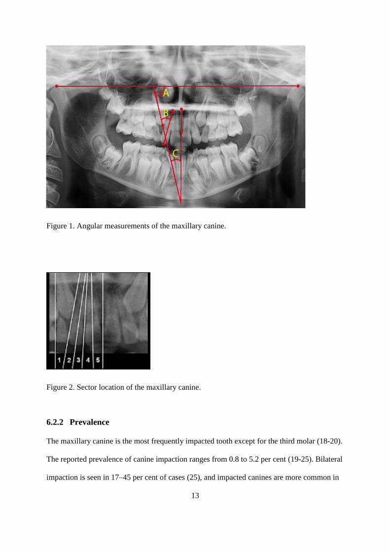
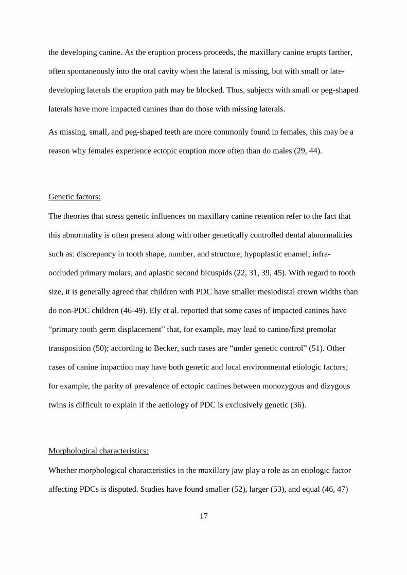
have been based on numerical values for the canine position. Bonetti et al. (9) defined a
palatally displaced canine as having an alpha angle ≥25° (Angle C, Figure 1) and as located in
sectors 2–5 (Figure 2), according to methods developed by Ericson and Kurol (10). Sigler et
al. (11) defined a PDC as having an alpha angle ≥15° and as located in sectors 2–5.
13
Figure 1. Angular measurements of the maxillary canine.
Figure 2. Sector location of the maxillary canine.
6.2.2 Prevalence
The maxillary canine is the most frequently impacted tooth except for the third molar (18-20).
The reported prevalence of canine impaction ranges from 0.8 to 5.2 per cent (19-25). Bilateral
impaction is seen in 17–45 per cent of cases (25), and impacted canines are more common in

14
females than males (8, 19, 25). More canines are reported to be displaced palatally (85%) in
contrast to labially (15%) (25-28).
The ectopic eruption of maxillary canines is 10–20 times more common than the ectopic
eruption of mandibular canines (29), and dental age estimation using Demirjian’s method has
been found to be lower than expected in subjects with maxillary canine impaction (30, 31). In
a Caucasian population, maxillary canine displacement is five times more common than in an
Asian population, and most canines in Caucasians are palatally displaced, while buccal
displacement is more common among Asians (32).
6.2.3 Aetiology
The aetiology of the ectopic eruption of maxillary canines is not fully understood.
Historically, the most common explanation for palatal ectopic eruption is based on the longer
and more difficult eruption path of the maxillary canine than those of other teeth. It was
hypothesized that due to this tortuous eruption path, the maxillary canine was more likely to
be retained than were other teeth (33). Later, the aetiology was divided into general and local
factors: general factors include endocrine diseases, febrile diseases, radiation, and vitamin D
deficiency (7); local factors, generally considered the most important causes of maxillary
canine retention, include space deficiency, blocked eruption pathway, a small or missing
lateral incisor, and morphological characteristics (34).
There is also discussion of the extent to which genetic factors influence ectopic canine
eruption (7, 31, 35-39).

15
Space deficiency:
For the labial canines, space deficiency is considered the primary etiologic factor, whereas for
palatally erupting canines, space deficiency is usually not present (28). Jacoby found that 85%
of palatally impacted canines had sufficient space for eruption, compared with 17% of labially
impacted canines (7).
When labial ectopia are observed in dentitions with sufficient space, possible etiologic factors
affecting impaction were reported to be lack of guidance from a lateral incisor and genetic
factors (40).
Blocked eruption pathway:
Retention of maxillary canines may happen due to the retention of primary teeth, odontomas,
or rotation of premolars that blocks the eruption pathway of the maxillary canine (36).
Follicular cyst development in the dental follicle covering the maxillary canine crown may
also block the eruption, as the hydrostatic pressure within the cyst can counteract the eruption
force, causing displacement of the maxillary canine (36). Also, chronic apical periodontitis
from deciduous canines may lead to retention of the permanent canine (36). Retention of the
maxillary canine may also happen secondary to the retention of other teeth, such as the
maxillary incisors. Chaushu et al. showed that the incidence of retained maxillary canines was
41.3% on the side with retained incisors as opposed to 4.7% on the contralateral side (41).
Small or missing lateral incisors:
The guidance theory suggests that palatal displacement is a result of lack of guidance along
the root of the lateral incisor due to a congenitally missing lateral or an abnormally shaped

16
lateral (42). In an Israeli study, 93% of the general population had normal lateral incisors as
opposed to 53% of the population with palatally displaced canines (43). Interestingly, the
association with PDCs was strongest for small laterals followed by peg-shaped and missing
laterals.
Figure 3. Lateral incisor morphology in the general population and PDC population. Based on
data from Brin et al. (43).
The findings of Brin et al. (43) suggest that local factors may be more important than genetic
factors in the aetiology of PDC, as the stronger genetic disturbance (missing laterals) seems to
be less correlated with PDC than is the minor genetic disturbance (small laterals). Becker (29)
presented a theory as to why small laterals and peg-shaped laterals are more correlated with
PDC than are missing laterals. He described several stages and outcomes of the impaction
process. The first step is that both late-developing and missing laterals will lead to a lack of
guidance for the permanent canine, as there are no lateral roots present at the critical time for
17
the developing canine. As the eruption process proceeds, the maxillary canine erupts farther,
often spontaneously into the oral cavity when the lateral is missing, but with small or late-
developing laterals the eruption path may be blocked. Thus, subjects with small or peg-shaped
laterals have more impacted canines than do those with missing laterals.
As missing, small, and peg-shaped teeth are more commonly found in females, this may be a
reason why females experience ectopic eruption more often than do males (29, 44).
Genetic factors:
The theories that stress genetic influences on maxillary canine retention refer to the fact that
this abnormality is often present along with other genetically controlled dental abnormalities
such as: discrepancy in tooth shape, number, and structure; hypoplastic enamel; infra-
occluded primary molars; and aplastic second bicuspids (22, 31, 39, 45). With regard to tooth
size, it is generally agreed that children with PDC have smaller mesiodistal crown widths than
do non-PDC children (46-49). Ely et al. reported that some cases of impacted canines have
“primary tooth germ displacement” that, for example, may lead to canine/first premolar
transposition (50); according to Becker, such cases are “under genetic control” (51). Other
cases of canine impaction may have both genetic and local environmental etiologic factors;
for example, the parity of prevalence of ectopic canines between monozygous and dizygous
twins is difficult to explain if the aetiology of PDC is exclusively genetic (36).
Morphological characteristics:
Whether morphological characteristics in the maxillary jaw play a role as an etiologic factor
affecting PDCs is disputed. Studies have found smaller (52), larger (53), and equal (46, 47)

18
palatal widths in patients with PDC compared with non-PDC controls. These contradictory
reports could be due to the use of different methods (i.e. CBCT, plaster models, and
cephalograms) to measure the size of the maxilla (54). Another reason could be differences in
study samples. The number of uni- or bilateral PDCs and age of children in the studied
samples could influence the results, as the absence of permanent canines causes a narrower
dental arch (55). One study excluded patients with space deficiency, which could mean that it
was not representative of the average population (54).
6.2.4 Root resorption of incisors
The most common adverse effect of ectopic canine eruption is external root resorption of
neighbouring teeth. Root resorption is defined as “a condition of dental complication
associated with either a physiological or pathological activity of the tooth resorbing cells,
which results in loss of cementum and/or dentine” (56). Most studies have found root
resorption to be more common in females, with the female/male ratio being 2:1–4:1 (4, 26,
57), though an equal male/female ratio has been reported for “ordinary” resorption and for the
severity and location of root resorption (58, 59). When patients with “severe” root resorption
(affecting more than a third of the root length) were studied, a 5:1 female to male ratio was
found (60). Possible reasons for the gender difference in susceptibility to root resorption could
be genetic or hormonal factors, or differences in skeletal and dental development (60).
The incidence of root resorption found in different studies is also dependent on the imaging
technique used to detect resorption, with new 3D techniques detecting approximately 50%
more resorption cases than conventional 2D radiographs (26, 61). The lateral incisor is most
prone to external root resorption, with reported frequencies of 27–67% versus 9–23% for
roots of central incisors (57, 61, 62). Ectopic canines may also cause root resorption in first

19
premolars, with an incidence rate of 10% reported in a Chinese population (63). External root
resorption leads to loss of tooth substance, which can cause some weakening. However, a
long-term study of resorbed incisor roots showed that the overall prognosis for these teeth was
good (64).
The exact etiologic factors causing root resorption by neighbouring teeth are still unknown.
Physical pressure from the erupting teeth is one theory (65-67), while others claim that the
dental follicle and not the tooth itself is the causative agent (68). Others point towards a
multifactorial explanation in which both systemic factors within the patient as well as local
factors around the ectopic canine (e.g., dental follicle, tooth shape, and physical contact
between teeth) work together (60). When severe root resorption was studied in a
multifactorial analysis, sex (female), enlarged dental follicle, and normal size of the lateral
incisor were found to significantly increase the risk of severe root resorption (60).
Other less common complications of ectopically erupting canines are loss of vitality of
adjacent incisors, shortening of the dental arch, formation of follicular cysts, canine ankylosis,
recurrent infections, recurrent pain, internal resorption, or a combination of these (69).
6.2.5 Diagnostics
There are three main methods for localizing the permanent canine: visual inspection,
palpation, and radiographic examination (70).
Visual inspection and palpation:
A bulge at the alveolar crest in the buccal sulcus is usually present 1–1.5 years before the
eruption of the canine (71). This bulge is a sign of a normally erupting canine and may
already be present at the age of 8 years and should generally be palpable in the buccal by the

20
age of 10 (8, 72). If the permanent canine cannot be palpated at that time, it may be a sign of a
developing eruption disturbance (72). The axial position of the adjacent lateral incisor may be
influenced by the eruption of the canine (72). This may be a normal physiological process
(“ugly duckling”), but it can also be an indication of the ectopic eruption of the canine. The
absence of a distal inclination or the proclination of the lateral incisor crown are reportedly
predictive signs of eruptive disorders of the canines (72, 73). If the deciduous canine is
mobile, it is a sign that the permanent canine is erupting normally, although some exceptions
may occur (8). However, the permanent canine may erupt ectopically even in cases displaying
varying degrees of deciduous canine root resorption, and ectopic eruption of permanent
maxillary canines has been reported in cases in which less than 1/3 of the root of the
deciduous canine remained (10). Based on findings from visual inspection and palpation,
Ericsson and Kurol suggested the following indications for radiographic examination when
eruption disturbances are suspected: 1. asymmetry on palpation; 2. the canine cannot be
palpated in a normal position at the expected time; and 3. the lateral incisor is late in eruption
or shows a pronounced buccal displacement or proclination (72). According to these criteria,
further radiographic examination was indicated in 12.8% of 10 year olds (72).
Radiographic examination:
In cases of ectopic teeth, radiographic examination is vital in order to visualize the tooth’s
position relative to neighbouring teeth and other skeletal structures. Radiograms also show the
severity of root resorption in teeth (61) and may indicate their long-term prognosis (74),
information which is vital for treatment planning (4).
In orthodontics today, the most commonly used radiographic methods are:

21
a) Periapical radiography Intraoral technique
b) Lateral cephalogram 2D
c) Orthopantomogram Extraoral technique
d) Conebeam CT 3D
Periapical radiography is an intraoral technique using standard intraoral radiographic sensors
or films (75). This method is inexpensive and the x-ray exposure is low. To determine the
position of an ectopic tooth, “Clark’s rule” or the “buccal object rule” is utilized (76) (Figure
4). In brief, two periapical images are taken, for example, of a canine and lateral, with
different projections. The object that is positioned more buccally will move more relative to
the object positioned more palatally and vice versa.
Figure 4. Clark’s rule (reprinted with permission from John Wiley & Sons Limited).

22
The lateral cephalogram is a radiographic imaging technique commonly used in orthodontics
for diagnosing anomalies, treatment planning, and evaluating growth and treatment results.
The images obtained can also be used to evaluate the anterior–posterior and vertical position
and inclination of the canine, though it is usually not the primary reason for using this
imaging technique (77).
The orthopantomogram is an extraoral technique that is routinely used in orthodontic
treatment (77). It is an excellent imaging technique if used with the understanding that it has
greater value for screening than diagnostic purposes. Orthopantomograms provide useful
information about: mandibular symmetry; present, missing, or supernumerary teeth; root
positions; dental age; eruption sequence and gross periodontal health; sinuses; and TMJ`s
(78). However, the orthopantomogram has shortcomings related to the reliability and accuracy
of the size, location, and form of the image obtained. These discrepancies arise because the
image is made by creating a focal trough or region of focus within a generic jaw form and size
(79). Any deviation from this generic jaw will result in structures that are not centred within
the focal trough. The resulting image will show differences in size, location, and form when
compared with the actual object. Generally, structures that are close to the x-ray beam will
appear magnified on the image relative to structures far from the x-ray beam. This can give an
indication of the position of an ectopic tooth, for example, as palatal canines will appear
larger on the image relative to laterals that are in “normal” positions (79).
The first 3D images were CT images used for medical applications. The effective radiation
dose from acquiring these images was much higher than from conventional 2D images. In
addition, these images were relatively expensive. Therefore, using CT for the routine analysis
of impacted canines seemed to be unjustified (80, 81).
23
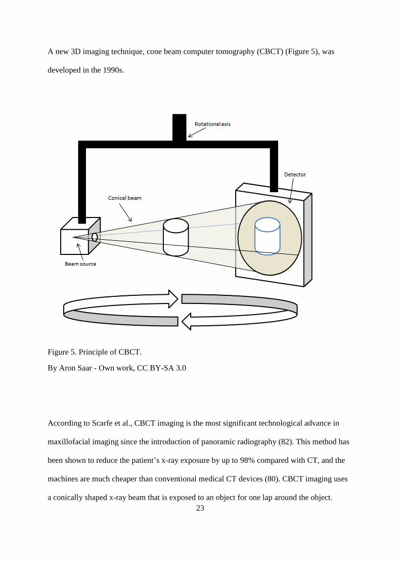
A new 3D imaging technique, cone beam computer tomography (CBCT) (Figure 5), was
developed in the 1990s.
Figure 5. Principle of CBCT.
By Aron Saar - Own work, CC BY-SA 3.0
According to Scarfe et al., CBCT imaging is the most significant technological advance in
maxillofacial imaging since the introduction of panoramic radiography (82). This method has
been shown to reduce the patient’s x-ray exposure by up to 98% compared with CT, and the
machines are much cheaper than conventional medical CT devices (80). CBCT imaging uses
a conically shaped x-ray beam that is exposed to an object for one lap around the object.

24
Using computer software, a 3D image is then reconstructed from the acquired data. In many
studies, CBCT has proven to be superior to 2D methods used in the craniofacial area (83, 84),
and its diagnostic accuracy and validity for the localization of ectopic teeth have been shown
to be better for 3D than 2D images (84). When evaluating changes in root surface
morphology, such as root resorption, CT images have been shown to increase the root
resorption detection rate by 50% (66). In particular, mild and early resorptions are detected
more accurately with 3D than 2D images (85). The sensitivity and specificity for detection of
root resorption using CBCT with a 0.3-mm voxel size are reportedly 97% and 94%,
respectively (86), versus 48% and 85% for 2D radiographs (87).
Although 3D imaging requires higher radiation doses (88), the difference in dosage between
CBCT and panoramic radiographs varies between studies. One study showed that CBCT had
a radiation dose 2–4 times higher than that of panoramic radiographs (84). Another study of
impacted canines using both panoramic radiographs and CBCT found that CBCT had a 15–
30-fold higher radiation dose (89). Comparing CBCT with standard periapical radiograph
imaging of a single impacted canine showed that the estimated effective dose would be 70–
140-fold higher for CBCT depending on the choice of CBCT device (89). The radiation dose
of CBCT may differ 15-fold between low- and high- dose resolution protocols for the same
field of view (90). Therefore, adhering to the as low as reasonably achievable (ALARA)
principle, 3D techniques should only be used on proper indications (75).
Radiographic prediction of root resorption:
Several studies have used CBCT to investigate radiographic predictors of incisor root
resorption in conjunction with an ectopic canine. Correlations between root resorption and 1)
canine position, 2) mesial overlap with adjacent teeth, 3) available space for the ectopic

25
canine, 4) closed canine apex, 5) contact relationship, and 6) dental follicle have been shown
(58, 60, 91, 92). However, other studies contradict these results (65, 67, 93, 94).
As panoramic radiography is more commonly used in orthodontics, and has a lower cost and
radiation dosage than does CBCT, Alqerban et al. investigated a possible prediction model for
root resorption based on panoramic radiographs (95). In the prediction model, patient gender
(female), canine apex (open apex), vertical canine crown position (above middle third of
incisor root), and canine magnification (magnified: yes, i.e., palatal) were the strongest
predictors of root resorption with an area under the curve (AUC) of 0.74 (i.e., fair accuracy).
This indicated that panoramic radiographs may be used to predict root resorption, particularly
as a helpful tool to justify the need for an additional CBCT (95).
6.2.6 Interceptive treatment
Interceptive orthodontic treatment can be defined as:
any procedure that eliminates or reduces the severity of malocclusion in the
developing dentition (96); and
all simple measures that eliminate the developing malocclusion (97).
Previous studies have mainly focused on the palatal ectopic canines (3, 8, 11-14, 49, 98-103).
The reason for this is not known, but is probably related to the fact that palatal ectopic canines
occur much more frequently than do buccal ectopic canines.
The most common orthodontic interceptive treatment for PDCs is to extract the deciduous
canine on the same side as the ectopic permanent canine. A prospective study by Ericson and
Kurol (103) in 1988 showed that this procedure was successful in improving the eruption path

26
of the permanent canine. The lack of a control group in this study raised questions about the
conclusions reached, though a similar study by Power and Short (8) confirmed the findings of
Ericson and Kurol and further concluded that crowding adversely affected the favourable
eruption of the canine.
Newer studies with a randomized controlled design (RCT) show that the success rate of
extraction of the primary canine is 67–69% as opposed to 39–42% in the control group (13,
14). Bazargani et al. (13) stated that the treatment effect is significantly better in younger age
groups (10–11 years old) than older age groups (12–14 years old), which they proposed was
due to the longer eruption time and greater deviation in the older subjects. Also, they
recommended maintaining the arch perimeter of the maxilla by means of a palatal arch in
order not to lose space after the extraction procedure.
Some studies have shown that the addition of headgear (HG) or a rapid maxillary expander
increases the effectiveness of the extraction procedure. Baccetti et al. found that the addition
of HG treatment increased the success rate from 65.2% to 87.5% (98). However, this study
was criticized for methodological weaknesses by Naoumova et al. (101). Silvola et al. (104)
investigated the effects of early HG treatment in 7-year-old children with Angle Class II
tendency and moderate crowding. They concluded that the eruption pattern of the maxillary
permanent canines was more vertical after 2 years of HG treatment than in the control group.
This finding may be related to the fact that HG treatment can expand the dental arch and
distalize first molars (105). Transseptal fibres (106) may apply a distal force on the posterior
dentition, increasing space for the erupting maxillary canine.
Sigler et al. (11) studied the effect of concomitant extraction of the deciduous canine and use
of a rapid maxillary expander followed by a transpalatal arch on the eruption of ectopic
canines. They found an 80% success rate in the treatment group as opposed to 28% in the
27
control group. This study included mild forms of displaced canines (alpha angle ≥15°) and a
heterogeneous sample (i.e., Angle Class II, Angle Class III, and mild space deficiency).
Bonetti et al. (9) compared the effect of extraction of both the primary canine and primary
first molar with extraction of the primary canine only on the emergence rate of PDCs as well
as angular changes in the canines. Their findings indicated that the double extraction
procedure was significantly more effective than the extraction of the primary canine only.
However, the study was criticized by Peck (107) for having a problematic sample, since the
prevalence of the bilateralism of ectopic canines was 2–3-fold higher than in other studies,
and also for including relatively young children (8 and 9 year olds).
Naoumova et al. analysed which PDC patients would benefit from the extraction of primary
canines (99) based on panorama radiographs. They concluded that canines located in sector 4
with an alpha angle exceeding 30° need immediate surgical exposure, and that canines located
in sector 2 with an alpha angle under 20° could be observed without extraction of the primary
canine. Patients with PDCs located in sectors 2 and 3 and an alpha angle of 20–30° would
likely benefit from primary canine extraction, according to their study. Power and Short (8)
reported that canines angulated 31° or more to the midline had a decreased chance of
successful eruption after primary canine extraction, whereas Ericson and Kurol (103) reported
that a more mesial location of the crown as well as a more horizontal position of the PDCs
reduced the chance of canine emergence. On the other hand, Alqerban et al. (108) found that
the prediction of maxillary canine impaction based on panorama radiographs was weak, and
that the best predictors to discriminate canine impaction for early intervention were the
canine–first premolar angle, canine cusp tip to midline distance, and canine cusp tip to
maxillary occlusal plane distance. Based on a CBCT study, Naoumova et al. (102) found that
a small mesio–angular angle, a long distance from the canine cusp tip to the midline, and a

28
short distance from the canine cusp tip to the midline were the best predictors of the
successful eruption of PDCs after primary canine extraction.
The definition of “success” varies between interceptive studies concerning displaced
maxillary canines. Most studies use “full eruption of the maxillary canine into the mouth
allowing bracket placement” as the criterion for success (9, 11, 12, 109). Other studies have
used: “canine emerged through the gingiva” (14), “eruption above the gingival margin in an
aesthetically acceptable location in the dental arch” (13), and “normalization of the path of
eruption and later clinically correct position” (103).
29
7 Objectives
The objectives of this thesis were to provide new knowledge of the root resorption of
maxillary incisors in conjunction with maxillary canine eruption and to determine the
influence of different interceptive treatment modalities in relation to the normal and ectopic
eruption of maxillary canines.
Specific aims:
1. Measure the prevalence and severity of the root resorption of maxillary incisors
caused by ectopically and normally erupting maxillary canines, and determine
predictors of root resorption of incisors using CBCT imaging
2. Assess whether HG treatment in young children affects the maxillary canine eruption
path, and determine whether the potential effect on the eruption pattern is related to
space conditions in the dental arch
3. Assess whether extraction of the primary canine and primary first molar is more
effective than extraction of the primary canine alone in improving the emergence rate
of PDCs, measure the positional changes of PDCs and find predictors of the
emergence of PDCs into the oral cavity.

30
8 Subjects and methods
8.1 Subjects
Paper I:
The inclusion criteria in this study were patients with eruption disturbances in the maxillary
canine region and subject to CBCT imaging in the period from January 2008 to December
2011 at the University Hospital of Oulu, Finland. All patients had been referred by their
general dentist or orthodontist after clinical and radiographic examination. The main reasons
for referral were suspicion of maxillary incisor root resorption and abnormal eruption pattern
of the maxillary canine. Ninety-seven patients were enrolled (Figure 6), 38 of whom were
excluded for the following reasons: insufficient image quality (n = 10), presence of
orthodontic appliances (n = 15), and other reasons (e.g., mesiodens and too early eruption
stage, n = 13). In total, 59 patients with 80 canines were entered into the study. The study
group was divided into the “Ectopic canine group” (46 canines, mean age 11.9 years, range
8.9–16.7 years) and “Normal canine group” (34 canines, mean age 10.7 years, range 8.8–16.7
years). An ectopic canine was defined as located in sectors 3–5, or located in sector 2 with an
alpha angle ≥25° (Figures 1 and 2). Canines with less severe displacements, erupting in
sectors 1 and 2 with an alpha angle <25°, and that, vertically, had reached the middle of the
lateral incisor root were defined as normal. No canines were located in transposition or
horizontally above the apex of the incisors.
The data were collected after approval from the Ethics Committee of Oulu University
Hospital, Finland, on 12 December 2011.

31
Figure 6. Grouping of patients and teeth. CBCT, Cone beam computed tomography; ECG,
Ectopic canine group; NCG, Normal canine group.
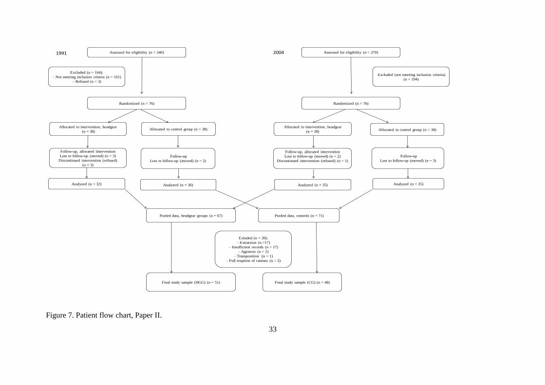
Paper II:
The data in this study were pooled from two RCTs performed in northern Finland studying
the outcomes of early HG treatment. The inclusion criteria were need for orthodontic
treatment due to moderate crowding and Angle Class II occlusion or tendency to Angle
Class II occlusion (cusp to cusp). Children with known syndromes and a cleft lip and palate
diagnosis were excluded. The first RCT (4, 11) included 71 seven-year-old children (mean
age 7.2 years, SD 0.6 years), and the second RCT included 67 seven-year-old children (mean
age 7.6 years, SD 0.3 years) (Figure 7). In both RCTs, the children were randomly divided
into two groups of equal size: the HG treatment group (HGG) and the control group (CG).
Thirty-nine individuals were excluded from the pooled sample for the following reasons:

32
interceptive extraction of primary teeth (n = 17), missing images (n = 15), poor image quality
(n = 2), full eruption of maxillary canines at T1 (n = 2), agenesis of lateral incisors (n = 2),
and transposition (canines and first premolars) (n = 1). The final study sample therefore
consisted of 99 subjects: 51 in the treatment group (HGG) and 48 in the CG. The HGG
comprised 39 per cent females and 61 per cent males, and the corresponding numbers for the
CG were 38 per cent females and 62 per cent males. The mean age of the pooled sample at T0
was 7.7 years (SD 0.4 years) in the HGG and 7.5 years (SD 0.4 years) in the CG. Interceptive
slicing of the mesial surface of the primary canines was performed in two children in the
HGG and three children in the CG; these cases were not excluded. No further interceptive
treatment was done in either group.
33
Randomized (n = 76)
Allocated to control group (n = 38)
Pooled data, headgear groups (n = 67)
Excluded (not meeting inclusion criteria)
(n = 194)
Assessed for eligibility (n = 270)
Pooled data, controls (n = 71)
Assessed for eligibility (n = 240)
Excluded (n = 164):
– Not meeting inclusion criteria (n = 161)
– Refused (n = 3)
1991 2004
Randomized (n = 76)
Follow-up, allocated Intervention
Lost to follow-up (moved) (n = 3)
Discontinued intervention (refused)
(n = 3)
Follow-up
Lost to follow-up (moved) (n = 2)
Allocated to intervention, headgear
(n = 38)Allocated to control group (n = 38)
Allocated to intervention, headgear
(n = 38)
Analyzed (n = 32) Analyzed (n = 36)
Follow-up, allocated intervention
Lost to follow-up (moved) (n = 2)
Discontinued intervention (refused) (n = 1)
Follow-up
Lost to follow-up (moved) (n = 3)
Analyzed (n = 35) Analyzed (n = 35)
Final study sample (HGG) (n = 51) Final study sample (CG) (n = 48)
Exluded (n = 39):
– Extraction (n =17)
– Insufficient records (n = 17)
– Agenesis (n = 2)
– Transposition (n = 1)
– Full eruption of canines (n = 2)
Fig1. Patient flow chart
Figure 7. Patient flow chart, Paper II.

34
Parental informed consent was obtained before randomization. The second RCT was
approved by the Ethics Committee of Oulu University Hospital, Finland (EETTMK:
46/2003), and registered at Clinicaltrials.gov, number NTC02010346.
Paper III:
This study took place at the Public Dental Health Competence Centre of Northern Norway
and one private clinic in Bryne, Norway, between 1 January 2013 and 31 December 2018.
Inclusion criteria were: children with dental age of at least 9.5 years (110) and the presence of
both primary maxillary canine and primary maxillary first molars and a palatally displaced
permanent maxillary canine (PDC). PDC was defined as eruption of the maxillary canine in
sectors III and IV according to Lindauer et al. (15) or eruption of the maxillary canine in
sector II with an angle between the long axis of the canine and the facial midline (Angle C) of
at least 25° (Figure 1).
Exclusion criteria were: previous orthodontic treatment, any disease not allowing local
anaesthesia or extraction, craniofacial syndromes, cleft lip palate, odontomas, cysts, and
agenesis of the maxillary lateral incisor.
Thirty-two children, 18 girls and 14 boys with mean ages (SD) of 10.7 (0.7) and 11.2 (1.0),
respectively, were invited to participate in the study, and all accepted. Sixteen children had
bilateral PDCs and each single canine served as a separate unit in the study; in total, 48 PDCs
were included in the study.

35
The data were collected after approval by the regional ethics committee (REC North) in June
2012 (2012/623/REK nord). Informed consent was obtained from the child and a parent or
from an adult with parental responsibilities and rights.
8.2 Methods
Paper I:
The study was designed as a retrospective study. CBCT images acquired in small (n = 56) and
medium (n = 3) field of view (FOV) were analysed.
The following measurements were performed for every subject on the CBCT images:
1. grading of root resorption severity according to Ericson and Kurol (61)
2. localization of the resorptions (i.e., apical, middle, or cervical third of the root)
3. position measurements of the canines:
a. palatal, labial, or in line with the arch (Figure 8)
b. distance from the most inferior point of the canine crown to the occlusal plane
(Figure 9)
c. canine angulation to the lateral (Figure 10)
d. canine angulation to the midline (Figure 10)
e. canine location in sectors (Figure 2) (panorama reconstruction view)

36
Figure 8. Maxillary canine location relative to the dental arch.
Figure 9. Maxillary canine angulation and distance to the occlusal plane.
Figure 10. Maxillary canine angulation to the midline and lateral incisor.

37
Paper II:
The data in this study were pooled from two RCTs studying the outcomes of early HG
treatment.
Children in the HGG were treated with cervical HG using 400–700-g force for 1 year or until
Angle Class I was achieved. The mean treatment time was 23.8 months (SD 5.6).
Panoramic radiographs and dental casts were taken before (T0) and after (T1) the study. The
radiographs were imported into the Facad® tracing programme (Ilexis, Linkoping, Sweden)
and angular measurements of the maxillary canine were performed (angles A–C, Figure 1)
Dental casts were digitized and analysed using Ortho AnalyzerTM computer software (3Shape,
Copenhagen, Denmark). Digital model measurements were performed along a constructed
occlusal plane, using the mesiobuccal cusp tips of the maxillary right and left first molars and
the incisal edges of the right or left central incisor. In cases with a deviating incisor position,
the incisor considered to be in the most “correct” position was used. Dental arch distances
were measured between the most buccal aspects of the contact points. For transpalatal
measurements, distances were measured between cusp tips (Figure 11).

38
Figure 11. Digital model analysis: (A) arch perimeter, (B) premolar space, (C) incisor space,
(D) premolar and canine space, (E) intercanine distance, and (F) intermolar space.
Paper III:
The study was designed as a randomized controlled clinical trial. The randomization was
performed using the block randomization method and the children were assigned in an
allocation ratio of 1:1 to either the double extraction group (DEG, n = 25) or single extraction
group (SEG, n = 23). The children were examined clinically, and by panoramic radiographic
imaging before extraction (T0) and every 6 months until the canine erupted into the mouth
(T1–Tx). If the canine position worsened or improvement was undetectable after 12 months,
alternative treatment was administered (i.e., surgical exposure, fixed orthodontic appliances,
and extractions). Clinical photos were taken of each participant before and at the end of the
study.
The panoramic radiographic images were imported into the Facad® tracing program (Ilexis,
Linkoping, Sweden) and angles A, B, and C (Figure 1) and sectors (Figure 2) were measured.

39
The clinical photos were visually inspected by one orthodontist (SHO) and each patient was
categorized according to the space conditions in the maxillary arch:
o crowding: one or more teeth are overlapping and displaced
o no crowding: all teeth are well aligned
o minor spacing: small open spaces between teeth (total ≤2 mm)
o major spacing: larger spaces between teeth (total >2 mm)
The following outcomes were assessed:
Primary outcome:
emergence of the maxillary canine into the oral cavity:
o “successful”: maxillary canine emerged through the gingiva
o “unsuccessful”: no eruption of the maxillary canine into the oral cavity
emergence of the maxillary canine in a “favourable position”:
o maxillary canine emerged in sector I in normal bucco–palatal relationship with
occluding teeth in the mandible (i.e., no crossbite)
Secondary outcome:
maxillary canine positional changes (angles A–C and sectors)
changes in maxillary arch space conditions
8.3 Reliability of measurements
Paper I:

40
Eighteen consecutive cases were evaluated by two observers to assess inter-rater agreement,
and 21 consecutive cases were evaluated with a time lapse of 1 month to assess intra-rater
agreement. Intraclass correlation was used for continuous variables and Kappa statistics
(Cohen’s kappa) for categorical variables. Both the inter- and intra-rater reliability showed
substantial agreement for the categorical variables (kappa = 0.64 – 0.88) and good–excellent
agreement for the continuous variables (ICC = 0.81 – 0.96).
Paper II:
Thirty panoramic radiographs and 20 digital models were measured and scored twice within 2
weeks by one investigator (SHO). The reliability analysis of the measurements of panoramic
radiographs indicated “acceptable” agreement for the measurement “canine to the maxillary
midline” (ICC = 0.745), and “excellent” and “almost perfect” agreement for all other
variables (ICC = 0.905–0.984, kappa = 0.92–1.00). For the 3D model analysis, the reliability
of all the variables was rated as “excellent” (ICC: 0.904–0.997).
Paper III:
The reliability of panoramic radiograph measurements was reported in Paper II.
The reliability of the space condition analysis was tested by measuring 20 randomly selected
plaster models (measured with sliding callipers) and 20 digital photos (measured visually).
The ICC was calculated to be 0.889, indicating excellent agreement.
8.4 Statistical analysis
41
The Statistical Package for the Social Sciences (SPSS) software for Windows, version 19, 24
and 25 (IBM, Armonk, NY, USA), and G*power, version 3.1.9.2 (copyright 2010–2013,
Heinrich-Heine Universität, Düsseldorf, Germany) were used for all calculations. Descriptive
statistics were calculated and presented as mean values and standard deviations. The
differences between various variables were tested using the Chi-square test, t-tests
(independent and paired), and the Mann–Whitney U test. To test the association between
various variables and root resorption, binary multiple logistic regression was used (Paper I).
To search for the best predictors of change in canine angulation (Paper II) and the emergence
of the maxillary canine (Paper III), the statistically significant variables from a univariate
calculation were entered into a stepwise regression model, and variables were excluded one
by one on the grounds of the P-value or the effect of beta. The level of significance was set at
p < .05.
Sample size calculation:
Paper II:
Based on an earlier published study, which used part of the present sample (104), 44 canines
each were needed in the HG and control groups. This calculation was based on the non-
significant changes observed in the alpha angle for the left maxillary canine after 2 years of
HG treatment, with alpha = 0.05, beta = 0.2, and power = 0.8.
Paper III:
Based on a comparable study by Bonetti et al. (109), 22 canines each were required in the
single and double extraction groups. This sample size is based on the differences in Angle C
(i.e., alpha angle) between the single and double extraction groups, with alpha = 0.05, beta =
0.2, and power = 0.8.

42
9 Results
Prevalence and severity of root resorption (Paper I):
When the maxillary canine was located ectopically, root resorption was found in 11% of
central incisors and 67 per cent of lateral incisors, versus 0% and 36%, respectively, when the
canine erupted normally. The difference in root resorption prevalence between the ectopic and
normal canine groups was statistically significant (p = .002). Most cases of resorption were
defined as “slight” and located in the middle third of the root. The best predictor of resorption
was the maxillary canine located mesial to the midline of the lateral incisor.
The impact of HG treatment on the eruption path of maxillary canines (Paper II):
The dental arch size increased in both the headgear group (HGG) and control group (CG)
from start (T0) to end (T1) of the study. HG treatment led to significantly greater increases in
arch perimeter (p = .002), intermolar distance (p < .001), and intercanine distance (p < .001)
than found in the CG.
The mean angular change of the permanent maxillary canine (Angle A) was significant in the
HGG (left: p = .012, right: p = .051), but not in the CG (left: p = .332, right: p = .295).
The space conditions in the dental arch affected the change in canine angulation in the HGG
but not in the CG. In the HGG, the maxillary canine angulation changed significantly more in
spaced than in crowded arches (left: p = .020, right: p = .031). A significant difference
between the HGG and CG was seen on the left side (i.e., a more vertical eruption pattern in
spaced than in crowded dental arches) (p = .025). In crowded arches, there was no difference
in canine angulation between the HGG and CG.

43
The best predictor of change in canine angulation (“Angle A, T1–T0”) was “incisor space” at
T0 (Figure 11).
The impact of double versus single extraction on the emergence of PDCs (Paper III):
Primary outcome:
No significant difference in the emergence rate of the maxillary canine was observed between
the DEG and SEG, i.e., 16/25 (64%) versus 18/23 (78%) (p = .283), and no significant
difference was found in emergence in a “favourable position”, i.e., 16/25 (64%) versus 13/23
(57%) (p = .600).
Of the PDCs that emerged into the oral cavity (34/48), significantly more canines emerged in
a “favourable position” in the DEG than in the SEG: 100 per cent versus 72 per cent (p =
.025).
Secondary outcome:
The angular and sector measurements indicated significant distal movement of the canines in
both groups (p < .001). However, no significant difference was found between the two groups
in changes in canine angle (A–C) or sector.
A significant reduction in estimated space was seen in both groups from T0 to Tend (p < .001),
but no significant difference was recorded between the groups (p = .727).
Predictive factors for the emergence of the maxillary canine into the oral cavity:
44
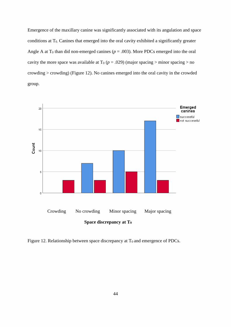
Emergence of the maxillary canine was significantly associated with its angulation and space
conditions at T0. Canines that emerged into the oral cavity exhibited a significantly greater
Angle A at T0 than did non-emerged canines (p = .003). More PDCs emerged into the oral
cavity the more space was available at T0 (p = .029) (major spacing > minor spacing > no
crowding > crowding) (Figure 12). No canines emerged into the oral cavity in the crowded
group.
Crowding No crowding Minor spacing Major spacing
Space discrepancy at T0
Figure 12. Relationship between space discrepancy at T0 and emergence of PDCs.

45
10 Discussion
10.1 Methodological considerations: strengths and weaknesses
Paper I:
This study was designed as an observational study with a cross-sectional, retrospective
design. The advantage of such studies is that subjects are not deliberately exposed, treated, or
not treated, so there are seldom ethical difficulties. They are also relatively cheap to perform
and it is possible to study multiple outcomes (111). A general problem with cross-sectional
studies is that they do not provide an explanation of the results, so only association and not
causation can be inferred. This is because it is impossible to control for confounding factors
that may affect the outcome in a retrospective study.
In this study, only patients with suspicion of or actual eruption disturbance in the maxillary
canine region were included. These patients were selected and referred by their general dentist
or orthodontist for CBCT imaging. Sampling bias may therefore be present, as the selection
was dependent on the doctor’s decision.
Ninety-seven children were included in the study, but x-ray images from 38 patients had to be
excluded for various reasons (e.g., patient movement, noise, syndromes, too early eruption
stage, and presence of orthodontic appliances). Observer selection may therefore have
occurred, as exclusion was dependent on the researcher’s opinion concerning which patients
to exclude (112).
When measuring the canine position in sagittal view, the occlusal plane was used as the
reference line (Figure 9). This is in accordance with several previous studies (9, 12, 103, 109),

46
but as the occlusal plane is an unstable structure, this may be inaccurate. A more reliable
reference line would be the “spina nasalis anterior to spina nasalis posterior”, which was used
by Naoumova et al. (14).
This study did not assess the validity of the CBCT measurements. However, previous studies
have found the linear and angular CBCT measurements of PDCs to be good (100), with a
mean difference between physical and 3D measurements of 0.5 ± 0.39 mm for the sagittal
angle and 0.22 ± 0.19 mm for the mesio–angular angle (100). These angles are comparable to
those used in this study (Figure 10). The reliability of the measurements was good in the
present study, as the intra- and inter-rater calculations indicated good to excellent agreement
for the localization of canines and substantial agreement for the assessment of root resorption.
The different voxel sizes of different CBCT devices can influence the detection of slight root
resorption (65). In this study, 56 images were taken with a 0.2-mm voxel size and three
images were taken with a 0.3-mm voxel size. According to Liedke et al. (86), the difference
between voxel sizes of 0.2, 0.3, and 0.4 mm in the detection of external root resorption using
CBCT imaging is not statistically significant. Therefore, different voxel sizes should not
represent a significant bias in this study.
In clinical practice, it is important to note that angles and sectors are displayed differently
between CBCT and panorama radiographs (84). Generally, the PDC position is exaggerated
on panorama radiographs relative to CBCT images (113). Therefore, there is a chance of
overestimating the root resorption risk, as clinicians mainly use panorama radiographs and
studies of root resorption generally use CBCT images.
Paper II:

47
The data were pooled from two randomized controlled trials concerning the outcomes of early
HG treatment. When pooling data from different studies, the combined results may contradict
the results of the individual studies. This effect, also known as “Simpson’s paradox”, may
arise when important subgroup characteristics in the different studies are not considered or
weighted (114). As these two studies were similar in design, weighting of the subgroups was
not considered necessary and therefore was not performed.
Lack of patient compliance records is a weakness of this study, but the endpoint of full Angle
Class I occlusion implies proper use of HG. However, large individual variation in how much
the HG was used and how the patients reported their compliance has been reported (115). The
treatment effect may therefore be underestimated for compliant patients and overestimated for
non-compliant patients. For future studies, objective monitoring of HG use is recommended.
Exclusion of patients from RCTs may induce selection bias (112). In the present study, 39
children were excluded from the study for the following reasons: extraction of primary teeth,
(n = 17), incomplete records (n = 17), and inappropriate sampling (i.e., agenesis, n = 2;
transposition, n = 1; full eruption, n = 2).
Exclusion of patients due to primary tooth extraction was necessary, as the extraction of
primary teeth affects the eruption path of permanent teeth (11, 14, 103). Incomplete records
were mainly due to insufficient radiographic image quality. Generally, panorama image
quality is operator dependent, and these images may not always correctly capture the patient’s
condition (116). Angular measurements were performed, as distortion and overlapping make
horizontal measurements unreliable in panorama radiographs (79). Blurring may accentuate
the upper incisor region due to ghost shadows of the cervical spine (117). This may be the
reason for the reduced reliability (ICC = 0.745) of the angle between the canine and facial
midline (alpha angle or Angle C, Figure 1). On the other hand, the angle between the canine

48
and the bicondylar line (Angle A, Figure 1) proved to be a more reliable variable, and was
therefore used as the dependent variable in the regression analysis. Parenti et al. also reported
Angle A to be the most reliable angle to measure for erupting maxillary canines (118).
However, Angle A may be unstable on a long-term basis as it is prone to changes in the
condyles, so caution should be taken in follow-up studies in this field.
In the present study, plaster models were digitized and analysed using Ortho AnalyzerTM
computer software (3Shape, Copenhagen, Denmark). Digital model measurements have been
found to be reliable, valid, and accurate in comparison to conventional impressions (119-121).
The digital model measurements (Figure 11) were performed along a constructed occlusal
plane, using the mesiobuccal cusp tips of the maxillary right and left first molars and the
incisal edges of the right or left central incisor. In cases with a deviating incisor, the incisor
considered to be in the “correct position” was used. As “correct position” may be a somewhat
subjective assessment, the construction of the occlusal plane may be inaccurate. Furthermore,
in cases of severe crowding, the arch perimeter (A, Figure 8) may be difficult to place, and
may be assessed differently between raters. The intra-rater reliability indicated excellent
agreement (ICC: 0.904-0.997) for the model analysis, but inter-rater agreement could have
been determined to ensure sufficient reliability.
Paper III:
This study was designed as a randomized controlled trial with a two-arm parallel group
design. This design is considered the gold standard for primary studies (112). In the
randomization process, each canine was used as one unit in this study. Each person could also
have been used as a unit, in view of the genetic aetiology theory of PDCs (25), as children
with bilateral ectopic canines may react similarly to treatment on both sides of the maxilla.

49
However, if each child was used as a unit, the study would have been more difficult to
perform as more children would have had to be included. Also, it would be impossible to see
whether changes in treatment outcome were dependent on the person or the treatment method.
According to our sample size calculations, 22 canines were needed in each of the two arms; to
compensate for possible dropouts, 48 canines were included in the study. During recruitment,
it was originally planned that information about the study would be given by an independent
person without a relationship to the patient (i.e., not a nurse or doctor treating the patient).
This proved difficult in practice, as questions regarding treatment were difficult for persons
not involved with the patient to answer. However, caution was taken not to persuade patients
to take part in the study, so that participation would be freely chosen.
Randomization was performed using the block randomization method (122). Block sizes
varied randomly among 2, 4, 6, and 8, as two closely balanced groups were needed at all
times in case the study had to be terminated. The study was unblinded, as the treatment was
impossible to hide for the patient and the doctor. In studies with an objective outcome, such
as this one, unblinded studies do not tend to be more biased than blinded ones (123). To
reduce bias, an independent person, not knowing the purpose of the study, measured the
radiographs.
Allocation was concealed by enclosing assignments in sequentially numbered sealed
envelopes. After randomization, children were assigned in an allocation ratio of 1:1 to either
the double extraction group (DEG) or single extraction group (SEG). None of the children
refused to take part in the study and there were no drop-outs, which is a great strength. A
control group was not included, as withholding treatment was considered unethical in this
patient group (9, 13, 14).

50
10.2 Statistical analysis
In statistical analysis there are many potential sources of bias. One essential factor is the
sample size used in the studies (124). Non-significant results may turn out to be statistically
significant with larger group sizes. Also, the division of samples into smaller subgroups may
weaken the chance of achieving statistically significant results. As a rule, multivariate
analysis needs a minimum of ten events per variable to ensure reliable modelling (125). In
Paper III, the variable “space discrepancy at T0” was not included as it contained groups of
fewer than 10 cases. The multivariate analysis in Paper III could therefore have had a
different outcome with a larger sample size.
When calculating the required sample size, it is important to evaluate which variable to use as
a basis for the power calculation. This is especially important in studies in which multiple
outcomes are studied. It may be that the sample size calculation is appropriate for one
outcome and not for another. In Paper III, observed change in the alpha angle was used
(Angle C) for the sample size calculation, in accordance with several previous studies of
PDCs (9, 13, 14). That means that our study had a sufficiently large sample to investigate
changes in alpha angle, but may have had too low a power to actually detect any difference in
some of the other outcome variables. Low power is unfortunately a common problem in
science. A meta-analysis of studies in neuroscience showed that the average study had a
power of approximately 21% (126). The problem with low power is that the likelihood of
detecting a difference, if there is one, is low (i.e., type II error). In retrospect, the sample size
in Paper III could have been larger to more reliably address some of the outcomes studied. To
achieve sufficient sample size for conditions with low prevalence, such as ectopic canines, a
multicentre study would have been preferable.

51
10.3 Ethical considerations
Children are considered a vulnerable group, so research involving children is strictly
regulated. Clinical studies of children should be performed in such a way that new useful
knowledge is generated. The aims of our studies were to improve our understanding of oral
health in children and to reinforce the scientific foundation for the treatment of ectopic
maxillary canines. These aims are in accordance with the United Nations Convention on the
Rights of the Child (Article 24):
1. States Parties recognize the right of the child to the enjoyment of the highest attainable
standard of health and to facilities for the treatment of illness and rehabilitation of
health. States Parties shall strive to ensure that no child is deprived of his or her right
of access to such health care services.
As the children in these studies were young, they had reduced autonomy and were dependent
on their parents or an adult with parental responsibilities and rights. Therefore, informed
written consent was obtained from the child by his or her caregiver, but if situations had
arisen in which the child did not want to participate, he or she would not have been included
in the study in order to respect the ethical principle of autonomy. Age-adapted written
information about the study was given as well as information about, for example, the study’s
purpose, benefits, potential negative effects, data handling, and examination method, as well
as the principle of voluntary participation.
Radiographs were taken during the studies, but no extra images were taken for the purpose of
the studies. If caries or other signs of oral disease were found, the children’s regular dental
clinics were informed.

52
The studies were performed according to the Declaration of Helsinki, which represents one of
the key ethical guidelines for research with human subjects (127).
No financial support influenced the studies and their results.
10.4 Discussion of main findings
Paper I:
The aims of this study were to measure the prevalence and severity of the root resorption of
maxillary incisors caused by ectopically and normally erupting maxillary canines, and to
determine predictors of the root resorption of incisors using CBCT imaging.
The prevalence of the root resorption of maxillary incisors varies with the population studied
(128). To the best of our knowledge, this is the first study of root resorption in Finnish
children, so we do not have exactly comparable data. However, comparing the results from
the ectopic canine group with the results of studies from other parts of the world reveals both
similar (57, 61) and lower (62) prevalences of incisor resorption. These differences likely
reflect variance in the malposition of the maxillary canine, female to male ratio, and age
group of the samples. In addition, recent studies have found that genetic background plays an
important role, as individual susceptibility to root resorption is considered a major factor, both
in orthodontic treatment and in other contexts (129, 130). One study claimed that genetic
influence accounted for approximately 50% of the observed variation in external root
resorption after orthodontic treatment, with variation in the gene for the inflammatory
cytokine Interleukin 1B determining 15% of the observed variation (129). Differences in the
definition of “ectopic canine” between studies, or even the lack of a definition, also make

53
comparison difficult. Some use numerical values in their definitions (9, 11), whereas others
are limited to a qualitative description of the canine position on radiographs and in clinical
examinations (14). These differences may cause bias when comparing studies of root
resorption.
Interestingly, approximately 1/3 of the patients with canines erupting normally exhibited
resorption, a significantly higher prevalence than the 5% previously reported (61). However,
studies of root resorption in normally erupting canines are scarce. Our finding suggests that
small root resorptions may be more common than previously assumed, but future studies are
needed to verify this finding, preferably in a less selective population.
The mesio–distal position of the canine (sectors 3–5, Figure 2) was found to be the best
predictor of root resorption in the present sample. As opposed to previous studies, we did not
find any significant association between canine angulation relative to the facial midline
(Angle C > 25°, Figure 1) (26) or between canine angulation relative to the lateral incisor
(Angle B > 54 º, Figure 1) and root resorption (131).
Paper II:
The aims of this study were to assess whether HG treatment in young children affects the
maxillary canine eruption path and to determine whether the potential effect on the eruption
pattern is related to space conditions in the dental arch.
This study showed that HG treatment in young children influences the eruption pattern of
maxillary canines. In a previous study, the same relationship was found, but only for the right
maxillary canine (104). Our study has a larger sample size and therefore a higher chance of
detecting a difference in canine angulation. Our study does not reveal the mechanisms by

54
which HG treatment influences the canine eruption pattern, but it may be related to transseptal
fibres (106) applying a distally directed force to the maxillary teeth as the first molars are
pulled distally. In addition, both papers II and III in our study indicate that increased space in
the maxillary arch promotes maxillary canine eruption. The HG treatment may increase the
space in the canine region by distalization, the lip bumper effect, and an increased transversal
dimension. Of special interest is the correlation between available space in the anterior part of
the maxilla and the vertical eruption of the maxillary canine. In spaced dental arches, the
maxillary canine changes its eruption pathway to become significantly more vertical than in
crowded arches. This is consistent with the findings of Paper III, showing that the amount of
space available in the maxillary arch is related to maxillary canine emergence. Other studies
have found that the emergence rate of PDCs increases when HG, rapid maxillary expanders,
or transpalatal arches are used in addition to primary canine extraction (11, 12), but they did
not quantify the association between the space discrepancy in the maxilla and canine angular
change.
The children in the present study sample were approximately 7.5 years old at T0. Canine
displacement cannot be diagnosed at such a young age (132), so we cannot conclude that HG
treatment influences displaced canines. To verify the influence of HG on ectopic canines,
studies of an older patient cohort with verified displaced canines are needed.
Paper III:
In Paper III, we tested two different interceptive treatment protocols (i.e., double vs. single
extractions) for PDCs, as previous studies have presented conflicting results about the
outcome (9, 109) and have been criticized for their methodology (107).

55
The two protocols did not identify any statistically significant difference in the emergence
rate of PDCs depending on whether the primary canine only was extracted or both the
primary canine and primary first molar were extracted. This is similar to the results of Bonetti
et al.’s first study (109) and in contrast to those of their second study (9). These two studies
also found that the angle of the maxillary canine (Angle C, Figure 1) changed significantly
more when double rather than single extractions were performed, which was not verified in
the present study. The discrepancy between the two studies by Bonetti et al. (9, 109) and the
present one is likely related to differences in the samples studied. Compared with previous
studies, the present study included older patients with more severe malpositioning of the
maxillary canine.
In the present study, significantly more of the emerged canines in the DEG erupted in a
“favourable position” versus in the SEG. The reason for this observation is unknown, but may
be related to a change in the first premolar eruption path (109) that secondarily affects the
canine eruption path. It may also be a random finding. To investigate this finding further, a
larger study cohort is needed.
We found an interesting significant association between canine emergence and space
conditions in the maxillary arch. No canines emerged in the “crowded” group but more
canines gradually emerged as the available space increased (Figure 12). A similar finding was
reported by Power and Short (8). This information could be valuable for clinicians, who
should be sure to facilitate canine emergence by increasing the maxillary dental arch space
when needed.
Bonetti et al. claimed that the extraction of two teeth is no more traumatic for the patient or
technically difficult than the extraction of one tooth (9). Based on experience from the present
study, I disagree with this statement. Compared with extracting just the single-rooted primary

56
canine, extracting both the primary canine and the multi-rooted first primary molar takes
longer, requires more anaesthesia, and entails more patient discomfort.
Papers I–III:
Overall, this study shows that it is important to monitor the ectopic maxillary canine eruption
in children, as root resorption is commonly found on maxillary incisors. HG treatment affects
the eruption path of normally erupting maxillary canines in such a way that it may be
beneficial for ectopically erupting maxillary canines, but this matter should be studied further.
Space conditions in the maxillary arch affect both the normal and ectopic eruption of
maxillary canines. The present study supports the currently most common interceptive
treatment method, primary canine extraction, as the best interceptive treatment modality for
PDCs.

57
11 Conclusions
In line with the objectives specified for the thesis, the following main conclusions were
drawn:
1. Lateral incisor root resorption was found in association with approximately 2/3 of
ectopically erupting canines and 1/3 of normally erupting canines in patients referred
for CBCT imaging due to maxillary canine eruption disturbances.
2. The best predictor of root resorption in our cohort was location of the canine mesial to
the midline of the lateral incisor root in the panoramic reconstruction view of a CBCT
image.
3. HG treatment in young children with Angle Class II occlusion may shift the eruption
path of maxillary canines to a more vertical direction, and the effect is related to space
conditions in the maxillary arch.
4. Double or single primary tooth extraction procedures are similar in supporting the
eruption of PDCs into the oral cavity and into a “favourable position” in the dental
arch.
5. Initial canine angulation and space conditions may be used as predictors of the
successful eruption of PDCs.

58
12 Future perspectives
Even though ectopic eruption of the maxillary canines affects many children each year, there
is still limited scientific evidence for interceptive treatment of this condition. An interesting
avenue for future research would be to further investigate the impact of single versus double
primary tooth extraction on PDC emergence in a larger sample. It would also be interesting to
investigate the effects of double extractions concomitant with orthodontic appliances such as
HG, the quad helix, or transpalatal arches on PDC emergence and on space conditions in the
maxillary arch. Cost/benefit analyses of different treatment approaches are needed, as are
investigations of the patient perspective on treatment. In addition, more basic research
employing genetic tests for predicting ectopic eruption as well as susceptibility to root
resorption would be interesting to pursue.

59
13 References
1. Stewart JA, Heo G, Glover KE, Williamson PC, Lam EW, Major PW. Factors that
relate to treatment duration for patients with palatally impacted maxillary canines. Am J
Orthod Dentofacial Orthop. 2001;119(3):216-25.
2. Gharaibeh TM, Al-Nimri KS. Postoperative pain after surgical exposure of palatally
impacted canines: closed-eruption versus open-eruption, a prospective randomized study.
Oral Surg Oral Med Oral Pathol Oral Radiol Endod. 2008;106(3):339-42.
3. Bazargani F, Magnuson A, Dolati A, Lennartsson B. Palatally displaced maxillary
canines: factors influencing duration and cost of treatment. Eur J Orthod. 2013;35(3):310-6.
4. Bjerklin K, Ericson S. How a computerized tomography examination changed the
treatment plans of 80 children with retained and ectopically positioned maxillary canines.
Angle Orthod. 2006;76(1):43-51.
5. Broadbent, BH. Ontogenic development of occlusion. Angle Orthodontist.
1941(11):223-41.
6. Coulter J, Richardson A. Normal eruption of the maxillary canine quantified in three
dimensions. Eur J Orthod. 1997;19(2):171-83.
7. Jacoby H. The etiology of maxillary canine impactions. Am J Orthod. 1983;84(2):125-
32.
8. Power SM, Short MB. An investigation into the response of palatally displaced
canines to the removal of deciduous canines and an assessment of factors contributing to
favourable eruption. Br J Orthod. 1993;20(3):215-23.
9. Alessandri Bonetti G, Zanarini M, Incerti Parenti S, Marini I, Gatto MR. Preventive
treatment of ectopically erupting maxillary permanent canines by extraction of deciduous
canines and first molars: A randomized clinical trial. Am J Orthod Dentofacial Orthop.
2011;139(3):316-23.
10. Ericson S, Kurol J. Radiographic examination of ectopically erupting maxillary
canines. Am J Orthod Dentofacial Orthop. 1987;91(6):483-92.
11. Sigler LM, Baccetti T, McNamara JA, Jr. Effect of rapid maxillary expansion and
transpalatal arch treatment associated with deciduous canine extraction on the eruption of
palatally displaced canines: A 2-center prospective study. Am J Orthod Dentofacial Orthop.
2011;139(3):235-44.

60
12. Baccetti T, Sigler LM, McNamara JA, Jr. An RCT on treatment of palatally displaced
canines with RME and/or a transpalatal arch. Eur J Orthod. 2011;33(6):601-7.
13. Bazargani F, Magnuson A, Lennartsson B. Effect of interceptive extraction of
deciduous canine on palatally displaced maxillary canine: a prospective randomized
controlled study. Angle Orthod. 2014;84(1):3-10.
14. Naoumova J, Kurol J, Kjellberg H. Extraction of the deciduous canine as an
interceptive treatment in children with palatal displaced canines - part I: shall we extract the
deciduous canine or not? Eur J Orthod. 2015;37(2):209-18.
15. Lindauer SJ, Rubenstein LK, Hang WM, Andersen WC, Isaacson RJ. Canine
impaction identified early with panoramic radiographs. Journal of the American Dental
Association. 1992;123(3):91-2, 5-7.
16. Becker A. The orthodontic treatment of impacted teeth. 2nd ed.
Abingdon,Oxon,England: Informa Healthcare; 2007.
17. Peck S, Peck L, Kataja M. Prevalence of tooth agenesis and peg-shaped maxillary
lateral incisor associated with palatally displaced canine (PDC) anomaly. Am J Orthod
Dentofacial Orthop. 1996;110(4):441-3.
18. Dewel BF. The upper cuspid: Its development and impaction. Angle Orthod.
1949(19):79-90.
19. Dachi SF, Howell FV. A survey of 3, 874 routine full-month radiographs. II. A study
of impacted teeth. Oral Surg Oral Med Oral Pathol. 1961;14:1165-9.
20. Grover PS, Lorton L. The incidence of unerupted permanent teeth and related clinical
cases. Oral Surg Oral Med Oral Pathol. 1985;59(4):420-5.
21. Bishara SE. Impacted maxillary canines: a review. Am J Orthod Dentofacial Orthop.
1992;101(2):159-71.
22. Baccetti T. A controlled study of associated dental anomalies. Angle Orthod.
1998;68(3):267-74.
23. Thilander B, Myrberg N. The prevalence of malocclusion in Swedish schoolchildren.
Scand J Dent Res. 1973;81(1):12-21.
24. Chu FC, Li TK, Lui VK, Newsome PR, Chow RL, Cheung LK. Prevalence of
impacted teeth and associated pathologies--a radiographic study of the Hong Kong Chinese
population. Hong Kong medical journal = Xianggang yi xue za zhi / Hong Kong Academy of
Medicine. 2003;9(3):158-63.
25. Peck S, Peck L, Kataja M. The palatally displaced canine as a dental anomaly of
genetic origin. Angle Orthod. 1994;64(4):249-56.
61
26. Ericson S, Kurol J. Resorption of maxillary lateral incisors caused by ectopic eruption
of the canines. A clinical and radiographic analysis of predisposing factors. Am J Orthod
Dentofacial Orthop. 1988;94(6):503-13.
27. Hitchen AD. The impacted maxillary canine. British Dental Journal. 1956;100:1-14.
28. Rayne J. The unerupted maxillary canine. Dent Pract Dent Rec. 1969;19(6):194-204.
29. Becker A. Orthodontic treatment of impacted teeth, third edition: Whiley-Blackwell;
2012.
30. Rozylo-Kalinowska I, Kolasa-Raczka A, Kalinowski P. Dental age in patients with
impacted maxillary canines related to the position of the impacted teeth. Eur J Orthod.
2011;33(5):492-7.
31. Peck S. Dental Anomaly Patterns (DAP). A new way to look at malocclusion. Angle
Orthod. 2009;79(5):1015-6.
32. Oliver RG, Mannion JE, Robinson JM. Morphology of the maxillary lateral incisor in
cases of unilateral impaction of the maxillary canine. Br J Orthod. 1989;16(1):9-16.
33. Broadbent BH. Ontogenic development of occlusion. The angle orthodontist.
1941;11:223-41.
34. Søndergaard M NSE. Retinerede maksillære permanente hjørnetænder-ætiologi og
diagnostik. Nor Tannlegeforen Tid 2018;128(128):780-7.
35. Peck S, Peck L, Kataja M. Concomitant occurrence of canine malposition and tooth
agenesis: evidence of orofacial genetic fields. Am J Orthod Dentofacial Orthop.
2002;122(6):657-60.
36. Becker A, Chaushu S. Etiology of maxillary canine impaction: a review. Am J Orthod
Dentofacial Orthop. 2015;148(4):557-67.
37. Becker A, Chaushu S. Authors' response. Am J Orthod Dentofacial Orthop.
2016;149(2):150-1.
38. Peck S. Misleading article on palatally displaced canines. Am J Orthod Dentofacial
Orthop. 2016;149(2):149-50.
39. Odeh R, Townsend G, Mihailidis S, Lahdesmaki R, Hughes T, Brook A.
Infraocclusion: Dental development and associated dental variations in singletons and twins.
Archives of oral biology. 2015;60(9):1394-402.
40. Chaushu S, Bongart M, Aksoy A, Ben-Bassat Y, Becker A. Buccal ectopia of
maxillary canines with no crowding. Am J Orthod Dentofacial Orthop. 2009;136(2):218-23.
41. Chaushu S, Zilberman Y, Becker A. Maxillary incisor impaction and its relationship to
canine displacement. Am J Orthod Dentofacial Orthop. 2003;124(2):144-50; discussion 50.
62
42. Richardson G, Russell KA. A review of impacted permanent maxillary cuspids--
diagnosis and prevention. J Can Dent Assoc. 2000;66(9):497-501.
43. Brin I, Becker A, Shalhav M. Position of the maxillary permanent canine in relation to
anomalous or missing lateral incisors: a population study. Eur J Orthod. 1986;8(1):12-6.
44. Brook AH. A unifying aetiological explanation for anomalies of human tooth number
and size. Archives of oral biology. 1984;29(5):373-8.
45. Bjerklin K, Kurol J, Valentin J. Ectopic eruption of maxillary first permanent molars
and association with other tooth and developmental disturbances. Eur J Orthod.
1992;14(5):369-75.
46. Naoumova J, Alfaro GE, Peck S. Space conditions, palatal vault height, and tooth size
in patients with and without palatally displaced canines: A prospective cohort study. Angle
Orthod. 2018.
47. Langberg BJ, Peck S. Adequacy of maxillary dental arch width in patients with
palatally displaced canines. Am J Orthod Dentofacial Orthop. 2000;118(2):220-3.
48. Fraga MR, Vitral RW, Mazzieiro ET. Tooth size reduction and agenesis associated
with palatally displaced canines. Pediatr Dent. 2012;34(3):216-9.
49. Zilberman Y, Cohen B, Becker A. Familial trends in palatal canines, anomalous lateral
incisors, and related phenomena. Eur J Orthod. 1990;12(2):135-9.
50. Ely NJ, Sherriff M, Cobourne MT. Dental transposition as a disorder of genetic origin.
Eur J Orthod. 2006;28(2):145-51.
51. Becker A, Gillis I, Shpack N. The etiology of palatal displacement of maxillary
canines. Clin Orthod Res. 1999;2(2):62-6.
52. Kim Y, Hyun HK, Jang KT. Interrelationship between the position of impacted
maxillary canines and the morphology of the maxilla. Am J Orthod Dentofacial Orthop.
2012;141(5):556-62.
53. Al-Nimri K, Gharaibeh T. Space conditions and dental and occlusal features in
patients with palatally impacted maxillary canines: an aetiological study. Eur J Orthod.
2005;27(5):461-5.
54. Naoumova J, Alfaro GE, Peck S. Space conditions, palatal vault height, and tooth size
in patients with and without palatally displaced canines: A prospective cohort study. The
Angle orthodontist. 2018;88(6):726-32.
55. Saiar M, Rebellato J, Sheats RD. Palatal displacement of canines and maxillary
skeletal width. Am J Orthod Dentofacial Orthop. 2006;129(4):511-9.

63
56. Tronstad L. Root resorption-etiology, terminology and clinical manifestations. Endod
Dent Traumatol. 1988;4(6):241-52.
57. Walker L, Enciso R, Mah J. Three-dimensional localization of maxillary canines with
cone-beam computed tomography. Am J Orthod Dentofacial Orthop. 2005;128(4):418-23.
58. Cernochova P, Krupa P, Izakovicova-Holla L. Root resorption associated with
ectopically erupting maxillary permanent canines: a computed tomography study. Eur J
Orthod. 2011;33(5):483-91.
59. Ericson S, Kurol J. Incisor resorption caused by maxillary cuspids. A radiographic
study. Angle Orthod. 1987;57(4):332-46.
60. Chaushu S, Kaczor-Urbanowicz K, Zadurska M, Becker A. Predisposing factors for
severe incisor root resorption associated with impacted maxillary canines. Am J Orthod
Dentofacial Orthop. 2015;147(1):52-60.
61. Ericson S, Kurol PJ. Resorption of incisors after ectopic eruption of maxillary canines:
a CT study. Angle Orthod. 2000;70(6):415-23.
62. Liu DG, Zhang WL, Zhang ZY, Wu YT, Ma XC. Localization of impacted maxillary
canines and observation of adjacent incisor resorption with cone-beam computed tomography.
Oral Surg Oral Med Oral Pathol Oral Radiol Endod. 2008;105(1):91-8.
63. Yan B, Sun Z, Fields H, Wang L. Maxillary canine impaction increases root resorption
risk of adjacent teeth: A problem of physical proximity. Orthod Fr. 2015;86(2):169-79.
64. Bjerklin K, Guitirokh CH. Maxillary incisor root resorption induced by ectopic
canines. Angle Orthod. 2011;81(5):800-6.
65. Lai CS, Bornstein MM, Mock L, Heuberger BM, Dietrich T, Katsaros C. Impacted
maxillary canines and root resorptions of neighbouring teeth: a radiographic analysis using
cone-beam computed tomography. Eur J Orthod. 2013;35(4):529-38.
66. Ericson S, Kurol J. Incisor root resorptions due to ectopic maxillary canines imaged by
computerized tomography: a comparative study in extracted teeth. Angle Orthod.
2000;70(4):276-83.
67. Ericson S, Bjerklin K, Falahat B. Does the canine dental follicle cause resorption of
permanent incisor roots? A computed tomographic study of erupting maxillary canines. Angle
Orthod. 2002;72(2):95-104.
68. Marks SC SH, Andreasen JO. Theories and mechanism of tooth eruption. St Louis:
Mosby; 1997.
69. Shafer WG HM, Levy BM. A textbook of oral pathology. WB Saunders, Philadelphia.
1984 (4th edn.).

64
70. Jacobs SG. Localization of the unerupted maxillary canine: how to and when to. Am J
Orthod Dentofacial Orthop. 1999;115(3):314-22.
71. Chalakkal P, Thomas AM, Chopra S. Displacement, location, and angulation of
unerupted permanent maxillary canines and absence of canine bulge in children. Am J Orthod
Dentofacial Orthop. 2011;139(3):345-50.
72. Ericson S, Kurol J. Longitudinal study and analysis of clinical supervision of
maxillary canine eruption. Community Dent Oral Epidemiol. 1986;14(3):172-6.
73. Bonetti GA, Parenti SI, De Rinaldis S, Marini I. Association between the inclination
of the lateral incisors and the position of the erupting canines on panoramic radiographs. Acta
odontologica Scandinavica. 2011;69(4):222-8.
74. Falahat B, Ericson S, Mak D'Amico R, Bjerklin K. Incisor root resorption due to
ectopic maxillary canines: a long-term radiographic follow-up. Angle Orthod.
2008;78(5):778-85.
75. Dental radiographic examinations: recommendations for patient selection and limiting
radiation exposure In: American Dental Association Cosa, editor. 2012.
76. Jacobs SG. Radiographic localization of unerupted maxillary anterior teeth using the
vertical tube shift technique: the history and application of the method with some case reports.
Am J Orthod Dentofacial Orthop. 1999;116(4):415-23.
77. Smith NJ. Orthodontic radiology: a review. Int Dent J. 1987;37(1):16-24.
78. Quintero JC, Trosien A, Hatcher D, Kapila S. Craniofacial imaging in orthodontics:
historical perspective, current status, and future developments. Angle Orthod.
1999;69(6):491-506.
79. Lurie A. Panoramic Imaging. In: White SC PM, editor. Oral radiology Principles and
interpretation. 5. China: Mosby; 2004. p. 191–209.
80. Scarfe WC, Farman AG, Sukovic P. Clinical applications of cone-beam computed
tomography in dental practice. J Can Dent Assoc. 2006;72(1):75-80.
81. Preda L, Di Maggio EM, Dore R, La Fianza A, Solcia M, Schifino MR, et al. Use of
spiral computed tomography for multiplanar dental reconstruction. Dentomaxillofac Radiol.
1997;26(6):327-31.
82. Scarfe WCF, A.G. Cone beam computed tomography: volume acquisition. Oral
Radiology, Principles and interpretations. 7 ed: Elsevier, Mosby; 2014. p. 185-98.
83. Haney E, Gansky SA, Lee JS, Johnson E, Maki K, Miller AJ, et al. Comparative
analysis of traditional radiographs and cone-beam computed tomography volumetric images
in the diagnosis and treatment planning of maxillary impacted canines. Am J Orthod
Dentofacial Orthop. 2010;137(5):590-7.

65
84. Alqerban A, Jacobs R, Fieuws S, Willems G. Comparison of two cone beam computed
tomographic systems versus panoramic imaging for localization of impacted maxillary
canines and detection of root resorption. Eur J Orthod. 2011;33(1):93-102.
85. Elefteriadis JN, Athanasiou AE. Evaluation of impacted canines by means of
computerized tomography. Int J Adult Orthodon Orthognath Surg. 1996;11(3):257-64.
86. Liedke GS, da Silveira HE, da Silveira HL, Dutra V, de Figueiredo JA. Influence of
voxel size in the diagnostic ability of cone beam tomography to evaluate simulated external
root resorption. J Endod. 2009;35(2):233-5.
87. Nance RS, Tyndall D, Levin LG, Trope M. Diagnosis of external root resorption using
TACT (tuned-aperture computed tomography). Endod Dent Traumatol. 2000;16(1):24-8.
88. Eslami E, Barkhordar H, Abramovitch K, Kim J, Masoud MI. Cone-beam computed
tomography vs conventional radiography in visualization of maxillary impacted-canine
localization: A systematic review of comparative studies. Am J Orthod Dentofacial Orthop.
2017;151(2):248-58.
89. Kadesjo N, Lynds R, Nilsson M, Shi XQ. Radiation dose from X-ray examinations of
impacted canines: cone beam CT vs two-dimensional imaging. Dento maxillo facial
radiology. 2018;47(3):20170305.
90. Ludlow JB, Timothy R, Walker C, Hunter R, Benavides E, Samuelson DB, et al.
Effective dose of dental CBCT-a meta analysis of published data and additional data for nine
CBCT units. Dento maxillo facial radiology. 2015;44(1):20140197.
91. Yan B, Sun Z, Fields H, Wang L. Maxillary canine impaction increases root resorption
risk of adjacent teeth: a problem of physical proximity. Am J Orthod Dentofacial Orthop.
2012;142(6):750-7.
92. Kim Y, Hyun HK, Jang KT. The position of maxillary canine impactions and the
influenced factors to adjacent root resorption in the Korean population. Eur J Orthod.
2012;34(3):302-6.
93. Schmuth GP, Freisfeld M, Koster O, Schuller H. The application of computerized
tomography (CT) in cases of impacted maxillary canines. Eur J Orthod. 1992;14(4):296-301.
94. Preda L, La Fianza A, Di Maggio EM, Dore R, Schifino MR, Campani R, et al. The
use of spiral computed tomography in the localization of impacted maxillary canines.
Dentomaxillofac Radiol. 1997;26(4):236-41.
95. Alqerban A, Jacobs R, Fieuws S, Willems G. Predictors of root resorption associated
with maxillary canine impaction in panoramic images. Eur J Orthod. 2016;38(3):292-9.
96. Hiles AM. Is orthodontic screening of 9-year-old school children cost effective? Br
Dent J. 1985;159(2):41-5.

66
97. Ackerman JL, Proffit WR. Preventive and interceptive orthodontics: a strong theory
proves weak in practice. Angle Orthod. 1980;50(2):75-87.
98. Baccetti T, Leonardi M, Armi P. A randomized clinical study of two interceptive
approaches to palatally displaced canines. Eur J Orthod. 2008;30(4):381-5.
99. Naoumova J, Kjellberg H. The use of panoramic radiographs to decide when
interceptive extraction is beneficial in children with palatally displaced canines based on a
randomized clinical trial. Eur J Orthod. 2018;40(6):565-74.
100. Naoumova J, Kjellberg H, Palm R. Cone-beam computed tomography for assessment
of palatal displaced canine position: a methodological study. Angle Orthod. 2014;84(3):459-
66.
101. Naoumova J, Kurol J, Kjellberg H. A systematic review of the interceptive treatment
of palatally displaced maxillary canines. Eur J Orthod. 2011;33(2):143-9.
102. Naoumova J, Kurol J, Kjellberg H. Extraction of the deciduous canine as an
interceptive treatment in children with palatally displaced canines - part II: possible predictors
of success and cut-off points for a spontaneous eruption. Eur J Orthod. 2015;37(2):219-29.
103. Ericson S, Kurol J. Early treatment of palatally erupting maxillary canines by
extraction of the primary canines. Eur J Orthod. 1988;10(4):283-95.
104. Silvola AS, Arvonen P, Julku J, Lahdesmaki R, Kantomaa T, Pirttiniemi P. Early
headgear effects on the eruption pattern of the maxillary canines. Angle Orthod.
2009;79(3):540-5.
105. Henriques FP, Janson G, Henriques JF, Pupulim DC. Effects of cervical headgear
appliance: a systematic review. Dental Press J Orthod. 2015;20(4):76-81.
106. Itoiz MC, FA. Carranza’s Clinical Periodontology. 9th ed: W.B. Saunders Company;
2002.
107. Peck S. Problematic sample in the study of interception of palatally displaced canines.
Am J Orthod Dentofacial Orthop. 2011;140(1):2-3; author reply -4.
108. Alqerban A, Storms AS, Voet M, Fieuws S, Willems G. Early prediction of maxillary
canine impaction. Dentomaxillofac Radiol. 2016;45(3):20150232.
109. Alessandri Bonetti G, Incerti Parenti S, Zanarini M, Marini I. Double vs single
primary teeth extraction approach as prevention of permanent maxillary canines ectopic
eruption. Pediatr Dent. 2010;32(5):407-12.
110. AlQahtani SJ, Hector MP, Liversidge HM. Accuracy of dental age estimation charts:
Schour and Massler, Ubelaker and the London Atlas. American journal of physical
anthropology. 2014;154(1):70-8.
67
111. Mann CJ. Observational research methods. Research design II: cohort, cross sectional,
and case-control studies. Emergency medicine journal : EMJ. 2003;20(1):54-60.
112. Pannucci CJ, Wilkins EG. Identifying and avoiding bias in research. Plastic and
reconstructive surgery. 2010;126(2):619-25.
113. Bjorksved M, Magnuson A, Bazargani SM, Lindsten R, Bazargani F. Are panoramic
radiographs good enough to render correct angle and sector position in palatally displaced
canines? Am J Orthod Dentofacial Orthop. 2019;155(3):380-7.
114. Bravata DM, Olkin I. Simple pooling versus combining in meta-analysis. Evaluation
& the health professions. 2001;24(2):218-30.
115. Cureton SL, Regennitter FJ, Yancey JM. The role of the headgear calendar in
headgear compliance. Am J Orthod Dentofacial Orthop. 1993;104(4):387-94.
116. Suomalainen A, Pakbaznejad Esmaeili E, Robinson S. Dentomaxillofacial imaging
with panoramic views and cone beam CT. Insights into imaging. 2015;6(1):1-16.
117. Molander B, Ahlqwist M, Grondahl HG. Image quality in panoramic radiography.
Dentomaxillofac Radiol. 1995;24(1):17-22.
118. Incerti Parenti S, Gatto MR, Gracco A, Alessandri Bonetti G. Reliability of different
methods for measuring the inclination of the maxillary canines on panoramic radiographs.
Orthod Craniofac Res. 2013;16(3):177-84.
119. Aragon ML, Pontes LF, Bichara LM, Flores-Mir C, Normando D. Validity and
reliability of intraoral scanners compared to conventional gypsum models measurements: a
systematic review. Eur J Orthod. 2016;38(4):429-34.
120. Camardella LT, Breuning H, de Vasconcellos Vilella O. Accuracy and reproducibility
of measurements on plaster models and digital models created using an intraoral scanner.
Journal of orofacial orthopedics = Fortschritte der Kieferorthopadie : Organ/official journal
Deutsche Gesellschaft fur Kieferorthopadie. 2017;78(3):211-20.
121. Zhang F, Suh KJ, Lee KM. Validity of Intraoral Scans Compared with Plaster Models:
An In-Vivo Comparison of Dental Measurements and 3D Surface Analysis. PloS one.
2016;11(6):e0157713.
122. Pandis N, Polychronopoulou A, Eliades T. Randomization in clinical trials in
orthodontics: its significance in research design and methods to achieve it. Eur J Orthod.
2011;33(6):684-90.
123. Wood L, Egger M, Gluud LL, Schulz KF, Juni P, Altman DG, et al. Empirical
evidence of bias in treatment effect estimates in controlled trials with different interventions
and outcomes: meta-epidemiological study. BMJ. 2008;336(7644):601-5.

68
124. Greenland S, Senn SJ, Rothman KJ, Carlin JB, Poole C, Goodman SN, et al. Statistical
tests, P values, confidence intervals, and power: a guide to misinterpretations. Eur J
Epidemiol. 2016;31(4):337-50.
125. Ogundimu EO, Altman DG, Collins GS. Adequate sample size for developing
prediction models is not simply related to events per variable. J Clin Epidemiol. 2016;76:175-
82.
126. Button KS, Ioannidis JP, Mokrysz C, Nosek BA, Flint J, Robinson ES, et al. Power
failure: why small sample size undermines the reliability of neuroscience. Nature reviews
Neuroscience. 2013;14(5):365-76.
127. World Medical A. World Medical Association Declaration of Helsinki: ethical
principles for medical research involving human subjects. JAMA. 2013;310(20):2191-4.
128. Alqerban A, Jacobs R, Lambrechts P, Loozen G, Willems G. Root resorption of the
maxillary lateral incisor caused by impacted canine: a literature review. Clinical oral
investigations. 2009;13(3):247-55.
129. Hartsfield JK, Jr., Everett ET, Al-Qawasmi RA. Genetic Factors in External Apical
Root Resorption and Orthodontic Treatment. Critical reviews in oral biology and medicine :
an official publication of the American Association of Oral Biologists. 2004;15(2):115-22.
130. Segal GR, Schiffman PH, Tuncay OC. Meta analysis of the treatment-related factors
of external apical root resorption. Orthod Craniofac Res. 2004;7(2):71-8.
131. Guarnieri R, Cavallini C, Vernucci R, Vichi M, Leonardi R, Barbato E. Impacted
maxillary canines and root resorption of adjacent teeth: A retrospective observational study.
Medicina oral, patologia oral y cirugia bucal. 2016;21(6):e743-e50.
132. Mitchell L. Assessing maxillary canine position. 4th ed: Oxford University Press, UK;
2013.

69
14 Appendix
REC application (paper III)
REC approval (paper III)
Papers I-III

Side 1 av 6
E-post adresse:
Mobiltelefon:
Telefon:
Stilling:
Kontaktperson:
Institusjon:
E-post adresse:
Telefon:
Sted:
Postnummer:
Arbeidsadresse:
Hovedarbeidssted:
Stilling:
Klinisk kompetanse:
Akademisk grad:
Navn:
Prosjektsøknad Skjema for søknad om godkjenning av forskningsprosjekt i deregionale komiteer for medisinsk og helsefaglig forskningsetikk (REK)
2012/623-1Dokument-id: 259434 Dokument mottatt 27.03.2012
Forebyggende behandling av overkjevens permanente hjørnetennermed frembruddsproblemer
1. Generelle opplysninger
a. Prosjekttittel
Forebyggende behandling av overkjevens permanente hjørnetenner med frembruddsproblemer
Interceptive study of ectopic eruption of permanent maxillary teeth
b. Prosjektleder
Sigurd Hadler-Olsen
Cand.Odont
Spesialist i Kjeveortopedi
Kjeveortoped
Tannhelsetjenestens kompetansesenter for Nord-Norge
Postboks 2406
9271
Tromsø
99532166
c. Forskningsansvarlig
1. Forskningsansvarlig
Institutt for klinisk odontologi, Universitetet i Tromsø
Heidi Kerosuo
Professor
77649124
45271959

Side 2 av 6
Prosjektrolle:
Akademisk rolle:
Institusjon:
Stilling:
Navn:
Prosjektrolle:
Akademisk rolle:
Institusjon:
Stilling:
Navn:
d. Andre prosjektopplysninger
Initiativtaker til prosjektet er prosjektleder eller forskningsansvarlig (bidragsforskning)
Utdanningsprosjekt/doktorgradsprosjekt PHD programmet ved Universitet i Tromsø, PHD
e. Prosjektmedarbeidere
1. Prosjektmedarbeider
Pertti Pirttiniemi
Professord
University of Oulu
PHD, DDS
Veileder
2. Prosjektmedarbeider
Raija Lahdesmaki
Førsteamanuensis
Universitetet i Oulu, Finland
PHD,DDS
Veileder
2. Prosjektopplysninger
a. Formål
Prosjektleders prosjektbeskrivelse
I 1-3% av befolkningen bryter hjørnetennene i overkjeven frem på feil plass. Dette kan føre til en rekkeproblemer for pasienten f.eks manglende frembrudd av hjørnetannen, skade på nabotenner, utvikling avcyster, smerter og et dårligere estetisk tannsett. Behandlingen er kostbar og omfattende og krever behandlingav flere typer tannlegespesialister. Forebyggende behandling skjer i dag først og fremst ved tidligdiagnostisering og ekstraksjon av melkehjørnetannen som står i relasjon til den displasserte hjørnetannen. Ennylig studie hvor man ekstraherte både melkehjørnetannen og første melkejeksel har vist høyere suksessrate.Vi ønsker å gjøre en prospektive randomisert klinisk studie hvor vi tester denne prosedyren på et størremateriale i Norge. Videre vil vi teste om denne nye behandlingsmetoden er assosiert med mer eller mindresmerte for pasienten.
b. Forskningsdata
Nye helseopplysninger
Funn på røntgenbilder samt spørsmål om pasientens tidligere erfaring med tannbehandling og erfaringer medden behandling som blir utført i studien.

Side 3 av 6
c. Forskningsmetode
Både statistiske og fortolkende analysemetoder
Klinisk undersøkelse
Undersøkelse av røntgenbilder. Ingen ekstra røntgen vil bli tatt som følge av studien
Spørreskjema
Begrunnelse for valg av data og metode
Ekstraksjon av melkehjørnetann er internasjonalt en standard prosedyre å utføre i tilfeller med displasseringav den permanente hjørnetannen. Ekstraksjon av både første melkejeksel og melkehjørnetann er ikke envanlig prosedyre å utføre i Norge, mens det i Sverige er tradisjon for dette. Det har vært utført få studier somhar sammenlignet ekstraksjon av melkehjørnetann med ekstraksjon av melkehjørnetann+ første melkejeksel itilfeller med displassering av den permanente hjørnetannen. En nylig studie fra Italis har vist at det er mereffektivt å ekstrahere både melkehjørnetannen og første melkejeksel i slike tilfeller. Samtidig ekstraksjon avmelkehjørnetann og første melkejeksel antas å ikke være noe mer teknisk vanskelig eller traumatisk forpasienten sammenlignet med ekstraksjon av bare melkehjørnetannen. Tannekstraksjonen utføres i lokalanestesi og samme mengde anestesi vil være krevet i begge studiegruppene. Behovet for røntgenbilder vilvære det samme i begge studiegruppene . Det vil ikke være noen kontrollgruppe da det regnes som uetisk åikke behandle denne tilstanden. For å sammenligne pasientens opplevelse av av de to forskjelligebehandlingsprosedyrene vil pasientene få 3 spørreskjema. 1. skjema før behandlingen starter som sier noe omtidligere erfaring med tannbehandling og angst for tannbehandling. 2. Et skjema som fylles ut samme dagsom ekstraksjon foretaes som omhandler smerte, ubehag og bruk av smertestillende samme dag somekstraksjonen(e) ble foretann 3. Et skjema som fylles ut 1 uke etter tannekstraksjonen(e) ble utført somomhandler smerte, ubehag og bruk av smertestillende den første uken etter tannekstraksjonen(e). Disse treskjemaer har vært brukt i tidligere internasjonale forskningsprosjekt og reliabilitet og validitet er vurdert somakseptabel.
d. Utvalg
Mindreårige med mangelfull samtykkekompetanse - under 12 år
Den aktuelle aldersgruppen i denne studien er barn i alderen 10-13 år. Dette er på grunn av at det er i dennealderen at hjørnetenner bryter frem i munnhulen, og dersom det skjer avvik i tannfrembruddet er det i dennealderen der er aktuelt å gjøre forebyggende tiltak
e. Antall forskningsdeltakere
Antall forskningsdeltakere i Norge 80
Avvik i frembrudd av hjørnetenner skjer i ca 2 % av befolkningen. Om vi inkluderer 3 årskull iTromsø første år, deretter to årskull de neste to årene, vil antall deltagere maksimalt bli 100. Styrkeberegningviser at vi bør ha minst 26 individer i hver av de to test gruppene. Med tanke på eventuelt frafall i gruppeneplanlegges antall forskningsdeltagere til 80.
Styrkeberegning
Ut i fra tidligere studier har vi funnet at vi må ha minimum 26 deltagere i hver gruppe når forskjellen i alphavinkel mellom gr 1 og gr 2 er 10 grader, ved en alfa nivå på 0,05 og med en styrke på 80%
3. Informasjon, samtykke og personvern
Side 4 av 6
Samtykke innhentes for alle data
Spesifikt informert aktivt skriftlig samtykke
Beskrivelse av rekrutteringsprosedyre
Alle pasienter ved Tannhelsetjenestens kompetansesenter for Nord Norge i Tromsø med den aktuelleproblemstillingen vil bli spurt om deltagelse i forskningsprosjektet. Informasjon vil bli gitt skriftlig ogmuntlig og pasienten vil få betenkningstid slik at de kan rådføre seg med andre.
For deltakere med mangelfull samtykkekompetanse innhentes samtykke:
Fra pårørende/foreldre alene
For barn under 12 år er det pårørende/foreldre som har samtykkekompetanseDersom noe av barna i studien er mellom 12 og 16 år, og av grunner som bør respekteres, ikke ønsker atforeldrene, andre med foreldreansvar eller barnevernstjenesten gjøres kjent med opplysninger om barnet, skaldette ivaretas
4. Forskningsetiske utfordringer ved prosjektet
a. Fordeler
Grupper av personer
Barn som i fremtiden vil ha avvik i frembrudd av permanente hjørnetenner
Studien vil gi økt kunnskap om forebyggelse av avvik i frembrudd av permanente hjørnetenner. Fremtidigepasienter vil få den mest effektive tilnærming til problemet og potesielt unngå mye omfattende behandling
Vitenskapen
Vitenskapen vil få økt kunnskap om forebygging av og tiltak mot frembruddsavvik av permanentehjørnetenner.
b. Ulemper
Den enkelte prosjektdeltaker
Individene i gruppen som skal trekke to melketenner (i forhold til gruppen som skal trekke en melketann) vilpotensielt oppleve noe mer ubehag. Siden melketennene sitter løst mener at den ekstra belastningen vil bliliten.
c. Tiltak
Det vil ikke være nødvendig med særlige tiltak for å beskytte deltagerne i forskningsprosjektet
d. Forsvarlighet
Forskningsprosjektet medfører ingen risiko eller ulempe for deltageren, samfunn eller miljø. Metodene iprosjektet er kjente og har ikke vist å gi bivirkninger.
5. Sikkerhet, interesser og publisering

Side 5 av 6
a. Personidentifiserbare opplysninger
Opplysninger som registreres i prosjektet er direkte personidentifserbare - Navn, adresse og/ellerfødselsdato
b. Internkontroll og sikkerhet
Personidentifiserbare opplysninger oppbevares:
På institusjonens server
c. Forsikringsdekning for deltakere
Pasientskadeerstatningsloven
d. Vurdering av andre instanser
Egen institusjon
Instanser i utlandet
Universitet i Oulu, Finland, hvor to av medarbeiderene i prosjektet er tilknyttet
e. Interesser
Finansieringskilder
Vertsinstitusjonen (tannhelsetjenestens kompetansesenter for nord-norge) finansierer lønn til forsker samtdriftsmidler til prosjektet.
Godtgjøring til institusjon
Ingen
Honorar prosjektleder/-medarbeidere
Ingen
Kompensasjon for forskningsdeltakere
Det vil bli søkt om eksterne midler for frikjøp av forsker i fra klinisk virksomhet i perioder med øktforskningsaktivitet
Eventuelle interessekonflikter for prosjektleder/-medarbeidere
Ingen
f. Publisering
Det er ikke restriksjoner med hensyn til offentliggjøring og publisering av resultantene fra prosjektet

Side 6 av 6
Publisering i internasjonale fagtidskrifter, samt formidling på internasjonale kongresser/møter
h. Tidsramme
Prosjektstart 01.06.2012
Prosjektslutt 01.06.2017
Etter prosjektslutt skal datamaterialet anonymiseres
Rådata vil bli oppbevart slik at resultatene kan etterprøves dersom det skulle bli behov for det.
6. Vedlegg
# Type Filnavn Lagt inndato
1. spørreskjema 2 Spørreskjema 2 smerte, ubehag, medisin første kveld ettertanntrekking.docx
15.03.12
2. spørreskjema 3 Spørreskjema 3 smerte,ubehag, fritid, medisin en uke ettertanntrekking.docx
15.03.12
3. Spørreskjema spørreskjema 1 MDAS oversatt til Norsk.doc 15.03.12
4. CV for prosjektleder CV sigurd.docx 14.03.12
5. Forespørsel omdeltakelse
innhenting samtykke.doc 14.03.12
6. Forskningsprotokoll Interceptive treatment of palatally displaced caninens.ppt.docx 14.03.12
7. Ansvarserklæring
Jeg erklærer at prosjektet vil bli gjennomført i henhold til gjeldende lover, forskrifter og retningslinjer
Jeg erklærer at prosjektet vil bli gjennomført i samsvar med opplysninger gitt i denne søknaden
Jeg erklærer at prosjektet vil bli gjennomført i samsvar med eventuelle vilkår for godkjenning gitt avREK eller andre instanser

Region: Saksbehandler: Telefon: Vår dato: Vår referanse:
REK nord Veronica Sørensen 77620758 15.06.2012 2012/623/REK nord
Deres dato: Deres referanse:
11.06.2012
Vår referanse må oppgis ved alle henvendelser
Besøksadresse:TANN-bygget Universitetet iTromsø 9037 Tromsø
Telefon: 77646140E-post: [email protected]: http://helseforskning.etikkom.no/
All post og e-post som inngår isaksbehandlingen, bes adressert tilREK nord og ikke til enkeltepersoner
Kindly address all mail and e-mailsto the Regional Ethics Committee,REK nord, not to individual staff
Sigurd Hadler-Olsen
Postboks 2406
2012/623 Forebyggende behandling av overkjevens permanente hjørnetenner medfrembruddsproblemer
Forskningsansvarlig institusjon: Heidi Kerosuo Sigurd Hadler-Olsen Prosjektleder:
Prosjektleders prosjektbeskrivelse
I 1-3% av befolkningen bryter hjørnetennene i overkjeven frem på feil plass. Dette kan føre til en rekkeproblemer for pasienten f.eks manglende frembrudd av hjørnetannen, skade på nabotenner, utvikling avcyster, smerter og et dårligere estetisk tannsett. Behandlingen er kostbar og omfattende og krever behandlingav flere typer tannlegespesialister. Forebyggende behandling skjer i dag først og fremst ved tidligdiagnostisering og ekstraksjon av melkehjørnetannen som står i relasjon til den displasserte hjørnetannen.En nylig studie hvor man ekstraherte både melkehjørnetannen og første melkejeksel har vist høyeresuksessrate.Vi ønsker å gjøre en prospektive randomisert klinisk studie hvor vi tester denne prosedyren på et størremateriale i Norge. Videre vil vi teste om denne nye behandlingsmetoden er assosiert med mer eller mindresmerte for pasienten.
Vurdering
Vi viser til prosjektendring av 11.06.2012.Endringen går ut på bytte av prosjektleder fra Sigurd Hadler-Olsen til Heidi Kerosuo.Rek har vurdert cv til Kerosuo og anser at ny prosjektleder har den nødvendige kompetanse for stå somprosjektleder.
Etter fullmakt er det fattet slikt:
Vedtak Med hjemmel i helseforskningsloven § 10 og forskningsetikkloven § 4 godkjennes prosjektendringen ogprosjektet.
Sluttmelding og søknad om prosjektendringProsjektleder skal sende sluttmelding på eget skjema senest et halvt år etterprosjektslutt,jf.helseforskningslovens § 12.

Prosjektleder skal sende søknad om prosjektendring til REK dersom det skal gjøres vesentlige endringer iforhold til de opplysninger som er gitt i søknaden, jf. helseforskningslovens § 11.
KlageadgangDu kan klage på komiteens vedtak, jf. helseforskningslovens §10,3 ledd og forvaltningslovens § 28 flg.Klagen sendes til REK nord. Klagefristen er tre uker fra du mottar dette e- brevet. Dersom vedtaketopprettholdes av REK nord, sendes klagen videre til Den nasjonale forskningsetiske komité for medisin oghelsefag for endelig vurdering.
Vi ber om at tilbakemeldinger til komiteen og prosjektendringer sendes inn på skjema via vår saksportal:
http://helseforskning.etikkom.no.
Øvrige henvendelser sendes på e-post til [email protected].
Med vennlig hilsen
May Britt Rossvollsekretariatsleder
Veronica Sørensen rådgiver
Kopi til: [email protected]

Paper I

Paper II

Paper III
“Double vs. single primary tooth extraction in interceptive treatment of palatally displaced canines – a randomized
controlled trial”
Journal: European Journal of Orthodontics
Manuscript ID Draft
Manuscript Type: Randomized Controlled Trial (RCT)
Research Topics: Clinical trials, RCT (randomized controlled trials), Treatment timing, interceptive orthodontics
For Peer Review
Manuscripts submitted to European Journal of Orthodontics, http://mc.manuscriptcentral.com/ejo

1
Summary:
Objective:
To compare the impact of primary canine and primary first molar extractions with extractions
of only the primary canine regarding the correction of palatally displaced canines (PDCs).
Subjects and methods:
A sample of 32 children aged 9.5–13.5 years (18 girls, 14 boys) with 48 PDCs was recruited
and randomly allocated to either the double extraction group (DEG) or single extraction group
(SEG) using block randomization. No dropouts were recorded during the study. Clinical
examinations including panoramic radiographs were performed at baseline (T0) and at 6-
month intervals until the canine emerged into the oral cavity or orthodontic treatment was
started. The mean observation time was 14.8 (SD 4.5) months. Outcome measures were:
emergence of the maxillary canine into the oral cavity (Y/N), emergence of maxillary canine
into a ‘favourable position’ (Y/N), maxillary canine positional change (angulation and sector),
and space conditions in the maxillary arch. Factors influencing PDC emergence were
analysed using logistic regression.
Results:
Sixty-four per cent of canines in the DEG emerged into the oral cavity versus 78 per cent in
the SEG (P = .283). Favourable PDC position at trial end was seen in 64 per cent of the DEG
versus 57 per cent of the SEG (P = .600). Significant distal movement of PDCs and reduction
of interdental space were recorded in both the DEG and SEG, though no significant difference
was observed between the groups. Significant predictors of canine emergence into the oral
cavity were initial canine angulation (Angle A) (P = .008) and space conditions at T0 (P =
.029).
Conclusions:
Double or single primary tooth extraction procedures are equivalent in supporting PDC
eruption into the oral cavity and into a favourable position in the dental arch. Initial canine
angulation and space assessments may be used as predictors of successful PDC eruption.
Registration:
This trial was registered at http://www.clinicaltrials.gov (NCT02675036).
Page 1 of 30
For Peer Review
Manuscripts submitted to European Journal of Orthodontics, http://mc.manuscriptcentral.com/ejo
123456789101112131415161718192021222324252627282930313233343536373839404142434445464748495051525354555657585960

2
Introduction
The maxillary canine is an important tooth from both the aesthetic and functional
perspectives. Ectopic eruption and impaction are not very frequent (1–3 per cent prevalence)
(1-3), but can create problems if left untreated. Such problems may be malpositioning and
retention of the ectopic tooth, external root resorption, migration of neighbouring teeth,
dentigerous cyst formation, referred pain, and other complications (4). To avoid
complications, interceptive treatment, including extraction of primary teeth, has been
suggested, and several studies have evaluated the effects of such treatment (5-12).
Considerable variation in diagnostic tools, study design, sample size, and research approach
has produced conflicting results, making it difficult to evaluate the conclusions drawn (13).
The most widely suggested interceptive treatment approach in cases of palatally displaced
maxillary canines (PDCs) is extraction of the deciduous maxillary canine (14). This approach
has been presented in several studies as an effective treatment modality that could increase the
emergence rate of PDCs from 39–42 per cent without extraction to 67–69 per cent with the
primary canine extracted (10, 11). A recent study compared the effects of extracting both the
primary canine and primary first molar with extracting the primary canine only. The study
reported that the double extraction procedure was significantly more beneficial than extracting
the primary canine only (12). The study has been criticized for having a problematic sample,
as the occurrence rate of bilateral PDCs was abnormally high and many of the children in the
sample were too young to be diagnosed with PDCs (15).
The main objective of this study was to investigate whether extracting the primary canine and
primary first molar is more beneficial than extracting only the primary canine in improving
the emergence rate of palatally displaced canines. Furthermore, changes in PDC position and
space conditions in the maxillary arch were evaluated and predictors of PDC emergence into
the oral cavity were analysed.
Our null hypothesis was that there is no significant difference in the emergence rate or
positional changes of PDCs depending on whether the primary canine and the primary first
molar are extracted at the same time or whether only the primary canine is extracted.
Subjects and methods
Page 2 of 30
For Peer Review
Manuscripts submitted to European Journal of Orthodontics, http://mc.manuscriptcentral.com/ejo
123456789101112131415161718192021222324252627282930313233343536373839404142434445464748495051525354555657585960

3
Trial design:
This study was designed as a randomized controlled clinical trial with equal allocation of
subjects to either:
1) the double extraction group (DEG): extraction of both the primary canine and the
primary first molar; or
2) the single extraction group (SEG): extraction of the primary canine only.
Ethics:
The regional ethical committee approved the study in June 2012 (2012/623/REK nord).
Informed consent was obtained from the child and parent or from an adult with parental
responsibilities and rights. All procedures were conducted in accordance with the Declaration
of Helsinki.
Subjects:
All patients, 9.5–13.5 years of age, examined at the Public Dental Health Competence Center
of Northern Norway and one private clinic in Bryne, Norway, between January 1th. 2013 and
December 31th. 2017, diagnosed with unilateral or bilateral PDC were invited by the
consultant orthodontist to participate in the study. Only patients with a dental age of at least
9.5 years (16) and the presence of both primary maxillary canines and primary maxillary first
molars were included in the study. In this study, PDC was defined as the eruption of the
maxillary canine in sectors III and IV according to Lindauer et al. (17) (Figure 1) or of the
maxillary canine in sector II with an angle between the long axis of the canine and the facial
midline (Angle C) of at least 25 degrees (Figure 2) (18). Exclusion criteria were: previous
orthodontic treatment, any disease not allowing local anaesthesia or extraction, craniofacial
syndromes, cleft lip palate, odontomas, cysts, and agenesis of maxillary incisor.
Thirty-two children, 18 girls and 14 boys with a mean age (SD) of 10.7 (0.7) and 11.2 (1.0),
respectively, were invited to participate in the study, and all accepted. Sixteen children had
Page 3 of 30
For Peer Review
Manuscripts submitted to European Journal of Orthodontics, http://mc.manuscriptcentral.com/ejo
123456789101112131415161718192021222324252627282930313233343536373839404142434445464748495051525354555657585960
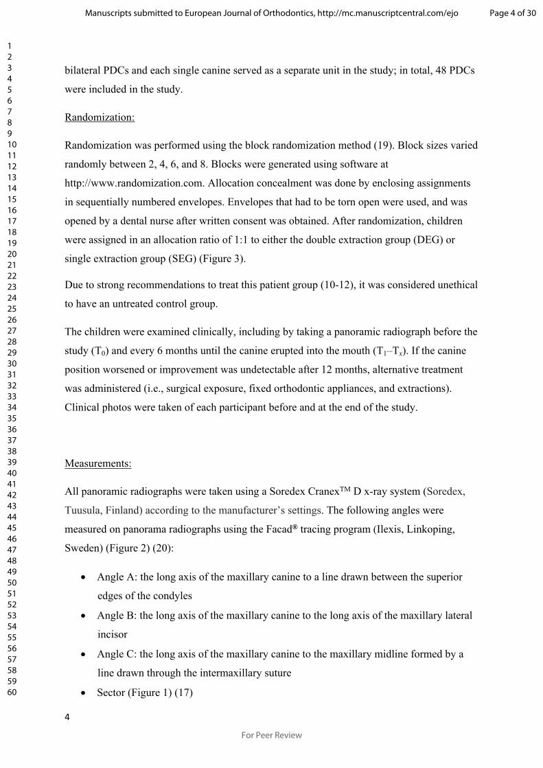
4
bilateral PDCs and each single canine served as a separate unit in the study; in total, 48 PDCs
were included in the study.
Randomization:
Randomization was performed using the block randomization method (19). Block sizes varied
randomly between 2, 4, 6, and 8. Blocks were generated using software at
http://www.randomization.com. Allocation concealment was done by enclosing assignments
in sequentially numbered envelopes. Envelopes that had to be torn open were used, and was
opened by a dental nurse after written consent was obtained. After randomization, children
were assigned in an allocation ratio of 1:1 to either the double extraction group (DEG) or
single extraction group (SEG) (Figure 3).
Due to strong recommendations to treat this patient group (10-12), it was considered unethical
to have an untreated control group.
The children were examined clinically, including by taking a panoramic radiograph before the
study (T0) and every 6 months until the canine erupted into the mouth (T1–Tx). If the canine
position worsened or improvement was undetectable after 12 months, alternative treatment
was administered (i.e., surgical exposure, fixed orthodontic appliances, and extractions).
Clinical photos were taken of each participant before and at the end of the study.
Measurements:
All panoramic radiographs were taken using a Soredex CranexTM D x-ray system (Soredex,
Tuusula, Finland) according to the manufacturer’s settings. The following angles were
measured on panorama radiographs using the Facad® tracing program (Ilexis, Linkoping,
Sweden) (Figure 2) (20):
Angle A: the long axis of the maxillary canine to a line drawn between the superior
edges of the condyles
Angle B: the long axis of the maxillary canine to the long axis of the maxillary lateral
incisor
Angle C: the long axis of the maxillary canine to the maxillary midline formed by a
line drawn through the intermaxillary suture
Sector (Figure 1) (17)
Page 4 of 30
For Peer Review
Manuscripts submitted to European Journal of Orthodontics, http://mc.manuscriptcentral.com/ejo
123456789101112131415161718192021222324252627282930313233343536373839404142434445464748495051525354555657585960

5
Measurement reliability has been reported in a previous study (20).
Space conditions:
Clinical photos taken before and after the study were evaluated visually by an experienced
orthodontist (SHO). Space conditions mesial to the first maxillary molar were estimated and
categorized into the following groups (Figure 4):
1. Crowding: one or more teeth are overlapping and displaced
2. No crowding: all teeth are well aligned
3. Minor spacing: small open spaces between teeth (total ≤ 2 mm)
4. Major spacing: larger spaces between teeth (total > 2 mm)
Reliability was tested by measuring 20 randomly selected plaster models (measured with
sliding callipers) and 20 digital photos (measured visually). ICC was calculated to be 0.889,
indicating excellent agreement.
Dental age assessment:
Panoramic radiographs of each patient were compared with dental stages at QMUL-Atlas
(16), and only patients with crown and root development comparable to or beyond a dental
age stage of 9.5 years were included in the study.
Blinding:
Measurements of panoramic radiographs were made by a faculty member (NLB) at the
Radiology Department of the University of Tromsø, Norway. The radiographs were not
blinded, but to reduce bias, this person was unaware of the purpose of the study.
Primary outcome:
Emergence of the maxillary canine into the oral cavity:
Page 5 of 30
For Peer Review
Manuscripts submitted to European Journal of Orthodontics, http://mc.manuscriptcentral.com/ejo
123456789101112131415161718192021222324252627282930313233343536373839404142434445464748495051525354555657585960
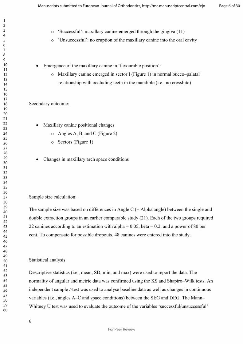
6
o ‘Successful’: maxillary canine emerged through the gingiva (11)
o ‘Unsuccessful’: no eruption of the maxillary canine into the oral cavity
Emergence of the maxillary canine in ‘favourable position’:
o Maxillary canine emerged in sector I (Figure 1) in normal bucco–palatal
relationship with occluding teeth in the mandible (i.e., no crossbite)
Secondary outcome:
Maxillary canine positional changes
o Angles A, B, and C (Figure 2)
o Sectors (Figure 1)
Changes in maxillary arch space conditions
Sample size calculation:
The sample size was based on differences in Angle C (= Alpha angle) between the single and
double extraction groups in an earlier comparable study (21). Each of the two groups required
22 canines according to an estimation with alpha = 0.05, beta = 0.2, and a power of 80 per
cent. To compensate for possible dropouts, 48 canines were entered into the study.
Statistical analysis:
Descriptive statistics (i.e., mean, SD, min, and max) were used to report the data. The
normality of angular and metric data was confirmed using the KS and Shapiro–Wilk tests. An
independent sample t-test was used to analyse baseline data as well as changes in continuous
variables (i.e., angles A–C and space conditions) between the SEG and DEG. The Mann–
Whitney U test was used to evaluate the outcome of the variables ‘successful/unsuccessful’
Page 6 of 30
For Peer Review
Manuscripts submitted to European Journal of Orthodontics, http://mc.manuscriptcentral.com/ejo
123456789101112131415161718192021222324252627282930313233343536373839404142434445464748495051525354555657585960

7
and ‘favourable position’. The marginal homogeneity test was used to test within-group
changes in ordinal data (i.e., sector). To evaluate the association between various factors, i.e.,
group, uni/bilateral canine, dental anomalies (Table 2), gender, angles A–C (T0), sector (T0),
and emergence of the maxillary canine into the oral cavity, a binary logistic regression was
performed to calculate odds ratios and two-sided 95-per cent confidence intervals. The level
of significance was set at p < .05. Statistical analysis was performed using version 24.0 of the
SPSS software package (SPSS Inc., Chicago, IL).
Harms:
No harms were detected during the study.
Results
Participant flow:
Successful cases (n = 34) were followed until the tooth had erupted in the mouth. For the
unsuccessful cases (n = 14), alternative treatment started at 12 months (6 children/7 cases), 18
months (1 child/1 case), and 24 months (3 children/3 cases). Two children (3 cases) were
observed for only 6 months because the canine position had worsened severely. The mean
observation time for the studied sample was 14.8 months (range 6–24 months). There were no
drop-outs in the study (Figure 3).
Baseline findings:
There were no significant differences in gender, age, sector, space conditions, or any of the
angular variables between the DEG and SEG (Table 1). Angular and metric data were
normally distributed in both groups. The distribution of dental anomalies in the study
population is presented in Table 2.
Page 7 of 30
For Peer Review
Manuscripts submitted to European Journal of Orthodontics, http://mc.manuscriptcentral.com/ejo
123456789101112131415161718192021222324252627282930313233343536373839404142434445464748495051525354555657585960

8
Primary outcome:
Emergence of the maxillary canine into the oral cavity:
Sixteen of 25 canines in the DEG emerged into the oral cavity versus 18 of 23 canines in the
SEG (64 per cent vs. 78 per cent; P = .283)
In the DEG, 40.0 per cent of canines emerged within 12 months, 53 per cent within 18
months, and 7 per cent within 24 months, versus 32 per cent, 63 per cent, and 5 per cent,
respectively, in the SEG. No significant difference was recorded between the groups (P =
.732).
Among the emerged canines (34/48), all canines in the DEG occurred in sector I, versus 77.8
per cent in sector I, 16.7 per cent in sector II, and 5.6 per cent in sector III in the SEG (P =
.048). No canines emerged in sector IV.
Uni- and bilateral canines were equally distributed among the PDCs with unsuccessful
eruption in the oral cavity (7/14). Two of 16 children with bilateral PDCs had an unsuccessful
outcome on both sides.
Emergence of the maxillary canine in ‘favourable position’:
Sixty-four per cent of the canines emerged in the ‘favourable position’ in the DEG versus 57
per cent in the SEG (16/25 vs. 13/23; P = .600).
Among the PDCs that emerged into the oral cavity (34/48), significantly more canines
emerged in a ‘favourable position’ in the DEG than in the SEG: 100 per cent versus 72 per
cent (P = .025).
A correct bucco–palatal relationship was present for all canines that emerged in the DEG,
whereas 4 of 18 canines emerged in an anterior crossbite position in the SEG.
Secondary outcome:
Maxillary canine positional change:
Page 8 of 30
For Peer Review
Manuscripts submitted to European Journal of Orthodontics, http://mc.manuscriptcentral.com/ejo
123456789101112131415161718192021222324252627282930313233343536373839404142434445464748495051525354555657585960

9
The angular and sector measurements indicated significant distal movement of the canines in
both groups (P < .001), with wide individual variation (Figure 5). However, no significant
difference was found between the two groups for changes in canine angle (i.e., A, B, or C) or
sector (Table 3).
Of the 14 unsuccessful cases, improvement in the eruption path (i.e., reduction in sector
and/or Angle A) was seen in 56 per cent (5/9) in the DEG and 60 per cent (3/5) in the SEG.
Six of all examined canines (n =48) exhibited a worsened eruption path, 4 in the DEG and 2
in the SEG (P = .449).
Space conditions in the maxillary arch
A significant reduction in estimated space was seen in both groups from T0 to Tend (P < .001),
but no significant difference was recorded between the groups (P = .727) (Table 3).
Predictive factors for the emergence of the maxillary canine into the oral cavity:
A significant relationship was observed between maxillary canine angulation at baseline
(Angle A) and the emergence of PDCs into the oral cavity (OR = 0.882, CI = 0.804–0.968; P
= .008).
Canines that emerged into the oral cavity exhibited a significantly greater Angle A at T0 (i.e.,
more vertical eruption pattern) than did non-emerged canines (mean 63.7°, SD 6.6 vs. mean
56.2°, SD 9.1; P = .003).
A significant relationship was also seen between space discrepancy at T0 and the emergence
of PDCs (P = .029). More PDCs emerged into the oral cavity the more space was available at
T0 (Major spacing > minor spacing > no crowding > crowding) (Figure 6). No canines
emerged into the oral cavity in the crowded group.
Discussion
Page 9 of 30
For Peer Review
Manuscripts submitted to European Journal of Orthodontics, http://mc.manuscriptcentral.com/ejo
123456789101112131415161718192021222324252627282930313233343536373839404142434445464748495051525354555657585960

10
The main objective of this randomized interceptive study was to compare the emergence rate
of PDCs when the primary canine and the primary first molar were extracted compared with
extraction of the primary canine only. This study could not find a significant difference in
emergence rate between these two procedures. In two previous studies examining double
versus single extraction, Bonetti et al. found no difference in emergence rate between the
groups in the first study (21), but a significantly higher emergence rate in the double
extraction group in the second study (12). In the present study, 64 per cent (DEG) and 78 per
cent (SEG) of PDCs emerged into the oral cavity compared with 96 per cent vs. 85 per cent
and 85 per cent vs. 79 per cent, respectively, in the two studies by Bonetti et al. (12, 21). One
reason for the dissimilarity between these studies could be differences in dental
developmental age in the studied samples. In the second study by Bonetti et al. (12), 43 per
cent of participants had a dental development age of 8.5 years (16), versus 100 per cent in the
9.5 years and older stage in the present study. McSherry et al. (22) demonstrated that the
maxillary canine normally appears radiographically palatal in children younger than 10 years.
It has been pointed out that it is too early to define canines as PDCs at 8.5 years of age, and
that some of the teeth in Bonetti et al.’s study would have emerged spontaneously without
extractions (15).
There were also differences in the initial canine position between the present study and
Bonetti’s first study (21), which could have influenced the emergence rate. In the present
study, no canines were situated in sector I, 1/4 in sector II, 2/3 in sector III, and 7/48 in sector
IV. In Bonetti’s study (21), 1/4 of canines were located in sector I and 2/3 in sector II. This
indicates that the present study had more severe cases. The higher success rate for canine
emergence in Bonetti’s studies versus the present study could also be related to a longer
observation time in the previous studies (i.e., 18 months vs. 14.8 months). Longer observation
time may, however, increase the risk of root resorption (11). Therefore, 6 patients (7 canines)
with no improvement in canine position after 12 months and 2 patients (3 canines) with severe
worsening of the canine position after 6 months were given alternative treatment in the
present study. The appropriate length of the observation period is still under debate.
Observation intervals of six months (5), 10 months (23), and 12–18 months (12) have been
proposed in previous studies. Based on the limited number of canines worsening their position
by the 6-month observation in the present study (3 of 48 canines, 6 per cent), radiographic
exposure of all children at the 6-month observation seems unjustified according to the ‘as low
as reasonably achievable’ (ALARA) principle (24). Therefore, 12-month intervals seem more
Page 10 of 30
For Peer Review
Manuscripts submitted to European Journal of Orthodontics, http://mc.manuscriptcentral.com/ejo
123456789101112131415161718192021222324252627282930313233343536373839404142434445464748495051525354555657585960

11
appropriate, though more studies including cone beam computed tomography images are
needed to determine the proper observation interval.
Bonetti et al. emphasizes that the ultimate criterion for successful outcome may not only be
the rate of eruption/non-eruption of PDCs into the dental arch (21). We agree with this
statement, as it is also very important where in the mouth the canines emerge, as the distance
from the emergence point and ‘favourable position’ in the dental arch affects the orthodontic
treatment length and cost (25). In addition, the risk of root resorption is substantially reduced
if the canine is localized in sector I (18). Emergence of the maxillary canine in a ‘favourable
position’ was therefore added to the primary outcome, defined as canines erupting in sector I
and having a normal bucco–palatal relationship with antagonists in the mandible. No
significant difference was seen in this respect between the two extraction groups when all
examined canines were analysed. However, for canines that emerged into the oral cavity,
significantly more canines emerged in a ‘favourable position’ in the DEG than the SEG. The
reason for this difference is unclear. Previous studies have found that the angle between the
first premolar and the facial midline increases more when double rather than single
extractions are performed (21), which may lead to a different eruption pattern for canines in
the DEG than the SEG.
Canines in both the DEG and SEG changed to a significantly more vertical position from the
initial to final observations in both the DEG and SEG, though no significant difference was
found between the groups. This is in contrast to two previous studies finding that the double
extraction groups experienced greater angular change than did the single extraction groups
(12, 21). The angulation of canines in their final position in the dental arch is not only
dependent on which interceptive treatment is given, but also dependent on space conditions in
the maxillary arch (26) and type of occlusion. Therefore, the angular position of emerged
canines may not be the best outcome variable, though it has been used in many PDC studies
(5, 6, 11, 12, 21). However, as a selection criterion for decisions regarding primary canine
extraction and as a predictive variable during the observation period, canine angulation may
be important. In a recent article, Naoumova et al. advocated guidelines for the interceptive
extraction of primary canines based on sector location and alpha angle (23). They suggested
that interceptive extraction is beneficial when the canine is located in sector II or III with an
alpha angle (Angle C) of 20–30 degrees. If the canine is located in sector IV with an alpha
angle of greater than 30 degrees, immediate surgical exposure is recommended, and
observation is recommended if the canine is located in sector II with an alpha angle of 20
Page 11 of 30
For Peer Review
Manuscripts submitted to European Journal of Orthodontics, http://mc.manuscriptcentral.com/ejo
123456789101112131415161718192021222324252627282930313233343536373839404142434445464748495051525354555657585960

12
degrees or less. If these guidelines had been applied to the present sample, they would have
worked very well for the recommendations to extract (23 of 26 canines emerged
successfully), but not as well for the recommendations to implement surgical exposure (3 of 4
canines emerged successfully without surgery). A problem with guidelines is that they have to
simplify and sometimes exclude a number of cases – in the present study, 38 per cent of the
canines. The definition of success is important when applying a clinical perspective to PDC
treatment, and one can argue whether the success definition (‘canine emerged through the
gingiva’) on which these guidelines are based is appropriate. As previously mentioned, the
emergence position of PDCs greatly influences the treatment length and cost (25), and should
therefore be incorporated into an updated definition of successful eruption.
Anterior dental arch space was significantly reduced from start to endpoint in both extraction
groups, though without an intergroup difference, which is in accordance with a previous study
(10). Particularly interesting is the finding that cases of successful eruption had significantly
more dental arch space before treatment than did unsuccessful cases. This is in line with
earlier studies showing that increased maxillary dental arch space positively affects the PDC
emergence rate (9, 27, 28). It has also been reported that space conditions in the maxillary
dental arch influence the canine eruption path (20, 26). Therefore, there is good reason to use
space conditions as an inclusion/exclusion criteria for studies of PDCs, as has previously been
done (10, 11).
The most important predictor of successful emergence into the oral cavity was the angle
between the maxillary canine and the bicondylar line (Angle A) at T0. Naoumova et al. also
found canine angulation (alpha angle), apart from primary canine extraction, to be the best
predictor of PDC emergence when they compared extraction of the primary canine with an
untreated control group (23). Power and Short (6) confirmed this, finding that PDCs angulated
more than 31 degrees to the maxillary midline had a reduced chance of successful eruption.
In contrast to the present findings, the sector location of the PDC has been found to be a good
predictor of successful emergence into the oral cavity (5, 6, 23). Other predictors of successful
canine eruption reported in previous studies are: the distance from the canine cusp tip to the
midline, the angle between the canine and first premolar, and the distance from the canine
cusp tip to the maxillary occlusal plane (29, 30). However, Alqerban et al. (29) found that the
ability to predict maxillary canine impaction based on panorama radiographs is weak, so some
caution is warranted regarding these predictive variables.
Page 12 of 30
For Peer Review
Manuscripts submitted to European Journal of Orthodontics, http://mc.manuscriptcentral.com/ejo
123456789101112131415161718192021222324252627282930313233343536373839404142434445464748495051525354555657585960

13
Dental anomalies such as peg-shaped or small laterals, missing maxillary and mandibular
second premolars, generalized small teeth, and localized or generalized delayed eruption have
long been seen as major etiologic factors for PDCs (31-33). In the present sample, 56 per cent
of the children had one or more of the listed dental anomalies, supporting the genetic theory
of PDC occurrence. The regression analysis, however, did not find any association between
these dental anomalies and PDC emergence, and PDCs seem to respond similarly to primary
tooth extraction whether or not dental anomalies are present.
Limitations:
Though the sample size was based on the best available data when the study was initiated
(21), the sample size is limited. The results and conclusions might well have been different if
the sample had been larger.
No cone beam computed tomography images were taken since no clinical or radiological
signs of root resorption were detected. Therefore, we have no data regarding root resorption
or other adverse effects. The observation period and success rates might have been different if
such information had been available.
This paper has not taken the patient perspective into account, and the two procedures might
differ in terms of pain and discomfort. This matter will be investigated further in a follow-up
study.
Generalization:
The present study was conducted using Caucasian children aged 9.5–13.5 years, and cannot
be generalized to all populations.
Registration:
The study was registered at ClinicalTrials.gov, number NCT02675036.
Page 13 of 30
For Peer Review
Manuscripts submitted to European Journal of Orthodontics, http://mc.manuscriptcentral.com/ejo
123456789101112131415161718192021222324252627282930313233343536373839404142434445464748495051525354555657585960

14
Conclusions:
Double or single primary tooth extraction procedures are equivalent in supporting
PDC eruption into the oral cavity and into a favourable position in the dental arch.
Initial canine angulation and space assessments may be used as predictors of
successful PDC eruption.
Funding:
No funding or other support was received to conduct this study.
Conflict of interest:
None to declare.
Reference List
1. Dachi SF, Howell FV. A survey of 3, 874 routine full-month radiographs. II. A study of
impacted teeth. Oral surgery, oral medicine, and oral pathology. 1961;14:1165-9.
2. Thilander B, Myrberg N. The prevalence of malocclusion in Swedish schoolchildren.
Scand J Dent Res. 1973;81(1):12-21.
3. Ericson S, Kurol J. Radiographic assessment of maxillary canine eruption in children with
clinical signs of eruption disturbance. European journal of orthodontics. 1986;8(3):133-40.
4. Shafer WG HM, Levy BM. A textbook of oral pathology. 2nd ed. Philadelphia: WB
Saunders; 1963.
5. Ericson S, Kurol J. Early treatment of palatally erupting maxillary canines by extraction of
the primary canines. European journal of orthodontics. 1988;10(4):283-95.
6. Power SM, Short MB. An investigation into the response of palatally displaced canines to
the removal of deciduous canines and an assessment of factors contributing to favourable
eruption. Br J Orthod. 1993;20(3):215-23.
Page 14 of 30
For Peer Review
Manuscripts submitted to European Journal of Orthodontics, http://mc.manuscriptcentral.com/ejo
123456789101112131415161718192021222324252627282930313233343536373839404142434445464748495051525354555657585960

15
7. Bruks A, Lennartsson B. The palatally displaced maxillary canine. A retrospective
comparison between an interceptive and a corrective treatment group. Swed Dent J.
1999;23(4):149-61.
8. Leonardi M, Armi P, Franchi L, Baccetti T. Two interceptive approaches to palatally
displaced canines: a prospective longitudinal study. The Angle orthodontist. 2004;74(5):581-
6.
9. Baccetti T, Leonardi M, Armi P. A randomized clinical study of two interceptive
approaches to palatally displaced canines. European journal of orthodontics. 2008;30(4):381-
5.
10. Bazargani F, Magnuson A, Lennartsson B. Effect of interceptive extraction of deciduous
canine on palatally displaced maxillary canine: a prospective randomized controlled study.
The Angle orthodontist. 2014;84(1):3-10.
11. Naoumova J, Kurol J, Kjellberg H. Extraction of the deciduous canine as an interceptive
treatment in children with palatal displaced canines - part I: shall we extract the deciduous
canine or not? Eur J Orthod. 2015;37(2):209-18.
12. Alessandri Bonetti G, Zanarini M, Incerti Parenti S, Marini I, Gatto MR. Preventive
treatment of ectopically erupting maxillary permanent canines by extraction of deciduous
canines and first molars: A randomized clinical trial. American journal of orthodontics and
dentofacial orthopedics : official publication of the American Association of Orthodontists, its
constituent societies, and the American Board of Orthodontics. 2011;139(3):316-23.
13. Naoumova J, Kurol J, Kjellberg H. A systematic review of the interceptive treatment of
palatally displaced maxillary canines. Eur J Orthod. 2011;33(2):143-9.
14. Bishara SE. Impacted maxillary canines: a review. American journal of orthodontics and
dentofacial orthopedics : official publication of the American Association of Orthodontists, its
constituent societies, and the American Board of Orthodontics. 1992;101(2):159-71.
15. Peck S. Problematic sample in the study of interception of palatally displaced canines.
American journal of orthodontics and dentofacial orthopedics : official publication of the
American Association of Orthodontists, its constituent societies, and the American Board of
Orthodontics. 2011;140(1):2-3; author reply -4.
16. AlQahtani SJ, Hector MP, Liversidge HM. Accuracy of dental age estimation charts:
Schour and Massler, Ubelaker and the London Atlas. American journal of physical
anthropology. 2014;154(1):70-8.
17. Lindauer SJ, Rubenstein LK, Hang WM, Andersen WC, Isaacson RJ. Canine impaction
identified early with panoramic radiographs. J Am Dent Assoc. 1992;123(3):91-2, 5-7.
Page 15 of 30
For Peer Review
Manuscripts submitted to European Journal of Orthodontics, http://mc.manuscriptcentral.com/ejo
123456789101112131415161718192021222324252627282930313233343536373839404142434445464748495051525354555657585960
16
18. Hadler-Olsen S, Pirttiniemi P, Kerosuo H, Bolstad Limchaichana N, Pesonen P, Kallio-
Pulkkinen S, et al. Root resorptions related to ectopic and normal eruption of maxillary canine
teeth - A 3D study. Acta odontologica Scandinavica. 2015;73(8):609-15.
19. Pandis N, Polychronopoulou A, Eliades T. Randomization in clinical trials in
orthodontics: its significance in research design and methods to achieve it. European journal
of orthodontics. 2011;33(6):684-90.
20. Hadler-Olsen S, Pirttiniemi P, Kerosuo H, Sjogren A, Pesonen P, Julku J, et al. Does
headgear treatment in young children affect the maxillary canine eruption path? European
journal of orthodontics. 2018.
21. Alessandri Bonetti G, Incerti Parenti S, Zanarini M, Marini I. Double vs single primary
teeth extraction approach as prevention of permanent maxillary canines ectopic eruption.
Pediatric dentistry. 2010;32(5):407-12.
22. McSherry P, Richardson A. Ectopic eruption of the maxillary canine quantified in three
dimensions on cephalometric radiographs between the ages of 5 and 15 years. European
journal of orthodontics. 1999;21(1):41-8.
23. Naoumova J, Kjellberg H. The use of panoramic radiographs to decide when interceptive
extraction is beneficial in children with palatally displaced canines based on a randomized
clinical trial. European journal of orthodontics. 2018;40(6):565-74.
24. Langlais RP, Rodriguez IE, Maselle I. Principles of radiographic selection and
interpretation. Dental clinics of North America. 1994;38(1):1-12.
25. Bazargani F, Magnuson A, Dolati A, Lennartsson B. Palatally displaced maxillary
canines: factors influencing duration and cost of treatment. European journal of orthodontics.
2013;35(3):310-6.
26. Silvola AS, Arvonen P, Julku J, Lahdesmaki R, Kantomaa T, Pirttiniemi P. Early
headgear effects on the eruption pattern of the maxillary canines. The Angle orthodontist.
2009;79(3):540-5.
27. Sigler LM, Baccetti T, McNamara JA, Jr. Effect of rapid maxillary expansion and
transpalatal arch treatment associated with deciduous canine extraction on the eruption of
palatally displaced canines: A 2-center prospective study. American journal of orthodontics
and dentofacial orthopedics : official publication of the American Association of
Orthodontists, its constituent societies, and the American Board of Orthodontics.
2011;139(3):e235-44.
Page 16 of 30
For Peer Review
Manuscripts submitted to European Journal of Orthodontics, http://mc.manuscriptcentral.com/ejo
123456789101112131415161718192021222324252627282930313233343536373839404142434445464748495051525354555657585960

17
28. Armi P, Cozza P, Baccetti T. Effect of RME and headgear treatment on the eruption of
palatally displaced canines: a randomized clinical study. The Angle orthodontist.
2011;81(3):370-4.
29. Alqerban A, Storms AS, Voet M, Fieuws S, Willems G. Early prediction of maxillary
canine impaction. Dento maxillo facial radiology. 2016;45(3):20150232.
30. Alqerban A, Jacobs R, Fieuws S, Willems G. Radiographic predictors for maxillary
canine impaction. American journal of orthodontics and dentofacial orthopedics : official
publication of the American Association of Orthodontists, its constituent societies, and the
American Board of Orthodontics. 2015;147(3):345-54.
31. Becker A, Smith P, Behar R. The incidence of anomalous maxillary lateral incisors in
relation to palatally-displaced cuspids. The Angle orthodontist. 1981;51(1):24-9.
32. Peck S, Peck L, Kataja M. The palatally displaced canine as a dental anomaly of genetic
origin. The Angle orthodontist. 1994;64(4):249-56.
33. Peck S, Peck L, Kataja M. Prevalence of tooth agenesis and peg-shaped maxillary lateral
incisor associated with palatally displaced canine (PDC) anomaly. American journal of
orthodontics and dentofacial orthopedics : official publication of the American Association of
Orthodontists, its constituent societies, and the American Board of Orthodontics.
1996;110(4):441-3.
Figure Legends:
Figure 1 Sector location of the maxillary canine.
Figure 2 Angular measurements of maxillary canines. Angle A: canine to bicondylar line,
Angle B: canine to lateral incisor, Angle C: canine to maxillary midline.
Figure 3 Consort flow diagram of participants in the study
Figure 4 Space discrepancy assessment on clinical photos. Crowding: one or more teeth are
overlapping and displaced, No crowding: all teeth are well aligned, Minor spacing: small open
spaces between teeth (total ≤2 mm), Major spacing: larger spaces between teeth (total >2
mm).
Page 17 of 30
For Peer Review
Manuscripts submitted to European Journal of Orthodontics, http://mc.manuscriptcentral.com/ejo
123456789101112131415161718192021222324252627282930313233343536373839404142434445464748495051525354555657585960

18
Figure 5 Sector change in palatally displaced canines (PDCs) from start (T0) to end (Tend) of
trial in the single and double extraction groups. SEG = single extraction group, DEG = double
extraction group.
Figure 6 Relationship between space discrepancy at T0 and emergence of PDCs.
Crowding: one or more teeth are overlapping and displaced, No crowding: all teeth are well
aligned, Minor spacing: small open spaces between teeth (total ≤2 mm), Major spacing: larger
spaces between teeth (total >2 mm).
Table 1 Baseline data (T0) for the single extraction group (SEG) and double extraction group
(DEG).
NS: not significant, * P-value < .05 is considered statistically significant, A Mann–Whitney U
test: P = .212, B Mann–Whitney U test: P = .586, C Mann–Whitney U test: P = .359.
Table 2 Dental anomalies in study population.
a Mesiodistal width greatest in cervical margin, b Mesiodistal width equal to or smaller than its
mandibular counterpart, c > 2 SD.
Table 3 Comparison of changes in canine angulation and space discrepancy between initial
and end observations; mean observation time, 14.8 months.
NS, not significant, * P-value < 0.05 is considered statistically significant, A Mann–Whitney U
test: P = 0.727
Page 18 of 30
For Peer Review
Manuscripts submitted to European Journal of Orthodontics, http://mc.manuscriptcentral.com/ejo
123456789101112131415161718192021222324252627282930313233343536373839404142434445464748495051525354555657585960

19
Page 19 of 30
For Peer Review
Manuscripts submitted to European Journal of Orthodontics, http://mc.manuscriptcentral.com/ejo
123456789101112131415161718192021222324252627282930313233343536373839404142434445464748495051525354555657585960

Table 1 Baseline data (T0) for the single extraction group (SEG) and double extraction group (DEG).
Variable SEG (n = 23)
Mean ± SD
DEG (n = 25)
Mean ± SD
P-value *
Age 11.0 ± 1.1 10.8 ± 0.7 0.621 NS
Angle A (°) 60.9 ± 7.8 62.1 ± 8.5 0.626 NS
Angle B (°) 36.9 ± 9.5 35.1 ± 6.6 0.485 NS
Angle C (°) 30.4 ± 9.1 27.1 ± 7.7 0.183 NS
n n
Crowding
No crowding
Minor spacing
Major spacing
Sector 2
1
5
11
6
7
2
5
4
14
4
A
A
A
A
B
NS
NS
NS
NS
NS
Sector 3 12 18 B NS
Sector 4 4 3 B NS
Female 15 13 C NS
Male 8 12 C NS
NS: not significant, * P-value < .05 is considered statistically significant, A Mann–Whitney U test: P = .212, B Mann–Whitney U test: P = .586, C Mann–Whitney U test: P = .359.
Page 20 of 30
For Peer Review
Manuscripts submitted to European Journal of Orthodontics, http://mc.manuscriptcentral.com/ejo
123456789101112131415161718192021222324252627282930313233343536373839404142434445464748495051525354555657585960

Table 2 Dental anomalies in study population.
Anomalies Study sample (n = 32)
Missing maxillary second premolar 3 (9.4%)
Missing mandibular second premolar 2 (6.3%)
Peg-shaped laterala 5 (15.6%)
Small lateralb 2 (6.3%)
Generalized small teeth 2 (6.3%)
Localized delayed eruptionc
Generalized delayed eruptionc
1 (3.1%)
3 (9.4%)a Mesiodistal width greatest in cervical margin, b Mesiodistal width equal to or smaller than its mandibular counterpart, c > 2 SD.
Page 21 of 30
For Peer Review
Manuscripts submitted to European Journal of Orthodontics, http://mc.manuscriptcentral.com/ejo
123456789101112131415161718192021222324252627282930313233343536373839404142434445464748495051525354555657585960

Table 3 Comparison of changes in canine angulation and space discrepancy between initial and end observations; mean observation time, 14.8 months.
Variable SEG DEG P-value*
Mean Min Max Mean Min Max
Angle A° –15.3 –29.9 1.7 –14.7 –36.3 6.7 0.846 NS
Angle B° 15.8 –2.5 38.6 14.9 –11.2 44.6 0.818 NS
Angle C° 17.7 –2.7 43.9 13.3 –10.6 33.7 0.203 NS
Crowding
No crowding
Minor spacing
Major spacing
T0 (n)
1
5
11
6
Tend (n)
5
7
6
3
Change (n)
4
3
–5
–3
T0 (n)
2
5
4
14
Tend (n)
7
3
7
8
Change (n)
5
–2
3
–6
A
A
A
A
NS
NS
NS
NS
NS, not significant, * P-value < 0.05 is considered statistically significant, A Mann–Whitney U test: P = 0.727
Page 22 of 30
For Peer Review
Manuscripts submitted to European Journal of Orthodontics, http://mc.manuscriptcentral.com/ejo
123456789101112131415161718192021222324252627282930313233343536373839404142434445464748495051525354555657585960

Figure 1. Sector location of the maxillary canine.
Page 23 of 30
For Peer Review
Manuscripts submitted to European Journal of Orthodontics, http://mc.manuscriptcentral.com/ejo
123456789101112131415161718192021222324252627282930313233343536373839404142434445464748495051525354555657585960

Figure 2. Angular measurements of maxillary canines. Angle A: canine to bicondylar line, Angle B: canine to lateral incisor, Angle C: canine to maxillary midline.
Page 24 of 30
For Peer Review
Manuscripts submitted to European Journal of Orthodontics, http://mc.manuscriptcentral.com/ejo
123456789101112131415161718192021222324252627282930313233343536373839404142434445464748495051525354555657585960

Assessed for eligibility
(n = 32 children, n = 48 canines)
Excluded (n = 0)
Analysed (n = 23) Excluded from analysis (n = 0)
Single extraction group (SEG) (n = 23) Received allocated intervention (n = 23) Did not receive allocated intervention (n = 0 )
Double extraction group (DEG) (n = 25) Received allocated intervention (n= 25) Did not receive allocated intervention (n = 0)
Analysed (n = 25) Excluded from analysis (n = 0)
Allocation
Analysis
Randomized (n = 48)
Enrollment
Figure 3. Consort flow diagram of participants in the study
Page 25 of 30
For Peer Review
Manuscripts submitted to European Journal of Orthodontics, http://mc.manuscriptcentral.com/ejo
123456789101112131415161718192021222324252627282930313233343536373839404142434445464748495051525354555657585960

Figure 4 Space discrepancy assessment on clinical photos. Crowding: one or more teeth are overlapping and displaced, No crowding: all teeth are well aligned, Minor spacing: small open spaces between teeth (total ≤2 mm), Major spacing: larger spaces between teeth (total >2 mm).
Page 26 of 30
For Peer Review
Manuscripts submitted to European Journal of Orthodontics, http://mc.manuscriptcentral.com/ejo
123456789101112131415161718192021222324252627282930313233343536373839404142434445464748495051525354555657585960

PDCs Tend
Sector 1 Sector 2 Sector 3 Sector 4
PDCs T0 SEG DEG SEG DEG SEG DEG SEG DEG
Sector 2
Sector 3
Sector 4
Sector 2 3 3 3 0 0 0 0 0
Sector 3 9 14 1 2 1 1 2 2
Sector 4 2 1 1 0 1 1 0 1
Total 14 18 5 2 2 2 2 3
Figure 5 Sector change in palatally displaced canines (PDCs) from start (T0) to end (Tend) of trial in the single and double extraction groups. SEG = single extraction group, DEG = double extraction group.
Page 27 of 30
For Peer Review
Manuscripts submitted to European Journal of Orthodontics, http://mc.manuscriptcentral.com/ejo
123456789101112131415161718192021222324252627282930313233343536373839404142434445464748495051525354555657585960

Crowing No crowing Minor Spacing Major spacing
Space discrepancy at T0
Figure 6 Relationship between space discrepancy at T0 and emergence of PDCs.Crowding: one or more teeth are overlapping and displaced, No crowding: all teeth are well aligned, Minor spacing: small open spaces between teeth (total ≤2 mm), Major spacing: larger spaces between teeth (total >2 mm).
Page 28 of 30
For Peer Review
Manuscripts submitted to European Journal of Orthodontics, http://mc.manuscriptcentral.com/ejo
123456789101112131415161718192021222324252627282930313233343536373839404142434445464748495051525354555657585960

CONSORT 2010 checklist Page 1
CONSORT 2010 checklist of information to include when reporting a randomised trial*
Section/TopicItem No Checklist item
Reported on page No
Title and abstract1a Identification as a randomised trial in the title 11b Structured summary of trial design, methods, results, and conclusions (for specific guidance see CONSORT for abstracts) 1
Introduction2a Scientific background and explanation of rationale 2Background and
objectives 2b Specific objectives or hypotheses 2
Methods3a Description of trial design (such as parallel, factorial) including allocation ratio 3Trial design3b Important changes to methods after trial commencement (such as eligibility criteria), with reasons -4a Eligibility criteria for participants 3Participants4b Settings and locations where the data were collected 3
Interventions 5 The interventions for each group with sufficient details to allow replication, including how and when they were actually administered
3
6a Completely defined pre-specified primary and secondary outcome measures, including how and when they were assessed
5,6Outcomes
6b Any changes to trial outcomes after the trial commenced, with reasons -7a How sample size was determined 6Sample size7b When applicable, explanation of any interim analyses and stopping guidelines
Randomisation:8a Method used to generate the random allocation sequence 4 Sequence
generation 8b Type of randomisation; details of any restriction (such as blocking and block size) 4 Allocation
concealment mechanism
9 Mechanism used to implement the random allocation sequence (such as sequentially numbered containers), describing any steps taken to conceal the sequence until interventions were assigned
4
Implementation 10 Who generated the random allocation sequence, who enrolled participants, and who assigned participants to interventions
3,4
Blinding 11a If done, who was blinded after assignment to interventions (for example, participants, care providers, those -
Page 29 of 30
For Peer Review
Manuscripts submitted to European Journal of Orthodontics, http://mc.manuscriptcentral.com/ejo
123456789101112131415161718192021222324252627282930313233343536373839404142434445464748495051525354555657585960

CONSORT 2010 checklist Page 2
assessing outcomes) and how11b If relevant, description of the similarity of interventions -12a Statistical methods used to compare groups for primary and secondary outcomes 6,7Statistical methods12b Methods for additional analyses, such as subgroup analyses and adjusted analyses 4,5
Results13a For each group, the numbers of participants who were randomly assigned, received intended treatment, and
were analysed for the primary outcome4, figure 3Participant flow (a
diagram is strongly recommended) 13b For each group, losses and exclusions after randomisation, together with reasons 7
14a Dates defining the periods of recruitment and follow-up 3Recruitment14b Why the trial ended or was stopped -
Baseline data 15 A table showing baseline demographic and clinical characteristics for each group Table 1Numbers analysed 16 For each group, number of participants (denominator) included in each analysis and whether the analysis was
by original assigned groups8,9,10
17a For each primary and secondary outcome, results for each group, and the estimated effect size and its precision (such as 95% confidence interval)
8,9,10Outcomes and estimation
17b For binary outcomes, presentation of both absolute and relative effect sizes is recommended -Ancillary analyses 18 Results of any other analyses performed, including subgroup analyses and adjusted analyses, distinguishing
pre-specified from exploratory-
Harms 19 All important harms or unintended effects in each group (for specific guidance see CONSORT for harms) 7
DiscussionLimitations 20 Trial limitations, addressing sources of potential bias, imprecision, and, if relevant, multiplicity of analyses 13Generalisability 21 Generalisability (external validity, applicability) of the trial findings 13Interpretation 22 Interpretation consistent with results, balancing benefits and harms, and considering other relevant evidence -
Other informationRegistration 23 Registration number and name of trial registry 13Protocol 24 Where the full trial protocol can be accessed, if available -Funding 25 Sources of funding and other support (such as supply of drugs), role of funders 14
*We strongly recommend reading this statement in conjunction with the CONSORT 2010 Explanation and Elaboration for important clarifications on all the items. If relevant, we also recommend reading CONSORT extensions for cluster randomised trials, non-inferiority and equivalence trials, non-pharmacological treatments, herbal interventions, and pragmatic trials. Additional extensions are forthcoming: for those and for up to date references relevant to this checklist, see www.consort-statement.org.
Page 30 of 30
For Peer Review
Manuscripts submitted to European Journal of Orthodontics, http://mc.manuscriptcentral.com/ejo
123456789101112131415161718192021222324252627282930313233343536373839404142434445464748495051525354555657585960


![Commented [R1] - SIIT · (1) Thesis Title DISSERTATION TITLE DISSERTATION TITLE DISSERTATION TITLE DISSERTATION TITLE Author Mr. International Institute Degree Doctor of Philosophy](https://img.pdfslide.net/doc/110x75/5f15f57a7867284d112eafe5/commented-r1-siit-1-thesis-title-dissertation-title-dissertation-title-dissertation.jpg)