Embed Size (px)
Citation preview

A Murine Model of the Eosinophilia-Myalgia SyndromeInduced by 1,1 '-Ethylidenebis(L-Tryptophan)Richard M. Silver, * Anna Ludwicka, * Marta Hampton,t Takashi Ohba, * Sarah A. Bingel,' Timothy Smith,tRussell A. Harley,t John Maize,t and Melvyn P. HeyesilFrom the Division ofRheumatology and Immunology, Departments of *Medicine, $Dermatology and Pathology, and OLaboratoryAnimal Resources ofthe Medical University ofSouth Carolina, Charleston, South Carolina 29425; and I"Laboratory ofClinical Science,Section on Analytical Biochemistry, National Institute ofMental Health, Bethesda, Maryland 20892
Abstract
The eosinophilia-myalgia syndrome (EMS) is a recently de-scribed disease that has been associated with the ingestion ofL-tryptophan containing trace amounts of several impurities.The first such contaminant to be identified and linked epidemio-logically to the EMS epidemic was 1,1'-ethylidenebis(L-tryp-tophan) (EBT), but its role in the etiology and pathogenesis ofthe syndrome has been controversial. We report the develop-ment of inflammation and fibrosis affecting the dermis and sub-cutis, including the fascia and perimyseal tissues, after thedaily intraperitoneal administration of EBT to female C57BL/6 mice. Such changes are accompanied by increased numbersof mast cells, many of which appear to be degranulating.Plasma levels of quinolinic acid, a metabolic product of L-tryp-tophan via the kynurenine pathway, are reduced initially, andthen become elevated when inflammation and fibrosis are morepronounced. The nature and location of the inflammatory cellinfiltrate and fibrosis, as well as the presence of mast cells andalterations of L-tryptophan metabolism, are consistent withfindings reported in patients with EMS. This murine modelsuggests that EBT may have been one of the mediators ofEMSand should facilitate studies of the pathogenesis of EMS. (J.Clin. Invest. 1994. 93:1473-1480.) Key words: EMS * L-tryp-tophan * EBT * fibrosis * mast cell
Introduction
The eosinophilia-myalgia syndrome (EMS)' occurred as anepidemic in the United States in 1989 (1-3). During the epi-demic the following surveillance case definition was estab-lished by the Centers for Disease Control (CDC): (a) bloodeosinophil count > 109/liter; (b) generalized myalgia of suffi-cient severity to limit a patient's usual activities; and (c) exclu-sion of neoplasm or infection to account for symptoms (4).
This work was presented in part at the Annual and Southeast RegionalMeetings of the American College of Rheumatology, October 12-15,1992, Atlanta, GA, and March 26-28, 1993, Arlington, VA, respec-tively.
Address correspondence to Dr. Richard M. Silver, ProfessorofMed-icine and Pediatrics, Medical University ofSouth Carolina, Room 912,Clinical Sciences Building, 171 Ashley Avenue, Charleston, SC 29425.
Receivedfor publication 10 July 1993 and in revisedform 29 Oc-tober 1993.
1. Abbreviations used in this paper: EBT, 1,1 -ethylidenebis(L-trypto-phan); EMS, eosinophelia-myalgia syndrome; GVHD, graft-vs.-hostdisease; TOS, toxic oil syndrome.
The Journal of Clinical Investigation, Inc.Volume 93, April 1994, 1473-1480
More than 1,500 cases and 38 deaths that fulfill the CDC crite-ria for EMS have been reported from the United States (5), butmany more individuals who did not fulfill the CDC criteria orwere not reported to the CDC are believed to have been af-fected (6).
In addition to eosinophilia and myalgia, many EMS pa-tients developed scleroderma-like skin changes frequently ac-companied by fasciitis (2, 3, 7, 8), peripheral neuropathy (9-I 1 ), and pneumonitis ( 12). Remarkably similar clinical andhistological changes were observed in patients affected by thetoxic oil syndrome (TOS) that occurred in Spain in 1981 (13).In each syndrome, epidemiologic studies showed a strong asso-ciation with the ingestion of products containing contami-nants. Adulterated rapeseed oil was implicated in TOS ( 14),although a precise etiologic agent was never identified. TheEMS epidemic has been linked to the ingestion ofL-tryptophanmanufactured by a single company ( 15, 16) and shown to con-tain several contaminants ( 16). The first to be identified was1,1 '-ethylidenebis(L-tryptophan) (EBT) ( 17). Recently, a sec-ond contaminant of L-tryptophan also linked to the EMS epi-demic was identified as 3-(phenylamino)alanine ( 18), whichresembles a putative contaminant of adulterated rapeseed oilimplicated in TOS (19).
Although epidemiologic studies support a link betweenthese contaminants and EMS, it is not known if they are theetiologic agent(s) or simply markers for a yet to be identifiedagent. Animal studies have been controversial (20), but onegroup has reported that female Lewis (LEW/N) rats fed L-tryptophan implicated in the EMS epidemic or LEW/N ratsfed synthesized EBT develop myofascial fibrosis similar to thatobserved in patients with EMS (21, 22). In this study we suc-ceeded in developing an animal model ofEMS and report theinduction of inflammation and fibrosis in skin and fascia offemale C57BL/6 mice treated with daily intraperitoneal injec-tions of EBT. In addition, we report the presence of increasednumbers of mast cells similar to that observed in patients withEMS (23). Furthermore, since altered L-tryptophan metabo-lism and elevated plasma quinolinic acid levels were reportedin patients with EMS (2), we investigated the effect of EBTadministration on kynurenine pathway metabolism in this mu-rine model.
Methods
Animals. 6-wk-old female C57BL/6 mice (The Jackson Laboratory,Bar Harbor, ME) weighing 20-22 g were maintained in animalquarters designed for pathogen-free mice and provided with food (Tek-lad Rodent Diet; Harlan Teklad Laboratory, Indianapolis, IN) andwater ad libitum. All studies were conducted under a protocol ap-proved by the institutional review board for animal studies.
Treatments. Groups of 10-12 animals received daily intraperito-neal injections consisting of 0.3 ml of the following substances: (a)
Murine Model ofthe Eosinophilia-Myalgia Syndrome 1473

4 %=6
a
D
4
1474 Silver et al.
0
saline; (b) L-tryptophan (Sigma Chemical Co., St. Louis, MO), 30mg/kg body weight dissolved in saline; (c) synthesized EBT (lot no.901015, kindly provided by Showa Denko K. K., Tokyo, shown byHPLC to have a purity of 98%, as previously reported [24]) 40 Ag/kgbody weight dissolved in saline; and (d) L-tryptophan combined withEBT (same doses as above). The doses ofEBT and L-tryptophan wereselected because they fall within the range ofthose ingested by patientswith EMS (2, 18, 25). At various time intervals, ranging from 3 d to 6wk, mid-morning blood and plasma were obtained and animals werekilled by exsanguination under ketamine anesthesia. The following tis-sues were obtained for further analysis: (a) skin and subcutaneous tis-sue from the interscapular area; (b) muscle from the proximal hind-limb; (c) lungs; (d) heart; (e) liver; and (f) kidneys.
Pathology. Peripheral blood eosinophil counts were performed onblood smears prepared with Wright's stain. Portions of all tissues werefixed in 10% buffered formalin, embedded in paraffin, and stained withhematoxylin-eosin and Masson's trichrome stains. Lungs from sevenanimals in each treatment group were inflated with 3 ml of 10% buff-ered formalin, fixed in the same solution at 4VC overnight, embeddedin paraffin, and stained as above. Skin and subcutaneous tissue werealso prepared with a specific esterase mast cell stain using naphtholAS-D chloroacetate and hematoxylin counterstain (26). Muscle tissuetaken from the proximal hind limb was quick-frozen in liquid nitro-gen-cooled isopentane. Mounted sections were cut at 5 usm using arotary microtome/cryostat. Sections were picked up on cover slips andstained with hematoxylin-eosin. All slides were coded and provided toinvestigators who were masked to the experimental conditions.
Biochemistry. Plasma quinolinic acid levels were determined usinggas chromatography/mass spectrometry as previously described (27).All samples were run as coded specimens within a single assay.
Data analysis. For analysis of skin and subcutaneous tissues, twodermatopathologists (M. Hampton and J. Maize) independentlygraded tissue sections according to extent and severity of inflamma-tion, destruction of cells, and fibrosis. An index of inflammation orfibrosis was calculated on the basis of the severity (none = 0; mild = 1;moderate = 2; severe = 3) x extent (focal = 1; diffuse = 3). The indexof inflammation or fibrosis varied up to 20% between experiments,each consisting of 10-12 animals/treatment group, and data from indi-vidual experiments are presented in each figure and table unless indi-cated otherwise. Mast cells were counted under high magnification(x200) with a light microscope equipped with a micrometer and areexpressed as the number ofmast cells per 1.5 mm2. A neuropathologist(T. Smith) graded muscle sections for thickness of fascia (,gm), ne-crotic and atrophic myocytes (no./ 2.5 mm2), and presence or absenceof islands of perimyseal inflammatory cells, perineural inflammation,and fibrosis. Group means±standard deviation were calculated for his-
xwazz0
-jU-z
3
2
0
m SALINE
= L-TRYP
_ EBT
M L.TRYP-EBT
6 - I Day3EJ Day 6
Day 21x
z 4z0
U-z
DERMIS SUPERFICIAL PANNICULUS DEEP ADIPOSEADIPOSE LAYER CARNOSUS LAYER
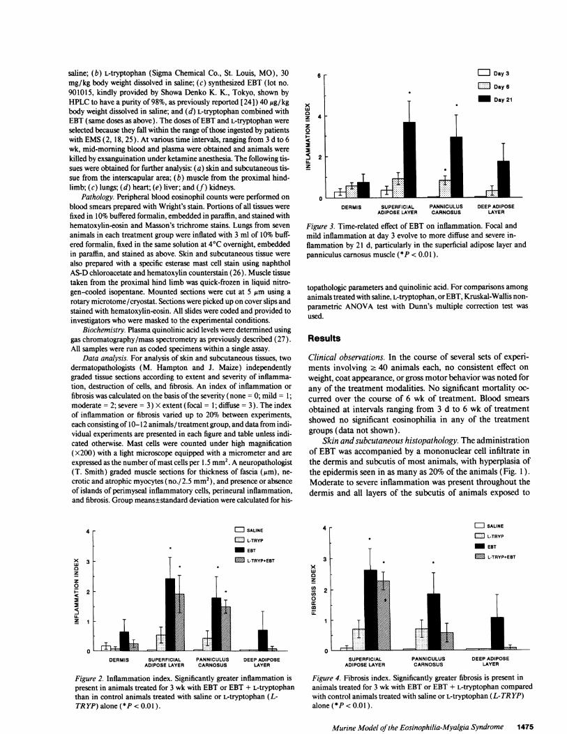
Figure 3. Time-related effect of EBT on inflammation. Focal andmild inflammation at day 3 evolve to more diffuse and severe in-flammation by 21 d, particularly in the superficial adipose layer andpanniculus carnosus muscle (*P < 0.01 ).
topathologic parameters and quinolinic acid. For comparisons amonganimals treated with saline, L-tryptophan, or EBT, Kruskal-Wallis non-parametric ANOVA test with Dunn's multiple correction test wasused.
Results
Clinical observations. In the course of several sets of experi-ments involving 2 40 animals each, no consistent effect onweight, coat appearance, or gross motor behavior was noted forany of the treatment modalities. No significant mortality oc-curred over the course of 6 wk of treatment. Blood smearsobtained at intervals ranging from 3 d to 6 wk of treatmentshowed no significant eosinophilia in any of the treatmentgroups (data not shown).
Skin and subcutaneous histopathology. The administrationof EBT was accompanied by a mononuclear cell infiltrate inthe dermis and subcutis of most animals, with hyperplasia ofthe epidermis seen in as many as 20% of the animals (Fig. 1).Moderate to severe inflammation was present throughout thedermis and all layers of the subcutis of animals exposed to
4 [
3-xw03z
C)0a:
I
DERMIS SUPERFICIAL PANNICULUS DEEP ADIPOSEADIPOSE LAYER CARNOSUS LAYER
Figure 2. Inflammation index. Significantly greater inflammation ispresent in animals treated for 3 wk with EBT or EBT + L-tryptophanthan in control animals treated with saline or L-tryptophan (L-TRYP)alone(*P<0.01).
= SALINE
= L-TRYP
_ EBT
M L-TRYP+EBTI.2h
0SUPERFICIAL PANNICULUS DEEP ADIPOSE
ADIPOSE LAYER CARNOSUS LAYER
Figure 4. Fibrosis index. Significantly greater fibrosis is present inanimals treated for 3 wk with EBT or EBT + L-tryptophan comparedwith control animals treated with saline or L-tryptophan (L-TR YP)alone (*P< 0.01).
Murine Model ofthe Eosinophilia-Myalgia Syndrome 1475
11

xW 40z
0
0DERMIS SUPERFICIAL PANNICULUS DEEP ADIPOSE
ADIPOSE LAYER CARNOSUS LAYER
Figure 5. Time-related effect ofEBT on fibrosis. Significant fibrosis isevident in the superficial adipose layer and panniculus carnosusmuscle by day 6, increasing by day 21 (*P < 0.01 compared withday 3).
EBT, with the most extensive inflammation occurring in thesuperficial adipose layer and the panniculus carnosus muscle(Figs. 1 and 2). The cellular infiltrate was composed predomi-nantly of lymphocytes and monocytes with occasional plasmacells and frequent mast cells (see below). Focal sites of inflam-mation were observed in the dermis and subcutis of animalstreated with saline or L-tryptophan (Fig. 1), but such changeswere never as severe as those observed in animals exposed toEBT. Focal cellular infiltrates were observed as early as 3 d afterbeginning EBT administration; diffuse changes were present inthe subcutis after 6 d oftreatment and were more prevalent andsevere after 3 wk (Fig. 3). Of interest was the observation thatanimals treated with L-tryptophan + EBT had less inflamma-tion than did animals treated with EBT alone (Figs. 1 and 2).
Cellular infiltration was accompanied by varying degrees offibrosis and destruction of the adipose layers and panniculuscarnosus muscle. As was the case with inflammation, suchchanges were always more prevalent and severe in animals ex-posed to EBT than in animals exposed to saline, L-tryptophan,or EBT combined with L-tryptophan (Figs. 4 and 5). WithEBT administration fibrotic changes were observed in all layersof the subcutis, and some animals developed diffuse fibrosis inthe dermis also. Fibrosis was less frequent and severe in ani-mals treated with L-tryptophan + EBT than in animals treatedwith EBT alone (Fig. 4). The fibrosis of the dermis and subcu-
tis evolved from focal to diffuse by 3 wk of exposure to EBT(Fig. 5).
EBT exposure was accompanied by a two- to nearly four-fold increase in the number of dermal and subcutaneous mastcells (Table I). The increase in mast cell number was evidentafter 3 d of exposure to EBT, when significant increases werenoted in the dermis, superficial adipose layer, and the pannicu-lus carnosus muscle (Table I and Fig. 6). In the control groupof saline-treated animals, mast cells were limited primarily tothe dermis. After 6 d ofEBT exposure the mast cell numbers insome skin layers appeared to decline to the control level ob-served in saline-treated animals and were significantly lowerthan at day 3. By 6 wk, the number of mast cells increased tolevels comparable to or greater than day 3.
Muscle andfascia. The endomysium, perimysium, and fas-cia of animals exposed to EBT showed progressive thickeningwith increasing treatment times (Table II). After 6 wk of expo-sure to EBT there was a significant increase in the thickness ofthe fascia of the muscle as compared with animals treated withsaline (168±22 ,m vs. 43±5 ,um; P < 0.01) or L-tryptophan( 168±22 Am vs. 69±7 tlm; P < 0.01 ) (Table II). L-Tryptophanalone resulted in a slight increase in fascia thickness comparedwith animals receiving saline, but the difference was not statis-tically significant.
Muscle from all treatment groups showed nonspecific myo-pathic changes consisting of basophilic myocytes, atrophicmyocytes, and myocytes with central nuclei. A significant in-crease in the number of necrotic muscle fibers was seen only inthe group of animals receiving EBT for 4 wk (2.92±0.95/2.5mm2) compared with animals receiving saline (0.18±0.13/2.5mm2) or L-tryptophan (0.27±0.19/2.5 mm2) (P < 0.01 each),but not at 6 wk. After 4 wk ofEBT exposure there was also anincrease in the number of atrophic myocytes compared withcontrols, but this was not apparent at 6 wk. Inflammatory is-lands were rarely identified within muscle tissue. Muscle spin-dles were only rarely inflamed. Endothelial cell hyperplasia waspresent in the endomysial septa of some but not all animalstreated with EBT for 6 wk.
Other pathology. No significant abnormalities of the heart,kidney, or liver were detected by light microscopy. Focal areasof inflammation and fibrosis were observed in the lungs ofanimals treated for 3 wk with L-tryptophan alone or in combi-nation with EBT, but such changes were mild and there was nosignificant difference in the inflammation index or fibrosis in-dex among the various control and treatment groups. Areas of
Table I. Mast Cell Numbers
Layer Exp. condition Day 3 Day 6 Day 21 Day 42
Dermis Saline 79.3±5.1* 130.4±12.8 79.7±6.1 NDtEBT 157.9±14.8§ 120.2±16.91 98.3±15.6 162.7±9.6
Superficial Adipose Saline 29.4±6.2 60.4±3.1 50.2±5.7 NDEBT 99.5±12.4§ 56.5±14.311 61.3±7.3 77.9±4.7
Panniculus Carnosus Saline 1.4±0.5 12.0±3.7 11.5±2.6 NDEBT 5.2±1.5¶ 10.2±1.8 12.0±2.7 16.5±3.5**
Deep Adipose Saline 2.2±1.0 9.4±3.4 4.5±1.1 NDEBT 4.2±0.8 7.2±1.4 7.0±1.4 17.0±2.4**
* Number of mast cells per linear 1.5 mm2 ofeach layer, mean±SD. t ND, not done. 1P < 0.01 EBT vs. saline, day 3. "lP < 0.05 day 3 vs. day6, EBT. tP = 0.05 EBT vs. saline, day 3. ** P < 0.01 day 3 vs. day 42, EBT.
1476 Silver et al.

IWA.~~~~
> . vvt E2t~~) < J Ir/Xjj8
I' -X* S ' ' ) / P E
' t ~ ~ ~ ) /> 1 tX.W'1
44~~!!44)tj >
Cc>~~~~tcr p.r da
4k AI|itiG/t O~tg d e E i
J 9 S~#4t )/ X
U &r jh t df -
A;1*t!tt l/1 tk, 2 E ¢
~~~~~~~~~I~~~~~~~~~~~~~~~~~!~ ~ ~ ~ ~ ~~I
Cu3
4Cd
C4u
a X~~~~~~~~~~~~~~~~~~~~~~Cuis /~~~~~~~~~~~~~~~~~~~~~~~~~~~c
SI~~~~~~~~~~~~~~~~~~~~I
Ails~~~~~~~A
X~
Ao~ IA'ssi <'7X aX 1 e s ~ r Si t3 8 t '
Murine Model ofthe Eosinophilia-Myalgia Syndrome 1477

Table IL Fascia and Muscle Histopathology ofEBT-treated and Control Mice
Saline* L-Tryp$ EBT (3 wk) EBT (4 wk) EBT (6 wk)
Fascia thickness (,m) 43±5 69±7 47±7 72±6 168±22§1Atrophic muscle fibers (no./2.5 mm2) 2.07±0.86 2.00±0.56 2.25±0.31 3.67±0.94 3.07±0.61Necrotic muscle fibers (no./2.5 mm2) 0.18±0.13 0.27±0.19 0.50±0.34 2.92±0.951 0.15±0.10
* Data pooled from 3- and 6-wk experiments. * L-Tryp, L-tryptophan. Significantly different (P < 0.01) from Isaline control or IIL-tryptophancontrol.
lymphoidEBT or tchanges m
Kynuilevels renbiphasic Ition of Elline valueof EBT, I
trols). QiEBT exp(controls)elevated cmals by 3return toiremained< 0.05 ea
Discuss
The ingeseral contalogic stud:An etiolol
400
j 300
-jo 200z
a 100
0
Figure 7.(nM) of t]treated wiltreated an
tissue within the lungs were seen in animals receiving the epidemic, has been suggested by studies of female Lewisthe combination of EBT + L-tryptophan, but such (LEW/N) rats fed implicated lots of L-tryptophan or syntheticvere always focal and mild. EBT, in which myofascial fibrosis with (21) or without inflam-renine pathway metabolites. Plasma quinolinic acid mation (22) was observed. Such studies have not been gener-nained stable in saline-treated animals. There was a ally reproducible (20), perhaps related to differences in meth-response of plasma quinolinic acid after administra- odology, so there has been considerable controversy over theBT (Fig. 7). Plasma quinolinic acid fell from a base- role of EBT in the etiology and pathogenesis of EMS. In theof206±62 to 110±24 and 131±35 nM after 3 and 6 d current study, we demonstrate that significant dermal and sub-respectively (P < 0.001 compared with saline con- cutaneous inflammation and fibrosis occur when femaleuinolinic acid returned toward baseline by 2 wk of C57BL/6 mice are administered synthetic EBT by intraperito-osure (180±55 nM; P = NS compared with saline neal injection. EBT appears to be capable ofinducing lesions ofand continued to rise to levels that were significantly the dermis and subcutis, including the fascia and perimysium,compared with baseline or saline-treated control ani- consistent with those seen in patients with EMS (2, 3,7,8, 11).3 wk (326±71 nM; P < 0.001). The levels began to Other contaminants, such as 3-(phenylamino)alanine, linkedward baseline by 4 and 6 wk of EBT exposure but by epidemiologic studies to the EMS epidemic (18), may alsohigh (299±96 and 253±45 nM, respectively; P be capable of inducing such changes. In addition, our study
Lch). and those of others (21, 22) indicate that L-tryptophan per semay induce fibrosis, albeit to a lesser extent than EBT.
lion In this murine model of EBT-induced inflammation andfibrosis, focal lesions were observed in the dermis and subcutis
,tion ofL-tryptophan containing trace amounts ofsev- as early as 3 d after initiation ofEBT injections and tended toLminants has been shown by a number of epidemio- become diffuse by 3-6 wk. Epidermal hyperplasia was ob-Lies to be associated with the EMS epidemic (15, 16). served in 20% of animals as early as day 6, becoming moregic role for EBT, the contaminant initially linked to pronounced by day 21 (see Fig. 1), and resolving after 6 wk.
Similar epidermal changes were reported in a murine model of* * graft-vs.-host disease (GVHD), where regression was also
noted as the cellular infiltrate lessened (28). It is possible thanin each ofthese murine models ofcutaneous inflammation andfibrosis the epidermal hyperplasia and subcutaneous fibrosisare mediated by growth factors and cytokines secreted by in-flammatory cells.
Fascial thickness was increased fourfold after 6 wk of EBTexposure. The degree of fascial thickness observed in the
- 1 current study is remarkably similar to that observed in LEW/-- - ------------- N rats by Crofford et al. (21), although the magnitude of in-
flammation appears to be greater in C57BL/6 mice. As wasobserved in patients with EMS, fascial and perimyseal changeswere more pronounced than myopathic changes per se.
The daily dose ofEBT administered in the present study isless than that of previously reported feeding studies (20-22),40 Ag/kg vs. 40 mg/kg, suggesting species sensitivity or, alterna-tively, greater systemic exposure related to route ofadministra-tion. The doses used in the present study are within the range of
0 9 18 27 36 45 estimated exposure to EBT and L-tryptophan by patients whodevelopedEMS (2, 18,25). It might be argued that the intraper-
EXPOSURE TO EBT (DAYS) itoneal administration ofEBT does not simulate the exposureEffect of EBT on quinolinic acid. Plasma concentration of individuals who ingested contaminated L-tryptophan per os,he kynurenine pathway metabolite quinolinic acid in mice since it has been shown that in an acid environment similarth EBT for 3 d to 6 wk. *P < 0.05 compared with saline- to gastric fluid EBT breaks down to L-tryptophan and twoLimals (-----). intermediate products, I-(hydroxyethylidene)-L-tryptophan
1478 Silver et al.

(HET) and 1-methyl- 1 ,2,3,4-tetrahydro-f,-carboline-3-carbox-ylic acid (MCAA) (24, 29). Nevertheless, the breakdown ofEBT in an acid milieu is not too fast to preclude some absorp-tion of EBT into the circulation (29). Furthermore, concomi-tant ingestion of supraphysiologic quantities of L-tryptophanmight have altered the intestinal epithelial border (30), per-haps facilitating absorption of EBT.
Eosinophilia was not observed in the current study or inthose involving the LEW/N rat (20-22). Eosinophils were re-ported to be increased in the blood ofBALB/c mice adminis-tered EBT-contaminated-L-tryptophan by intraperitoneal in-jection in an oil vehicle, but control animals receiving unimpli-cated L-tryptophan also developed eosinophilia ( 31 ).Although eosinophils and eosinophil-derived proteins havebeen postulated to play a role in the pathophysiology of EMS(1), this and other studies (21, 22) suggest that significant in-flammation and fibrosis may occur after EBT exposure with-out an increase in blood or tissue eosinophils.
In the present murine model, dermal and subcutaneousmast cells were noted to be increased as early as 3 d after EBTexposure. Oral feeding of L-tryptophan containing EBT toLEW/N rats also was associated with an increase in the num-ber of degranulating mast cells within the intestinal mucosa(32) and in the dermis, fascia, and muscle (L. Love, personalcommunication). The apparent decline in mast cell numberobserved after 6 d in the current study may represent degranu-lation of mast cells, since marked extracellular staining wasapparent as early as day 3. By 6 wk, dermal and superficialsubcutaneous mast cell numbers were comparable to mast celllevels at day 3, whereas deeper subcutaneous mast cell num-bers remained significantly elevated. In a murine model ofchronic GVHD, Claman et al. (33) showed that dermal mastcells seemingly disappear due to degranulation and loss of in-tracellular granules, yet "phantom mast cells" can be identifiedby ultrastructural methods. The changes observed in the pres-ent model are consistent with those observed in skin sectionsabove the level ofthe panniculus carnosus in the murine modelof GVHD (34). Mast cells are prominent in many chronicfibrosing disorders, including scleroderma (35, 36). Further-more, mast cells were noted to be increased in the skin of pa-tients with EMS (37) and a similar condition, TOS (36). Therole of the mast cell in the pathogenesis of fibrosis remainsunclear, but the presence ofdegranulating mast cells in fibroticlesions ofmice treated with EBT is additional evidence that thecurrent model resembles EMS.
Of interest is our observation that inflammation and fibro-sis were less intense when animals were treated with EBT plusL-tryptophan compared with EBT alone. One explanation forthis observation is that L-tryptophan and EBT compete forbinding at unknown but important sites. Although it has beenreported that EBT does not inhibit the binding ofL-tryptophanto hepatic nuclear envelopes (38), EBT and L-tryptophan maycompete for incorporation into proteins. The latter effect hasbeen shown to be due to direct competition rather than aninhibitory effect of EBT on in vitro protein synthesis (H. Si-dransky, personal communication). It is also possible that L-tryptophan may modulate the effect ofEBT on connective tis-sue metabolism. TGF-#3 has been shown to be present in EMSlesions and is postulated to play a role in the development ofthe connective tissue alterations present in EMS-associatedfasciitis (39). In vitro studies ofhuman dermal fibroblasts sug-gest that L-tryptophan can block the stimulatory effect ofTGF-
3 on collagen synthesis (V. Falanga, personal communication)and can upregulate collagenase gene expression (40). Animalstudies reported here and by others (21, 22), however, suggestthat L-tryptophan per se induces rather than inhibits fibrosis invivo. The serotonin pathway ofL-tryptophan metabolism maybe an important mediator of such fibrosis, since serotonin hasbeen shown to induce fibrosis in vivo (41 ) and fibroblast prolif-eration in vitro (42, 43).
Plasma quinolinic acid levels were significantly reduced inthe first week ofEBT exposure. Such a decrease in kynureninepathway metabolism might be explained by loss oftryptophandioxygenase activity due to a hepatotoxic effect ofEBT or oneofits breakdown products, yet no significant hepatic abnormali-ties were observed at the light microscopic level. Alternatively,competitive inhibition of tryptophan dioxygenase by EBTmight explain the observed decline in quinolinic acid levels inthe early days after EBT exposure. The significant elevation inquinolinic acid observed later, when inflammation and fibrosiswere more pronounced, is similar to what we reported in pa-tients during the active phase ofEMS (2, 23), and may be theresult ofinduction ofindoleamine 2,3-dioxygenase by interfer-ons (44). The present finding of decreased plasma quinolinicacid at the earliest time points followed subsequently by ele-vated quinolinic acid may indicate that quinolinic acid is not ofprimary importance in the pathogenesis ofEMS, but merely amarker of ongoing inflammation.
The availability of a model for EMS using an animal strainwithout inherent immunologic abnormalities should facilitatestudies ofthe early cellular events central to the pathogenesis ofEMS. Such studies may lead to a better understanding ofEMSand perhaps related conditions, such as eosinophilic fasciitisand TOS. Ultimately such studies may lead to the developmentof more effective treatment strategies for such diseases.
Acknowledgments
We acknowledge the expert assistance of P. Briggs, K. Prioleau, T.Ruth, J. Nicholson, and A. Donaldson.
This work was supported by grants from K. K. Showa Denko (To-kyo, Japan) and the Medical University ofSouth Carolina InstitutionalResearch Funds (Charleston, SC).
References
1. Hertzman, P. A., W. L. Blevins, J. Mayer, B. Greenfield, M. Ting, and G. J.Gleich. 1990. Association of the eosinophilia-myalgia syndrome with the inges-tion of tryptophan. N. Engl. J. Med. 322:869-873.
2. Silver, R. M., M. P. Heyes, J. C. Maize, B. Quearry, M. Vionnet-Fuasset,and E. M. Sternberg. 1990. Scleroderma, fasciitis, and eosinophilia associatedwith the ingestion of L-tryptophan. N. Engl. J. Med. 322:874-881.
3. Clauw, D. J., D. J. Nashel, and P. Katz. 1990. Tryptophan-associatedeosinophilic connective-tissue disease. A new clinical entity? J. Am. Med. Assoc.263:1502-1506.
4. Kilbourne, E. M., L. A. Swygert, R. M. Philen, R. K. Sun, S. B. Auerbach,L. Miller, D. E. Nelson, and H. Falk. 1990. Interim guidance on the eosinophilia-myalgia syndrome. Ann. Int. Med. 112:85-87.
5. Auerbach, S. B., and H. Falk. 1991. Eosinophilia-myalgia syndrome: CDCupdate. Clevel. Clin. J. Med. 58:215-217.
6. Ahmed, S. R., and D. Clauw. 1991. USA: EMS and L-tryptophan. Lancet.338:15 12.
7. Bulpitt, K. J., M. A. Verity, P. J. Clements, and H. E. Paulus. 1990. Associa-tion of L-tryptophan and an illness resembling eosinophilic fasciitis. Clinical andhistopathologic findings in four patients with eosinophilia-myalgia syndrome.Arthritis Rheum. 33:918-929.
8. Varga, J., J. Peltonen, J. Uitto, and S. Jimenez. 1990. Development ofdiffuse fasciitis with eosinophilia during L-tryptophan treatment: demonstration
Murine Model ofthe Eosinophilia-Myalgia Syndrome 1479

ofelevated type I collagen gene expression in affected tissues. A clinicopathologicstudy of four patients. Ann. Int. Med. 112:344-351.
9. Smith, B. E., and P. J. Dyck. 1990. Peripheral neuropathy in the eosino-philia-myalgia syndrome associated with L-tryptophan ingestion. Neurology.40:1035-1040.
10. Heiman-Patterson, T., S. J. Bird, G. J. Parry, J. Varga, M. E. Shy, N. W.Culligan, L. Edelsohn, G. T. Tatarian, M. P. Heyes, C. A. Garcia, and A. J.Tahmoush. 1990. Peripheral neuropathy associated with eosinophilia-myalgiasyndrome. Ann. Neurol. 28:522-528.
1 1. Kaufman, L. D., R. J. Seidman, and B. L. Gruber. 1990. L-Tryptophan-associated eosinophilic perimyositis, neuritis, and fasciitis. A clinicopathologicand laboratory study of 25 patients. Medicine (Baltimore). 69:187-199.
12. Read, C. A., D. Clauw, C. Weir, A. T. Da Silva, and P. Katz. 1992.Dyspnea and pulmonary function in the L-tryptophan-associated eosinophilia-myalgia syndrome. Chest. 101:1282-1286.
13. Kilbourne, E. M., M. Posada de la Paz, I. Abaitua Borda, M. D. Ruiz-Na-varro, R. M. Philen, and H. Falk. 1991. Toxic oil syndrome: acurrent clinical andepidemiologic summary, including comparisons with eosinophilia-myalgia syn-drome. J. Am. Coll. Cardiol. 18:711-717.
14. Kilbourne, E. M., M. Posada de la Paz, and I. Abaitua Borda. 1992.Epidemiological studies. In Toxic Oil Syndrome: Current Knowledge and FuturePerspectives. WHO Regional Publications, European Series, Copenhagen. 42:5-25.
15. Slutsker, L., F. C. Hoesly, L. Miller, L. P. Williams, J. C. Watson, andD. W. Fleming. 1990. Eosinophilia-myalgia syndrome associated with exposureto tryptophan from a single manufacturer. J. Am. Med. Assoc. 264:213-217.
16. Belongia, E. A., C. W. Hedberg, G. J. Gleich, K. E. White, A. N. Mayeno,D. A. Loegering, S. L. Dunnette, P. L. Pirie, K. L. MacDonald, and M. T. Oster-holm. 1990. An investigation of the cause of the eosinophilia-myalgia syndromeassociated with tryptophan use. N. Engl. J. Med. 323:357-365.
17. Mayeno, A. N., F. Lin, C. S. Foote, D. A. Loegering, M. M. Ames, C. W.Hedberg, and G. J. Gleich. 1990. Characterization of "Peak E," a novel aminoacid associated with eosinophilia-myalgia syndrome. Science (Wash. DC).250:1707-1708.
18. Mayeno, A. N., E. A. Belongia, F. Lin, S. K. Lundy, and G. J. Gleich.1992. 3-(Phenylamino)alanine, a novel aniline-derived amino acid associatedwith the eosinophilia-myalgia syndrome: a link to the toxic oil syndrome? MayoClin. Proc. 67:1134-1139.
19. Vazques Roncero, A., C. Janer del Valle, R. Maestro Duran, and E.Graciani Constante. 1983. New aniline derivatives in cooking oils associated withthe toxic oil syndrome (letter to the editor). Lancet. ii: 1024-1025.
20. Clauw, D. J., L. Scott, H. J. Manz, E. Kagan, and P. Katz. 1992. A ratfeeding study using tryptophan implicated as causal in the eosinophilia myalgiasyndrome (EMS). Arthritis Rheum. 35:25S. (Abstr.)
21. Crofford, L. J., J. I. Rader, M. C. Dalakas, R. H. Hill, Jr., S. W. Page, L. L.Needham, L. S. Brady, M. P. Heyes, R. L. Wilder, P. W. Gold, et al. 1990.L-Tryptophan implicated in human eosinophilia-myalgia syndrome causesfasciitis and perimyositis in the Lewis rat. J. Clin. Invest. 86:1757-1763.
22. Love, L. A., J. I. Rader, L. J. Crofford, R. B. Raybourne, M. A. Principato,S. W. Page, M. W. Trucksess, M. J. Smith, E. M. Dugan, M. L. Turner, et al.1993. Pathological and immunological effects ofingesting L-tryptophan and 1,1'-ethylidenebis(L-tryptophan) in Lewis rats. J. Clin. Invest. 91:804-811.
23. Silver, R. M., K. McKinley, E. A. Smith, B. Quearry, Y. Harati, E. M.Steinberg, and M. P. Heyes. 1992. Tryptophan metabolism via the kynureninepathway in patients with the eosinophilia-myalgia syndrome. Arthritis Rheum.35:1097-1105.
24. Ito, J., Y. Hosaki, Y. Torigoe, and K. Sakimoto. 1992. Identification ofsubstances formed by decomposition of peak E substance in tryptophan. Food.Chem. Toxicol. 30:71-81.
25. Kamb, M. L., J. J. Murphy, J. L. Jones, J. C. Caston, K. Nederlof, L. F.Homey, L. A. Swygert, H. Falk, and E. M. Kilbourne. 1992. Eosinophilia-myal-gia syndrome in L-tryptophan-exposed patients. J. Am. Med. Assoc. 267:77-82.
26. Leder, L. D. 1964. Uber die fermentcytochemische Darstellung von neu-
trophilen myeloischen Zellen und Gewebsmastzellen im Paraffinschnitt. Kin.Wochenschr. 42:553.
27. Heyes, M. P., and S. P. Markey. 1988. Quantification ofquinolinic acid inrat brain, whole blood, and plasma by gas chromatography and negative chemicalionization mass spectrometry: effects of systemic L-tryptophan administrationon brain and CSF quinolinic acid. Anal. Biochem. 174:349-359.
28. Claman, H. N., B. D. Jaffee, J. C. Huff, and R. A. F. Clark. 1985. Chronicgraft-versus-host disease as a model for scleroderma. II. Mast cell depletion withdeposition of immunoglobulins in the skin and fibrosis. Cell. Immunol. 94:73-84.
29. Driskell, W. J., D. L. Ashley, J. Grainger, S. R. Sirimanne, E. P. Mazzola,S. W. Page, L. L. Needham, and R. H. Hill, Jr. 1992. Identification ofdecomposi-tion products of 1,1 '-ethylidenebis[L-tryptophan], a compound associated withthe eosinophilia-myalgia syndrome. Bull. Environ. Contam. Toxicol. 48:679-687.
30. Madara, J., and S. Carlson. 1991. Supraphysiologic L-tryptophan elicitscytoskeletal and macromolecular permeability alterations in hamster small intes-tinal epithelium in vitro. J. Clin. Invest. 87:454-462.
31. Jones, M. M., X-D. Wu, R. Schelper, A. Bergold, and T. J. Waldschmidt.1991. L-tryptophan induces pneumonitis and eosinophilia in BALB/c mice. Ar-thritis Rheum. 34:S1 16. (Abstr.)
32. DeSchryver-Kecskemeti, K., T. L. Gramlick, L. J. Crofford, J. I. Rader,S. W. Page, L. L. Needham, R. H. Hill, Jr., and E. M. Sternberg. 1991. Mast celland eosinophil infiltration in intestinal mucosa of Lewis rats treated with L-tryp-tophan implicated in human eosinophilia myalgia syndrome. Mod. Pathol.4:354-357.
33. Claman, H. N., K. L. Choi, W. Sujansky, and A. E. Vatter. 1986. Mast cell"disappearance" in chronic murine graft-vs-host disease (GVHD): ultrastruc-tural demonstration of "phantom mast cells." J. Immunol. 137:2009-2013.
34. Claman, H. N. 1985. Mast cell depletion in murine chronic graft-versus-host disease. J. Invest. Dermatol. 84:246-248.
35. Hawkins, R. A., H. N. Claman, R. A. F. Clark, andJ. C. Steigerwald. 1985.Increased dermal mast cell populations in progressive systemic sclerosis: a link inchronic fibrosis? Ann. Int. Med. 102:182-186.
36. Fonseca, E., and J. Solis. 1985. Mast cells in the skin: progressive systemicsclerosis and the toxic oil syndrome. Ann. Int. Med. 102:864-865.
37. Kaufman, L., R. Seidman, M. Phillips, and B. Gruber. 1990. Cutaneousmanifestations of the L-tryptophan-associated eosinophilia-myalgia syndrome: aspectrum of sclerodermatous skin disease. J. Am. Acad. Dermatol. 23:1063-1069.
38. Sidransky, H., E. Verney, J. W. Cosgrove, and A. M. Schwartz. 1992.Studies with compounds that compete with tryptophan binding to rat hepaticnuclei. J. Nutr. 122:1085-1095.
39. Peltonen, J., J. Varga, S. Sollberg, J. Uitto, and S. A. Jimenez. 1991.Elevated expression of the genes for transforming growth factor-#, and type VIcollagen in diffuse fasciitis associated with the eosinophilia-myalgia syndrome. J.Invest. Dermatol. 96:20-25.
40. Varga, J., L. Li, J. Jeffrey, H. Welgus, and S. Jimenez. 1992. Stimulationof metalloproteinase gene expression in vitro by L-tryptophan (LTRP). ArthritisRheum. 35:S48. (Abstr.)
41. MacDonald, R. A., S. L. Robbins, and G. K. Mallory. 1958. Dermalfibrosis following subcutaneous injections of serotonin creatinine sulphate. Proc.Soc. Exp. Biol. Med. 97:334-337.
42. Boucek, R. J., and T. R. Alvarez. 1970. 5-Hydroxytryptamine: a cytospe-cific growth stimulator of cultured fibroblasts. Science (Wash. DC). 167:898-899.
43. Seuwen, K., I. Magnaldo, and J. Pouyssegur. 1987. Serotonin stimulatesDNA synthesis in fibroblasts acting through 5-HT1B receptors coupled to a Gi-protein. Nature (Lond.). 335:254-256.
44. Ozaki, Y., M. P. Edelstein, and D. S. Duch. 1987. The actions of inter-feron and antiinflammatory agents on induction ofindoleamine 2,3-dioxygenasein human peripheral blood monocytes. Biochem. Biophys. Res. Commun.144:1147-1153.
1480 Silver et al.