Embed Size (px)
Citation preview
Autologous plasma coating - a new approach for improvement of the
biocompatibility of mesh implants
Holger Gerullis M.D.
Ph.D. Thesis
Institute of Surgical Research
University of Szeged
2014
1
CONTENT
LIST OF PAPERS RELATED TO THE SUBJECT OF THE THESIS 2
List of full papers 2
List of abstracts relating to the subject of the thesis 2
LIST OF ABBREVIATIONS 3
1. SUMMARY 4
2. INTRODUCTION 5
2.1. Biocompatibility and surgical meshes 5
2.2. Foreign body reaction 6
2.3. Application of surgical meshes and current controversies 7
2.4. The IDEAL recommendations 8
3. MAIN GOALS 9
4. MATERIALS AND METHODS 10
4.1. In vitro experiments 10
4.1.1 Meshes/Patients 10
4.1.2. Tissue preparation and mesh incubation 11
4.1.3. Mesh coating 12
4.1.4. Morphological studies 12
4.2. In vivo experiments 13
4.2.1. Animals 13
4.2.2. Mesh implantation 13
4.2.3. Mesh explantation 14
4.2.4. Morphological studies 14
4.2.5. Statistics 15
5. RESULTS 16
5.1. In vitro experiments 16
5.1.1. Macroscopic results 16
5.1.2. Microscopic results 16
5.2. In vivo experiments 18
6. DISCUSSION 29
6.1. In vitro test system 29
6.2. In vitro plasma coating 31
6.3. In vivo validation 33
6.4. Foreign body reaction in the short term and the long term 35
6.5. The project with regard to the IDEAL recommendations and future perspectives 37
7. SUMMARY OF FINDINGS 39
8. REFERENCES 40
9. ACKNOWLEDGMENTS 46
2
LIST OF FULL PAPERS RELATED TO THE SUBJECT OF THE THESIS
I H. Gerullis, E. Georgas, C. Eimer, P. Goretzki, B. Lammers, B. Klosterhalfen, M.
Boros, M. Wishahi, G. Heusch, T. Otto (2011) Evaluation of biocompatibility of
alloplastic materials: development of a tissue culture in vitro test system.
Surg Technol Int. 2011 Dec 1;XXI:21-27 IF: 0
II H. Gerullis, B. Klosterhalfen, M. Boros, B. Lammers, C. Eimer, E. Georgas, T. Otto.
IDEAL in meshes for prolapse, urinary incontinence and hernia repair.
Surg Innov. 2013;20(5):502-8 IF: 1.537
III H. Gerullis, Georgas E, Eimer C, Arndt C, Barski D, Lammers B, Klosterhalfen B,
Boros M, Otto T. Coating with autologous plasma improves biocompatibility of mesh
grafts in vitro: development stage of a surgical innovation.
Biomed Res Int. 2013;2013:536814. doi: 10.1155/2013/536814 IF: 2.88
IV H. Gerullis, Georgas E, Boros M, Klosterhalfen B, Eimer C, Arndt C, Otto S, Barski
D, Ysebaert D, Ramon A, Otto T. Inflammatory reaction as determinant of foreign
body reaction is an early and susceptible event after mesh implantation.
Biomed Res Int. 2014;2014:510807. doi: 10.1155/2014/510807 IF: 2.88
V D. Barski, T. Otto, H. Gerullis. Systematic Review and Classification of
complications after anterior, posterior, apical, and total vaginal mesh implantation for
prolapse repair.
Surg Technol Int. 2014 Mar;24:217-24 IF: 0
LIST OF SELECTED ABSTRACTS RELATED TO THE SUBJECT OF THE THESIS
1 H. Gerullis, C. Eimer, E. Georgas, B. L. Ammers, P. Goretzki, B. Klosterhalfen, M.
Boros, A. Ramon, T. Otto. Improved biocompatibility of meshes used for hernia,
incontinence and organ prolapse repair by plasma coating-results of in vitro and in vivo
studies. Eur Urol Suppl 11(1) pp. E1068-U229 (2012)
2 H. Gerullis, E. Georgas, C. Eimer, B. Lammers, P. Goretzki, B. Klosterhalfen, M.
Boros, A. Ramon, T. Otto. Evaluation of biocompatibility of alloplastic materials -
development of a tissue culture in vitro test system. Eur Urol Suppl, 11(1), 2012, e807a
3 H. Gerullis, C. Eimer, J. Bagner, T. Otto. In vitro test pattern for determination of
biocompability of alloplastic materials used for treatment of urinary incontinence. Urology,
Volume 74, Issue 4, Suppl, October 2009, S232
3
LIST OF ABBREVIATIONS
FBR foreign body reaction
FDA Food and Drug Administration
POP pelvic organ prolapse
SUI stress urinary incontinence
PVDF polyvinylidene fluoride
CD cluster of differentiation
PBMCs peripheral blood mononuclear cells
ATR Advanced Tissue Regeneration System
TVT Tension-free Vaginal Tape
DAAD Deutscher Akademischer Austauschdienst (German Academic Exchange
Service)
DFG Deutsche Forschungsgemeinschaft (German Alliance of Science
Organizations)
PTFE polytetrafluorethyle
MMP-2 matrix metalloproteinases-2
IDEAL Innovation, Development Exploration, Assessment and Long-term
PP polypropylene
PET polyethylene terephthalate
4
1. SUMMARY
Two recent warnings by the US Food and Drug Administration (FDA) relating to
severe side-effects led to discussions concerning the biocompatibility requirements of
surgical meshes. There are currently no standardized tools for the comparison of surgical
meshes.
Our aim was to develop a standardized and manufacturer-independent in vitro test
system for the adherence performance of tissue clusters (fibroblasts, endothelial cells and
muscle-derived cells) as a marker for the biocompatibility of commercially available meshes.
In this test system, we could establish a repeatable ranking of meshes with regard to their
biocompatibility. The adherence behavior was independent of the individual patient features,
suggesting that the biological behavior of a mesh is probably conditioned by the structure of
the biomaterial or/and its chemical composition rather than by individual host
characteristics/features. This in vitro test system has been shown to be a feasible pattern for
the investigation of different mesh coating strategies. The coating of meshes prior to
cultivation, e.g. with peripheral blood mononuclear cells (PBMCs), did not affect the
adherence score, whereas coating with platelets and blood plasma increased the score,
suggesting improved biocompatibility in vitro. Plasma coating exhibited the greatest potential
to improve the in vitro adherence score. The previous ranking of native meshes remained
consistent after coating, but established at a higher level.
In order to explore the predictive value and validity of the test system and also newly
tested coating strategies, we translated the preliminary in vitro results into in vivo
circumstances and conducted a large-animal experiment on sheep. The entire experimental
approach followed the recently developed recommendations of the IDEAL (Innovation,
Development Exploration, Assessment and Long-term) study for surgical innovations. In this
long-term animal study, we demonstrated that our recently developed in vitro test system may
predict the in vivo performances of the meshes. This effect was independent of the location of
the mesh in the body, although its particular extent varies with the site of implantation. The
coating of meshes with autologous plasma prior to implantation had positive effects on the
biocompatibility of meshes in vivo. Investigation of the ultra-short-term determinants of the
foreign body reaction (FBR) at the implant site in vivo revealed that the local inflammatory
reaction is an early and susceptible event after mesh implantation. It cannot be influenced by
prior plasma coating and does not depend on the localization of implantation. The project is
continuing, the method currently being implemented in humans.
5
2. INTRODUCTION
2.1. Biocompatibility and surgical meshes
The mechanical and biocompatibility properties of synthetic materials used to replace
or support native tissues play a crucial role in their in vivo function. An ideal graft material is
expected to be chemically inert, non-toxic, non-allergic, non-inflammatory, resistant to
infection, non-carcinogenic, solid, sterilizable and convenient and affordable.2
Biocompatibility is defined as “the ability of a material to perform with an appropriate
host response in a specific situation”, implying a symbiotic relationship of acceptance
between the host and the respective implanted material.3 For a material to perform best, it
needs to be integrated properly into the tissue, to generate an appropriate inflammatory
response and to maintain mechanical integrity (keep its shape). These qualities are discussed
along with additional and host-related factors that contribute to biocompatibility. As
biocompatibility is described by the FBR at the host-tissue/biomaterial interface, three crucial
steps have been identified that describe the time course of the FBR: protein absorption, cell
recruitment and, finally, fibrotic encapsulation and extracellular matrix formation. The
dynamics of the FBR is greatly influenced by the biomaterial composition, and in particular
the type of polymer, the material weight, the filament structure and the pore size. In this
regard, Amid proposed a standardized classification system to distinguish between different
types of synthetic meshes, in particular emphasizing the physical properties of biomaterials
and their predictable impact on possible adverse events (Table 1).4 They classified meshes
into 4 different groups, focussing mainly on the respective pore size. They assumed that the
utilization of meshes from the different groups has a prognostic value for different adverse
events, such as the risk of infection, seroma formation and fistula formation, and may
therefore be used as predictors for biocompatibility. They recommended that the
classification should influence the clinical decision as to which alloplastic material to apply
in a particular clinical situation. In accordance with those early results, it is currently accepted
that large porous meshes lead to the best tissue integration and, thanks to the reduced surface
area, produce the least FBR, inflammation and fibrosis. Small-pore or microporous and foil-
or layer-like mesh modifications, however, are associated with a significantly greater FBR
and inflammation frequently related with the phenomenon of bridging, which finally may
cause a significant contraction or shrinkage of the mesh. Polypropylene (PP), polyethylene
terephthalate (PET) and polytetrafluoroethylene (PTFE) are currently the most commonly
used mesh materials, whereas PP seems to be the most frequently used polymer for the
construction of surgical meshes.5 There appears to be a trend in current mesh development
6
toward minimization of the mesh density and the use of macroporous weave patterns of
monofilament PP.6 In a review analyzing the mechanical properties of and the tolerance to
synthetic implants for stress urinary incontinence (SUI) and pelvic organ prolapsed (POP),
Cosson et al. identified PP (known to offer durability and elasticity) as the most promising
material for those indications.7 Biocompatibility assessment of alloplastic materials through
the use of appropriate cell cultures in vitro is a valid and accepted method which furnishes
information about the toxicity of the investigated material, and the possible effects on the
metabolism and growth of the cells.8 Although cells can be sensitively characterized with this
method, and the conditions can easily be standardized, no complex tissue representative of
the human body has yet been investigated.
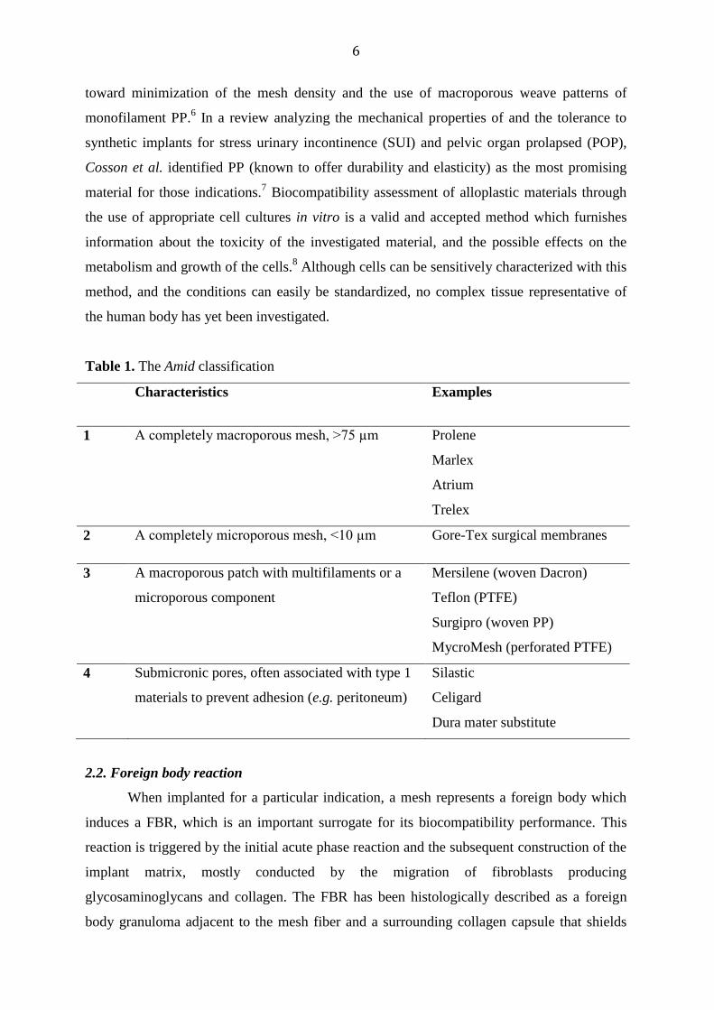
Table 1. The Amid classification
Characteristics Examples
1 A completely macroporous mesh, >75 µm Prolene
Marlex
Atrium
Trelex
2 A completely microporous mesh, <10 µm Gore-Tex surgical membranes
3 A macroporous patch with multifilaments or a
microporous component
Mersilene (woven Dacron)
Teflon (PTFE)
Surgipro (woven PP)
MycroMesh (perforated PTFE)
4 Submicronic pores, often associated with type 1
materials to prevent adhesion (e.g. peritoneum)
Silastic
Celigard
Dura mater substitute
2.2. Foreign body reaction
When implanted for a particular indication, a mesh represents a foreign body which
induces a FBR, which is an important surrogate for its biocompatibility performance. This
reaction is triggered by the initial acute phase reaction and the subsequent construction of the
implant matrix, mostly conducted by the migration of fibroblasts producing
glycosaminoglycans and collagen. The FBR has been histologically described as a foreign
body granuloma adjacent to the mesh fiber and a surrounding collagen capsule that shields
7
the host from the foreign material. It seems likely that such a chronic inflammatory process
impairs normal wound healing and tissue regeneration and may result in reduced
functionality and increased side-effects when applied clinically.9 A considerable influence on
the dynamics of the FBR is exerted by the biomaterial composition, and in particular the type
of polymer, the material weight, the filament structure and the pore size.
However, the process of FBR does not necessarily reduce the proposed mesh function
of restoring mechanical functionality in a particular region of the body. Several attempts have
been made to improve the biocompatibility of meshes and reduce the FBR.10, 11
The exact
FBR mechanisms and respective time flow in vivo are not entirely understood, but the rapid
accumulation of huge numbers of phagocytic cells, and especially blood monocytes and
tissue-derived macrophages, play a crucial role in it.12-14
Additionally, the transcriptionally-
induced overexpression of the matrix metalloproteinases-2 (MMP-2) seems to play a key role
in the FBR.15-18
New therapeutic strategies may target cellular and molecular interactions
during these phases, influencing the complex cascade of immune modulators, soluble
mediators and different cell types. A beneficial effect of gentamycin on a chronic FBR by
modulation of MMP-2 gene transcription has recently been described, which may be a
feasible approach to the optimization of mesh integration into the abdominal wall, and
ultimately to improvement of the long-term outcome following hernia mesh repair.19
2.3. Application of surgical meshes and current controversies
Alloplastic materials such as meshes are widely applied in surgical approaches for
hernia, incontinence and prolapse situations. The development of meshes is an ongoing
process characterized by changes in polymer structure, biocompatibility, operative handling
and costs. A good mesh should provoke a negligible FBR with no pathologic fibrosis at a
decreased risk of infection. In a Public Health Notification issued in 2008, the FDA reported
more than 1000 unexpected and severe adverse events, mostly associated with transvaginal
placement of a surgical mesh to treat POP and SUI.20
In 2011, a second FDA warning was
announced on the basis of 2,874 newly identified Medical Device Reports: 1,503 associated
with POP repairs, and 1,371 associated with SUI repairs.21
Factors involved included the
overall health of the patient, the surgical technique used and concomitant procedures
undertaken; particular importance was attached to the mesh material and the size and shape of
the mesh as causes of adverse events. However, the search for the optimal mesh for a
particular indication, with high functionality and biocompatibility and a minimized side-
effect profile in the short-term and long-term follow-up remains difficult. These two FDA
8
warnings have led to several regulatory changes for surgical meshes, including the upgrading
of risk classifications, requirements for clinical studies to address the risks and benefits of
meshes used to treat POP and SUI and the expanded post-market monitoring of device
performance.21
2.4. The IDEAL recommendations
In the assessment of quality standards for surgical meshes, comparability with other
meshes with regard to quality and stage of development should be possible. Despite the
existence of several models for the assessment of different meshes with regard to their
particular biomechanical characteristics, there are currently virtually no standardized tools for
comparisons among meshes.22, 23
Many alloplastic materials are still being used without
proper trials and are recommended by manufacturers rather than on the basis of data arising
from in vitro or in vivo experiences. Once a product is on the market, financial support for
further investigations decreases and ongoing evaluation with unknown results is often not
desired. Thus, as compared with the strict regulations for drug development and market
implementation, the process of adopting and improving surgical innovations (e.g. meshes) is
still unregulated, unstructured and variable. In 2009, the Lancet dedicated a series to the topic
of ”Surgical Innovation and Evaluation” and its current status.24-26
A five-stage description of
the surgical development process has been proposed, the IDEAL model which allows the
assignment of every surgical innovation, e.g. surgical technique, alloplastic materials, etc., to
its particular corresponding step of development. The IDEAL framework, has so far been
used in retrospective studies, and in particular in the description of surgical procedures.27, 28
However, this framework is highly recommended for the application in the development of
surgical innovations different from surgeries. This is important as the innovations are often
introduced and developed only by manufacturers. Research ethics committees and device
regulatory bodies could help by requiring a declaration of the IDEAL stage that the
investigators/manufacturers feel the device or procedure has reached, with supporting
evidence. Innovations in the idea stage, for instance, would then be expected to lead to
proposals for a prospective development study. Thus, application of IDEAL would allow the
setting of minimum requirements for developmental steps prior to market entry for any
innovative product or device. As the current studies cover the topic of a surgical innovation,
we followed the IDEAL recommendations in the entire investigation strategy and course in
order to ensure comparability, visibility and confirmability.
9
3. MAIN GOALS
There are currently no standardized tools for the comparison of surgical meshes. The
initial purpose of our studies was to develop and standardize an in vitro test system with
which to investigate biocompatibility features of surgical meshes. Once having developed
this test system, we aimed to implement mesh-coating strategies and subsequently to
investigate their influence on biocompatibility in in vitro performance. In order to explore the
predictive value and validity of the test system and also newly tested coating strategies, we
conducted a large-animal experiment in sheep. The entire approach followed the recently
developed recommendations of IDEAL for surgical innovations.
1. Study I. The main purpose was to investigate and develop an in vitro approach for an
assessment of the biocompatibility features of surgical meshes. Seven different mesh types,
currently used in various indications, were randomly selected and microscopically
investigated after incubation for 6 weeks with regard to their adherence performance, using a
tissue culture approach, with tissues representative of fibroblasts, muscle cells and
endothelial cells, originating from 10 different patients.
2. Study II. We aimed to investigate different mesh coating modalities with autologous blood
components and their impact on the biocompatibility performance of the meshes in vitro,
using the previously developed test system. Seven different mesh types were therefore coated
prior to cultivation with autologous PBMCs, platelets and blood plasma and subsequently
incubated for 6 weeks in a minced tissue assay. The adherence performance of the tissues on
the meshes was investigated microscopically, assessed semi-quantitatively and compared
with the native counterparts, using a previously developed scoring system.
3. The goals of Study III and Study IV were to translate the preliminary in vitro results into
an animal model in order to validate the in vitro test system and to explore its predictive
value for in vivo surroundings. Three different meshes [TVT (Tension-free Vaginal Tape),
UltraPro® and polyvinylidene fluoride PVDF] with different previous in vitro performance
scores were implanted in female sheep in a native or a plasma-coated version. In the ultra-
short-term study, meshes were explanted and investigated histochemically for inflammatory
infiltrate, macrophage infiltration, vessel formation, myofibroblast invasion and connective
tissue accumulation at the implant site at 5 min, 20 min, 60 min and 120 min. In the long-
term study, meshes were explanted after 3, 6, 12 or 24 months and processed.
10
4. MATERIAL AND METHODS
4.1. In vitro experiments
4.1.1. Meshes/Patients
We randomly identified alloplastic materials currently applied as implants for
different surgical indications covering hernia repair, POP and SUI. A total of 7 different
meshes were investigated in this study. The meshes and their biomechanical/material
characteristics are listed in Table 2. The alloplastic materials were prepared in 2 x 2 cm
fragments for further investigations. Additionally, we harvested tissue probes of muscle,
fascia and renal vein from 10 patients undergoing right-side nephrectomy. All patients gave
their informed consent previously. The tissues and cells were processed identically in all
patients. Each mesh was tested with the tissue and cells of each patient for comparison
purposes.
Table 2. Investigated meshes and their characteristics
Mesh name Material Biomechanical characteristics
Vitamesh®,
ProxyBiomedical
large-pore
monofilament
polypropylene
knitted PP, pore size 2410 µm, thickness
250 µm, tear resistance (Fmax N) 33.7
Dynamesh®, FEG
Textiltechnik
monofilament PVDF effective porosity: 58%, reactive surface:
1.97 m²/m², suture pull-out strength: 31
N, tear propagation resistance: 28 N, pore
size: 3000 μm
TFT Motifmesh®,
ProxyBiomedical
micromachined PTFE pore size 235 µm, thickness 150 µm, tear
resistance (Fmax N) 15.1
TVT PP
PP non-absorbable, permanent PP suture,
pore size 164 x 96 μm
UltraPro®, Hernia
System Medium
UHSM®, Ethicon
PP reinforced with
polyglecaprone fibers
filament thickness 0.09 mm, mesh
thickness 0.5 mm, Fmax N 69 N, pore size
300 µm
Proceed surgical
mesh®, Ethicon
monofilament PP
encapsulated with
polydioxanone (PDS)
closely knitted with small pores < 1000
µm size, high tensile strength
Mersilene, Johnson &
Johnson
multifilament mesh,
PET
density 0.19 g/cm3, pore size 120-85 µm
11
4.1.2. Tissue preparation and mesh incubation
In an initial cell culture approach involving investigation of the cells of the different
tissues, we could not identify adherence on the mesh microscopically. In contrast, the cells
grew on the bottom of the cell culture device. When performing a tissue culture, we observed
tissue adherence both on the meshes and on the bottom of the cell culture device. As the cells
did not grow on the respective mesh matrices, we decided to use tissue cultures for the
following investigations. In addition, we considered tissue cultures more appropriate because
of the reduced artificial modification processes due to the shorter culture processing.
We extracted tissue probes originating from muscle, fascia and renal vein at a length
of 0.5-1.0 cm each from 10 different patients. After crushing, we incubated the tissue with
phosphate-buffered saline and, after 2 additional washing procedures, incubation was
performed with DMEM/F12 plus 10% serum and 1% glutamine + 1% penicillin/stretomycin.
After successful expansion and growth (80-90% adherent growth) of the tissue pellets, the
different alloplastic materials were added. Thus, the prepared and expanded tissue probes
consisting of myoblast, endothelial cells and fibroblasts presenting relevant tissues of the
pelvic floor were used in vitro in order to create a model for investigation of the integrity of
the different mesh types. Myoblasts were detected by α-sarcomeric actin and desmin as
markers of myogene differentiation. Fibroblasts were stained with antibodies targeting
vimentin, whereas antibodies against cluster of differentiation 34 (CD34) were used for the
verification of endothelial cells. We reproduced every single approach 10 times with tissue
probes from the 10 different patients.
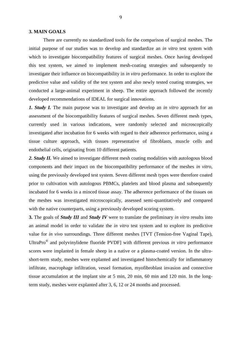
Figure 1. Microscopic adhesion course on PVDF: A) native prior to tissue culture, B) after a
3-week tissue culture and C) after a 6-week tissue culture (time point of assessment). In B)
and C) the spaces between the mesh filaments are increasingly occupied by fibroblasts,
collagen deposits and capillaries. As adherence appeared 3-dimensionally, entirely defined
pictures cannot be shown.29
12
4.1.3. Mesh coating
PBMCs, platelets and plasma were used. PBMCs were separated through density
gradient centrifugation using Ficoll.30
For the isolation of platelets and the respective
mediators, the Advanced Tissue Regeneration System (ATR® by Curasan Inc) was used
(http://www.curasan.de/de/produkte/dental/atr/atr.php). The plasma preparation procedure
followed the classical method of Crowley.31
After isolation of the 3 different blood
components of each patient, we incubated the meshes (2 x 2 cm) with 10 ml of the respective
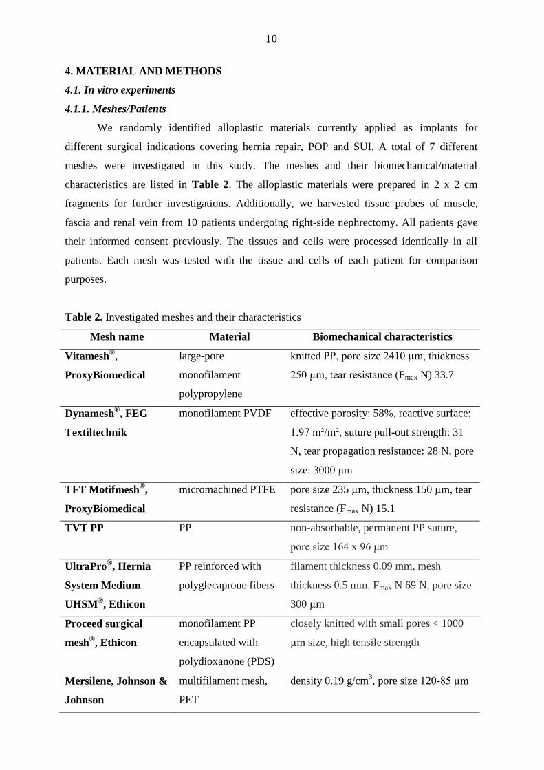
suspension and incubated them for 12 h prior to testing with tissue. Successful plasma
coating is exemplified in Figure 2.
Figure 2. PVDF: A) native, B) after a 12-h plasma incubation, C) after a 12-h plasma
incubation and trypan blue staining. In B and C the plasma is adherent to the mesh filaments,
whereas the non-covered parts of the mesh appear native as in A.32
4.1.4. Morphological study
The adherence and the cell count (if possible) were assessed microscopically and
through the use of immunohistochemistry after co-incubation of the cells with different types
of alloplastic meshes. The test duration was 6 weeks. Meshes were investigated with regard
to interstructural tissue connections and the quantity of mesh-adherent cells. Tissue cultures
were maintained up to 4 months, with frequent changes of the medium, and assessment was
repeated if possible.
A descriptive/semiquantitive assessment pattern was used in order to describe the
adherence of tissue to the investigated mesh materials. The assessment pattern was based on
the maximum identifiable quantity of mesh-adherent cells within a tissue cluster per vision
field. Adherence performance was ranked after assessment of the quality and quantity of the
tissue clusters/cells as none, fair, good or excellent.33
13
4.2. In vivo experiments
4.2.1. Animals
The animal experiments were conducted at the Institute for Surgical Research of the
University of Szeged, Hungary, in accordance with the NIH Guidelines (Guide for the Care
and Use of Laboratory Animals). The experimental protocol was approved by the Animal
Welfare Committee at the University of Szeged (license/permission No. V01353/2010).
Fourteen 6-month old, female sheep weighing 20-25 kg were housed and cared for at
the University farm for experimental animal studies. We included 2 animals more than the 12
needed for safety calculations. All the animals had free access to food and water, and were
cared for by an educated keeper and routinely inspected by a veterinarian. On the basis of the
previously described test system and the resulting ranking, we selected 3 meshes representing
good, intermediate and poor in vitro performance; PVDF, UltraPro® and TVT (Table 3a).
4.2.2. Mesh implantation
Operations were performed on sheep in a supine position. The animals were intubated
and an aspiration tube was introduced into the stomach. Anesthesia with Isoflurane 2% mixed
with air and O2 (50/50%) was then established. Surgery was performed by using a
longitudinal laparotomy. We chose 3 different locations in the sheep to implant the meshes
via open surgery. In order to represent different in vivo surroundings, 3 meshes were placed
in the following localizations: a) interaperitoneally, b) as fascia onlay and c) as muscle onlay
(fascia sublay). The size of the implanted meshes was 3 x 5 cm. Then, 3 plasma-coated
versions of the same mesh type were implanted in equivalent localizations on the
contralateral side of the torso. Meshes had to be incubated with autologous plasma at least 12
h prior to implantation. This procedure was performed in 14 animals, resulting in 4 animals
per mesh type (plus 2 animals with PP TVT/PVDF). The mean operation duration was 1.5 h.
For the short-term study, 3 additional female sheep weighing from 20 - 25 kg and at
least 6 months old were included. The protocol for housing and veterinary maintenance was
as in the long-term study. The size of the implanted meshes was 3 x 12 cm. For every native
mesh implant, a respective plasma-coated version was implanted in an equivalent localization
on the contralateral side of the torso. The length of incubation prior to surgery was at least 12
h. We selected 1 sheep per investigated mesh, i.e. resulting in 3 animals. The investigated
meshes were again PVDF, UltraPro® and TVT. The chosen time points for explantation were
5 min, 20 min, 60 min and 120 min. At each explantation time point, we dissected a piece
about 3 x 3 cm in size from the initially implanted mesh. During implantation, the meshes
14
were fixed with 2 sutures at both ends. The mean operation duration for the implantation was
50 min.
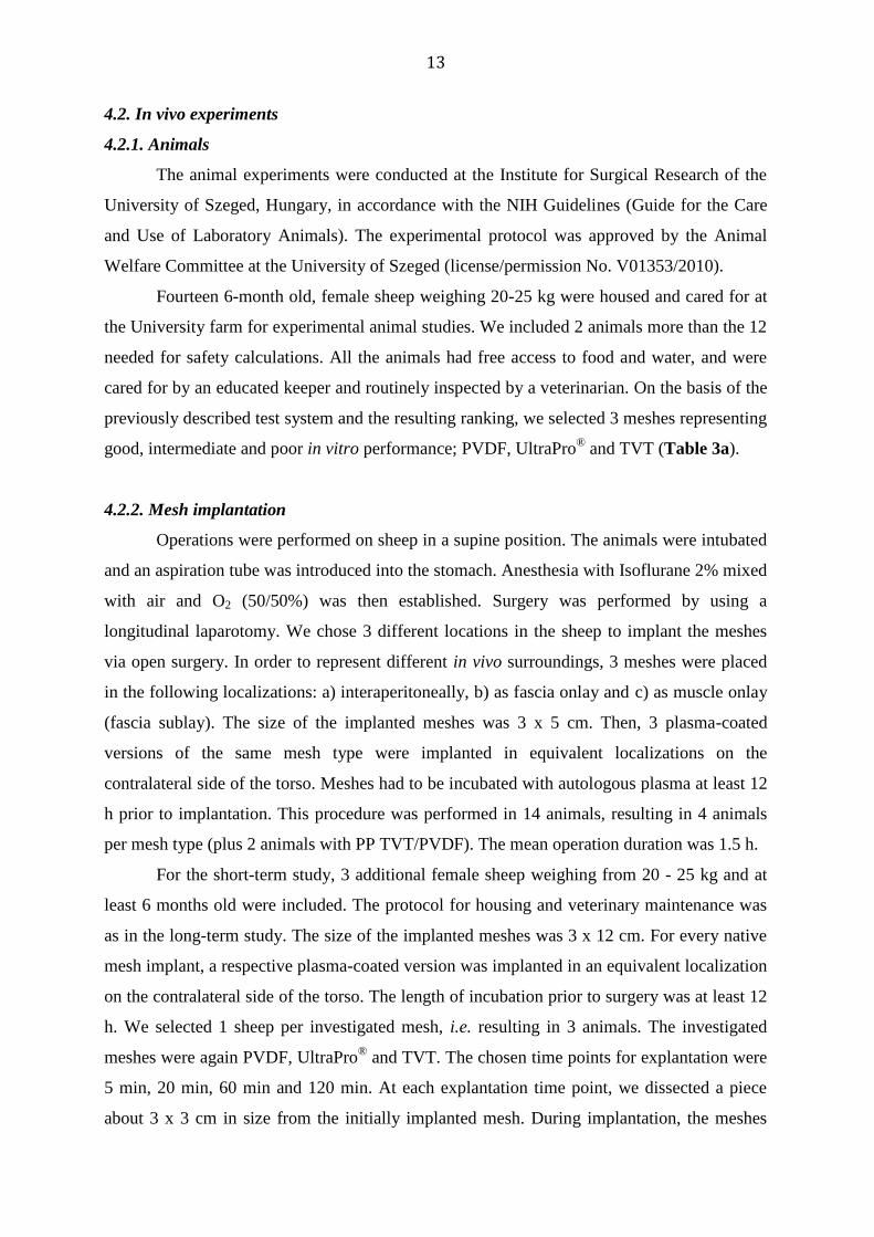
Figure 3. Intraoperative sites during implantation: (A) intraperitoneal, (B) fascia onlay, (C)
muscle onlay. 12 h prior to implantation the meshes were coated and incubated with
autologous plasma. Meshes were implanted bilaterally into the torso to allow intraindividual
comparison of coated versus uncoated meshes per animal.
4.2.3. Mesh explantation
After 3, 6, 12 and 24 months, three animals, respectively, underwent surgery for mesh
explantation. The meshes were identified and then harvested, extent of local reactions was
described macroscopically. The animals were sacrificed directly after mesh explantation and
harvesting of probes of parenchymatous organs (liver, intestine, kidney, lung, heart). The
harvested material was then assessed for foreign body reaction, scar formation and
inflammatory reaction. For the short term study, explantation time points were 5 min, 20 min,
60 min, 120 min, respectively.
4.2.4. Morphological studies
A single longitudinal section of mesh and adhesive tissue was obtained from each
explanted mesh. Tissue samples were fixed in 10% formalin, and then sliced into 0.3 x 1 cm
pieces and embedded in paraffin. In each case, 10 to 15 sections of 4 µm thickness were
stained with hematoxylin and eosin (H&E), and with periodic acid-Schiff plus diastase and
Elastica van Gieson. All mesh specimens were studied by light microscopy which was
controlled by immunohistochemistry performed on the material embedded in paraffin, using
15
the avidin-biotin complex method with diaminobenzidine as a chromogen. The procedure
was repeated twice for every sample. The antibodies used in this study included polyclonal
rabbit anti-human CD3, 1:50, as pan-marker for T-lymphocytes (DAKO, Hamburg,
Germany), polyclonal rabbit anti-human CD138, 1:50 as pan-marker for plasma cells
(DAKO, Hamburg, Germany), monoclonal mouse anti-porcine CD68, 1:50 (DAKO,
Hamburg, Germany), as pan-marker for macrophages, monoclonal anti-human CD15, 1:10
(Becton Dickinson, Heidelberg, Germany) as marker for polymorphonuclear granulocytes,
polyclonal rabbit anti-actin protein, 1:200 (DAKO, Hamburg, Germany) and monoclonal
anti-CD34 1:200 (BIOMOL, Hamburg, Germany), as markers for fibromyocytes, and
monoclonal porcine CD31, 1:10 (DIANOVA, Hamburg, Germany), as marker for endothelial
cells. The morphometric evaluation consisted of a quantitative cell analysis of the
inflammatory reaction and soft-tissue reaction. The cells were counted in each of in 5 H&E-
slides in 10 fields in a grid of 10 points (100x, area 0.1 mm2) and in the interface (0-300 mm,
400x, area 625 mm2). The parameters measured were the inflammatory infiltrate (µm),
connective tissue (µm), vessels (%), macrophages (%), leukocytes (%), polymorphonuclear
granulocytes (%), and fibroblasts (%), and TUNEL, Ki67 and HSP 70 expressing cells (%).
4.2.5. Statistics
All data were tested by ANOVA with the LSD modification according to Bonferroni.
Statistical significance was set at p < 0.05.
16
5. RESULTS
5.1. In vitro experiments
5.1.1. Macroscopic results
Overall, the macroscopic evaluation after tissue culturing did not disclose differences
in the gross appearance of the meshes. Tissue culturing was successful in 100% of the probes.
No signs of infection were observed throughout the entire cultivation course.
5.1.2. Microscopic results
The tissue growth was comparable in all approaches within a test duration of 6 weeks. The
testing of the biocompatibility of myoblasts, endothelial cells and fibroblasts following the
addition of BioGlue®
revealed unchanged tissue adherence to the mesh. Interindividual
differences were not observed as concerns the growth and adherence performance after
incubation with the different meshes in the investigated 10 patients.
After 6 weeks, the investigated meshes were ranked according to the
descriptive/semiquantitative approach described by Melman et al.33
The Melman score
classifies tissue/cellular ingrowth on meshes as follows: none (0 point): no tissue ingrowth;
fair (1 point): thin bands of fibroblasts and small collagen deposits between the mesh
filaments; good (2 points): moderately thick bands of fibroblasts / collagen deposits between
mesh filaments; excellent (3 points): nearly all the spaces between the mesh filaments are
occupied by fibroblasts, collagen deposits and capillaries.
The ranking is shown in Table 3a. Interestingly, after 4 months of tissue culturing the
adhesion performance was comparable for all the meshes. Table 3b and Figure 4 present the
ranking of the investigated native meshes and the different coating modifications.
The entire experiment was reproduced as described and a modified Melman score was
subsequently used for the 3 different coating approaches for each patient. Analysis of the
PBMC-incubated meshes indicated tissue ingrowth comparable to that for the native mesh.
Interestingly, the meshes previously incubated with ATR® (Curasan Inc) and the plasma-
coated meshes exhibited a slightly better performance. This trend was reproduced after 4
months of tissue culturing. All individuals displayed comparable effects of tissue ingrowth in
the native state and after coating with the different blood components.
17
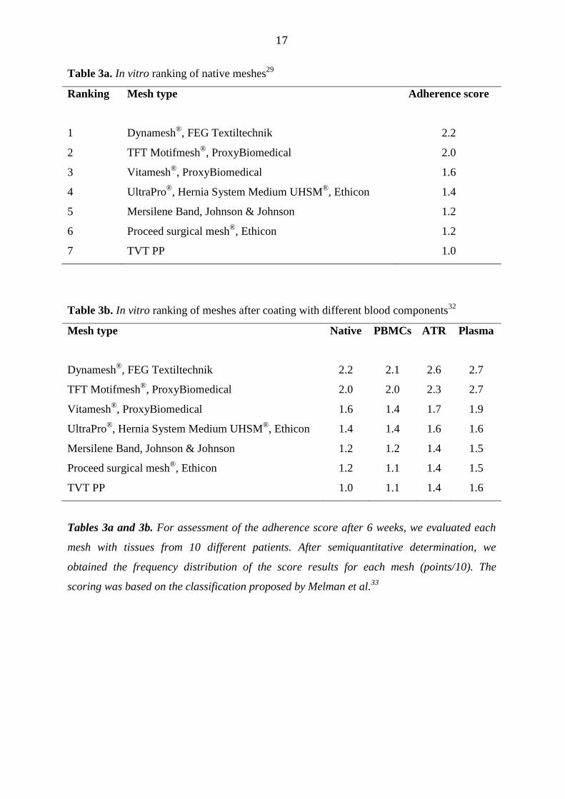
Table 3a. In vitro ranking of native meshes29
Ranking Mesh type Adherence score
1 Dynamesh®, FEG Textiltechnik 2.2
2 TFT Motifmesh®, ProxyBiomedical 2.0
3 Vitamesh®, ProxyBiomedical 1.6
4 UltraPro®
, Hernia System Medium UHSM®, Ethicon 1.4
5 Mersilene Band, Johnson & Johnson 1.2
6 Proceed surgical mesh®, Ethicon 1.2
7 TVT PP 1.0
Table 3b. In vitro ranking of meshes after coating with different blood components32
Mesh type Native PBMCs ATR Plasma
Dynamesh®, FEG Textiltechnik 2.2 2.1 2.6 2.7
TFT Motifmesh®, ProxyBiomedical 2.0 2.0 2.3 2.7
Vitamesh®, ProxyBiomedical 1.6 1.4 1.7 1.9
UltraPro®, Hernia System Medium UHSM
®, Ethicon 1.4 1.4 1.6 1.6
Mersilene Band, Johnson & Johnson 1.2 1.2 1.4 1.5
Proceed surgical mesh®, Ethicon 1.2 1.1 1.4 1.5
TVT PP 1.0 1.1 1.4 1.6
Tables 3a and 3b. For assessment of the adherence score after 6 weeks, we evaluated each
mesh with tissues from 10 different patients. After semiquantitative determination, we
obtained the frequency distribution of the score results for each mesh (points/10). The
scoring was based on the classification proposed by Melman et al.33
18
0
0,5
1
1,5
2
2,5
3
Dynamesh ®, FEG
Textiltechnik
TFT Motifmesh ®,
ProxyBiomedical
Vitamesh ®,
ProxyBiomedical
UltraPro Hernia System
Medium UHSM®,
Ethicon
Mersilene Band,
Johnson & Johnson
Proceed surgical mesh
®, Ethicon
TVT Polypropylene
Mesh-Type
Melm
an
-Sco
re
native
PBMC
ATR
Plasma
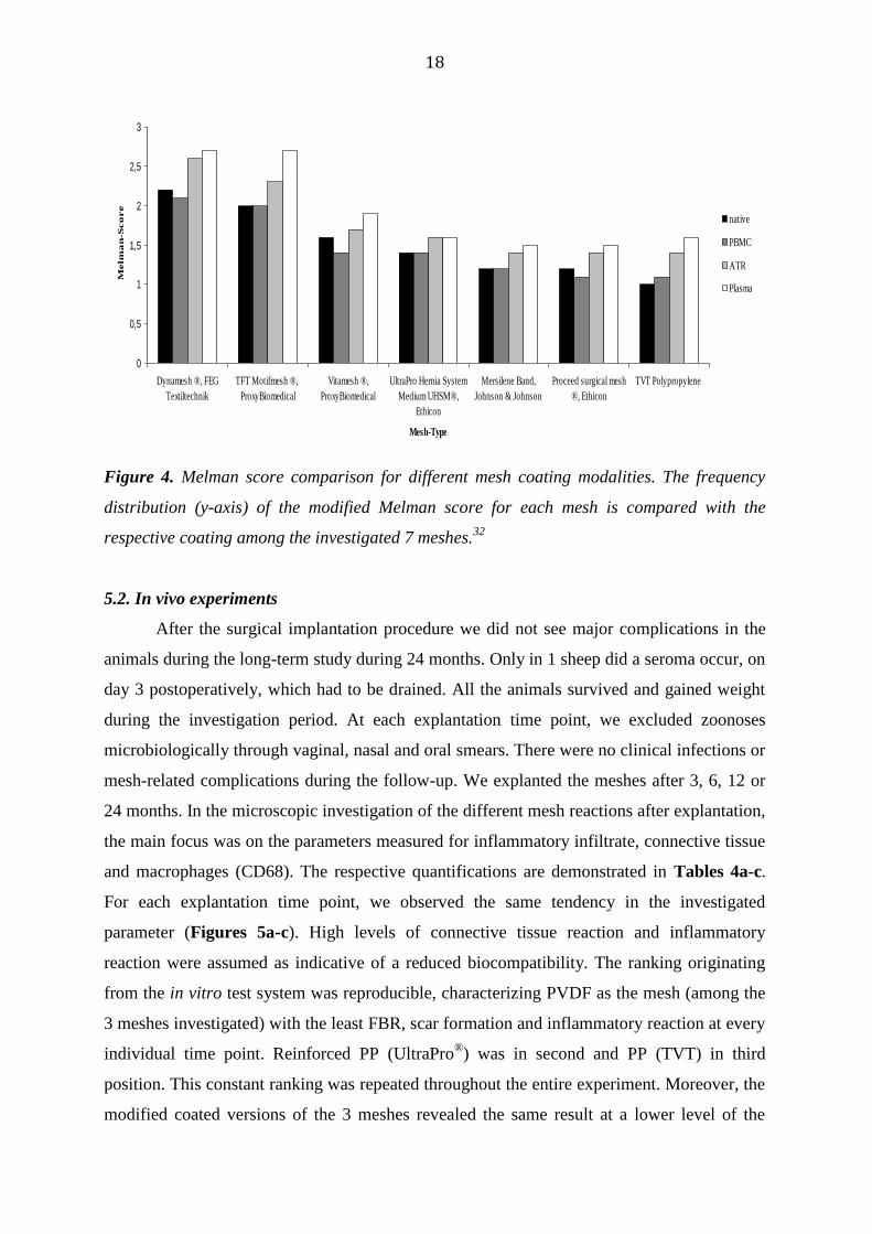
Figure 4. Melman score comparison for different mesh coating modalities. The frequency
distribution (y-axis) of the modified Melman score for each mesh is compared with the
respective coating among the investigated 7 meshes.32
5.2. In vivo experiments
After the surgical implantation procedure we did not see major complications in the
animals during the long-term study during 24 months. Only in 1 sheep did a seroma occur, on
day 3 postoperatively, which had to be drained. All the animals survived and gained weight
during the investigation period. At each explantation time point, we excluded zoonoses
microbiologically through vaginal, nasal and oral smears. There were no clinical infections or
mesh-related complications during the follow-up. We explanted the meshes after 3, 6, 12 or
24 months. In the microscopic investigation of the different mesh reactions after explantation,
the main focus was on the parameters measured for inflammatory infiltrate, connective tissue
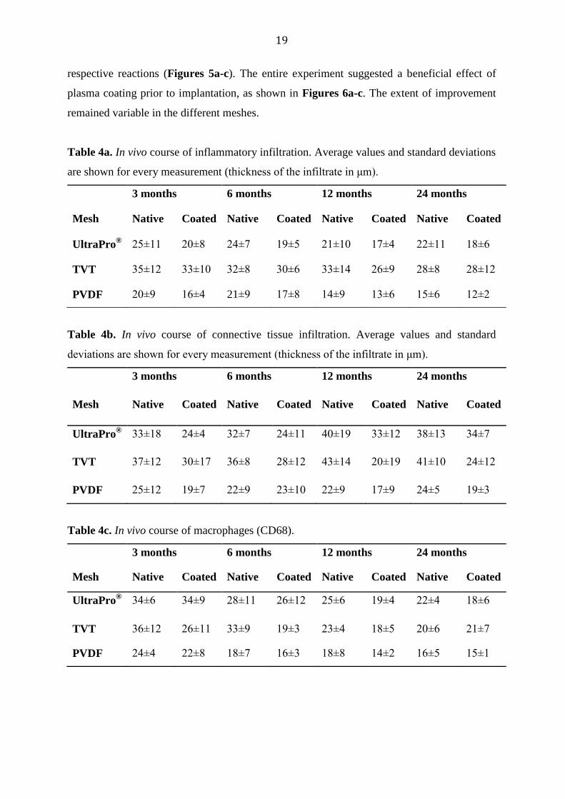
and macrophages (CD68). The respective quantifications are demonstrated in Tables 4a-c.
For each explantation time point, we observed the same tendency in the investigated
parameter (Figures 5a-c). High levels of connective tissue reaction and inflammatory
reaction were assumed as indicative of a reduced biocompatibility. The ranking originating
from the in vitro test system was reproducible, characterizing PVDF as the mesh (among the
3 meshes investigated) with the least FBR, scar formation and inflammatory reaction at every
individual time point. Reinforced PP (UltraPro®) was in second and PP (TVT) in third
position. This constant ranking was repeated throughout the entire experiment. Moreover, the
modified coated versions of the 3 meshes revealed the same result at a lower level of the
19
respective reactions (Figures 5a-c). The entire experiment suggested a beneficial effect of
plasma coating prior to implantation, as shown in Figures 6a-c. The extent of improvement
remained variable in the different meshes.
Table 4a. In vivo course of inflammatory infiltration. Average values and standard deviations
are shown for every measurement (thickness of the infiltrate in μm).
Table 4b. In vivo course of connective tissue infiltration. Average values and standard
deviations are shown for every measurement (thickness of the infiltrate in μm).
Table 4c. In vivo course of macrophages (CD68).
3 months 6 months 12 months 24 months
Mesh Native Coated Native Coated Native Coated Native Coated
UltraPro®
25±11 20±8 24±7 19±5 21±10 17±4 22±11 18±6
TVT 35±12 33±10 32±8 30±6 33±14 26±9 28±8 28±12
PVDF 20±9 16±4 21±9 17±8 14±9 13±6 15±6 12±2
3 months 6 months 12 months 24 months
Mesh Native Coated Native Coated Native Coated Native Coated
UltraPro®
33±18 24±4 32±7 24±11 40±19 33±12 38±13 34±7
TVT 37±12 30±17 36±8 28±12 43±14 20±19 41±10 24±12
PVDF 25±12 19±7 22±9 23±10 22±9 17±9 24±5 19±3
3 months 6 months 12 months 24 months
Mesh Native Coated Native Coated Native Coated Native Coated
UltraPro® 34±6 34±9 28±11 26±12 25±6 19±4 22±4 18±6
TVT 36±12 26±11 33±9 19±3 23±4 18±5 20±6 21±7
PVDF 24±4 22±8 18±7 16±3 18±8 14±2 16±5 15±1
20
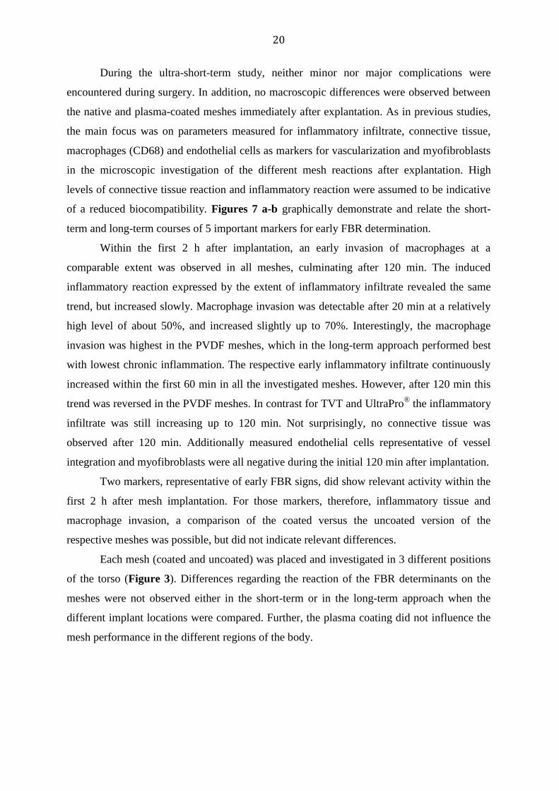
During the ultra-short-term study, neither minor nor major complications were
encountered during surgery. In addition, no macroscopic differences were observed between
the native and plasma-coated meshes immediately after explantation. As in previous studies,
the main focus was on parameters measured for inflammatory infiltrate, connective tissue,
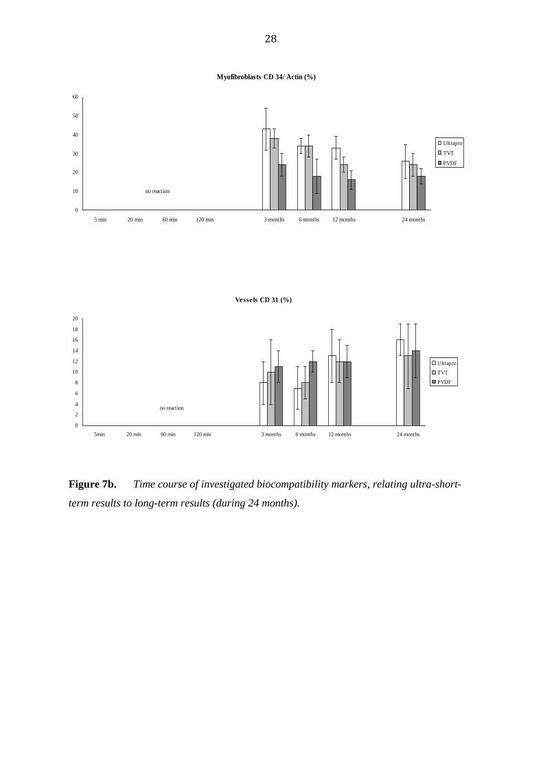
macrophages (CD68) and endothelial cells as markers for vascularization and myofibroblasts
in the microscopic investigation of the different mesh reactions after explantation. High
levels of connective tissue reaction and inflammatory reaction were assumed to be indicative
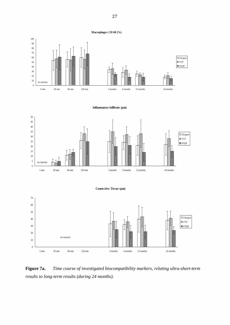
of a reduced biocompatibility. Figures 7 a-b graphically demonstrate and relate the short-
term and long-term courses of 5 important markers for early FBR determination.
Within the first 2 h after implantation, an early invasion of macrophages at a
comparable extent was observed in all meshes, culminating after 120 min. The induced
inflammatory reaction expressed by the extent of inflammatory infiltrate revealed the same
trend, but increased slowly. Macrophage invasion was detectable after 20 min at a relatively
high level of about 50%, and increased slightly up to 70%. Interestingly, the macrophage
invasion was highest in the PVDF meshes, which in the long-term approach performed best
with lowest chronic inflammation. The respective early inflammatory infiltrate continuously
increased within the first 60 min in all the investigated meshes. However, after 120 min this
trend was reversed in the PVDF meshes. In contrast for TVT and UltraPro®
the inflammatory
infiltrate was still increasing up to 120 min. Not surprisingly, no connective tissue was
observed after 120 min. Additionally measured endothelial cells representative of vessel
integration and myofibroblasts were all negative during the initial 120 min after implantation.
Two markers, representative of early FBR signs, did show relevant activity within the
first 2 h after mesh implantation. For those markers, therefore, inflammatory tissue and
macrophage invasion, a comparison of the coated versus the uncoated version of the
respective meshes was possible, but did not indicate relevant differences.
Each mesh (coated and uncoated) was placed and investigated in 3 different positions
of the torso (Figure 3). Differences regarding the reaction of the FBR determinants on the
meshes were not observed either in the short-term or in the long-term approach when the
different implant locations were compared. Further, the plasma coating did not influence the
mesh performance in the different regions of the body.
21
Inflammatory Infiltrate in µm uncoated
0
5
10
15
20
25
30
35
40
3 months 6 months 12 months 24 months
TVT
Ultrapro
PVDF
Inflammatory Infiltrate in µm coated
0
5
10
15
20
25
30
35
40
3 months 6 months 12 months 24 months
TVT
Ultrapro
PVDF
Figure 5a. In vivo ranking and validation. In vivo ranking of PVDF, PP (TVT) and
reinforced PP (UltraPro®
) meshes with regard to the extent of the inflammatory reaction.
The ranking is depicted for the coated and native versions of the meshes.
22
Macrophages CD68 (%) coated
0
5
10
15
20
25
30
35
40
3 months 6 months 12 months 24 months
TVT
Ultrapro
PVDF
Macrophages CD68 (%) uncoated
0
5
10
15
20
25
30
35
40
3 months 6 months 12 months 24 months
TVT
Ultrapro
PVDF
Figure 5b. In vivo ranking and validation. In vivo ranking of PVDF, PP (TVT) and
reinforced PP (UltraPro®
) meshes with regard to the extent of the macrophage count. The
ranking is depicted for the coated and native versions of the meshes.
23
Connective Tissue in µm coated
0
5
10
15
20
25
30
35
40
45
50
3 months 6 months 12 months 24 months
TVT
Ultrapro
PVDF
Connective Tissue in µm uncoated
0
5
10
15
20
25
30
35
40
45
50
3 months 6 months 12 months 24 months
TVT
Ultrapro
PVDF
Figure 5c. In vivo ranking and validation. In vivo ranking of PVDF, PP (TVT) and
reinforced PP (UltraPro®
) meshes with regard to the extent of connective tissue formation. The
ranking is depicted for the coated and native versions of the meshes.
24
Connective Tissue in µm
0
5
10
15
20
25
30
35
40
45
50
3 months 6 months 12 months 24 months
coated
uncoated
**
**
Connective Tissue in µm
0
5
10
15
20
25
30
35
40
45
50
3 months 6 months 12 months 24 months
coated
uncoated
**
**
Connective Tissue in µm
0
5
10
15
20
25
30
35
40
45
50
3 months 6 months 12 months 24 months
coated
uncoated
***
ULTRAPRO
TVT
PVDF
* = p<0,05
Figure 6a. Effect of plasma coating prior to implantation. A high level of connective tissue
is related to a reduced biocompatibility. Statistically significant differences in the thickness of
the connective tissue are indicated by an asterisk (*), corresponding to p < 0.05.
25
Inflammatory Infiltrate in µm
0
5
10
15
20
25
30
35
40
3 months 6 months 12 months 24 months
coated
uncoated
*
***
Inflammatory Infiltrate in µm
0
5
10
15
20
25
30
35
40
3 months 6 months 12 months 24 months
coated
uncoated
*
Inflammatory Infiltrate in µm
0
5
10
15
20
25
30
35
40
3 months 6 months 12 months 24 months
coated
uncoated
*
**
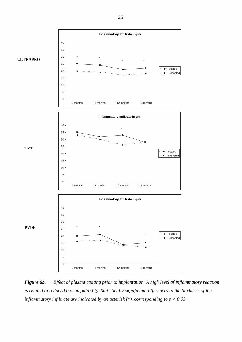
Figure 6b. Effect of plasma coating prior to implantation. A high level of inflammatory reaction
is related to reduced biocompatibility. Statistically significant differences in the thickness of the
inflammatory infiltrate are indicated by an asterisk (*), corresponding to p < 0.05.
ULTRAPRO
TVT
PVDF
26
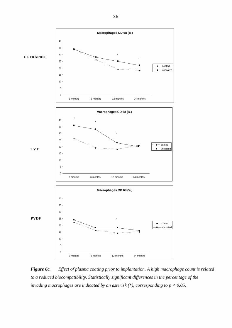
Macrophages CD 68 (%)
0
5
10
15
20
25
30
35
40
3 months 6 months 12 months 24 months
coated
uncoated
*
*
Macrophages CD 68 (%)
0
5
10
15
20
25
30
35
40
3 months 6 months 12 months 24 months
coated
uncoated
*
*
*
Macrophages CD 68 (%)
0
5
10
15
20
25
30
35
40
3 months 6 months 12 months 24 months
coated
uncoated
*
Figure 6c. Effect of plasma coating prior to implantation. A high macrophage count is related
to a reduced biocompatibility. Statistically significant differences in the percentage of the
invading macrophages are indicated by an asterisk (*), corresponding to p < 0.05.
ULTRAPRO
TVT
PVDF
27
Macrophages CD 68 (%)
0
10
20
30
40
50
60
70
80
90
100
5 min 20 min 60 min 120 min 3 months 6 months 12 months 24 months
no reaction
Ultrapro
TVT
PVDF
Inflammatory Infiltrate (µm)
0
5
10
15
20
25
30
35
40
45
50
5 min 20 min 60 min 120 min 3 months 6 months 12 months 24 months
no reaction
Ultrapro
TVT
PVDF
Connective Tissue (µm)
0
10
20
30
40
50
60
70
5 min 20 min 60 min 120 min 3 months 6 months 12 months 24 months
no reaction
Ultrapro
TVT
PVDF
Figure 7a. Time course of investigated biocompatibility markers, relating ultra-short-term
results to long-term results (during 24 months).
28
Myofibroblasts CD 34/ Actin (%)
0
10
20
30
40
50
60
5 min 20 min 60 min 120 min 3 months 6 months 12 months 24 months
no reaction
Ultrapro
TVT
PVDF
Vessels CD 31 (%)
0
2
4
6
8
10
12
14
16
18
20
5min 20 min 60 min 120 min 3 months 6 months 12 months 24 months
no reaction
Ultrapro
TVT
PVDF
Figure 7b. Time course of investigated biocompatibility markers, relating ultra-short-
term results to long-term results (during 24 months).
29
6. DISCUSSION
6.1. In vitro test system
At present, many alloplastic materials are used without proper trials and are
recommended by manufacturers rather than on the basis of data arising from in vitro or in
vivo experiences. The standardized classification put forward by Amid in 1997 is one of the
few tools which allows prediction of the biocompatibility performance of a surgical mesh.4 In
that classification, importance is attributed to the pore size of the meshes as predictive of the
expectable adverse event rate, suggesting that the classification should be taken into account
when clinical decisions are made. Large porous meshes are currently regarded as the best
tissue-integrative, with the least FBR, inflammation and fibrosis, whereas small-pore mesh
modifications are associated with a significantly greater FBR and inflammation frequently
related with the phenomenon of bridging, which may finally cause significant contraction or
shrinkage of the mesh.4
The aim of Study I was to investigate whether different tissues of the pelvic floor
demonstrate different in vitro interaction characteristics with alloplastic materials currently
used as meshes in different clinical indications, i.e. biocompatibility features rather than
mechanical characteristics. Most of the meshes investigated in the present study consist of PP
as basic material. We searched for a feasible and reproducible test system that allows the
assessment and comparison of meshes with regard to their in vitro adherence scores to
different tissues, as markers of their biocompatibility.
The biocompatibility assessment of alloplastic materials through the use of
appropriate cell cultures in vitro is a valid and accepted method which yields information
about the toxicity of the investigated material, and possible effects on the metabolism and
growth of the cells.8 Langer et al. investigated the cellular response of human fibroblasts
cultured on different PP meshes, in particular with regard to the mesh material and
structure.34
They used a method comparable to the one described here, implementing
scanning electron microscopy. Their major conclusion was that the polymer surface and
structure had a paramount influence on the biocompatibility of the meshes, as they identified
fibroblasts preferably growing on low-weight meshes, thin filaments and mesh nodes. In
contrast heavy-weight meshes were revealed to induce degenerative cell reactions resulting in
a reduced biocompatibility. In contrast with Langer et al. we used a tissue culture approach,
as our initial results with cell cultures did not reveal sufficient cell growth. Although
investigation of the adherence of specific cells is useful, we consider that the co-incubation of
implants with tissue clusters is comparable to in vivo processes. We can support the thesis of
30
Langer et al. that the microstructure of the meshes has a relevant impact on the growth and
adherence behavior of cells imitating in vivo surroundings. Besides the presence of
fibroblasts, we investigated muscle-derived and endothelial cells presenting relevant tissues
of the pelvic floor. These observations resulted in a ranking of the investigated meshes as
concerns their affinity for the co-incubated tissues/cells. Our ranking is in good accordance
with the suggestions of Amid.4 However, material features other than pore size seem to play a
role for the in vitro performance, explaining the different scores. Moreover, it emerged
reproducibly that the adherence behavior was independent of the individual patient features,
thereby supporting the idea that the biological behavior of a mesh in contact with host tissues,
is mostly conditioned by the structure of the biomaterial or/and its chemical composition
rather than by individual host characteristics/features. PVDF, a polymer with good textile and
biological properties, displayed the best adherence performance in our test system. This
polymer is currently applied as PVDF-coated PP mesh for intraperitoneal-only repair.35, 36
The main characteristics of this mesh are its macroporosity, a decreased adhesion rate to the
bowel and a favorable biocompatibility in vivo, with low rates of inflammation and fibrosis.5
The good performance of PVDF in our test system is therefore not surprising. To date,
reported investigations of the cellular reaction on meshes in humans are based on explanted
meshes after complicated postoperative courses, resulting in negatively preselected
alloplastic explants.37
The test system presented here is far from being representative of an in vivo situation
as the tissue culture was sterile and no probable physiological in vivo reaction, such as a
FBR, inflammation, etc. was imitated. Another criticism may be aimed at the unselective
investigation of the alloplastic materials. Meshes for POP, SIU or hernia repair definitely do
not have identical biocompatibility/mechanical requirements. However, assumption of the
adherence performance of tissue on a mesh as a possible marker of its biocompatibility seems
logically independent of the respective clinical use of the implant. The test system may help
to select particular alloplastic materials for further investigations, such as animal
experiments, coating approaches (in vivo and in vitro), etc., as we reproduce comparable
extents of cell adherence on specific meshes independently of the individual patient. Thus, at
least in vitro, specific patient features do not seem to influence the adherence performance
and respective biocompatibility of the alloplastic material. Although an exaggerated FBR
tissue response is assumed to be related to clinical complications, a positive role in mesh
incorporation at the implant site may be triggered by bioactive mediators such as epidermal
growth factor, basic fibroblast growth factor or transforming growth factor and others
31
produced by fibroblasts or smooth muscle cells, for example. Thus the cultivation and
positive adherence of cell clusters consisting of these cell types and the respective assessment
and comparison, as shown here, may be helpful in the consideration of a mesh as regards its
possible tissue ingrowth and capacity to form connective tissue.
The in vivo behavior of a particular alloplastic material cannot be reliably extrapolated
from in vitro studies, and appropriate in vivo approaches are therefore needed. The possible
predictive value of these in vitro results with respect to cell and tissue adherence in vivo was
the target in the following animal investigations.
6.2. In vitro plasma coating
Preclinical investigations of surgical innovations, e.g. meshes, in terms of bench
and/or animal testing, are likely to represent a new standard requirement to confirm that
engineering specifications are met and that the material and/or specific modification chosen
for a mesh is sufficiently biocompatible. To date, there have been numerous reports of mesh
modification approaches in order to improve their biocompatibility. The permanent character
of a foreign body implant may cause persistent and increased inflammation, with ongoing
collagen deposition leading to extensive fibrosis. The impaired host-acceptance of the
implanted mesh is likely to appear through chronic inflammation and extensive fibrosis.16
In
order to tackle the problem of extensive FBR initiated by early local inflammation, several
researchers have modified the chemical and physical properties of meshes by different
coating approaches resulting in altered local reactions and tissue responses, mostly by using
in vivo experiments. Numerous compounds have been tested so far for mesh-coating
purposes, the majority of them in in vivo models, mostly after setting a pathological defect to
be repaired by the investigated meshes.10, 11, 38-40
Besides numerous in vivo experiments,
Bryan et al. provide an in vitro model to facilitate the mesh choice in uncomplicated hernia
repair by quantitatively determining neutrophil activation and degranulation in different mesh
types.41
Their approach represents one of the few in vitro assessment tools for meshes
currently available in the literature. In their experiments, reactive oxygen species (ROS),
released by activated neutrophils and leading to non-specific host tissue damage and potential
mechanical weakening were measured on the surface of 6 different meshes. They
investigated native, non-modified meshes. They concluded that the mesh structure is a greater
determinant of ROS release than the chemical composition. It seems likely that their
sophisticated assay could also be used for mesh assessment after different coating
approaches. This would be a conclusive further development comparable to the approach
32
presented here, which represents an advance on the initially described in vitro assessment
tool for native meshes.
The aim of our study was to implement and assess a facile mesh-coating procedure in
vitro and to investigate whether the coating of meshes with autologous blood components
shows different in vitro interaction characteristics with different tissues types as compared
with native meshes. We used autologous blood components as they are relatively easy to
obtain from the respective patients and contain relevant cells and substances involved in the
humoral immune defense. This approach was based on the assumption that the extent to
which an implanted alloplastic material elicits an acute local inflammatory response has an
impact on the long-term outcome when applied in vivo.42
In order to investigate cellular and
non-cellular components, we separately investigated PBMC, plasma and platelets with the
respective mediators. Incubation with PBMCs did not result in modification of the adherence
score for the investigated tissues. This may be explained by the reduced ability of these cells
to maintain permanent contact with the polymer surface of the meshes, as previously shown.
In contrast, blood plasma and ATR resulted in a better adherence performance and increased
biocompatibility in all meshes. An interesting observation in the current study is that all
meshes previously ranked with regard to their biocompatibility performance displayed an
increased score after plasma coating and maintained their position in the ranking relative
compared to the other investigated meshes. This supports the thesis that coating with plasma
may have an effect independent of the mesh, and, at least in vitro, all meshes could improve
their performance, but low-ranked meshes could not increase their position relative to
natively better-positioned counterparts. The thesis of Bryan et al. can thereby be supported:
the mesh structure seems to be an important determinant of the in vitro performance in the
native and coated configuration of a mesh.41
Mesh-related complications are known to be
related to extensive local inflammation, representing the first step of a FBR. This FBR after
implantation of a mesh is assumed to be triggered by the secretion of a variety of proteins that
attract inflammatory cells to migrate to the site of injury, finally leading to extracellular
matrix regulation and collagen deposition. In a recent study, Brandt et al. investigated the
effect of mesh coating (PVDF) with different substances affecting the cortisone metabolism.
In their in vivo approach, they found that hydrocortisone and spironolactone protected from
inflammatory response, ended up in smaller granuloma at the implant site of the mesh and
decreased the collagen formation.43
Their approach suggested that the respective coating
approaches are a possible way to attenuate local inflammatory processes in order to reduce
the FBR. This is supported by other research groups who have reported altered local cell
33
activation and tissue responses after modifying the chemical and/or physical properties of
meshes via coating, leading to the hypothesis that the coating of polymer surfaces may be an
opportunity to improve mesh integration and biocompatibility.44
The assumption of the
adherence performance of tissues on a mesh as a possible marker of its biocompatibility
seems logical, independently of the respective clinical use of the implant. Although an
exaggerated FBR/tissue response has been suggested to be related to clinical complications, a
positive role in mesh incorporation at the implant site may be triggered by bioactive
mediators such as epidermal growth factor, basic fibroblast growth factor or transforming
growth factor and others produced by fibroblasts or smooth muscle cells, for instance. Thus,
the cultivation and positive adherence of cell clusters consisting of these cell types and the
respective assessment and comparison, as shown here, may be helpful for considering the
possible tissue ingrowth and capacity of a mesh to form connective tissue. The coating of
meshes with plasma and ATR appears to have a positive effect on those features.
A main limitation of this study is that no inflammatory reaction as normally cascading
in vivo was imitated as the in vitro approach was sterile. In conclusion, we conducted an
animal investigation (Studies III and IV) in order to validate the in vitro results, for both
native and coated meshes.
6.3. In vivo validation
The next step in the current approach was to translate the previous in vitro results into
in vivo surroundings. In vitro models to investigate the biocompatibility features of alloplastic
materials like meshes are limited with regard to their predictibility for in vivo surroundings. A
mesh, per se, is a foreign body which induces a FBR. This FBR is triggered by the initial
acute phase reaction and the subsequent construction of the implant matrix, mostly conducted
by the migration of fibroblasts producing glycosaminoglycans and collagen. There is ongoing
debate as to which implant-induced reactions are desirable and which are not. The
development of new meshes should be based on a firm understanding of the mechanisms of a
FBR.45
In our in vivo study, the histologic investigations for inflammatory infiltrates
indicated a slight reaction associated with PVDF, which was increased in reinforced PP
(UltraPro®
) and even more so in PP (TVT). This reduced inflammatory reaction can be
considered an expression of good biocompatibility. However, this observed postoperative
sign of an inflammatory reaction was non-infectious, as counts for cells involved in the
infectious immune defense, e.g. CD3, remained unaltered at low levels.46
In addition, when
the connective tissue investigated, the same trend was observed: PVDF exhibited the thinnest
layer of connective tissue, followed by reinforced PP (UltraPro®) and PP (TVT). There was a
34
macrophage decrease in all meshes during the postoperative follow-up, but the highest
number of macrophages was seen in the TVT meshes and the in vitro ranking was consistent
as regards this marker. Macrophages are key mediators involved in the foreign body immune
reaction, suggesting that this reaction was stronger in PP (TVT) than in the other two applied
meshes. As concerns the investigated parameters, macrophage invasion, inflammatory tissue
and connective tissue formation, this study confirmed the previously established in vitro
ranking of the 3 investigated meshes repeatedly throughout the entire animal experiment,
after 3, 6, 12 and 24 months. Moreover, when the meshes were modified by pre-implant
coating with autologous plasma, the ranking remained constant. This supports the assumption
that the recently developed tissue culture in vitro test system for meshes is able to predict the
in vivo performance of the meshes. Practically, the test system helps to distinguish between
meshes with good and poorer healing performances. The previously described in vitro test
system was sterile, and thus no physiological in vivo reaction such as FBR or inflammation
could be imitated. This indicates that the adherence ability of a mesh is crucial for subsequent
FBRs or inflammatory processes which determine the in vivo performance of the meshes.
Moreover, as in the in vitro approach, we did not see individual recipient features that
influenced the performance of the meshes. Besides material quality issues, we assume that
the process which caused FBR to the meshes must have occurred in the early period, before 3
months after implantation, since there was no further tendency to change during the
following explantations.
In a recent comparable long-term study in sheep, Zinther et al. investigated the
shrinkage of an intraperitoneal onlay mesh using a coated polyester mesh versus a covered
PP mesh.47
Besides the individual differences of the investigated meshes, they described a
peak for shrinkage at 3 months, without additional shrinkage in the following 15 months,
suggesting an early effect. This is in accordance with our results which indicate that an early
process is responsible for the extent of a FBR and the mid- and long-term performance of an
implanted mesh. This tendency is independent of the location of the mesh in the body,
although its particular extent varies, depending on the site of implantation. Although those
results must be confirmed in a larger series, this could be a novel approach to predict the
bioperformance and integration of any available mesh, using a standardized in vitro
experiment.
Several animal studies have been proposed and reported for the investigation of local
reactions after the implantation of a mesh graft. The present study is the first experimental
study conducted in sheep, with a 2-year observation period. The use of sheep as an animal
35
model has various advantages. The biological behavior of human cells is comparable to that
of the cells in the sheep model. As compared with other large animals, sheep demonstrate a
limited growth potential, while the tendency to adhesion formation (intra-abdominally) is
similar to that in humans.48, 49
In our study, we did not observe a specific reaction triggered
by lymphocytes (B-lymphocytes and T-lymphocytes). Thus, it is very unlikely that the
different lymphocyte status of sheep vs. humans may have had important influence on the in
vivo biocompatibility performance. However, to exclude this potential bias, experiments in
primates would be necessary, although very unrealistic. Given the mentioned advantages, the
sheep model has the potential to serve as a template in future experimental mesh studies, in
particular for the assessment of meshes in the abdominal cavity, but also other intracorporal
locations. Data on adequate functional performance and material safety are currently at the
focus of premarket reviews for mesh devices. Thus, preclinical investigations in terms of
bench and/or animal testing are currently used to confirm that engineering specifications are
met and that the material chosen for a mesh is biocompatible. Unfortunately, clinical
performance data are rarely used to support clearance for meshes for whatever indication.
In the study presented here, we could demonstrate the predictive value of our recently
developed in vitro cell culture approach for the biocompatibility assessment of meshes when
translated to in vivo circumstances. In a second attempt, we investigated coating approaches
for meshes in order to improve their biocompatibility. In preliminary experiments, mesh
coating with autologous plasma was shown to reduce FBRs both in vitro and in vivo. A
plasma coating seems to have a consistent improving effect on the performance of the mesh
as regards connective tissue development and inflammatory local reactions at the implant
site, suggesting an improved biocompatibility.
6.4. Foreign body reaction in the short term and the long term
The main purpose of our large-animal study was to investigate in vivo
biocompatibility predictors for 3 different meshes by measuring early and long-term signs of
a FBR such as macrophage invasion, and inflammatory reaction and connective tissue
determination at the implant site of the meshes. By relating the ultra-short-term data to the
long-term data in the same species (i.e. sheep), we could show that the process of
determination of a FBR is defined early in the course after implantation for markers of local
acute inflammation. In contrast, myofibroblast invasion, vascularization and connective
tissue adhesion are not relevantly presented in the ultra-short-term course. The extent of
macrophage invasion and inflammatory tissue does not relevantly increase after 120 min as
compared with the values for 3 months after explantation or later. Our previously described
36
method to improve the biocompatibility performance of meshes in vivo and in vitro by
autologous plasma coating before implantation did not have an effect on the early
inflammatory events, as the respective values for inflammatory infiltrate and macrophage
invasion did not differ from coated to native meshes.1, 29, 32
However, markers such as
connective tissue organization, myofibroblast invasion and endothelial cells, characteristic of
vascularization, are detectable after 3 months post-implantation and show different extents in
the 3 investigated meshes.
To the best of our knowledge, our results reflect the longest combined short and long-
term in vivo approach to the investigation of biocompatibility issues on meshes. In addition,
no ultra-short-term investigations in vivo have ever been reported so far, as most of the
currently available studies investigated effects on meshes at the earliest after 7-21 days.50
It
has been shown that an acute inflammatory reaction occurs at 7 days after implantation of a
mesh, dominated by macrophage invasion.51
Over time, this early inflammatory process
transforms into a chronic, at times granulomatous reaction, promoting wound healing, but
also forming small granulomas.16
It is known that the extent of collagen formation may vary
during this process, whereas a severe inflammatory reaction, with disordered fibrin and
collagen deposition, is likely to compromise the integration process and functional outcome.
In an investigation of prolene and a porcine dermal collagen implant (Pelvicol®), Zheng et al.
identified a first acute-phase reaction after 48 h, peaking on day 7-14.51
Our data add ultra-
short-term information, suggesting that this reaction starts even earlier in the course, after a
matter of minutes. Zheng et al. described that the acute reaction diminished and finally
reached negligible levels by 90 days, which can be partially supported in our current study.
FBRs to alloplastic mesh material are primarily induced by inflammatory cells such as
macrophages and T-lymphocytes.52, 53
Macrophages have a critical role in acute inflammation
and early vascularization, and also in the subsequent chronic phase of the host response, as
they are known to be capable of differentiating toward two pathways. This M1/M2
polarization enhances macrophages, leading to an immediate and/or persistent inflammation
or to a constructive remodeling and new tissue generation.54
However, this polarization was
not investigated in the current study. From the aspect of wound healing, it is known that
CD68-positive macrophages reach their maximum level on the second day after injury and
slowly decline thereafter.55
We have shown that high percentages of CD68-positive
macrophages are detectable on the meshes after only minutes or hours. This could be of
interest when investigating and developing mesh modification strategies to influence this
early acute reaction. A previously developed plasma-coating strategy to optimize the
37
biocompatibility of meshes does not seem to influence this early inflammatory reaction and
inflammatory infiltrate formation, but rather to influence mid- and long-term processes which
lead to neovascularization and collagen fiber organization. It has been found that premature
type III collagen is predominantly synthesized in the early phases of wound healing and in
the presence of inflammatory cells.56
This type III collagen is then replaced by highly cross-
linked and stable type I collagen later after implantation. Delayed wound healing and
immature scar development due to persistent chronic inflammation may be predicted by a
lowered type I/III collagen ratio.37, 57
A favorable type I/III collagen ratio is known to
improve biocompatibility and can be positively influenced by the pre-implantation of mesh
modifications such as a gentamycin coating.39
As concerns the results of the present study, an
increase in inflammatory infiltrate was revealed for all 3 meshes. After 120 min, PVDF was
observed to increase considerably more slowly than TVT and UltraPro®. The previously
shown good long-term biocompatibility performance of PVDF may hypothetically be
triggered by an early decrease of the acute inflammatory reaction and subsequent
modification of the micro-environment meshes, leading to an improved type I/III collagen
ratio, for instance. We did not observe a direct influence of the localization of the implanted
mesh in either the body in the short- or the long-term study as regards the reproducibility for
the coated and uncoated meshes. Although the localizations were chosen to cover different
structural parts of the body with different immunologic potentials and physical/mechanical
strains, the localization does not seem to be of utmost importance for the biocompatibility
performance of a mesh in vivo.
6.5. The project with regard to the IDEAL recommendations and future perspectives
A plethora of commercially available meshes currently make the decision as to which
mesh to apply very difficult. Two FDA warnings in 2008 and 2011 reported more than 3,500
severe adverse events after mesh applications, mostly in POP and SIU patients. As a
consequence, the FDA recommended the consideration of regulatory changes, including an
upgrading in risk classifications for meshes, clinical studies to address the risks and benefits
of meshes and expanded the post-market monitoring of device performance.58
Our preclinical
in vivo study was initially inspired by the first FDA warning of unexpected and severe
adverse events during the use of mesh devices.20
We raised the question of whether the
performance of a mesh would be predictable prior to its implantation in order to reduce the
probability of unexpected mid- and long-term events as reported and complemented in
2011.21
Although we did not selectively investigate meshes for the indications reported as
POP and SIU, our system (in vivo and in vitro) may easily be used with every available mesh.
38
However, in a considerably narrow time frame as a reaction to the first FDA warning, we
developed an in vitro approach, a subsequent animal study, and are now translating our
results into a clinical trial. As a first step with this regard, we have analyzed and classified
specific complications after vaginal mesh implantation approaches for prolapse repair.59
This
analysis comprises the basis for a currently ongoing pilot study which is investigating the
feasibility of the autologous plasma coating of meshes prior to implantation in humans. Thus,
on the basis of the results presented in this work, we are applying for the first time plasma-
coated meshes in the human setting (Barski, Gerullis, Otto et al., 2014, unpublished data). As
a next and most important step, a prospective randomized trial, possibly proving the positive
effect of the plasma coating on the biocompatibility and morbidity outcome, is planned on the
basis of the positive preliminary results. Both studies would fulfill stage 3/A of the IDEAL
classification and consistently sustain the initially chosen way of implementing this new
technology of plasma coating following IDEAL.
In addition, and in order to provide the final stage 4/L step of the IDEAL approach,
we are already creating and establishing an online platform for registration and outcome
measurement of mesh application with clear definitions and classifications, which will be
offered to the uro-gynecological community. We are therefore using well-described
definitions for risk factors and possible complications based on ICS-IUGA standards in
terminology and guidelines (Barski, Gerullis, Otto et al., 2014, unpublished data).
These steps conform to the IDEAL recommendations and show how surgical research
may be concluded (independently of any result) when strictly driven following standardized
recommendations. McCulloch et al. specified recommendations concerning IDEAL for the
field of urology.60
Although not mentioned in their review, we would add mesh-
implementing procedures for an interventional option as a topic of current controversy and
debate in urology/uro-gynecology, not only for safety purposes, but also for effectiveness
considerations.61-64
In conclusion, the current work displays the experimental in vitro and in
vivo basis for the surgical innovation of a plasma coating on surgical meshes. The already
completed and all planned stages of this innovation strictly follow the IDEAL
recommendations.
39
7. SUMMARY OF FINDINGS
1. We have developed a manufacturer-independent in vitro test system for the adherence
performance of tissue clusters (fibroblasts, endothelial cells and muscle-derived cells) as a
marker for the biocompatibility of commercially available meshes. In this test system, we
established a repeatable ranking of meshes with regard to their biocompatibility. The
adherence behavior was independent of the individual patient features, suggesting that the
biological behavior of a mesh is probably conditioned by the structure of the biomaterial
or/and its chemical composition rather than by individual host characteristics/features.
2. The in vitro test system is a feasible pattern for the investigation of different coating
strategies of meshes. The coating of meshes prior to cultivation with PBMCs, for instance,
did not affect the adherence score, whereas coating with platelets and blood plasma increased
the score, suggesting an improved biocompatibility in vitro. The plasma coating displayed the
greatest potential to improve the in vitro adherence score. The previous ranking of the native
meshes remained consistent after coating, but was established at a higher level.
3. In a long-term animal study in sheep, we demonstrated that the developed in vitro test
system for the biocompatibility of meshes may predict the in vivo performance of the meshes.
This effect is independent of the location of the mesh in the body, although its particular
extent varies depending on the site of implantation. The coating of meshes with autologous
plasma prior to implantation seems to have a positive effect on the biocompatibility of
meshes in vivo.
4. Investigations of the ultra-short-term determinants of the FBR at the implant site in vivo
revealed that the local inflammatory reaction is an early and susceptible event after mesh
implantation. It cannot be influenced by prior plasma coating and does not depend on the
localization of implantation.
The development of this surgical innovation (the plasma coating of meshes prior to
implantation) strictly followed the IDEAL recommendations at every step in order to ensure
comparability and transparency.
40
REFERENCES
1. Gerullis H, Klosterhalfen B, Boros M, Lammers B, Eimer C, Georgas E, et al. IDEAL in
meshes for prolapse, urinary incontinence, and hernia repair. Surg Innov
2013;20(5):502-8.
2. Gomelsky A, Dmochowski RR. Biocompatibility assessment of synthetic sling materials
for female stress urinary incontinence. J Urol 2007;178(4 Pt 1):1171-81.
3. Black J. Biological Performance of Materials. Fundamentals of Biocompatibility, New
York: Marcel Dekker, Inc 1999.
4. Amid PK. Classification of biomaterials and their related complications in abdominal wall
hernia surgery. Hernia 1997;1:15-21.
5. Klinge U, Klosterhalfen B, Ottinger AP, Junge K, Schumpelick V. PVDF as a new
polymer for the construction of surgical meshes. Biomaterials 2002;23(16):3487-93.
6. de Tayrac R, Deffieux X, Gervaise A, Chauveaud-Lambling A, Fernandez H. Long-term
anatomical and functional assessment of trans-vaginal cystocele repair using a
tension-free polypropylene mesh. Int Urogynecol J Pelvic Floor Dysfunct
2006;17(5):483-8.
7. Cosson M, Debodinance P, Boukerrou M, Chauvet MP, Lobry P, Crepin G, et al.
Mechanical properties of synthetic implants used in the repair of prolapse and urinary
incontinence in women: which is the ideal material? Int Urogynecol J Pelvic Floor
Dysfunct 2003;14(3):169-78.
8. Pizzoferrato A, Vespucci A, Ciapetti G, Stea S. Biocompatibility testing of prosthetic
implant materials by cell cultures. Biomaterials 1985;6(5):346-51.
9. Szpaderska AM, DiPietro LA. Inflammation in surgical wound healing: friend or foe?
Surgery 2005;137(5):571-3.
10. Emans PJ, Schreinemacher MH, Gijbels MJ, Beets GL, Greve JW, Koole LH, et al.
Polypropylene meshes to prevent abdominal herniation. Can stable coatings prevent
adhesions in the long term? Ann Biomed Eng 2009;37(2):410-8.
11. Junge K, Rosch R, Klinge U, Saklak M, Klosterhalfen B, Peiper C, et al. Titanium
coating of a polypropylene mesh for hernia repair: effect on biocompatibilty. Hernia
2005;9(2):115-9.
12. Ziats NP, Miller KM, Anderson JM. In vitro and in vivo interactions of cells with
biomaterials. Biomaterials 1988;9(1):5-13.
13. Bhardwaj RS, Henze U, Klein B, Zwadlo-Klarwasser G, Klinge U, Mittermayer C, et al.
Monocyte-biomaterial interaction inducing phenotypic dynamics of monocytes: a
41
possible role of monocyte subsets in biocompatibility. J Mater Sci Mater Med
1997;8(12):737-42.
14. Schachtrupp A, Klinge U, Junge K, Rosch R, Bhardwaj RS, Schumpelick V. Individual
inflammatory response of human blood monocytes to mesh biomaterials. Br J Surg
2003;90(1):114-20.
15. Eckes B, Zigrino P, Kessler D, Holtkotter O, Shephard P, Mauch C, et al. Fibroblast-
matrix interactions in wound healing and fibrosis. Matrix Biol 2000;19(4):325-32.
16. Klinge U, Klosterhalfen B, Muller M, Schumpelick V. Foreign body reaction to meshes
used for the repair of abdominal wall hernias. Eur J Surg 1999;165(7):665-73.
17. Jansen PL, Kever M, Rosch R, Krott E, Jansen M, Alfonso-Jaume A, et al. Polymeric
meshes induce zonal regulation of matrix metalloproteinase-2 gene expression by
macrophages and fibroblasts. FASEB J 2007;21(4):1047-57.
18. Ravanti L, Kahari VM. Matrix metalloproteinases in wound repair (review). Int J Mol
Med 2000;6(4):391-407.
19. Binnebosel M, von Trotha KT, Ricken C, Klink CD, Junge K, Conze J, et al. Gentamicin
supplemented polyvinylidenfluoride mesh materials enhance tissue integration due to
a transcriptionally reduced MMP-2 protein expression. BMC Surg 2012;12:1.
20. www.fda.gov/MedicalDevices/Safety/AlertsandNotices/ucm262435.htm.
21.www.fda.gov/medicaldevices/safety/alertsandnotices/publichealthnotifications/ucm06197
6.htm.
22. Dietz HP, Vancaillie P, Svehla M, Walsh W, Steensma AB, Vancaillie TG. Mechanical
properties of urogynecologic implant materials. Int Urogynecol J Pelvic Floor
Dysfunct 2003;14(4):239-43; discussion 43.
23. Afonso JS, Martins PA, Girao MJ, Natal Jorge RM, Ferreira AJ, Mascarenhas T, et al.
Mechanical properties of polypropylene mesh used in pelvic floor repair. Int
Urogynecol J Pelvic Floor Dysfunct 2008;19(3):375-80.
24. Ergina PL, Cook JA, Blazeby JM, Boutron I, Clavien PA, Reeves BC, et al. Challenges in
evaluating surgical innovation. Lancet 2009;374(9695):1097-104.
25. McCulloch P, Altman DG, Campbell WB, Flum DR, Glasziou P, Marshall JC, et al. No
surgical innovation without evaluation: the IDEAL recommendations. Lancet
2009;374(9695):1105-12.
26. Barkun JS, Aronson JK, Feldman LS, Maddern GJ, Strasberg SM, Altman DG, et al.
Evaluation and stages of surgical innovations. Lancet 2009;374(9695):1089-96.
42
27. Blazeby JM, Blencowe NS, Titcomb DR, Metcalfe C, Hollowood AD, Barham CP.
Demonstration of the IDEAL recommendations for evaluating and reporting surgical
innovation in minimally invasive oesophagectomy. Br J Surg 2011;98(4):544-51.
28. Heikens JT, Gooszen HG, Rovers MM, van Laarhoven CJ. Stages and evaluation of
surgical innovation: a clinical example of the ileo neorectal anastomosis after
ulcerative colitis and familial adenomatous polyposis. Surg Innov 2013;20(5):459-65.
29. Gerullis H, Georgas E, Eimer C, Goretzki PE, Lammers BJ, Klosterhalfen B, et al.
Evaluation of Biocompatibility of Alloplastic Materials: Development of a Tissue
Culture In Vitro Test System. Surg Technol Int 2011;XXI:21-27.
30. Fotino M, Merson EJ, Allen FH, Jr. Micromethod for rapid separation of lymphocytes
from peripheral blood. Ann Clin Lab Sci 1971;1(2):131-3.
31. Crowley RT. A Method for Plasma Preparation and Preservation for Intravenous Use.
Ann Surg 1941;113(6):1088-9.
32. Gerullis H, Georgas E, Eimer C, Arndt C, Barski D, Lammers B, et al. Coating with
autologous plasma improves biocompatibility of mesh grafts in vitro: development
stage of a surgical innovation. Biomed Res Int 2013;2013:536814.
33. Melman L, Jenkins ED, Hamilton NA, Bender LC, Brodt MD, Deeken CR, et al.
Histologic and biomechanical evaluation of a novel macroporous
polytetrafluoroethylene knit mesh compared to lightweight and heavyweight
polypropylene mesh in a porcine model of ventral incisional hernia repair. Hernia
2011;15(4):423-31.
34. Langer C, Schwartz P, Krause P, Mohammadi H, Kulle B, Schaper A, et al. In-vitro study
of the cellular response of human fibroblasts cultured on alloplastic hernia meshes.
Influence of mesh material and structure. Chirurg 2005;76(9):876-85.
35. Berger D, Bientzle M. Polyvinylidene fluoride: a suitable mesh material for laparoscopic
incisional and parastomal hernia repair! A prospective, observational study with 344
patients. Hernia 2009;13(2):167-72.
36. Conze J, Junge K, Weiss C, Anurov M, Oettinger A, Klinge U, et al. New polymer for
intra-abdominal meshes--PVDF copolymer. J Biomed Mater Res B Appl Biomater
2008;87(2):321-8.
37. Klinge U, Si ZY, Zheng H, Schumpelick V, Bhardwaj RS, Klosterhalfen B. Abnormal
collagen I to III distribution in the skin of patients with incisional hernia. Eur Surg
Res 2000;32(1):43-8.
43
38. Schonleben F, Reck T, Tannapfel A, Hohenberger W, Schneider I. Collagen foil
(TissuFoil E) reduces the formation of adhesions when using polypropylene mesh for
the repair of experimental abdominal wall defects. Int J Colorectal Dis
2006;21(8):840-6.
39. Binnebosel M, Ricken C, Klink CD, Junge K, Jansen M, Schumpelick V, et al. Impact of
gentamicin-supplemented polyvinylidenfluoride mesh materials on MMP-2
expression and tissue integration in a transgenic mice model. Langenbecks Arch Surg
2010;395(4):413-20.
40. van 't Riet M, de Vos van Steenwijk PJ, Bonthuis F, Marquet RL, Steyerberg EW, Jeekel
J, et al. Prevention of adhesion to prosthetic mesh: comparison of different barriers
using an incisional hernia model. Ann Surg 2003;237(1):123-8.
41. Bryan N, Ahswin H, Smart NJ, Bayon Y, Hunt JA. In vitro activation of human
leukocytes in response to contact with synthetic hernia meshes. Clin Biochem
2012;45(9):672-6.
42. Clave A, Yahi H, Hammou JC, Montanari S, Gounon P, Clave H. Polypropylene as a
reinforcement in pelvic surgery is not inert: comparative analysis of 100 explants. Int
Urogynecol J 2010;21(3):261-70.
43. Brandt CJ, Kammer D, Fiebeler A, Klinge U. Beneficial effects of hydrocortisone or
spironolactone coating on foreign body response to mesh biomaterial in a mouse
model. J Biomed Mater Res A 2011;99(3):335-43.
44. MacEwan MR, Brodbeck WG, Matsuda T, Anderson JM. Student Research Award in the
Undergraduate Degree Candidate category, 30th Annual Meeting of the Society for
Biomaterials, Memphis, Tennessee, April 27-30, 2005. Monocyte/lymphocyte
interactions and the foreign body response: in vitro effects of biomaterial surface
chemistry. J Biomed Mater Res A 2005;74(3):285-93.
45. Weyhe D, Belyaev O, Muller C, Meurer K, Bauer KH, Papapostolou G, et al. Improving
outcomes in hernia repair by the use of light meshes--a comparison of different
implant constructions based on a critical appraisal of the literature. World J Surg
2007;31(1):234-44.
46. Kaupp HA, Matulewicz TJ, Lattimer GL, Kremen JE, Celani VJ. Graft infection or graft
reaction? Arch Surg 1979;114(12):1419-22.
47. Zinther NB, Wara P, Friis-Andersen H. Shrinkage of intraperitoneal onlay mesh in sheep:
coated polyester mesh versus covered polypropylene mesh. Hernia 2010;14(6):611-5.
44
48. Ewoldt JM, Anderson DE, Hardy J, Weisbrode SE. Evaluation of a sheep laparoscopic
uterine trauma model and repeat laparoscopy for evaluation of adhesion formation
and prevention with sodium carboxymethylcellulose. Vet Surg 2004;33(6):668-72.
49. Moll HD, Wolfe DF, Schumacher J, Wright JC. Evaluation of sodium
carboxymethylcellulose for prevention of adhesions after uterine trauma in ewes. Am
J Vet Res 1992;53(8):1454-6.
50. Ulrich D ES, White JF, Supit T, Ramshaw JA, Lo C, Rosamilia A, Werkmeister JA,
Gargett CE. A preclinical evaluation of alternative synthetic biomaterials for fascial
defect repair using a rat abdominal hernia model. PLoS One. 2012;7(11):e50044.
51. Zheng F, Lin Y, Verbeken E, Claerhout F, Fastrez M, De Ridder D, et al. Host response
after reconstruction of abdominal wall defects with porcine dermal collagen in a rat
model. Am J Obstet Gynecol 2004;191(6):1961-70.
52. Postlethwait RW, Schauble JF, Dillon ML, Morgan J. Wound healing. II. An evaluation
of surgical suture material. Surg Gynecol Obstet 1959;108(5):555-66.
53. Barbul A, Breslin RJ, Woodyard JP, Wasserkrug HL, Efron G. The effect of in vivo T
helper and T suppressor lymphocyte depletion on wound healing. Ann Surg
1989;209(4):479-83.
54. Mantovani A, Sica A, Sozzani S, Allavena P, Vecchi A, Locati M. The chemokine
system in diverse forms of macrophage activation and polarization. Trends Immunol
2004;25(12):677-86.
55. Engelhardt E, Toksoy A, Goebeler M, Debus S, Brocker EB, Gillitzer R. Chemokines IL-
8, GROalpha, MCP-1, IP-10, and Mig are sequentially and differentially expressed
during phase-specific infiltration of leukocyte subsets in human wound healing. Am J
Pathol 1998;153(6):1849-60.
56. Lynen Jansen P, Klinge U, Mertens PR. Hernia disease and collagen gene regulation: are
there clues for intervention? Hernia 2006;10(6):486-91.
57. White JF, Werkmeister JA, Darby IA, Bisucci T, Birk DE, Ramshaw JA. Collagen fibril
formation in a wound healing model. J Struct Biol 2002;137(1-2):23-30.
58. Menchen LC, Wein AJ, Smith AL. An appraisal of the Food and Drug Administration
warning on urogynecologic surgical mesh. Curr Urol Rep 2012;13(3):231-9.
59. Barski D, Otto T, Gerullis H. Systematic Review and Classification of Complications
after Anterior, Posterior, Apical, and Total Vaginal Mesh Implantation for Prolapse
Repair. Surg Technol Int 2014;XXIV.
45
60. McCulloch P. The IDEAL recommendations and urological innovation. World J Urol
2011;29(3):331-6.
61. Iglesia CB, Sokol AI, Sokol ER, Kudish BI, Gutman RE, Peterson JL, et al. Vaginal mesh
for prolapse: a randomized controlled trial. Obstet Gynecol 2010;116(2 Pt 1):293-303.
62. Sung VW, Rogers RG, Schaffer JI, Balk EM, Uhlig K, Lau J, et al. Graft use in
transvaginal pelvic organ prolapse repair: a systematic review. Obstet Gynecol
2008;112(5):1131-42.
63. Sand PK, Koduri S, Lobel RW, Winkler HA, Tomezsko J, Culligan PJ, et al. Prospective
randomized trial of polyglactin 910 mesh to prevent recurrence of cystoceles and
rectoceles. Am J Obstet Gynecol 2001;184(7):1357-62; discussion 62-4.
64. Carey M, Higgs P, Goh J, Lim J, Leong A, Krause H, et al. Vaginal repair with mesh
versus colporrhaphy for prolapse: a randomised controlled trial. BJOG
2009;116(10):1380-6.

46
ACKNOWLEDGMENTS
I would like to express my deepest gratitude to my scientific and clinical mentor, Professor
Thomas Otto, head of the Urology Department at Lukas Hospital Neuss, for his scientific guidance
and at the same time for offering such immense scientific opportunities. Discussing, projecting and
realizing new scientific ideas with him have always been a deep fountain of inspiration.
I am very especially grateful to Professor Mihály Boros, head of the Institute of Surgical
Research at the University of Szeged, for his permanent support and encouragement throughout this
entire project. His optimistic and fruitful attitude has helped me fundamentally to establish and
conclude this international collaboration project.
I express my thanks to the entire staff of the Institute of Surgical Research at the University of
Szeged for the excellent technical assistance and amicable collaboration during the experiments. In
particular, Associate Professors József Kaszaki and Andrea Szabó have always been extremely
cooperative during the animal experiments.
High appreciation is due to Professor Bernd Klosterhalfen for his technical help and
continuous scientific input in the course of the entire project.
I am also very grateful to my co-workers and friends, Christoph Eimer and Evangelos
Georgas, for their outstanding help and valuable suggestions, which have contributed considerably to
the improvement of the scientific value of the studies included in this PhD thesis.
Special appreciation is to be paid to Albert Ramon from ITERA for his invaluable networking
support over the last decade and in particular during this project.
I am very grateful to Dieter Obolenski and Andreas Müllen, who have helped by providing
material and supported me with insight from a manufacturer/engineer´s point of view.
I also highly appreciate the continuous help and clinical and scientific input from a general
surgeon´s point of view from Peter Goretzki and Bernhard Lammers.
Annette Wiggen-Krämer deserves special gratitude for her persistent motivation and excellent
accomplishment in the majority of the in vitro experiments.
In general, I express my thanks to all doctoral candidates who participated in this project for
their reliability, support and persistence.
Very special thanks are reserved for my wife Camila and our children Maria and Gael.
Without their continuous support, patience, optimistic attitude and love, this PhD study could hardly
have been completed.
Last, but not least, I wish to thank my parents for their love, patience and trust.
This work was supported by the DAAD/DFG (German Research Foundation) ID50753662.