Embed Size (px)
Citation preview
Fornaciari Gino
Cancer and Infectious Diseases: the Challenge of Soft Tissue
Paleopathology
Mummies, Bones, and Ancient Pathogens7-8 September 2012
Stintino, Sardinia, Italy

Histology, histochemistry and immunohistochemistry techniques can again be useful, in particular as preliminary approach.
For almost more than 15 years the detection, identification, and characterization of tumors and pathogens in ancient human specimens have emerged on the basis of ancient DNA (aDNA) analyses. Over the last years, aDNA limitations due to potential contamination by modern DNA and altered aDNA have led to the development of alternative methods for the detection and characterization of non nucleotidic biomolecules, including proteins.
The diagnoses of ancient tumors and infectious diseases depend on the specific detection of cancer cells and pathogens in buried individuals; these fields of research are known as paleo-oncology and paleo-microbiology, two emerging disciplines which have benefited from technological advances.
from Thi-Nguyen-Ny Tran et al, 2011

The Basilica of San Domenico Maggiore, which dates back to the beginning of the 14th century, is one of the largest and important churches in Naples.
Apse of S. Domenico The nave
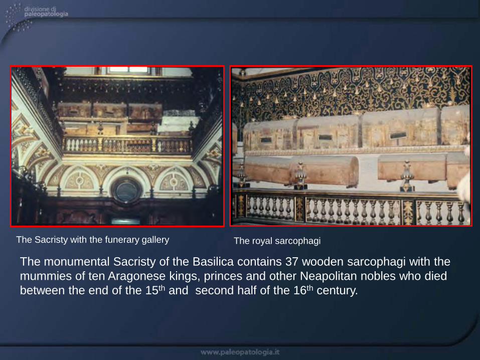
The monumental Sacristy of the Basilica contains 37 wooden sarcophagi with the mummies of ten Aragonese kings, princes and other Neapolitan nobles who died between the end of the 15th and second half of the 16th century.
The Sacristy with the funerary gallery The royal sarcophagi

The sarcophagi of San Domenico Maggiore were carefully examined by a team of the Institute of Pathology of Pisa University.The tombs revealed many well preserved (natural and artificial) mummies.
Natural mummy of Ferdinando Orsini, 5th duke of Gravina in Apulia (+1549)
Natural mummy of Antonello Petrucci (+1585) Artificial mummy of Giovanni d’Aragona (1566-1571)

Anthropological, radiological and autoptic examinations of the mummies were carried out on site.
The first X-ray of a mummy of S. Domenico with a portable apparatus, in February 1983
Autopsy of the natural mummy of Pietro d’Aragona, 3rd Duke of Montalto (1539-1552)
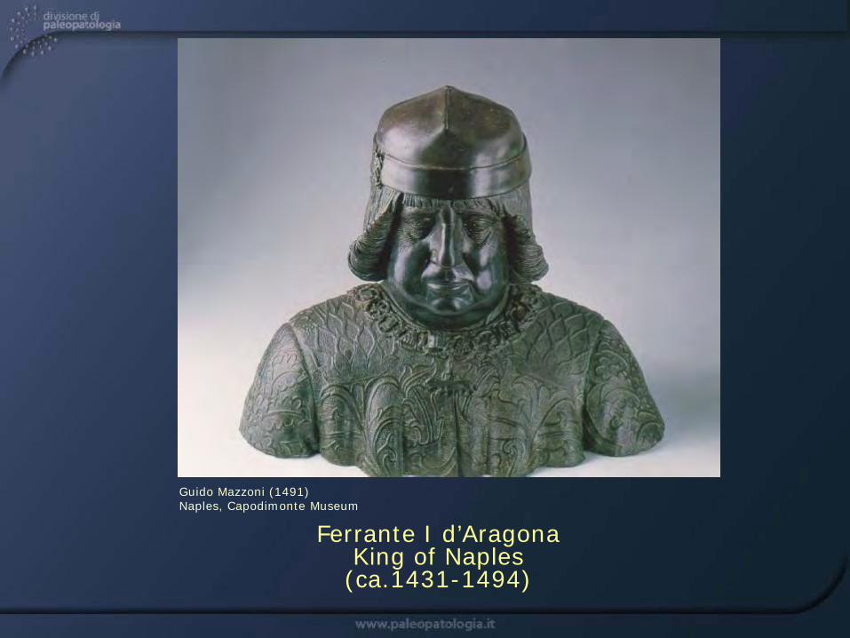
Ferrante I d’AragonaKing of Naples
(ca.1431-1494)
Guido Mazzoni (1491) Naples, Capodimonte Museum

Ferrante I was King of Naples from 1458 to 1494 and died at the age of 63 years.
Low relief of Ferrante at Porta Capuana, Naples
Sarcophagus of Ferrante (Basilica of S. Domenico Maggiore)

Autopsy of the mummy of Ferrante I revealed a colon adenocarcinoma extensively infiltrating the muscles of the small pelvis.
The pelvic cavity of Ferrante at moment ofautopsy, with the anal canal and the siteof tumor (yellow arrow)
Section of sample of tumor after rehydration, with the tumor massively infiltrating the internal tissues of the abdomen (roundish yellow-white formations)
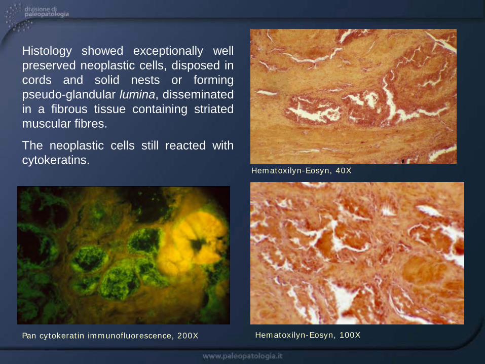
Histology showed exceptionally wellpreserved neoplastic cells, disposed incords and solid nests or formingpseudo-glandular lumina, disseminatedin a fibrous tissue containing striatedmuscular fibres.
The neoplastic cells still reacted withcytokeratins.
Hematoxilyn-Eosyn, 40X
Pan cytokeratin immunofluorescence, 200X Hematoxilyn-Eosyn, 100X
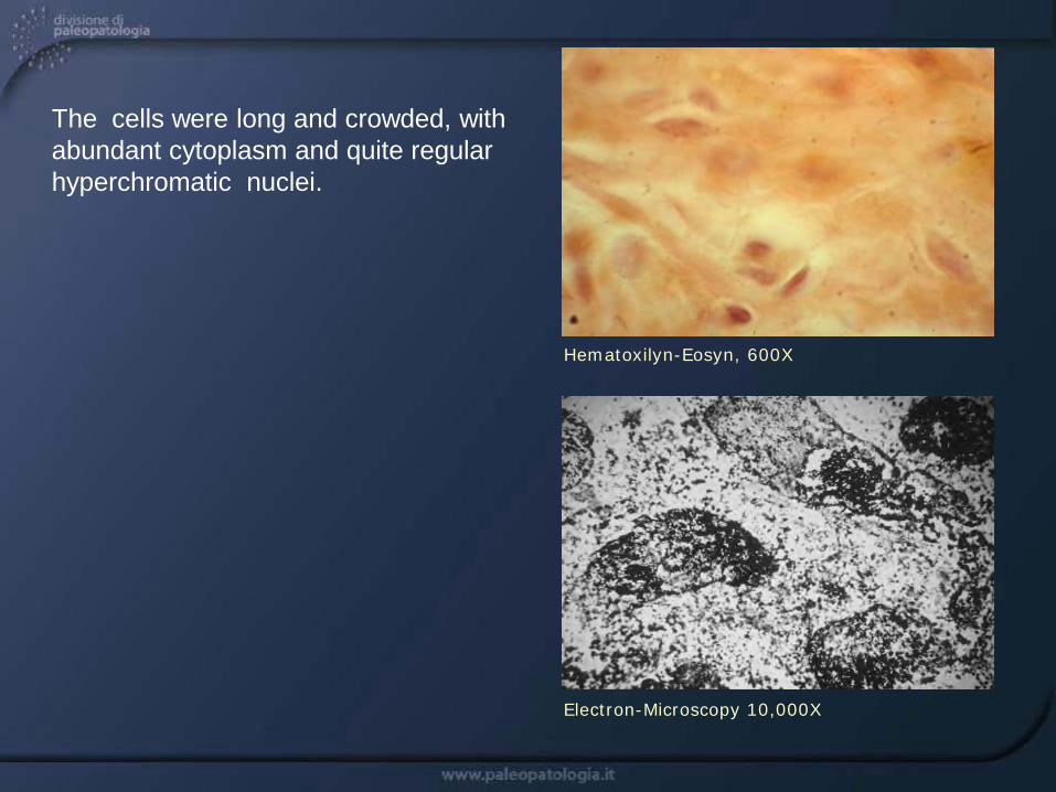
The cells were long and crowded, with abundant cytoplasm and quite regular hyperchromatic nuclei.
Hematoxilyn-Eosyn, 600X
Electron-Microscopy 10,000X
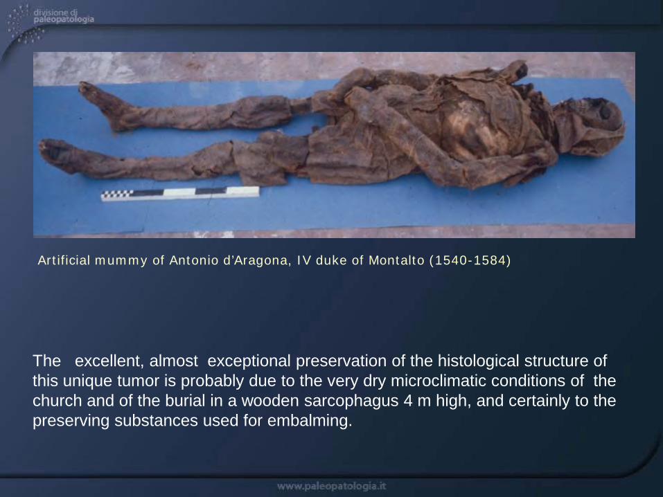
The excellent, almost exceptional preservation of the histological structure of this unique tumor is probably due to the very dry microclimatic conditions of the church and of the burial in a wooden sarcophagus 4 m high, and certainly to the preserving substances used for embalming.
Artificial mummy of Antonio d’Aragona, IV duke of Montalto (1540-1584)

The normal sequence GGT encoding the amino acid glycin was turned into GAT encoding the amino acid aspartic acid.
Ancient DNA amplification of the neoplastic cells by polymerase chain reaction (PCR) in the mummy of Ferrante I evidenced a typical point mutation of the K-ras gene codon 12.
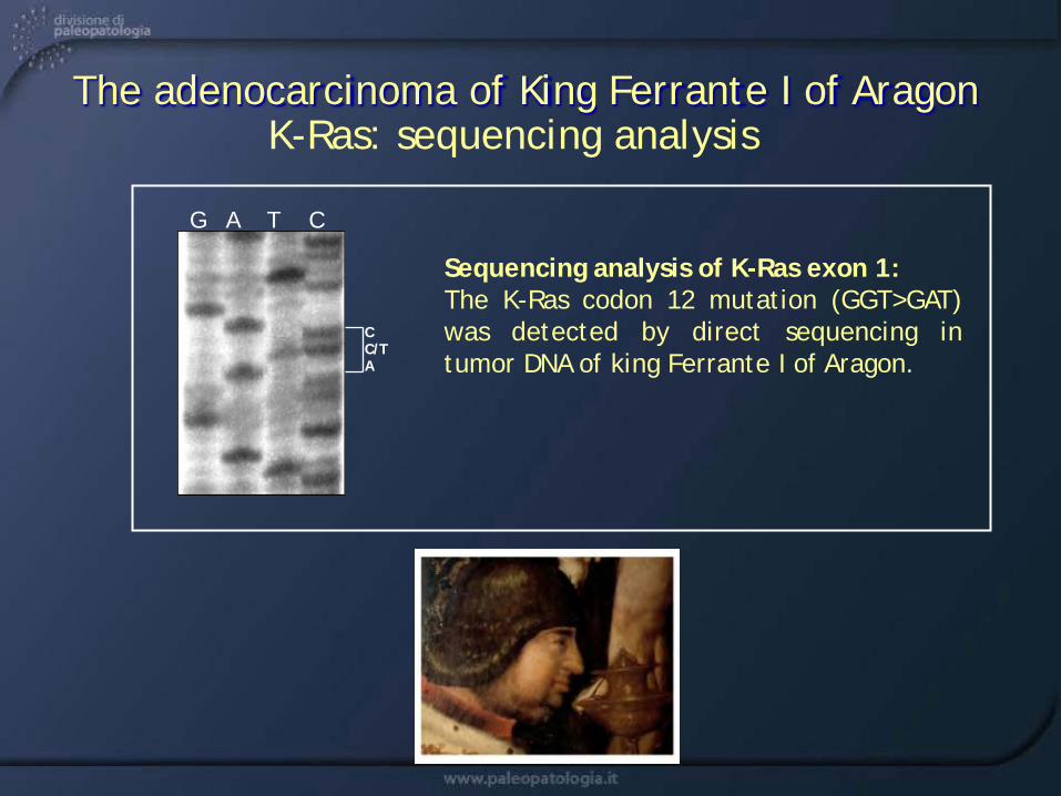
K-Ras: sequencing analysisThe adenocarcinoma of King Ferrante I of Aragon
Sequencing analysis of K-Ras exon 1:The K-Ras codon 12 mutation (GGT>GAT)was detected by direct sequencing intumor DNA of king Ferrante I of Aragon.
C C/TA
G A T C
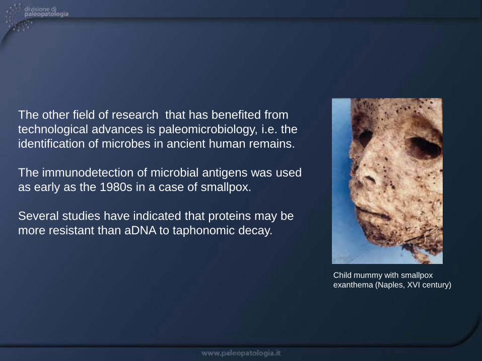
The other field of research that has benefited from technological advances is paleomicrobiology, i.e. the identification of microbes in ancient human remains.
The immunodetection of microbial antigens was used as early as the 1980s in a case of smallpox.
Several studies have indicated that proteins may be more resistant than aDNA to taphonomic decay.
Child mummy with smallpox exanthema (Naples, XVI century)

Immunohistochemistry
Immunohistochemistry combines morphology and immunochemical detection of antigenic determinants in tissues.
Detection of microbial antigens can be done using immunofluorescence analysis of frozen tissues and immunoperoxidase applied to formalin-fixed, paraffin-embedded tissues.
Immunohistochemistry is easy to perform and avoids any laboratory contamination.Direct visualization of ancient pathogens is also possible with this technique.
from Thi-Nguyen-Ny Tran et al, 2011

Among the mummies of S. Domenico, one of the most interesting was the artificial mummy of a 2-year-old child, wearing the monastic clothes of the Dominican order. The child died around the mid 16th century (14C: 1569±60)
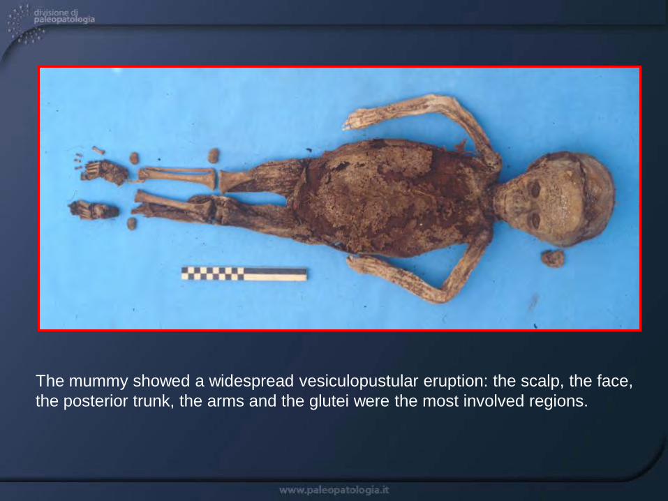
The mummy showed a widespread vesiculopustular eruption: the scalp, the face, the posterior trunk, the arms and the glutei were the most involved regions.

The impressive preservation of the mummy’s facial exanthema: the scalp, the face with the palpebral regions and the lips are covered with vesicles and pustules.

The right forearm with large, confluent pustules: the vesicles and pustules (10-20 mm in size), completely dry, with crystalline content, show a sort of central umbilication and a pale, peripheral halo.

At light microscopy the vesicles showed an evident separation of the superficial layers of the epidermis from the basal layer.
Indirect immunofluorescence with anti-variola virus antibody showed a focal positivity.

Negative staining with osmium tetroxide showed many tetrahedral viral-like particles (so-called “brick-like”), peculiar to orthopox viruses.
Scale bar = 300 nm
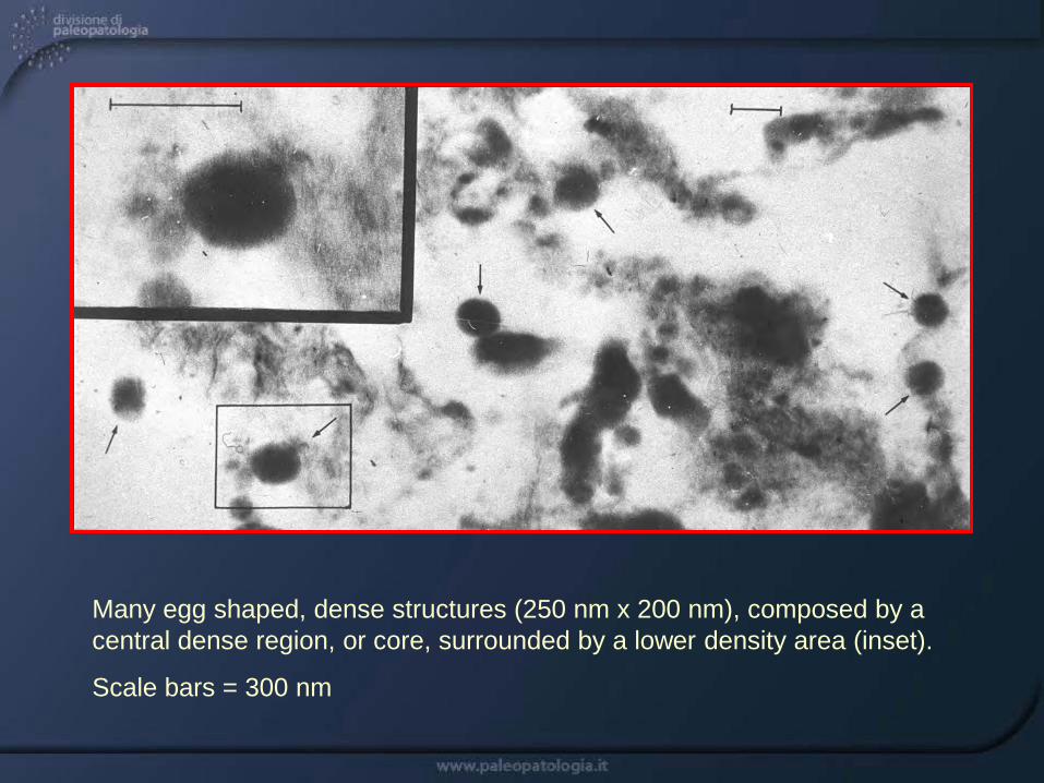
Many egg shaped, dense structures (250 nm x 200 nm), composed by a central dense region, or core, surrounded by a lower density area (inset).
Scale bars = 300 nm

Following incubation with anti-variola virus antiserum, after protein A-gold complex, some of these particles were completely covered by the A-gold protein (a), others were partially labelled (b, c).
Scale bars = 300 nm

The artificial mummy of Maria d'Aragona (1503-1568), marquise of Vasto (southern Italy), a noble woman of the Italian Renaissance, was exhumed in the Basilica of Saint Domenico Maggiore.
Famed for her beauty, Maria d'Aragona belonged to the intellectual and religious circles of Ischia, whose leading figures were Giulia Gonzaga and the well-known poetess Vittoria Colonna, friend of Michelangelo.
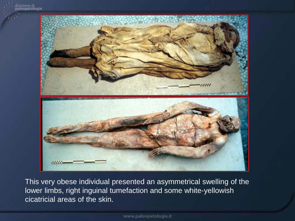
This very obese individual presented an asymmetrical swelling of the lower limbs, right inguinal tumefaction and some white-yellowish cicatricial areas of the skin.
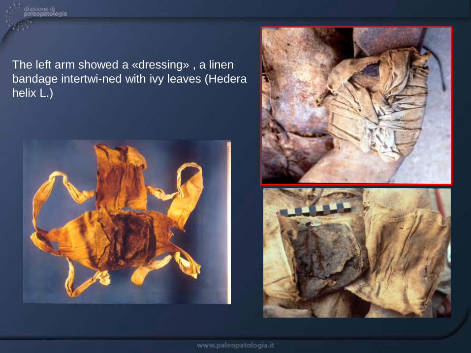
The left arm showed a «dressing» , a linen bandage intertwi-ned with ivy leaves (Hedera helix L.)
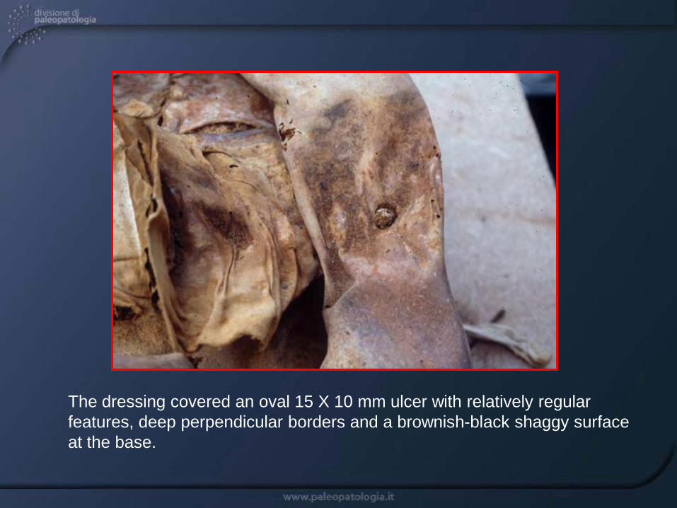
The dressing covered an oval 15 X 10 mm ulcer with relatively regular features, deep perpendicular borders and a brownish-black shaggy surface at the base.

Inside the ulcer there was a free hard spheroidal 5-mm diameter formation, with irregular surface, which gave off an intense aromatic smell when cut.
At light microscope the formation appeared composed by vegetal filaments immersed in an amorphous substance. This substance, microanalysed by EDS with a SEM Stereoscan 250 coupled to a microsound EDS link, revealed to be prevalently sulphur, a substance largely utilized at that time for skin diseases.
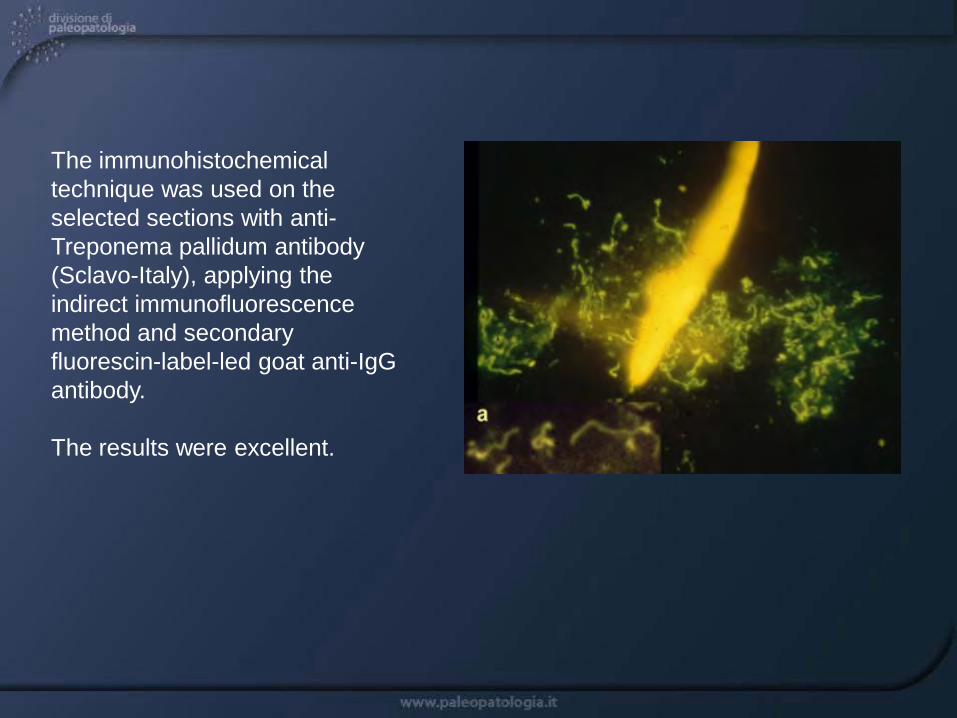
The immunohistochemical technique was used on the selected sections with anti-Treponema pallidum antibody (Sclavo-Italy), applying the indirect immunofluorescence method and secondary fluorescin-label-led goat anti-IgG antibody.
The results were excellent.

A large number of filaments with intense yellow-green fluorescence and lying between 10 and 20 µm. were identified.
The dimensions and the morpho-logical characteristics of the fluorescent treponemes were well defined (a).
These sometimes formed a regular spire with a round formation at each extremity (3c,e), at other times they were heaped (a) or isolated (d).
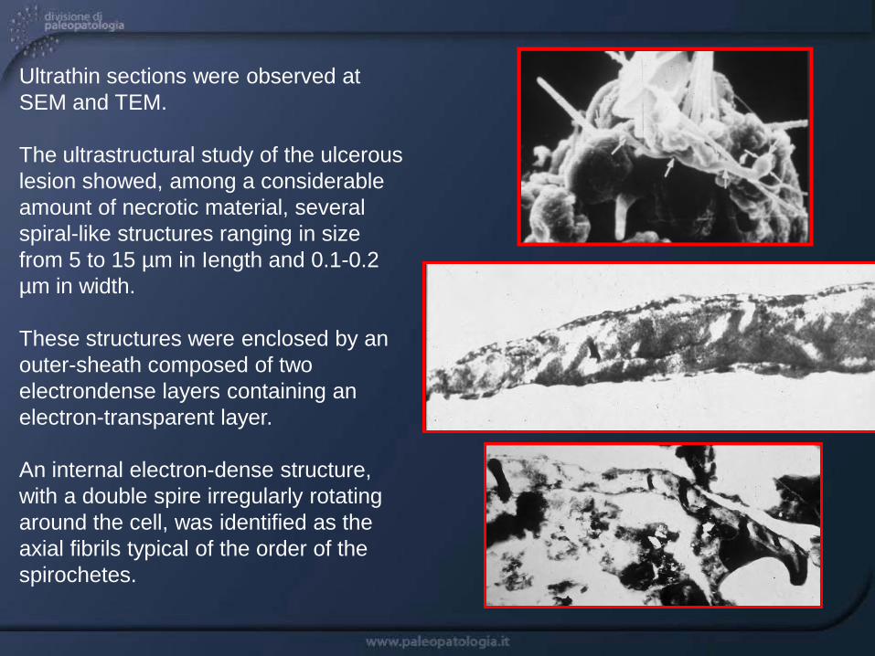
Ultrathin sections were observed at SEM and TEM.
The ultrastructural study of the ulcerous lesion showed, among a considerable amount of necrotic material, several spiral-like structures ranging in size from 5 to 15 µm in Iength and 0.1-0.2 µm in width.
These structures were enclosed by an outer-sheath composed of two electrondense layers containing an electron-transparent layer.
An internal electron-dense structure, with a double spire irregularly rotating around the cell, was identified as the axial fibrils typical of the order of the spirochetes.

Conclusions
The histological, immunological and ultrastructural findings clearly show that we are dealing with a treponemal infection.
The macroscopic characteristics of the cutaneous ulcer are those typical of a third stage luetic gumma containing numerous treponemes.
Owing to the historical period (16th century) and geographical position (Naples), a diagnosis of tertiary venereal syphilis (the Neapolitan disease of the Renaissance!), seems appropriate.

This was the first example of identification of treponemes in the soft tissues of ancient human remains, as until now the paleopathological diagnosis of syphilis has only been carried out on skeletal remains.
The discovery of this mummy is even more important since it dates back to the 16th century when the disease reached epidemic levels.
Further ultrastructural, immunological, biochemical and molecular investigations, so far impossible, should help clarify the biology of treponema in the epidemic phase of the disease.
Physicians treating patients infected with syphilis using mercury (woodcut , Wien, 1497)
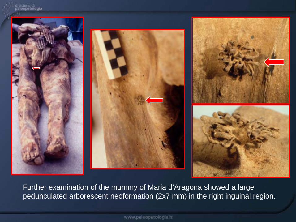
Further examination of the mummy of Maria d’Aragona showed a large pedunculated arborescent neoformation (2x7 mm) in the right inguinal region.
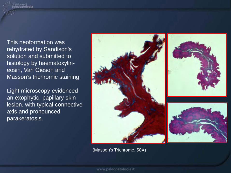
This neoformation was rehydrated by Sandison's solution and submitted to histology by haematoxylin-eosin, Van Gieson and Masson’s trichromic staining.
Light microscopy evidenced an exophytic, papillary skin lesion, with typical connective axis and pronounced parakeratosis.
(Masson’s Trichrome, 50X)
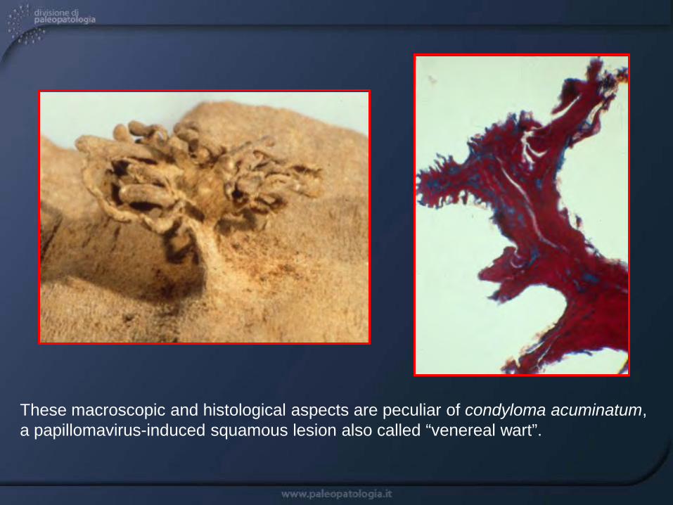
These macroscopic and histological aspects are peculiar of condyloma acuminatum, a papillomavirus-induced squamous lesion also called “venereal wart”.

In order to detect the presence of HPV (Human papillomavirus) nucleic acid, DNA extracted from a single 5 µm paraffin-embedded section was amplified with L1 consensus primers GP5+/GP6+, promoting the amplification of a 141-151 bp sequence from 25 distinct genital HPVs.
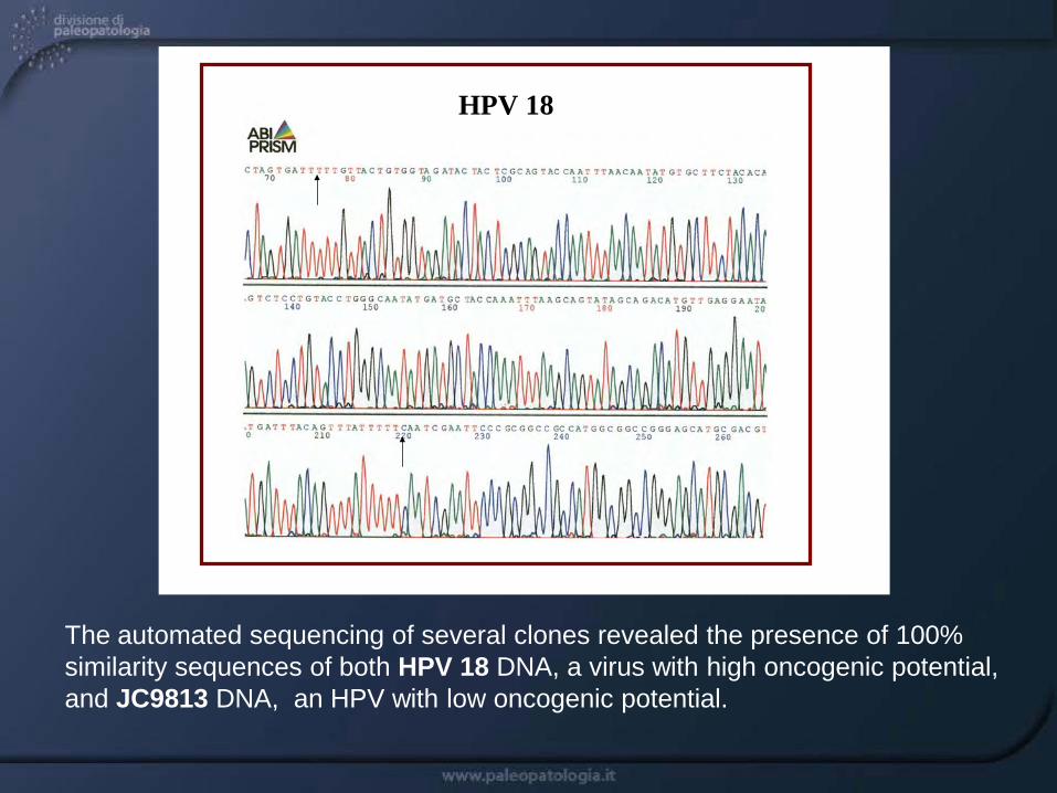
HPV 18
The automated sequencing of several clones revealed the presence of 100% similarity sequences of both HPV 18 DNA, a virus with high oncogenic potential, and JC9813 DNA, an HPV with low oncogenic potential.

HPV is a very ancient virus that has evolved with mankind. Nowadays, HPV represents one of the most common viruses sexually transmitted, responsible for numerous lesions at the level of the external and internal genitals and, in particular, of cervix cancer.
This study represents the first molecular diagnosis of HPV in mummies and could pave the way for further research on the secular evolution of these viruses, very important in human oncology.

A Peruvian mummy, a young woman 20±3 years old from Cuzco (Peru) from the National Museum of Anthropology and Ethnology of Florence, was examined.The bundle in twisted grass cords is typical of the late intermediate period (1000-1476 A.D.) of the Andean highlands.

After preliminary X-ray, showing good preservation of the body, the mummy was submitted to autopsy.

Macroscopically, a megavisceral syndrome in the form of cardiomegaly, megaesophagus, gastric ectasia and megacolon,with enormous amounts of feces, was found.
Megacolon with abundant feces Cardiomegaly

Giemsa staining evidenced in the myocardium and esophagus rare roundish intratissutal nests, about 15-20 µm large, containing several ovular formations (1-2 µm) with small nuclei. The findings corresponded morphologically to possible intratissutal nests of amastigotes of Trypanosoma cruzi.
(Esophagus, Giemsa, 50X)
(Giemsa, 600X)(Giemsa, 900X)

For the immunohistochemical study we adopted the immunoperoxidase method, and avidine-biotine system.
Monoclonal antibodies anti-T. cruzi a-flagellar FCH-F8-1 and FCH-F8-4, produced at the Institute Fatala Chaben of Buenos Aires, were used.
The nests of ovular formations and the single formations, if separately examined, showed intense, selective immunoreactivity.
(Immunoperoxidase, 400X)
(Esophagus, immunoperoxidase, 50X)
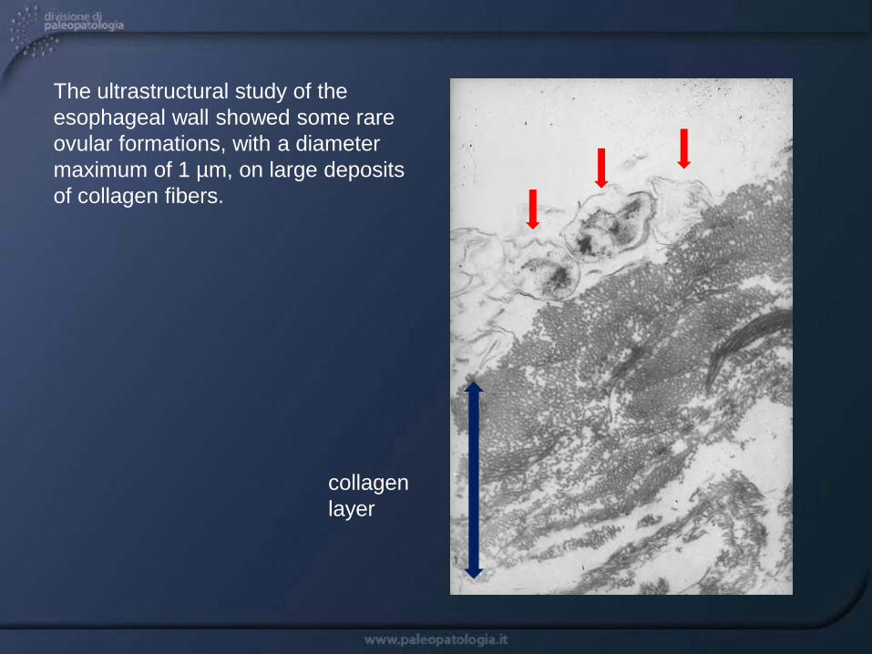
The ultrastructural study of the esophageal wall showed some rare ovular formations, with a diameter maximum of 1 µm, on large deposits of collagen fibers.
collagen layer
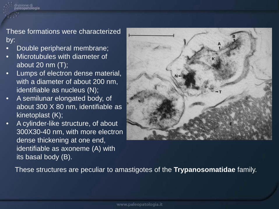
These formations were characterized by: • Double peripheral membrane;• Microtubules with diameter of
about 20 nm (T); • Lumps of electron dense material,
with a diameter of about 200 nm, identifiable as nucleus (N);
• A semilunar elongated body, of about 300 X 80 nm, identifiable as kinetoplast (K);
• A cylinder-like structure, of about 300X30-40 nm, with more electron dense thickening at one end, identifiable as axoneme (A) with its basal body (B).
These structures are peculiar to amastigotes of the Trypanosomatidae family.

The macroscopic, histological, immunohistochemical and ultrastructural findings clearly showed that we were in presence of an old case of chronic Chagas’ disease.
We thus obtained the first direct demonstration of the existence of this disease and its etiological agent in the southern American continent, during the pre-Inca civilization.
This specimen was used as positive ancient control in a large study about the diffusion of American trypanosomiasis in pre-Columbian south America.
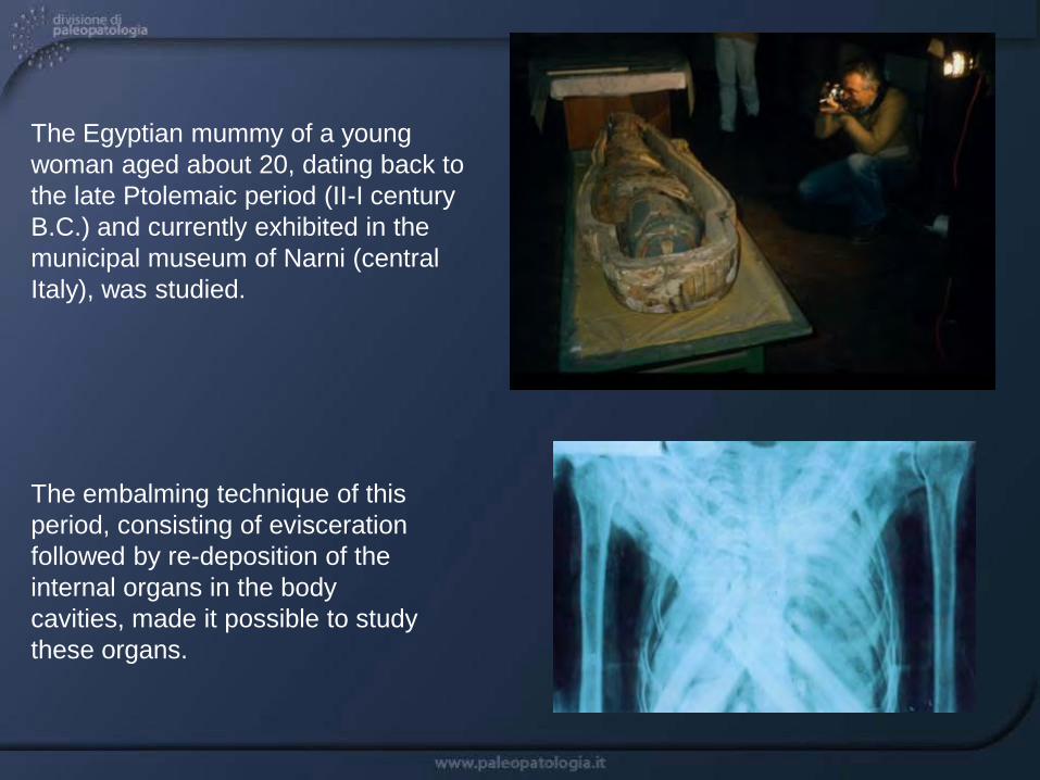
The Egyptian mummy of a young woman aged about 20, dating back to the late Ptolemaic period (II-I century B.C.) and currently exhibited in the municipal museum of Narni (central Italy), was studied.
The embalming technique of this period, consisting of evisceration followed by re-deposition of the internal organs in the body cavities, made it possible to study these organs.
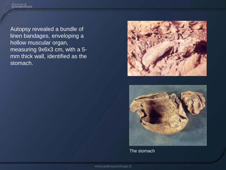
Autopsy revealed a bundle of linen bandages, enveloping a hollow muscular organ, measuring 9x6x3 cm, with a 5-mm thick wall, identified as the stomach.
The stomach

Light microscopy showed a 6x4 mm cystic structure, with a wall of about 80 µm, humped by externally projected protrusions.
A rostellum, with two rows of hooklets and two roundish structures, identifiable as suckers, were visible under a layer of loose fibrous tissue.
Morphology strongly suggested a cysticercus of Taenia solium(or “pig tapeworm”).
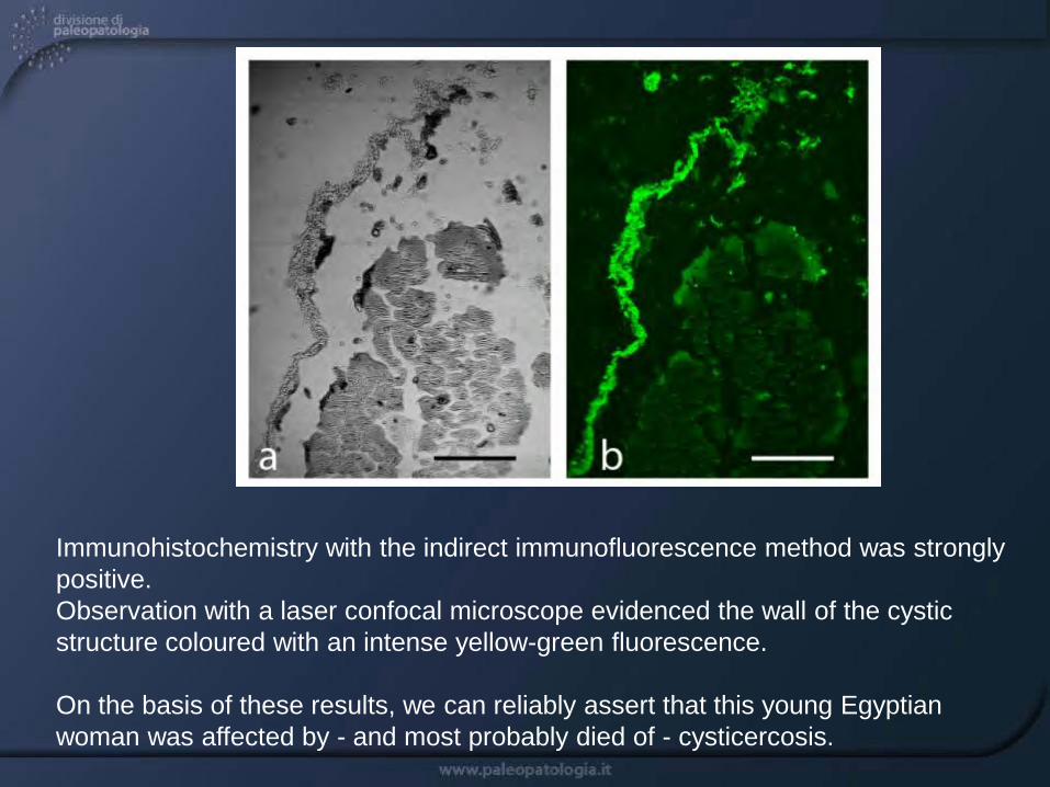
Immunohistochemistry with the indirect immunofluorescence method was strongly positive. Observation with a laser confocal microscope evidenced the wall of the cystic structure coloured with an intense yellow-green fluorescence.
On the basis of these results, we can reliably assert that this young Egyptian woman was affected by - and most probably died of - cysticercosis.

This discovery was of great scientific importance because, as far as tapeworms are concerned, only the presence of ova has been documented in the intestinal tract of an Egyptian male buried in the first half of the 12th century BC.
This is the first paleopathological diagnosis of cysticercosis, i.e. of human tissue invasion by T. solium larvae, and confirms the wide diffusion in Egypt of pig farming, representing the most common intermediate hosts of T. solium.
Histology Histochemistry
Immunohistochemistry
ElectronMicroscopy
aDNA
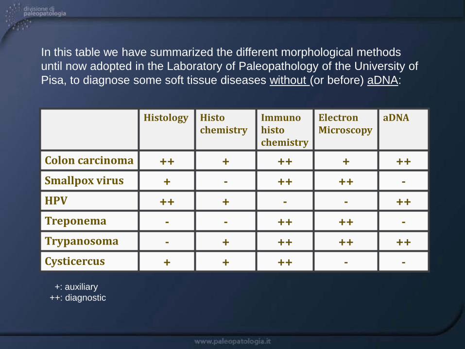
Colon carcinoma ++ + ++ + ++Smallpox virus + - ++ ++ -HPV ++ + - - ++Treponema - - ++ ++ -Trypanosoma - + ++ ++ ++Cysticercus + + ++ - -
In this table we have summarized the different morphological methods until now adopted in the Laboratory of Paleopathology of the University of Pisa, to diagnose some soft tissue diseases without (or before) aDNA:
+: auxiliary++: diagnostic

In conclusion, immunohistochemistry and electron microscopy, two morphological disciplines in these last years rather neglected compared to ancient DNA studies, can be still useful and have to be revaluated in Paleopathology.
Histological study of the tumor of King Ferrante I of Aragon