Embed Size (px)
Citation preview
BioOne sees sustainable scholarly publishing as an inherently collaborative enterprise connecting authors, nonprofit publishers, academic institutions, researchlibraries, and research funders in the common goal of maximizing access to critical research.
A THYMIC CARCINOID IN A BENGAL TIGER (PANTHERA TIGRIS)Author(s): Joshua PoweB.V.Sc., M.Vet.Stud., William CastlemanD.V.M., Ph.D., Dipl. A.C.V.P.,Christine FiorelloD.V.M., Ph.D.Source: Journal of Zoo and Wildlife Medicine, 36(3):531-533. 2005.Published By: American Association of Zoo VeterinariansDOI: http://dx.doi.org/10.1638/04-035.1URL: http://www.bioone.org/doi/full/10.1638/04-035.1
BioOne (www.bioone.org) is a nonprofit, online aggregation of core research in the biological, ecological, andenvironmental sciences. BioOne provides a sustainable online platform for over 170 journals and books publishedby nonprofit societies, associations, museums, institutions, and presses.
Your use of this PDF, the BioOne Web site, and all posted and associated content indicates your acceptance ofBioOne’s Terms of Use, available at www.bioone.org/page/terms_of_use.
Usage of BioOne content is strictly limited to personal, educational, and non-commercial use. Commercial inquiriesor rights and permissions requests should be directed to the individual publisher as copyright holder.
531
Journal of Zoo and Wildlife Medicine 36(3): 531–533, 2005Copyright 2005 by American Association of Zoo Veterinarians
A THYMIC CARCINOID IN A BENGAL TIGER (PANTHERA TIGRIS)
Joshua Powe, B.V.Sc., M.Vet.Stud., William Castleman, D.V.M., Ph.D., Dipl. A.C.V.P., andChristine Fiorello, D.V.M., Ph.D.
Abstract: An 18-yr-old Bengal tiger (Panthera tigris) presented with acute onset hind limb paresis. Radiographicand ultrasonographic imaging revealed a caudal abdominal aortic thrombus and a cranial mediastinal mass. Necropsyconfirmed aortic thrombosis. Necrotizing enteritis and multifocal renal thrombosis were also noted. The cranial medi-astinum contained a bilobed mass that histologically and ultrastructurally was consistent with a carcinoid.
Key words: Carcinoid, Panthera tigris, thymus, tiger.
BRIEF COMMUNICATION
Carcinoids are rare tumors derived from neuro-endocrine tissue. In animals, carcinoids have beenreported in the gastrointestinal tract of dogs, cats,horses, and cows,4 as well as in the liver and gallbladder of dogs and cats, and one cow.2,6 A pul-monary carcinoid has been reported in a dog,3 andsinonasal carcinoids have been reported in horses11
and a dog.12 In humans, carcinoids occur through-out the gastrointestinal tract as well as in the lungsand thymus, and on rare occasion the gonads.5 Inthis report, we describe the finding of a thymic car-cinoid in an 18-yr-old male tiger (Panthera tigris).
An 18-yr-old intact male Bengal tiger (Pantheratigris) presented to the Veterinary Medical Teach-ing Hospital (VMTH), University of Florida, witha 2-day history of acute onset hind limb paresis,inappetence, and lethargy. On presentation he wasdepressed and had bilateral neurologic deficits inthe rear limbs. On physical examination, femoralpulses could not be palpated, and the hind limbfootpads were cool and grey. Severe bloody diar-rhea was noted during hospitalization. Completeblood count revealed a neutrophilia with a left shift,lymphopenia, and hyperfibrinogenemia. Serial se-rum chemistry revealed moderate elevations inALT, AST, and phosphorus, and increasing moder-ate azotemia. Radiographically, a 4 3 7 cm2 oval,smooth-margined soft tissue mass was seen in theright cranial mediastinum. An ultrasonographicexam revealed a possible thrombus and poor flowin the iliac artery. A clinical diagnosis of iliac ar-terial thrombosis was made. Because of the poorprognosis, euthanasia prior to recovery from anes-thesia was elected.
From the Department of Pathobiology, College of Vet-erinary Medicine, University of Florida, P.O. Box 110880,Gainesville, Florida 32611, USA (Castleman, Powe); andthe Department of Small Animal Clinical Sciences, Col-lege of Veterinary Medicine, University of Florida, P.O.Box 100126, Gainesville, Florida 32610, USA (Fiorello).Corresponding should be directed to Dr. Powe.
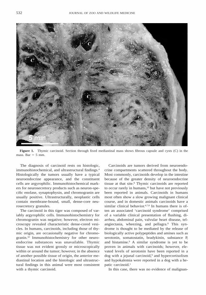
At necropsy, there was a large thrombus in thecaudal abdominal aorta that extended into the ex-ternal iliac arteries. There were large areas of is-chemic myonecrosis in the musculature of the hindlimbs. Both kidneys had multifocal vascular throm-boses and infarctions. In the mid jejunum, therewas a 15 cm locally extensive area of transmuralnecrotizing enteritis. The cranial mediastinum con-tained two closely apposed 4 3 3 3 2 cm3 and 2.53 2.5 3 0.5 cm3 dark red to pale tan, multilobular,mildly fluctuant masses that on cut surface werefinely multilocular and oozed a small amount ofwatery clear fluid (Fig. 1).
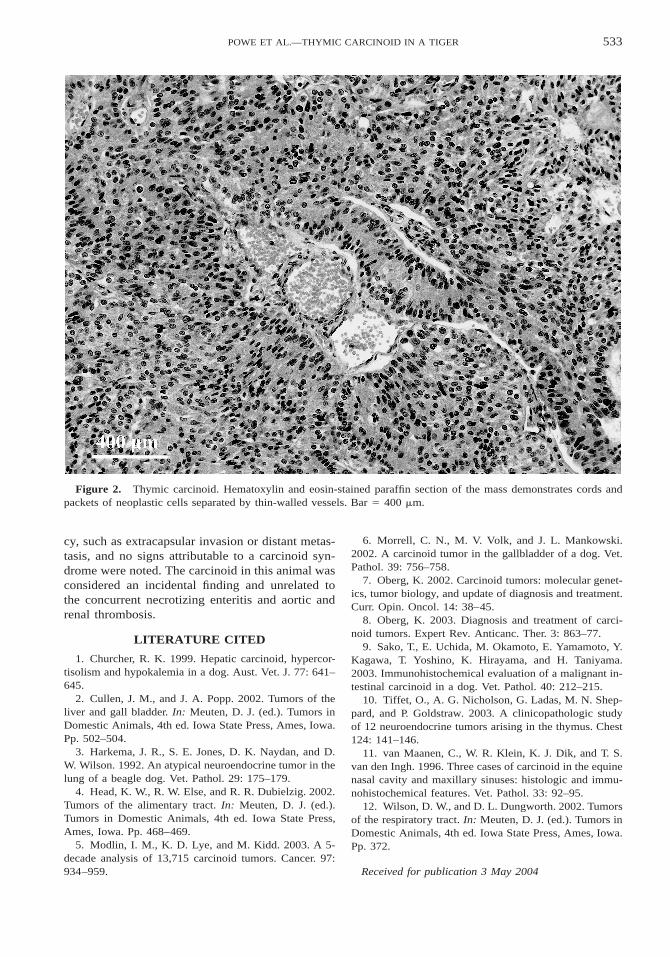
Histologically the cranial mediastinum containedtwo thickly encapsulated, densely cellular massescomprised of tightly packed lobules, palisadingcords, and perivascular rosettes supported by a thinfibrovascular stroma (Fig. 2). Lobules often con-tained large spaces filled with erythrocytes or oc-casionally macrophages admixed with necrotic ormineralized debris. The cells were cuboidal to co-lumnar with moderate amounts of homogenous am-phophilic cytoplasm; however, there were multifo-cal clusters of cells with a more feathered dispersedpale amphophilic cytoplasm. Nuclei were oval, ec-centric to basal, and with a hyperchromatic, denselyreticular chromatin distribution and up to twosmall, irregular basophilic nucleoli. There wasmoderate anisocytosis and anisokaryosis, and mi-toses were rare. Throughout the tissue were smallmultifocal areas of necrosis and hemorrhage. Oc-casional organizing thrombi were present withinblood-filled spaces lined by neoplastic cells, andthere were low numbers of scattered hemosidero-phages.
Multifocally the cells had weakly argyrophiliccytoplasmic granules with Grimelius and FontanaMasson stains. Using immunohistochemistry, thecells were weakly positive for S-100 and cytoker-atin, but negative for chromogranin and thyroglob-ulin. Transmission electron microscopy demonstrat-ed rare 135–200 nm2 diameter cytoplasmic dense-cored vesicles.
532 JOURNAL OF ZOO AND WILDLIFE MEDICINE
Figure 1. Thymic carcinoid. Section through fixed mediastinal mass shows fibrous capsule and cysts (C) in themass. Bar 5 5 mm.
The diagnosis of carcinoid rests on histologic,immunohistochemical, and ultrastructural findings.4
Histologically the tumors usually have a typicalneuroendocrine appearance, and the constituentcells are argyrophilic. Immunohistochemical mark-ers for neurosecretory products such as neuron-spe-cific enolase, synaptophysin, and chromogranin areusually positive. Ultrastructurally, neoplastic cellscontain membrane-bound, small, dense-core neu-rosecretory granules.
The carcinoid in this tiger was composed of var-iably argyrophilic cells. Immunohistochemistry forchromogranin was negative; however, electron mi-croscopy revealed characteristic dense-cored vesi-cles. In humans, carcinoids, including those of thy-mic origin, are occasionally negative for chromo-granin.10 Immunohistochemistry for other neuro-endocrine substances was unavailable. Thymictissue was not evident grossly or microscopicallywithin or around the tumor; however, in the absenceof another possible tissue of origin, the anterior me-diastinal location and the histologic and ultrastruc-tural findings in this animal were most consistentwith a thymic carcinoid.
Carcinoids are tumors derived from neuroendo-crine compartments scattered throughout the body.Most commonly, carcinoids develop in the intestinebecause of the greater density of neuroendocrinetissue at that site.8 Thymic carcinoids are reportedto occur rarely in humans,10 but have not previouslybeen reported in animals. Carcinoids in humansmost often show a slow growing malignant clinicalcourse, and in domestic animals carcinoids have asimilar clinical behavior.4,7,8 In humans there is of-ten an associated ‘carcinoid syndrome’ comprisedof a variable clinical presentation of flushing, di-arrhea, abdominal pain, valvular heart disease, tel-angiectasia, wheezing, and pellagra.8 This syn-drome is thought to be mediated by the release ofbiologically active polypeptides and amines such asserotonin, somatostatin, bradykinin, substance P,and histamine.9 A similar syndrome is yet to beproven in animals with carcinoids; however, ele-vated levels of serotonin have been reported in adog with a jejunal carcinoid,9 and hypercortisolismand hypokalemia were reported in a dog with a he-patic carcinoid.1
In this case, there was no evidence of malignan-
533POWE ET AL.—THYMIC CARCINOID IN A TIGER
Figure 2. Thymic carcinoid. Hematoxylin and eosin-stained paraffin section of the mass demonstrates cords andpackets of neoplastic cells separated by thin-walled vessels. Bar 5 400 mm.
cy, such as extracapsular invasion or distant metas-tasis, and no signs attributable to a carcinoid syn-drome were noted. The carcinoid in this animal wasconsidered an incidental finding and unrelated tothe concurrent necrotizing enteritis and aortic andrenal thrombosis.
LITERATURE CITED
1. Churcher, R. K. 1999. Hepatic carcinoid, hypercor-tisolism and hypokalemia in a dog. Aust. Vet. J. 77: 641–645.
2. Cullen, J. M., and J. A. Popp. 2002. Tumors of theliver and gall bladder. In: Meuten, D. J. (ed.). Tumors inDomestic Animals, 4th ed. Iowa State Press, Ames, Iowa.Pp. 502–504.
3. Harkema, J. R., S. E. Jones, D. K. Naydan, and D.W. Wilson. 1992. An atypical neuroendocrine tumor in thelung of a beagle dog. Vet. Pathol. 29: 175–179.
4. Head, K. W., R. W. Else, and R. R. Dubielzig. 2002.Tumors of the alimentary tract. In: Meuten, D. J. (ed.).Tumors in Domestic Animals, 4th ed. Iowa State Press,Ames, Iowa. Pp. 468–469.
5. Modlin, I. M., K. D. Lye, and M. Kidd. 2003. A 5-decade analysis of 13,715 carcinoid tumors. Cancer. 97:934–959.
6. Morrell, C. N., M. V. Volk, and J. L. Mankowski.2002. A carcinoid tumor in the gallbladder of a dog. Vet.Pathol. 39: 756–758.
7. Oberg, K. 2002. Carcinoid tumors: molecular genet-ics, tumor biology, and update of diagnosis and treatment.Curr. Opin. Oncol. 14: 38–45.
8. Oberg, K. 2003. Diagnosis and treatment of carci-noid tumors. Expert Rev. Anticanc. Ther. 3: 863–77.
9. Sako, T., E. Uchida, M. Okamoto, E. Yamamoto, Y.Kagawa, T. Yoshino, K. Hirayama, and H. Taniyama.2003. Immunohistochemical evaluation of a malignant in-testinal carcinoid in a dog. Vet. Pathol. 40: 212–215.
10. Tiffet, O., A. G. Nicholson, G. Ladas, M. N. Shep-pard, and P. Goldstraw. 2003. A clinicopathologic studyof 12 neuroendocrine tumors arising in the thymus. Chest124: 141–146.
11. van Maanen, C., W. R. Klein, K. J. Dik, and T. S.van den Ingh. 1996. Three cases of carcinoid in the equinenasal cavity and maxillary sinuses: histologic and immu-nohistochemical features. Vet. Pathol. 33: 92–95.
12. Wilson, D. W., and D. L. Dungworth. 2002. Tumorsof the respiratory tract. In: Meuten, D. J. (ed.). Tumors inDomestic Animals, 4th ed. Iowa State Press, Ames, Iowa.Pp. 372.
Received for publication 3 May 2004