Embed Size (px)
Citation preview
THE JOURNAL 01' I NVESTIGATIVE D ERMATOLOGY. 65:423- 428. 1975 Copyri ght © 1975 by The Willi a ms & Wilkins Co.
Vol. 65 , No.5 Print ed in U.S.A.
ACTION OF STAPHYLOCOCCAL EPIDERMOLYTIC TOXIN ON MOUSE SKIN: AN ELECTRON MICROSCOPIC STUDY
A. L. C . M cLAY , M .B. , C H.B. , J. P. AHBUTHNOTT, PH .D ., AND ALAN LYELL, M.D .
Departments of Pathalopy. Bacteriology , and Dermatology, University of Glas/{ow, Royal Infirmary , Glasgow. Scotland
The ul t rastructure of t he skin of 3-day-old mice cha llenged wi t h small doses of highly purified staphylococcal ep idermolytic toxin was examined at various t ime interva ls . Up to 130 min few changes were evident, bu t at this t ime wide gaps developed between cells in the horizonta l planes of the stratum granulosum, and " bubbles" norma lly present in the intercellula r space were no longer apparent . Spli tti ng of t he desmosomes occurred after the development of distended in tercellu lar spaces. After 20 hr, t hat is, in t he " healing" phase, t he appearance suggested t hat normal maturation of keratinocytes wa altered. Also at this t ime a degree of cell separation was still apparent in the outermost actively maturing layer of the st ratum granulosum . The prote inase inhibitor Trasylol was tested for its effect on epidermolytic toxin in 3-day-old mice and adul t hairl ess (hrhr) mice. No convincing ev idence was obta ined for its inhibi to ry effect on the tox in when administered up to 45 min after cha llenge.
In the clinical syndrome known as tox ic epidermal necrolysis (TEN) t he skin looks and fee ls as t hough it had been scalded , though it has not suffered thermal injury. It has become clear t hat this condition is induced by ce rtai n drugs or by certa in strains of Staphy lococcus aureus. T he histology of staphylococcal TEN, also referred to as t he sca lded skin syndrome, is cha racterized by an intraepidermal spli t, as opposed to t he subepidermal sp li t seen in other forms of TEN. Staphylococcal TEN has been studied actively by several groups a nd it is now well established tha t t he intraepidermal spli t is caused by the action of a diffusib le staphylococca l exotoxin known as epidermolytic tox in , or exfoliatin [1- 5]. Most experimental work has been performed in neonata l mice, though it has been shown that cell -free prepa rations of epidermolytic toxin induce epidermal split~ing in adul t human volun teers [6,7 ]. Strains of ha ired mice become progress ively less sensit ive
f to t he action of ep idermolytic tox in with age, and by 6 or 7 days appea r to be resistant. By contrast, adul t hai rl ess mice show epidermal splitting when injected with tox in by t he subcutaneous or in travenouS route [8,9 ].
Epidermolytic tox in has been purifi ed and shown to be a protein hav ing a molecular weight of 25,000 [3-5,7,10]. Isoelectric focusing studies have
/ revealed the presence of mul tiple molecular forms and have shown t hat the main component has an isoelectr ic point of 7.0 [4,5,7 ].
This work was supported by gra nts fro m t he Royal Society a nd Dist illers Company L t d .
Reprin t requests to: Dr. A. Lyell , Depa rt men t of Dermatology, U nive rs ity of Glasgow, Royal Infi rmary, 86 Castle Street, Glasgow G4 OSF, Scotla nd .
423
Lilli bridge, Melish , and Glasgow [11) described certa in electron microscopic features of t he effects of epidermolytic toxin in neonatal mouse skin and in biopsy material from human pat ients. They concluded that epidermolytic tox in acts primarily by ca using spli tting of desmosomes connecting the cells of the stratum granulosum. This group also noted t he disappearance of intercellular " bubbles" in affected a reas of epidermis and suggested that this led to the release of a hydrolytic enzyme, wh ich t hen acted on desmosomes . More recent ly, Elias, Fritsch, and Wolff [1 2 ] showed that cells in the region of the epidermal spli t did not show cytolysis and retained the mucopolysaccharide of intercell ul ar cement.
In the present work we studied ce llular changes in the epidermis of 3-day-old mice at different times after administration of highly purified epidermolytic toxin , and investigated the mode of action of epidermolytic toxin indirectly by testing t he effects of possible inhibitors.
MATERIALS AND METHODS
Preparat l:on of Epidermolytic Toxin
Toxin was prepared as d escr ibed previous ly from a phage group II Staphy lococcus aureus isolate (Stra in 123A) by preparative isoelectric focus ing [7 ]. P uri fied tox in was dia lyzed aga inst p hosp hate- buffered sa line. pH 7.4 , and con ta ined approximately 2,500 uni ts/m g protein; one uni t of activity was d efin ed as t he small est dose of toxin t hat gave rise to a posit ive N ikolsky s ign 6 hr a fte r subcutaneous injection in 3-day-old mice.
To fo llow t he development of ul t rastructura l cha nges in 3-d ay-old CFLP stra in mi ce, grou ps of 6 a nima ls (a verage weight 2.0 gm ) were injected with 1 to 2 uni ts of epide rmolytic toxin , and individ ua l an im a ls were sacrificed by ether a nesthetizat ion at t ime in tervals . Dorsolatera l skin sa mples were ta ken a nd suitab ly t rimmed
424 MCLAY , ARBUTH NOTT, AN D LYELL
spec im ens were imm ediately fix ed in chill ed 2% gluta ra ldehyde, pH 7.4, in phosphate buffer, osmicated , dehydrated in etha nol, a nd embedded in Epon 812. Sections were cut, usin g glass knives, on a n LKB Vltratome III and coll ected on 200-mesh Form var-coated grids. Sections were sta in ed routin ely wit h ura nyl ace tate a nd lead c itrate and a ll sect ions were viewed and photogra phed on a P hilips EM 301 G elect ron microscope. In this way in severa l exper iments we exam ined spec im ens at 10, 20, 40, 70,90, 120, and 130 min a nd 20 hr after injec tion of toxin. S imil arly, sa mples of skin were prepared from 7-day-old CFLP mi ce cha ll enged with tox in .
In add it ion, skin sa mples we re prepared for electron mi croscopy from ad ult ha irl ess mice (Stra in hrhr) 4 hr a fter in t ravenous inject ion of epidermolytic tox in from Stage II of t he purification procedure.
Effect of Tra sylol and Antitoxin on Epiderm.olytic Toxin
Trasylol (Bayer Pha rm aceut icals Ltd) , a concentrated form (10,000 KJ V/ml) of a na tu rally occurring prote inase in h ibitor from bov ine lung was tested in several ex peri ments as a possible inhib itor of ep idermolytic tox in in both 3-day-old CFLP mice and 25- to 30-gm adu lt hrhr hairless mice. In experim ents with newborn mice, doses of Trasylol (1,500 KJV) were administered subcuta neously at different t imes prior to and foll ow ing cha llenge with epidermolytic tox in; anim a ls were observed at in tervals and the t ime of onset of epidermal spli tt ing was noted . In ex periments with adul t ha irless mice, Trasy lol (10,000- 20,000 KlU) was ad ministered in single and multiple doses a t different t imes and by different routes (intrave nous, in t rape ritonea l, subcutaneous) from 6 hr before to 1 hr a fte r cha ll enge with a pprox imately 30 units of epidermolytic tox in by the subcutaneous route.
Ant itox in raised in rabbi ts [7] aga inst purified epidermoly t ic tox in was also tested for t he ability to inhibit t he act ion of epidermolyti c tox in when administered at different t imes before a nd afte r tox in in 3-day-old CFLP a nd adu lt ha irless mice.
RESU LTS
Electron Microscopic S tudies
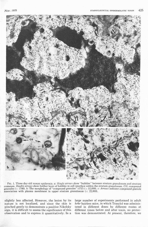
Control 3-day-old mice. The epidermis, from basa l layer to t he upper ce ll layer of t he st ratum granulosum consisted of 5 ce ll laye rs. The stratum gra nulosum itself a ppeared to contain not less t han 3 cell layers in which close cell membrane ap position and a normal desmosomal pattern were seen. As described by Lillibridge et al [11 ] the interce llu la r spaces in t he stratum granulosum and stratum corneum conta ined irregula rl y shaped vesicles t ermed " bubbl es" (Fig. 1a) . These were most prominent between layers of maturing keratin. Moreover, within cells of t he stratum granulosum, structures of approx imate ly 180 nm diameter were frequently seen. They occupied predominantly t he ap ical poles of cell s and were made up of severa l smaller "granules" (Fig . 1b) ; for t his reason they were termed compound granules. They differed from typ ical Odland (membrane-coating) granules in t hat they we re vesicul ar rather t han lamellated. As can be seen in Figure 1c s imila r structures we re seen in association with t he plasma membrane, ap parently discha rging t heir contents into t he intercellular space. Compound granules were a lso observed in epidermal cells in 7 -day-old and 18-
Vol. 65, No . 5
day-old mice. However, in the 18-day-old anim al more typica l Odland bodies were a lso seen . It seems not unreasonab le to suggest t hat in neonata l mice the compound granule may perform a fun ction s imila r to t hat proposed for t he Odland body [13 ].
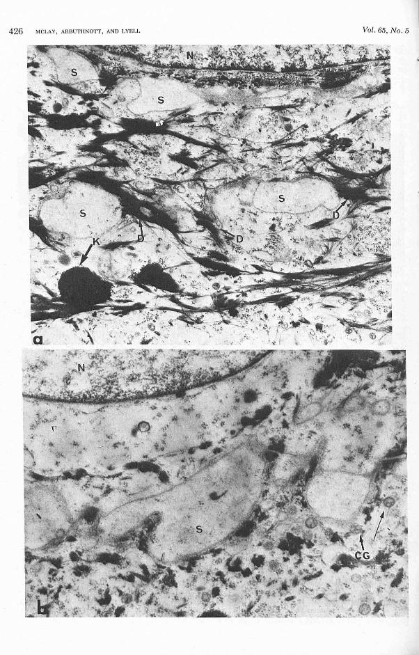
Three -day -o ld mice treated with epidermoly tic toxin. After cha ll enge with 1 unit of highly purified epidermolytic tox in no cha nges were apparent unt il 130 m in, when fo ci showing areas of ce ll separation were found . As can be seen from Figure 2a separation occurred within t he stratum gran ulosum and involved anyone or more of its com ponent cell layer in te rfaces. At t his time, although opposing membranes were widely separated , desmosomal contact was mainta ined . Despite close examinat ion no cha nges preceding spli tti ng were seen . Also, as noted by Lillibridge et a l [11], discrete bubbles were not readily discernibl e in t he distended space which was occupied by irregularly dispersed, fin ely granular material (Fig. 2b) . Moreover , t here is no apparent damage to plasma membrane or intrace llular organelles and it is worth noting here t hat, in t he fully developed split ( i. e., when desmosomal contact has been disrupted), membranes remain intact and no features assoc iated with cell death are ev ident eit her a bove or below the split, at t his early stage (i.e., 3 to 4 hr).
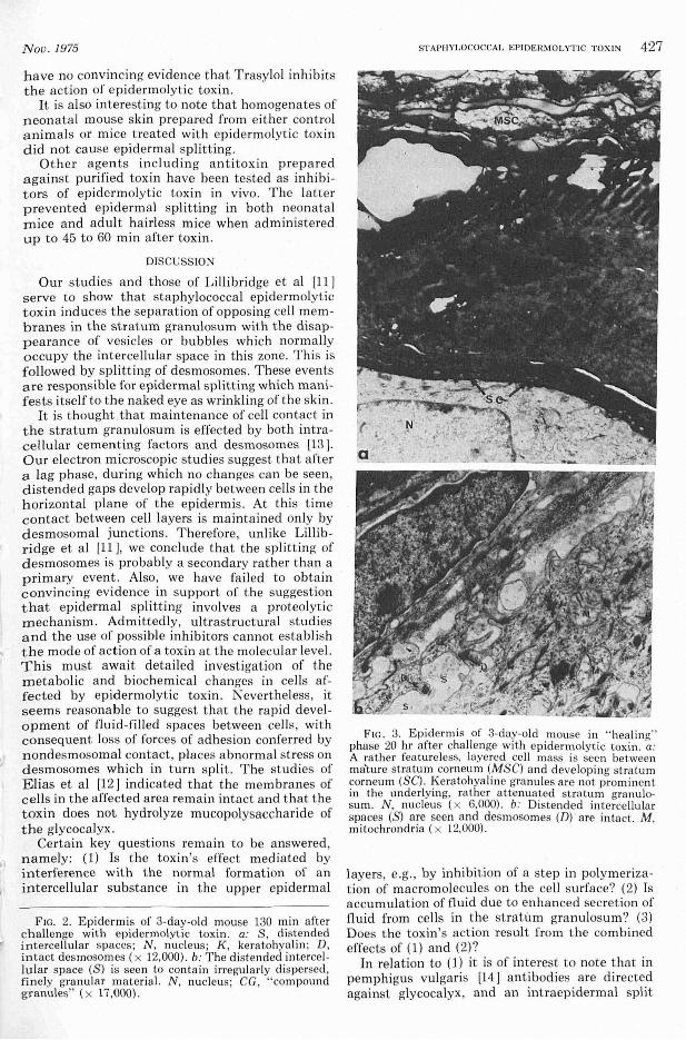
Three-day -old mice treated with epidermoly tic toxin after spontaneous " healing," 20 hI' after injection. In t hese anim als the ep iderm is ap peared abnormal in t hat a layered mass of cell showing high electron density on routine sta ining a nd lacking ultrastructura l features of li ving keratinizing cells (Fig. 3a) was observed between mature s tratum corneum and an already keratinized layer overlyi ng an attenuated stratum gran ul osum. Within the uppermost layers of t he newly maturing stratum granulosum (Fig. 3b) there was ev idence of cell separation s imilar to t hat seen in Figure 2a. These cha nges suggest t hat susceptible ce lls, once affected by epidermolytic toxin , fa il to mature in t he normal manner .
Attempts to Inhib it Epidermoly tic Toxin
In an earlier study of epidermolytic tox in , Lillibrid ge et al [11] speculated that bubbles seen in t he intercellula r space conta in a hydrolytic enzyme, possibly protease. It was suggested t hat disappearance of intercellular bubbles following the action of epidermolytic toxin is accompanied by the release of hydrolytic enzy me, which in t urn causes spli tting of desmosomes. This hypothes is was investigated indirectly by test ing t he protease inhibitor Trasylol as an inhibitor of epidermal spli tt ing.
A seri es of 9 experiments was performed using 3-day-old mice. Trasylol was admini stered subcutaneously at different t imes in s ingle and mul tiple doses prior to and following chall enge with 1 to 2 units of ep idermolytic tox in . In no case did Trasy-101 protect aga inst epidermal splitting, a lt hough in 5 of t he 9 experim ents t reated mice appeared
Nou.1975 STA PH YLOCO CCAL EPIOERMO LYT IC TOX IN 425
FIG. 1. Three-day-old mouse epidermis. a: ingle arrows show " bubbles" between t ratum gran ulosum and strat um corneum . Double arrows show further layer of bubbles in cell interface within the stratum granul osum. CG, compound granules ( x 1700). b: The morphology of "compound granul es" (CG) (x 52,000) . c: Arrows indicate compound granu le associat ion with plasma membrane in upper stratum granulosum ( x 22,000).
s light ly less affected. However, the les ion by its nature is not localized, and since the skin is p inched gent ly to demonstrate a positive Nikolsky sign , it is difficul t to assess t he signifi cance of t his observation and to express it quantitatively. In a
large number of experiments performed in adu lt hrhr hairl ess mice, in which Trasylol was administered in different doses by different routes at differen t times before and afte r tox in , no protect ion was demonstrated. At present, therefore, we
426 MCLAY, ARBUTHNOTT, AND LYELL Vol. 65, No .5
J'
Nov. 1975
have no convincing evidence that Trasylol inhibits t he action of epidermolyt ic toxin .
It is a lso interesting to note that homogenates of neonatal mouse skin prepared from either control animals or mice treated with epidermolytic toxin did not cause epidermal splitting.
Other age nts including antitoxin prepared against purified toxin have been tested as inhibitors of epidermolytic toxin in vivo. The latter prevented epidermal splitting in both neonatal mice and adult hairless mice when administered up to 45 to 60 min after toxin.
DISCUSSION
Our studies and those of Lillibridge et a l [11] serve to show that staphylococca l epidermolytic toxin induces t he separation of opposing cell membranes in the stratum granulosum with the disappearance of vesicles or bubbles which normally occupy the intercellular space in this zone. This is followed by splitting of desmosomes . These events are responsible for epidermal splitting which mani fests itself to the naked eye as wrinkling of the skin .
It is thought that maintenance of cell contact in the stratum granulosum is effected by both intracellula r cementing factors and desmosomes [13 ]. Our electron microscopic studies suggest that after a lag phase , during which no changes can be seen, distended gaps develop rapidly between cells in the horizontal plane of the epidermis. At this time contact between cell layers is maintained only by desmosomal junctions. T herefore, unlike Lillib ridge et al [11], we conclude that the splitting of desmosomes is probably a secondary rather than a primary event. Also, we have fai led to obtain convincing evidence in support of the suggestion that epidermal splitting involves a proteolytic mechanism. Admittedly, ultrastructura l studies and the use of possible inhibitors cannot establish the mode of action of a toxin at the molecular leve l. This must await detailed investiga tion of the metabolic and biochemical changes in cell s affected by epidermolytic toxin. Nevertheless , it seems reasonable to suggest that the rapid development of fluid-filled spaces between cells, wi t h consequent loss of forces of adhesion conferred by nondesmosomal contact, places abnormal stress on desmosomes which in turn split. The studies of Elias et al [12 ] indicated that the membranes of cells in the affected area remain intact and that the toxin does not hydrolyze mucopolysaccharide ' of the glycocalyx .
Certain key questions remain to be answered, namely: (1) Is the toxin 's effect mediated by interference with the normal formation of an intercellular substance in the upper epidermal
FIG. 2. Epidermis of 3-day-old mouse 130 min after challenge with epidermolytic toxin . a: S, distended intercellular spaces; N , nucleus; K , keratohyalin ; D, intact desrnosomes (x 12,000). b: The distended intercellular space (S) is seen to contain irregularly dispersed, finely granular materia l. N, nucleus; CG , "compound granules" (x 17,000).
STAPH YLOCOCCA L EP IDERMOLYTIC TOX IN 427
FIG. 3. Epidermis of 3-day-old mouse in '"healinl'(" phase 20 hr after challenge with epidermolytic toxin . a: A rather feature less, layered cell mass is seen between ma"ture stratum corneum (MSC) and developing stratum corneum (SC). Keratohya line granules are not prominent in the underlying, rather attenuated stratum granulosum. N , nucleus ( x 6,000) . b: Distended interce llular spaces (S) are seen and desmosomes (Dr are in tact. M , mitochrondria ( x 12,000).
layers, e.g., by inhibit ion of a step in polymerization of macromolecules on the cell surface? (2) Is accumulation of fluid due to enhanced secret ion of fluid from cells in the stratum granu losum? (3) Does the toxin 's action result from the combined effects of (1) and (2)?
In relation to (1) it is of interest to note that in pemphigus vulgaris [14 ] antibodies are directed against glycocalyx, and an intraepidermal spli t
428 MCLAY, ARBUTH NOTT, AN D LYELL
occurs at the first level a t which antibody can combine wit h intercellular substance. On the other hand , in pemphigus foliaceus [15 ], in which t he spli t may occur at a level compara bl e to that seen in staphylococca l TEN, it has been shown that in approx imately 50% of cases, antibodies a re a lso directed towards an intercellular substance other tha n glycocalyx.
Attempts to elucidate the mode of action of epidermolytic tox in will probably involve the use of iso lated epidermis and skin , and epidermal explants [16] ma intained for short periods in organ cul t ure.
The aut hors gratefully t hank Dr. W. C. Noble for his ass istance in ex periments wit h hairless mice, and acknowledge th e skilled technical ass istance of Bronwen Billcliffe and Lesley M ulra ine .
REFERENCES
1. Melish ME, Gl asgow LA: The staphylococcal scalded skin syndrome: development of an experi menta l model. N Engl J M ed 282: 1114- 1119, 1970
2. Arbuthnott J P, Kent J, Lyell A, Ge mmell CG: T ox ic epiderm al necrolys is produced by a n extracellul ar product of Staphy lococcus aureus. Br J Dermatol 85:145- 149, 1971
3. Kapral FA, Miller MM : Product of S taphy lococcus aureus responsible for the scalded skin syndrome. Infect Immunity 4:541- 545, 1971
4. ArbUlhnott J P, Kent J , Lyell A, Gemmell CG: Studies of sta phylococcal toxins in relat ion to tox ic epiderm a l necrolys is (the scald ed skin syndrome) . Br J Derm atol 86(S uppl 8):35- 39, 1972
5. Melish ME , Glasgow LA, Turner MD: The sta phylococcal scalded skin syndrome: isolation and pa rtial
Vol. 65, No.5
characterisation of the exfoliative tox in . J Infect Dis 125: 129- 140, 1972
6. Wiley BB, Allm an S, Rogolsky M , Norden CW, Gl asgow LA: Staphylococcal sca lded skin syndrome: potent iat ion by immunosuppress ion in mi ce; tox in mediated exfoliat ion in a hea lt hy adult. Infect Immuni ty 9:636- 640, 1974
7. Arbut hnott JP , Billcliffe B, Thompson WD : Isoelectric focusing studies of staphylococcal epidermoly tic toxin . FEBS Let te rs 46:92- 95, 1974
8. Ka pra l FA, Miller MM: Skin les ions produced by S taphy lococcus aureus exfoliatin in ha irless mi ce . Infect Immunity 6:877- 879, 1972
9. Arbut hnott JP, Kent J, Noble WC: T he response of ha irless mice to staphylococcal e pidermolytic toxin . Br J Dermatol 88:481- 485, 1973
10. K ondo I , Sakura i S , Sa ra i Y: Purification of exfoliatin produced by S taphy lococcus aU.reus of bacte riophage group 2 and its phys icochem ical propert ies. Infect Immunity 8: 156-164, 1973
11 . Lillibridge CA, Melish ME, Glasgow LA: Si te of ac tion of exfoli at ive toxin in the staphylococcal scalded skin sy ndrome: Pedi at ri cs 50: 728- 738, 1972
12. E li as P , Fritsch P, WolffK : Subcellul a r a nd molecular sites of staphylococca l exfolia t ive tox in act ivi ty (a bstr). J Invest Dermatol 62:546, 1974
13. Montagna W, P arakka l PF: The Structure and Function of Skin. Third Edit ion . New York, Academic, 1971, pp 52- 57
14. Hashimoto K , King LE, Ya manishi Y, Beachy E H, Mayens E : Identifica tion of t he substance binding pemph igus antibody and conconava lin A in the skin . J Invest Derm atol 52:423- 435, 1974
15. Bystryn J-C, Abel E , De Feo C: Epidermal ant ibodies of unique specificity in pe mphi gus foliaceus (abstr). J Invest Dermatol 62:543, 1974
16. M cCallum HM : Action of sta phylococcal epidermoly t ic tox in on mouse skin in orga n cul ture . Br J
dermatol 86(Suppl 8): 40, 1972