Embed Size (px)
Citation preview
EDITORIAL
Advanced endoscopic technologies for colorectal cancer screening
Keith L Obstein, Pietro Valdastri
World J Gastroenterol 2013 January 28; 19(4): 431-439ISSN 1007-9327 (print) ISSN 2219-2840 (online)
© 2013 Baishideng. All rights reserved.
Online Submissions: http://www.wjgnet.com/esps/[email protected]:10.3748/wjg.v19.i4.431
431 January 28, 2013|Volume 19|Issue 4|WJG|www.wjgnet.com
Keith L Obstein, Department of Medicine, Division of Gas-troenterology, Hepatology and Nutrition, Vanderbilt University Medical Center, Nashville, TN 37232, United StatesPietro Valdastri, STORM Lab, Department of Mechanical En-gineering, Vanderbilt University, Nashville, TN 37235, United StatesAuthor contributions: Obstein KL and Valdastri P contributed equally to this work. Supported by The National Science Foundation, Grant No. CNS-1239355; the National Center for Research Resources, Grant No. UL1 RR024975-01; and the National Center for Ad-vancing Translational Sciences, Grant No. 2 UL1 TR000445-06 (the content is solely the responsibility of the authors and does not necessarily represent the official views of the NIH)Correspondence to: Pietro Valdastri, PhD, Assistant Profes-sor, Director of the STORM Lab, Department of Mechanical Engineering, Vanderbilt University, 2301 Vanderbilt Place PMB 351592, Nashville, TN 37235, United States. [email protected] Telephone: +1-615-8756955 Fax: +1-615-3436687Received: June 20, 2012 Revised: August 23, 2012 Accepted: August 26, 2012Published online: January 28, 2013
AbstractColorectal cancer is the third most common cancer in men and the second most common cancer in women worldwide. Diagnosing colorectal has been increas-ingly successful due to advances in technology. Flex-ible endoscopy is considered to be an effective method for early diagnosis and treatment of gastrointestinal cancer, making it a popular choice for screening pro-grams. However, millions of people who may benefit from endoscopic colorectal cancer screening fail to have the procedure performed. Main reasons include psychological barriers due to the indignity of the proce-dure, fear of procedure related pain, bowel preparation discomfort, and potential need for sedation. Therefore, an urgent need for new technologies addressing these issues clearly exists. In this review, we discuss a set of advanced endoscopic technologies for colorectal cancer
screening that are either already available or close to clinical trial. In particular, we focus on visual-inspection-only advanced flexible colonoscopes, interventional colonoscopes with alternative propulsion mechanisms, wireless capsule colonoscopy, and technologies for in-traprocedural bowel cleansing. Many of these devices have the potential to reduce exam related patient discomfort, obviate the need for sedation, increase di-agnostic yield, reduce learning curves, improve access to screening, and possibly avert the need for a bowel preparation.
© 2013 Baishideng. All rights reserved.
Key words: Endoscopy; Technology; Capsule colonos-copy; Colorectal cancer; Screening
Obstein KL, Valdastri P. Advanced endoscopic technologies for colorectal cancer screening. World J Gastroenterol 2013; 19(4): 431-439 Available from: URL: http://www.wjgnet.com/1007-9327/full/v19/i4/431.htm DOI: http://dx.doi.org/10.3748/wjg.v19.i4.431
INTRODUCTIONColorectal cancer (CRC) is the third most common cancer in men and the second most common cancer in women worldwide with approximately 608 000 people dying each year[1]. In the United States alone there are approximately 1.14 million people alive who have a history of CRC and 1 in 20 will be diagnosed with cancer of the colon or rectum in their lifetime[2]. Unfortunately, this number is projected to increase by 62% by the year 2030[3].
Diagnosing CRC has been increasingly successful due to numerous advances in technology. One of the paramount technological advances has been the ability to directly visualize the gastrointestinal (GI) tract (and provide therapy) with the flexible endoscope. The earliest flexible endoscope, completely based on optical fibers, was
Obstein KL et al . Advanced endoscopic technologies for CRC screening
432 January 28, 2013|Volume 19|Issue 4|WJG|www.wjgnet.com
presented at the American Gastroscopy Society annual meeting in May 1957 by Hirschowitz[4]. This achievement was inspired by a paper published in 1954, entitled “a flexible fiberscope using static scanning”, by Hopkins et al[5] at the Imperial College of Science and Technology in London. Building on this history of technological innovation, and driven by breakthroughs in electronics, material science, computational capabilities, sensing, and actuation strategies, many novel GI devices and diagnostic techniques have emerged. In this review, we will discuss a set of advanced endoscopic technologies for CRC screening that are either already available or close to clinical trial; and have the potential to reduce procedure related patient discomfort and increase diagnostic yield.
LIMITATION OF STANDARD GI ENOSCOPYFlexible endoscopy is considered to be an effective method for early diagnosis and treatment of GI cancer, thus is a primary choice for screening programs. Few complications are associated with this technique, with cardiorespiratory problems related to sedation and analgesia being the most common (0.03%20% incidence[6]). Less frequent complications include, infection (0.2% incidence[7]), bleeding (0.2%2.1% incidence) or perforation (0.1% incidence[8]), which potentially require subsequent medications, transfusions, or endoscopic/surgical intervention to correct.
Based on the efficacy and low complication rate of flexible endoscopy, it is clear that the main clinical challenge facing GI endoscopy is one of distribution. Millions of people who may benefit from endoscopic CRC screening fail to have the procedure performed. The reasons cited include psychological barriers due to the indignity of the procedure, fear of procedure related pain, bowel preparation discomfort, and potential need for sedation[9].
How rational are these fears? From a mechanical perspective, the endoscope consists of a long and fairly stiff (compared to the compliant colon) tube with a steerable head. The colonoscope must easily navigate the colon curves and traverse the intestinal environment efficiently meaning that a colonoscope must be simultaneously stiff and compliant. If the colonoscope is too stiff, it will deform the colon wall significantly at turns; yet, if it is too compliant there will be undesired buckling[10]. Since the colonoscope must be pushed from the back, while the tip is aimed along the lumen center, when the intestine bends, the shaft pushes against the colon wall until the lumen and its surroundings provide sufficient counter pressure to force the endoscope shaft to bend. This stretches the colon and often leads to “loop” formation, thus potentially causing substantial discomfort. In particular, looping occurs when the colonoscope continues to be advanced into the colon without a corresponding progression of the tip. This displaces the colon from its native configuration and stretches mes
entery muscles. Looping of the endoscope has been shown to be responsible for 90% of the pain episodes in colonoscopy and increases the chance of tissue damage and perforation[11]. Special maneuvers can be performed to minimize this effect, making colonoscopy a procedure that requires a great degree of training, technical skill, and experience to safely perform[12,13]. Despite these techniques, even expert endoscopists can not always prevent all challenges or complications; partially because the flexible endoscope design is one of compromise and is not perfect for its intended purpose[10]. Additionally, the need for a bowel preparation acts as a potential deterrent due to the unpleasantness of ingesting powerful medications to clean the intestine. A detailed discussion on bowel preparation is beyond the scope of this review and will only be discussed briefly.
TOWARD MECHANISMS ENABLING PAINLESS COLONOSCOPYSeveral colonoscope modifications have recently been presented with the common goal of preventing excessive force application to the colon wall and consequent looping. These devices can be categorized into two groups: those designed purely for visual inspection (visual inspection devices), and those that contain internal channels through which interventional devices (i.e. biopsy, snare, needle, etc.) can be passed (interventional devices). In addition to flexible colonoscope modifications, wireless capsule colonoscopy is emerging as a “patient-friendly” alternative technique for visual inspection of the colon. In this section we will provide an overview of recent advancements in these three device categories.
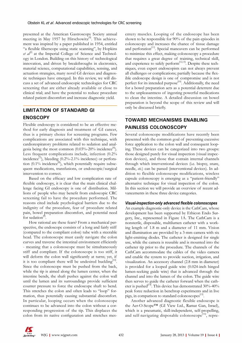
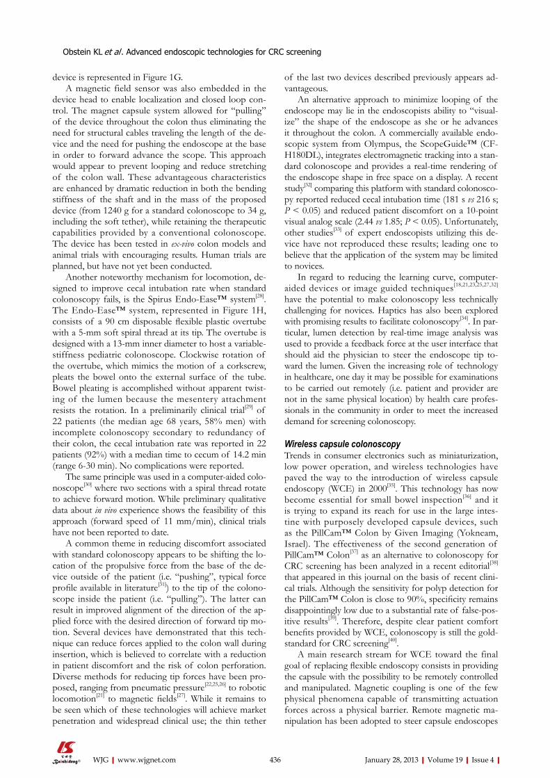
Visual-inspection-only advanced flexible colonoscopesAn example diagnosticonly device is the CathCam, whose development has been supported by Ethicon Endo Surgery, Inc., represented in Figure 1A. The CathCam is a nonsterile, disposable, multilumen catheter with a working length of 1.8 m and a diameter of 11 mm. Vision and illumination are provided by a 3mm camera with six lightemitting diodes. The catheter is designed for single use, while the camera is reusable and is mounted into the catheter tip prior to the procedure. The channels of the CathCam accommodate the cables of the video camera and enable the system to provide suction, irrigation, and visualization. An accessory channel (2.8 mm in diameter) is provided for a looped guide wire (0.024inch hinged lumen-seeking guide wire) that is advanced through the channel and into the lumen of the colon. The guide wire then serves to guide the catheter forward when the catheter is pushed[14]. This device has demonstrated 30%40% peak force reduction in benchtop experiments and in live pigs, in comparison to standard colonoscopes[15].
Another advanced diagnostic flexible endoscope is the AerOScope™ (GI View Ltd., Ramat Gan, Israel), which is a pneumatic, skill-independent, self-propelling, and selfnavigating disposable colonoscope[16], repre
433 January 28, 2013|Volume 19|Issue 4|WJG|www.wjgnet.com
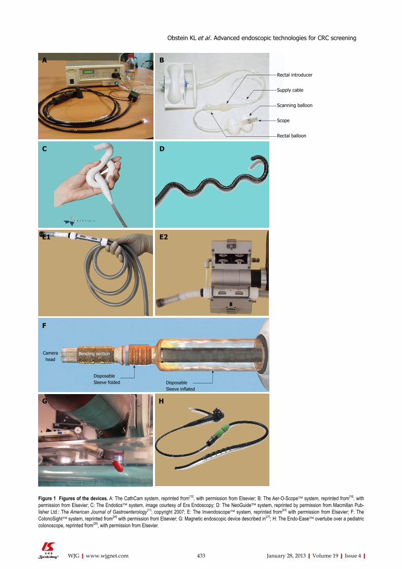
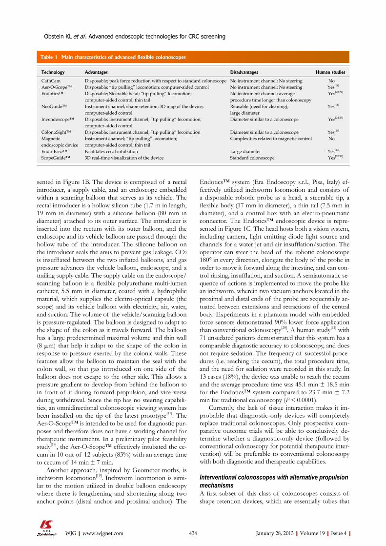
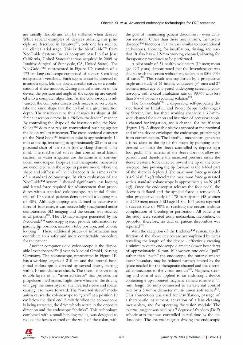
Figure 1 Figures of the devices. A: The CathCam system, reprinted from[15], with permission from Elsevier; B: The Aer-O-Scope™ system, reprinted from[18], with permission from Elsevier; C: The Endotics™ system, image courtesy of Era Endoscopy; D: The NeoGuide™ system, reprinted by permission from Macmillan Pub-lisher Ltd.: The American Journal of Gastroenterology[11], copyright 2007; E: The Invendoscope™ system, reprinted from[24] with permission from Elsevier; F: The ColonoSight™ system, reprinted from[26] with permission from Elsevier; G: Magnetic endoscopic device described in[27]; H: The Endo-Ease™ overtube over a pediatric colonoscope, reprinted from[28], with permission from Elsevier.
Disposable Sleeve inflated
Disposable Sleeve folded
Rectal introducer
Supply cable
Scanning balloon
Scope
Rectal balloon
A B
C D
E1 E2
F
G H
Bending sectionCamera head
Obstein KL et al . Advanced endoscopic technologies for CRC screening
434 January 28, 2013|Volume 19|Issue 4|WJG|www.wjgnet.com
sented in Figure 1B. The device is composed of a rectal introducer, a supply cable, and an endoscope embedded within a scanning balloon that serves as its vehicle. The rectal introducer is a hollow silicon tube (1.7 m in length, 19 mm in diameter) with a silicone balloon (80 mm in diameter) attached to its outer surface. The introducer is inserted into the rectum with its outer balloon, and the endoscope and its vehicle balloon are passed through the hollow tube of the introducer. The silicone balloon on the introducer seals the anus to prevent gas leakage. CO2 is insufflated between the two inflated balloons, and gas pressure advances the vehicle balloon, endoscope, and a trailing supply cable. The supply cable on the endoscope/scanning balloon is a flexible polyurethane multi-lumen catheter, 5.5 mm in diameter, coated with a hydrophilic material, which supplies the electrooptical capsule (the scope) and its vehicle balloon with electricity, air, water, and suction. The volume of the vehicle/scanning balloon is pressureregulated. The balloon is designed to adapt to the shape of the colon as it travels forward. The balloon has a large predetermined maximal volume and thin wall (8 µm) that help it adapt to the shape of the colon in response to pressure exerted by the colonic walls. These features allow the balloon to maintain the seal with the colon wall, so that gas introduced on one side of the balloon does not escape to the other side. This allows a pressure gradient to develop from behind the balloon to in front of it during forward propulsion, and vice versa during withdrawal. Since the tip has no steering capabilities, an omnidirectional colonoscopic viewing system has been installed on the tip of the latest prototype[17]. The AerOScope™ is intended to be used for diagnostic purposes and therefore does not have a working channel for therapeutic instruments. In a preliminary pilot feasibility study[18], the AerOScope™ effectively intubated the cecum in 10 out of 12 subjects (83%) with an average time to cecum of 14 min ± 7 min.
Another approach, inspired by Geometer moths, is inchworm locomotion[19]. Inchworm locomotion is similar to the motion utilized in double balloon endoscopy where there is lengthening and shortening along two anchor points (distal anchor and proximal anchor). The
Endotics™ system (Era Endoscopy s.r.l., Pisa, Italy) effectively utilized inchworm locomotion and consists of a disposable robotic probe as a head, a steerable tip, a flexible body (17 mm in diameter), a thin tail (7.5 mm in diameter), and a control box with an electropneumatic connector. The Endotics™ endoscopic device is represented in Figure 1C. The head hosts both a vision system, including camera, light emitting diode light source and channels for a water jet and air insufflation/suction. The operator can steer the head of the robotic colonoscope 180° in every direction, elongate the body of the probe in order to move it forward along the intestine, and can control rinsing, insufflation, and suction. A semiautomatic sequence of actions is implemented to move the probe like an inchworm, wherein two vacuum anchors located in the proximal and distal ends of the probe are sequentially actuated between extensions and retractions of the central body. Experiments in a phantom model with embedded force sensors demonstrated 90% lower force application than conventional colonoscopy[20]. A human study[21] with 71 unsedated patients demonstrated that this system has a comparable diagnostic accuracy to colonoscopy, and does not require sedation. The frequency of successful procedures (i.e. reaching the cecum), the total procedure time, and the need for sedation were recorded in this study. In 13 cases (18%), the device was unable to reach the cecum and the average procedure time was 45.1 min ± 18.5 min for the Endotics™ system compared to 23.7 min ± 7.2 min for traditional colonoscopy (P < 0.0001).
Currently, the lack of tissue interaction makes it improbable that diagnosticonly devices will completely replace traditional colonoscopes. Only prospective comparative outcome trials will be able to conclusively determine whether a diagnosticonly device (followed by conventional colonoscopy for potential therapeutic intervention) will be preferable to conventional colonoscopy with both diagnostic and therapeutic capabilities.
Interventional colonoscopes with alternative propulsion mechanismsA first subset of this class of colonoscopes consists of shape retention devices, which are essentially tubes that
Table 1 Main characteristics of advanced flexible colonoscopes
Technology Advantages Disadvantages Human studies
CathCam Disposable; peak force reduction with respect to standard colonoscope No instrument channel; No steering NoAer-O-Scope™ Disposable; “tip pulling” locomotion; computer-aided control No instrument channel; No steering Yes[18]
Endotics™ Disposable; Steerable head; “tip pulling” locomotion; computer-aided control; thin tail
No instrument channel; average procedure time longer than colonoscopy
Yes[20,21]
NeoGuide™ Instrument channel; shape retention; 3D map of the device; computer-aided control
Reusable (need for cleaning); large diameter
Yes[11]
Invendoscope™ Disposable; instrument channel; “tip pulling” locomotion; computer-aided control
Diameter similar to a colonoscope Yes[24,25]
ColonoSight™ Disposable; instrument channel; “tip pulling” locomotion Diameter similar to a colonoscope Yes[26]
Magnetic endoscopic device
Instrument channel; “tip pulling” locomotion; computer-aided control; thin tail
Complexities related to magnetic control No
Endo-Ease™ Facilitates cecal intubation Large diameter Yes[29]
ScopeGuide™ 3D real-time visualization of the device Standard colonoscope Yes[32,33]
Obstein KL et al . Advanced endoscopic technologies for CRC screening
435 January 28, 2013|Volume 19|Issue 4|WJG|www.wjgnet.com
are initially flexible and can be stiffened when desired. While several examples of devices utilizing this principle are described in literature[10], only one has reached the clinical trial stage. This is the NeoGuide™ from NeoGuide Systems Inc. (a company based in San Jose, California, United States that was acquired in 2009 by Intuitive Surgical of Sunnyvale, CA, United States). The NeoGuide™, represented in Figure 1D, consists of a 173 cmlong endoscope composed of sixteen 8 cmlong independent vertebrae. Each segment can be directed to assume a right, left, up, down, circular curve, or a combination of these motions. During manual insertion of the device, the position and angle of the scope tip are encoded into a computer algorithm. As the colonoscope is advanced, the computer directs each successive vertebra to take the same shape that the tip had at a given insertion depth. The insertion tube thus changes its shape at different insertion depths in a “follow-the-leader” manner. By controlling the shape of the insertion tube, the NeoGuide™ does not rely on conventional pushing against the colon wall to maneuver. The crosssectional diameter of the NeoGuide™ insertion tube is approximately 14 mm at the tip, increasing to approximately 20 mm at the proximal shaft of the scope (the working channel is 3.2 mm). The mechanical valves that control insufflation, suction, or water irrigation are the same as in conventional endoscopes. Biopsies and therapeutic maneuvers are conducted with the scope in passive mode; where the shape and stiffness of the endoscope is the same as that of a standard colonoscope. In vitro evaluation of the NeoGuide™ system[22] showed significantly less looping and lateral force required for advancement than procedures with a standard colonoscope. An initial clinical trial of 10 sedated patients demonstrated a looping rate of 40%. Although looping was defined as extensive in three of four cases, it was successfully straightened under computerized 3D imaging and the cecum was reached in all patients[11]. The 3D map images generated by the NeoGuide™ endoscopy system provide information regarding tip position, insertion tube position, and colonic looping[23]. These additional pieces of information may contribute to a safer and more comfortable procedure for the patient.
Another computeraided colonoscope is the disposable Invendoscope™ (Invendo Medical GmbH, Kissing, Germany). The colonoscope, represented in Figure 1E, has a working length of 210 cm and the internal functional endoscope is covered by several layers, starting with a 10mm diameter sheath. The sheath is covered by double layers of an “inverted sleeve” that provides the propulsion mechanism. Eight drive wheels in the driving unit grip the inner layer of the inverted sleeve and rotate, causing it to move forward. The “inverted sleeve” mechanism causes the colonoscope to “grow” at a position 10 cm below the distal end. Similarly, when the colonoscope is being retracted, the drive wheels rotate in the opposite direction and the endoscope “shrinks”. This technology, combined with a small bending radius, was designed to reduce the forces exerted on the walls of the colon, with
the goal of minimizing patient discomfort even without sedation. Other than these mechanisms, the Invendoscope™ functions in a manner similar to conventional endoscopes, allowing for insufflation, rinsing, and suction. It also has a 3.2-mm working channel, allowing for therapeutic procedures to be performed.
A pilot study of 34 healthy volunteers (19 men; mean age 49.7 years) demonstrated that the Invendoscope was able to reach the cecum without any sedation in 80%90% of cases[24]. This result was supported by a prospective singlearm study of 61 healthy volunteers (34 men and 27 women; mean age 57.5 years) undergoing screening colonoscopy, with a cecal intubation rate of 98.4% with less than 5% of patients requiring sedation[25].
The ColonoSight™, a disposable, selfpropelling device based on IntraPull and ProtectiScope technologies by Stryker, Inc., has three working channels: a 3.7-mm-wide channel for suction and insertion of accessory tools, a channel for irrigation, and a channel for insufflation (Figure 1F). A disposable sleeve anchored at the proximal end of the device envelopes the endoscope, protecting it from contamination. The IntraPull mechanism generates a force close to the tip of the scope by pumping compressed air inside the sleeve controlled by depressing a foot pedal. The material of the sleeve does not allow expansion, and therefore the increased pressure inside the sleeve creates a force directed toward the tip of the colonoscope, thus pushing the tip forward as the folded part of the sleeve is deployed. The maximum force generated is 4.9 N (0.5 kgf) whereby the maximum force generated with a standard colonoscope is approximately 44 N (4.5 kgf). Once the endoscopist releases the foot pedal, the sleeve is deflated and the applied force is removed. A pilot prospective study of 178 participants (48 women and 130 men; mean ± SD age 51.8 ± 10.7 years) reported a success rate of 90% in reaching the cecum without complication of bleeding or perforation. All patients in the study were sedated using midazolam, mepiridine, or propofol; therefore, no data on patient discomfort was reported[26].
With the exception of the Endotics™ system, tip deflection of the above devices are accomplished by wires travelling the length of the device effectively creating a minimum outer endoscope diameter (lower boundary) of approximately 10 mm. If however, one could “pull” rather than “push” the endoscope, the outer diameter lower boundary may be reduced further; limited by the space needed for the therapeutic channel and the electrical connections to the vision module[21]. Magnetic steering and control was applied to an endoscopic device containing a tipmounted magnetic camera (diameter 11 mm, length 26 mm) connected to an external control box by a 5.4mm diameter multilumen soft tether[27]. This connection was used for insufflation, passage of a therapeutic instrument, activation of a lens cleaning mechanism, and for operating the vision module. The external magnet was held by a 7 degreeoffreedom (DoF) robotic arm that was controlled in realtime by the endoscopist. The external magnet driving the endoscopic
Obstein KL et al . Advanced endoscopic technologies for CRC screening
436 January 28, 2013|Volume 19|Issue 4|WJG|www.wjgnet.com
device is represented in Figure 1G.A magnetic field sensor was also embedded in the
device head to enable localization and closed loop control. The magnet capsule system allowed for “pulling” of the device throughout the colon thus eliminating the need for structural cables traveling the length of the device and the need for pushing the endoscope at the base in order to forward advance the scope. This approach would appear to prevent looping and reduce stretching of the colon wall. These advantageous characteristics are enhanced by dramatic reduction in both the bending stiffness of the shaft and in the mass of the proposed device (from 1240 g for a standard colonoscope to 34 g, including the soft tether), while retaining the therapeutic capabilities provided by a conventional colonoscope. The device has been tested in ex-vivo colon models and animal trials with encouraging results. Human trials are planned, but have not yet been conducted.
Another noteworthy mechanism for locomotion, designed to improve cecal intubation rate when standard colonoscopy fails, is the Spirus EndoEase™ system[28]. The EndoEase™ system, represented in Figure 1H, consists of a 90 cm disposable flexible plastic overtube with a 5mm soft spiral thread at its tip. The overtube is designed with a 13mm inner diameter to host a variablestiffness pediatric colonoscope. Clockwise rotation of the overtube, which mimics the motion of a corkscrew, pleats the bowel onto the external surface of the tube. Bowel pleating is accomplished without apparent twisting of the lumen because the mesentery attachment resists the rotation. In a preliminarily clinical trial[29] of 22 patients (the median age 68 years, 58% men) with incomplete colonoscopy secondary to redundancy of their colon, the cecal intubation rate was reported in 22 patients (92%) with a median time to cecum of 14.2 min (range 630 min). No complications were reported.
The same principle was used in a computeraided colonoscope[30] where two sections with a spiral thread rotate to achieve forward motion. While preliminary qualitative data about in vivo experience shows the feasibility of this approach (forward speed of 11 mm/min), clinical trials have not been reported to date.
A common theme in reducing discomfort associated with standard colonoscopy appears to be shifting the location of the propulsive force from the base of the device outside of the patient (i.e. “pushing”, typical force profile available in literature[31]) to the tip of the colonoscope inside the patient (i.e. “pulling”). The latter can result in improved alignment of the direction of the applied force with the desired direction of forward tip motion. Several devices have demonstrated that this technique can reduce forces applied to the colon wall during insertion, which is believed to correlate with a reduction in patient discomfort and the risk of colon perforation. Diverse methods for reducing tip forces have been proposed, ranging from pneumatic pressure[22,25,26] to robotic locomotion[21] to magnetic fields[27]. While it remains to be seen which of these technologies will achieve market penetration and widespread clinical use; the thin tether
of the last two devices described previously appears advantageous.
An alternative approach to minimize looping of the endoscope may lie in the endoscopists ability to “visualize” the shape of the endoscope as she or he advances it throughout the colon. A commercially available endoscopic system from Olympus, the ScopeGuide™ (CFH180DL), integrates electromagnetic tracking into a standard colonoscope and provides a realtime rendering of the endoscope shape in free space on a display. A recent study[32] comparing this platform with standard colonoscopy reported reduced cecal intubation time (181 s vs 216 s; P < 0.05) and reduced patient discomfort on a 10point visual analog scale (2.44 vs 1.85; P < 0.05). Unfortunately, other studies[33] of expert endoscopists utilizing this device have not reproduced these results; leading one to believe that the application of the system may be limited to novices.
In regard to reducing the learning curve, computeraided devices or image guided techniques[18,21,23,25,27,32] have the potential to make colonoscopy less technically challenging for novices. Haptics has also been explored with promising results to facilitate colonoscopy[34]. In particular, lumen detection by realtime image analysis was used to provide a feedback force at the user interface that should aid the physician to steer the endoscope tip toward the lumen. Given the increasing role of technology in healthcare, one day it may be possible for examinations to be carried out remotely (i.e. patient and provider are not in the same physical location) by health care professionals in the community in order to meet the increased demand for screening colonoscopy.
Wireless capsule colonoscopyTrends in consumer electronics such as miniaturization, low power operation, and wireless technologies have paved the way to the introduction of wireless capsule endoscopy (WCE) in 2000[35]. This technology has now become essential for small bowel inspection[36] and it is trying to expand its reach for use in the large intestine with purposely developed capsule devices, such as the PillCam™ Colon by Given Imaging (Yokneam, Israel). The effectiveness of the second generation of PillCam™ Colon[37] as an alternative to colonoscopy for CRC screening has been analyzed in a recent editorial[38] that appeared in this journal on the basis of recent clinical trials. Although the sensitivity for polyp detection for the PillCam™ Colon is close to 90%, specificity remains disappointingly low due to a substantial rate of falsepositive results[39]. Therefore, despite clear patient comfort benefits provided by WCE, colonoscopy is still the gold-standard for CRC screening[40].
A main research stream for WCE toward the final goal of replacing flexible endoscopy consists in providing the capsule with the possibility to be remotely controlled and manipulated. Magnetic coupling is one of the few physical phenomena capable of transmitting actuation forces across a physical barrier. Remote magnetic manipulation has been adopted to steer capsule endoscopes
Obstein KL et al . Advanced endoscopic technologies for CRC screening
437 January 28, 2013|Volume 19|Issue 4|WJG|www.wjgnet.com
by several research groups worldwide. Given Imaging has investigated the use of a handheld external magnet to translate and orient a capsule in the upper GI tract using a modified version of PillCam™ Colon, which was half-filled with magnets[41]. This demonstrated the feasibility of magnetic steering, but revealed that more research was required to increase the reliability and accuracy of magnetic control[42]. An alternative technique for generating the external magnetic field, jointly developed by Olympus and Siemens, involved use of a magnetic resonance imaging scanner to create the field and field gradients[43]. In a recent nonrandomized blinded pilot study comparing a capsule device to traditional endoscopy for evaluation of the stomach, the overall diagnostic yield was similar for both methods[44].
A robotic navigation system commonly used for cardiovascular clinical procedures (Niobe™, Stereotaxis, Inc, United States) was used to control the orientation of a wireless capsule endoscope throughout the entire GI tract in several ex-vivo and in-vivo animal trials[4547]. This technique was validated by equipping a PillCam™ with a custommade coaxial magnetic shell[48] glued to its external surface. In vivo tests performed with 3D fluoroscopic localization demonstrated an accuracy of 1° in orientation[47]; however, the Niobe™ does not allow for field-gradient control, thus controlled translations of the capsule are not possible this is an important limitation for this approach.
Magnetic control over position and orientation of a capsule inside the colon was demonstrated by coupling permanent magnets one embedded inside the endoscopic capsule and the other connected as the end effector of a 6 DoF industrial robotic arm in an animal pilot study[49]. The same kind of magnetic coupling was used by the first ever reported wireless therapeutic endoscopic capsule[50]. This pilot study in a porcine model demonstrated the feasibility of wireless controlled deployment of a surgical clip to close an iatrogenic bleed in the colon. In a different study[51], the combined use of external static magnetic fields and internal actuation to move small embedded permanent magnets allowed for wirelessly controlled precise camera steering. This approach was used to obtain a full 360° view in the gastric cavity and a 45° span inside the colon.
Overall, this body of literature indicates that precise capsule manipulation can be achieved by means of magnetic coupling and that, if relevant clinical evidence will further support this approach, the next generation of wireless capsule endoscopes may eventually have a concrete potential to replace traditional colonoscopy, at least for CRC screening.
TOWARD MECHANISMS ENABLING UNPREPARED COLONOSCOPYA significant common disadvantage of both conventional and novel colonoscopes is the need for bowel preparation. An innovative device addressing this challenge is
the ClearPath™ system[52] that consists of a control cabinet and a disposable unit. The control cabinet includes a peristaltic pump, a controller, and a pinch valve that enables control of suction. The disposable element consists of a multilumen, custommade, extruded tube. The tube has two channels: one that supplies water for irrigation and one that provides suction. When attached to the standard colonoscope, the ClearPath™ system adds an additional 6 mm to the outer diameter of the standard colonoscope. Water for irrigation flows through four 0.6 mm nozzles in the distal head and debris is evacuated through a single 18 mm2 crosssectional aperture. Preliminary animal trials on partially prepared domestic swine demonstrated effective intraprocedural colon cleaning with no immediate mucosal damage, acute complication (i.e. perforation), or delayed adverse consequence.
An alternative approach[53] for cleaning the colon during colonoscopy consists of a disposable softtipped catheter with a water jet spray that can be advanced, under direct vision, through the accessory channel of the standard colonoscope and into fecal matter. When the water jet is activated the solid stool can be broken apart for removal by suction. The water jet catheter tip has 4 radial nozzles through which the water is pumped. Feasibility studies in the unprepared colons of anesthetized pigs demonstrated effectiveness; however, mucosal trauma, bleeding, perforation, clogging of the colonoscope suction channel, and electrolyte imbalances may limit the overall impact.
A similar approach for intraprocedural bowel cleansing[54], still based on a disposable catheter, has been developed by Medjet Ltd., Tel Aviv, Israel. The MedJet digestive tract lumen cleaning device provides controlled delivery of a supersonic twophase jet of microdroplets consisting of minimal amounts of saline solution and CO2. The solution is accelerated through the catheter, and enhances visibility by clearing away stool, secretions, or blood; and by disintegrating small particles for suctioning through the working channel of the endoscope during colonoscopy. In a recent clinical study[54], the MedJet was reported to be effective - offering significant improvement in bowel cleansing in 32 patients with no devicerelated adverse events.
In summary, there have been promising advances in the development of devices utilized for CRC screening with the devices designed to achieve a common goal to improve the way we directly view the colon (and possible intervene on any pathology present). The main principle that must be kept in mind with any new device is patient safety. Secondary principles that continue to be addressed include ease of use, reduction of the learning curve, improvement in colonic visualization, improvement in patient procedural comfort, better access to endoscopic screening, and possibly obviation of the need for a bowel preparation. With technology improving at a rapid pace, it may not be long before a disruptive innovation takes hold and we see the conventional colonoscope as an item on the shelf in a medical museum.
Obstein KL et al . Advanced endoscopic technologies for CRC screening
438 January 28, 2013|Volume 19|Issue 4|WJG|www.wjgnet.com
REFERENCES1 Ferlay J, Shin HR, Bray F, Forman D, Mathers C, Parkin
DM. Estimates of worldwide burden of cancer in 2008: GLOBOCAN 2008. Int J Cancer 2010; 127: 2893-2917 [PMID: 21351269 DOI: 10.1002/ijc.25516]
2 Howlader N, Noone AM, Krapcho M, Neyman N, Aminou R, Altekruse SF, Kosary CL, Ruhl J, Tatalovich Z, Cho H, Mariotto A, Eisner MP, Lewis DR, Chen HS, Feuer EJ, Cro-nin KA, editors. SEER Cancer Statistics Review, 1975-2009 (Vintage 2009 Populations). Bethesda, MD: National Cancer Institute. Available from: ULR: http: //seer.cancer.gov/csr/1975_2009_pops09
3 O’Callaghan T. Introduction: The prevention agenda. Na-ture 2011; 471: S2-S4 [PMID: 21430716 DOI: 10.1038/471S2a]
4 Classen M, Tytgat GNJ, Lightdale CJ. Gastroenterological Endoscop. Stuttgart, Germany: George Thieme Verlag, 2002
5 Hopkins HH, Kapany NS. A flexible fiberscope, using static scanning. Nature 1954; 173: 39-41 [DOI: 10.1038/173039b0]
6 Nelson DB, McQuaid KR, Bond JH, Lieberman DA, Weiss DG, Johnston TK. Procedural success and complications of large-scale screening colonoscopy. Gastrointest Endosc 2002; 55: 307-314 [PMID: 11868001 DOI: 10.1067/mge.2002.121883]
7 Dafnis G, Ekbom A, Pahlman L, Blomqvist P. Complica-tions of diagnostic and therapeutic colonoscopy within a defined population in Sweden. Gastrointest Endosc 2001; 54: 302-309 [PMID: 11522969 DOI: 10.1067/mge.2001.117545]
8 Varadarajulu S, Banerjee S, Barth BA, Desilets DJ, Kaul V, Kethu SR, Pedrosa MC, Pfau PR, Tokar JL, Wang A, Wong Kee Song LM, Rodriguez SA. GI endoscopes. Gastrointest Endosc 2011; 74: 1-6.e6 [PMID: 21704803 DOI: 10.1016/j.gie.2011.01.061]
9 Bujanda L, Sarasqueta C, Zubiaurre L, Cosme A, Muñoz C, Sánchez A, Martín C, Tito L, Piñol V, Castells A, Llor X, Xicola RM, Pons E, Clofent J, de Castro ML, Cuquerella J, Medina E, Gutierrez A, Arenas JI, Jover R. Low adherence to colonoscopy in the screening of first-degree relatives of patients with colorectal cancer. Gut 2007; 56: 1714-1718 [PMID: 17400596 DOI: 10.1136/gut.2007.120709]
10 Loeve A, Breedveld P, Dankelman J. Scopes too flexible...and too stiff. IEEE Pulse 2010; 1: 26-41 [PMID: 21097368 DOI: 10.1109/MPUL.2010.939176]
11 Eickhoff A, van Dam J, Jakobs R, Kudis V, Hartmann D, Damian U, Weickert U, Schilling D, Riemann JF. Computer-assisted colonoscopy (the NeoGuide Endoscopy System): results of the first human clinical trial (“PACE study”). Am J Gastroenterol 2007; 102: 261-266 [PMID: 17156149 DOI: 10.1111/j.1572-0241.2006.01002.x]
12 Lee SH, Chung IK, Kim SJ, Kim JO, Ko BM, Hwangbo Y, Kim WH, Park DH, Lee SK, Park CH, Baek IH, Park DI, Park SJ, Ji JS, Jang BI, Jeen YT, Shin JE, Byeon JS, Eun CS, Han DS. An adequate level of training for technical competence in screening and diagnostic colonoscopy: a prospective multicenter evaluation of the learning curve. Gastrointest Endosc 2008; 67: 683-689 [PMID: 18279862 DOI: 10.1016/j.gie.2007.10.018]
13 Obstein KL, Patil VD, Jayender J, San José Estépar R, Spof-ford IS, Lengyel BI, Vosburgh KG, Thompson CC. Evalua-tion of colonoscopy technical skill levels by use of an objec-tive kinematic-based system. Gastrointest Endosc 2011; 73: 315-321 [PMID: 21111413 DOI: 10.1016/j.gie.2010.09.005]
14 Fritscher-Ravens A, Fox S, Swain CP, Milla P, Long G. Cath Cam guide wire-directed colonoscopy: first pilot study in patients with a previous incomplete colonoscopy. En-doscopy 2006; 38: 209-213 [PMID: 16528644 DOI: 10.1055/s-2006-925138]
15 Long G, Fritscher-Ravens A, Mosse CA, Mills T, Swain P. The Cath-Cam: a new concept in colonoscopy. Gastrointest Endosc 2006; 64: 997-1001 [PMID: 17140912 DOI: 10.1016/j.gie.2006.06.033]
16 Pfeffer J, Grinshpon R, Rex D, Levin B, Rösch T, Arber N, Halpern Z. The Aer-O-Scope: proof of the concept of a pneu-matic, skill-independent, self-propelling, self-navigating colonoscope in a pig model. Endoscopy 2006; 38: 144-148 [PMID: 16479421 DOI: 10.1055/s-2006-925089]
17 Arber N, Grinshpon R, Pfeffer J, Maor L, Bar-Meir S, Rex D. Proof-of-concept study of the Aer-O-Scope omnidirectional colonoscopic viewing system in ex vivo and in vivo porcine models. Endoscopy 2007; 39: 412-417 [PMID: 17516347 DOI: 10.1055/s-2007-966452]
18 Vucelic B, Rex D, Pulanic R, Pfefer J, Hrstic I, Levin B, Hal-pern Z, Arber N. The aer-o-scope: proof of concept of a pneumatic, skill-independent, self-propelling, self-navigat-ing colonoscope. Gastroenterology 2006; 130: 672-677 [PMID: 16530508 DOI: 10.1053/j.gastro.2005.12.018]
19 Phee L, Accoto D, Menciassi A, Stefanini C, Carrozza MC, Dario P. Analysis and development of locomotion devices for the gastrointestinal tract. IEEE Trans Biomed Eng 2002; 49: 613-616 [PMID: 12046707 DOI: 10.1109/TBME.2002.1001976]
20 Cosentino F, Tumino E, Passoni GR, Morandi E, Capria A. Functional evaluation of the endotics system, a new dis-posable self-propelled robotic colonoscope: in vitro tests and clinical trial. Int J Artif Organs 2009; 32: 517-527 [PMID: 19844894]
21 Tumino E, Sacco R, Bertini M, Bertoni M, Parisi G, Capria A. Endotics system vs colonoscopy for the detection of polyps. World J Gastroenterol 2010; 16: 5452-5456 [PMID: 21086563 DOI: 10.3748/wjg.v16.i43.5452]
22 Eickhoff A, Jakobs R, Kamal A, Mermash S, Riemann JF, van Dam J. In vitro evaluation of forces exerted by a new computer-assisted colonoscope (the NeoGuide Endoscopy System). Endoscopy 2006; 38: 1224-1229 [PMID: 17163323 DOI: 10.1055/s−2006−945014]
23 Striegel J, Jakobs R, Van Dam J, Weickert U, Riemann JF, Eickhoff A. Determining scope position during colonoscopy without use of ionizing radiation or magnetic imaging: the enhanced mapping ability of the NeoGuide Endoscopy Sys-tem. Surg Endosc 2011; 25: 636-640 [PMID: 20730449]
24 Rösch T, Adler A, Pohl H, Wettschureck E, Koch M, Wie-denmann B, Hoepffner N. A motor-driven single-use colo-noscope controlled with a hand-held device: a feasibility study in volunteers. Gastrointest Endosc 2008; 67: 1139-1146 [PMID: 18355823 DOI: 10.1016/j.gie.2007.10.065]
25 Groth S, Rex DK, Rösch T, Hoepffner N. High cecal intuba-tion rates with a new computer-assisted colonoscope: a fea-sibility study. Am J Gastroenterol 2011; 106: 1075-1080 [PMID: 21386833 DOI: 10.1038/ajg.2011.52]
26 Shike M, Fireman Z, Eliakim R, Segol O, Sloyer A, Cohen LB, Goldfarb-Albak S, Repici A. Sightline ColonoSight sys-tem for a disposable, power-assisted, non-fiber-optic colo-noscopy (with video). Gastrointest Endosc 2008; 68: 701-710 [PMID: 18501356 DOI: 10.1016/j.gie.2007.12.062]
27 Valdastri P, Ciuti G, Verbeni A, Menciassi A, Dario P, Arez-zo A, Morino M. Magnetic air capsule robotic system: proof of concept of a novel approach for painless colonoscopy. Surg Endosc 2012; 26: 1238-1246 [PMID: 22179445 DOI: 10.1007/ s00464-011-2054-x]
28 Akerman PA, Agrawal D, Chen W, Cantero D, Avila J, Pang-tay J. Spiral enteroscopy: a novel method of enteroscopy by using the Endo-Ease Discovery SB overtube and a pediatric colonoscope. Gastrointest Endosc 2009; 69: 327-332 [PMID: 19100974 DOI: 10.1016/j.gie.2008.07.042]
29 Schembre DB, Ross AS, Gluck MN, Brandabur JJ, McCor-mick SE, Lin OS. Spiral overtube-assisted colonoscopy after incomplete colonoscopy in the redundant colon. Gastrointest Endosc 2011; 73: 515-519 [PMID: 21353848 DOI: 10.1016/j.gie.2010.11.047]
30 Trovato G, Shikanai M, Ukawa G, Kinoshita J, Murai N, Lee JW, Ishii H, Takanishi A, Tanoue K, Ieiri S, Konishi K, Hashizume M. Development of a colon endoscope robot
Obstein KL et al . Advanced endoscopic technologies for CRC screening
439 January 28, 2013|Volume 19|Issue 4|WJG|www.wjgnet.com
that adjusts its locomotion through the use of reinforcement learning. Int J Comput Assist Radiol Surg 2010; 5: 317-325 [PMID: 20480247 DOI: 10.1007/s11548-010-0481-0]
31 Dogramadzi S, Virk GS, Bell GD, Rowland RS, Hancock J. Recording forces exerted on the bowel wall during colo-noscopy: in vitro evaluation. Int J Med Robot 2005; 1: 89-97 [PMID: 17518409 DOI: 10.1002/rcs.61]
32 Szura M, Bucki K, Matyja A, Kulig J. Evaluation of magne tic scope navigation in screening endoscopic examination of colorectal cancer. Surg Endosc 2012; 26: 632-638 [PMID: 21959687 DOI: 10.1007/s00464-011-1930-8]
33 Shah SG, Thomas-Gibson S, Brooker JC, Suzuki N, Willi-ams CB, Thapar C, Saunders BP. Use of video and magnetic endoscope imaging for rating competence at colonoscopy: validation of a measurement tool. Gastrointest Endosc 2002; 56: 568-573 [PMID: 12297780 DOI: 10.1016/S0016-5107(02) 70449-5]
34 Reilink R, Stramigioli S, Kappers AM, Misra S. Evaluation of flexible endoscope steering using haptic guidance. Int J Med Robot 2011; 7: 178-186 [PMID: 21462290 DOI: 10.1002/rcs.386]
35 Iddan G, Meron G, Glukhovsky A, Swain P. Wireless cap-sule endoscopy. Nature 2000; 405: 417 [PMID: 10839527 DOI: 10.1038/35013140]
36 Ladas SD, Triantafyllou K, Spada C, Riccioni ME, Rey JF, Niv Y, Delvaux M, de Franchis R, Costamagna G. European Society of Gastrointestinal Endoscopy (ESGE): recommen-dations (2009) on clinical use of video capsule endoscopy to investigate small-bowel, esophageal and colonic diseases. Endoscopy 2010; 42: 220-227 [PMID: 20195992 DOI: 10.1055/s-0029-1243968]
37 Eliakim R, Yassin K, Niv Y, Metzger Y, Lachter J, Gal E, Sapoznikov B, Konikoff F, Leichtmann G, Fireman Z, Kopel-man Y, Adler SN. Prospective multicenter performance eval-uation of the second-generation colon capsule compared with colonoscopy. Endoscopy 2009; 41: 1026-1031 [PMID: 19967618 DOI: 10.1055/s-0029-1215360]
38 Riccioni ME, Urgesi R, Cianci R, Bizzotto A, Spada C, Costa-magna G. Colon capsule endoscopy: Advantages, limita-tions and expectations. Which novelties? World J Gastrointest Endosc 2012; 4: 99-107 [PMID: 22523610 DOI: 10.4253/wjge.v4.i4.99]
39 Spada C, De Vincentis F, Cesaro P, Hassan C, Riccioni ME, Minelli Grazioli L, Bolivar S, Zurita A, Costamagna G. Ac-curacy and safety of second-generation PillCam COLON capsule for colorectal polyp detection. Therap Adv Gastroen-terol 2012; 5: 173-178 [PMID: 22570677 DOI: 10.1177/1756283X12438054]
40 Spada C, Hassan C, Galmiche JP, Neuhaus H, Dumonceau JM, Adler S, Epstein O, Gay G, Pennazio M, Rex DK, Bena-mouzig R, de Franchis R, Delvaux M, Devière J, Eliakim R, Fraser C, Hagenmuller F, Herrerias JM, Keuchel M, Macrae F, Munoz-Navas M, Ponchon T, Quintero E, Riccioni ME, Ron-donotti E, Marmo R, Sung JJ, Tajiri H, Toth E, Triantafyllou K, Van Gossum A, Costamagna G. Colon capsule endoscopy: European Society of Gastrointestinal Endoscopy (ESGE) Guideline. Endoscopy 2012; 44: 527-536 [PMID: 22389230 DOI: 10.1055/s-0031-1291717]
41 Keller J, Fibbe C, Volke F, Gerber J, Mosse AC, Reimann-Zawadzki M, Rabinovitz E, Layer P, Swain P. Remote mag-netic control of a wireless capsule endoscope in the esopha-gus is safe and feasible: results of a randomized, clinical trial
in healthy volunteers. Gastrointest Endosc 2010; 72: 941-946 [PMID: 20855064 DOI: 10.1016/j.gie.2010.06.053]
42 Keller J, Fibbe C, Volke F, Gerber J, Mosse AC, Reimann-Zawadzki M, Rabinovitz E, Layer P, Schmitt D, Andresen V, Rosien U, Swain P. Inspection of the human stomach using remote-controlled capsule endoscopy: a feasibility study in healthy volunteers (with videos). Gastrointest Endosc 2011; 73: 22-28 [PMID: 21067740 DOI: 10.1016/j.gie.2010.08.053]
43 Rey JF, Ogata H, Hosoe N, Ohtsuka K, Ogata N, Ikeda K, Aihara H, Pangtay I, Hibi T, Kudo S, Tajiri H. Feasibility of stomach exploration with a guided capsule endoscope. Endoscopy 2010; 42: 541-545 [PMID: 20593331 DOI: 10.1055/s-0030-1255521]
44 Rey JF, Ogata H, Hosoe N, Ohtsuka K, Ogata N, Ikeda K, Aihara H, Pangtay I, Hibi T, Kudo SE, Tajiri H. Blinded non-randomized comparative study of gastric examination with a magnetically guided capsule endoscope and standard videoendoscope. Gastrointest Endosc 2012; 75: 373-381 [PMID: 22154417 DOI: 10.1016/j.gie.2011.09.030]
45 Carpi F, Galbiati S, Carpi A. Controlled navigation of en-doscopic capsules: concept and preliminary experimental investigations. IEEE Trans Biomed Eng 2007; 54: 2028-2036 [PMID: 18018698 DOI: 10.1109/TBME.2007.894729]
46 Carpi F, Pappone C. Magnetic maneuvering of endoscopic capsules by means of a robotic navigation system. IEEE Trans Biomed Eng 2009; 56: 1482-1490 [PMID: 19174328 DOI: 10.1109/TBME.2009.2013336]
47 Carpi F, Kastelein N, Talcott M, Pappone C. Magnetically controllable gastrointestinal steering of video capsules. IEEE Trans Biomed Eng 2011; 58: 231-234 [PMID: 20952324 DOI: 10.1109/TBME.2010.2087332]
48 Carpi F, Galbiati S, Carpi A. Magnetic shells for gastroin-testinal endoscopic capsules as a means to control their mo-tion. Biomed Pharmacother 2006; 60: 370-374 [PMID: 16935464 DOI: 10.1016/j.biopha.2006.07.001]
49 Ciuti G, Donlin R, Valdastri P, Arezzo A, Menciassi A, Morino M, Dario P. Robotic versus manual control in mag-netic steering of an endoscopic capsule. Endoscopy 2010; 42: 148-152 [PMID: 20017088 DOI: 10.1055/s-0029-1243808]
50 Valdastri P, Quaglia C, Susilo E, Menciassi A, Dario P, Ho CN, Anhoeck G, Schurr MO. Wireless therapeutic endoscopic capsule: in vivo experiment. Endoscopy 2008; 40: 979-982 [PMID: 19065478 DOI: 10.1055/s-0028-1103424]
51 Valdastri P, Quaglia C, Buselli E, Arezzo A, Di Lorenzo N, Morino M, Menciassi A, Dario P. A magnetic internal mech-anism for precise orientation of the camera in wireless en-doluminal applications. Endoscopy 2010; 42: 481-486 [PMID: 20506065 DOI: 10.1055/s-0029-1244170]
52 Moshkowitz M, Hirsch Y, Carmel I, Duvdevany T, Fabian I, Willenz EP, Cohen J. A novel device for rapid cleaning of poorly prepared colons. Endoscopy 2010; 42: 834-836 [PMID: 20886401 DOI: 10.1055/s-0030-1255777]
53 Fritscher-Ravens A, Mosse CA, Mills T, Ikeda K, Swain P. Colon cleaning during colonoscopy: a new mechanical cleaning device tested in a porcine model. Gastrointest Endosc 2006; 63: 141-143 [PMID: 16377331 DOI: 10.1016/j.gie.2005. 05.023]
54 Kiesslich R, Schuster N, Hoffman A, Goetz M, Galle PR, Santo E, Halpern Z. MedJet--a new CO2-based disposable cleaning device allows safe and effective bowel cleans-ing during colonoscopy: a pilot study. Endoscopy 2012; 44: 767-771 [PMID: 22438188 DOI: 10.1055/s-0031-1291703]
P- Reviewers Bujanda L, Rodriguez DC, Cichoż-Lach HS- Editor Lv S L- Editor A E- Editor Xiong L
Obstein KL et al . Advanced endoscopic technologies for CRC screening