Embed Size (px)
Citation preview
An Introduction to ocular therapeutics
Lid infections, conjunctivitis, sterile and corneal ulcers, and trauma
Dr Simon Barnard
PhD BSc FCOptom FAAO DCLP DipClinOptom Certificated in Ocular Therapeutics
Pennsylvania College of Optometry, USA & City University, UK
Institute of Optometry 18
th June 2002
An Introduction to Ocular Therapeutics Dr Simon Barnard
2
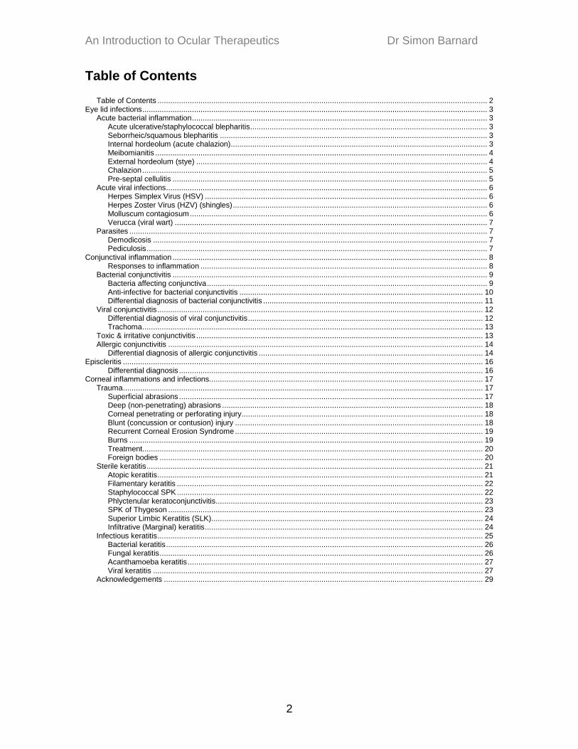
Table of Contents
Table of Contents ......................................................................................................................................................... 2
Eye lid infections ................................................................................................................................................................ 3 Acute bacterial inflammation ......................................................................................................................................... 3
Acute ulcerative/staphylococcal blepharitis .............................................................................................................. 3
Seborrheic/squamous blepharitis ............................................................................................................................ 3 Internal hordeolum (acute chalazion) ....................................................................................................................... 3
Meibomianitis .......................................................................................................................................................... 4
External hordeolum (stye) ....................................................................................................................................... 4 Chalazion ................................................................................................................................................................ 5
Pre-septal cellulitis .................................................................................................................................................. 5
Acute viral infections ..................................................................................................................................................... 6 Herpes Simplex Virus (HSV) ................................................................................................................................... 6
Herpes Zoster Virus (HZV) (shingles) ...................................................................................................................... 6
Molluscum contagiosum .......................................................................................................................................... 6
Verucca (viral wart) ................................................................................................................................................. 7
Parasites ...................................................................................................................................................................... 7
Demodicosis ........................................................................................................................................................... 7 Pediculosis .............................................................................................................................................................. 7
Conjunctival inflammation .................................................................................................................................................. 8
Responses to inflammation ..................................................................................................................................... 8 Bacterial conjunctivitis .................................................................................................................................................. 9
Bacteria affecting conjunctiva .................................................................................................................................. 9
Anti-infective for bacterial conjunctivitis ................................................................................................................. 10 Differential diagnosis of bacterial conjunctivitis ...................................................................................................... 11
Viral conjunctivitis ....................................................................................................................................................... 12
Differential diagnosis of viral conjunctivitis ............................................................................................................. 12 Trachoma .............................................................................................................................................................. 13
Toxic & irritative conjunctivitis ..................................................................................................................................... 13
Allergic conjunctivitis .................................................................................................................................................. 14 Differential diagnosis of allergic conjunctivitis ........................................................................................................ 14
Episcleritis ....................................................................................................................................................................... 16
Differential diagnosis ............................................................................................................................................. 16 Corneal inflammations and infections............................................................................................................................... 17
Trauma ....................................................................................................................................................................... 17
Superficial abrasions ............................................................................................................................................. 17 Deep (non-penetrating) abrasions ......................................................................................................................... 18
Corneal penetrating or perforating injury................................................................................................................ 18
Blunt (concussion or contusion) injury ................................................................................................................... 18 Recurrent Corneal Erosion Syndrome ................................................................................................................... 19
Burns .................................................................................................................................................................... 19
Treatment.............................................................................................................................................................. 20 Foreign bodies ...................................................................................................................................................... 20
Sterile keratitis ............................................................................................................................................................ 21
Atopic keratitis ....................................................................................................................................................... 21 Filamentary keratitis .............................................................................................................................................. 22
Staphylococcal SPK .............................................................................................................................................. 22
Phlyctenular keratoconjunctivitis ............................................................................................................................ 23
SPK of Thygeson .................................................................................................................................................. 23
Superior Limbic Keratitis (SLK) .............................................................................................................................. 24 Infiltrative (Marginal) keratitis ................................................................................................................................. 24
Infectious keratitis ....................................................................................................................................................... 25
Bacterial keratitis ................................................................................................................................................... 26 Fungal keratitis ...................................................................................................................................................... 26
Acanthamoeba keratitis ......................................................................................................................................... 27
Viral keratitis ......................................................................................................................................................... 27 Acknowledgements .................................................................................................................................................... 29
An Introduction to Ocular Therapeutics Dr Simon Barnard
3
Eye lid infections Acute bacterial inflammation
Acute ulcerative/staphylococcal blepharitis Brittle crusty, yellow scales along lid margin. Lid margins are tender and red
2 keratoconjunctivitis with SPK + infiltrates at 2- 4- 8- & 10 o‟clock positions.
Treatment of acute ulcerative blepharitis
Lid hygiene: Lid scrubs (Baby Shampoo or Lid Care); Warm compresses qid
Broad spectrum antibiotics (e.g., Brolene, Polyfax (bacitracin + polymyxin B)
For SPK/infiltrates consider steroid/antibiotic combo (e.g., framycetin + gramicidin + dexamethasone)
Follow up in two weeks
Consider adding oral antibiotic (e.g., oxytetracycline) Seborrheic/squamous blepharitis
Greasy, oily dandruff-like scales along the lid margin. No symptoms / or “burning” due to acidic tears and short BUT. Foaming of tears. Seborrheic dermatitis may be present.
Treatment of seborrheic blepharitis
Generalised lid hygiene
Control seborrhea with shampoo
Artificial tears or eye wash for burning
Refer to dermatologist if symptoms persist
Follow-up in 2-4 weeks
Internal hordeolum (acute chalazion) Acute abscess infection of meibomian gland. Focal area of pain, redness and swelling. Possible pre-auricular node involvement. Rule out cellulites. May be associated with chronic blepharitis. Rule out blepharitis or dacryocystitis
An Introduction to Ocular Therapeutics Dr Simon Barnard
4
Treatment of internal hordeolum
Hot compresses minimum qid
Antibiosis (e.g., Brolene ointment qid)
Follow up 2 days and continue treatment for 5 days; then recheck
Watch for cellulitis (usually pre-septal) and treat if occurs with oral antibiosis
Meibomianitis
Congestion of meibomian glands due to build of meibum (fatty acids). Associated with seborrhoea. A probable staphylococcal or immunological aetiology. Ocular surface symptoms (dry eye burning). Inflammatory appearance to posterior lid margin and palpebral conjunctiva with congested orifices of meibomian glands. Ocular surface involvement due to blepharokeratoconjunctivitis. Note “frothy tears”.
Treatment of meibomianitis
Hot compresses bid
Lid massage bid
Oral oxytetracycline 250 m qid for 10 to 30 days
Review 2 weeks
Advise patient to continue hot compresses and lid scrubs
External hordeolum (stye) Acute abscess infection of lash follicle (gland of Zeiss or Moll). Commonly staphylococcus. Unilateral pain, redness and swelling along lid margin. There may be external suppuration.
Treatment of external hordeolum
Hot compresses qid
Antibiosis may help (e.g. Brolene ointment qid)
Removal of lash may help
Do not squeeze. Spreads infection
Instruct patient on future lid hygiene
An Introduction to Ocular Therapeutics Dr Simon Barnard
5
Chalazion Common lid lump or granuloma. External chalazion - Zeiss gland Internal chalazion – Meibomian gland Diagnostic considerations
If recurrent, rule out neoplasia.
Rule out systemic causes
Rule out acute lid infection
Rule out acne rosacea
Treatment of chalazia
8 mm, hot compresses minimum qid
>8 mm, consider surgical removal
Treat infections before surgery
For heat therapy, follow-up in two weeks
If resolving, continue treatment
If no improvement, consider surgery
Pre-septal cellulitis Acute, diffuse spread of inflammatory cells & debris through loose tissue of eyelid. Lid is diffusely swollen, red and painful. May be sequel of trauma or a sequel of acute internal hordeolum caused by, for example, Stretococcus pneumoniae, S. pyogenes or Haemophilus influenza
Treatment of cellulitis
Pre-septal treated with hot compresses and systemic antibiotics (e.g., erythromycin; penicillin)
Post-septal involvement is serious and potentially life threatening Look out for proptosis; impaired ocular motility, pain on eye movement; fever. Septa of babies are fragile
Hospitalise
Intravenous antibiosis
An Introduction to Ocular Therapeutics Dr Simon Barnard
6
Acute viral infections Viruses consist of particles containing DNA or RNA and can only replicate in host cells. Strongly antigenic producing monocyte proliferation and folliculosis
Herpes Simplex Virus (HSV) Over 90% of adults have HSV antigens HSV 1 – oral HSV 2 – genital Lid inflammations are primary herpetic infections. The lesions appear as a cluster of clear, pearl-like vesicles which do not respect midline and do not scar.
Treatment of HSV blepharitis
Keep clean with pure soap and water
Cold compresses
Calamine lotion
“Tincture of Time”
Acyclovir topical cream q4h
Herpes Zoster Virus (HZV) (shingles)
Varicella virus. Vesicles appear along branch(es) of trigeminal nerve in immunosuppressed adults (old, HIV, chemotherapy). The ophthalmic division is commonly involved. Severe pain occurs 3 to 5 days before eruption. Look out for Hutchinson's sign in which the tip of nose involved. This alerts you that the nasociliary branch is affected and there will be corneal involvement.
Treatment of HZV lid and facial lesions
Topical and systemic steroid (e.g., prednisilone)
Acyclovir 800 mg 5 x daily for up to 10 days
NSAID and narcotic analgesics
Molluscum contagiosum White, round, waxy non-inflammed umbillicated lesion which will contain yellow cheesy material when virus is active. May shed active virus into cul-de-sac giving a follicular conjunctivitis.
An Introduction to Ocular Therapeutics Dr Simon Barnard
7
Treatment of molluscum contagiosum
When quiet (dry central core), leave alone
If cheesy material present, loosen with sterile needle and squeeze out contents
Follow-up one week and repeat as necessary
Verucca (viral wart) Single or multiple non-secreting papillomatous warts.
Parasites
Demodicosis Demodex follicularum. Endemic in older adults. An arachnid mite that live in hair follicles and sebaceous glands. Associated with acne rosacea. Excessive numbers produce toxic or hypersensitive marginal type reaction. Symptoms and signs of demodicosis include acute itchiness of eyelids especially on awakening and tubular/pyramidal collarettes extending from base of lash.
Treatment of demodicosis
Lid scrubs and ointment (e.g., vaseline) for 10 days
Review in 10 days
Pediculosis pediculosis corporis, pediculosis capatis, pthirius pubis. Transmitted by contact or contaminated bed linen etc. Symptoms and signs of lice include itching and burning of eyelids, a crusty appearance to lid margins as well as the presence of lice and nits.
Treatment of lice
Remove nits and lice with forceps or debride with alcohol soaked cotton-wool bud
Vaseline applied to lids tid for 5 days
Lice specific shampoo to rid scalp
Review in 5 days
An Introduction to Ocular Therapeutics Dr Simon Barnard
8
Conjunctival inflammation
Responses to inflammation
A. Exudation /discharge
1. Purulence Characterised by copious pus. Occurs in hyperacute conjunctivitis e.g. gonococcal.
2. Mucopurulence
Characterised by a combination of mucus and fibrin. Occurs in acute conjunctivitis e.g., staphylococcal.
3. Fibrinosis (pseudomembrane) Characterised by a fibrin discharge, trapping inflammatory cells and debris attached to conjunctival surface. Peel away. May be uncomfortable even with Benoxinate. May be caused by streptococcus pyogenes or adenovirus type 8 which is thought to be a specific cause of EKC.
4. Haemorrhage Characterised by subconjunctival haemorrhage and usually caused by pneumococcus or haemophilus.
5. Catarrhal Characterised by a white, stringy discharge may be caused by allergic conjunctivitis
6. Serous
Characterised by a clear, watery discharge. May be caused by a viral conjunctivitis
B. Tissue Alteration
1. Follicles Clear cysts with blood vessel at the base (“rice grain”). Prevalent in viral and chlamydial conjunctivitis.
An Introduction to Ocular Therapeutics Dr Simon Barnard
9
2. Papillae
Nodular elevations with central vascular tufts and prevalent in allergic conjunctivitis.
Bacterial conjunctivitis
Bacteria affecting conjunctiva
Staphylococcus epidermis & aureus (+ive cocci) Most common infective organism of lids and conjunctiva. s. epidermis = normal flora; s. aureus = normal & pathogenic. Not aggressively invasive but pyogenic and produces exotoxins > infiltrates
Streptococcus pyogenes (+ive cocci) An infrequent cause of conjunctivitis. Is invasive and toxinogenic producing a much redder eye than staphylococcus. Can cause cellulitis and pseudomembranes.
Streptococcus pneumoniae (pneumococcus) (+ive cocci) This organism produces a very acute conjunctivitis and sometimes haemorrhages. Can cause cellulites and is a leading cause of acute dacryocystitis.
Pseudomonas aeroginosa (- ive rod) Very virulent. Can destroy eye in 24 hours. Produces blue-green pus, is smelly and has predilection for central cornea.
Morax Axenfeld bacillus ( - ive bacillus) This produces angular conjunctivitis in children.
Haemophilus influenza (Koch-Weeks bacillus) ( -ve) Commonest cause of bacterial conjunctivitis in children between 5 weeks and 5 years. Causes patchy subconjunctival haemorrhages and purplish discolouration of lids.
Neisseria gonorrhea (- ive diplococci)
An Introduction to Ocular Therapeutics Dr Simon Barnard
10
This produces a hyperacute conjunctivitis with extreme purulence and puffy lids. It can invade intact cornea. The most common cause of neonatal conjunctivitis. Chlamydia trachomatis (TRIC organism) (gram - ive) Has a 2 - 4 week history and produces mixed bacterial, viral and immune signs with follicles, mucopurulence and lymphadenopathy. A positive genitourinary history may be present. Needs oral antibiotics (e.g. oxytetracycline).
Anti-infective for bacterial conjunctivitis
Sodium sulphacetamide A broad spectrum but NOT active against pseudomonas. Nor is it effective in the presence of pus. There is a high prevalence of sensitivity to the drug. Not now available in the UK (except in combination)
Gentamycin (and Fucidic acid) Broad spectrum ( gram + ive & - ive). Effective against pseudomonas but not not as effective against some streptococcal strains. Some allergy reactions.
Tobramycin Similar efficacy to gentamycin but more effective against pseudomonas than gentamycin. Not as effective against some streptococcal strains as other agents.
Neomycin Broad spectrum and produces the highest prevalence of allergic response.
Framycetin (Soframycin) Similar to Neomycin
Chloramphenicol Broad spectrum. There have been very rare, idiosyncratic bone marrow disease following use of this drug (usually following oral use).
An Introduction to Ocular Therapeutics Dr Simon Barnard
11
Whilst this is the most commonly used topical ophthalmic antibiotic in the UK it is shunned in the USA
Fluoroquinilones e.g. ciprofloxacin Broad spectrum (with similar activity to Gentamycin). Effective in cases of severe infection but not good against chlamydia.
Polyfax = Polymyxin B (against gram - ive) plus bacitracin (against gram - ive) effective against + ive, - ive and pseudomonas. Available only in ointment form.
Polytrim = Polymyxin B (against gram - ive) plus trimethoprim (a sulpha drug) (against gram +ive). Very broad spectrum drug and is effective against pseudomonas. Available on as drops.
Neosporin = polymyxin B plus gramicidin plus neomycin. A very broad acting antibiotic. It is expensive and available only in drops form.
Differential diagnosis of bacterial conjunctivitis It usually occurs over 2 to 3 days compared to viral which takes longer. The eye appear meaty red with a mucopurulent discharge. The lashes may be stuck in the morning. Papillae may be present. Rule out chlamydia. Not a painful condition and vision is normal.
Treatment of conjunctivitis Treat with lid hygiene, irrigation, warm compresses and antibiosis (start with a loading dose). If you are suspicious then take a conjunctival swab BEFORE commencing treatment.
general treatment Following a loading dose, treat qid for 5 to 7 days minimum. If the organism is resistant then take a culture and change to another antibiotic and/or extend course. Consider oral antibiotics.
An Introduction to Ocular Therapeutics Dr Simon Barnard
12
specific treatment Staphylococcus Topicals: polysporin, polytrim, neosporin , chloramphenicol, gentamycin, neomycin, tobramycin, framycetin, ciprofloxacins. Orals: (for hyperacute infections only) e.g., erythromycin, tetracycline. Streptococcus Topicals: as above Orals: e.g., erythromycin, penicillins. Haemophilus Topicals: gentamycin, polymyxin B, or ciprofloxacin (not children) Orals: erythromycin, cephalosporins. Moraxella Topicals: ciprofloxacin, gentamycin, neomycin, Polymyxin B, Sulphonamides, tetracycline, tobramycin, zinc sulphate. Orals: not indicated. Chlamydia Topicals: not indicated Orals: Doxycline, tetracycline (not children or pregnant women), or eryrthromycin.
adjunct steroids Avoid in mild to moderate cases but consider combination use with caution in cases of severe inflammatory response:- q4h for 3 to 5 days then taper. Check IOP.
Viral conjunctivitis
Differential diagnosis of viral conjunctivitis Occurs over a 3 to 7 day period. Commonly an adenovirus. Eyes look pink/purple with a watery discharge and the lids can look puffy with the conjunctiva appearing“boggy”/plica enlarged (hypertrophy). Follicles often present and the cornea may show SPK. The preauricular lymph node is often enlarged.
An Introduction to Ocular Therapeutics Dr Simon Barnard
13
Treatment of viral conjunctivitis
Lubrication
warm compresses
vasoconstrictors
follow up 7 days. It is very rare for a secondary corneal infection so prophylactic antibiotics are not usually indicated.
Trachoma
Caused by TRIC (chlamydia trachomatis). An oculogenital disease endemic to many underdeveloped countries and spread by poor hygiene. Common in the western world in the ocular form. The severe corneal scarring forms more sight threatening than milder conjunctival forms. Signs of trachoma include upper tarsal follicles, papillary hypertrophy, trichiasis and entropion. These may progress to superior pannus, ulceration, limbal follicles and subsequent Herbert‟s pits.
Treatment of trachoma
Treat ALL cases, mild or severe, with oral tetracycline (not children or pregnant women) or erythromycin.
Some advocate topical tetracyline ointment bid for two to three months prophylactic treatment of family.
Toxic & irritative conjunctivitis Low grade chronic hyperaemia with no exudates and minimal follicles. Due to e.g., smoke, UV light, pollutants, dry eyes, toxins e.g. staphylococcus, molluscum, verrucae, medications, soaps, perfumes and contact lenses.
Treatment of toxic & irritative conjunctivitis
Remove or treat primary cause e.g., staphylococcal blepharitis or reduce exposure to UV by wear UV protection.
cold compresses q3-4 hourly for 2 weeks
lubricants 6 to 10 times daily for two weeks
An Introduction to Ocular Therapeutics Dr Simon Barnard
14
Allergic conjunctivitis Type 1 allergy is the most common of ocular reactions. Allergens include dust, pollen, foods, insect bites, and drugs. The allergen attaches itself to an IgE molecule on mast cell or basophil and histamines and leukotrienes are released. These produce erythema, chemosis, tearing, itching, and heat. Examples of Type 1 allergy include atopic dermatitis, hay fever, asthma and anaphylactic shock A reaction occurs within minutes of exposure (compare to Type 4). There is often a family history of allergy. The disease is often seasonal and the specific allergen known. Signs and symptoms include itching (the primary symptom). It may be unilateral (contact) or bilateral. Vision is unaffected or fluctuates. The bulbar conjunctiva appears pink, red, “boggy” and glassy. A stringy mucoid discharge may be present and the lids oedematous and red. Whilst papillae are more likely to occur, sometimes follicles are present.
Differential diagnosis of allergic conjunctivitis Rule out other causes of red eye and conjunctivitis. Try and determine whether Type 1 or 4 and what the causative agent might be. Causes include hay fever (plant allergens). The allergy may present as seasonal conjunctivitis or spring catarrh and be caused by tree and flower allergens, mould spores or grasses. General allergic conjunctivitis can be caused by pets, house mites, household products, personal hygiene and perfumery products. General irritative conjunctivitis can occur from smog (ozone, particulates), chemicals or livestock.
Aims of management of non-infectious conjunctivitis
determine when the symptoms are worse
investigate the cause
remove the allergens if possible
clean the area (wash)

An Introduction to Ocular Therapeutics Dr Simon Barnard
15
counter the itchy symptoms
reduce the hyperaemia (vasoconstrict)
reduce tissue reactions
prevent oedematous damage and infiltration
Management and treatment Possible treatment and management interventions include
remove the allergen if possible cure
wash the eye with saline eye wash
apply frequent cold compresses
remove pollution and bacteria from lid margins by carrying out lid scrubs
wash the eyes with astringent drops or eye wash
instil decongestant drops
counter lacrimation and itchiness with anti-histamine drops
counter lacrimation, itchiness and vasodilation with oral anti-histamines
reduce conjunctival response to allergens with mast cell stabilisers
steroids Saline eye washes and drops are useful to remove irritants or allergens. Examples include:- Minims saline 0.9%; Normasol (sachet); Ami-dose (plastic ampule). Astringent eye washes and drops include:- Optrex Eye Lotion (with eye cup). This contains Witch Hazel and is also available as drops. Sootheye eye drops is available as a multidose bottle (10 ml) and contains zinc sulphate. Lid hygiene The patient should be instructed to use a cotton wool bud dipped in a solution of 1/10 J & J baby shampoo/boiled water to carefully „scrub‟ lid margins or use CIBA Lid Care. Astringent-decongestant-antihistamine eye drops Otrivine-Antistin (CIBA Vision) (xylometazoline 0.05% & antazoline 0.5%) Murine eye drops (Abbott) (naphazoline 0.012%) Isopto Frin (Alcon) (phenylephrine hyd. 0.12% & naphazoline 0.01%)
An Introduction to Ocular Therapeutics Dr Simon Barnard
16
Optrex Clearine eye drops (phenylephrine hyd. 0.12% & naphazoline Oral antihistamines Discuss the possible use with adult patients. The advice of pharmacist can be useful. Oral anti-histamines available OTC. Mast cell stabilisers Optrex Hay Chrom; Opticrom; Hay-crom (Cromyln sodium) are available OTC. Advise patient that the drops are designed for prophylactic use. Other drugs include Alomide and Rapatil (POM). NSAIDs NSAIDs such as Voltaren Ophtha eye drops (diclofenac sodium 0.1%) and Acular (ketorolac) are used in the USA for allergic conjunctivitis. Alrex (lotepredenol etabonate). Topical steroids Topical steroids may be required for severe allergic conjunctivitis
Episcleritis Characterised by acute onset of redness and mild pain/discomfort in one or both eyes. Occurs typically young adults and recurrent episodes are common. Clinical signs and symptoms include sectorial (less commonly diffuse) redness of one or both eyes. The redness is due to engorgement of the episcleral vessels. There is tenderness over affected area of episcleral injection. May be a nodule. Nodule can be moved slightly over sclera. The cornea is rarely affected and the vision is normal. The cause is usually “idiopathic”, There may be a relationship with collagen-vascular disease (e.g., rheumatoid arthritis; polyarteritis nodosa; Systemic Lupus Erythematous; gout; HZV and others).
Differential diagnosis With scleritis there will severe deep pain radiating to ipsilateral side of head and no vessel blanching with phenylephrine 2.5%. There will also be a bluish hue to the sclera. In conjunctivitis there will be a discharge, follicles or and papillae.
An Introduction to Ocular Therapeutics Dr Simon Barnard
17
With iritis there will be anterior chamber involvement. To aid examination and diagnosis you may wish to use 2.5% phenylephrine topically. This will blanch vessels after 15 minutes. Also, you can anaesthetise eye and move conjunctival vessels with Q-tip to determine depth. Check AC. Treatment
Cold compresses
Artificial tears
Decongestant
Moderate severe cases may need topical steroid e.g. FML qid
Rare cases may require NSAID e.g., Ibuprofen or aspirin
Follow-up Check weekly if on steroids Otherwise after several weeks
Warn that it may reoccur
Reassure that it will resolve
Corneal inflammations and infections
Trauma
Superficial abrasions Superficial abrasions are commonly seen in practice and the patient will often volunteer a direct acute history. There may be trauma to the ocular surface from a transient foreign body which is usually particulate in nature. Fibreglass can be problematic. The patient complains of grittiness and, usually mild pain May be due to an embedded foreign body particularly upper tarsus (so always evert lids), trichiasis or entropion, be contact lens induced (edge or trapped foreign matter), EBMD or “dry eye”, Examination should consist of Vas followed by slit lamp with fluorescein examination of lids, lashes, conjunctivae and cornea. Single evert the superior lid for tarsus and double evert for the cul-de-sac.
Management of superficial abrasions
Firstly remove foreign body by irrigation, swabbing or with a blunt spatula.
Prophylactic antibiosis
An Introduction to Ocular Therapeutics Dr Simon Barnard
18
Pain management
Recheck in 18 to 24 hours
Add warm compresses
Add Hypertonic saline (5%) q4h for 4 weeks
Review again 24 hours then 1 week
Deep (non-penetrating) abrasions There is usually a distinct history of gouging or laceration from fingernail, paper cuts and mascara brushes. The patient will be in moderate to severe pain with lid spasm, oedema and erythema. VA will be reduced and the bulbar conjunctiva hyperaemic. There will be AC cells and flare. Examination should include VA s and the use of a topical anaesthetic may be required. Slit lamp examination with fluorescein must be used to rule out penetration (stroma) and perforation (full thickness puncture). Perpendicular sharp objects (e.g., pencil; pine needle) are frequently involved. Look for stromal or endothelial involvement and Seidel‟s sign.
Management deep (non-penetrating) abrasions
Never allow patient to take topical anaesthetic for home use.
Dilate with cycloplegic (Homatropine 5% or cyclopentolate 1% bid).
Instil broad spectrum antibiotic eye ointment.
Analgesics (Aspirin, Ibuprofen; Paracetamol; Codeine).
Only pressure patch severe abrasions if patient is likely to rub eye or where most of cornea is involved
Or bandage contact lens
Follow-up next day
Introduce warm compresses & hypertonic saline
Follow every 24 to 48 hours
Corneal penetrating or perforating injury Refer to Eye Casualty Dept. Eye shield not pressure patch.
Blunt (concussion or contusion) injury
These are often sports injuries. Other causes include a punch, blast (closed lid), industrial or from birth trauma (vertical folds/tears in Descemet‟s membrane).
An Introduction to Ocular Therapeutics Dr Simon Barnard
19
Common findings include ecchymosis, subconjunctival haemorrhage and chemosis, corneal abrasion/laceration, Corneal oedema, and anterior uveitis. All ocular tissues may be involved so rule hyphaema, iris dialysis and retinal tears.
Management of blunt (concussion or contusion) injury
Treat all involved structures appropriately
Mild to moderate corneal involvement usually resolves without treatment
Warm compresses
Hypertonic saline q 4 hours
Reassurance Recurrent Corneal Erosion Syndrome
Any damage to EBM or Bowman‟s membrane causes weakening or loss of adhesion between epithelium and stroma. The basal epithelial cells take weeks to repair EBM and weak adhesions may persist indefinitely. Usually one of two classic histories: Previous corneal injury or spontaneous development (no history of trauma). History symptoms include painful eye often on awakening. It is usually unilateral and often of a recurring nature. There may be a history of associated disease process (EBMD; Diabetes). A tangential injury (v. perpendicular) e.g., fingernail; paper cut rather than a perpendicular one may lead to RCE syndrome. Signs include mild erosions, corneal oedema and epithelial flaps.
Management of RCE syndrome
As corneal abrasions
Burns Thermal burns can arise from cinders, a match head or hair curling irons. The burn leaves an eschar (white charring of epithelium).
Treatment
Debride and treat as abrasion
An Introduction to Ocular Therapeutics Dr Simon Barnard
20
Radiation Most commonly related to UV sources (sun, UV lamps, welding arcs). The exposure produces focal or diffuse SPK and acute discomfort.
Treatment
Manage as with infectious keratitis Chemical burns A true emergency that may arise from an accident with: Aromatic compounds such as benzene, turpentine or petrol Acids such as battery acids Alkalis such as ammonia; drain cleaners; cement (lime); magnesium hydroxide (fireworks); chlorine (bleach, pool cleaners) The amount of damage is correlated with amount of chemical but whilst acid burns are rapid, they carry better prognosis Alkali burns appear initially innocuous, progress rapidly, and are more threatening to deeper tissue. Penetration continues by collagenolytic reaction for hours or day. This can result in unrelenting ulceration, melting, stromal scarring or perforation.
Management
Often incoming phone call for advice
Obtain details of chemical
If in practice, topical anaesthesia
Acute stage
Copious continued irrigation with first available bland fluid (telephone) or with sterile irrigating solution
Delays of 3 - 5 mins or discontinuation sooner than 20 – 30 mins reduces prognosis significantly
Debride or remove caustic particles
Wet cotton wool swab
Antibiotic ointment
Cycloplegia
Foreign bodies There is usually an obvious history. Try and ascertain if the entry was of high speed ? Discomfort ranges from mild to severe. Frequently the event happened 1 – 3 days before presentation.
An Introduction to Ocular Therapeutics Dr Simon Barnard
21
Signs include a distinct immovable embedded particle(s) on corneal epithelium, Coat‟s white ring (oedema/infiltrates), a rust ring and foreign body tracking on the cornea. A topical anaesthetic should be used to enable slit lamp microscopy to determine depth of foreign body. The lid should be everted. Assess for secondary infection and/or anterior uveitis.
Management
Remove foreign body and any rust ring
Antibiosis
Cycloplegia for 24 hours
Oral Analgesic
Pressure patch (?)
Review 24 hours
Sterile keratitis
Atopic keratitis This occurs in males > females. There is usually a history of atopic dermatitis and very itchy eyes. It occurs year round and is not “seasonal”. There is concurrent irritation and burning. The condition is bilateral and there is a watery mucoid discharge, mild lid swelling and papillary hypertrophy in inferior palpebral conjunctival. There may be superior (more common) corneal findings with circumlimbal infiltrates, Trantas‟ dots and SPK. This can lead to pannus and sterile ulceration. Rule out GPC (more superior tarsus); SLK (no dermatitis) and Vernal KC (not seasonal. Management
OTC antihistamines
Cold compresses
Topical prednisilone 1%; q 4 h for 7 days
Taper to Opticrom
Ocular lubricants
Counsel patient on chronic nature May self limit. Reserve steroids for acute phases and taper quickly.
An Introduction to Ocular Therapeutics Dr Simon Barnard
22
This condition can cause scarring and particularly for children should be managed by a specialist ophthalmologist.
Filamentary keratitis This condition produces mild to severe foreign body sensation/pain. Mucin debris combines with dead epithelial cells to form threads 1 – 3 mm long with the filament end attaching itself to a dry corneal area. The unattached end moves with blink. It is often associated with wide range of other corneal disease. Management
Treat underlying disease
Remove or reduce filaments
Heavy lubrication
Acetylcystein
Bandage contact lens
Staphylococcal SPK Often associated with a history of blepharitis. The patient reports a gritty sensation and vision may be slightly reduced. There is variable photophobia and or lacrimation. More common in contact lens wearers and dry eye patients. SPK can be of variable amount and size but is most common situated inferiorly. Dense patches at 4- or 8-o‟clock area are a prodromal sign of marginal infiltrative and subsequent infiltrative keratitic response. There may be variable bulbar hyperaemia, inferior injection and a localised leash of vessels. There is a moderate papillary response on palpebral conjunctiva and no discharge in the mild to moderate condition. However, if chronic, it may produce peripheral corneal scarring.
Management
Rule out infectious keratitis
Ocular lubricants
Skin/lid hygiene
Antibiotic eye ointment
Recheck in 7 days
An Introduction to Ocular Therapeutics Dr Simon Barnard
23
Phlyctenular keratoconjunctivitis
A localised, superficial, infiltrative reaction produced by superficial exotoxins Whilst usually staphylococcus, TB can also produce this condition. Phlyctens can occur on any ocular surface and are usually unilateral. The patient complains of a gritty sensation.
Treatment The condition should respond quickly to treatment.
Topical steroids q4h to reduce infiltrate quickly
Prophylactic antibiosis qid 5 days
Treat blepharitis
Review 3 days
Encourage good lid hygiene
Review in 6/12
SPK of Thygeson
Unknown cause, possibly a virus. It occurs most commonly in young adult females (15 to 40 years) and produces occasional periods of gritty sensation and photophobia. There are remissions and exacerbations. It usually self limits over 4 to 7 years. Generally bilateral. Signs include diffuse SPK which is greatest centrally. It will affect vision when dense. There may be confluent NaFL staining. The other important sign are the presence intraepithelial infiltrates on exacerbations. These stain negatively to NaFl . The eyes are white and quiet despite keratitis.
Treatment Mild to moderate Thygeson‟s may be treated with
Ocular lubricants
Moderate to severe
Heavy lubrication 6 – 10 times/day
Topical steroid q4h for 14 days
Antivirals (?)
Bandage contact lens
An Introduction to Ocular Therapeutics Dr Simon Barnard
24
Superior Limbic Keratitis (SLK) This produces a non-specific ocular irritation and is more prevalent in women aged 20 – 50 years. It is usually bilateral and can occur secondary to CLs. There is also a correlation with thyroid disease. There is superior bulbar conjunctival hyperaemia that follows the path of the superior rectus muscle. Chemosis can develop over injected area and limbal border. There may superior tarsal papillae. Superior SPK may coalesce and there can be sub-epithelial infiltrates (chronic) and occasionally pseudo-dendrites Usually self-limiting.
Management
Moderate to severe presentations may require topical steroids qid for 14 days
Mild to moderate presentations – Opticrom qid 3 to 4 weeks
Discontinue contact lenses
Cold compresses
Infiltrative (Marginal) keratitis Infiltrative immune response to staphylococcal exotoxins (alpha toxin). This is a sterile response to bacteria. The 4- and 8-o‟clock limbal areas most vulnerable and the toxins produce intraepithelial infiltrates on corneal periphery. These are accumulations of cells and debris. “Island” lesions with clear interval between limbus and distal border of lesion are pathonomonic of this condition. The size of the infiltrates may range from 0.5 mm to 1.5 mm.
Management
Mild
Warm compresses
Lid hygiene
Antibiotic ointment tid
Gentamicin drops tid for 7 days
Moderate (lesions up to 1 mm)
Add prednisilone 1% qid
Cycloplegia
An Introduction to Ocular Therapeutics Dr Simon Barnard
25
Severe
No steroids in first 24 h
Loading dose of gentamicin 1 drop every minute for 5 minutes
3 drops gentamicin every waking hour, q 2h overnight
Review 24 hours
Infectious keratitis
Bacterial (most common aetiology)
Fungal (mycotic)
Protozoan (acanthamoeba)
Viral Symptoms include red eye, mild-to-severe ocular pain, photophobia, decreased vision and discharge. Critical signs include a focal white opacity in corneal stroma (infiltrate). An ulcer exists if there as an overlying epithelial defect that stains with NaFl with stromal loss. If you cannot see the iris through the lesion with a lit lamp then this is a sign that it is an ulcer rather than simply stromal oedema. Other signs include conjunctival hyperaemia, corneal thinning, stromal oedema and inflammation surrounding the infiltrate, folds in Descemet‟s membrane, anterior chamber reaction and hypopyon. A mucopurulent discharge is likely and there may be upper eyelid oedema. Your examination should include a careful history:
CL wear and care regimen ? Swim with lenses ? Trauma or foreign body ? Systemic illness ? Previous treatment ?
Slit lamp exam with NaFl may show epithelial loss overlying an infiltrate. Document the size, depth and location. Note any AC reaction and check IOP. Corneal scrapings for smears and cultures should be done for all ulcers (Kimura spatula). However, non-staining infiltrates may be treated with antibiotics without a scrape.
An Introduction to Ocular Therapeutics Dr Simon Barnard
26
Bacterial keratitis
Treatment Ulcers and infiltrates are generally treated as bacterial initially unless high index of suspicion of another form of infection e.g., Fungal, acanthamoeba or HSV.
Cycloplegics (e.g. scopolamine 0.25% tid)
Topical antibiotics
Pain medication For small non-staining infiltrate with minimal AC reaction and no/minimal discharge and where patient is a Non-contact lens wearer
Broad spectrum topical e.g.,polymixinB/Bacitracin ointment qid or ciprofloxacin drops q 2-6 h
Contact lens wearer
Ciprofloxacin or tobramycin drops q 2-6 h
Can add tobramycin ointment In the presence of large or staining infiltrate or moderate to severe AC reaction or purulent discharge
Fortified tobramycin or gentamycin every hour alternating with ciprofloxacin every hour (half hourly)
Consider subconjunctival injection
Protect with shield (do NOT patch)
Consider hospitalisation if sight threatening, there is a likelihood of non-compliance, or if systemic antibiotics are required.
Follow up - Daily evaluation
Reculture ulcer if not responding to treatment
Fungal keratitis Critical history - history of trauma, particularly from vegetable matter (e.g., tree branch) or chronic eye disease.
An Introduction to Ocular Therapeutics Dr Simon Barnard
27
Critical signs - corneal stromal grey-white infiltrate with a feathery border. There may be either elevation or stromal thinning. Other signs may include satellite lesions surrounding primary infiltrate, conjunctival injection, mucopurulent discharge, AC reaction and hypopyon. In previously unhealthy eyes the cause is usually non-filamentous fungi e.g., candida. The likeliest cause following trauma with vegetable matter are the filamentous fungi such as fusarium or aspergillus.
Treatment
Admission to hospital
Natamycin
Cycloplegia
Amphoteracin
Fluconazole
Miconazole or clotrimazole
Acanthamoeba keratitis Symptoms include severe ocular pain, redness and photophobia over several weeks. A critical sign is that there is less corneal and AC segment inflammation than would be expected for degree of pain experienced. Epithelial and sub-epithelial infiltrates occur (sometimes along corneal nerves) Later a corneal stromal infiltrate in the form of a ring may manifest.
Treatment of acanthamoeba
Prompamidine isethionate 0.1%
Neosporin
Pain medication
Viral keratitis Epidemic Keratoconjunctivitis (EKC) This is the most common contagious disease in optometric practice. Highly contagious, this virus is transmitted by towel, hand instrument.
An Introduction to Ocular Therapeutics Dr Simon Barnard
28
Rules (or guidelines) of 8
Most commonly Adenovirus 8
1st 8 days (conjunctivitis) most contagious
Day 8 – corneal SPK presents
8 days later – Epithelial infiltrates The symptoms may be mild to severe and include burning, photophobia, lacrimation. Signs include a possible reduction in vision, purplish pink conjunctiva, oedematous lids, watery eye, reduced BUT, follicles and preauricular involvement. In severe cases, a pseudomembrane may develop. Signs include diffuse SPK and subepithelial infiltrates which do not stain.
Treatment
“Contagious” ! – Advice
Cold compresses
Analgesics
Ocular lubricants
Recheck in 7 days HSV Note that corneal involvement may lead to a reduced corneal sensitivity. Corneal epithelial HSV disease may include SPK, stellate, dendritic and or geographic lesions. The edges of these lesions stain to rose Bengal and the central areas stain to fluorescein. A neurotrophic HSV corneal ulcer is an area of interpalpebral stromal disease that persists despite anti-viral therapy. It may lead to stromal melting and perforation. It is sterile and has smooth margins. Corneal stromal HSV disease can manifest as disciform keratitis in which there is a disc-shaped stromal oedema and intact epithelium. There may be an associated mild iritis with granulomatous KP. There is no necrosis or neovascularisation. Another manifestation is necrotising interstitial keratitis in which there are multiple or diffuse grey stromal infiltrates with neovascularisation.
An Introduction to Ocular Therapeutics Dr Simon Barnard
29
Treatment of corneal HSV epithelial disease
Cycloplegic
Acyclovir eye ointment HZV Symptoms and signs include skin rash, skin pain, headache, fever, malaise, blurred vision, eye pain, and red eye. A critical sign is an acute vesicular skin rash that follows the dermatome of 5th N. This condition is unilateral respects the midline and commonly involves upper lid only. Hutchinson‟s sign – lesion at tip of nose predicts wider ocular involvement of lower eye lid, conjunctivitis, SPK, corneal pseudodendrites, immune stromal keratitis, neurotrophic keratitis and uveitis.
Treatment
Adults with acute moderate to severe skin rash present less than 3-5 days in which active skin lesions are present
Acyclovir 800 mg po 5 x a day
Antibiotic ointment to skin lesions
Warm compresses to skin lesions Over 60‟s who are not immunocompromised
Add prednisilone 60 mg po for 3 days; 40 mg po for 3 days; 20 mg po for 4 days
Acknowledgements Catania L. (1995) Primary Care of the Anterior Chamber, Appleton & Lange Norwalk Cullom RD and Chang B (1994) The Wills Eye Manual 2nd Ed,
JB Lippincott Company, Philadelphia I would also like to acknowledge use of various aspects of lecture notes from the Pennsylvania College of Optometry and City University ocular therapy courses.
Dr Simon Barnard 12th June 2002