Embed Size (px)
Citation preview
Heekin et al. Parasites & Vectors 2012, 5:162http://www.parasitesandvectors.com/content/5/1/162
RESEARCH Open Access
Analysis of Babesia bovis infection-induced geneexpression changes in larvae from the cattle tick,Rhipicephalus (Boophilus) microplusAndrew M Heekin1*, Felix D Guerrero1, Kylie G Bendele1, Leo Saldivar2, Glen A Scoles3, Cedric Gondro4,Vishvanath Nene5, Appolinaire Djikeng5 and Kelly A Brayton6
Abstract
Background: Cattle babesiosis is a tick-borne disease of cattle that has severe economic impact on cattleproducers throughout the world’s tropical and subtropical countries. The most severe form of the disease is causedby the apicomplexan, Babesia bovis, and transmitted to cattle through the bite of infected cattle ticks of the genusRhipicephalus, with the most prevalent species being Rhipicephalus (Boophilus) microplus. We studied the reaction ofthe R. microplus larval transcriptome in response to infection by B. bovis.
Methods: Total RNA was isolated for both uninfected and Babesia bovis-infected larval samples. Subtracted librarieswere prepared by subtracting the B. bovis-infected material with the uninfected material, thus enriching forexpressed genes in the B. bovis-infected sample. Expressed sequence tags from the subtracted library weregenerated, assembled, and sequenced. To complement the subtracted library method, differential transcriptexpression between samples was also measured using custom high-density microarrays. The microarray probeswere fabricated using oligonucleotides derived from the Bmi Gene Index database (Version 2). Array results wereverified for three target genes by real-time PCR.
Results: Ticks were allowed to feed on a B. bovis-infected splenectomized calf and on an uninfected control calf.RNA was purified in duplicate from whole larvae and subtracted cDNA libraries were synthesized from Babesia-infected larval RNA, subtracting with the corresponding uninfected larval RNA. One thousand ESTs were sequencedfrom the larval library and the transcripts were annotated. We used a R. microplus microarray designed from a R.microplus gene index, BmiGI Version 2, to look for changes in gene expression that were associated with infectionof R. microplus larvae. We found 24 transcripts were expressed at a statistically significant higher level in ticksfeeding upon a B. bovis-infected calf contrasted to ticks feeding on an uninfected calf. Six transcripts wereexpressed at a statistically significant lower level in ticks feeding upon a B. bovis-infected calf contrasted to ticksfeeding on an uninfected calf.
* Correspondence: [email protected], Knipling Bushland US Livestock Insect Research Laboratory, 2700Fredericksburg Rd, Kerrville, TX 78028, USAFull list of author information is available at the end of the article
© 2012 Heekin et al.; licensee BioMed Central Ltd. This is an Open Access article distributed under the terms of the CreativeCommons Attribution License (http://creativecommons.org/licenses/by/2.0), which permits unrestricted use, distribution, andreproduction in any medium, provided the original work is properly cited.
Heekin et al. Parasites & Vectors 2012, 5:162 Page 2 of 12http://www.parasitesandvectors.com/content/5/1/162
Conclusion: Our experimental approaches yielded specific differential gene expression associated with theinfection of R. microplus by B. bovis. Overall, an unexpectedly low number of transcripts were found to bedifferentially expressed in response to B. bovis infection. Although the BmiGI Version 2 gene index (http://compbio.dfci.harvard.edu/tgi/cgi-bin/tgi/gimain.pl?gudb=b_microplus) was a useful database to help assign putative functionto some transcripts, a majority of the differentially expressed transcripts did not have annotation that was useful forassignment of function and specialized bioinformatic approaches were necessary to increase the information fromthese transcriptome experiments.
Keywords: Cattle tick, Rhipicephalus (Boophilus) microplus, Babesia bovis, Larva, Transcriptome, Serine proteinaseinhibitor
BackgroundThe cattle tick, Rhipicephalus (Boophilus) microplus, is aone-host tick parasitizing cattle in most of the world’stropical and subtropical countries. This tick has a hugeimpact on cattle producers, large and small, with lossesdue to R. microplus infestations in Brazil alone estimatedto be over $2 billion annually [1]. Perhaps the major im-pact is due to losses attributable to pathogens and theirassociated diseases transmitted by the tick’s bite. R.microplus is known to frequently harbor Anaplasmamarginale, the causative agent of anaplasmosis, andBabesia bovis and Babesia bigemina, the apicomplexanagents that cause cattle babesiosis. The cattle-R. micro-plus-Babesia complex has been described as the mostimportant agricultural host-arthropod-pathogen complexglobally [2]. B. bovis is generally responsible for the moreserious cases of bovine babesiosis, with infection of naivehosts often causing pulmonary edema, central nervoussystem problems, and death. The severity of this diseasein Australia is such that an anti-B. bovis vaccine is inwidespread use across northern Australia in response toseasonal outbreaks in the major cattle producing areas[3].When R. microplus ingests blood from a B. bovis-
infected animal, the ingested merozoites undergo de-velopmental changes in the tick midgut until the zyg-ote stage of the apicomplexan enters the digestivecells of the tick’s gut where further multiplication anddevelopment to the kinete stage occurs. This kinetestage eventually enters the hemolymph and spreads tothe rest of the tick’s tissues [3]. We are interested intranscriptional changes that accompany the variousstages of infection by B. bovis as it interacts with itstick host. As our biological system, we allowed R.microplus to feed upon a splenectomized calf sufferingfrom bovine babesiosis due to infection with B. bovis.We compared gene expression in larvae that hatchedfrom eggs oviposited by adult female ticks that hadfed on this infected calf with gene expression in lar-vae that hatched from eggs oviposited by adult femaleticks that had fed on an uninfected calf. Ourapproaches included sequencing subtracted libraries
synthesized from infected larvae, microarray analysis,and quantitative real-time polymerase chain reaction(qRT-PCR) to identify and annotate transcripts asso-ciated with B. bovis infection of R. microplus larvae.
MethodsTicksR. microplus larvae were from the f16 generation of theB. bovis-free La Minita strain. The La Minita strain wasstarted from engorged female ticks collected from anoutbreak in Starr County, Texas in 1996 and propagatedat the USDA Cattle Fever Tick Research Laboratory atMoore Field, Texas before being transferred to theUSDA-ARS Animal Disease Research Unit (ADRU) inPullman, WA. Two splenectomized Holstein calves, 5-6months of age, were used in these studies. One of thecalves was infected by inoculating with frozen blood sta-bilate of the Texas Strain (2nd passage), of B. bovis. Theinfection status of this calf was verified by blood testingand subsequently used to provide B. bovis infected ticklarvae for this study. The second calf was maintainedinfection-free and was used to provide uninfected ticklarvae for this study. All animal use was conducted atADRU facilities at the University of Idaho Holm Re-search Center (Moscow, ID) while following protocolsapproved by the University of Idaho Institutional AnimalCare and Use Committee.B. bovis-infected larvae were obtained by placing unin-
fected larvae that hatched from 1.0 g of eggs from the B.bovis-free La Minita colony ticks into feeding patchesglued to the shaved back and sides of the B. bovis-infected calf (designated as day 1). After feeding, all lar-vae were removed before they molted to the nymphalstage and frozen at -80oC immediately.Uninfected larvae were obtained by placing uninfected
larvae that hatched from 1.0 g of eggs from the B. bovis-free La Minita colony ticks into feeding patches glued tothe shaved back and sides of the infection-free calf(designated as day 1). After feeding, all larvae wereremoved before they molted to the nymphal stage andfrozen at -80oC immediately.
Heekin et al. Parasites & Vectors 2012, 5:162 Page 3 of 12http://www.parasitesandvectors.com/content/5/1/162
RNA protocolsSeparately, for both the uninfected and Babesia bovis-infected larval samples, total RNA was isolated from apool of several thousand tick larvae using the FastPrep-24Tissue and Cell Homogenizer and Lysing Matrix D (Qbio-gene, Irvine, CA, USA) as described in Saldivar et al.(2008) [4]. Total RNA was treated with Turbo DNAse perTurbo DNA-free kit protocols (Ambion Inc.). RNA integ-rity was verified by formaldehyde gel electrophoresis andstaining in GelStar Nucleic Acid Gel Stain (Lonza, Rock-land, ME, USA). Twenty μg of DNA-free total RNA wassent to NimbleGen Systems Inc. (Madison, WI, USA) foruse in microarray hybridization. Express Genomics Inc.(Frederick, MD, USA) was used to produce subtracted li-braries from 0.15 mg each of B. bovis-infected and unin-fected larval total RNA. The subtracted libraries,produced by subtracting the B. bovis-infected materialwith the uninfected material, were enriched for expressedgenes in the B. bovis-infected sample.
Sequencing and bioinformaticsLibrary sequencing was performed at the J. Craig VenterInstitute (Rockville, MD). Bacterial colonies were pickedfor template preparation using colony-picking robots(Genetix, Boston, MA), inoculated into 384 well platescontaining liquid medium and grown overnight. A ro-botic workstation was used to prepare sequencing gradeplasmid DNA using an alkaline lysis method modifiedfor high throughput processing [5]. Beckman Multimek96 or Biomek FX automated pipetting robot work sta-tions (Beckman Coulter, Fullerton, CA) were used tocombine pre-aliquoted templates and reaction mixesconsisting of deoxy- and fluorescently labeled dideoxy-nucleotides, Taq DNA polymerase, sequencing primers,and sequencing reaction buffer. Linear amplificationsteps were performed on MJ Research Tetrads PTC-225(MJ Research, Inc., Watertown, MA) and sequencing re-action products purified by ethanol precipitation andresolved on ABI 3730xl sequencing machines (AppliedBiosystems, Foster City, CA).The larval subtracted library expressed sequence tags
(ESTs) were assembled into 469 contigs with ParacelTranscript Assembler (2002) with the default assemblyparameters (http://www.paracel.com/), and submitted toGenBank TSA with accession numbers JT844344-JT844812. The remaining 306 singleton reads, represent-ing paired-end sequences that did not assemble, weresubmitted to GenBank dbEST with the accession num-bers of FG579553-FG579858. All ESTs were screened forvector contamination with the SeqClean vector trim-ming utility downloaded from the Dana Farber Institute(http://compbio.dfci.harvard.edu/tgi/software/). Multiplesequence alignments were performed with the onlinetool PRRN using the default settings except for the gap
open penalty, which was lowered to 7.0 to achieve betterscoring alignments (http://www.genome.jp/tools/prrn/).To determine presence of rRNA in the subtracted li-brary, a blastn search was performed against aeukaryotic rRNA database assembled from the Europeanribosomal RNA database (http://www.psb.ugent.be/rRNA/) with the recommended expect value (e-value)cutoff of 1e-65 [6]. Samples were similarly screened formitochondrial RNA by searching a database of mito-chondrial proteins curated by the National Center forBiotechnology and Information (NCBI) using the e-valuecutoff of 1e-08 (ftp://ftp.ncbi.nlm.nih.gov/blast/db/FASTA/mito.aa.gz). All preprocessed EST contigs and singletonsless than 200 base pairs in length were discarded. The term“unigene” will be used to refer to a contig or a singletonthroughout this study.
EST annotationAnnotations were assigned to ESTs in this study in threestages. Similarity search methods of extant protein data-bases generally produce more accurate annotations thande novo prediction methods [6]. We therefore annotatedsequences using similarity searches of the Uniref100database as the first stage. UniRef100 contains all therecords in the UniProt knowledgebase and merges iden-tical sequences and subfragments as a single entry, andthus increases speed and accuracy of homology searches[7]. Homology searches of Uniref100 were performedwith the BLAST tool BLASTX [8], which translates thequery to all 6 possible reading frames using an e-valuecutoff of 1e-07. Sequences with no high-scoring pairs(HSPs) from the Uniref100 BLAST search were passedto the second annotation stage.The second stage analysis was performed with two
open-source software platforms: annot8r and prot4EST.Detecting the correct reading frame for each EST is es-sential for robust de novo function prediction. The pro-t4EST software package includes a pipeline to correctEST datasets for frame shifts [6]. Sequence data wererun through the prot4EST pipeline prior to annotation.The annot8r package is a tool for assignment of GeneOntology (GO), Enzyme Commission (EC) and KyotoEncyclopaedia of Genes and Genomes (KEGG) annota-tions [9]. GO constitutes a controlled vocabulary to de-scribe function and location of gene products (The GeneOntology Consortium, 2000). EC is a hierarchical en-zyme classification based on the type of reaction cata-lyzed [10]. KEGG annotates biochemical pathways forwholly sequenced genomes [11]. In a preprocessing step,annot8r automatically downloads relevant files and gen-erates a reference database that stores UniRef100 entrieswith GO, EC and KEGG annotations. For each of theGO, EC and KEGG entries, annot8r extracts a specificsequence subset from the UniRef dataset based on
Heekin et al. Parasites & Vectors 2012, 5:162 Page 4 of 12http://www.parasitesandvectors.com/content/5/1/162
matches to the reference database. These three subsetsare then formatted for BLAST searches. To annotate se-quence data, annot8r conducts BLASTX searches withthe recommended minimum cutoff bit score of 55against these three subsets. The BLAST results are thenparsed and the corresponding annotations retrievedfrom the reference database.In the final stage of our analysis we used InterProScan,
which is a software package that integrates the commonmethodologies in the area of protein family, domain andmotif detection. The InterProScan package identifies sig-natures from the InterPro member databases by applyingdisparate algorithms [12]; see Additional file 1 for acomplete listing of available databases and applicableanalysis tools. This additional annotation was attemptedon each EST regardless of whether it appeared in thefirst or second stage BLAST searches, and thereforeallowed the identification of more distant evolutionaryrelationships.To quantify the effect of B. bovis on infected larvae,
we focused on unigenes that were annotated with one ofthree terms from the GO ontology. The GO term “im-mune response” (GO:0006955) includes both innate andadaptive immune responses. The GO term “stress re-sponse” (GO:0006950) is defined as an exogenouschange in state or activity of a cell or an organism as aresult of a disturbance in its homeostasis. The GO term“defense response” (GO:0006952) is a specific (child)term of stress response defined as a triggered responseto the presence of a foreign body or the occurrence ofan injury; it also includes some responses of the innateimmune system.
Microarray design and analysisCustom high-density single channel oligonucleotidearrays were constructed by NimbleGen Systems Inc.using 13,601 of the 13,642 members of BmiGI Version 2and 14 perfect-match 50-mer probes per BmiGI target;these microarrays are described in detail by Saldivaret al. [4]. Probes with randomly generated sequenceswere designed into the arrays, but no mismatch probeswere included. Each array chip includes two in-slidereplicates, called spot replicates which have the sameprobe spotted on different locations on the chip, and areconsidered technical replicates as each of the probes forthe 13,601 Gene Index members are spotted on differentlocations within the chip. The spot replicates increaseprecision and provide a basis for testing differenceswithin treatment groups. Because of the status of R.microplus as an arthropod requiring adherence to strictUnited States Department of Agriculture (USDA) quar-antine and handling restrictions and because of the needto sacrifice the calves at the end of each experiment, theideal independent biological replicates were not available.
Instead, we utilized pooled tick samples sourcing froman experiment with a single calf for each of the infectedand uninfected feeding experiments. Our array experi-mental design consisted of four technical replicates, i.e.repeated measurements of the same R. microplus mRNAisolated from the feeding larval sample recovered andpooled from the single B. bovis infected or uninfectedcalf. In total, the design consisted of two chip replicates,each chip containing two spot replicates that are locatedon each chip. Samples were labeled before hybridizationto the microarrays. After scanning the arrays, image ana-lysis was conducted at NimbleGen Systems Inc. asdescribed by Saldivar et al. [4]. Quality control measuresand preprocessing were performed using the statisticalcomputing language R and Bioconductor [13,14]. Allmicroarray images and quality control measurementswere within recommended limits, although one of thetwo replicate larval arrays was marginal. The quality ofthe arrays was assessed through standard quality controlmeasures including: pseudo-images of the arrays (to de-tect spatial effects), scatter plots of the arrays versus apseudo-median reference chip, and additional summarystatistics including histogram, box plots of raw and nor-malized log intensities.The intensity raw values were normalized using quin-
tile normalization, gene calls generated using the RobustMultichip Average (RMA) algorithm [15,16], and raw in-tensity data log base 2 (log2) transformations [4]. Themicroarray datasets have been submitted to the GEOdatabase (www.ncbi.nlm.nih.gov/geo/; GEO accessionnumber GSE10816). The data files were loaded intoMicroarray Experiment Viewer (MeV Version 4.0, Dana-Farber Cancer Institute, Boston, MA, USA) and Signifi-cance Analysis of Microarrays (SAM) [17,18] selectedstatistically significant differentially expressed genes. Athreshold value delta equal to 1.8 and a fold change ≥ 2.0were used to separate significant from non-significantlydifferentially expressed ESTs. With the selected deltaand fold change parameters, SAM estimated the propor-tion of false positives as < 0.00001.
Verification by Real-Time PCRArray results were selected for verification based ontheir level of differential expression and the amount ofannotation available for their corresponding BmiGI se-quence. The same total RNA samples used for themicroarrays were also used for the real-time PCRs. Fourmicrograms of DNA-free total RNA for each sample wasused according to manufacturer’s recommendations withthe RETROscript Kit Reverse Transcription for RT-PCR(Ambion) to produce cDNA for each sample. Primersand TaqMan probes were designed using Beacon De-signer 7.5 (PREMIER BioSoft International, Palo Alto,CA; Table 1) and synthesized by Sigma-Aldrich Inc.
Heekin et al. Parasites & Vectors 2012, 5:162 Page 5 of 12http://www.parasitesandvectors.com/content/5/1/162
(Atlanta, GA) for each EST selected and for the R.microplus 18S rRNA gene, which was used as referencegene for normalization [4]. Validation and primeroptimization experiments were run on each EST andreference gene to determine the efficiencies of the targetESTs and reference gene and optimal concentrations tobe used in individual experiments. All real-time reac-tions were carried out in clear low 96 well plates (no.MLL9601, BioRad, Hercules, CA) and sealed withMicroseal B film (BioRad) using 25μL total reactionvolumes including primers, 250nM TaqMan probe, Taq-Man Universal Master Mix No AmpErase UNG (Ap-plied Biosystems Inc., Foster City, CA) andcorresponding RETROscript cDNA. The final primerconcentration for the 18S rRNA reference gene and theESTs were 900nM for both the forward and reverse pri-mers with the exception of 300nM for the TC9020 re-verse primer. All primer and probe sequences are listedin Table 1. The BioRad CFX96 Real-Time System wasused with a cycling protocol of 95oC for 10 minutes, 50cycles of 95oC for 15 seconds, and 60oC for 1 minuteplus plate read. The fluorescence emission data analysiswas done using the baseline subtracted curve fit modewith CFX Manager Software version 1.0 (BioRad).
Results and discussionWe studied differential gene expression in R. microplusassociated with B. bovis infection to better understandthe interplay between the larval life stage of the tick hostand the invading apicomplexan pathogen as the infec-tion process initially takes place. We infected tick larvaeby allowing B. bovis-free larvae to feed upon a B. bovis-infected calf and for comparison, we repeated theprotocol, allowing uninfected larvae to feed upon an un-infected calf. We used various analytical approaches tocharacterize infection-induced differential gene expres-sion in these larvae.
Table 1 Relative quantitative real-time PCR primers and prob
EST Primers
TC293 FW 5’ AACATCTACAGCAAGTTTGACACC 3’
RV 5’ CCCGTCGATGCAGGTTTTGG 3'
BEAI101TR FW 5’ CGGAAGAAACGAGAAATACGAGAC 3’
RV 5’ TACATGAGAACAGTAGCATATAGGG 3’
TC12256 FW 5’ CTTCACATTCAACACGCCCTAC 3’
RV 5’ AAAACCGCTACGGCAAATGC 3’
TC14244 FW 5’ TTAAACATTCTTTCGCTCATCAGTC 3’
RV 5’ TACATGAGAACAGTAGCATATAGGG 3’
18S FW 5’ CCTGAGAAACGGCTACCACATC 3’
RV 5’ GTGCCGGGAGTGGGTAATT 3’a Based on BmiGI Version 2 designations (http://compbio.dfci.harvard.edu/tgi/cgi-bib Forward (FW) & reverse (RV).
Microarray resultsFrom the microarray experiments, 24 transcripts wereexpressed in larval tissues at a statistically significant(adjusted p-value <0.01) higher level in ticks feedingupon a B. bovis-infected calf contrasted to ticks feedingon an uninfected calf (Additional file 2). Six transcriptswere expressed in larval tissues at a statistically signifi-cant (adjusted p-value <0.01) lower level in ticks feedingupon a B. bovis-infected calf contrasted to ticks feedingon an uninfected calf (Additional file 2). Tables 2 and 3show the greatest up- and down-regulated transcripts,respectively, with fold-change and annotation data. Assimilarly reported by Saldivar et al. [4], a number of thesignificantly differentially expressed genes have no usefulannotation. Three of the 20 transcripts in Table 2 and 4of the transcripts in Table 3 lacked significant (e-value <0.001) BLASTX hits.We attempted to identify genes whose transcripts
played significant roles in the host-pathogen interactionsbetween B. bovis and R. microplus. Our microarray ap-proach identified infection-associated transcripts from apreexisting EST database, BmiGI Version 2.0. Interest-ingly, the second greatest up-regulated transcript in thelarval microarrays, TC11482, had sequence similarity tocytochrome P-450, a family of genes whose products areoften involved in a rapid response to external environ-mental stresses, including detoxification of xenobiotics(Table 2). The unigene TC9012 was also up-regulated inthe microarrays and shared sequence similarity to gluta-thione S-transferase, another gene family often involvedin detoxification or stress response. In fact, TC9012 hassignificant sequence similarity to the glutathione S-transferase DvGST2, which was reported as differentiallyup-regulated in Dermacentor variabilis in response toinfection by Rickettsia montanensis [19]. DvGST2 wasalso reported as up-regulated upon blood-feeding in thesame tick species [20]. The glutathione S-transferase
es
Taqman Probe
5’ FAM- AGGTGACCGCCTGGATACTCCGCA-TAMRA 3’
5’ FAM- TGCGTCGGCACACACTGGTACAGC-TAMRA 3’
5’ FAM-AGCCACAGCAACGCCATCGCCG-TAMRA 3’
5’ FAM-CCGCACGACGCAAGCCGAAA-TAMRA 3’
5’ FAM-AGGAAGGCAGCAGGCGCGC-TAMRA 3’
n/tgi/gimain.pl?gudb=b_microplus).
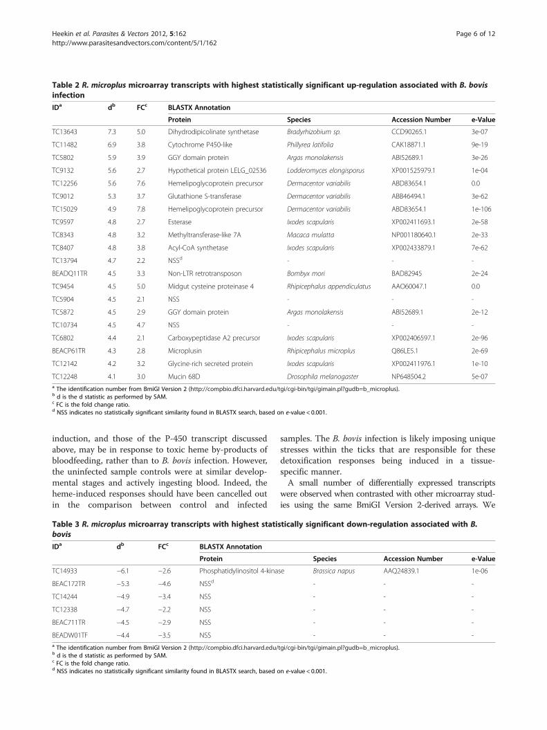
Table 2 R. microplus microarray transcripts with highest statistically significant up-regulation associated with B. bovisinfection
IDa db FCc BLASTX Annotation
Protein Species Accession Number e-Value
TC13643 7.3 5.0 Dihydrodipicolinate synthetase Bradyrhizobium sp. CCD90265.1 3e-07
TC11482 6.9 3.8 Cytochrome P450-like Phillyrea latifolia CAK18871.1 9e-19
TC5802 5.9 3.9 GGY domain protein Argas monolakensis ABI52689.1 3e-26
TC9132 5.6 2.7 Hypothetical protein LELG_02536 Lodderomyces elongisporus XP001525979.1 1e-04
TC12256 5.6 7.6 Hemelipoglycoprotein precursor Dermacentor variabilis ABD83654.1 0.0
TC9012 5.3 3.7 Glutathione S-transferase Dermacentor variabilis ABB46494.1 3e-62
TC15029 4.9 7.8 Hemelipoglycoprotein precursor Dermacentor variabilis ABD83654.1 1e-106
TC9597 4.8 2.7 Esterase Ixodes scapularis XP002411693.1 2e-58
TC8343 4.8 3.2 Methyltransferase-like 7A Macaca mulatta NP001180640.1 2e-33
TC8407 4.8 3.8 Acyl-CoA synthetase Ixodes scapularis XP002433879.1 7e-62
TC13794 4.7 2.2 NSSd - - -
BEADQ11TR 4.5 3.3 Non-LTR retrotransposon Bombyx mori BAD82945 2e-24
TC9454 4.5 5.0 Midgut cysteine proteinase 4 Rhipicephalus appendiculatus AAO60047.1 0.0
TC5904 4.5 2.1 NSS - - -
TC5872 4.5 2.9 GGY domain protein Argas monolakensis ABI52689.1 2e-12
TC10734 4.5 4.7 NSS - - -
TC6802 4.4 2.1 Carboxypeptidase A2 precursor Ixodes scapularis XP002406597.1 2e-96
BEACP61TR 4.3 2.8 Microplusin Rhipicephalus microplus Q86LE5.1 2e-69
TC12142 4.2 3.2 Glycine-rich secreted protein Ixodes scapularis XP002411976.1 1e-10
TC12248 4.1 3.0 Mucin 68D Drosophila melanogaster NP648504.2 5e-07a The identification number from BmiGI Version 2 (http://compbio.dfci.harvard.edu/tgi/cgi-bin/tgi/gimain.pl?gudb=b_microplus).b d is the d statistic as performed by SAM.c FC is the fold change ratio.d NSS indicates no statistically significant similarity found in BLASTX search, based on e-value < 0.001.
Heekin et al. Parasites & Vectors 2012, 5:162 Page 6 of 12http://www.parasitesandvectors.com/content/5/1/162
induction, and those of the P-450 transcript discussedabove, may be in response to toxic heme by-products ofbloodfeeding, rather than to B. bovis infection. However,the uninfected sample controls were at similar develop-mental stages and actively ingesting blood. Indeed, theheme-induced responses should have been cancelled outin the comparison between control and infected
Table 3 R. microplus microarray transcripts with highest statisbovis
IDa db FCc BLASTX Annotation
Protein
TC14933 −6.1 −2.6 Phosphatidylinositol 4-kina
BEAC172TR −5.3 −4.6 NSSd
TC14244 −4.9 −3.4 NSS
TC12338 −4.7 −2.2 NSS
BEAC711TR −4.5 −2.9 NSS
BEADW01TF −4.4 −3.5 NSSa The identification number from BmiGI Version 2 (http://compbio.dfci.harvard.edu/b d is the d statistic as performed by SAM.c FC is the fold change ratio.d NSS indicates no statistically significant similarity found in BLASTX search, based o
samples. The B. bovis infection is likely imposing uniquestresses within the ticks that are responsible for thesedetoxification responses being induced in a tissue-specific manner.A small number of differentially expressed transcripts
were observed when contrasted with other microarray stud-ies using the same BmiGI Version 2-derived arrays. We
tically significant down-regulation associated with B.
Species Accession Number e-Value
se Brassica napus AAQ24839.1 1e-06
- - -
- - -
- - -
- - -
- - -
tgi/cgi-bin/tgi/gimain.pl?gudb=b_microplus).
n e-value < 0.001.

Table 4 GO annotation summary by domain
Cellular component (C)
GO ID Description Occurrences
GO:0016020 Membrane 52
GO:0005623 Cell 15
GO:0005576 Extracellular region 15
GO:0005622 Intracellular region 132
Molecular function (F)
GO ID Description Occurrences
GO:0003774 Motor activity 0
GO:0016874 Ligase activity 3
GO:0016829 Lyase activity 4
GO:0004871 Signal transducer activity 6
GO:0016491 Oxiodoreductase activity 24
GO:0016853 Isomerase activity 1
GO:0030234 Enzyme regulator activity 8
GO:0003824 Catalytic activity 36
GO:0005488 Binding 113
GO:0016740 Transferase activity 22
GO:0005198 Structural molecule activity 18
GO:0005215 Transporter activity 12
Biological process (P)
GO ID Description Occurrences
GO:0006944 Cellular membrane fusion 3
GO:0050896 Response to stimulus 18
GO:0007610 Behavior 0
GO:0006810 Transport 26
GO:0030154 Cell differentiation 17
GO:0008152 Metabolic process 87
GO:0050789 Regulation of biological process 52
GO:0043062 Extracellular structure organization 0
GO:0007275 Multicellular organismal development 23
GO:0009987 Cellular process 40
GO:0007154 Cell communication 3
GO:0008219 Cell death 3
GO:0006139 Nucleobase, nucleoside, nucleotide and nucleic acid metabolic process 49
Heekin et al. Parasites & Vectors 2012, 5:162 Page 7 of 12http://www.parasitesandvectors.com/content/5/1/162
found 30 statistically significant differentially expressed tran-scripts in our larval microarray experiments. Mercado-Curiel and colleagues measured the temporal response ofgene expression in adult male R. microplus in response toAnaplasma marginale infection [21]. When they comparedinfected ticks with uninfected controls using microarrayassays, they determined that 888 genes were differentiallyexpressed in midgut tissue 2 days post-infection and 146genes were differentially expressed in the salivary glands 9days post-infection. In contrast, Ribeiro and colleaguesfound only 10 differentially expressed genes in the salivary
glands of Ixodes scapularis nymphs in response to Borreliaburgdorferi infection [22]. Rodriguez-Valle et al. found over300 differentially expressed BmiGI transcripts in their studyof R. microplus feeding upon Bos indicus and Bos taurus cat-tle [23]. Feeding upon tick resistant cattle as opposed to ticksusceptible cattle evidently creates greater perturbationscompared with feeding upon B. bovis-infected blood asopposed to uninfected cattle blood. Saldivar et al. discovered76, 32, 80, and 83 differentially expressed BmiGI Version 2transcripts in their microarray analysis of gene expressionchanges in response to larval exposure to the acaricides:

Heekin et al. Parasites & Vectors 2012, 5:162 Page 8 of 12http://www.parasitesandvectors.com/content/5/1/162
coumaphos, permethrin, ivermectin and amitraz, respect-ively [4].
Subtracted library results and discussionESTs from the B. bovis-infected larval subtracted librar-ies were assembled and annotated. Additional file 1 con-tains data pertaining to annotated unigenes. Of the 791total transcripts sequenced, 30 were classified as comingfrom B. bovis and removed from analysis. Three add-itional sequences, one from a mitochondrial gene, onerRNA sequence, and one putative transcript from BosTaurus were also removed. Of the remaining 758sequences, 193 had little sequence similarity to BmiGIVersion 2 transcripts and therefore appear to be novel.The Uniref100 database searches revealed 381 sequenceswith similarity to known proteins.
GO annotationsThe annot8r application provides a synopsis of the GOannotation process by categorizing the unigenes into 3domains consisting of 29 high-level GO terms (Table 4).In the cellular component domain (C), most of the dif-ferentially expressed transcripts were deemed intracellu-lar. The predominant annotation in the molecularfunction domain (F) was protein binding followed bycatalytic activity and oxidoreductase activity. Metabolicprocesses were the largest component of the biologicalprocess domain (P).We further utilized the GO annotations to find uni-
genes with possible roles in the infection process [24,25].Gene products identified via BLASTX searches with GOannotations of defense response (GO:000692) or stressresponse (GO:006950) are listed in Tables 5 and 6, re-spectively. Each unique annotation comprises the studysequence identifier, the source (database) of the annota-tion, a description of the annotation, the genus and spe-cies of the HSP, the NCBI accession number of theprotein, and the corresponding e-value for the HSP.Annotations for 6 unigenes from the assembled sub-
tracted library sequencing were related to defense re-sponse by similarity to proteins in the Uniprot databaseannotated by GO (Table 5). Among the 6 ESTs, 2 were
Table 5 Unigenes from subtracted library with BLAST databa
Unigene #a Database BLASTX Annotation
Protein Annotation
45 Uniref100 Salivary lipocalin
92 KEGG Toll-like receptor signaling pathway
113 Uniref100 Interferon gamma-inducible protein
402 Uniref100 Glycoprotein 3-alpha-l-fucosyltransferase A
468 Uniref100 Putative defense protein precursor
673 Uniref100 FYN binding proteina Unigene identification number as listed in Additional file 1.
homologous to proteins in Ixodus scapularis. Unigene 402showed significant similarity to 3-alpha-1-fucosyltransferase,which has been demonstrated to increase microbialpathogenesis in I. scapularis [26]. Unigene 468 was identi-fied as a putative defense protein precursor by similarityto a protein from Bombyx mori. Two defense relatedhydrolases were also identified. Valacyclovir hydrolase(unigene 79) is designated as “response to toxin” by theGO ontology (GO:0009636). In addition, Bleomycinhydrolase (unigene 637) inactivates bleomycin B2 (a cyto-toxic glycometallopeptide: GO:0009636). Unigene 113 dis-played similarity to an Interferon gamma-inducible proteinthat in humans has a role in antigen processing and epi-tope presentation [27], but in ticks may upregulate cathe-psins that activate serine proteases as an immuneresponse. A putative salivary lipocalin (unigene 45) sug-gests a role in the tick’s immune response since lipocalinsare involved in many inflammation and detoxification pro-cesses in mammals [28]. Unigene 673 is a putative FYNbinding protein; the GO Ontology annotates this proteinwith the term “defense response”. As determined byBLAST search similarity to the KEGG database, Unigene92 exhibited strong homology (e-value =5e-176) to an I.scapularis protein within the Toll-like receptor-signalingpathway—an important component of the host innate im-mune system.Annotations for 9 unigenes from the assembled sub-
tracted library sequencing subsumed the GO term “re-sponse to stress” (GO:0006950) by similarity to proteinsin the Uniprot database (Table 6). Moreover, 6 unigeneswere homologous to proteins in I. scapularis. Unigenes244 and 489 showed significant similarity to two I. sca-pularis cytochrome P450 proteins, which are potentdetoxifiers of xenobiotics. Other observed detoxificationproteins include: thioredoxin (unigene 416) and glutathi-one peroxidase (unigene 40), which facilitate the reduc-tion of proteins and lipids, respectively. Two putativeantioxidant peroxidase-related proteins (unigenes 89 and187) were also identified. Two putative stress response-related hydrolases were detected in the subtraction li-brary. Valacyclovir hydrolase (unigene 79) is annotatedwith the biological process “response to toxin”
se GO annotation of defense response (GO:0006952)
Species Accession Number e-Value
Amblyomma variegatum DAA34698.1 2e-07
Ixodes scapularis XP002404081.1 5e-176
Amblyomma americanum AAK82985.1 2e-92
Ixodes scapularis XP002413615.1 3e-70
Bombyx mori NP001091819.1 2e-20
Ixodes scapularis XP002412475.1 1e-22

Table 6 Unigenes from subtracted library with BLAST database GO annotation of stress response (GO:0006950)
BLASTX Annotation
Unigene #a Database Protein Annotation Species Accession Number e-Value
40 Uniref100 Glutathione Peroxidase Ixodes scapularis XP002399259.1 1e-41
79 Uniref100 Valacyclovir hydrolase Ixodes scapularis XP002404467.1 2e-56
89 GO Hydrogen peroxide catabolism Homo sapiens A1KZ92 1e-11
187 Enzyme class Peroxidase Caenorhabditis elegans Q1EN18 4e-10
215 Uniref100 Heat shock protein Locusta migratoria AAO21473.1 4e-10
244 Uniref100 Cytochrome P450 Ixodes scapularis B7PSW2 1e-81
416 Uniref100 Thioredoxin domain-containing protein Ixodes scapularis XP002436084.1 9e-28
489 Uniref100 Cytochrome P450 Ixodes scapularis XP002414034.1 2e-44
637 Uniref100 Bleomycin hydrolase Ixodes scapularis XP002399259.1 1e-78a Unigene identification number as listed in Additional file 1.
Heekin et al. Parasites & Vectors 2012, 5:162 Page 9 of 12http://www.parasitesandvectors.com/content/5/1/162
(GO:0009636). In addition, Bleomycin hydrolase (uni-gene 637) inactivates bleomycin B2 (a cytotoxic glyco-metallopeptide). Unigene 215 is homologous to aputative heat shock protein discovered in Locusta migra-toria. Heat shock protein expression levels are generallyupregulated when the organism is stressed.The InterProScan algorithms yielded an additional 190
unique sequence annotations, which brought the totalnumber of annotated ESTs to 571 out of 758 (75%). Nounigenes were annotated with the term “immune re-sponse” (GO:006950) by homology searches. However,Table 7 lists unigenes associated with immune functionidentified by InterProScan. Each unique annotationcomprises the study sequence identifier, the protein,source (method) of the annotation, and the InterProidentifier assigned to this protein. InterProScan anno-tated 11 unigenes with immune or defense response-related function including an additional lipocalin (uni-gene 443) that was not identified in the BLAST searches.Six of these unigenes were classified as immunoglobulin
Table 7 Unigenes from subtracted library with InterProScan a
InterProScan Annotation
Unigene #a Protein Annotation Met
298 Serine Protease Inhibitor HM
371 Serine Protease Inhibitor HM
520 Serine Protease Inhibitor HM
89 Immunoglobulin-like HM
187 Immunoglobulin-like HM
216 Immunoglobulin Sup
288 Immunoglobulin-like fold HM
520 Immunoglobulin-like HM
601 Immunoglobulin-like HM
242 Lectin/glucanase Gen
443 Lipocalin Supa Unigene identification number as listed in Additional file 1.
or immunoglobulin-like proteins. Since the tick has noadaptive immune system, we speculate that these pro-teins are either false positives or may serve a similar roleto proteins that have homologs in vertebrates. For ex-ample, the human Down Syndrome cell adhesion mol-ecule (DSCAM) has an immunoglobulin-like domainwith several known homologs in arthropods that mayhave thousands of splice variants and help drive theclearance of pathogens [29,30].The remaining three unigenes were characterized as
serine protease inhibitors (serpins). Recent interest intick serpins stems from their significant antimicrobialand antifungal activity [31-33]. Serpins are cysteine-richantimicrobial peptides and are components of the im-mune system of many invertebrates. Serpins have beenused as target antigens for recombinant tick vaccines[34]. Unigenes 298 and 520 had sequence similarity(HSPs) to proteins that belong to the Trypsin inhibitorlike cysteine rich domain (TIL) family. Huang et al.characterized a protein, C/E1 from Ascaris suum, with a
nnotation of immune response
hods Interpro ID
MPfam, superfamily IPR002919
MPfam, superfamily IPR002919
MPfam, superfamily IPR002919
MPfam, HMMSmart, Gene3D, ProfileScan IPR007110
MPfam, HMMSmart, Gene3D, ProfileScan IPR002919
erfamily
MPfam, HMMSmart, Gene3D, ProfileScan IPR013783
MPfam, HMMSmart, Gene3D, ProfileScan IPR002919
MPfam, HMMSmart, ProfileScan, Superfamily IPR007110
e3D, Superfamily IPR008985
erfamily IPR011038
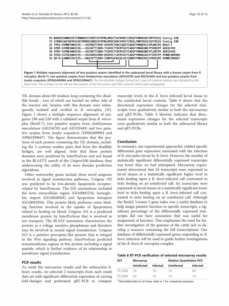
Figure 1 Multiple sequence alignment of two putative serpins identified in the subtracted larval library with a known serpin from R.microplus (BmSI-7), two putative serpins from Amblyomma maculatum (AEO34783 and AEO32449) and two putative serpins fromIxodes scapularis (XP002409984 and XP002399667). The five disulfide bridges formed by 5 pairs of cysteine residues are indicated by theblack lines. The numbers to the left are the position of the first amino acid (first column) within each polypeptide.
Table 8 RT-PCR verification of selected microarray results
EST Microarray Relative Quantitative PCR
Uninfected Infected Uninfected Infected
TC12256 1.0 7.6 1.0 8.4
TC14244 3.4 1.0 1.4 1.0a Normalized data to set lower value to 1 for comparison purposes.
Heekin et al. Parasites & Vectors 2012, 5:162 Page 10 of 12http://www.parasitesandvectors.com/content/5/1/162
TIL domain about 60 residues long containing five disul-fide bonds - two of which are located on either side ofthe reactive site. Serpins with this domain were subse-quently isolated and verified in R. microplus [35].Figure 1 shows a multiple sequence alignment of uni-genes 298 and 520 with a validated serpin from R. micro-plus (BmSI-7), two putative serpins from Amblyommamaculatum (AEO34783 and AEO32449) and two puta-tive serpins from Ixodes scapularis (XP002409984 andXP002399667). The figure demonstrates that the por-tions of each protein containing the TIL domain, includ-ing the 5 cysteine residue pairs that form the disulfidebridges, are well aligned. Note that these proteindomains were predicted by InterProScan and not foundin the BLASTX search of the Uniprot100 database, thusunderscoring the utility of de novo domain predictionalgorithms.Other noteworthy genes include three novel unigenes
involved in signal transduction pathways. Unigene 192was predicted to be low-density lipoprotein receptor-related by InterProscan. The GO annotations includedthe term extracellular region (GO:0005576), hemoglo-bin import (GO:0020028) and lipoprotein transport(GO:0042954). This protein likely performs some bind-ing function involved in the uptake of lipoproteinsrelated to feeding on blood. Unigene 331 is a predictedmembrane protein by InterProScan that is involved inion transport. The BLAST annotation characterizes theprotein as a voltage sensitive phosphatase and thereforemay be involved in neural signal transduction. Unigene412 is a putative porcupine-like protein that is integralto the Wnt signaling pathway. InterProScan predictedtransmembrane regions in this protein including a signalpeptide, which is further evidence of its relationship tomembrane signal transduction.
PCR resultsTo verify the microarray results and the subtraction li-brary results, we selected 2 transcripts from each resultdata set with significant differential expression of varyingfold-changes and performed qRT-PCR to compare
transcript levels in the B. bovis infected larval tissue tothe uninfected larval controls. Table 8 shows that thedirectional expression changes for the selected tran-scripts were qualitatively similar in both the microarraysand qRT-PCRs. Table 9 likewise indicates that direc-tional expression changes for the selected transcriptswere qualitatively similar in both the subtracted libraryand qRT-PCRs.
ConclusionIn summary, our experimental approaches yielded specificdifferential gene expression associated with the infectionof R. microplus larvae by B. bovis. However, the number ofstatistically significant differentially expressed transcriptswas lower than we had anticipated. Microarray experi-ments determined that 24 transcripts were expressed inlarval tissues at a statistically significant higher level inticks feeding upon a B. bovis-infected calf contrasted toticks feeding on an uninfected calf. Six transcripts wereexpressed in larval tissues at a statistically significant lowerlevel in ticks feeding upon a B. bovis-infected calf con-trasted to ticks feeding on an uninfected calf. Althoughthe BmiGI Version 2 gene index was a useful database tohelp assign putative function to specific transcripts, a sig-nificant percentage of the differentially expressed tran-scripts did not have annotation that was useful forassignment of function. This emphasizes the need for fur-ther investigation of the genome of the cattle tick to de-velop a resource containing the full transcriptome. Ourdatabase of differentially expressed genes responding to B.bovis infection will be used to guide further investigationsof the B. bovis-R. microplus complex.
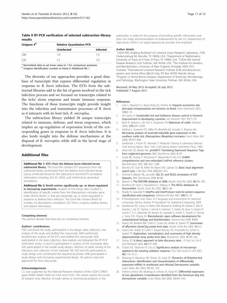
Table 9 RT-PCR verification of selected subtraction libraryresults
Unigene #b Relative Quantitative PCR
Uninfected Infected
298 1.0 1.9
520 2.1 1.0a Normalized data to set lower value to 1 for comparison purposes.b Unigene identification number as listed in Additional file 1.
Heekin et al. Parasites & Vectors 2012, 5:162 Page 11 of 12http://www.parasitesandvectors.com/content/5/1/162
The diversity of our approaches provides a good data-base of transcripts that express differential regulation inresponse to B. bovis infection. The ESTs from the sub-tracted libraries add to the list of genes involved in the tickinfection process and we focused on transcripts related tothe ticks’ stress response and innate immune response.The functions of these transcripts might provide insightinto the infection and transmission processes of B. bovisas it interacts with its host tick, R. microplus.The subtraction library yielded 28 unique transcripts
related to immune, defense, and stress responses, whichimplies an up-regulation of expression levels of the cor-responding genes in response to B. bovis infection. It isalso lends insight into the defense mechanisms at thedisposal of R. microplus while still in the larval stage ofdevelopment.
Additional files
Additional file 1: ESTs from the Babesia bovis-infected larvaesubtracted library. This Excel file contains EST sequences from thesubtracted library synthesized from the Babesia bovis-infected larvae(using uninfected larvae for the subtraction) and BLASTX annotationinformation including: GO, EC, KEGG, and InterProScan generatedannotations.
Additional file 2: BmiGI entries significantly up- or down-regulatedin microarray experiments. Analysis of microarray data resulted inidentification of specific entries from the Rhipicephalus microplus geneindex, BmiGI Version 2, that are statistically up- or down-regulated inresponse to Babesia bovis infection. This Excel file contains BmiGI IDnumber, hit descriptions, annotation, GO Terms, e-values, reading frames,and related information.
Competing interestsThe authors declare that there are no competing interests.
Authors’ contributionsFDG conceived the study, participated in the design, data collection, andanalysis of the study and drafted the manuscript. AMH performedbioinformatic analysis of the ESTs and drafted the manuscript. KGBparticipated in the data collection, data analysis, and designed the RT-PCRverification study. LS and CG participated in analysis of the microarray data;GAS participated in the overall study design, infection of cattle, timing of tickinfections and collection of tick materials. VN, SED and AD participated instudy design and coordinated the sequencing phases. KAB participated instudy design and microarray experimental design. All authors read andapproved the final manuscript.
AcknowledgementsL.S. was supported by the National Research Initiative of the USDA CSREESgrant #2005-35604-15440 (to K.A.B. and F.D.G.). This article reports the resultsof research only. Mention of trade names or commercial products in this
publication is solely for the purpose of providing specific information anddoes not imply recommendation of endorsement by the U.S. Department ofAgriculture. USDA is an equal opportunity provider and employer.
Author details1USDA-ARS, Knipling Bushland US Livestock Insect Research Laboratory, 2700Fredericksburg Rd, Kerrville, TX 78028, USA. 2Department of Mathematics,University of Texas at El Paso, El Paso, TX 79968, USA. 3USDA-ARS AnimalDisease Research Unit, Pullman, WA 99164, USA. 4The Institute for Geneticsand Bioinformatics, University of New England, Armidale, NSW 2351,Australia. 5International Livestock Research Institute (ILRI) and Bioscienceseastern and central Africa (BecA) Hub, PO Box 30709, Nairobi, Kenya.6Program in Vector-Borne Diseases, Department of Veterinary Microbiologyand Pathology, Washington State University, Pullman, WA 99164, USA.
Received: 24 May 2012 Accepted: 26 July 2012Published: 7 August 2012
References1. Grisi L, Massard CL, Moya Borja GE, Pereira JB: Impacto economico das
principais ectoparasitoses em bovinos no Brasil. Hora Veterinaria 2002,125:8–10.
2. de Castro JJ: Sustainable tick and tickborne disease control in livestockimprovement in developing countries. Vet Parasitol 1997, 71:77–97.
3. Bock R, Jackson L, de Vos A, Jorgensen W: Babesiosis of cattle. Parasitol2004, 129:S247–S269.
4. Saldivar L, Guerrero FD, Miller RJ, Bendele KG, Gondro C, Brayton KA:Microarray analysis of acaricide-inducible gene expression in thesouthern cattle tick, Rhipicephalus (Boophilus) microplus. Insect Molec Biol2008, 17:597–606.
5. Sambrook J, Fritsch EF, Maniatis T: Molecular Cloning A Laboratory Manual.Cold Spring Harbor, New York: Cold Spring Harbor Laboratory Press; 1989.
6. Wasmuth JD, Blaxter ML: prot4EST: Translating Expressed Sequence Tagsfrom neglected genomes. BMC Bioinforma 2004, 5:187.
7. Suzek BE, Huang H, McGarvey P, Mazumder R, Wu CH: UniRef:comprehensive and non-redundant UniProt reference clusters.Bioinformatics 2007, 23:1282–1288.
8. Altschul SF, Gish W, Miller W, Myers EW, Lipman DJ: Basic local alignmentsearch tool. J Mol Biol 1990, 215:403–410.
9. Schmid R, Blaxter ML, annot8r: GO, EC and KEGG annotation of ESTdatasets. BMC Bioinforma 2008, 9:180.
10. Bairoch A: The ENZYME database in 2000. Nucleic Acids Res 2000, 28:304–305.11. Kanehisa M, Goto S, Kawashima S, Nakaya A: The KEGG databases at
GenomeNet. Nucleic Acids Res 2002, 30:42–46.12. Mulder N, Apweiler R: InterPro and InterProScan: tools for protein sequence
classification and comparison. Methods Mol Biol 2007, 396:59–70.13. R Development Core Team. R: A language and environment for statistical
computing. Vienna, Austria: R Foundation for Statistical Computing; 2009.14. Gentleman RC, Carey VJ, Bates DM, Bolstad B, Dettling M, Dudoit S, Ellis B,
Gautier L, Ge YC, Gentry J, Hornik K, Hothorn T, Huber W, Iacus S, Irizarry R,Leisch F, Li C, Maechler M, Rossini AJ, Sawitzki G, Smith C, Smyth G, TierneyL, Yang JYH, Zhang JH: Bioconductor: open software development forcomputational biology and bioinformatics. Genome Biol 2004, 5:R80.
15. Irizarry RA, Bolstad BM, Collin F, Cope LM, Hobbs B, Speed TP: Summariesof affymetrix GeneChip probe level data. Nucleic Acids Res 2003, 31:e15.
16. Irizarry RA, Hobbs B, Collin F, Beazer-Barclay YD, Antonellis KJ, Scherf U,Speed TP: Exploration, normalization, and summaries of high densityoligonucleotide array probe level data. Biostatistics 2003, 4:249–264.
17. Storey JD: A direct approach to false discovery rates. J R Stat Soc Ser BStat Methodol 2002, 64:479–498.
18. Tusher VG, Tibshirani R, Chu G: Significance analysis of microarraysapplied to the ionizing radiation response. Proc Natl Acad Sci USA 2001,98:5116–5121.
19. Mulenga A, Macaluso KR, Simser JA, Azad AF: Dynamics of Rickettsia-tickinteractions: identification and characterization of differentiallyexpressed mRNAs in uninfected and infected Dermacentor variabilis.Insect Molec Biol 2003, 12:185–193.
20. Dreher-Lesnick SM, Mulenga A, Simser JA, Azad AF: Differential expressionof two glutathione S-transferases identified from the American dog tick,Dermacentor variabilis. Insect Molec Biol 2006, 15:445–453.

Heekin et al. Parasites & Vectors 2012, 5:162 Page 12 of 12http://www.parasitesandvectors.com/content/5/1/162
21. Mercado-Curiel RF, Palmer GH, Guerrero FD, Brayton KA: Temporalcharacterization of the organ-specific Rhipicephalus microplustranscriptional response to Anaplasma marginale infection. Int J Parasitol2011, 41:851–860.
22. Ribeiro JMC, Alarcon-Chaidez F, Francischetti IMB, Mans BJ, Mather TN,Valenzuela JG, Wikel SK: An annotated catalog of salivary glandtranscripts from Ixodes scapularis ticks. Insect Biochem Molec Biol 2006,36:111–129.
23. Rodriguez-Valle M, Lew-Tabor A, Gondro C, Moolhuijzen P, Vance M,Guerrero FD, Bellgard M, Jorgensen W: Comparative microarray analysis ofRhipicephalus (Boophilus) microplus expression profiles of larvae pre-attachment and feeding adult female stages on Bos indicus and Bostaurus cattle. BMC Genomics 2010, 11:437.
24. Ashburner M, Ball CA, Blake JA, Botstein D, Butler H, Cherry JM, Davis AP,Dolinski K, Eppig JT, Harris MA, Hill DP, Issel-Traver L, Kasarskis A, Lewis S,Matese JC, Richardson JE, Ringwald M, Rubin GM, Sherlock G: Geneontology: tool for the unification of biology. The Gene Ontologyconsortium. Nat Genet 2000, 25:25–29.
25. Camon E, Magrane M, Barrell D, Lee V, Dimmer E, Maslen J, Binns D, HarteN, Lopez R, Apweiler R: The Gene Ontology Annotation (GOA) Database:sharing knowledge in Uniprot with Gene Ontology. Nucleic Acids Res2004, 32:D262–D266.
26. Pedra JHF, Narasimhan S, Rendic D, DePonte K, Bell-Sakyi L, Wilson IBH,Fikrig E: Fucosylation enhances colonization of ticks by Anaplasmaphagocytophilum. Cellular Microbioology 2010, 12:1222–1234.
27. Goldstein OG, Hajiaghamohseni LM, Amria S, Sundaram K, Reddy SV, HaqueA: Gamma-IFN-inducible-lysosomal thiol reductase modulates acidicproteases and HLS class II antigen processing in melanoma. CancerImmunol Immunother 2008, 57:1461–1470.
28. Flower DR, Attwood TK, North AC: The lipocalin protein family: structureand function. Biochem J 1996, 318:1–14.
29. Dong YM, Taylor HE, Dimopoulos G: AgDscam, a hypervariableimmunoglobulin domain-containing receptor of the Anopheles gambiaeinnate immune system. Plos Biology 2006, 4:1137–1146.
30. Watson FL, Puttmann-Holgado R, Thomas F, Lamar DL, Hughes M, KondoM, Rebel VI, Schmucker D: Extensive diversity of Ig-superfamily proteins inthe immune system of insects. Science 2005, 309:1874–1878.
31. Fogaça AC, Lorenzini DM, Kaku LM, Esteves E, Bulet P, Daffre S: Cysteine-rich antimicrobial peptides of the cattle tick Boophilus microplus:isolation, structural characterization and tissue expression profile.Develop Comp Immunol 2004, 28:191–200.
32. Gronenborn AM, Nilges M, Peanasky RJ, Clore GM: Sequential resonanceassignment and secondary structure determination of the Ascaris trypsininhibitor, a member of a novel class of proteinase inhibitors. Biochemistry1990, 29:183–189.
33. Silva FD, Rezende CA, Rossi DC, Esteves E, Dyszy FH, Schreier S, Gueiros-Filho F,Campos CB, Pires JR, Daffre S: Structure and mode of action of microplusin,a copper II-chelating antimicrobial peptide from the cattle tickRhipicephalus (Boophilus) microplus. J Biol Chem 2009, 284:34735–34746.
34. Sugino M, Imamura S, Mulenga A, Nakajima M, Tsuda A, Ohashi K: A serineproteinase inhibitor (serpin) from ixodid tick Haemaphysalis longicornis;cloning and preliminary assessment of its suitability as a candidate for atick vaccine. Vaccine 2003, 21:2844–2851.
35. Sasaki SD, de Lima CA, Lovato DV, Juliano MA, Torquato JS, Tanaka AS:BmSI-7, a novel subtilisin inhibitor from Boophilus microplus, with activitytoward Pr1 proteases from the fungus Metarhizium anisopliae. ExpParasitol 2008, 118:214–220.
doi:10.1186/1756-3305-5-162Cite this article as: Heekin et al.: Analysis of Babesia bovis infection-induced gene expression changes in larvae from the cattle tick,Rhipicephalus (Boophilus) microplus. Parasites & Vectors 2012 5:162.
Submit your next manuscript to BioMed Centraland take full advantage of:
• Convenient online submission
• Thorough peer review
• No space constraints or color figure charges
• Immediate publication on acceptance
• Inclusion in PubMed, CAS, Scopus and Google Scholar
• Research which is freely available for redistribution
Submit your manuscript at www.biomedcentral.com/submit