Embed Size (px)
DESCRIPTION
Staphylococcus aureus (S. aureus) is the key organism for food poisoning due to massive production of heat stable exotoxins. The current study was attempted to investigate the effect of Mr. Trivedi’s biofield treatment on S aureus. S
Citation preview

American Journal of BioScience 2015; 3(6): 212-220
Published online October 15, 2015 (http://www.sciencepublishinggroup.com/j/ajbio)
doi: 10.11648/j.ajbio.20150306.13
ISSN: 2330-0159 (Print); ISSN: 2330-0167 (Online)
Antibiogram, Biochemical Reactions and Genotyping Characterization of Biofield Treated Staphylococcus aureus
Mahendra Kumar Trivedi1, Alice Branton
1, Dahryn Trivedi
1, Gopal Nayak
1,
Sambhu Charan Mondal2, Snehasis Jana
2, *
1Trivedi Global Inc., Henderson, USA 2Trivedi Science Research Laboratory Pvt. Ltd., Hall-A, Chinar Mega Mall, Chinar Fortune City, Hoshangabad Rd., Bhopal, Madhya
Pradesh, India
Email address: [email protected] (S. Jana)
To cite this article: Mahendra Kumar Trivedi, Alice Branton, Dahryn Trivedi, Gopal Nayak, Sambhu Charan Mondal, Snehasis Jana. Antibiogram, Biochemical
Reactions and Genotyping Characterization of Biofield Treated Staphylococcus aureus. American Journal of BioScience.
Vol. 3, No. 6, 2015, pp. 212-220. doi: 10.11648/j.ajbio.20150306.13
Abstract: Staphylococcus aureus (S. aureus) is the key organism for food poisoning due to massive production of heat stable
exotoxins. The current study was attempted to investigate the effect of Mr. Trivedi’s biofield treatment on S. aureus. S. aureus
(ATCC 25923) was divided into two parts, Group (Gr.) I: control and Gr. II: treatment. After biofield treatment, Gr. II was
further subdivided into two parts, Gr. IIA and Gr. IIB. Gr. IIA was analyzed on day 10, while Gr. IIB was stored and analyzed
on day 159 after revival (Study I). The revived sample (Gr. IIB) were retreated on day 159 (Study II), and divided into three
separate tubes. Tube 1 was analyzed on day 5, likewise, tube 2 and 3 were analyzed on day 10 and 15, respectively. All the
experimental parameters were studied using automated MicroScan Walk-Away® system. The 16S rDNA sequencing was
carried out in Gr. IIA sample to correlate the phylogenetic relationship of S. aureus with other bacterial species. The
antimicrobial susceptibility and minimum inhibitory concentration showed significant alteration i.e. 92.86% and 90.00%
respectively in treated cells of S. aureus as compared to control. The biochemical reactions also showed the significant
(35.71%) alteration in treated sample with respect to control. The biotype number and microbial species were substantially
changed in Gr. IIA (767177; Staphylococcus cohnii subsp. urealyticum) on day 10, while only the biotype numbers were
changed in rest of the treated samples as compared to control (307016; S. aureus). The 16S rDNA analysis showed that the
identified strain in this experiment was S. aureus (GenBank Accession No.: L37597) after biofield treatment. However, the
nearest homolog genus-species was found as Staphylococcus simiae (GenBank Accession No.: DQ127902). These results
suggested that biofield treatment has a significant impact on S. aureus in lyophilized as well as revived state.
Keywords: Staphylococci, Staphylococcus aureus, Antimicrobial Sensitivity, Biofield Treatment, Biochemical Reaction,
Biotype, 16S rDNA, Gram-Positive Bacteria
1. Introduction
Staphylococci are the important class of pyogenic Gram-
positive spherical bacteria resembling to the grapes like
structure. They are considered as the third most important
cause of food-borne disorders in the world [1]. It is the
main pathogen for mastitis in the milch animals [2]. It is
estimated that in US alone food-borne illnesses affect 6 to
80 million people each year, causing up to 9000 deaths [3].
Based on literature various genes have been found as a
target for identification of S. aureus with the help of 16S
rDNA sequence viz. heat shock protein 60 (hsp60) [4],
superoxide dismutase A (sodA) [5], and RNA polymerase B
(rpoB) [6]. S. aureus has developed resistance to the most
classes of the antimicrobial agents. Penicillin is the drug of
choice to treat against Staphylococcus infection but due to
penicillinase or β-lactamase enzyme that destroy the
penicillin, leads to resistance against S. aureus [7].
Therefore, some alternative strategies are needed to treat
against staphylococci infections.
National Institute of Health/National Center for
Complementary and Alternative Medicine (NIH/NCCAM)
have reported that biofield (putative energy fields) or
electromagnetic based energy therapies used to promote
American Journal of BioScience 2015; 3(6): 212-220 213
health and healing [8]. Biofield energy treatment has been
known as an alternative approach that may be useful to alter
the sensitivity pattern of the antimicrobials. Harold Saxton
Burr had performed the detailed studies on the correlation
of electric current with physiological processes and
suggested that every single process in the human body had
an electrical significance [9]. The electrical process that
happening in the human body have strong relationship with
magnetic field as required by Ampere’s law, which stated
that the moving charge produces magnetic field in
surrounding space [10, 11]. Thus, the human body emits the
electromagnetic waves in the form of bio-photons, which
surrounds the body and it is commonly known as biofield.
Therefore, the biofield consists of an electromagnetic field,
being generated by moving electrically charged particles
(ions, cell, molecule, etc.) inside the human body. Prakash
et al. 2015, reported that the various scientific instruments
such as Kirlian photography, polycontrast interference
photography and resonance field imaging can be
extensively used to measure the biofield of human body
[12]. Thus, a human has the ability to harness the energy
from environment or universe and can transmit into any
living or nonliving object(s) around the Globe. The objects
always receive the energy and respond into useful way that
is called biofield energy and the process is known as
biofield treatment. Mr. Trivedi’s biofield treatment (The
Trivedi Effect®
) has been known to alter the structural,
physical and thermal properties of several metals in
materials science [13-15], improved the overall productivity
of crops [16, 17], altered characteristics features of
microbes [18-20] and improved growth and anatomical
characteristics of various medicinal plants [21, 22]. Due to
the clinical significance of this organism and literature
reports on biofield treatment, the present work was
undertaken to evaluate the impact of biofield treatment
modality on S. aureus in relation to the antimicrobials
susceptibility, biochemical reactions, biotyping and 16S
rDNA sequencing.
2. Materials and Methods
S. aureus, American Type Culture Collection (ATCC
25923) strain was procured from MicroBioLogics, Inc.,
USA and stored with proper storage conditions until further
use. All the tested antimicrobials and biochemicals were
procured from Sigma-Aldrich (MA, USA). The
antimicrobial susceptibility, biochemical reactions and
biotype number were estimated with the help of MicroScan
Walk-Away®
(Dade Behring Inc., West Sacramento, CA,
USA) using Positive Breakpoint Combo 20 (PBPC 20)
panel. The 16S rDNA sequencing analysis was carried out
using ultrapure genomic DNA prep kit; Cat KT 83
(Bangalore Genei, India).
2.1. Experimental Design
The impact of biofield treatment on tested bacterium S.
aureus was evaluated in two groups-
Group I: ATCC strain in lyophilized state was considered
as control. No treatment was given and analyzed for
antimicrobial sensitivity, biochemical reactions and biotype
number as per the standard protocol.
Group II: The lyophilized state of ATCC strain was
divided into two parts named as Gr. IIA and Gr. IIB. Both the
groups of ATCC strain of S. aureus in lyophilized state were
assigned to the Mr. Trivedi’s unique biofield treatment (first
treatment). Gr. IIA was analyzed on day 10 while Gr. IIB
sample was stored in lyophilized state for 159 days at -70ºC.
Gr. IIB was further sub-divided in two separate parts named
as Gr. IIB - Study I and Gr. IIB - Study II.
Group IIB - Study I
After 159 days, antimicrobial sensitivity, MIC,
biochemical reactions and biotyping were performed as per
the standard protocol.
Group IIB - Study II
The stored strain was revived from -70ºC and the revived
culture was again provided to Mr. Trivedi’s biofield
treatment (re-treatment) on day 159. After biofield
retreatment, the sample was sub-cultured into three separate
tubes and analyzed on Day 5, Day 10 and Day 15 of its sub-
culturing.
2.2. Biofield Treatment Strategy
The lyophilized sample of S. aureus was subjected to Mr.
Trivedi’s biofield treatment (first treatment) and then
stored, analyzed on day 10 (Gr. IIA) followed by
retreatment on 159 days in revived state (Gr. IIB, Study II).
In details, the treatment groups in sealed pack were handed
over to Mr. Trivedi for biofield treatment under laboratory
conditions. Mr. Trivedi provided the treatment through his
energy transmission process to the treated group without
touching the samples. After first treatment, the analysis of
Gr. IIA lyophilized sample was done on day 10 for
antimicrobial sensitivity along with minimum inhibitory
concentration (MIC), biochemical reactions with biotype
number and 16S rDNA analysis as per the standard
protocol. While handing over these cultures to Mr. Trivedi
for retreatment purposes, optimum precautions were taken
to avoid contamination.
2.3. Antimicrobial Susceptibility Test
Investigation of antimicrobial susceptibility of S. aureus
was carried out with the help of automated instrument,
MicroScan Walk-Away®
using PBPC 20 panel. The panel
can be stored at 2 to 25ºC for analysis. The panel was
allowed to equilibrate to room temperature prior to
rehydration. All opened panels were used on the same day.
The tests carried out on MicroScan were miniaturized of the
broth dilution susceptibility test that has been dehydrated.
Briefly, 0.1 mL of the standardized suspension of S. aureus
was pipetted into 25 mL of inoculum water using pluronic
and inverted 8 to 10 times and inoculated, rehydrated, and
then subjected to incubation for 16 hours at 35°C.
Rehydration and inoculation were performed using the

214 Mahendra Kumar Trivedi et al.: Antibiogram, Biochemical Reactions and Genotyping
Characterization of Biofield Treated Staphylococcus aureus
RENOK®
system with inoculators-D (B1013-4). 25 mL of
standardized inoculum suspension was poured into
inoculum tray. The detailed experimental procedure and
conditions were followed as per the manufacturer's
instructions. The antimicrobial susceptibility pattern (S:
Susceptible, R: Resistant; and BLAC: β-lactamase positive)
and MIC were determined by observing the lowest
antimicrobial concentration showing inhibition of growth
[23].
2.4. Biochemical Reaction Studies
Biochemical reactions of S. aureus were determined
using MicroScan Walk-Away®
, system with PBPC 20 panel.
Preparation of PBPC 20 panel, inoculum followed by
dehydration and rehydration were performed in a similar
way as mentioned in antimicrobial susceptibility assay for
analysis of biochemical reactions followed by biotype
number. The detailed experimental procedures and
conditions were followed as per the manufacturer's
instructions [23].
2.5. Identification of Organism by Biotype Number
The biotype number of S. aureus was determined on
MicroScan Walk-Away® processed panel data report with the
help of biochemical reactions data [23, 24].
2.6. Amplification and Gene Sequencing of 16S rDNA
Genomic DNA was isolated from S. aureus cells (Gr. IIA,
sample coded as 9A) using genomic purification kit, according
to the manufacturer instructions. 16S rDNA gene (~1.5 kb)
fragment was amplified with the help of high-fidelity
polymerase chain reaction (PCR) using universal primers;
forward primer (5ˊ-AGAGTTTGATCCTGGCTCAG-3ˊ) and
reverse primer (3ˊ-ACGGTCATACCTTGTTACGACTT-5ˊ).
Amplified products were subjected to gel electrophoresis in
1.0% agarose gel, stained with ethidium bromide and
visualized under UV light in a gel documentation unit (BioRad
Laboratories, USA). The PCR amplified fragment was purified
from the agarose gel using a DNA gel extraction kit.
Sequencing of amplified product was done on a commercial
basis from Bangalore Genei, India. The 16S rDNA sequences
obtained were aligned and compared with the sequences stored
in GenBank database available from National Center for
Biotechnology Information (NCBI) using the algorithm
BLASTn program. Multiple sequence alignment /
phylogenetic tree were established using MEGA3.1 molecular
software [25].
3. Results and Discussion
3.1. Antimicrobial Susceptibility Test
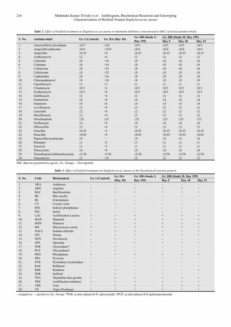
The results of S. aureus susceptibility pattern and MIC
values of tested antimicrobials after biofield treatment are
summarized in Table 1 and 2 respectively. The data were
analyzed and compared with respect to control.
Antimicrobial sensitivity assay and MIC were performed in
twenty-eight and thirty antimicrobials respectively. The
treated cells of S. aureus showed a significant (85.71%)
alteration (twenty-four out of twenty-eight) in antimicrobial
sensitivity pattern from susceptible (S) to resistance (R) in
lyophilized treated Gr. IIA on day 10 after first-time
biofield treatment as compared with control. However,
these twenty-four antimicrobials did not show any change
of sensitivity pattern on day 159 as well as in revived state
even after retreatment as compared to control. Out of
twenty-eight antimicrobials two antibiotics i.e. ampicillin
and penicillin were changed from S to β-lactamase positive
(BLAC) in lyophilized treated Gr. IIA on day 10 while
showed similar response in rest of treated groups even after
second-time biofield treatment as compared with control. S.
aureus has the ability to produce β-lactamases or
penicillinase enzyme which breakdown the β-lactam ring
present in penems and cephems heteronucleus [26]. Two,
out of twenty eight (7.14%) tested antimicrobials such as
piperacillin/tazobactam and linezolid did not show any
responses in lyophilized treated cells (Gr. IIA) on day 10
while they exhibited susceptible in rest of treated samples
of S. aureus. Overall, 92.86% antimicrobial susceptibility
pattern was altered after biofield treatment as compared to
control. MIC values of several antimicrobials viz.
ampicillin/sulbactam, azithromycin, cefazolin, cefepime,
cephalothin, chloramphenicol, ciprofloxacin, gatifloxacin,
gentamicin, levofloxacin, linezolid, moxifloxacin,
norfloxacin, ofloxacillin, rifampin, tetracycline, and
synercid showed an alteration about two-fold in Gr. IIA on
day 10 as compared to control. The MIC value of
cefotaxime, ceftriaxone and clindamycin were changed
about four-fold in Gr. IIA on day 10 while remained
unchanged in rest of the groups as compared to control.
Certain antimicrobials such as erythromycin, oxacillin and
vancomycin showed eight-fold, while ampicillin showed
thirty two-fold (≤0.25 to >8 µg/mL) and penicillin showed
around two hundred sixty seven-fold (≤0.03 to >8 µg/mL)
alteration of MIC values in Gr. IIA on day 10 as compared to
control. Amoxicillin / k-clavulanate and trimethoprim /
sulfamethoxazole were slightly altered the MIC values in Gr.
IIA on day 10. Overall, 90% out of thirty tested
antimicrobials showed an alteration of MIC values as
compared to control. Three out of thirty (10%) antimicrobials
did not show any alteration of MIC values in all the treated
groups as compared to control (Table 2) except piperacillin /
tazobactam in Gr. IIA, value not reported. Overall, the
antimicrobial resistance pattern (S to R) and corresponding
MIC values were significantly altered in lyophilized strain S.
aureus after first-time biofield treatment as compared to
control.
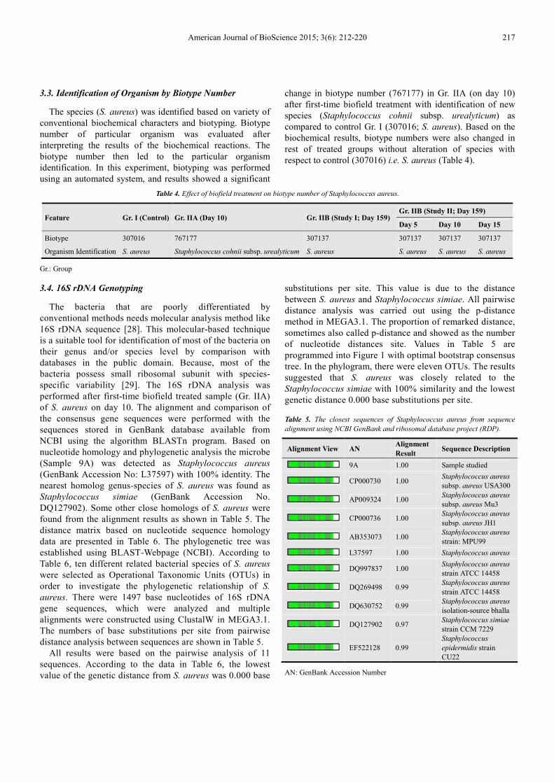
3.2. Biochemical Reactions Studies
Data obtained from biochemical reactions studies for
distinction of S. aureus are illustrated in Table 3. Study of
biochemical reactions can be utilized to identify the
enzymatic and metabolic characteristic feature of microbes.
American Journal of BioScience 2015; 3(6): 212-220 215
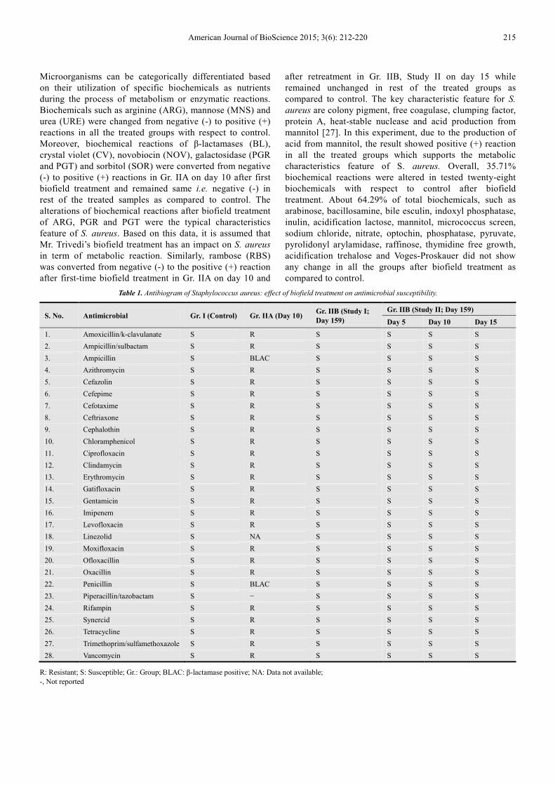
Microorganisms can be categorically differentiated based
on their utilization of specific biochemicals as nutrients
during the process of metabolism or enzymatic reactions.
Biochemicals such as arginine (ARG), mannose (MNS) and
urea (URE) were changed from negative (-) to positive (+)
reactions in all the treated groups with respect to control.
Moreover, biochemical reactions of β-lactamases (BL),
crystal violet (CV), novobiocin (NOV), galactosidase (PGR
and PGT) and sorbitol (SOR) were converted from negative
(-) to positive (+) reactions in Gr. IIA on day 10 after first
biofield treatment and remained same i.e. negative (-) in
rest of the treated samples as compared to control. The
alterations of biochemical reactions after biofield treatment
of ARG, PGR and PGT were the typical characteristics
feature of S. aureus. Based on this data, it is assumed that
Mr. Trivedi’s biofield treatment has an impact on S. aureus
in term of metabolic reaction. Similarly, rambose (RBS)
was converted from negative (-) to the positive (+) reaction
after first-time biofield treatment in Gr. IIA on day 10 and
after retreatment in Gr. IIB, Study II on day 15 while
remained unchanged in rest of the treated groups as
compared to control. The key characteristic feature for S.
aureus are colony pigment, free coagulase, clumping factor,
protein A, heat-stable nuclease and acid production from
mannitol [27]. In this experiment, due to the production of
acid from mannitol, the result showed positive (+) reaction
in all the treated groups which supports the metabolic
characteristics feature of S. aureus. Overall, 35.71%
biochemical reactions were altered in tested twenty-eight
biochemicals with respect to control after biofield
treatment. About 64.29% of total biochemicals, such as
arabinose, bacillosamine, bile esculin, indoxyl phosphatase,
inulin, acidification lactose, mannitol, micrococcus screen,
sodium chloride, nitrate, optochin, phosphatase, pyruvate,
pyrolidonyl arylamidase, raffinose, thymidine free growth,
acidification trehalose and Voges-Proskauer did not show
any change in all the groups after biofield treatment as
compared to control.
Table 1. Antibiogram of Staphylococcus aureus: effect of biofield treatment on antimicrobial susceptibility.
S. No. Antimicrobial Gr. I (Control) Gr. IIA (Day 10) Gr. IIB (Study I;
Day 159)
Gr. IIB (Study II; Day 159)
Day 5 Day 10 Day 15
1. Amoxicillin/k-clavulanate S R S S S S
2. Ampicillin/sulbactam S R S S S S
3. Ampicillin S BLAC S S S S
4. Azithromycin S R S S S S
5. Cefazolin S R S S S S
6. Cefepime S R S S S S
7. Cefotaxime S R S S S S
8. Ceftriaxone S R S S S S
9. Cephalothin S R S S S S
10. Chloramphenicol S R S S S S
11. Ciprofloxacin S R S S S S
12. Clindamycin S R S S S S
13. Erythromycin S R S S S S
14. Gatifloxacin S R S S S S
15. Gentamicin S R S S S S
16. Imipenem S R S S S S
17. Levofloxacin S R S S S S
18. Linezolid S NA S S S S
19. Moxifloxacin S R S S S S
20. Ofloxacillin S R S S S S
21. Oxacillin S R S S S S
22. Penicillin S BLAC S S S S
23. Piperacillin/tazobactam S − S S S S
24. Rifampin S R S S S S
25. Synercid S R S S S S
26. Tetracycline S R S S S S
27. Trimethoprim/sulfamethoxazole S R S S S S
28. Vancomycin S R S S S S
R: Resistant; S: Susceptible; Gr.: Group; BLAC: β-lactamase positive; NA: Data not available;
-, Not reported
216 Mahendra Kumar Trivedi et al.: Antibiogram, Biochemical Reactions and Genotyping
Characterization of Biofield Treated Staphylococcus aureus
Table 2. Effect of biofield treatment on Staphylococcus aureus to minimum inhibitory concentration (MIC) of tested antimicrobials.
S. No. Antimicrobial Gr. I (Control) Gr. IIA (Day 10) Gr. IIB (Study I;
Day 159)
Gr. IIB (Study II; Day 159)
Day 5 Day 10 Day 15
1. Amoxicillin/k-clavulanate ≤4/2 ˃4/2 ≤4/2 ≤4/2 ≤4/2 ≤4/2
2. Ampicillin/sulbactam ≤8/4 ˃16/8 ≤8/4 ≤8/4 ≤8/4 ≤8/4
3. Ampicillin ≤0.25 ˃8 ≤0.25 ≤0.25 ≤0.25 ≤0.25
4. Azithromycin ≤2 ˃4 ≤2 ≤2 ≤2 ≤2
5. Cefazolin ≤8 ˃16 ≤8 ≤8 ≤8 ≤8
6. Cefepime ≤8 ˃16 ≤8 ≤8 ≤8 ≤8
7. Cefotaxime ≤8 ˃32 ≤8 ≤8 ≤8 ≤8
8. Ceftriaxone ≤8 ˃32 ≤8 ≤8 ≤8 ≤8
9. Cephalothin ≤8 ˃16 ≤8 ≤8 ≤8 ≤8
10. Chloramphenicol ≤8 ˃16 ≤8 ≤8 ≤8 ≤8
11. Ciprofloxacin ≤1 ˃2 ≤1 ≤1 ≤1 ≤1
12. Clindamycin ≤0.5 ˃2 ≤0.5 ≤0.5 ≤0.5 ≤0.5
13. Erythromycin ≤0.5 ˃4 ≤0.5 ≤0.5 ≤0.5 ≤0.5
14. Gatifloxacin ≤2 ˃4 ≤2 ≤2 ≤2 ≤2
15. Gentamicin ≤4 ˃8 ≤4 ≤4 ≤4 ≤4
16. Imipenem ≤4 ≤4 ≤4 ≤4 ≤4 ≤4
17. Levofloxacin ≤2 ˃4 ≤2 ≤2 ≤2 ≤2
18. Linezolid ≤2 ˃4 ≤2 ≤2 ≤2 ≤2
19. Moxifloxacin ≤2 ˃4 ≤2 ≤2 ≤2 ≤2
20. Nitrofurantoin ≤32 ≤32 ≤32 ≤32 ≤32 ≤32
21. Norfloxacin ≤4 ˃8 ≤4 ≤4 ≤4 ≤4
22. Ofloxacillin ≤2 ˃4 ≤2 ≤2 ≤2 ≤2
23. Oxacillin ≤0.25 ˃2 ≤0.25 ≤0.25 ≤0.25 ≤0.25
24. Penicillin ≤0.03 ˃8 ≤0.03 ≤0.03 ≤0.03 ≤0.03
25. Piperacillin/tazobactam ≤4 − ≤4 ≤4 ≤4 ≤4
26. Rifampin ≤1 ˃2 ≤1 ≤1 ≤1 ≤1
27. Synercid ≤1 ˃2 ≤1 ≤1 ≤1 ≤1
28. Tetracycline ≤4 ˃8 ≤4 ≤4 ≤4 ≤4
29. Trimethoprim/sulfamethoxazole ≤2/38 ˃2/38 ≤2/38 ≤2/38 ≤2/38 ≤2/38
30. Vancomycin ≤2 ˃16 ≤2 ≤2 ≤2 ≤2
MIC data are presented in µg/mL; Gr.: Group; -, Not reported
Table 3. Effect of biofield treatment on Staphylococcus aureus to the biochemical reaction pattern.
S. No. Code Biochemical Gr. I (Control) Gr. IIA
(Day 10)
Gr. IIB (Study I;
Day 159)
Gr. IIB (Study II; Day 159)
Day 5 Day 10 Day 15
1. ARA Arabinose - - - - - -
2. ARG Arginine - + + + + +
3. BAC Bacillosamine + + + + + +
4. BE Bile esculin - - - - - -
5. BL β-lactamases - + - - - -
6. CV Crystal violet - + - - - -
7. IDX Indoxyl phosphatase - - - - - -
8. INU Inulin - - - - - -
9. LAC Acidification Lactose + + + + + +
10. MAN Mannitol + + + + + +
11. MNS Mannose - + + + + +
12. MS Micrococcus screen + + + + + +
13. NACL Sodium chloride + + + + + +
14. NIT Nitrate + + + + + +
15. NOV Novobiocin - + - - - -
16. OPT Optochin + + + + + +
17. PGR Glycosidase* - + - - - -
18. PGT Glycosidase# - + - - - -
19. PHO Phosphatase + + + + + +
20. PRV Pyruvate - - - - - -
21. PYR Pyrolidonyl arylamidase - - - - - -
22. RAF Raffinose - - - - - -
23. RBS Rambose - + - - - +
24. SOR Sorbitol - + - - - -
25. TFG Thymidine free growth + + + + + +
26. TRE Acidification trehalose + + + + + +
27. URE Urea - + + + + +
28. VP Voges-Proskauer + + + + + +
-, (negative); +, (positive); Gr.: Group; *PGR: p-nitro phenyl β-D- glucuronide; #PGT: p-nitro phenyl β-D-galactopyranoside
American Journal of BioScience 2015; 3(6): 212-220 217
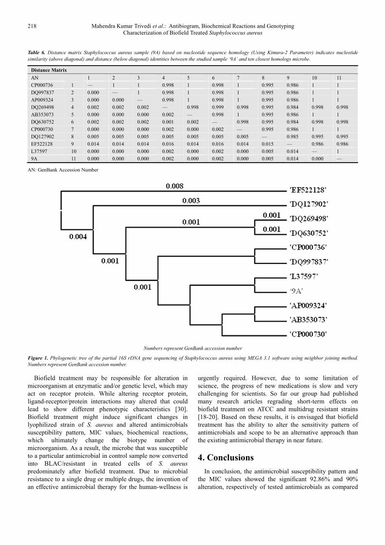
3.3. Identification of Organism by Biotype Number
The species (S. aureus) was identified based on variety of
conventional biochemical characters and biotyping. Biotype
number of particular organism was evaluated after
interpreting the results of the biochemical reactions. The
biotype number then led to the particular organism
identification. In this experiment, biotyping was performed
using an automated system, and results showed a significant
change in biotype number (767177) in Gr. IIA (on day 10)
after first-time biofield treatment with identification of new
species (Staphylococcus cohnii subsp. urealyticum) as
compared to control Gr. I (307016; S. aureus). Based on the
biochemical results, biotype numbers were also changed in
rest of treated groups without alteration of species with
respect to control (307016) i.e. S. aureus (Table 4).
Table 4. Effect of biofield treatment on biotype number of Staphylococcus aureus.
Feature Gr. I (Control) Gr. IIA (Day 10) Gr. IIB (Study I; Day 159) Gr. IIB (Study II; Day 159)
Day 5 Day 10 Day 15
Biotype 307016 767177 307137 307137 307137 307137
Organism Identification S. aureus Staphylococcus cohnii subsp. urealyticum S. aureus S. aureus S. aureus S. aureus
Gr.: Group
3.4. 16S rDNA Genotyping
The bacteria that are poorly differentiated by
conventional methods needs molecular analysis method like
16S rDNA sequence [28]. This molecular-based technique
is a suitable tool for identification of most of the bacteria on
their genus and/or species level by comparison with
databases in the public domain. Because, most of the
bacteria possess small ribosomal subunit with species-
specific variability [29]. The 16S rDNA analysis was
performed after first-time biofield treated sample (Gr. IIA)
of S. aureus on day 10. The alignment and comparison of
the consensus gene sequences were performed with the
sequences stored in GenBank database available from
NCBI using the algorithm BLASTn program. Based on
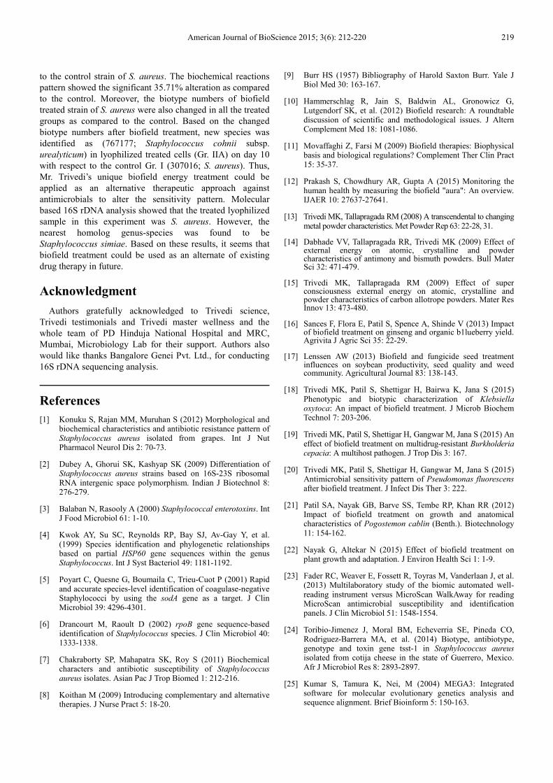
nucleotide homology and phylogenetic analysis the microbe
(Sample 9A) was detected as Staphylococcus aureus
(GenBank Accession No: L37597) with 100% identity. The
nearest homolog genus-species of S. aureus was found as
Staphylococcus simiae (GenBank Accession No.
DQ127902). Some other close homologs of S. aureus were
found from the alignment results as shown in Table 5. The
distance matrix based on nucleotide sequence homology
data are presented in Table 6. The phylogenetic tree was
established using BLAST-Webpage (NCBI). According to
Table 6, ten different related bacterial species of S. aureus
were selected as Operational Taxonomic Units (OTUs) in
order to investigate the phylogenetic relationship of S.
aureus. There were 1497 base nucleotides of 16S rDNA
gene sequences, which were analyzed and multiple
alignments were constructed using ClustalW in MEGA3.1.
The numbers of base substitutions per site from pairwise
distance analysis between sequences are shown in Table 5.
All results were based on the pairwise analysis of 11
sequences. According to the data in Table 6, the lowest
value of the genetic distance from S. aureus was 0.000 base
substitutions per site. This value is due to the distance
between S. aureus and Staphylococcus simiae. All pairwise
distance analysis was carried out using the p-distance
method in MEGA3.1. The proportion of remarked distance,
sometimes also called p-distance and showed as the number
of nucleotide distances site. Values in Table 5 are
programmed into Figure 1 with optimal bootstrap consensus
tree. In the phylogram, there were eleven OTUs. The results
suggested that S. aureus was closely related to the
Staphylococcus simiae with 100% similarity and the lowest
genetic distance 0.000 base substitutions per site.
Table 5. The closest sequences of Staphylococcus aureus from sequence
alignment using NCBI GenBank and ribosomal database project (RDP).
Alignment View AN Alignment
Result Sequence Description
9A 1.00 Sample studied
CP000730 1.00 Staphylococcus aureus
subsp. aureus USA300
AP009324 1.00 Staphylococcus aureus
subsp. aureus Mu3
CP000736 1.00 Staphylococcus aureus
subsp. aureus JH1
AB353073 1.00 Staphylococcus aureus
strain: MPU99
L37597 1.00 Staphylococcus aureus
DQ997837 1.00 Staphylococcus aureus
strain ATCC 14458
DQ269498 0.99 Staphylococcus aureus
strain ATCC 14458
DQ630752 0.99 Staphylococcus aureus
isolation-source bhalla
DQ127902 0.97 Staphylococcus simiae
strain CCM 7229
EF522128 0.99
Staphylococcus
epidermidis strain
CU22
AN: GenBank Accession Number
218 Mahendra Kumar Trivedi et al.: Antibiogram, Biochemical Reactions and Genotyping
Characterization of Biofield Treated Staphylococcus aureus
Table 6. Distance matrix Staphylococcus aureus sample (9A) based on nucleotide sequence homology (Using Kimura-2 Parameter) indicates nucleotide
similarity (above diagonal) and distance (below diagonal) identities between the studied sample ‘9A’ and ten closest homologs microbe.
Distance Matrix
AN
1 2 3 4 5 6 7 8 9 10 11
CP000736 1 — 1 1 0.998 1 0.998 1 0.995 0.986 1 1
DQ997837 2 0.000 — 1 0.998 1 0.998 1 0.995 0.986 1 1
AP009324 3 0.000 0.000 — 0.998 1 0.998 1 0.995 0.986 1 1
DQ269498 4 0.002 0.002 0.002 — 0.998 0.999 0.998 0.995 0.984 0.998 0.998
AB353073 5 0.000 0.000 0.000 0.002 — 0.998 1 0.995 0.986 1 1
DQ630752 6 0.002 0.002 0.002 0.001 0.002 — 0.998 0.995 0.984 0.998 0.998
CP000730 7 0.000 0.000 0.000 0.002 0.000 0.002 — 0.995 0.986 1 1
DQ127902 8 0.005 0.005 0.005 0.005 0.005 0.005 0.005 — 0.985 0.995 0.995
EF522128 9 0.014 0.014 0.014 0.016 0.014 0.016 0.014 0.015 — 0.986 0.986
L37597 10 0.000 0.000 0.000 0.002 0.000 0.002 0.000 0.005 0.014 — 1
9A 11 0.000 0.000 0.000 0.002 0.000 0.002 0.000 0.005 0.014 0.000 —
AN: GenBank Accession Number
Numbers represent GenBank accession number
Figure 1. Phylogenetic tree of the partial 16S rDNA gene sequencing of Staphylococcus aureus using MEGA 3.1 software using neighbor joining method.
Numbers represent GenBank accession number.
Biofield treatment may be responsible for alteration in
microorganism at enzymatic and/or genetic level, which may
act on receptor protein. While altering receptor protein,
ligand-receptor/protein interactions may altered that could
lead to show different phenotypic characteristics [30].
Biofield treatment might induce significant changes in
lyophilized strain of S. aureus and altered antimicrobials
susceptibility pattern, MIC values, biochemical reactions,
which ultimately change the biotype number of
microorganism. As a result, the microbe that was susceptible
to a particular antimicrobial in control sample now converted
into BLAC/resistant in treated cells of S. aureus
predominately after biofield treatment. Due to microbial
resistance to a single drug or multiple drugs, the invention of
an effective antimicrobial therapy for the human-wellness is
urgently required. However, due to some limitation of
science, the progress of new medications is slow and very
challenging for scientists. So far our group had published
many research articles regrading short-term effects on
biofield treatment on ATCC and multidrug resistant strains
[18-20]. Based on these results, it is envisaged that biofield
treatment has the ability to alter the sensitivity pattern of
antimicrobials and scope to be an alternative approach than
the existing antimicrobial therapy in near future.
4. Conclusions
In conclusion, the antimicrobial susceptibility pattern and
the MIC values showed the significant 92.86% and 90%
alteration, respectively of tested antimicrobials as compared
American Journal of BioScience 2015; 3(6): 212-220 219
to the control strain of S. aureus. The biochemical reactions
pattern showed the significant 35.71% alteration as compared
to the control. Moreover, the biotype numbers of biofield
treated strain of S. aureus were also changed in all the treated
groups as compared to the control. Based on the changed
biotype numbers after biofield treatment, new species was
identified as (767177; Staphylococcus cohnii subsp.
urealyticum) in lyophilized treated cells (Gr. IIA) on day 10
with respect to the control Gr. I (307016; S. aureus). Thus,
Mr. Trivedi’s unique biofield energy treatment could be
applied as an alternative therapeutic approach against
antimicrobials to alter the sensitivity pattern. Molecular
based 16S rDNA analysis showed that the treated lyophilized
sample in this experiment was S. aureus. However, the
nearest homolog genus-species was found to be
Staphylococcus simiae. Based on these results, it seems that
biofield treatment could be used as an alternate of existing
drug therapy in future.
Acknowledgment
Authors gratefully acknowledged to Trivedi science,
Trivedi testimonials and Trivedi master wellness and the
whole team of PD Hinduja National Hospital and MRC,
Mumbai, Microbiology Lab for their support. Authors also
would like thanks Bangalore Genei Pvt. Ltd., for conducting
16S rDNA sequencing analysis.
References
[1] Konuku S, Rajan MM, Muruhan S (2012) Morphological and biochemical characteristics and antibiotic resistance pattern of Staphylococcus aureus isolated from grapes. Int J Nut Pharmacol Neurol Dis 2: 70-73.
[2] Dubey A, Ghorui SK, Kashyap SK (2009) Differentiation of Staphylococcus aureus strains based on 16S-23S ribosomal RNA intergenic space polymorphism. Indian J Biotechnol 8: 276-279.
[3] Balaban N, Rasooly A (2000) Staphylococcal enterotoxins. Int J Food Microbiol 61: 1-10.
[4] Kwok AY, Su SC, Reynolds RP, Bay SJ, Av-Gay Y, et al. (1999) Species identification and phylogenetic relationships based on partial HSP60 gene sequences within the genus Staphylococcus. Int J Syst Bacteriol 49: 1181-1192.
[5] Poyart C, Quesne G, Boumaila C, Trieu-Cuot P (2001) Rapid and accurate species-level identification of coagulase-negative Staphylococci by using the sodA gene as a target. J Clin Microbiol 39: 4296-4301.
[6] Drancourt M, Raoult D (2002) rpoB gene sequence-based identification of Staphylococcus species. J Clin Microbiol 40: 1333-1338.
[7] Chakraborty SP, Mahapatra SK, Roy S (2011) Biochemical characters and antibiotic susceptibility of Staphylococcus aureus isolates. Asian Pac J Trop Biomed 1: 212-216.
[8] Koithan M (2009) Introducing complementary and alternative therapies. J Nurse Pract 5: 18-20.
[9] Burr HS (1957) Bibliography of Harold Saxton Burr. Yale J Biol Med 30: 163-167.
[10] Hammerschlag R, Jain S, Baldwin AL, Gronowicz G, Lutgendorf SK, et al. (2012) Biofield research: A roundtable discussion of scientific and methodological issues. J Altern Complement Med 18: 1081-1086.
[11] Movaffaghi Z, Farsi M (2009) Biofield therapies: Biophysical basis and biological regulations? Complement Ther Clin Pract 15: 35-37.
[12] Prakash S, Chowdhury AR, Gupta A (2015) Monitoring the human health by measuring the biofield "aura": An overview. IJAER 10: 27637-27641.
[13] Trivedi MK, Tallapragada RM (2008) A transcendental to changing metal powder characteristics. Met Powder Rep 63: 22-28, 31.
[14] Dabhade VV, Tallapragada RR, Trivedi MK (2009) Effect of external energy on atomic, crystalline and powder characteristics of antimony and bismuth powders. Bull Mater Sci 32: 471-479.
[15] Trivedi MK, Tallapragada RM (2009) Effect of super consciousness external energy on atomic, crystalline and powder characteristics of carbon allotrope powders. Mater Res Innov 13: 473-480.
[16] Sances F, Flora E, Patil S, Spence A, Shinde V (2013) Impact of biofield treatment on ginseng and organic b1lueberry yield. Agrivita J Agric Sci 35: 22-29.
[17] Lenssen AW (2013) Biofield and fungicide seed treatment influences on soybean productivity, seed quality and weed community. Agricultural Journal 83: 138-143.
[18] Trivedi MK, Patil S, Shettigar H, Bairwa K, Jana S (2015) Phenotypic and biotypic characterization of Klebsiella oxytoca: An impact of biofield treatment. J Microb Biochem Technol 7: 203-206.
[19] Trivedi MK, Patil S, Shettigar H, Gangwar M, Jana S (2015) An effect of biofield treatment on multidrug-resistant Burkholderia cepacia: A multihost pathogen. J Trop Dis 3: 167.
[20] Trivedi MK, Patil S, Shettigar H, Gangwar M, Jana S (2015) Antimicrobial sensitivity pattern of Pseudomonas fluorescens after biofield treatment. J Infect Dis Ther 3: 222.
[21] Patil SA, Nayak GB, Barve SS, Tembe RP, Khan RR (2012) Impact of biofield treatment on growth and anatomical characteristics of Pogostemon cablin (Benth.). Biotechnology 11: 154-162.
[22] Nayak G, Altekar N (2015) Effect of biofield treatment on plant growth and adaptation. J Environ Health Sci 1: 1-9.
[23] Fader RC, Weaver E, Fossett R, Toyras M, Vanderlaan J, et al. (2013) Multilaboratory study of the biomic automated well-reading instrument versus MicroScan WalkAway for reading MicroScan antimicrobial susceptibility and identification panels. J Clin Microbiol 51: 1548-1554.
[24] Toribio-Jimenez J, Moral BM, Echeverria SE, Pineda CO, Rodriguez-Barrera MA, et al. (2014) Biotype, antibiotype, genotype and toxin gene tsst-1 in Staphylococcus aureus isolated from cotija cheese in the state of Guerrero, Mexico. Afr J Microbiol Res 8: 2893-2897.
[25] Kumar S, Tamura K, Nei, M (2004) MEGA3: Integrated software for molecular evolutionary genetics analysis and sequence alignment. Brief Bioinform 5: 150-163.
220 Mahendra Kumar Trivedi et al.: Antibiogram, Biochemical Reactions and Genotyping
Characterization of Biofield Treated Staphylococcus aureus
[26] Milazzo I, Blandino G, Caccamo F, Musumeci R, Nicoletti G, et al. (2003) Faropenem, a new oral penem: Antibacterial activity against selected anaerobic and fastidious periodontal isolates. J Antimicrob Chemother 51: 721-725.
[27] Ishii Y, Alba J, Maehara C, Murakami H, Matsumoto T, et al. (2006) Identification of biochemically atypical Staphylococcus aureus clinical isolates with three automated identification systems. J Med Microbiol 55: 387-392.
[28] Drancourt M, Bollet C, Carlioz A, Martelin R, Gayral JP, et al. (2000) 16S ribosomal DNA sequence analysis of a large collection of environmental and clinical unidentifiable bacterial isolates. J Clin Microbiol 38: 3623-3630.
[29] Vandamme P, Pot B, Gillis M, de Vos P, Kersters K, et al. (1996) Polyphasic taxonomy, a consensus approach to bacterial systematics. Microbiol Rev 60: 407-438.
[30] Lindstrom E, Mild KH, Lundgren E (1998) Analysis of the T cell activation signaling pathway during ELF magnetic field exposure, p56lck and [Ca2+]i-measurements. Bioeletrochem Bioenerg 46: 129-137.