Embed Size (px)
Citation preview
Association of Campylobacter spp. Levels between Chicken Grow-Out Environmental
Samples and Processed Carcasses
Matthew W. Schroeder
Thesis submitted to the faculty of the Virginia Polytechnic Institute and State University in
partial fulfillment of the requirements for the degree of
Master of Science
In
Food Science and Technology
Joseph D. Eifert, Chair
Monica A. Ponder
David G. Schmale, III
April 20, 2012
Blacksburg, VA
Keywords: Campylobacter, environmental sampling, single flock, poultry
Association of Campylobacter spp. Levels between Chicken Grow-Out Environmental Samples
and Processed Carcasses
Matthew W. Schroeder
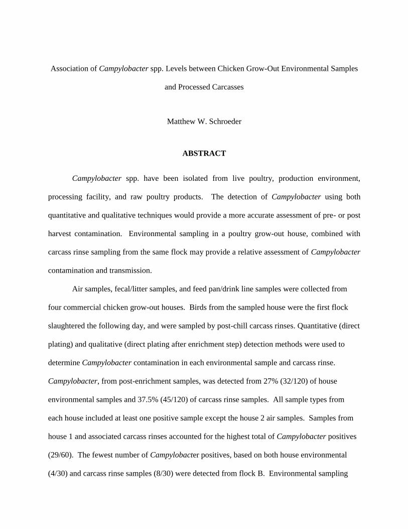
ABSTRACT
Campylobacter spp. have been isolated from live poultry, production environment,
processing facility, and raw poultry products. The detection of Campylobacter using both
quantitative and qualitative techniques would provide a more accurate assessment of pre- or post
harvest contamination. Environmental sampling in a poultry grow-out house, combined with
carcass rinse sampling from the same flock may provide a relative assessment of Campylobacter
contamination and transmission.
Air samples, fecal/litter samples, and feed pan/drink line samples were collected from
four commercial chicken grow-out houses. Birds from the sampled house were the first flock
slaughtered the following day, and were sampled by post-chill carcass rinses. Quantitative (direct
plating) and qualitative (direct plating after enrichment step) detection methods were used to
determine Campylobacter contamination in each environmental sample and carcass rinse.
Campylobacter, from post-enrichment samples, was detected from 27% (32/120) of house
environmental samples and 37.5% (45/120) of carcass rinse samples. All sample types from
each house included at least one positive sample except the house 2 air samples. Samples from
house 1 and associated carcass rinses accounted for the highest total of Campylobacter positives
(29/60). The fewest number of Campylobacter positives, based on both house environmental
(4/30) and carcass rinse samples (8/30) were detected from flock B. Environmental sampling

iii
techniques provide a non-invasive and efficient way to test for foodborne pathogens.
Correlating qualitative or quantitative Campylobacter levels from house and plant samples may
enable the scheduled processing of flocks with lower pathogen incidence or concentrations, as a
way to reduce post-slaughter pathogen transmission.
iv
ACKNOWLEDGMENTS
I would first like to thank all the faculty and staff of the Department of Food Science
and Technology at Virginia Tech, especially my committee members Dr. Ponder and Dr.
Schmale. I would like to especially thank Dr. Eifert for giving me the opportunity to work
with him. I could never thank you enough for your unending support and help throughout
my project and thesis preparation. Why you still brave the chicken houses after all these
years is still a mystery to me. I would also like to thank Hassan Masri and Mohammad
Alshuniaber for their help on sampling and plating in the house and processing facility.
Though we did make 40,000 friends on those days, I know it was not the most pleasant
experience, but thank you so much for your friendship and help. I would also like to thank
Govind for your help with the latex agglutination testing procedures. Thanks goes to all the
QA technicians at the processing facility for allowing me to work in their area. Dr. Wang,
thank you so much for your help with the statistics behind my research.
I would also like to send a special thanks to all my fellow graduate students in the
department. The friendships I have made here at Virginia Tech are something I will cherish
and remember for the rest of my life. The many, many nights we spent together, whether it
was a Tech football game, Monster Jam, or late nights at my apartment are times I will never
forget. Thank you for making the past two years here some of the best of my life. I cannot
wait to spend the next three years here with some of you and come visit others in various
cities. Finally, I would like to thank all the players and coach from the Virginia Tech Club
Lacrosse Team, especially Dr. Joel Nachlas. Thank you for the opportunity to work with you
and most importantly, the stress relief. The long trips all over the East coast with all of you,
aside from being hilarious, were way too much fun.

v
DEDICATION
I dedicate this body of work to my sister, Emily Schroeder. Thank you for always
being there for me. I would not be where I am today without you. I love you!
vi
TABLE OF CONTENTS
Abstract…………………………………………………………………………………. ii
Acknowledgments………………………………………………………………………
Dedication………………………………………………………………………………...
List of tables and figures…………………………………………………………………
List of abbreviations……………………………………………………………………...
Introduction………………………………………………………………………………
Literature Review………………………………………………………………………...
Campylobacter in Food Systems…………………………………………………
Growth and Survival of Campylobacter…………………………………………
Infection and Transmission of Campylobacter in Poultry Grow-out Houses……
Transmission of Campylobacter during Transport………………………………
Transmission of Campylobacter during Processing……………………………...
Control of Campylobacter during Grow-out…………………………………......
Control of Campylobacter during Processing……………………………………
Detection Methods for Campylobacter from Foods………………………….......
References……………………………………………………………………………......
Materials and Methods……………………………………………………………….......
Grow-out house sample collection and analysis ………………………………………
Air sampling……………………………………………………………………...….
Fecal and litter environmental samples ……………………………………………..
Feeder and drinker environmental samples …………………………………………
Transportation sample collection and analysis………………………………………...
iv
v
viii
ix
1
4
4
5
6
13
14
16
17
20
24
35
36
36
36
37
38
vii
Processing plant sample collection and analysis………………………………………
Carcass Rinse ……………………………………………………………..................
Chill tank and scald tank water ……………………………………………………...
Campylobacter confirmation………………………………………………………...…...
Statistical analysis………………………………………………………………………...
Results……………………………………………………………………………….…...
Discussion……………………………………………………………………………..…
References……………………………………………………………………………..…
Tables and Figures……………………………………………………………………..…
39
39
40
40
41
41
43
48
51
viii
LIST OF TABLES AND FIGURES
Table 1.Campylobacter samples percent positive (post-enrichment), by sample type, from
grow-out houses and processing plant……………………………………………................
Table 2.Grow-out house and flock information…………………………………………….
Figure 1.Number of Campylobacter positive samples (post-enrichment) from houses and
plant………………………………………………………………………………………….
Figure 2.Grow-out house sampling flow chart……………………………………………..
Figure 3.Flow chart for processing facility analysis………………………………………..
Figure 4.Campylobacter transmission through chicken processing………………………..
Figure 5.Example of sampling locations within a grow-out house……………………........
51
52
53
54
55
56
57
ix
LIST OF ABBREVIATIONS
Abbreviation Word
CFU colony forming unit(s)
ft feet
g gram(s)
l liter(s)
lbs pound(s)
m meter(s)
mg milligram(s)
min minute(s)
ml milliliter(s)
ppm parts per million (10-6
)
μl microliter(s) (10-6
l)

1
INTRODUCTION
Campylobacter bacteria are Gram negative, slender, spiral-curved rods that cause a majority
of the intestinal infectious diseases worldwide (Snelling et al. 2005; Keener et al. 2004). In
particular, Campylobacter jejuni, one of the “thermophilic” campylobacters, is one of the leading
causes of bacterial diarrheal illness reported in the United States (Altekruse et al. 1999; Keener et
al. 2004). An estimated 40,000 cases are documented annually in the United States (Keener et
al. 2004). In 2011, Campylobacter caused 0.8 million illnesses total (Scallan et al.
2011).Campylobacter jejuni has a low infectious dose (~500-10,000 organisms), which is
normally followed by symptoms including fever, abdominal pain, and diarrhea (Snelling et al.
2005; Kenner et al. 2004). C. jejuni grows best between 37oC to 42
oC, the approximate body
temperature of a chicken, and in a low oxygen (3-15%), or microaerophilic environment
(Altekruse et al. 1999).
Reservoirs for Campylobacter include wildlife such as ducks and geese, contaminated water,
insects, raw milk, and meat; however, 50-70% of Campylobacter illness comes from consuming
poultry and poultry products (Altekruse et al. 1999; Allos 2001). Chicken has the greatest
Campylobacter risk, in part, because of the large quantities consumed (Humphrey et al. 2007).
Though colonization is not detectable until at least 10 days of age, once infected, Campylobacter
has been found in up to 100% of birds tested in a given flock (Keener et al. 2004; Moore et al.
2005). This pre-harvest infection, along with the high likelihood of cross contamination inside
the poultry processing plant, has caused the United States Department of Agriculture-Food
Safety Inspection Service(USDA-FSIS) to revise their regulations to require poultry processors
to monitor and control Campylobacter (Keener et al. 2004; Moore et al. 2005; USDA 2011b).
2
Suzuki and Yamamoto (2009) conducted a literature survey of prevalence of Campylobacter
on retail poultry in the United States. From 1167 samples, Campylobacter was found on 71.5%
of retail chicken carcasses in the United States (Suzuki and Yamamoto 2009). However, in 2010
less than 10% of U.S. processing facilities were specifically testing for Campylobacter in a
survey of 167 processors (Alvarado 2011). Because most processors are still concerned about
Salmonella with less emphasis on Campylobacter, the USDA-FSIS has devised new
performance standards that focus on both quantitative and qualitative detection methods of
Campylobacter. These regulations require commercial poultry processors to isolate, identify,
and enumerate Campylobacter from poultry rinses by direct plating onto selective agars or by
enrichment culture (Williams et al. 2009). From post-chill carcass rinses, a direct plating method
(1 ml sample split among four plates) will be utilized for enumeration of Campylobacter. An
additional 30 ml sample from the rinse with an enrichment broth will be used for the qualitative
analysis (USDA 2011a). According to the new regulation, acceptable percentages of positive
carcasses for the 1 ml samples and combination of the 1 ml and 30 ml samples are 10.4 and 46.4,
respectively (Alvarado 2011). That is to say, in a sample set of 51 carcasses, 8 can test positive
for Campylobacter and still be acceptable (Alvarado 2011; USDA 2011b). If the 1 ml samples
are negative, the 30 ml samples are tested with 21 out of 51 samples for the combination
considered acceptable (Alvarado 2011). Knowing that 100% of boiler flock chickens entering
the processing facility could contain Campylobacter, processors now not only need to worry
about Salmonella, but also Campylobacter to ensure the safety of their product (Moore et al.
2005).
In this study, air samples and other environmental swabs in grow-out houses will be tested,
while carcasses and chill tank water will be tested before and during slaughter for possible
3
Campylobacter contamination. The samples taken from the same flock in the grow house, and
processing facility may show a relationship between pre- and post harvest contamination. Most
previous research has focused on either the grow-out house or the processing facility only. Little
research has been done on a single flock continuously from farm through slaughter completion.
And, with the adoption of the new government standards concerning Campylobacter and the
little amount of environmental sampling that has been done in grow-out houses and processing
facilities, this project will be valuable to large or small food processors.
The purpose of this study was to 1) determine Campylobacter numbers in poultry houses
through environmental sampling (i.e.- air samples, shoe bootie samples, and chicken feed
pan/drinker samples), 2) determine Campylobacter contamination levels in the processing
facility after the previously sampled flock is processed, and 3) determine if there is an
association between Campylobacter levels in commercial chicken production and processing
environment through pre- and post- harvest cycles.
Based on the objectives described, the bootie samples will account for the highest amount
of Campylobacter positives through environmental sampling techniques. The processing facility
samples (carcass rinses) will have a lower number of Campylobacter positives than the
environmental grow-out houses samples; however, there will be an association between these
levels.
4
LITERATURE REVIEW
A. Campylobacter in Food Systems
Within the past 25 years, Campylobacter has been recognized as a cause for human
illness (Keener et al. 2004). In the United States alone, 50-70% of human Campylobacter illness
is attributed to poultry, with a reported 40,000 cases documented annually (Keener et al. 2004).
Though deaths from Campylobacter infection are uncommon (680-730 per year), children less
than the age of one and young adults aged fifteen to twenty-five more frequently acquire
infection (Keener et al., 2004). Most infections are sporadic with clinical symptoms including
fever, abdominal pain, and diarrhea (Keener et al. 2004). Though outbreaks with Campylobacter
are uncommon, Campylobacter associated illness and infections still occur. In 2011 in England,
the Health Protection Agency (HPA) found that 90% of Campylobacter infection associated with
catering venues was due to the consumption of chicken liver plate, a popular dish in England and
Wales (Health Protection Agency 2011). In 2005, a Campylobacter outbreak was also identified
in an Australian restaurant where chicken was the source of the problem (Black et al., 2006).
Campylobacter contamination can occur in a variety of areas including production,
processing, distribution, retail marketing, and preparation (Zhao et al. 2001). However,
Campylobacter can also be found in the food supply at retail markets (Altekruse et al. 1999).
Retail chicken meat has been isolated with Campylobacter at a rate of 98% (Stern and Line
1992). A study was conducted evaluating the prevalence of Campylobacter spp., E. coli, and
Salmonella in retail chicken, turkey, pork, and beef in the Washington, D.C. area (Zhao et al.
2001). From the 92 sampling visits from supermarket chains, it was found that 91% of the stores
had Campylobacter contaminated chicken (Zhao et al. 2001). The potential for transmission of
5
foodborne pathogens from raw retail meats further emphasizes the importance of consumer and
food safety knowledge (Zhao et al. 2001).
The USDA Food Safety and Inspection Service (FSIS) recently changed their
regulations regarding the control of Campylobacter within processing facilities (USDA 2011b).
These new regulations specify that only 8 out of 51 chicken carcasses can test positive for
Campylobacter and still be considered acceptable (Alvarado 2011; USDA 2011b). If a facility
fails to meet these standards, they will be moved to the second highest priority for scheduling to
conduct a follow-up (USDA 2011b). This is an important change when considering most
processing facilities and growers focus on Salmonella and E. coli (Northcutt 2003). Most
growers use vaccination to control Salmonella, but once infected, an entire flock can become
infected with Campylobacter on the farm (Fielding 2012). Although at processing Salmonella
has been seen at 13% positive, the majority of carcass contamination is due to Campylobacter
(Rasschaert 2007). The main focus for controlling Campylobacter should be the farm; however,
other places such as hatcheries and processing facilities also need to implement control strategies
to reduce contamination (Shanker et al. 1990; Bull et al. 2006). These initiatives set forth by the
USDA-FSIS hopefully will begin to reduce contamination due to Campylobacter to ensure
public health safety.
B. Growth and Survival of Campylobacter
Campylobacter jejuni, a micro-aerophile, survives best in a low oxygen atmosphere, such
as 5% oxygen, 10% carbon dioxide, and 85% nitrogen at a temperature around 42oC (Altekruse
et al. 1999; Keener et al. 2004). In the United States, over 99% of the reported infections are
with Campylobacter jejuni, although 14 species of Campylobacter have been identified (Allos
6
2001). A majority of human infections are from C. jejuni and C. coli, which is why they are
studied more frequently (Keener et al. 2004). The optimal atmosphere for the growth of various
Campylobacter jejuni and coli strains was found to be 5-10% oxygen and 1-10% carbon dioxide
(Bolton and Coates 1983). Rates of inactivation can be influenced by strain, temperature,
humidity, and suspension medium; however, Campylobacter cannot survive below an acidic pH
of 4.9 (Keener et al. 2004). The optimal pH range for Campylobacter is 6.5-7.5, but at low pH,
survival of the organism is temperature dependent (Curtis 2007). Campylobacter has been
shown to grow best at temperatures between 37oC and 42
oC (Keener et al. 2004). Minimal
growth temperatures for Campylobacter jejuni strains 104 and ATCC 33560 were found to be
32oC and 31
oC, respectively (Hazeleger et al. 1998). However, around 30
oC, a sudden growth
rate decline was observed (Hazeleger et al. 1998). Campylobacter is also sensitive to freezing
and salinity (Altekruse et al. 1999). Doyle and Roman (1982) found that three strains of
Campylobacter jejuni could grow at 1.5% NaCl, but not 2.0% NaCl at 42oC.
C. Infection and Transmission of Campylobacter in Poultry Grow-out Houses
Initial Colonization
Although it is impossible to determine the exact moment a single bird in a flock becomes
contaminated with Campylobacter due to flock size and necessary sampling frequency,
understanding the mechanism by which grow-out houses becomes colonized proves important
for controlling the bacteria and establishing appropriate control measures (van Gerwe et al.
2009). Estimation of transmission and dynamics of colonization of Campylobacter proves very
difficult and is not well understood (Conlan et al. 2007). However, initial flock colonization
with Campylobacter is age dependent with normally no detection when birds are less than two
7
weeks old (Conlan et al. 2007). A dose as low as 40 CFUs have been shown experimentally to
colonize 1-day-old chicks, although this is dependent on the strain of Campylobacter jejuni and
breed of the bird (Conlan et al. 2007).
Campylobacter colonizes the intestinal mucus layer in the epithelium of chickens
(Keener et al. 2004). Once infected, the Campylobacter numbers will remain high in the
intestine (Potturi-Venkata et al. 2007). Horizontal transmission seems to be the most likely
mechanism by which Campylobacter is introduced into a flock, with rapid colonization of a
flock (3-7 days) once colonization occurs (van Gerwe et al. 2009; Shanker et al. 1990). Possible
sources of initial colonization include: wild birds, other farm animals, rodents, insects,
contaminated groundwater, carryover from previous flock and farm workers (Conlan et al. 2007;
Jacobs-Reitsma et al. 1995; Humphrey et al. 2007).
Age Dependence
Contamination of birds, on the farm, by Campylobacter is normally not detectable until at
least 10 days of age (Newall et al. 2000; Byrd et al. 1998). Chickens less than two weeks of age
normally are not colonized due to a “lag phase” derived from maternal antibodies that are
prevalent in young chicks (Conlan et al. 2007). Colonization in broiler chickens is the highest in
the mucosal crypts of the caeca, but also invades the small intestine (Conlan et al. 2007).
Normally the transmission of Campylobacter results from fecal-oral transmission and can often
contaminate the entire flock within 5 weeks (Keener et al. 2004; Jacobs-Reitsma et al. 1995). In
2009, van Gerwe et al. estimated that one colonized bird could infect 2.37 birds per day on
average. Theoretically, at this rate, 95% of the flock could be contaminated in one week. Age
and bioaerosol concentration also play an important role in Campylobacter contamination
8
(Northcutt et al. 2003). Birds aged 42 days tested 100% positive for Campylobacter, while birds
aged 56 days showed a 90% infection rate (Northcutt et al. 2003). Berndtson et al. (1996) found
that the rate of positive flocks increased with age when following flocks from week one to week
five. Since bioaerosol concentration increases with bird age and air hygiene is considered an
important factor for animal health, Campylobacter infection in the flock may be spread through
the air (Vucemilo et al. 2007; Saleh et al. 2005).
Housing Environmental Factors
Broiler chickens are raised in grow-out houses with dimensions between 40’ and 60’ (12-
18 m) wide and 300’ to 600’ (90-180 m) long. Each house holds approximately 12,000 to
40,000 birds (Kuntz 2009). Initially, one-day-old chicks are placed in the house where the
temperature is heated to around 32oC (90
oF), but as the birds grow and begin to produce their
own heat, the temperature of the house decreases, eventually reaching ambient temperatures (70-
75oF) (Kuntz 2009; Donald et al. 2005).
Farms widely differ in their infection rates (Humphrey et al. 2007). Though these
differences could be due to hygiene within the house, birds raised in a poor environment are
more susceptible to campylobacters (Humphrey et al. 2007). Environmental factors such as
humidity, ventilation, temperature, and bioaerosols can play an important role in the transmission
of Campylobacter.
Campylobacter are susceptible to dry conditions. Thus, in a grow-out house with higher
humidity, Campylobacter could survive (Ishihara et al. 2012). In some instances, relative
humidity within a grow-out house can reach 75 to 80% (Line 2006; Choct 2010). Line (2006)
studied C. jejuni colonization under high (80%) and low (30%) relative humidity conditions. He
9
found that there were differences in rates of colonization of Campylobacter on litter held under
high and low relative humidity. The artificially dry, low humidity pen showed a colonization
delay in comparison to the high relative humidity pen (Line 2006). In Japan, Ishihara et al.
(2012) found that locations with higher humidity and shorter periods of sunshine were associated
with increased colonization of broiler flocks. All Campylobacter positive locations had a higher
mean humidity than the Campylobacter negative locations (Ishihara et al. 2012).
Many grow-out houses in the United States today utilize tunnel ventilation
(Chinivasagam et al. 2009). In this system, large volumes of air are circulated throughout the
house. This mass movement of air is beneficial for maintaining proper temperature and humidity
for the birds inside the house; however, the moving air can also contain pathogens such as
Campylobacter and/or Salmonella (Chinivasagam et al. 2009). Air-quality related illnesses in
humans such as cough, runny nose, sore throat and eye irritation have been shown to decrease
when ventilation is effective (Shale and Lues 2007). Air circulation within a grow-out house
increases over the chicken cycle, which also increases the rate at which microbes are distributed
(Shale and Lues 2007; Chinivasagam et al. 2009). This could contribute to the spread of
Campylobacter contamination in a grow-out house.
Seasonality and temperature also have been reported to affect Campylobacter incidence
(Ishihara et al. 2012; Humphrey et al. 2007). Aside from temperature contributing to the eating
habits of the birds, the temperature outside as well as inside the house have shown a correlation
to Campylobacter colonization (Donald et al. 2005; Humphrey et al. 2007). Ishihara et al.
(2012) found that higher air temperatures during chicken rearing was associated with increased
Campylobacter contamination in the house as well as during processing. In the United States,
10
increases in the level of human Campylobacteriosis have been seen during the summer months
(Sopwith et al. 2003).
With increased bird densities within grow-out houses due to large scale production,
exposure to organic dust from chicken feces, litter, feed, and feather formation are leading to
human health problems (Oppliger et al. 2008). Air hygiene and exposure to these bioaerosols,
which increase during the fattening period in birds, impacts both the health of the birds as well as
the humans working in the industry (Oppliger et al. 2008; Vucemilo et al. 2007). Vucemilo et al.
(2006 and 2007) found that all pollutants within the housing environment increased with
increasing poultry age and body weight, ranging from 3.22 x 10³
CFU/m3in the first week to 6.40
x 107
CFU/m3 in the fifth week. Airborne dust, which is influenced by poultry age, litter, and
poultry activity, also increased with age, reaching 4.8 mg/m3 air (Vucemilo et al. 2007).
Chicken bedding is normally comprised of peanut hulls, sawdust, and wood shavings. In
the United States, typically dozens of flocks will be raised on a single bed of layered litter
(Dumas et al. 2011). Thus, bedding is a possible reservoir of disease-causing bacteria (Dumas et
al. 2011). Montrose et al. (1985) studied the role of litter in the transmission of Campylobacter.
They found that uninfected chickens placed on infected autoclaved and nonautoclaved litter shed
Campylobacter jejuni after only several days (Montrose et al. 1985). This research indicates the
possible transmission of Campylobacter from flock to flock from litter.
Campylobacter Detection in the Pre-Harvest Environment
Most studies of the chicken grow-out houses analyze the chicken’s surroundings, such as
its feed, water, or litter (Shanker et al. 1990). Studies have implicated these items for possible
sources of horizontal transmission, but the full epidemiology is still not fully understood (Jacobs-
11
Reitsma et al. 1995). Campylobacter in the environment is detected intermittently; typically,
detection doesn’t occur until it the flock has been colonized, but the direction of the spread is
unclear (Bull et al. 2006). Research has begun focusing on environmental sampling methods
inside and around grow-out houses as opposed to live bird sampling due to speed and efficiency.
Campylobacter has even been detected outside of chicken houses, in some cases up to 30
m downwind of the broiler house (Bull et al. 2006; Hansson et al. 2007). Campylobacter has
also been detected from the air exiting the broiler sheds, which could be important for the
transmission of infection (Bull et al. 2006). Chickens attain harvest weight around 40 days of
age (Humphrey et al. 2007). Within the last week of fattening, bioaerosols are at their highest
concentration and prevalence of contamination is highest (Saleh et al. 2005). Since bioaersol
concentration includes poultry feces, litter, and feathers, which are all known sources of
Campylobacter contamination, air could affect contamination levels inside the house (Vucemilo
et al. 2007). As chicks mature, their size increases, subsequently increasing the amount of
bioaerosols that may serve as a vehicle for infection in other birds. Aside from the normal fecal,
intestinal, and swabbing techniques used to isolate Campylobacter, environmental studies
including air sampling have become popular to detect presence in grow-out houses (Keener et al.
2004; Berndtson et al. 1996). Olsen et al. (2009) determined that Campylobacter colonization
could be detected prior to detection in the traditional sock (bootie) sampling techniques and only
required 1800ml of air. In 2009, Kuntz et al. found that 28% of air samples within a chicken
grow-out house tested positive for Campylobacter.
The transmission of Campylobacter is normally fecal-oral. Once a flock has become
Campylobacter-positive, 100% of fecal samples tested positive (Potturi-Venkata et al. 2007).
Environmental sampling of poultry litter or feces is common, but a number of different
12
techniques exist (Keener et al. 2004). Eifert et al. (2003) compared various sampling techniques
(fecal swabs and environmental surface “drag” samples) and found that environmental swabs of
the litter yielded the highest percentage recovery. Bull et al. (2006) collected fresh fecal samples
and found that 83% (189/229) tested positive for Campylobacter. A similar technique was used
by Stern and Robach (2003) and found that 96.4% of fecal samples tested positive. Williams et
al. (2009) and Hansson et al. (2007) placed a bandage or sock over a normal shoe and walked
around the house for fecal sampling. Using this method, 23 out of 131 flocks (18%) tested
positive for Campylobacter.
Campylobacter is most often transmitted by the fecal-oral route and can be spread by
direct contact on food and water. Adult chickens consume about 0.05 to 0.16 gallons of water
per day depending on temperature and eat about 2.5 lbs of food/lb of weight gain (Frame 2008).
Chickens have free access to their feed trays and water lines. Chickens can defecate on their
feed and water lines; therefore swab samples of these can be taken to test for the presence of
Campylobacter (Berndtson et al. 1996). Berndtson et al. (1996) found 90 out of 300 pooled
swab samples from water lines (30%) tested positive for Campylobacter. Sampling of feed
devices in a study conducted by Johnsen et al. (2006) found 25% (4/16) tested positive for
Campylobacter.
Flying insects, such as flies, have also been implicated as a vector for Campylobacter
transmission, especially during the summer months (Hansson et al. 2007). Insects are able to
carry Campylobacter on their exoskeleton (Altekruse et al. 1999). A study showed that flies
caught in Campylobacter-negative chickens were also negative, but with chickens that were
Campylobacter-positive, flies were positive as well (Berndtson et al. 1996). Though insects may
pose a risk of introduction of Campylobacter into a flock based on amount and ventilation
13
patterns within the house, 1% (3/291) of insects were Campylobacter positive (Hansson et al.
2007). Water and various other environmental samples (feces from cows, straw, mud) from
surrounding farms were found to contain Campylobacter around six of the seven colonized
flocks (Bull et al. 2006).
D. Transmission of Campylobacter during Transport
When animals are transported from farm to processing facility, the animals are under a
tremendous amount of stress (Keener et al. 2004). This stress may increase spreading of
intestinal bacteria such as Campylobacter (Keener et al. 2004). Bacterial counts on carcasses
have shown 1,000-fold increase during transportation (Altekruse et al. 1999). During crating or
transport, any pathogen, such as Campylobacter, could still colonize the ceca of birds, which
would be retained during processing (Keener et al. 2004). Aside from the birds themselves,
crates that are not properly cleaned could increase contamination levels (Stern et al. 2001). A
survey of over 10,000 poultry companies of varying sizes conducted by Auburn University
discovered that 80% of poultry growers don’t sanitize their crates and only 18.3% sanitize their
trucks and trailers (Fielding 2012). One study found 53% of batches of crates tested positive for
Campylobacter spp.; the same subtype that was found on the farm and before slaughter (Hansson
et al. 2007). In addition to the possible contamination from crates during transport, over 50% of
bird catchers’ boots, drivers’ boots, and truck wheels have tested positive for Campylobacter
(Ramabu et al. 2004). Following the same flock from farm to the processing plant can provide a
more accurate assessment of cross contamination that can occur during transportation.
Contamination will likely increase once birds are in transit from farm to processing plant.
14
E. Transmission of Campylobacter during Processing
Cross contamination is highly likely to occur within poultry processing facilities (Moore
et al. 2005). The status of Campylobacter infection at the conclusion of processing is related to
the Campylobacter- status of the arriving flock (Berrang et al. 2007). Birds entering the facility
normally already have high rates of infection around 93% (Jozwiak et al. 2006). By the end of
processing, the number of positive carcasses has been shown to increase on occasion, but most
often bacterial populations on carcasses have been shown to decrease as processing progresses
(Jozwiak et al. 2006; Berrang and Dickens 2000). However, more data is needed to understand
the mechanism by which Campylobacter contamination changes during processing (Guerin et al.
2010). Poultry processors, now given the current regulations, are required to monitor the
presence of Campylobacter in their facilities.
Carcass Sampling and Detection
Most previous research has focused on examining whole carcasses or parts of carcasses
during different periods of processing (Berrang and Dickens 2000; Berrang et al. 2007).
Rasschaert et al. (2007) conducted a study to examine the best carcass sampling site (duodenum,
ceca, and crop) for detection of Campylobacter and Salmonella. He found that only sampling
the duodena of the chicken at the slaughterhouse level was sufficient to determine
Campylobacter infection (Rasschaert et al. 2007). Other studies have sampled the respiratory
tract, cloacae, and neck skin of chickens (Berrang et al. 2003; Hansson et al. 2007). Berrang et
al. (2003), who examined the respiratory tracts of chickens, found Campylobacter in about half
the thoraco-abdominal cavities before and after scald. Campylobacter was also detected in the
15
cloacae at slaughter in 23% (30/131) of flocks and 30% (39/131) of neck skins (Hansson et al.
2007).
Post-chill carcass rinses have also been studied. This method of carcass sampling
involves removing the entire carcass from the processing line, placing the carcass in a sterile
plastic bag with a sterile solution, massaging, and placing the liquid back into a sterile cup
(Berrang et al. 2007). Berrang et al. (2000 and 2007) used whole carcass rinses to examine the
effectiveness of different processing stages in reducing Campylobacter populations. Cason et al
(1997) evaluated Campylobacter contamination postpick, pre-chill, and post-chill using whole
carcass rinses. Campylobacter was identified in 94% (198/210) of all carcasses sampled (Cason
et al. 1997).
Environmental Sampling and Detection
In processing facilities, much Campylobacter research has focused on detection and
prevalence on the carcass with less emphasis on the processing environment (Bashor et al. 2004).
The water used in the process and the chill tank is also a possible location for cross
contamination due to the extensive use of potable water as well as the high speed of processing
(Wempe et al. 1983; Peyrat et al. 2008). Processing facilities can use two different methods to
chill carcasses to reduce carcass temperature: immersion chilling or air chilling (Berrang et al.
2008). Immersion-chilled carcasses were found to have significantly lower bacterial (E. coli and
Campylobacter) numbers per milliliter than air chilled carcasses (Berrang et al. 2008). Some
immersion-chill tanks use sanitizers such as chlorine (50 ppm maximum) to reduce other
contaminants such as blood and tissue fragments (Jakarta 2006; Keener et al. 2004). The use of
chlorine in the chill tank significantly reduced Campylobacter numbers, but does not completely
16
eliminate bacteria (Berrang et al. 2007). In 1995, the USDA required the addition of 20 to 50
ppm chlorine to prevent cross contamination (Keener et al. 2004). Carcasses that entered the
chill tank without Campylobacter may become contaminated, whereas carcasses that were
heavily contaminated with Campylobacter may show a reduced concentration of organisms upon
exiting the chill tank (Wempe et al. 1983).
Peyrat et al. (2008) conducted a study that analyzed cleaning and disinfection practices
within a poultry slaughterhouse specifically targeting Campylobacter jejuni. Among the
numerous places they tested, they determined that 60% of the scald tank water tested positive for
Campylobacter before cleaning. Scalding water is used to ease the plucking procedure and is
controlled based on a time/temperature relationship (Jakarta 2006). Scald water temperatures of
49oC, 53
oC, and 60
oC did not contribute to a lower prevalence of Campylobacter (Wempe et al.
1983). Other environmental sampling locations such as the processing equipment and workers
are also a likely source of cross contamination, testing 100% positive for Campylobacter on
staff’s hands, slaughtering equipment, and transport boxes (Jozwiak et al. 2006; Berndtson et al.
1996).
F. Control of Campylobacter during Grow-out
Due to mass processing, there is near-universal contamination and bacterial burden of
Campylobacter in flocks. This makes the elimination of Campylobacter near impossible (Allos
2001). Several simple strategies have been employed in the grow-out house as well as the
processing facility to help prevent the spread of Campylobacter.
It seems farmers may be less concerned about flock contamination with foodborne
pathogens due to control measures set forth by slaughter or further processing facilities (Kuntz
17
2009). However, strategies have evolved in order to aid this burden. These include: limiting
animals’ consumption of antibiotics, disinfection of their food and water, chlorination of
drinking water, vaccination, or selective breeding (Allos 2001; Keener et al. 2004). Treatment of
chicks with commensal bacteria and immunization of older birds has been shown experimentally
to reduce Campylobacter colonization on the farm (Altekruse et al. 1999). Irradiation of food for
animals has also been suggested as a possible strategy; however, it has not been accepted by the
public as of yet (Allos 2001).
G. Control of Campylobacter during Processing
Processing Effectiveness
Berrang and Dickens (2000), Berrang et al. (2007) and Guerin et al. (2010) have studied
and reviewed the effectiveness of different stages of processing in reducing the prevalence of
Campylobacter. The sites sampled included pre-scald, post-scald/pre-pick, post-pick, post-
evisceration, pre-chill/post-final washer, and post-chill. Post-pick steps such as evisceration,
final wash, and chilling were found to decrease Campylobacter counts (Berrang and Dickens
2000). As a general rule, processing reduces the bacterial population numbers, except for the
defeathering step due to the rubber fingers being contaminated (Berrang et al. 2007; Berrang and
Dickens 2000; Wempe et al. 1983). The prevalence of Campylobacter after scalding (20.0-
40.0% decrease) and chilling (26.6-100.0% decrease) decreased in addition to the concentrations
of Campylobacter (Guerin et al. 2010). Berrang et al. (2007) found a mean concentration
decrease after chill of 0.43 log CFU/ml. Guerin et al. (2010) saw a maximum concentration
decrease after scalding of 2.9 CFU/ml and after chilling of 1.7 CFU/carcass. Once a carcass has
18
been chilled, the concentration and prevalence of Campylobacter has been considerably reduced
(Berrang et al. 2007).
Antimicrobial Rinses
Processors implore several treatments to reduce microbial contamination. These methods
include physical methods, chemical methods, and irradiation (Keener et al. 2004). One research
project reported that 100% of processing equipment tested positive for Campylobacter, therefore,
using an antimicrobial agent would most likely reduce this number to some degree (Jozwiak et
al. 2006). Contamination can be reduced by 90-99% by washing with potable water, an example
of a physical method (SCVMRPH 1998). Using a chemical agent, such as chlorine, acidified
sodium chlorite (ASC), trisodium phosphate (TSP), or cetylpyridinium chloride (CPC) has been
shown to reduce the level of Campylobacter contamination on poultry carcasses, but will not
completely eliminate the bacteria (Bashor et al. 2004; Arritt et al. 2002; Berrang et al. 2007).
TSP rinse is more active on Gram-negative pathogens such as Campylobacter, eliminating the
need for off-line reprocessing (Keener et al. 2004). TSP has several modes of action including
surfactant properties, removal of bacteria not firmly attached to skin, and disrupting bacterial cell
membrane (Keener et al. 2004). TSP has a maximum dosage of 41.5 mg/l for use in drinking
water (Keener et al. 2004). Bashor et al. (2004) reported a 1.03 log CFU/ml reduction of
Campylobacter using a concentration of 12% TSP rinse at pH 11.0. Waldroup et al. (2010)
looked at the effects of different application techniques and concentrations of CPC on the
reduction of Campylobacter. The 0.5% CPC 10 second immersion dip was found to completely
eliminate Campylobacter, while 1.7-2.2 logs/ml reductions were determined for the other
application techniques (0.2 or 0.5% as a mist or 0.2% as a 10-sec spray) (Waldroup et al. 2010).
19
Logistic Scheduling
Logistic scheduling can also be used to reduce contamination in the poultry processing
plant (Potturi-Venkata et al. 2007). Logistic scheduling involves the slaughter of negative flocks
before positive flocks to avoid cross contamination (Clements 2011). However, sampling to test
for possible bacteria needs to be done as close to slaughter as possible to avoid flocks becoming
positive after testing (Clements 2011). Normally, scheduling has been based on Salmonella
samples because contamination can be determined weeks in advance (Clements 2011). With
Campylobacter, determination of flock contamination only could come a few days before
slaughter, making it much harder to schedule flocks with low prevalence (Clements 2011). This
is especially true given the possibility of flocks becoming more contaminated as they are
transported to the processing facility. However, logistic scheduling offers a simple system to
preserve the negative status of a flock (Potturi-Venkata et al. 2007).
Additional Processing Strategies
In addition to antimicrobial rinse steps and logistic scheduling of flocks, other sanitation
and food safety practices can be used to reduce pathogens. Viator et al. (2008) reported a survey
of meat and poultry processors that looked at very basic practices, which would undoubtedly aid
in the control of bacterial contamination. The survey found 74.5% of processors wash their
hands after contact with meat or poultry, while only 45.5% use antimicrobials on food contact
equipment on not ready-to-eat products (Viator et al. 2008). The hygienic design of equipment,
especially during the evisceration step, could also reduce contamination (Clements 2011). The
viscera could rupture if the evisceration machine is not suited to variation in carcass sizes, which
20
would release intestinal fluids and contaminate remaining carcasses to be processed (Clements
2011). Freezing to -20oC has been used in some countries, but requires a tremendous amount of
space and cost of frozen storage (Clements 2011).
H. Detection Methods for Campylobacter from Foods
Cultural Methods
Most scientific research regarding microbiology has focused on qualitative analysis.
Recently, because of quantitative microbial risk assessments, quantitative data based on direct
enumeration of Campylobacter without an enrichment step have been reported (Habib et al.
2008; Oyarzabal et al. 2005). Direct plating may be a faster and more cost-effective isolation
method for Campylobacter, especially with fecal sampling (Potturi-Venkata et al. 2007;
Altekruse et al. 1999) and for enumerating postchill carcass rinses (Oyarzabal et al. 2005).
Selective media with antimicrobials, oxygen quenching agents, and a low oxygen atmosphere
can be used to isolate the microorganism (Altekruse et al. 1999). Numerous selective agar
media, including Campy-Cefex, modified Campy-Cefex, mCCDA, Karmali, CAMPY, and
Campy-Line agars have been developed to isolate and enumerate Campylobacter (Oyarzabal et
al. 2005). Studies have shown that Campy-Cefex and modified Campy-Cefex produced the best
results for isolation and enumeration of Campylobacter (Oyarzabal et al. 2005; Potturi-Venkata,
et al. 2007). Stern and Line (1992) found that recovery of Campylobacter spp. was most
successful with Campy-Cefex agar without enrichment. Plates with either blood or charcoal had
better recovery rates in inoculation studies and Campy-Cefex was more efficient than Campy-
Line when culturing Campylobacter spp. from the cecum and colon (Potturi-Venkata et al.
2007). Since the supplements drive the price, the modified Campy-Cefex employs lysed horse
21
blood, which drastically reduces cost without altering recovery (Oyarzabal et al. 2005). The
Campy-Cefex agar is also used in the new USDA-FSIS Laboratory Manual for both qualitative
and quantitative isolation, identification, and enumeration of Campylobacter in poultry rinses
and sponge samples (USDA 2011a).
Using an enrichment broth prior to plating usually provides better recovery when target
cells are either low in number, injured, or stressed (Williams et al. 2009; Richardson et al. 2009).
Several selective enrichment broths have been used for Campylobacter detection including:
Preston broth, Bolton broth, Campylobacter enrichment broth, blood-free enrichment broth,
buffered peptone water, Hunt enrichment broth, TECRA broth, Park and Sanders, and Doyle and
Roman enrichment broth (Richardson et al. 2009; Stern and Line 1992). These enrichment
broths may also increase detection sensitivity compared to direct plating (Richardson et al.
2009). Richardson et al. (2009) compared TECRA and Bolton broths on postchill carcasses and
found no statistical difference between the two. Using the TECRA broth, 74% of postchill
carcasses were positive for Campylobacter, while 71% were positive using Bolton broth
(Richardson et al. 2009). Research has also been conducted with slight alterations to the
selective enrichment broths. For example, a blood-free Bolton broth as well as a double-strength
Bolton broth has been developed (Williams et al. 2009; Line 2006). In a comparison of Bolton
broth with and without blood, no statistical difference was determined in isolation of
Campylobacter from bootsock and caeca samples, but there was a statistical difference with
chicken carcass rinse samples (Williams et al. 2009). Line (2006) used a double-strength Bolton
broth to examine Campylobacter jejuni colonization under high and low relative humidity.
When seeder birds were introduced as the only source of Campylobacter, no difference in
22
colonization rate was observed between the high and low relative humidity conditions (Line
2006).
PCR
Conventional PCR, first developed for Campylobacter jejuni and coli in 1992, detects
chromosomal gene sequences and is able to detect cells at low numbers (Moore et al. 2005).
This method detects DNA from live and dead bacteria that are multiplied and then visualized
(Humphrey et al. 2007). Real-time PCR (rt-PCR) has also been investigated with
Campylobacter and is based on mRNA or DNA as a target (Moore et al. 2005). Hunter et al.
(2009) analyzed the short variable region (SVR) of the flagellin locus on carcasses at rehang and
postchill by PCR. As carcasses moved through processing, genetic diversity of Campylobacter
decreased; however, 1478 isolates were identified (Hunter et al. 2009). Olsen et al. (2009) used
rt-PCR to detect Campylobacter from airborne samples in the processing facility.
Confirmation of Campylobacter
Culture-based procedures for the confirmation of Campylobacter have limitations.
Because these procedures are slow and complicated, alternative methods of detection have been
developed (Olsen et al. 2009; Moore et al. 2005). Many of these new methods are molecular-
based, in particular PCR and rt-PCR. In addition to molecular-based methods, latex
agglutination is gradually replacing traditional culture-based methods due to their speed and
effectiveness (Moore et al. 2005).
23
Microscopy
Campylobacter spp. have a unique spiral shape and motility (Ng et al. 1985). Other
forms of the organism such as S-shapes, gull shapes, commas, dimpled, and coccoid shapes have
also been reported (Ng et al. 1985). Using electron microscopy, these morphological differences
can be studied to verify Campylobacter. Ng et al. (1985) found that young cells have a spiral
shape, but as the cells grow older, they change to a coccoid form. This finding was confirmed by
Holler et al. (1998) in a study that analyzed the effect of low temperature on Campylobacter coli
SP10. Percentages of coccoid cells at day 51 at 37oC, 20
oC, 10
oC and 4
oC were 98%, 94%, 71%
and 4%, respectively (Holler et al. 1998). Though this technique is not utilized as often to
confirm Campylobacter, this is not a practical method of detection directly from food.
Latex Agglutination
Latex agglutination tests, which can provide more rapid species confirmation than
conventional phenotypic tests, use polyclonal antibodies to detect antigenic proteins or epitopes
from flagella (Moore et al. 2005; Miller et al. 2008). The principle of the latex agglutination test
is that the latex particles agglutinate and are easily visible when mixed with Campylobacter
antigens. Though there are many commercial latex agglutination tests available, the Microgen
Campylobacter M46 test reacted with all eight Campylobacter spp., while the CAMPY (jcl)
reacted only with C. jejuni, C. coli, and C. lari (Miller et al. 2008).
24
REFERENCES
Allos, B.M. 2001. Campylobacter jejuni infections: update on emerging issues and trends. Clin.
Infect. Dis. 32:1201-1206.
Altekruse, S. F., N. J. Stern, P.I. Fields, D.L. Swerdlow. 1999. Campylobacter jejuni - An
emerging foodborne pathogen. Emerg. Infect. Dis. 5:28-35.
Alvarado, C. 2011. Dealing with new Campylobacter performance standards for poultry.
Available at: http://www.meatingplace.com.Accessed 10 July 2011.
Arritt, F.M, J.D. Eifert, M.D. Pierson, and S.S. Sumner. 2002. Efficacy of antimicrobials against
Campylobacter jejuni on chicken breast skin. J. Appl. Poult. Res. 11:358-366.
Bashor, M.P, P.A. Curtis, K.M. Keener, B.W. Sheldon, S. Kathariou, and J.A. Osborne. 2004.
Effects of carcass washers on Campylobacter contamination in large broiler processing plants.
Poult. Sci. 83:1232-1239.
Berndtson, E., M. L. Danielsson-Tham, and A. Engvall. 1996. Campylobacter incidence on a
chicken farm and the spread of Campylobacter during the slaughter process. Int. J. Food
Microbiol. 32:35-47.
Berrang, M.E. and J.A. Dickens. 2000. Presence and level of Campylobacter spp. on broiler
carcasses throughout the processing plant. J. Appl. Poult. Res. 9:43-47.
Berrang, M.E., R.J. Meinersmann, R.J. Buhr, N.A. Reimer, R.W. Philips, and M.A. Harrison.
2003. Presence of Campylobacter in the respiratory tract of broiler carcasses before and after
commercial scalding. Poult. Sci. 82:1995-1999.
25
Berrang, M.E., J.S. Bailey, S.F. Altekruse, B. Patel, W.K. Shaw, R.J. Meinersmann, and P.J.
Fedorka-Cray. 2007. Prevalence and numbers of Campylobacter on broiler carcasses collected at
rehang and postchill in 20 U.S. processing plants. J. Food Prot. 70:1556-1560.
Berrang, M.E., R.J. Meinersmann, D.P. Smith, and H. Zhuang. 2008. The effect of chilling in
cold air or ice water on the microbiological quality of broiler carcasses and the population of
Campylobacter. Poult. Sci. 87:992-998.
Black, A.P., M.D. Kirk, and G. Millard. 2006. Campylobacter outbreak due to chicken
consumption at an Australian Capital Territory restaurant. Commun. Dis. Intell. 30:373-377.
Bolton, F.J. and D. Coates. 1983. A study of the oxygen and carbon dioxide requirements of the
thermophilic Campylobacters. J. Clin. Pathol. 36:829-834.
Bull, S.A., V.M. Allen, G. Domingue, F. Jorgensen, J.A. Frost, R. Ure, R. Whyte, D. Tinker, J.E.
Corry, J. Gillard-King, and T.J. Humphrey. 2006. Sources of Campylobacter spp. colonizing
housed broiler flocks during rearing. Appl. Environ. Microbiol. 72:645-652.
Byrd, J.A., D.E. Corrier, M.E. Hume, R.H. Bailey, L.H. Stanker, and B.M. Hargis. 1998. Effect
of feed withdrawal on Campylobacter in the crops of market-age broiler chickens. Avian Dis.
42:802-806.
Cason, J.A., J.S. Bailey, N.J. Stern, A.D. Whittemore, and N.A. Cox. 1997. Relationship
between aerobic bacteria, Salmonellae, and Campylobacter on broiler carcasses. Poult. Sci.
76:1037-1041.
Chinivasagam, H.N., T. Tran, L. Maddock, A. Gale, and P.J. Blackall. 2009. Mechanically
ventilated broiler sheds: a possible source of aerosolized Salmonella, Campylobacter, and
Escherichia coli. Appl. Environ. Microbiol. 75:7417-7425.

26
Choct, M. 2010. Climate in poultry houses. Available at: http://www.poultryhub.org/chicken-
meat-industry/climate-in-poultry-houses/. Accessed 10 July 2011.
Clements, M. 2011. Campylobacter control during poultry slaughter and processing. Available
at: www.WATTAgNet.com.Accessed 14 August 2011.
Conlan, A.J., C. Coward, A.J. Grant, D.J. Maskell, and J.R.Gog. 2007. Campylobacter jejuni
colonization and transmission in broiler chickens: A modeling perspective. J.R. Soc. Interface.
4:819-829.
Curtis, L. 2007. Campylobacter. Available at:
http://www.foodsafetywatch.com/public/498print.cfm. Accessed 15 February 2012.
Donald, J.O., J. Blakely, J. Garmon, and B. Rochelle. 2005. Environmental management in the
broiler breeder laying house. Available at:
http://en.aviagen.com/assets/Uploads/EnvMgtLaying.pdf. Accessed 30 January 2012.
Doyle, M.P. and D.J. Roman. 1982. Response of Campylobacter jejuni to sodium chloride. Appl.
Environ. Microbiol.43:561-565.
Dumas M.D., S.W. Polson, D. Ritter, J. Ravel, J. Gelb J, R. Morgan, K.E. Wommack.
2011.Impacts of poultry house environment on poultry litter bacterial community composition.
PLoS ONE 6(9): e24785.
Eifert, J.D., R.M. Castle, F.W. Pierson, C.T. Larsen, and C.R. Hackney. 2003. Comparison of
sampling techniques for detection of Arcobacter butzleri from chickens. Poult. Sci. 82:1898-
1902.
27
Frame, D. 2008. Principles of feeding small flocks of chickens at home. Available at:
http://extension.usu.edu/files/publications/publication/AG_Poultry_2008-02pr.pdf.Accessed 9
February 2012.
Fielding, M. 2012. Most poultry farms don’t clean crates for pathogens. Available at:
http:www.meatingplace.com. Accessed 30 January 2012.
Guerin, M.T., C. Sir, J.M. Sargeant, L. Waddell, A.M. O’Connor, R.W. Wills, R.H. Bailey, and
J.A. Byrd. 2010. The change in prevalence of Campylobacter on chicken carcasses during
processing: A systemic review. Poult. Sci. 89:1070-1084.
Habib, I., I. Sampers, M. Uyttendaele, D. Berkvens, and L. De Zutter. 2008. Performance
characteristics and estimation of measurement uncertainty of three plating procedures for
Campylobacter enumeration in chicken meat. Food Microbiol. 25:65-74.
Hansson, I., I. Vagsholm, L. Svensson, and E.O. Engvall. 2007. Correlations between
Campylobacter spp. prevalence in the environment and broiler flocks. J. Appl. Microbiol.
103:640-649.
Hazeleger, W.C., J.A. Wouters, F.M. Rombouts, and T. Abee. 1998. Physiological activity of
Campylobacter jejuni far below the minimal growth temperature. Appl. Environ. Microbiol.
64:3917-3922.
Health Protection Agency. 2011. Over 90 per cent of Campylobacter outbreaks at catering
venues in 2011 linked to undercooked chicken liver pate. Available at:
http://www.hpa.org.uk/webw/HPAweb&HPAwebStandard/HPAweb_C/1317131748084?p=128
7147958032. Accessed 8 March 2012.
28
Hӧller, C., D. Witthuhn, and B. Janzen-Blunck. 1998. Effect of low temperatures on growth,
structure, and metabolism of Campylobacter coli SP10. Appl. Environ. Microbiol.64:581-587.
Humphrey, T., S. O’Brien, and M. Madsen. 2007. Campylobacters as zoonotic pathogens: A
food production perspective. Int. J. Food Microbiol. 117:237-257.
Hunter, S.M., M.E. Berrang, R.J. Meinersmann, and M.A. Harrison. 2009. Genetic diversity of
Campylobacter on broiler carcasses collected preevisceration and postchill in 17 U.S. poultry
processing plants. J. Food Prot. 72:49-54.
Ishihara, K., R. Takahashi, M. Andoh, K. Makita, S. Kamiji, H. Ueno, Y. Muramatsu, and Y.
Tamura. 2012. Effects of climatic elements on Campylobacter-contaminated chicken products in
Japan. Epidemiol. Infect.140:991-996.
Jacobs-Reitsma, W.J., A.W van de Giessen, N.M. Bolder, and R.W. Mulder. 1995.
Epidemiology of Campylobacter spp. at two Dutch broiler farms. Epidemiol. Infect. 114:413-
421.
Jakarta. 2006. Guidelines on chicken slaughtering and chicken meat handling in small scale
chicken slaughterhouses. Available at:
http://aitoolkit.org/site/DefaultSite/filesystem/documents/Guidelines_on_Slaughter_in_Small%2
0Scale%20Chicken%20SH__E.pdf.Accessed 14 February 2012.
Johnsen, G., H. Kruse, and M. Hofshagen. 2006. Genetic diversity and description of
transmission routes for Campylobacter on broiler farms by amplified-fragment length
polymorphism. J. Appl. Microbiol. 101:1130-1139.
Jozwiak, A., O. Reichart, and P. Laczay. 2006. The occurrence of Campylobacter species in
Hungarian broiler chickens from farm to slaughter. J. Vet. Med. 53:291-294.
29
Keener, K. M., M. P. Bashor, P.A. Curtis, B.W. Sheldon, and S. Kathariou. 2004.
Comprehensive review of Campylobacter and poultry processing. Comp. Rev. Food Sci. Food
Saf. 3:105-116.
Kuntz, T. 2009.Campylobacter jejuni and Salmonella spp. detection in chicken grow-out houses
by environmental sampling methods. M.S. thesis, Virginia Tech.
Line, J.E. 2006. Influence of relative humidity on transmission of Campylobacter jejuni in
broiler chickens. Poult. Sci. 85:1145-1150.
Miller, R.S., L. Speegle, O.A. Oyarzabal, A.J. Lastovica. 2008. Evaluation of three commercial
latex agglutination tests for identification of Campylobacter spp. J. Clin. Microbiol. 46:3546-
3547.
Montrose, M.S., S.M. Shane, and K.S. Harrington. 1985. Role of litter in the transmission of
Campylobacter jejuni. Avian Diseases. 29:392-399.
Moore, J.E., D. Corcoran, J.S. Dooley, S. Fanning, B. Lucey, M. Matsuda, D.A. McDowell, F.
Megraud, B.C. Millar, R. O’Mahony, L. O’Riordan, M. O’Rourke, J.R. Rao, P.J. Rooney, A.
Sails, and P. Whyte. 2005. Campylobacter: Review Article. Vet. Res. 36:351-382.
Newall, D.G., J.A. Wagenaar, et al. 2000. Poultry infections and their control at the farm level.
In: Nachamkin I, Blaser MJ, editors. Campylobacter. 2nd
ed. Washington, D.C.: ASM Press. 497-
510.
Ng, L.K., R. Sherburne, D.E. Taylor, M.E. Stiles. 1985. Morphological forms and viability of
Campylobacter species studied by electron microscopy. J. Bacteriol. 164:338-343.
30
Northcutt, J.K., M.E. Berrang, J.A. Dickens, D.L. Fletcher, and N.A. Cox. 2003. Effect of broiler
age, feed withdrawal and transportation on the levels of Campylobacter, Escherichia coli, and
Salmonella on carcasses before and after immersion chilling. Poult. Sci. 82:169-172.
Olsen, K.N., M. Lund, J. Skov, L.S. Christensen, and J. Hoorfar. 2009. Detection of
Campylobacter bacteria in air samples for continuous real-time monitoring of Campylobacter
colonization in broiler flocks. Appl. Environ. Microbiol. 75:2074-2078.
Oppliger, A., N. Charrière, P.O. Droz, and T. Rinsoz. 2008. Exposure to bioaerosols in poultry
houses at different stages of fattening; use of real-time PCR for airborne bacterial quantification.
Ann. Occup. Hyg. 52:405-412.
Oyarzabal, O.A., K.S. Macklin, J.M. Barbaree, and R.S. Miller. 2005. Evaluation of agar plates
for direct enumeration of Campylobacter spp. from poultry carcass rinses. Appl. Environ.
Microbiol. 71:3351-3354.
Peyrat, M.B., C. Soumet, P. Maris, and P. Sanders. 2008. Recovery of Campylobacter jejuni
from surfaces of poultry slaughterhouses after cleaning and disinfection procedures: Analysis of
a potential source of carcass contamination. Int. J. Food Microbiol.124:188-194.
Potturi-Venkata, L.P., S. Backert, A.J. Lastovica, S.L. Vieira, R.A. Norton, R.S. Miller, S.
Pierce, and O.A. Oyarzabal. 2007. Evaluation of different plate media for direct cultivation of
Campylobacter species from live broilers. Poult. Sci. 86:1304-1311.
Potturi-Venkata, L.P., S. Backert, S.L. Vieira, and O.A. Oyarzabal. 2007. Evaluation of logistic
processing to reduce cross-contamination of commercial broiler carcasses with Campylobacter
spp. J. Food Prot. 70:2549-2554.
31
Ramabu, S.S., N.S. Boxall P. Madie, and S.G. Fenwick. 2004. Some potential sources for
transmission of Campylobacter jejuni to broiler chickens. Lett. Appl. Microbiol. 39:252-256.
Rasschaert, G., K. Houf, J. van Hende, and L. De Zutter. 2007. Investigation of the concurrent
colonization with Campylobacter and Salmonella in poultry flocks and assessment of the
sampling site for status determination at slaughter. Vet. Microbiol. 123:104-109.
Richardson, L.J., N.A. Cox, J.S. Bailey, M.E. Berrang, J.M. Cox, R.J. Buhr, P.J. Fedorka-Cray,
and M.A. Harrrison. 2009. Evaluation of TECRA broth, Bolton broth, and direct plating for
recovery of Campylobacter spp. from broiler carcass rinsates from commercial processing
plants. J. Food Prot. 72:972-977.
Saleh, M., J. Seedorf, and J. Hartung. 2005. Influence of animal age and season on bio-aerosol
concentrations in a broiler house. Int. Soc. Anim. Hyg. 2:37-40.
Scallan, E., R. Hoekstra, F.J. Angulo, R.V. Tauxe, M.A. Widdowson, S.L. Roy, J.L. Jones, and
P.M. Griffin. 2011. Fooborne illness acquired in the United States- Major pathogens. Emerg.
Infect. Dis. 17:7-15.
SCVMRPH. Scientific Committee on Veterinary Measures Relating to Public Health. 1998.
Benefits and limitations of antimicrobial treatments for poultry carcasses. Available at:
http://europa.eu.int/comm/food/fs/sc/scv/out14_en.html. Brussels, Belgium: European
Commission. Accessed 14 July 2011.
Shale, K. and J.F.R. Lues. 2007. The etiology of bioaersols in food environments. Food Rev.
Intern. 23:73-90.
Shanker, S., A. Lee, and T.C. Sorrell. 1990. Horizontal transmission of Campylobacter jejuni
amongst broiler chicks: experimental studies. Epidemiol. Infect. 104:101-110.
32
Snelling, W.J., M. Matsuda, J.E. Moore, and J. Dooley. 2005. Under the microscope:
Campylobacter jejuni. Lett. Appl. Microbiol. 41:297-302.
Sopwith, W., M. Ashton, J.A. Frost, K. Tocque, S. O’Brien, M. Regan, Q. Syed. 2003. Enhanced
surveillance of Campylobacter infection in the North West of England 1997-1999. J. Infect.
46:35-45.
Stern, N.J., P.J. Fedorka-Cray, J.S. Bailey, N.A. Cox, S.E. Craven, K.L. Hiett, M.T. Musgrove,
S. Ladely, D. Cosby, and G.C. Mead. 2001. Distribution of Campylobacter spp. in selected U.S.
poultry production and processing operations. J. Food Prot. 64:1705-1710.
Stern, N.J. and J.E. Line. 1992. Comparison of three methods of recovery of Campylobacter spp.
from broiler carcasses. J. Food Prot. 55:663-666.
Stern, N.J., M.C. Robach. 2003. Enumeration of Campylobacter spp. in broiler feces and in
corresponding processed carcasses. J. Food Prot. 66:1557-1563.
Suzuki, H. and S. Yamamoto. 2009. Campylobacter Contamination in Retail Poultry Meats and
By-Products in the World: A Literature Survey. J. Vet. Med. Sci. 71:255–261.
USDA.2011a. Isolation, identification, and enumeration of Campylobacter jejuni/coli/lari from
poultry rinse and sponge samples. Available at: http://www.fsis.usda.gov/PDF/MLG_41_00.pdf.
Accessed 10 July 2011.
USDA. 2011b. New performance standards for Salmonella and Campylobacter in chilled
carcasses at young chicken and turkey slaughter establishments. Available at:
www.fsis.usda.gov/OPPDE/rdad/FSISNotices/31-11.pdf. Accessed 11 July 2011.
33
van Gerwe, T., J.K. Miflin, J.M. Templeton, A. Bouma, J.A. Wagenaar, W.F. Jacobs-Reitsma,
A. Stegeman, and D. Klinkenberg. 2009. Quantifying transmission of Campylobacter jejuni in
commercial broiler flocks. Appl. Environ. Microbiol.75:625-628.
Viator, C.L., S.C. Cates, S.A. Karns, and M.K. Muth. 2008. Adoption of interventions to
improve food safety at meat and poultry processing plants in the United States. Food Prot.
Trends 28:917-927.
Vucemilo, M., B. Vinkovic, and K. Matkovic. 2006. Influence of broiler age on airborne
pollutant content in poultry house. Krmiva 48:3-6.
Vucemilo, M., K. Matkovic, B. Vinkovic, S. Jaksic, K. Granic, and N. Mas. 2007. The effect of
animal age on air pollutant concentration in a broiler house. Czech J. Anim. Sci. 6:170-174.
Waldroup, A.L., K.L Beers, P.E. Cook, E.A. Dell, R. Odglen, R.A. Baker, C.W. Coleman, B.A.
Smith, and B.W. Maingi. 2010. The effects of cetylpyridinium chloride (Cecure CPC
antimicrobial) on Campylobacter spp. on raw poultry: a review. Int. J. Poult. Sci. 9:305-308.
Wempe, J.M., C.A. Genigeorgis, T.B. Farver, and H.I. Yusufu. 1983. Prevalence of
Campylobacter jejuni in two California chicken processing plants. Appl. Environ. Microbiol.
45:355-359.
Williams, L.K., F. Jөrgensen, R. Grogono-Thomas, and T.J. Humphrey. 2009. Enrichment
culture for the isolation of Campylobacter spp: Effects of incubation conditions and the inclusion
of blood in selective broths. Int. J. Food Microbiol. 130:131-134.

34
Zhao, C., B. Ge, J. De Villena, R. Sudler, E. Yeh, S. Zhao, D.G. White, D. Wagner, and J. Meng.
2001. Prevalence of Campylobacter spp, Escherichia coli, and Salmonella serovars in retail
chicken, turkey, pork, and beef from the Greater Washington, D.C. area. Appl. Environ.
Microbiol.67:5431-5436.
35
MATERIALS AND METHODS
Overview
One commercial chicken grow out house from four farms in western Virginia were
sampled the day before the chickens were slaughtered (35-38 days of age). Houses averaged
~37,500 birds and humidity ranged from 39-81%, depending on house and time of year sampled.
House samples were collected between October, 2011 and January, 2012, so outside temperature
averaged 14oC (Table 2). Inside house temperatures averaged 23
oC. Houses or flocks were
selected as the first flock of the day to be slaughtered at the associated processing plant. Ten
environmental swabs of feed pans or water lines, ten fecal/litter samples, and ten air samples
were collected each time from each house, producing a total of 30 samples per house per visit.
All samples utilized Campy-Cefex agar for both quantitative and qualitative analysis.
Qualitative analysis included an enrichment step prior to spread plating on the selective agar.
Air samples were collected initially onto gelatin filters, dissolved in a buffer solution, and then
spread plated for quantitative analysis.
The same flock sampled in the grow-out house was also sampled while en route to
processing and during processing. Several environmental samples (~4-5 samples) from the
transportation vehicle crates used to carry the birds from the grow house to the processing
facility were analyzed while the birds were waiting to be processed. At the processing facility,
thirty post chill carcasses were analyzed both quantitatively and qualitatively for Campylobacter
using Campy-Cefex agar. Environmental samples of chill and scald tank water before and
during the flock being processed were also collected and analyzed for Campylobacter as a
possible source of cross contamination.
36
Grow-out House Sample Collection and Analysis
1. Air Sampling
Ten air samples were collected onto Sartorius Stedim gelatin disposable filters using a
Sartorius AirPort MD8 Air Sampler (Microbiology International, Edgewood, NY). Each 1,000
liter sample was collected over a 10 minute span (100 liters of air per minute). Samples were
collected by hanging air sampler from a wall in the house (~1.5 meters high). Once collected,
sample gelatin filters were dissolved in 99 ml buffered peptone water (BPW) and stored in a
cooler with ice packs until further analysis the following day. For quantitative analysis, 0.1 ml
of solution was pippetted onto two Campy-Cefex plates (~100 μl per plate) and incubated for 48
hours at 42oC in an anaerobe jar under 5% O2 using OxoidCampyGen modified atmosphere
packs (USDA, 2011a) prior to enumeration. Campy-Cefex agar was supplemented with
0.033g/L cefoperazone (Neogen, Lansing, MI) and 5% laked horse blood (Remel, Lenexa, KS).
For qualitative analysis, 25 ml of sample was added to 25 ml of double strength blood-free
Bolton’s enrichment broth (2X BF-BEB) and incubated for 48 hours at 42oC (USDA, 2011a) in
an anaerobe jar under 5% O2 using OxoidCampyGen modified atmosphere packs. 2X BF-BEB
was supplemented with cefoperazone, vancomycin, trimethoprim, and cycloheximide (Oxoid
supplement SR0183E). After incubation, samples were spread plated onto a single Campy-
Cefex agar plate and incubated as above (USDA 2011a).
2. Fecal and Litter Environmental Samples
Ten environmental samples at the chicken grow out house were collected from the litter
using shoe coverings (DuPont Gripper clean room shoe covers; Fisher Scientific). These
coverings were placed over outer protective footwear and worn for about 1 min by the researcher
37
walking in a 10 ft x 10 ft designated section of the house. Shoe coverings were removed from
the outer protective footwear, placed in a large whirl-pack bag with 99 ml buffered peptone
water, sealed, and massaged for 1 min to evenly wash the bootie in buffer solution. At least 45
ml of this rinse was transferred to a sterile cup which was then placed in a cooler with ice packs
until further analysis. Quantitative and qualitative analysis was performed on the samples the
following day. Sample solutions from house 2, 3 and 4 were diluted 1:10 in BPW before spread
plating or enrichment due to overgrowth of plates that was observed in house 1. Quantitative
analysis was performed in the same manner as described above, with 0.1 ml of solution plated
onto two Campy-Cefex agar plates (USDA, 2011a). These plates incubated for 48 hours at 42oC
in an anaerobe jar under 5% O2 using OxoidCampyGen modified atmosphere packs before
enumeration. A 25 ml aliquot of the original sample was mixed with 25 ml 2X BF-BEB,
incubated for 48 hours at 42oC in an anaerobe jar under 5% O2, and plated onto Campy-Cefex
agar for qualitative analysis. After another 48 hours at 42oC in an anaerobe jar, plate was
analyzed for presence/absence of Campylobacter (USDA, 2011a).
3. Feeder and Drinker Environmental Samples
Ten sponge samples of feed pans and drink water lines were collected by directly
swabbing feed pans or water dispensers using a stick sponge wetted with 10 ml of BPW (3M
Food Safety, St. Paul, MN). Samples were designated as either feed or water samples. No single
sample had both feed and water lines. Samples were placed in Whirl-pak bags and stored in a
cooler with ice packs until further analysis. The following day, 99 ml BPW was added to a
Whirl-pak bag before quantitative and qualitative analysis was performed in the same manner as
described above. A 0.1 ml aliquot was plated onto two Campy-Cefex plates and allowed to
38
incubate for 48 hours at 42oC in an anaerobe jar under 5% O2 using OxoidCampyGen modified
atmosphere packs prior to enumeration. For qualitative analysis, 25 ml of the diluted sample was
mixed with 25 ml of double strength blood-free Bolton’s enrichment broth, incubated for 48
hours at 42oC in an anaerobic jar under 5% O2, and plated onto a single Campy-Cefex plate.
After another 48 hours at 42oC in an aerobic jar, the plate was analyzed for presence/absence of
Campylobacter (USDA, 2011a).
Transportation Sample Collection and Analysis
Since transport cages or vehicles are a known source of cross contamination of
Campylobacter, the crates used to carry the birds from grow out house to processing were tested
(Keenor et al., 2004; Ramabu, et al., 2004). Each truck that transports birds from the farm to the
processing plant carried about 5,000 birds, and there were approximately seven trucks of birds
for each flock. While the birds were waiting to be processed, five environmental swabs were
taken from the bird crates using a stick sponge wetted with 10 ml of BPW (3M Food Safety, St.
Paul, MN). Sponges were used to swab the entire bird crate. Crates on individual trucks were
selected at random to produce a total of five samples. Samples were stored in a cooler with ice
packs until further analysis the following day. Due to the high bacterial load expected from
these samples, only qualitative analysis was performed. After samples were collected, 25 ml of
original sample was added to 25 ml of 2X BF-BEB and allowed to incubate for 48 hours at 42oC
in an anaerobic jar under 5% O2. After 48 hours, samples were plated onto a single Campy-
Cefex plate, allowed to incubate for 48 hours at 42oC in an anaerobic jar, and examined for the
Campylobacter colonies.
39
Processing Plant Sample Collection and Analysis
All processing plant samples were collected from the same plant between October, 2011
and January, 2012. The same flock from the grow-out house was sampled at the processing plant.
Because each night the processing facility was thoroughly cleaned, the sampling in the
processing facility only took place on the first flock that was on schedule for that particular day.
1. Carcass Rinse
Whole carcass rinses were obtained post chill. Each trip, thirty birds were randomly
collected within a 2 to 3 hour period (between 6:30 AM to 8:30 AM) during which only the flock
that’s grow-out house was sampled were being processed. Once collected, carcasses were placed
in a 3500 ml BioPro Bird Rinse bag (3M Food Safety, St. Paul, MN) and rinsed with 400 ml of
BPW (3M Food Safety, St. Paul, MN). The bag was massaged for 1-2 minutes and a portion
(~100 ml) of the rinse was placed back into the original BPW bottle. For quantitative analysis,
0.1 ml samples were plated within an hour of collection in the processing facility laboratory onto
two Campy-Cefex agar plates (~100 μl per plate) and immediately incubated in an anaerobic box
with Oxoid CampyGen packs and placed in a portable incubator (Thermotote 24, Scientific
Device Laboratory #FS 7146). Another portion (25 ml) of the sample was enriched with 25 ml
of 2X BF-BEB and immediately incubated in an anaerobic box under 5% O2 and placed in the
Thermotote 24 portable incubator in the processing facility laboratory (Scientific Device
Laboratory #FS7146). Both qualitative and quantitative samples incubated for 48 hours at 42oC.
Quantitative samples were immediately enumerated after incubation, while qualitative samples
were plated onto a Campy-Cefex agar plate and presence/absence was determined after 48 hours
at 42oC.
40
2. Chill Tank and Scald Tank Water
Water samples from the scald tank and each chill tank were collected prior to birds
arriving at processing facility (~ 5 A.M.). In addition to these pre-operational samples, three
water samples of the chill tank were taken throughout the bird processing cycle near the carcass
entry end of the tank. Two additional scald tank samples were also taken while birds were being
processed. All water sample types were collected using 250 ml Bibby Sterile plastic cups
attached to a dipping rod (BibbySterilin Ltd 193A). Similar to the quantitative analysis for other
samples, these water samples (0.1 ml) were plated onto two Campy-Cefex agar plates within one
hour of collection in the processing facility laboratory. Samples were incubated for 48 hours at
42oC in an anaerobe jar under 5% O2 using OxoidCampyGen modified atmosphere packs prior to
enumeration. Qualitative analysis was performed in the same manner listed above.
Campylobacter Confirmation
After plating on Campy-Cefex selective agar and appropriate incubation periods for both
quantitative and qualitative analysis, Campylobacter colonies were confirmed in two different
ways. Upon visual inspection, if colonies were translucent or mucoid, glistening and pink in
color with no particular size requirements, colonies were reported positive for Campylobacter
(USDA, 2011a). For each sample type, selected colonies were tested using the Microgen M46
Campylobacter rapid latex agglutination test (Microbiology International, Frederick, MD).
Approximately three samples of each sample type per 10 were tested for Campylobacter using
latex agglutination.
41
Statistical Analysis
Qualitative samples where Campylobacter was detected were totaled and analyzed using
a chi-square analysis. Fisher’s exact test was used for analysis that involved a 2x2 table. All
tests of difference were at a statistical significance level ɑ= 0.05. Calculations were performed
using JMP 9 statistical software (SAS Institute Inc., Cary, N.C.).
RESULTS
Campylobacter was detected across all sample types in each house and plant, except the
air samples in house 2. Campylobacter was identified in 32% (77/240) of samples collected
across each sampling method (air filter, bootie, sponge, and carcass rinse) post-enrichment.
Campylobacter was discovered in 27% (32/120) of all house samples (air filter, bootie, and
sponge) and 37.5% (45/120) of carcass rinse samples after enrichment. Quantitative results
frequently found both bootie samples and sponge samples to be too numerous to count; however,
air samples showed 1-85 CFU/l. Campylobacter enumeration from carcass rinse samples was
found to be 10- 3.6x10³ CFU/ml. The proportion of Campylobacter positive samples in each
house and plant, by sample type and location, is shown in Table 1.
With the use of chi-square statistical analysis, no significant difference (ɑ>0.05) was
found between each sample type across all four houses. However, the sponge sample method
was found to have a significantly higher (ɑ<0.05) proportion of Campylobacter positive samples
(45%) than the bootie method (20%) and air method (15%) when sample types of all the houses
were compared. The highest number of positive sponge samples was found in house 3. When
each sample type (air, bootie, sponge, and carcass rinse) was compared separately from each
42
flock, only the carcass rinse samples showed a statistically significant difference (ɑ<0.05) (Table
1).
All house samples (air, bootie, and sponge) were compared to all carcass rinses taken
from the same flock. A statistically significant (ɑ<0.05) difference was only identified from
flock A, indicating a lower association between the corresponding house and carcass rinse
samples. No significant difference (ɑ>0.05) was found between flocks B, C, and D, indicating
an association between house samples and corresponding carcass rinses. Samples from house 1
and associated carcass rinses accounted for the highest total of Campylobacter positives (29/60).
And, the fewest number of Campylobacter positives, from both house environmental (4/30) and
carcass rinse samples (8/30) were detected from flock B.
A total of 42 environmental samples (chill tank water, scald/defeathering tank water, and
crate) in the processing plant were also tested for Campylobacter. For only flocks C and D, crate
samples using the same swabbing technique as the feed/water lines in the house, were tested for
Campylobacter. Quantitative results found 10-100 CFU/ml for chill tank water and 10-700
CFU/ml for scald/defeathering tank water. Quantitative analysis from crate samples was not
performed in this experiment. Recovery of Campylobacter was found to be 0% (0/20), 25%
(3/12), and 50% (5/10) for chill tank water, scald/defeathering tank water, and crate samples,
respectively after enrichment.
43
DISCUSSION
According to several published surveys, Campylobacter has been found on 71.5% of
retail chicken carcasses in the United States (Suzuki and Yamamoto 2009). The new USDA-
FSIS guidelines specify that out of 51 carcasses sampled, no more than 8 can test positive for
Campylobacter (Alvarado 2011). Thus, no more than 16% of carcasses can be contaminated
with Campylobacter after the last antimicrobial rinse according to the USDA. In this study,
Campylobacter was isolated from 32% of all samples tested from both house and plant, which is
not a surprising result. Carcasses in this study were tested prior to the final antimicrobial rinse
(CPC rinse). However, prior to carcasses entering the chill tank, a trisodium phosphate (TSP)
rinse was performed. Assuming this final antimicrobial rinse is effective, the percentage
reported in this study would decrease.
During the last week of fattening, bioaersols, which include poultry feces, litter, and
feathers, are at their highest concentration (Saleh et al. 2005; Vucemilo et al. 2007).
Campylobacter has been shown to survive in the air at higher relative humidities, but only in
negligible amounts (Vucemilo et al. 2007; Olsen et al. 2009). The concentration of
microorganisms in a poultry house varies greatly: ranging from 360 to 5860 CFU/l air (Vucemilo
et al. 2007). Kuntz et al. (2009) reported 28% of air samples tested positive for Campylobacter
in a chicken grow-out house and Johnsen et al (2006) found Campylobacter in 31% (5/16) of air
samples. This study found that 15% of air samples tested positive post-enrichment with a
majority of the positives coming from flock C and D (5 of 6). In addition, quantitative results
from direct plating yielded 1-85 CFU/l air. The low incidence of positive air samples from
flocks A and B were most likely due the low relative humidities (39-44%) observed in house 1
and 2. Relative humidities in grow-out houses can reach 75-80% and studies have shown a dry,
44
low humidity pen had a colonization delay in comparison to a high relative humidity pen (Choct
2010; Line 2006). These low humidities coupled with differences in ventilation techniques could
explain the low enumeration numbers as well as the low post-enrichment Campylobacter
positives.
Chickens are known to be coprophagic, meaning they ingest their own feces (Keener et
al. 2004). This could be a mode of cross contamination, especially since once a flock has
become Campylobacter-positive, 80-100% of fecal samples have tested positive (Bull et al.
2006; Potturi-Venkata et al. 2007). Many different fecal sampling methods are used; however,
this study placed a “sock” over the shoe and proceeded to walk in a designated area. Williams et
al. (2009) and Hansson et al. (2007) utilized this method and found 18% (23/131) of flocks tested
positive for Campylobacter.
Quantitative results from the bootie samples were inconclusive. Many of the plates were
overgrown, or had growth of other microorganisms. Though Campylobacter was found after
enrichment, dilutions will be necessary for future use in direct plating. A 1:10 dilution slightly
helped the direct plating method, but further dilutions are necessary. Campylobacter positive
samples were found in 20% (8/40) of bootie samples post- enrichment. This result does seem
slightly lower than normal, but can most likely be explained by the dry bedding conditions,
sampling techniques, and low humidities. House 1, which had the highest number of positives
(3/10) had unusually wet conditions in the house. The other houses showed drier conditions, and
since Campylobacter can survive 2-4 weeks in wet conditions, this could explain the low number
of positives (Hunt et al. 2001). In addition, feces were not directly sampled.
45
Chicken feed and water lines run the entire length of the house. Because chickens have
free access to their feed and water lines, cross contamination from feces on chicken feed is
another possible mode of transmittance (Berndtson et al. 1996). Feces were observed in some of
the chicken feed pans, so environmental sampling of feed and water lines is plausible method to
access Campylobacter contamination. Berndtson et al. (1996) found 30% (90/300) of water lines
tested positive for Campylobacter, while Johnsen et al. (2006) found 25% (4/16) of feed lines
and 63% (10/16) of water lines tested positive for Campylobacter. In this study, feed pans and
water lines were sampled separately. Campylobacter was isolated from 45% of feed pans or
water lines, which is not surprising based on previous research. Similar to the bootie samples,
quantitative results were found to be inconclusive. This could be explained by the tremendous
amount of dust on the feed pans and drink lines. The amount of airborne particles coupled with
the contamination from the transport of microorganisms from the chickens themselves, make
quantitative analysis difficult to execute. Dilution schemes would most likely aid in this type of
analysis, but were not done in this experiment.
Most research has focused on examining whole birds or part of birds to check for
Campylobacter contamination during processing. Carcass rinse samples, similar to the sampling
method used in this study, have been used by Berrang et al. (2007) and Johnson (2010) during
different stages of processing. Johnson (2010) found that 56.3% of post-chill carcasses were
contaminated with Campylobacter. Quantitative results from carcass rinses yielded 10- 3.6x10³
CFU/ml. Carcass rinses showed 37.5% (45/120) post-enrichment for Campylobacter positives in
this study. Campylobacter was isolated from 23% (30/131) of flocks when the cloacae was
studied at slaughter (Hansson et al. 2007).
46
Environmental sampling in the processing facility included water samples from the chill
and scald/defeathering tank as well as truck samples from flock C and D. The first water
samples were taken before any birds entered the processing line. Interestingly, 2 positives out of
12 samples observed from the scald and defeathering tanks were before the birds entered. Both
water (chill and scald/defeathering) tanks are thoroughly cleaned each night after the final flock
has been processed. However, Peyrat et al. (2008) found 60% of scald tank water tested positive
for Campylobacter before cleaning. Therefore, it is certainly possible that some Campylobacter
remained in the tank after cleaning. The final positive came from the defeathering water.
Bacterial counts of Campylobacter have been shown to increase following defeathering (Berrang
et al. 2000). No Campylobacter was found in any of the chill tank water samples across the four
flocks. This particular facility used chlorine in their chill tank, which is known to reduce
Campylobacter numbers (Berrang et al. 2007). This study found 50% (5/10) of crate samples
tested positive post-enrichment. Transportation from the farm to processing facility is certainly a
possible source of cross contamination as one study found 53% of batches of crates tested
positive for Campylobacter (Hansson et al. 2007). Because of the amount of stress during
transportation, increase spreading of intestinal material can occur, which could lead to an
increase in bacterial contamination upon arrival at processing (Keener et al. 2004). This is
especially a problem with a Campylobacter-heavy flock since a survey found only 18.3% of
processors sanitize their trucks and trailers (Fielding 2012). This indicates the possibility for
cross contamination that can occur while birds are in transport from grow-out house to
processing facility.
47
Continuing Research
Potential continuation of this research may include dilution schemes to better quantify
Campylobacter within the grow-out house. Direct plating of environmental samples in the house
appears feasible, but more efficient dilutions would make this process easier. An additional
study could look at carcass rinses pre-chill and/or before antimicrobial rinse to better understand
the effectiveness of the chill tank in reducing bacterial loads. More environmental sampling
such as air and equipment sampling in the plant could also provide more information about
possible sources of cross contamination within a flock. In the house, environmental sampling
such as bedding sampling could offer another possible source of Campylobacter transmission.
An additional study could compare different heights of air sampling as it relates to humidity and
Campylobacter contamination. Campylobacter levels may differ between carcass rinses taken
from the first birds of a flock in comparison to carcasses at the end of the flock. This comparison
may provide valuable information possible cross contamination within a flock. Hopefully this
research coupled with future studies can provide more safe poultry products to the general
public.
48
REFERENCES
Altekruse, S. F., N. J. Stern, P.I. Fields, D.L. Swerdlow. 1999. Campylobacter jejuni - An
emerging foodborne pathogen. Emerg. Infect. Dis. 5:28-35.
Alvarado, C. 2011. Dealing with new Campylobacter performance standards for poultry.
Available at: http://www.meatingplace.com.Accessed 10 July 2011.
Berndtson, E., M. L. Danielsson-Tham, and A. Engvall. 1996. Campylobacter incidence on a
chicken farm and the spread of Campylobacter during the slaughter process. Int. J. Food
Microbiol. 32:35-47.
Berrang, M.E. and J.A. Dickens. 2000. Presence and level of Campylobacter spp. on broiler
carcasses throughout the processing plant. J. Appl. Poult. Res. 9:43-47.
Berrang, M.E., J.S. Bailey, S.F. Altekruse, B. Patel, W.K. Shaw, R.J. Meinersmann, and P.J.
Fedorka-Cray. 2007. Prevalence and numbers of Campylobacter on broiler carcasses collected at
rehang and postchill in 20 U.S. processing plants. J. Food Prot. 70:1556-1560.
Bull, S.A., V.M. Allen, G. Domingue, F. Jorgensen, J.A. Frost, R. Ure, R. Whyte, D. Tinker, J.E.
Corry, J. Gillard-King, and T.J. Humphrey. 2006. Sources of Campylobacter spp. colonizing
housed broiler flocks during rearing. Appl. Environ. Microbiol. 72:645-652.
Choct, M. 2010. Climate in poultry houses. Available at: http://www.poultryhub.org/chicken-
meat-industry/climate-in-poultry-houses/. Accessed 10 July 2011.
Fielding, M. 2012. Most poultry farms don’t clean crates for pathogens. Available at:
http:www.meatingplace.com. Accessed 30 January 2012.
49
Hansson, I., I. Vagsholm, L. Svensson, and E.O. Engvall. 2007. Correlations between
Campylobacter spp. prevalence in the environment and broiler flocks. J. Appl. Microbiol.
103:640-649.
Hunt, J.M., C. Abeyta, and T. Tran. 2001. Bacteriological Analytical Manual: Isolation of
Campylobacter species from food and water. Available at:
http://www.fda.gov/Food/ScienceResearch/LaboratoryMethods/BacteriologicalAnalyticalManua
lBAM/ucm072616.htm.Accessed 13 March 2012.
Johnsen, G., H. Kruse, and M. Hofshagen. 2006. Genetic diversity and description of
transmission routes for Campylobacter on broiler farms by amplified-fragment length
polymorphism. J. Appl. Microbiol. 101:1130-1139.
Johnson, A. 2010.Airborne Campylobacter in a poultry processing plant. M.S. thesis, Virginia
Tech.
Keener, K. M., M. P. Bashor, P.A. Curtis, B.W. Sheldon, and S. Kathariou. 2004.
Comprehensive review of Campylobacter and poultry processing. Comp. Rev. Food Sci. Food
Saf. 3:105-116.
Kuntz, T. Campylobacter jejuni and Salmonella spp. Detection in chicken grow-out houses by
environmental sampling methods. M.S. thesis, Virginia Tech, 2009.
Line, J.E. 2006. Influence of relative humidity on transmission of Campylobacter jejuni in
broiler chickens. Poult. Sci. 85:1145-1150.
Olsen, K.N., M. Lund, J. Skov, L.S. Christensen, and J. Hoorfar. 2009. Detection of
Campylobacter bacteria in air samples for continuous real-time monitoring of Campylobacter
colonization in broiler flocks. Appl. Environ. Microbiol. 75:2074-2078.
50
Peyrat, M.B., C. Soumet, P. Maris, and P. Sanders. 2008. Recovery of Campylobacter jejuni
from surfaces of poultry slaughterhouses after cleaning and disinfection procedures: Analysis of
a potential source of carcass contamination. Int. J. Food Microbiol.124:188-194.
Potturi-Venkata, L.P., S. Backert, S.L. Vieira, and O.A. Oyarzabal. 2007. Evaluation of logistic
processing to reduce cross-contamination of commercial broiler carcasses with Campylobacter
spp. J. FoodProt. 70:2549-2554.
Saleh, M., J. Seedorf, and J. Hartung. 2005. Influence of animal age and season on bio-aerosol
concentrations in a broiler house. Int. Soc. Anim. Hyg. 2:37-40.
Suzuki, H. and S. Yamamoto. 2009. Campylobacter contamination in retail poultry meats and
by-products in theworld: a literature survey. J. Vet. Med. Sci. 71:255–261.
Vucemilo, M., K. Matkovic, B. Vinkovic, S. Jaksic, K. Granic, and N. Mas. 2007. The effect of
animal age on air pollutant concentration in a broiler house. Czech J. Anim. Sci. 6:170-174.
Williams, L.K., F. Jөrgensen, R. Grogono-Thomas, and T.J. Humphrey. 2009. Enrichment
culture for the isolation of Campylobacter spp: Effects of incubation conditions and the inclusion
of blood in selective broths. Int. J. Food Microbiol. 130:131-134.
Zhao, C., B. Ge, J. De Villena, R. Sudler, E. Yeh, S. Zhao, D.G. White, D. Wagner, and J. Meng.
2001. Prevalence of Campylobacter spp., Escherichia coli, and Salmonella serovars in retail
chicken, turkey, pork, and beef from the Greater Washington, D.C. area. Appl. Environ.
Microbiol.67:5431-5436.
51
TABLES AND FIGURES
Table 1.Campylobacter samples percent positive (post-enrichment),by sample type, from grow-
out houses and processing plant
Location Sample
Type
Flock A Flock B Flock C Flock D Flock
Total
House Air 10% Ab
0%Aa
30%Ab
20%Aa
15%a
House Bootie 30% Ab
10%Aa
20%Ab
20%Aa
20% a
House Sponge 60% Aa
30%Ba
70%Aa
20%Ba
45% b
House Air, Bootie,
Sponge Total
33% ABb
13%Aa
40%Bb
20%ABa
27%ABa
Plant Carcass
Rinse
63% Aa
27% Ba
33% Bb
27% Ba
37.5% Ba
n=10 for house samples, n=30 for plant samples
Significant differences (p<0.05) in chi-square statistic between rows are designated with
a lower case superscript letter.
Significant differences (p<0.05) in chi-square statistic between columns are designated
with an upper case superscript letter.
52
Table 2. Grow-out house and flock information
House 1 House 2 House 3 House 4
Date started 9/15/11 9/29/11 11/7/11 12/6/11
Date collected 10/18/11 11/3/11 12/12/11 1/11/12
Age 34 days 35 days 36 days 36 days
Bird count 38,147 31,007 44,974 36,548
Humidity 42-44% 39% 68-84% 70-81%
All information was obtained from farmer.
Humidity measurement with Fisher Scientific Thermo-Hygro (S01560) hygrometer.
53
Figure 1.Number of Campylobacter positive samples (post-enrichment) from houses and plant
n=30 for both house and plant
0
5
10
15
20
25
30
35
A B C D
Campylobacter
po
siti
ves
Flock
House Plant
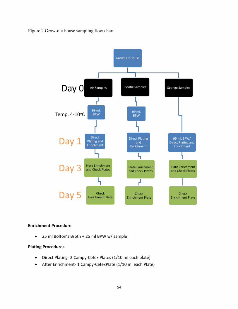
54
Figure 2.Grow-out house sampling flow chart
Enrichment Procedure
25 ml Bolton’s Broth + 25 ml BPW w/ sample
Plating Procedures
Direct Plating- 2 Campy-Cefex Plates (1/10 ml each plate)
After Enrichment- 1 Campy-CefexPlate (1/10 ml each Plate)
Day 5
Day 3
Day 1
Temp. 4-10oC
Day 0
Grow Out House
Air Samples
99 mL BPW
Direct Plating and Enrichment
Plate Enrichment and Check Plates
Check Enrichment Plate
Bootie Samples
99 mL BPW
Direct Plating and
Enrichment
Plate Enrichment and Check Plates
Check Enrichment Plate
Sponge Samples
99 mL BPW/ Direct Plating and
Enrichment
Plate Enrichment and Check Plates
Check Enrichment Plate
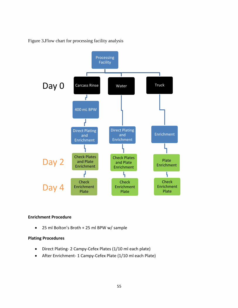
55
Figure 3.Flow chart for processing facility analysis
Enrichment Procedure
25 ml Bolton’s Broth + 25 ml BPW w/ sample
Plating Procedures
Direct Plating- 2 Campy-Cefex Plates (1/10 ml each plate)
After Enrichment- 1 Campy-Cefex Plate (1/10 ml each Plate)
Day 4
Day 2
Day 0
Processing Facility
Carcass Rinse
400 mL BPW
Direct Plating and
Enrichment
Check Plates and Plate
Enrichment
Check Enrichment
Plate
Water
Direct Plating and
Enrichment
Check Plates and Plate
Enrichment
Check Enrichment
Plate
Truck
Enrichment
Plate Enrichment
Check Enrichment
Plate
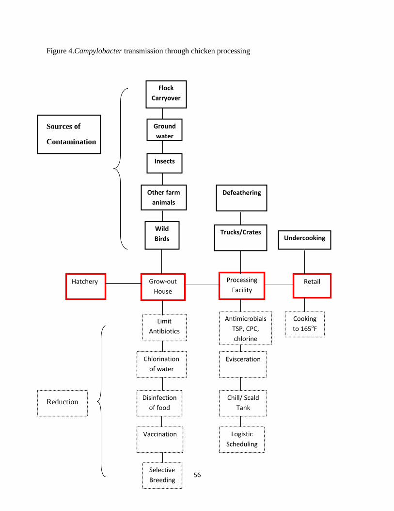
56
Figure 4.Campylobacter transmission through chicken processing
Sources of
Contamination
Reduction
Grow-out
House
Hatchery Retail Processing
Facility
Wild
Birds
Other farm
animals
Insects
Ground
water
Flock
Carryover
Limit
Antibiotics
Chlorination
of water
Disinfection
of food
Vaccination
Selective
Breeding
Antimicrobials
TSP, CPC,
chlorine
Evisceration
Chill/ Scald
Tank
Trucks/Crates
Defeathering
Logistic
Scheduling
Undercooking
Cooking
to 165oF
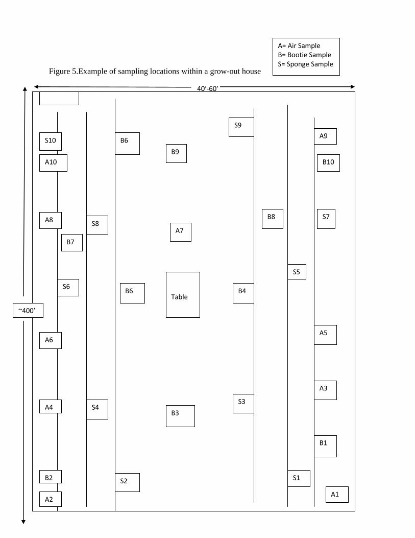
57
Figure 5.Example of sampling locations within a grow-out house
40’-60’
Table
B9
B6 S10
A10
S9
A9
B10
A7
A8
B7
S8 B8 S7
S5
B4 B6 S6
A6
A4
A2
B2
S4 B3
S3
A5
A3
S1 S2
B1
A1
~400’
A= Air Sample B= Bootie Sample S= Sponge Sample