Embed Size (px)
Citation preview

Benign Tumours of Epithelial Origin
• Papilloma• C/f
• Age 2-5th decade of life• More in males, can affect an sex• Most common site is tongue, followed by buccal mucosa, lip, gingiva• Clinically- cauliflower like growth, having finger like projections, usually
pedunculated, may be sessile, pinkish in colour; if keratinized, then colour is greyish white
• Usually painless unless secondarily infected
• Resembles verucca vulgaris occuring on fingers, caused by HPV

Papilloma Contd..
• Histological FeaturesEach finger like process has a connective tissue core covered by epithelium which is hyperplasticIf keratinized, then para or ortho keratinization is found on the surface of projectionsInflammatory cells may be present if ulcerated surface

Keratoacanthoma or self healing carcimoma
• It is a benign epithelial neoplasm which histologically resembles carcinoma
• Etiology unknown, suggested to be viral, genetic, chemical carcinogens etc.

Keratoacanthoma
•CLINICAL FEATURES:
• Usually occurs on external surface like lip, nose, cheek, Zygoma etc.
• Age 5th to 7th decade of life• More common in males• 1.5 to 2 cm in diameter

Keratoacanthoma: Clinical course
• Initially appears as nodule, which ulcerates and becomes crater like ulcerated nodule, keratin is present within the ulcer
• It grows to its full size in 4-8 weeks, remains static, then in following 6-8 weeks, it expels the central keratin core and heals, hence called as self healing carcinoma, recurrence is rare.

Keratoacanthoma
• Histological Features:• Hyperplastic squamous epithelium
growing into underlying connective tissue• Surface covered by thick layer of ortho &
para Keratin with central plugging• Occasional dysplastic features are seen in
the epithelial cells• At the leading margin of tumour, islands
of epithelium appear to be invading connective tissue and it is almost impossible to differentiate it from Squamous cell Carcinoma
• Perineural invasion has also been reported, but does not have an adverse effect on biological nature of tumour

Keratoacanthoma H/F Contd..• Connective tissue is infiltrated with chronic inflammatory cells• At the margin of the lesion, the normal adjacent epithelium is elevated towards the central
portion of crater, then an abrupt change of normal epithelium occurs into Hyperplastic Acanthotic epithelium
• Hence, Inclusion of the adjacent border of specimen is must in the biopsy to reach a conclusion.

Benign Epithelial Tumors Contd..
• Pigmented Cellular Nevus
• Nevus is a developmental tumor like malformation, composed of nevus cells.
• Melanin pigment is present in tumor cells

Pigmented Cellular Nevus
• Two types• Congenital
• Small - 3-5 cms in size• Garment - more than 10 cms in size
• Acquired• IntradermalJunctional• Compound• Spindle cell type• Blue Nevus

Pigmented Cellular Nevus
Congenital Nevus• These are macules, Pale or
coloured and are raised or elevated
• These subsequently show hair growth on their surface
• Are present at birth
Acquired Nevus• Present 2-3 yrs after birth• Number increases till 3rd decade of life,
then decrease in number• Usually small in size• Pigmented• With or without hair growth on
surface• This type is mostly seen in oral cavity,
common sites are palate & gingiva

Histopathology:
• Main cell is nevus cell. These are large ovoid cells with vesiculated nuclei, and pale cytoplasm
• They are derived from neural crest cells

Melanocytes in Normal epithelium

Normal & benign lesions (melanocyte related)
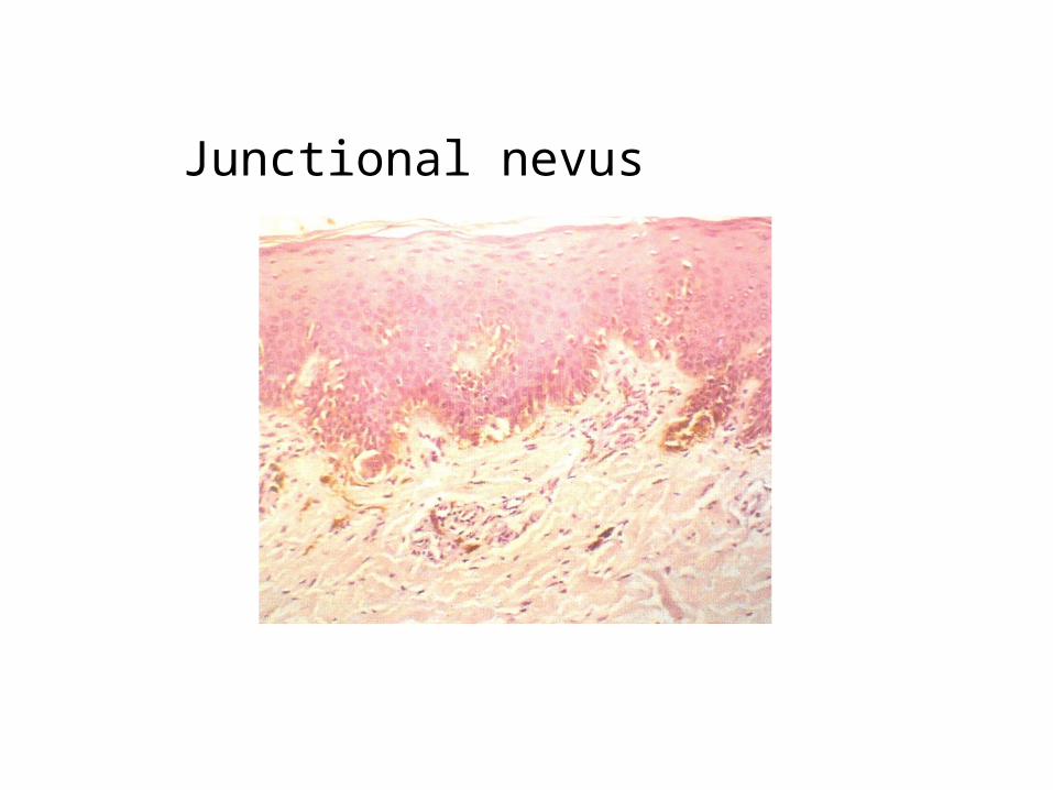
Junctional nevus

Compound Nevus-Shows junctional activity-& nests of nevus cells in connective tissue.

Intradermal Nevus

Blue Nevus