Embed Size (px)
Citation preview

Biochimica et Biophysica Acta 1782 (2008) 559–565
Contents lists available at ScienceDirect
Biochimica et Biophysica Acta
j ourna l homepage: www.e lsev ie r.com/ locate /bbadis
Changes in protein structure and distribution observed at pre-clinical stages ofscrapie pathogenesis
Ariane Kretlow a,b, Qi Wang b, Michael Beekes c, Dieter Naumann a, Lisa M. Miller b,⁎a P25, Robert Koch-Institut, Nordufer 20, 13353 Berlin, Germanyb National Synchrotron Light Source, Bldg 725D Brookhaven National Laboratory, 75 Brookhaven Avenue Upton, NY 11973, USAc P24, Robert Koch-Institut, Nordufer 20, 13353 Berlin, Germany
Abbreviations: TSE, Transmissible Spongiform Enceganglia; fcs, first clinical signs; dpi, days post-infectioInfrared Microspectroscopy⁎ Corresponding author. Tel.: +1 631 344 2091; fax: +
E-mail address: [email protected] (L.M. Miller).
0925-4439/$ – see front matter © 2008 Elsevier B.V. Aldoi:10.1016/j.bbadis.2008.06.004
a b s t r a c t
a r t i c l e i n f oArticle history:
Scrapie is a neurodegenera Received 31 January 2008Received in revised form 2 June 2008Accepted 3 June 2008Available online 14 June 2008Keywords:PrionScrapieDorsal root gangliaProtein structureProtein-foldingInfrared microspectroscopy
tive disorder that involves the misfolding, aggregation and accumulation of theprion protein (PrP). The normal cellular PrP (PrPC) is rich in α-helical secondary structure, whereas thedisease-associated pathogenic form of the protein (PrPSc) has an anomalously high β-sheet content. In thisstudy, protein structural changes were examined in situ in the dorsal root ganglia from perorally 263Kscrapie-infected and mock-infected hamsters using synchrotron Fourier Transform InfraRed Microspectro-scopy (FTIRM) at four time points over the course of the disease (pre-clinical, 100 and 130 days post-infection(dpi); first clinical signs (∼145 dpi); and terminal (∼170 dpi)). Results showed clear changes in the totalprotein content, structure, and distribution as the disease progressed. At pre-clinical time points, the scrapie-infected animals exhibited a significant increase in protein expression, but the β-sheet protein content wassignificantly lower than controls. Based on these findings, we suggest that the pre-clinical stages of scrapieare characterized by an overexpression of proteins low in β-sheet content. As the disease progressed, the β-sheet content increased significantly. Immunostaining with a PrP-specific antibody, 3F4, confirmed that thisincrease was partly – but not solely – due to the formation of PrPSc in the tissue and indicated that otherproteins high in β-sheet were produced, either by overexpression or misfolding. Elevated β-sheet wasobserved near the cell membrane at pre-clinical time points and also in the cytoplasm of infected neurons atlater stages of infection. At the terminal stage of the disease, the protein expression declined significantly,likely due to degeneration and death of neurons. These dramatic changes in protein content and structure,especially at pre-clinical time points, emphasize the possibility for identifying other proteins involved inearly pathogenesis, which are important for a further understanding of the disease.
© 2008 Elsevier B.V. All rights reserved.
1. Introduction
A characteristic feature of scrapie, a neurodegenerative disordernaturally occurring in sheep and goats, is the accumulation of a β-sheet rich protein termed PrPSc, or scrapie-associated prion protein,predominantly in the brain and spinal cord. First described in 1732 insheep, scrapie belongs to the family of Transmissible SpongiformEncephalopathies (TSEs), which can also occur in cattle (bovinespongiform encephalopathy), deer (chronic wasting disease), mink(transmissible mink encephalopathy) and other animals. The Creutz-feldt–Jakob disease in humans can occur sporadically but may alsohave genetic and infectious origins, which is a unique feature of thisgroup of diseases. According to the prion hypothesis, the causativeagent of TSEs is believed to be a proteinaceous infectious particle
phalopathy; DRG, dorsal rootn; FTIRM, Fourier Transform
1 631 344 3238.
l rights reserved.
(“prion”) that lacks agent-specific nucleic acid and consists mainly – ifnot entirely – of misfolded and pathologically aggregated prionprotein [1]. Normal PrP and its pathological isoform have an identicalamino acid sequence but different secondary (and tertiary) structures.PrPC, the cellular prion protein, mainly expressed by neuronal cells, ishigh in α-helix (42%) and consists of only 3% β-sheet, whereas themisfolded or disease-associated form PrPSc shows a high amount of β-sheet structure (43%) and has less α-helix (30%) [2].
The nervous system of hamsters orally infected with scrapie hasbeen studied extensively for the temporal–spatial course of PrPSc
deposition (for review see: [3]). After centripetally reaching the spinalcord, subsequent centrifugal spread of PrPSc depositionproceeds to thecorresponding afferent dorsal root ganglia (DRG, nodules on a dorsalspinal root that contain cell bodies of afferent nerve fibers), where theprotein can first be detected 76 days post-infection in half of theexamined hamsters [4–6]. Immunostained sections have shown thatPrPSc does not accumulate in all neurons of theDRG,where only certaincells stain strongly. In prion research, conventional techniques likeWestern Blot analysis are sensitive for examining the total amount ofPrPSc in homogenized tissue. In situ, PET blotting (paraffin embedded

Fig. 1. Mean values±average deviation for the relative (A) β-sheet protein content, (B)α-helix protein content, and (C) total protein content for scrapie-infected (black lines)and control (gray lines) hamster dorsal root ganglia at the four investigated time points.Asterisks mark significant differences (pb0.05) between scrapie and control.
560 A. Kretlow et al. / Biochimica et Biophysica Acta 1782 (2008) 559–565
tissue blot) [7] labels the misfolded prion protein directly within thetissue, providing spatially-resolved information on the location ofPrPSc. However, both techniques are limited to the detection of PrPSc,and do not provide information on other compositional changes thatoccur throughout the disease, especially at early time points inpathogenesis before PrPSc shows detectable accumulation.
In contrast, Fourier Transform InfraredMicrospectroscopy (FTIRM) isan in situ technique that is not restricted to the detection of PrPSc. In fact,an infrared spectrum of a biological sample is composed of character-istic absorption bands that originate from all tissue components, e.g.proteins, lipids, nucleic acids, and carbohydrates. Since the combinationof all molecular parameters (structure, composition, and/or interac-tions) in a specific tissue, cell type, or subcellular component is unique,FTIRM is an in situ technique for examining the molecular compositionof biological materials [8]. No labels, stains or dyes are required for FTIRmicrospectroscopy and it is a non-destructive, sensitive and fastanalytical tool. Thus, coupled with PrPSc-specific imaging techniques,FTIRM can be used to determine compositional changes in prion-infected tissue associated with scrapie pathogenesis [9,10].
In this study, protein content, structure, and distribution through-out scrapie pathogenesis were investigated using synchrotron FTIRM.Syrian hamsters (Mesocricetus auratus) perorally infected with scrapiestrain 263K were studied at 4 different time points: 100 days post-infection (dpi), 130 dpi, at first clinical signs (fcs, ∼145 dpi) and at theterminal stage (∼170 dpi) of the disease. The total protein, the α-helical protein, and the β-sheet protein contents and distributionswere determined as a function of disease progression and correlatedwith PrPSc immunostaining. The aim of this study was to examinechanges in protein composition throughout scrapie pathogenesis, andto correlate these findings with the temporal–spatial course of PrPSc
deposition. Identification of protein changes involved in early scrapiepathogenesis is important for understanding the disease process andfor identifying new approaches for early disease detection andtreatment with other, target-oriented techniques.
2. Materials and methods
2.1. Animal experiments and tissue preparation
All animal experiments were carried out in accordance withEuropean, German, and USA legal and ethical regulations. 20 outbredSyrian hamsters at an age of approximately 8 weeks were fed foodpellets containing 100 μl of a 10% (w/v) hamster brain homogenate(corresponding to 10 mg of tissue) from terminally ill 263K scrapie–infected donors or uninfected controls as described elsewhere [11].Twelve mock-infected hamsters of the same age were similarly fedwith normal brain homogenate and served as controls. Five infectedand three control hamsters each were euthanized with CO2, at each offour time points: 100 dpi, 130 dpi, fcs (∼145 dpi), and at the terminalstage of the disease (∼170 dpi). While the first two time points werefixed dates, the fcs and terminal stages depend on the progression ofthe disease in each individual animal. Fcs is defined as a stage wherethe animals start to show symptoms that are specific for a 263K scrapieinfection in hamsters. These are most commonly a hypersensitivity totouch and noise, where the animals often twitch, or have difficulties inmaintaining balance and rising from a supine position. At the terminalstage, animals showheadbobbing, ataxia of gait, and generalized tremor.
After euthanizing the animals, the dorsal root ganglia attached tothe thoracic spinal cord were removed and stored at −70 °C. Afterembedding the samples in Jung tissue freezing medium (LeicaInstruments, Germany), 10 μm thick cryosections were cut at atemperature of −20 °C and mounted on IR transparent slides (CaF2,1 mm in thickness) (Korth Kristalle GmbH, Altenholz, Germany).Adjacent sections were cut at the same thickness and mounted onstandard glass microscope slides for immunostaining. The CaF2 slideswere kept in a dry and dark environment until the FTIR microspectro-
scopic measurements were carried out. One animal died beforereaching the stage of fcs, resulting in a total of 19 infected and 12mock-infected control animals included in the study.
2.2. Immunohistochemistry
Adjacent sections on glass slides were stained for PrP using themonoclonal antibody 3F4 as described previously [12]. In short, theslideswerefixed three times: 5min each in 50,100 and50% ethanol, andthen rehydrated in distilled water. The ganglia were then treated with10% phosphate buffered formalin for 15 min, 100 mM NH4Cl and 0.5%Igepal in PBS (pH 7.8) for 5 min each. Sections were denatured in 3 MGuanidiniumthiocyanate/10 mM Tris–HCl (pH 7.8). They were blockedovernightwith fetal calf serum inTBST/0.2% Tween 20 at 4 °C, incubatedwith antibody 3F4 (1:100) for 1 h 45 min and then with biotinylatedsecondary antibody Goat-anti-Mouse (Dako E 0433) (1:200) in PBS/1%BSA. Finally the sections were incubated with Vectastain ABC Kit andstained with preincubation and incubation solution containing 3,3-Diaminobenzidin (DAB) to visualize the reaction product for 10min and3 min, respectively. For negative controls, ganglia slices were incubatedin normal mouse serum (Dako) (1:1000) instead of mAb 3F4 prior toincubationwith the secondary antibody. After FTIRMdata collection, theganglia on CaF2 windows were also immunostained accordingly.
2.3. FTIRM data collection
The FTIRM experiments were conducted at beamline U10B at theNational Synchrotron Light Source, Brookhaven National Laboratory(Upton, NY). AThermoNicoletMagna 860 FTIR spectrometer, coupled to

561A. Kretlow et al. / Biochimica et Biophysica Acta 1782 (2008) 559–565
a Continuum FTIR microscope (ThermoNicolet, Madison, WI), was usedwith synchrotron light as the infrared source. The microscope wasequipped with matching 32×Schwarzschild objectives, a motorized x–ymapping stage, an adjustable rectangular aperture, and a mercurycadmium telluride (MCT-A) detector. The aperture of the microscopewas set to 10×10 μm. A pre-defined area of approximately 100×100 μmencompassing about 15–20 cells was raster-scanned with a step size of4 μm using Omnic software (ThermoNicolet). At each point, anabsorbance spectrum was recorded in transmission mode. Eachspectrum was collected in the mid-infrared spectral range (800–4000 cm−1) with a spectral resolution of 8 cm−1 and 128 scans co-added. Happ–Genzel apodization and a zero-filling of level 2 wereapplied, resulting in approximately 1 data point per wave number. Abackground spectrumon clean substratewas also recordedbyco-adding512 scans. For each animal, one or two infrared maps were collected,yielding an average of 10,500 spectra from 138 cells (∼29 cells peranimal) per time point from the infected animals and 8887 spectra from110 cells (∼37 cells per animal) per time point from the controls.
2.4. Data analysis
The FTIRM data were analyzed using Thermo Nicolet's softwareOmnic 7.3. Absorbances from 1616 to 1636 cm−1 of protein infraredspectra are attributed primarily to β-sheet structures, which havebeen shown to increase in studies of recombinant and protease-resistant Syrian hamster PrP in vitro [13−16]. Thus, to examine therelative content of β-sheet structure in the tissue, each absorbancespectrum was integrated from 1624–1628 cm−1, applying a linearbaseline from 1480–1715 cm−1. To normalize for potential differencesin tissue thickness among samples, the β-sheet area was divided bythe area under the Amide I protein band (1600–1700 cm−1), applyingthe same linear baseline. Accordingly, the relative α-helical contentwas calculated by integrating each absorbance spectrum from 1654–1658 cm−1 and normalizing to the area under the Amide I band. Forboth α-helix and β-sheet content, narrow regions were integrated in
Fig. 2. (A) Photomicrographs of unstained cryosections (1st and 3rd column) and correspondinfected (right) ganglia at different time points. Scale bar: 20 μm.
order to prevent overlap with other secondary structure elements[17]. Chemical images in α-helix and β-sheet content were generatedusing Transform (Fortner Software, USA). To estimate the relativeprotein content at each pixel, the integrated area under the Amide Iprotein band was divided by the area under the entire infraredspectrum, in order to normalize for potential differences in tissuethickness among samples. The total integrated area was calculated asthe integrated area from 2500–3700 cm−1 (baseline from 2500 to3700 cm−1) plus the integrated area from 1760–1000 cm−1 (baselinefrom 1760–1000 cm−1). The whole spectral regionwas chosen since itincludes the vibrational modes of C–H, N–H and O–H bonds of allmajor biomolecules and therefore represents the total biomass. Thus,by dividing the total protein by the total biomass, a relative estimate oftotal protein content was determined.
For each animal, theα-helix,β-sheet, and total protein content weredetermined for every pixel in each map. These values were averaged toobtain a mean and standard deviation for each animal. Then theindividual animal values were averaged at their respective time points.An unpaired, two-sided t-test (Student's t-test) was performed on alldata to test for significant differences (pb0.05) between scrapie andcontrol at each time point, and differences between time points. In theanalysis, those spectra derived from areas outside the ganglion or fromtissue artifacts were excluded from the calculation.
3. Results
To examine changes in the secondary structure of proteins as afunction of disease severity, the mean values±standard deviation forβ-sheet content were calculated and plotted in Fig. 1A. As diseaseprogressed, results showed that the amount of β-sheet remainedrelatively constant in control hamsters (gray line), but substantiallyincreased with time in the scrapie-infected animals (black line). Themost dramatic difference between the control and infected animalswas observed at 100 dpi, where the β-sheet content in scrapie-infected ganglia was significantly lower than in the control ganglia
ing FTIRM images (2nd and 4th column) of the β-sheet distribution for control (left) and

Fig. 3. Original and second derivative spectra derived from near the cell membrane (black line) and within the cytoplasm (gray line) of a 100 dpi scrapie-infected animal.
562 A. Kretlow et al. / Biochimica et Biophysica Acta 1782 (2008) 559–565
(p=0.013). Over the course of the disease, we found a significantincrease in β-sheet content in the infected animals of about 8%(p=0.003). In contrast, the β-sheet content in the control gangliaremained unchanged over the time course studied here.
In contrast to the dramatic changes in β-sheet, Fig. 1B shows thatthe α-helical content was unaffected by the disease (black line) or byage (control, gray line).
To determine how the changes in protein structure were related tototal protein expression, the total protein content was determined as afunction of disease severity (Fig. 1C). At 100 dpi, the scrapie-infected
Fig. 4. Photomicrographs of antibody 3F4-stained adjacent sections. PrPSc can be detected aneuronal and satellite cells at later stages of incubation. Scale bar: 30 μm.
animals (black line) showed a significant increase in relative proteincontent when compared to the control animals (p=0.020). Moreover,the total protein content of scrapie-infected animals (black line)declined gradually during pathogenesis and reached a significantlylower level at the terminal stage of the disease (p=0.013). The controlanimals did not exhibit a change in total protein expressionthroughout the time course studied here.
In addition to measuring relative protein content and structure,the spatial distribution of β-sheet protein within the tissue wasalso examined. In Fig. 2, photomicrographs and corresponding
s granular deposits of immunoreactive material (arrows) in many – but not all –of the
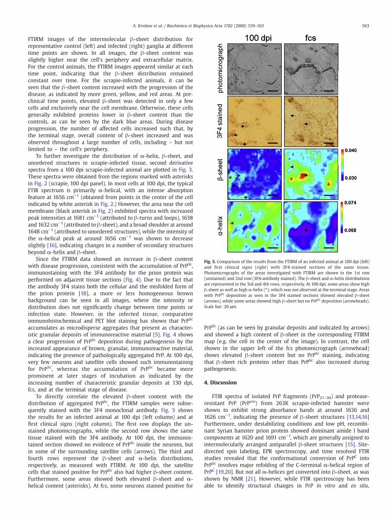
Fig. 5. Comparison of the results from the FTIRM of an infected animal at 100 dpi (left)and first clinical signs (right) with 3F4-stained sections of the same tissue.Photomicrographs of the areas investigated with FTIRM are shown in the 1st row(unstained) and 2nd row (3F4-antibody stained). The β-sheet and α-helix distributionsare represented in the 3rd and 4th rows, respectively. At 100 dpi, some areas show highβ-sheet as well as high α-helix (⁎), which was not observed at the terminal stage. Areaswith PrPSc deposition as seen in the 3F4 stained sections showed elevated β-sheet(arrows), while some areas showed high β-sheet but no PrPSc deposition (arrowheads).Scale bar: 20 μm.
563A. Kretlow et al. / Biochimica et Biophysica Acta 1782 (2008) 559–565
FTIRM images of the intermolecular β-sheet distribution forrepresentative control (left) and infected (right) ganglia at differenttime points are shown. In all images, the β-sheet content wasslightly higher near the cell's periphery and extracellular matrix.For the control animals, the FTIRM images appeared similar at eachtime point, indicating that the β-sheet distribution remainedconstant over time. For the scrapie-infected animals, it can beseen that the β-sheet content increased with the progression of thedisease, as indicated by more green, yellow, and red areas. At pre-clinical time points, elevated β-sheet was detected in only a fewcells and exclusively near the cell membrane. Otherwise, these cellsgenerally exhibited proteins lower in β-sheet content than thecontrols, as can be seen by the dark blue areas. During diseaseprogression, the number of affected cells increased such that, bythe terminal stage, overall content of β-sheet increased and wasobserved throughout a large number of cells, including – but notlimited to – the cell's periphery.
To further investigate the distribution of α-helix, β-sheet, andunordered structures in scrapie-infected tissue, second derivativespectra from a 100 dpi scrapie-infected animal are plotted in Fig. 3.These spectra were obtained from the regions marked with asterisksin Fig. 2 (scrapie, 100 dpi panel). In most cells at 100 dpi, the typicalFTIR spectrum is primarily α-helical, with an intense absorptionfeature at 1656 cm−1 (obtained from points in the center of the cellindicated by white asterisk in Fig. 2.) However, the area near the cellmembrane (black asterisk in Fig. 2) exhibited spectra with increasedpeak intensities at 1681 cm−1 (attributed to β-turns and loops), 1638and 1632 cm−1 (attributed to β-sheet), and a broad shoulder at around1648 cm−1 (attributed to unordered structures), while the intensity ofthe α-helical peak at around 1656 cm−1 was shown to decreaseslightly [16], indicating changes in a number of secondary structuresbeyond α-helix and β-sheet.
Since the FTIRM data showed an increase in β-sheet contentwith disease progression, consistent with the accumulation of PrPSc,immunostaining with the 3F4 antibody for the prion protein wasperformed on adjacent tissue sections (Fig. 4). Due to the fact thatthe antibody 3F4 stains both the cellular and the misfolded form ofthe prion protein [18], a more or less homogeneous brownbackground can be seen in all images, where the intensity ordistribution does not significantly change between time points orinfection state. However, in the infected tissue, comparativeimmunohistochemical and PET blot staining has shown that PrPSc
accumulates as microdisperse aggregates that present as character-istic granular deposits of immunoreactive material [5]. Fig. 4 showsa clear progression of PrPSc deposition during pathogenesis by theincreased appearance of brown, granular, immunoreactive material,indicating the presence of pathologically aggregated PrP. At 100 dpi,very few neurons and satellite cells showed such immunostainingfor PrPSc, whereas the accumulation of PrPSc became moreprominent at later stages of incubation as indicated by theincreasing number of characteristic granular deposits at 130 dpi,fcs, and at the terminal stage of disease.
To directly correlate the elevated β-sheet content with thedistribution of aggregated PrPSc, the FTIRM samples were subse-quently stained with the 3F4 monoclonal antibody. Fig. 5 showsthe results for an infected animal at 100 dpi (left column) and atfirst clinical signs (right column). The first row displays the un-stained photomicrographs, while the second row shows the sametissue stained with the 3F4 antibody. At 100 dpi, the immunos-tained section showed no evidence of PrPSc inside the neurons, butin some of the surrounding satellite cells (arrows). The third andfourth rows represent the β-sheet and α-helix distributions,respectively, as measured with FTIRM. At 100 dpi, the satellitecells that stained positive for PrPSc also had higher β-sheet content.Furthermore, some areas showed both elevated β-sheet and α-helical content (asterisks). At fcs, some neurons stained positive for
PrPSc (as can be seen by granular deposits and indicated by arrows)and showed a high content of β-sheet in the corresponding FTIRMmap (e.g. the cell in the center of the image). In contrast, the cellshown in the upper left of the fcs photomicrograph (arrowhead)shows elevated β-sheet content but no PrPSc staining, indicatingthat β-sheet rich proteins other than PrPSc also increased duringpathogenesis.
4. Discussion
FTIR spectra of isolated PrP fragments (PrP27–30) and protease-resistant PrP (PrPres) from 263K scrapie-infected hamster wereshown to exhibit strong absorbance bands at around 1636 and1626 cm−1, indicating the presence of β-sheet structures [13,14,16]Furthermore, under destabilizing conditions and low pH, recombi-nant Syrian hamster prion protein showed dominant amide I bandcomponents at 1620 and 1691 cm−1, which are generally assigned tointermolecularly arranged antiparallel β-sheet structures [15]. Site-directed spin labeling, EPR spectroscopy, and time resolved FTIRstudies revealed that the conformational conversion of PrPC intoPrPSc involves major refolding of the C-terminal α-helical region ofPrPC [19,20]. But not all α-helices get converted into β-sheet, as wasshown by NMR [21]. However, while FTIR spectroscopy has beenable to identify structural changes in PrP in vitro and ex situ,

564 A. Kretlow et al. / Biochimica et Biophysica Acta 1782 (2008) 559–565
identification of structural changes within scrapie-infected tissuehas been difficult.
Recently, a FTIRM study of terminally-diseased, 263K scrapiehamsters was the first to show a higher β-sheet content and a lowerα-helical content in infected DRG cells [22,23]. Here, this study wasextended to pre-clinical time points, and we find significant changesin protein structure, composition, and distribution throughout thecourse of the disease, while age-matched control animals did notexhibit any changes over the investigated time period. Specifically, wefind that elevated β-sheet content was detectable earlier in thedisease, i.e. with the onset of the first clinical signs. These findings areconsistent with work by McBride, et al., who used immunostaining toshow PrPSc accumulation as early as 76 days post-infection in the 263Kscrapie hamster model [5].
Over the course of the disease, results showed that the increasein β-sheet was ∼8%, well above the amount attributable to PrPSc,which was shown to represent only 0.1% of all proteins in the tissueat terminal stage [24]. Furthermore, FTIRM and correspondingimmunostain images revealed that not all areas with elevated β-sheet stained positive for PrPSc, indicating that other β-sheet richproteins also play a role in scrapie pathogenesis. Gene profiling of263K scrapie-infected hamster identified several upregulated genesat the terminal time point [25,26]. Among those were proteins withpredominantly β-sheet content, such as metallothionein [27],apolipoprotein J [28], IP-10 [29,30] and β2-microglobulin, the lastof which was shown to have amyloidogenic properties [31].However, since FTIRM spectra represent the average proteinstructure within the volume of tissue illuminated by the infraredlight, specific identification of these proteins is beyond the scope ofthis study.
While an increase in β-sheet content towards the terminal stage ofthe disease confirmed our initially proposed hypothesis, a signifi-cantly lower amount of β-sheet protein at pre-clinical time points wasan unexpected finding. Since the total protein content was signifi-cantly increased in the scrapie-infected animals at 100 dpi, we suggestthat the early scrapie infection is characterized by an overexpressionof proteins low in β-sheet content. Second derivative analysisconfirmed that most spectra showed predominantly high α-helicalcontent; however some spectra recorded near the cell's peripheryexhibited an increase in random coil and β-sheet content. Immuno-histochemistry studies have previously shown that the deposition ofpathological PrPSc starts in satellite cells and in the cytoplasm andplasmalemma of neurons [6]. PrPSc accumulation near the cellmembrane has also been observed in scrapie-infected mouseneuroblastoma cells [32], in cultured Chinese hamster ovary cells[33], in the medulla oblongata, pons, and astrocytes of sheep [34], andmice with natural scrapie [35,36].
At later stages of pathogenesis, the β-sheet content increasedthroughout the cell, which is consistent with (but not exclusivelyattributed to) the appearance of PrPSc aggregates in the cytoplasm ofneurons, primarily in secondary lysosomes [37], aggresomes [38], andin the nucleus [39]. Interestingly, accumulation of PrP in the cytosolhas been shown to be neurotoxic [40,41].
In summary, this study showed that pronounced protein-relatedchanges occur at the very early stages of scrapie pathogenesis, wellbefore clinical symptoms appear. Moreover, these changes go wellbeyond the transformation of PrPC to PrPSc. In the future, efforts toidentify these specific proteins that are involved at pre-clinical timepoints may provide an avenue for early disease detection andpossible targets for treatment of scrapie infection that are notavailable today.
Acknowledgements
The authors like to thank Marion Joncic and Kristin Kampf(Robert Koch-Institute) for skillful technical assistance with the
animal experiments. We are also grateful to the technical and safetystaff at the NSLS, especially to Randy Smith, Larry Carr, and AndrewAckerman. M. B. is grateful for ongoing support by the EU-fundedNetwork of Excellence “NeuroPrion”. This work was funded by theNational Institutes of Health Grant R01-GM66873. The NationalSynchrotron Light Source is funded by the U.S. Department of Energy,Office of Science, Office of Basic Energy Sciences, under Contract No.DE-AC02-98CH10886.
References
[1] S.B. Prusiner, Prion diseases and the BSE crisis, Science 278 (1997) 245–251.[2] K.M. Pan, M. Baldwin, J. Nguyen, M. Gasset, A. Serban, D. Groth, I. Mehlhorn, Z.
Huang, R.J. Fletterick, F.E. Cohen, et al., Conversion of alpha-helices into beta-sheets features in the formation of the scrapie prion proteins, Proc. Natl. Acad. Sci.U. S. A. 90 (1993) 10962–10966.
[3] M. Beekes, P.A. McBride, The spread of prions through the body in naturallyacquired transmissible spongiform encephalopathies, FEBS J. 274 (2007) 588–605.
[4] M. Beekes, P.A. McBride, E. Baldauf, Cerebral targeting indicates vagal spread ofinfection in hamsters fed with scrapie, J. Gen. Virol. 79 (Pt 3) (1998) 601–607.
[5] P.A. McBride, W.J. Schulz-Schaeffer, M. Donaldson, M. Bruce, H. Diringer, H.A.Kretzschmar, M. Beekes, Early spread of scrapie from the gastrointestinal tract tothe central nervous system involves autonomic fibers of the splanchnic and vagusnerves, J. Virol. 75 (2001) 9320–9327.
[6] P.A. McBride, M. Beekes, Pathological PrP is abundant in sympathetic and sensoryganglia of hamsters fed with scrapie, Neurosci. Lett. 265 (1999) 135–138.
[7] W.J. Schulz-Schaeffer, S. Tschoke, N. Kranefuss, W. Drose, D. Hause-Reitner, A.Giese, M.H. Groschup, H.A. Kretzschmar, The paraffin-embedded tissue blotdetects PrP(Sc) early in the incubation time in prion diseases, Am. J. Pathol. 156(2000) 51–56.
[8] D.L. Wetzel, S.M. LeVine, Imaging molecular chemistry with infrared microscopy,Science 285 (1999) 1224–1225.
[9] J. Kneipp, M. Beekes, P. Lasch, D. Naumann, Molecular changes of preclinicalscrapie can be detected by infrared spectroscopy, J. Neurosci. 22 (2002)2989–2997.
[10] J. Kneipp, P. Lasch, E. Baldauf, M. Beekes, D. Naumann, Detection of pathologicalmolecular alterations in scrapie-infected hamster brain by Fourier transforminfrared (FT-IR) spectroscopy, Biochim. Biophys. Acta. 1501 (2000) 189–199.
[11] E. Baldauf, M. Beekes, H. Diringer, Evidence for an alternative direct route of accessfor the scrapie agent to the brain bypassing the spinal cord, J. Gen. Virol. 78 (Pt 5)(1997) 1187–1197.
[12] Q. Wang, A. Kretlow, M. Beekes, D. Naumann, L. Miller, In situ characterization ofprion protein structure and metal accumulation in Scrapie-infected cells bysynchrotron infrared and X-ray imaging, Vibr. Spectrosc. 38 (2005) 61–69.
[13] A. Troullier, D. Reinstadler, Y. Dupont, D. Naumann, V. Forge, Transient non-nativesecondary structures during the refolding of alpha-lactalbumin detected byinfrared spectroscopy, Nat. Struct. Biol. 7 (2000) 78–86.
[14] B. Caughey, G.J. Raymond, R.A. Bessen, Strain-dependent differences in beta-sheetconformations of abnormal prion protein, J. Biol. Chem. 273 (1998) 32230–32235.
[15] F. Sokolowski, A.J. Modler, R. Masuch, D. Zirwer, M. Baier, G. Lutsch, D.A. Moss, K.Gast, D. Naumann, Formation of critical oligomers is a key event duringconformational transition of recombinant Syrian hamster prion protein, J. Biol.Chem. 278 (2003) 40481–40492.
[16] S. Spassov, M. Beekes, D. Naumann, Structural differences between TSEs strainsinvestigated by FT-IR spectroscopy, Biochim. Biophys. Acta. 1760 (2006)1138–1149.
[17] H. Fabian, D. Naumann, Methods to study protein folding by stopped-flow FT-IR,Methods 34 (2004) 28–40.
[18] R.J. Kascsak, R. Rubenstein, P.A. Merz, M. Tonna-DeMasi, R. Fersko, R.I. Carp, H.M.Wisniewski, H. Diringer, Mouse polyclonal and monoclonal antibody to scrapie-associated fibril proteins, J. Virol. 61 (1987) 3688–3693.
[19] N.J. Cobb, F.D. Sonnichsen, H. McHaourab, W.K. Surewicz, Molecular architectureof human prion protein amyloid: a parallel, in-register beta-structure, Proc. Natl.Acad. Sci. U. S. A. 104 (2007) 18946–18951.
[20] J. Ollesch, E. Kunnemann, R. Glockshuber, K. Gerwert, Prion protein alpha-to-betatransition monitored by time-resolved Fourier transform infrared spectroscopy,Appl. Spectrosc. 61 (2007) 1025–1031.
[21] J. Watzlawik, L. Skora, D. Frense, C. Griesinger, M. Zweckstetter, W.J. Schulz-Schaeffer, M.L. Kramer, Prion protein helix1 promotes aggregation but is notconverted into beta-sheet, J. Biol. Chem. 281 (2006) 30242–30250.
[22] J. Kneipp, L.M. Miller, M. Joncic, M. Kittel, P. Lasch, M. Beekes, D. Naumann, In situidentification of protein structural changes in prion-infected tissue, Biochim.Biophys. Acta. 1639 (2003) 152–158.
[23] J. Kneipp, L.M. Miller, S. Spassov, F. Sokolowski, P. Lasch, M. Beekes, D. Naumann,Scrapie-infected cells, isolated prions, and recombinant prion protein: acomparative study, Biopolymers 74 (2004) 163–167.
[24] M. Beekes, E. Baldauf, S. Cassens, H. Diringer, P. Keyes, A.C. Scott, G.A. Wells, P.Brown, C.J. Gibbs Jr., D.C. Gajdusek, Western blot mapping of disease-specificamyloid in various animal species and humans with transmissible spongiformencephalopathies using a high-yield purification method, J. Gen. Virol. 76 (Pt 10)(1995) 2567–2576.
[25] C. Riemer, I. Queck, D. Simon, R. Kurth, M. Baier, Identification of upregulatedgenes in scrapie-infected brain tissue, J. Virol. 74 (2000) 10245–10248.

565A. Kretlow et al. / Biochimica et Biophysica Acta 1782 (2008) 559–565
[26] M.J. Stobart, D. Parchaliuk, S.L. Simon, J. Lemaistre, J. Lazar, R. Rubenstein, J.D. Knox,Differential expression of interferon responsive genes in rodent models oftransmissible spongiform encephalopathy disease, Mol. Neurodegener. 2 (2007) 5.
[27] Y. Boulanger, C.M. Goodman, C.P. Forte, S.W. Fesik, I.M. Armitage, Model formammalian metallothionein structure, Proc. Natl. Acad. Sci. U. S. A. 80 (1983)1501–1505.
[28] M. Calero, T. Tokuda, A. Rostagno, A. Kumar, B. Zlokovic, B. Frangione, J. Ghiso,Functional and structural properties of lipid-associated apolipoprotein J (clus-terin), Biochem. J. 344 (Pt. 2) (1999) 375–383.
[29] V. Booth, D.W. Keizer, M.B. Kamphuis, I. Clark-Lewis, B.D. Sykes, The CXCR3binding chemokine IP10/CXCL10: structure and receptor interactions, Biochem-istry 41 (2002) 10418–10425.
[30] G.J. Swaminathan, D.E. Holloway, R.A. Colvin, G.K. Campanella, A.C. Papageorgiou,A.D. Luster, K.R. Acharya, Crystal structures of oligomeric forms of the IP10/CXCL10chemokine, Structure 11 (2003) 521–532.
[31] Z. Zhang, H. Chen, L. Lai, Identification of amyloid fibril-forming segments basedon structure and residue-based statistical potential, Bioinformatics 23 (2007)2218–2225.
[32] B. Caughey, G.J. Raymond, The scrapie-associated form of PrP is made from a cellsurface precursor that is both protease- and phospholipase-sensitive, J. Biol.Chem. 266 (1991) 18217–18223.
[33] S. Lehmann, D.A. Harris, Mutant and infectious prion proteins display commonbiochemical properties in cultured cells, J. Biol. Chem. 271 (1996) 1633–1637.
[34] L.J. van Keulen, B.E. Schreuder, R.H. Meloen, M. Poelen-van den Berg, G. Mooij-Harkes, M.E. Vromans, J.P. Langeveld, Immunohistochemical detection andlocalization of prion protein in brain tissue of sheep with natural scrapie, Vet.Pathol. 32 (1995) 299–308.
[35] M. Jeffrey, C.M. Goodsir, M.E. Bruce, P.A. McBride, N. Fowler, J.R. Scott, Murinescrapie-infected neurons in vivo release excess prion protein into the extracellularspace, Neurosci. Lett. 174 (1994) 39–42.
[36] M. Jeffrey, C.M. Goodsir, M.E. Bruce, P.A. McBride, J.R. Scott, Infection-specific prionprotein (PrP) accumulates on neuronal plasmalemma in scrapie-infected mice,Ann. N. Y. Acad. Sci. 724 (1994) 327–330.
[37] M. Jeffrey, G. McGovern, C.M. Goodsir, K.L. Brown,M.E. Bruce, Sites of prion proteinaccumulation in scrapie-infected mouse spleen revealed by immuno-electronmicroscopy, J. Pathol. 191 (2000) 323–332.
[38] E. Cohen, A. Taraboulos, Scrapie-like prion protein accumulates in aggresomes ofcyclosporin A-treated cells, EMBO J. 22 (2003) 404–417.
[39] A. Mange, C. Crozet, S. Lehmann, F. Beranger, Scrapie-like prion protein istranslocated to the nuclei of infected cells independently of proteasome inhibitionand interacts with chromatin, J. Cell. Sci. 117 (2004) 2411–2416.
[40] J. Ma, R. Wollmann, S. Lindquist, Neurotoxicity and neurodegeneration when PrPaccumulates in the cytosol, Science 298 (2002) 1781–1785.
[41] A.S. Rambold, M. Miesbauer, D. Rapaport, T. Bartke, M. Baier, K.F. Winklhofer, J.Tatzelt, Association of Bcl-2 with misfolded prion protein is linked to the toxicpotential of cytosolic PrP, Mol. Biol. Cell. 17 (2006) 3356–3368.