Embed Size (px)
Citation preview
Biochimica et Biophysica Acta 1792 (2009) 14–26
Contents lists available at ScienceDirect
Biochimica et Biophysica Acta
j ourna l homepage: www.e lsev ie r.com/ locate /bbadis
Review
Alternative splicing and disease
Jamal Tazi a, Nadia Bakkour a, Stefan Stamm b,⁎a University of Montpellier II, Institute of Molecular Genetics, Centre Nationale de Recherche Scientifique, 1919 Route de Mende, Franceb Department of Molecular and Cellular Biochemistry, University of Kentucky, College of Medicine, Lexington, KY 40536, USA
⁎ Corresponding author. Fax: +1 859 323 1037.E-mail address: [email protected] (S. Stamm).
0925-4439/$ – see front matter © 2008 Elsevier B.V. Adoi:10.1016/j.bbadis.2008.09.017
a b s t r a c t
a r t i c l e i n f oArticle history:
Almost all protein-coding ge Received 11 June 2008Received in revised form 19 September 2008Accepted 30 September 2008Available online 17 October 2008Keywords:Alternative splicingDiseaseSplicing codeMutation
nes are spliced and their majority is alternatively spliced. Alternative splicing is akey element in eukaryotic gene expression that increases the coding capacity of the human genome and anincreasing number of examples illustrates that the selection of wrong splice sites causes human disease. Afine-tuned balance of factors regulates splice site selection. Here, we discuss well-studied examples thatshow how a disturbance of this balance can cause human disease. The rapidly emerging knowledge ofsplicing regulation now allows the development of treatment options.
© 2008 Elsevier B.V. All rights reserved.
1. General principles of alternative splicing
1.1. Alternative pre-mRNA splicing regulates the function of the majorityof protein-coding genes
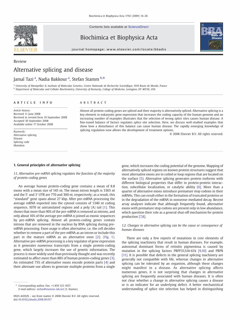
An average human protein-coding gene contains a mean of 8.8exons with a mean size of 145 nt. The mean intron length is 3365 ntand the 5′ and 3′ UTR are 770 and 300 nt, respectively; as a result, this“standard” gene spans about 27 kbp. After pre-mRNA processing theaverage mRNA exported into the cytosol consists of 1340 nt codingsequence, 1070 nt untranslated regions and a poly (A) tail [1]. Thisshows that more than 90% of the pre-mRNA is removed as introns, andonly about 10% of the average pre-mRNA is joined as exonic sequencesby pre-mRNA splicing. Almost all protein-coding genes containintrons that are removed in the nucleus by RNA splicing during pre-mRNA processing. Exon usage is often alternative, i.e. the cell decideswhether to remove a part of the pre-mRNA as an intron or include thispart in the mature mRNA as an alternative exon [2]; (Fig. 1).Alternative pre-mRNA processing is a key regulator of gene expressionas it generates numerous transcripts from a single protein-codinggene, which largely increases the use of genetic information. Theprocess is more widely used than previously thought andwas recentlyestimated to affect more than 88% of human protein-coding genes [3].An estimated 75% of alternative exons encode protein parts [4] andtheir alternate use allows to generate multiple proteins from a single
ll rights reserved.
gene, which increases the coding potential of the genome. Mapping ofalternatively spliced regions on known protein structures suggest thatmost alternative exons are in coiled or loop regions that are located onthe surface [5]. Alternative splicing generates protein isoforms withdifferent biological properties that differ in protein:protein interac-tion, subcellular localization, or catalytic ability [6]. More than aquarter of alternative exons introduce premature stop codons in theirmRNAs. This can result either in the formation of truncated proteins orin the degradation of the mRNA in nonsense-mediated decay. Recentarray analyses indicate that although frequently found, alternativeexons with premature stop codons are present only in low abundance,which question their role as a general shut-off mechanism for proteinproduction [7,8].
1.2. Changes in alternative splicing can be the cause or consequence ofhuman diseases
There are only a few reports of mutations in core elements ofthe splicing machinery that result in human diseases. For example,autosomal dominant forms of retinitis pigmentosa is caused bymutation in the splicing factors PRPF31/U4-61k [9,10] and PRP8[11]. It is possible that defects in the general splicing machinery aregenerally not compatible with life, whereas changes in alternativesplicing can be tolerated by an organism, although these changesmight manifest in a disease. As alternative splicing affectsnumerous genes, it is not surprising that changes in alternativesplicing are frequently associated with human diseases. It is oftennot clear whether a change in alternative splicing causes a diseaseor is an indicator for an underlying defect. A better mechanisticalunderstanding of splice site selection has helped in distinguishing
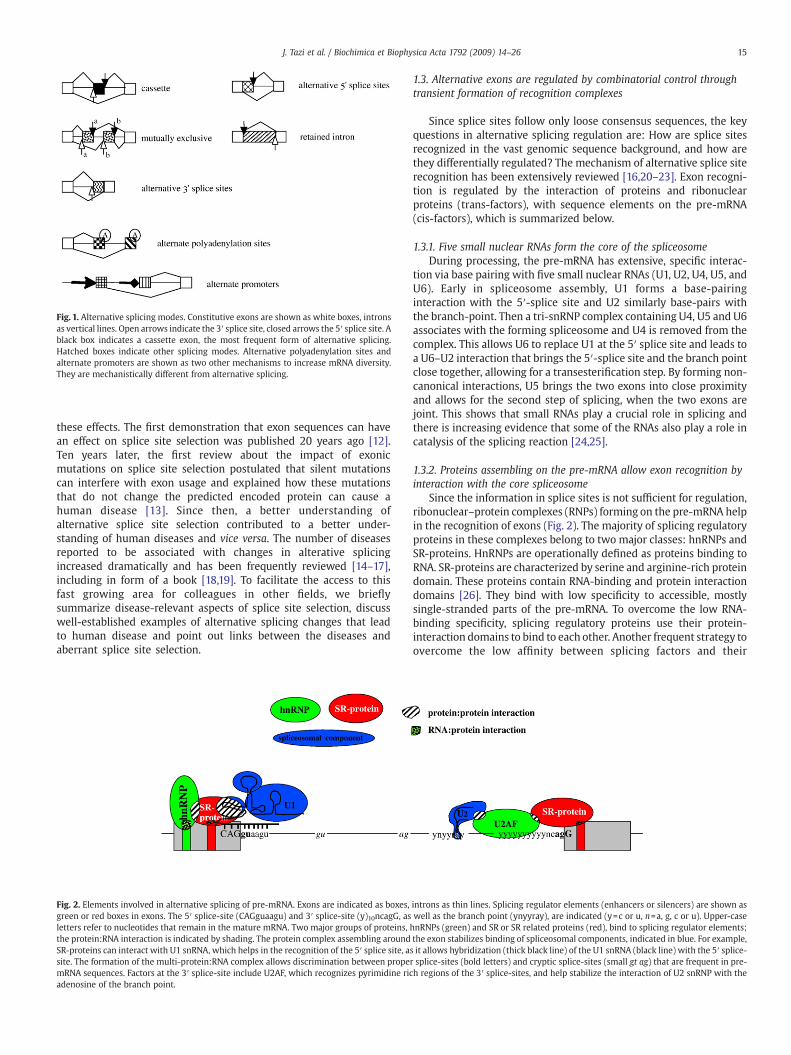
Fig. 1. Alternative splicing modes. Constitutive exons are shown as white boxes, intronsas vertical lines. Open arrows indicate the 3′ splice site, closed arrows the 5′ splice site. Ablack box indicates a cassette exon, the most frequent form of alternative splicing.Hatched boxes indicate other splicing modes. Alternative polyadenylation sites andalternate promoters are shown as two other mechanisms to increase mRNA diversity.They are mechanistically different from alternative splicing.
15J. Tazi et al. / Biochimica et Biophysica Acta 1792 (2009) 14–26
these effects. The first demonstration that exon sequences can havean effect on splice site selection was published 20 years ago [12].Ten years later, the first review about the impact of exonicmutations on splice site selection postulated that silent mutationscan interfere with exon usage and explained how these mutationsthat do not change the predicted encoded protein can cause ahuman disease [13]. Since then, a better understanding ofalternative splice site selection contributed to a better under-standing of human diseases and vice versa. The number of diseasesreported to be associated with changes in alterative splicingincreased dramatically and has been frequently reviewed [14–17],including in form of a book [18,19]. To facilitate the access to thisfast growing area for colleagues in other fields, we brieflysummarize disease-relevant aspects of splice site selection, discusswell-established examples of alternative splicing changes that leadto human disease and point out links between the diseases andaberrant splice site selection.
Fig. 2. Elements involved in alternative splicing of pre-mRNA. Exons are indicated as boxes,green or red boxes in exons. The 5′ splice-site (CAGguaagu) and 3′ splice-site (y)10ncagG, asletters refer to nucleotides that remain in the mature mRNA. Two major groups of proteins,the protein:RNA interaction is indicated by shading. The protein complex assembling aroundSR-proteins can interact with U1 snRNA, which helps in the recognition of the 5′ splice site, asite. The formation of the multi-protein:RNA complex allows discrimination between propemRNA sequences. Factors at the 3′ splice-site include U2AF, which recognizes pyrimidine ricadenosine of the branch point.
1.3. Alternative exons are regulated by combinatorial control throughtransient formation of recognition complexes
Since splice sites follow only loose consensus sequences, the keyquestions in alternative splicing regulation are: How are splice sitesrecognized in the vast genomic sequence background, and how arethey differentially regulated? The mechanism of alternative splice siterecognition has been extensively reviewed [16,20–23]. Exon recogni-tion is regulated by the interaction of proteins and ribonuclearproteins (trans-factors), with sequence elements on the pre-mRNA(cis-factors), which is summarized below.
1.3.1. Five small nuclear RNAs form the core of the spliceosomeDuring processing, the pre-mRNA has extensive, specific interac-
tion via base pairing with five small nuclear RNAs (U1, U2, U4, U5, andU6). Early in spliceosome assembly, U1 forms a base-pairinginteraction with the 5′-splice site and U2 similarly base-pairs withthe branch-point. Then a tri-snRNP complex containing U4, U5 and U6associates with the forming spliceosome and U4 is removed from thecomplex. This allows U6 to replace U1 at the 5′ splice site and leads toa U6–U2 interaction that brings the 5′-splice site and the branch pointclose together, allowing for a transesterification step. By forming non-canonical interactions, U5 brings the two exons into close proximityand allows for the second step of splicing, when the two exons arejoint. This shows that small RNAs play a crucial role in splicing andthere is increasing evidence that some of the RNAs also play a role incatalysis of the splicing reaction [24,25].
1.3.2. Proteins assembling on the pre-mRNA allow exon recognition byinteraction with the core spliceosome
Since the information in splice sites is not sufficient for regulation,ribonuclear–protein complexes (RNPs) forming on the pre-mRNA helpin the recognition of exons (Fig. 2). The majority of splicing regulatoryproteins in these complexes belong to two major classes: hnRNPs andSR-proteins. HnRNPs are operationally defined as proteins binding toRNA. SR-proteins are characterized by serine and arginine-rich proteindomain. These proteins contain RNA-binding and protein interactiondomains [26]. They bind with low specificity to accessible, mostlysingle-stranded parts of the pre-mRNA. To overcome the low RNA-binding specificity, splicing regulatory proteins use their protein-interaction domains to bind to each other. Another frequent strategy toovercome the low affinity between splicing factors and their
introns as thin lines. Splicing regulator elements (enhancers or silencers) are shown aswell as the branch point (ynyyray), are indicated (y=c or u, n=a, g, c or u). Upper-casehnRNPs (green) and SR or SR related proteins (red), bind to splicing regulator elements;the exon stabilizes binding of spliceosomal components, indicated in blue. For example,s it allows hybridization (thick black line) of the U1 snRNA (black line) with the 5′ splice-r splice-sites (bold letters) and cryptic splice-sites (small gt ag) that are frequent in pre-h regions of the 3′ splice-sites, and help stabilize the interaction of U2 snRNP with the
16 J. Tazi et al. / Biochimica et Biophysica Acta 1792 (2009) 14–26
recognition sequence is a repetitive arrangement of regulatorysequences. For example, exon 4 of the doublesex pre-mRNA containssix repeats, each 13 nt long, that bind to the SR-proteins tra, tra2 and9G8 [27]. Once these protein complexes have been formed around anexon, they aid ribonuclear protein components of the core spliceosomein establishing an RNA:RNA interaction at the 5′ splice site and at thebranchpoint. A well-studied interaction occurs between SR-proteinsand the U1 snRNP, which is part of the spliceosome. The RNAcomponent of the U1 snRNP interacts with the 5′ splice site, whichdefines one border of an exon. Since SR-proteins that bind to an exonstabilize the interaction with U1 snRNP and the 5′ splice site, theygenerally promote inclusion of exons to which they bind. The highfidelity of exon recognition is thus achieved by the combination ofmultiple weak protein:protein, protein:RNA and RNA:RNA interac-tions, which are schematically shown in Fig. 2. Different pre-mRNAsseem to associate with a unique arrangement of proteins, whichconstitutes the ‘splicing- or mRNP code’ [28]. By interacting with thespliceosome, the protein complexes forming on the pre-mRNA, i.e. thesplicing code, determine which RNA parts will be removed as intronsandwhich parts will be included in themature mRNA. The low affinityof each interaction is an intrinsic property of pre-mRNAprocessing andallows the transient formation of a specific protein:RNA complex fromseveral intrinsically weak interactions. This has several advantages: (i)it allows a high sequenceflexibility of exonic regulatory sequences thatputs no constrains on coding requirements, (ii) the protein interactioncan be influenced by small changes in the concentration of regulatoryproteins which allows the alternative usage of exons depending on atissue and/or developmental-specific concentration of regulatoryfactors, (iii) phosphorylation of regulatory factors that alter protein:protein-interactions can influence splice site selection, (iv) theregulatory proteins can be exchanged with other proteins after thesplicing reaction, allowing a dynamic processing of the RNA.
Alternative splice site selection is connected to other processingsteps, such as transcription, 5′ end capping, 3′end polyadenylation andnuclear export. Splicing regulatory proteins often function in multipleprocessing events while they remain bound to ‘their’ pre-mRNA [29].
1.3.3. The secondary structure of pre-mRNA influences splicesite selection
Secondary structures of the pre-mRNA can influence splice siteselection (reviewed in [30]). For example, the stability of a predictedstem–loop structure at the 5′ splice site of tau exon 10 regulates usageof this exon and the alternative exon of the fibronectin gene isinfluenced by secondary RNA structure [31]. Bioinformatic analysisindicate that the splicing regulatory sequences are preferentially in asingle-stranded conformation, which allows these sequences tointeract with RNA binding proteins that mostly have a preferencefor single strand RNA [32].
1.3.4. Small nucleolar RNAs regulate splice site selectionTiling array data and detailed expression analysis of parts of the
human genome in the ENCODE project showed that the expressiondata and gene structures collected in current databases are largelyincomplete [33]. Numerous noncoding regions are transcribed intopolyadenylated, stable RNAs, which are named TUF (transcript ofunknown function). Within most previously well-characterizedprotein coding genes, there are large numbers of transcribedfragments (transfrags) that could represent new exons or shortRNAs of unknown sequence. The most recent study of human geneexpression using tiling arrays demonstrates that about 57% of thetransfrags are not annotated in Genbank or EnsEMBL databases [34].Recently, it was discovered that a snoRNA, HBII-52, regulatesalternative splicing by binding to an alternative exon of the serotoninreceptor. This snoRNA is not expressed in peoplewith the Prader–Willisyndrome and it is likely that the lack of HBII-52 snoRNA expressioncontributes to the disease [35]. In support of this theory, a child with a
174,584 bp long microdeletion was reported. The deletion encom-passes only snoRNAs: HBII-438A, all snoRNAs of the HBII-85 clusterand 23 of the 47 HBII-52 snoRNAs. Since this child showsmost clinicalfeatures of Prader–Willi syndrome, it is now very clear that the loss ofsnoRNAs cause the disease [36], which is in agreement with geneticstudies and reflected by two recent mouse models [37–39]. A possiblelink between snoRNPs and splice site selection was mechanisticallyhard to understand, as snoRNPs reside in the nucleolus and splicingtakes place in the nucleoplasma. Recently, it was shown that nuclearexport and import factors are directly involved in U8 C/D box snoRNAbiogenesis, suggesting that C/D box snoRNPs are transported throughthe nucleoplasmawhile being assembled [40]. Furthermore, the 15.5Kprotein that was originally identified as part of the C/D box snoRNPcomplex where it binds to a conserved kink turn, binds also to asimilar structure in the U4 snRNA where it interacts with the splicingfactor hPrp31 [41]. These findings raise the possibility that 15.5Kprotein bound to snoRNPs interferes with the U4/U6 rearrangementduring splicing by interacting with hPrp31. It is therefore possible thatother small RNAs are also involved in splice site selection.
1.3.5. Exon-recognition complexes are targeted by cellular signaltransduction pathways
Cells frequently regulate alternative splicing events, often inresponse to external stimuli (reviewed in [42–44]). In most casesstudied, rapid changes observed in splice site selection are influencedby phosphorylation events that are regulated by known signaltransduction pathways. Reversible phosphorylation is a key regulatorystep in the formation of protein complexes on pre-mRNA (reviewed in[45]). SR-proteins are phosphorylated by several kinases, including theClk/Sty kinase family (cdc2 like kinases, Clk1-4), the SRPK family(SRPK1,2), mammalian PRP4, topoisomerase I, CDC2 kinase 2 [46], andGSK3 [47]. A change in phosphorylation of the RS-domain influencesits ability to interact with other proteins. For example, phosphoryla-tion of SRp38 decreases its binding to U170K snRNP, but increases itsbinding to tra2-alpha [48]. Phosphorylation of the SR-protein SF2/ASFincreased its binding to U1 70K snRNP [49] and decreased its bindingto the RNA export factor TAP/NXF1 [50]. Finally, the ability of the RS-domain to bind to RNA depends on its phosphorylation [49,51].
The phosphorylation is reversed by phosphatases; only about 25serine/threonine protein phosphatases are known [52]. This is incontrast to the estimated 474–518 protein kinases representing 1.7% ofhuman proteins [53]. The low number of protein phosphatases is theresult of a combinatorial control. The protein phosphatase representsonly the catalytically active subunit that forms numerous regulatorycomplexes with other proteins. Splicing regulatory proteins aredephosphorylated by protein phosphatase 1 (PP1), PP2A and proteinphosphatase 2Cgamma [54,55]. Complete blocking of phosphatasesinhibits splicing as dephosphorylation is necessary for the transester-ification step [54,56]. However, modulation of phosphatase activity invitro [57] and in vivo [58,59] influences alternative exon usage,demonstrating that adjusting phosphatase activity can be used bycells to control alternative splicing.
1.4. Alternative exons are generated during evolution and their usage canbe changed by point mutations located outside the splice sites
Alternative exons can be generated by three mechanisms(reviewed by [60]): (i) exon shuffling, where an existing exon isduplicated within the same gene and is then alternatively spliced, (ii)exonisation of mobile genetic elements, such as Alu elements [61] and(iii) a transformation of formally constitutive exons into alternativeones [62]. Since the approximately one million human Alu elementsare primate-specific elements that account for 10% of the humangenome [63], their exonisation provides a large reservoir to generatenew alternative exons. Numerous studies showed that synonymousmutations in coding regions can influence splice site selection. There
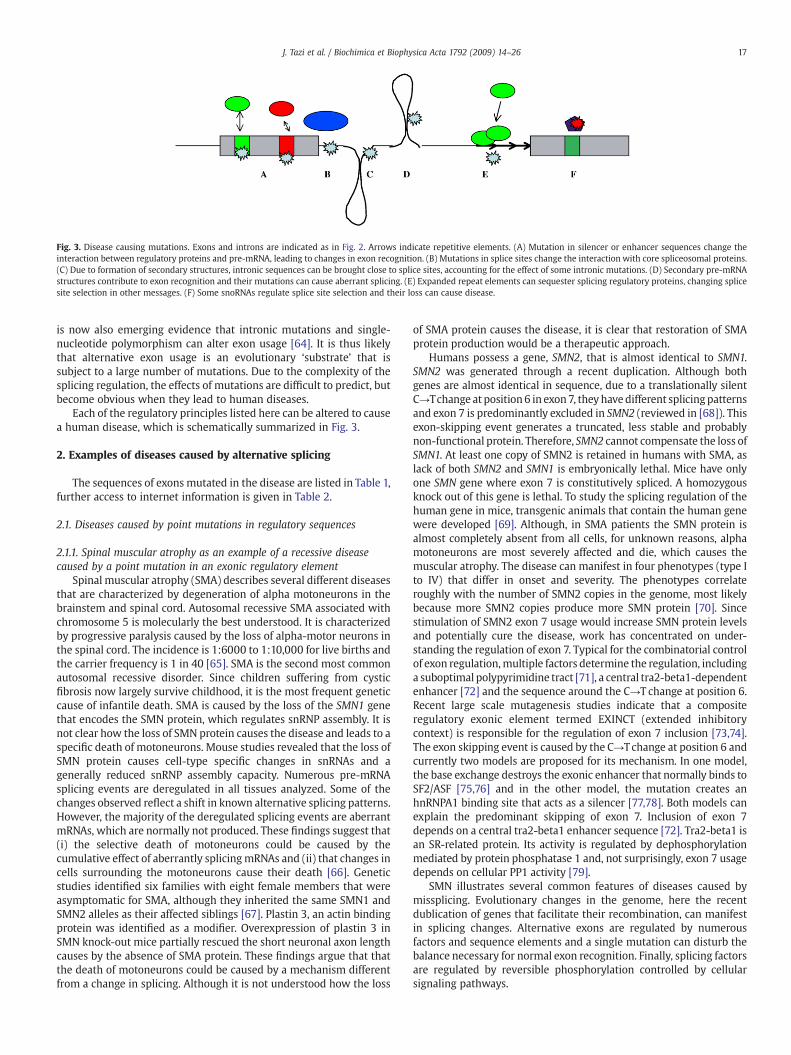
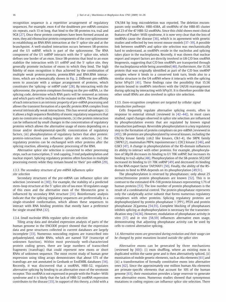
Fig. 3. Disease causing mutations. Exons and introns are indicated as in Fig. 2. Arrows indicate repetitive elements. (A) Mutation in silencer or enhancer sequences change theinteraction between regulatory proteins and pre-mRNA, leading to changes in exon recognition. (B) Mutations in splice sites change the interaction with core spliceosomal proteins.(C) Due to formation of secondary structures, intronic sequences can be brought close to splice sites, accounting for the effect of some intronic mutations. (D) Secondary pre-mRNAstructures contribute to exon recognition and their mutations can cause aberrant splicing. (E) Expanded repeat elements can sequester splicing regulatory proteins, changing splicesite selection in other messages. (F) Some snoRNAs regulate splice site selection and their loss can cause disease.
17J. Tazi et al. / Biochimica et Biophysica Acta 1792 (2009) 14–26
is now also emerging evidence that intronic mutations and single-nucleotide polymorphism can alter exon usage [64]. It is thus likelythat alternative exon usage is an evolutionary ‘substrate’ that issubject to a large number of mutations. Due to the complexity of thesplicing regulation, the effects of mutations are difficult to predict, butbecome obvious when they lead to human diseases.
Each of the regulatory principles listed here can be altered to causea human disease, which is schematically summarized in Fig. 3.
2. Examples of diseases caused by alternative splicing
The sequences of exons mutated in the disease are listed in Table 1,further access to internet information is given in Table 2.
2.1. Diseases caused by point mutations in regulatory sequences
2.1.1. Spinal muscular atrophy as an example of a recessive diseasecaused by a point mutation in an exonic regulatory element
Spinal muscular atrophy (SMA) describes several different diseasesthat are characterized by degeneration of alpha motoneurons in thebrainstem and spinal cord. Autosomal recessive SMA associated withchromosome 5 is molecularly the best understood. It is characterizedby progressive paralysis caused by the loss of alpha-motor neurons inthe spinal cord. The incidence is 1:6000 to 1:10,000 for live births andthe carrier frequency is 1 in 40 [65]. SMA is the second most commonautosomal recessive disorder. Since children suffering from cysticfibrosis now largely survive childhood, it is the most frequent geneticcause of infantile death. SMA is caused by the loss of the SMN1 genethat encodes the SMN protein, which regulates snRNP assembly. It isnot clear how the loss of SMN protein causes the disease and leads to aspecific death of motoneurons. Mouse studies revealed that the loss ofSMN protein causes cell-type specific changes in snRNAs and agenerally reduced snRNP assembly capacity. Numerous pre-mRNAsplicing events are deregulated in all tissues analyzed. Some of thechanges observed reflect a shift in known alternative splicing patterns.However, the majority of the deregulated splicing events are aberrantmRNAs, which are normally not produced. These findings suggest that(i) the selective death of motoneurons could be caused by thecumulative effect of aberrantly splicingmRNAs and (ii) that changes incells surrounding the motoneurons cause their death [66]. Geneticstudies identified six families with eight female members that wereasymptomatic for SMA, although they inherited the same SMN1 andSMN2 alleles as their affected siblings [67]. Plastin 3, an actin bindingprotein was identified as a modifier. Overexpression of plastin 3 inSMN knock-out mice partially rescued the short neuronal axon lengthcauses by the absence of SMA protein. These findings argue that thatthe death of motoneurons could be caused by a mechanism differentfrom a change in splicing. Although it is not understood how the loss
of SMA protein causes the disease, it is clear that restoration of SMAprotein production would be a therapeutic approach.
Humans possess a gene, SMN2, that is almost identical to SMN1.SMN2 was generated through a recent duplication. Although bothgenes are almost identical in sequence, due to a translationally silentC→Tchange at position6 in exon7, they have different splicingpatternsand exon 7 is predominantly excluded in SMN2 (reviewed in [68]). Thisexon-skipping event generates a truncated, less stable and probablynon-functional protein. Therefore, SMN2 cannot compensate the loss ofSMN1. At least one copy of SMN2 is retained in humans with SMA, aslack of both SMN2 and SMN1 is embryonically lethal. Mice have onlyone SMN gene where exon 7 is constitutively spliced. A homozygousknock out of this gene is lethal. To study the splicing regulation of thehuman gene in mice, transgenic animals that contain the human genewere developed [69]. Although, in SMA patients the SMN protein isalmost completely absent from all cells, for unknown reasons, alphamotoneurons are most severely affected and die, which causes themuscular atrophy. The disease can manifest in four phenotypes (type Ito IV) that differ in onset and severity. The phenotypes correlateroughly with the number of SMN2 copies in the genome, most likelybecause more SMN2 copies produce more SMN protein [70]. Sincestimulation of SMN2 exon 7 usage would increase SMN protein levelsand potentially cure the disease, work has concentrated on under-standing the regulation of exon 7. Typical for the combinatorial controlof exon regulation,multiple factors determine the regulation, includinga suboptimal polypyrimidine tract [71], a central tra2-beta1-dependentenhancer [72] and the sequence around the C→T change at position 6.Recent large scale mutagenesis studies indicate that a compositeregulatory exonic element termed EXINCT (extended inhibitorycontext) is responsible for the regulation of exon 7 inclusion [73,74].The exon skipping event is caused by the C→Tchange at position 6 andcurrently two models are proposed for its mechanism. In one model,the base exchange destroys the exonic enhancer that normally binds toSF2/ASF [75,76] and in the other model, the mutation creates anhnRNPA1 binding site that acts as a silencer [77,78]. Both models canexplain the predominant skipping of exon 7. Inclusion of exon 7depends on a central tra2-beta1 enhancer sequence [72]. Tra2-beta1 isan SR-related protein. Its activity is regulated by dephosphorylationmediated by protein phosphatase 1 and, not surprisingly, exon 7 usagedepends on cellular PP1 activity [79].
SMN illustrates several common features of diseases caused bymissplicing. Evolutionary changes in the genome, here the recentdublication of genes that facilitate their recombination, can manifestin splicing changes. Alternative exons are regulated by numerousfactors and sequence elements and a single mutation can disturb thebalance necessary for normal exon recognition. Finally, splicing factorsare regulated by reversible phosphorylation controlled by cellularsignaling pathways.
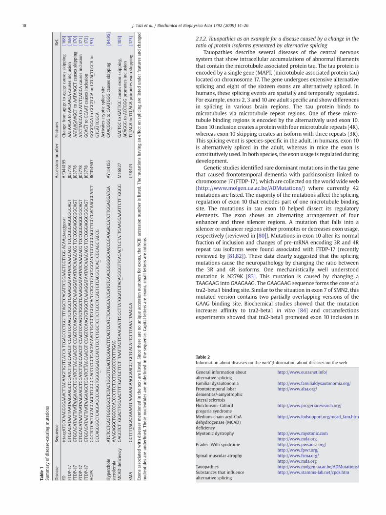
Table1
Summaryof
disease-caus
ingmutations
Disea
seSe
quen
ceAccession
numbe
rFe
atures
Ref.
FDtttaag
ATG
CCAAGGGGAAACT
TAGAAGTT
GTT
CATC
ATC
GAGCC
CTGGTT
TTAGCT
CAGATT
CGGAAGTG
GTT
GGACA
Agtaa
g tgc
cat
AF0
4419
5Ch
ange
from
agtgcto
agcg
ccaus
esskipping
[168
]FT
DP-17
GTG
CAGATA
ATT
AATA
AGAAGCT
GGATC
TTAGCA
ACG
TCC
AGTC
CAAGTG
TGGCT
CAAAGGATA
ATA
TCAAACA
CGTC
CCGGGAGGCG
GCA
GT
J037
78AATA
AGAto
AAGAAGAcaus
esinclus
ion
[169
]FT
DP-17
GTG
CAGATA
ATT
AAT A
AGAAGCT
GGATC
TTAGCA
ACG
TCC
AGTC
CAAGTG
TGGCT
CAAAGGATA
ATA
TCAAACA
CGTC
CCGGGAGGCG
GCA
GT
J037
78AATA
AGAAGCT
toAATA
AGCT
caus
esskipping
[170
]FT
DP-17
GTG
CAGATA
ATT
AATA
AGAAGCT
GGATC
T TAGCA
ACG
TCC
AGTC
CAAGTG
TGGCT
CAAAGGATA
ATA
TCAAACA
CGTC
CCGGGAGGCG
GCA
GT
J037
78ATC
TTAGCA
toATC
TCAGCA
caus
esinclus
ion
[171
]FT
DP-17
GTG
CAGATA
ATT
AATA
AGAAGCT
GGATC
TTAGCA
ACG
TCC
AGTC
CAAGTG
TGGCT
CAAAGGATA
ATA
TCAAACA
CGTC
CCGGGAGGCG
GCA
GT
J037
78GCA
GTto
GCA
ATcaus
esinclus
ion
[172
]HGPS
GGCT
CCCA
CTGCA
GCA
GCT
CGGGGGACC
CCGCT
GAGTA
CAACC
TGCG
CTCG
CGCA
CCGTG
CTGTG
CGGGACC
TGCG
GGCA
GCC
TGCC
GACA
AGGCA
TCT
GCC
AGCG
GCT
CAGGAGCC
CAGGTG
GGCG
GACC
CATC
TCCT
CTGGCT
CTTC
TGCC
TCCA
GTG
TCACG
GTC
ACT
CGCA
GCT
ACC
GBC
0145
07GGGCG
GAto
GGGTG
GAor
GTC
ACT
CGCA
toGTC
ATT
CGCA
[93]
Activates
cryp
ticsp
licesite
Hyp
erch
ole
sterolem
iaATC
TCCT
CAGTG
GCC
GCC
TCTA
CTGGGTT
GACT
CCAAACT
TCACT
CCATC
TCAAGCA
TCGATG
TCAACG
GGGGCA
ACC
GGAAGACC
ATC
TTGGAGGATG
AAAAGAGGCT
GGCC
CACC
CCTT
CTCC
TTGGCC
GTC
TTTG
AG
AY1141
55CA
ACG
GG
toCA
ATG
GG
caus
esskipping
[94,95
]
MCA
Dde
ficien
cyGAGGTC
TTGGACT
TGGAACT
TTTG
ATG
CTTG
TTTA
ATT
AGTG
AAGAATT
GGCT
TATG
GATG
TACA
GGGGTT
CAGACT
GCT
ATT
GAAGGAAATT
CTTT
GGGG
M16
827
GACT
GCto
GATT
GCcaus
esex
onskipping
,AC A
GGGto
ACC
GGG
prom
otes
inclus
ion
[103
]
SMA
GGTT
TTAGACA
AAATC
AAAAAGAAGGAAGGTG
CTCA
CATT
CCTT
AAATT
AAGGA
U18
423
TTTA
GAto
TTCA
GAprom
otes
exon
skipping
[173
]
Exon
sassociated
withdiseases
men
tion
edin
thetext
arelis
ted.
Sinc
ethereareno
unique
accessionnu
mbe
rsforex
ons,theNCB
Iaccession
numbe
ris
listed.
Themutations
having
aneffect
onsp
licingarelis
tedun
derfeatures
andch
ange
dnu
cleo
tide
sareun
derlined
.The
senu
cleo
tide
sareun
derlined
inthesequ
ence.C
apital
lettersareex
ons,sm
allletters
areintron
s.
18 J. Tazi et al. / Biochimica et Biophysica Acta 1792 (2009) 14–26
2.1.2. Tauopathies as an example for a disease caused by a change in theratio of protein isoforms generated by alternative splicing
Tauopathies describe several diseases of the central nervoussystem that show intracellular accumulations of abnormal filamentsthat contain the microtubule associated protein tau. The tau protein isencoded by a single gene (MAPT, (microtubule associated protein tau)located on chromosome 17. The gene undergoes extensive alternativesplicing and eight of the sixteen exons are alternatively spliced. Inhumans, these splicing events are spatially and temporally regulated.For example, exons 2, 3 and 10 are adult specific and show differencesin splicing in various brain regions. The tau protein binds tomicrotubules via microtubule repeat regions. One of these micro-tubule binding regions is encoded by the alternatively used exon 10.Exon 10 inclusion creates a proteinwith fourmicrotubule repeats (4R),whereas exon 10 skipping creates an isoform with three repeats (3R).This splicing event is species-specific in the adult. In humans, exon 10is alternatively spliced in the adult, whereas in mice the exon isconstitutively used. In both species, the exon usage is regulated duringdevelopment.
Genetic studies identified rare dominant mutations in the tau genethat caused frontotemporal dementia with parkinsonism linked tochromosome 17 (FTDP-17), which are collected on theworld wideweb(http://www.molgen.ua.ac.be/ADMutations/) where currently 42mutations are listed. The majority of the mutations affect the splicingregulation of exon 10 that encodes part of one microtubule bindingsite. The mutations in tau exon 10 helped dissect its regulatoryelements. The exon shows an alternating arrangement of fourenhancer and three silencer regions. A mutation that falls into asilencer or enhancer regions either promotes or decreases exon usage,respectively (reviewed in [80]). Mutations in exon 10 alter its normalfraction of inclusion and changes of pre-mRNA encoding 3R and 4Rrepeat tau isoforms were found associated with FTDP-17 (recentlyreviewed by [81,82]). These data clearly suggested that the splicingmutations cause the neuropathology by changing the ratio betweenthe 3R and 4R isoforms. One mechanistically well understoodmutation is N279K [83]. This mutation is caused by changing aTAAGAAG into GAAGAAG. The GAAGAAG sequence forms the core of atra2-beta1 binding site. Similar to the situation in exon 7 of SMN2, thismutated version contains two partially overlapping versions of theGAAG binding site. Biochemical studies showed that the mutationincreases affinity to tra2-beta1 in vitro [84] and cotransfectionsexperiments showed that tra2-beta1 promoted exon 10 inclusion in
Table 2Information about diseases on the web";Information about diseases on the web
General information aboutalternative splicing
http://www.eurasnet.info/
Familial dysautonomia http://www.familialdysautonomia.org/Frontotemporal lobardementias/-amyotrophiclateral sclerosis
http://www.alsa.org/
Hutchinson–Gilfordprogeria syndrome
http://www.progeriaresearch.org/
Medium-chain acyl-CoAdehydrogenase (MCAD)deficiency
http://www.fodsupport.org/mcad_fam.htm
Myotonic dystrophy http://www.myotonic.comhttp://www.mda.org
Prader–Willi syndrome http://www.pwsausa.org/http://www.fpwr.org/
Spinal muscular atrophy http://www.fsma.org/http://www.mda.org
Tauopathies http://www.molgen.ua.ac.be/ADMutations/Substances that influencealternative splicing
http://www.stamms-lab.net/cpds.htm
19J. Tazi et al. / Biochimica et Biophysica Acta 1792 (2009) 14–26
reporter gene constructs [85]. In vitro studies showed that theasparagine to lysine exchange in the mutation does not alter thebinding between tau and tubulin, the tau aggregation or microtubuleassembly [86]. These data suggested that mainly the change in theratio of expressed isoforms is responsible for the disease. Testing thishypothesis in mouse models was difficult, as the mouse tau geneconstitutively expresses the 4R isoform in the adult. Therefore, aminigene of human tau, containing the promoter and all exons flankedby shortened intronic regions was expressed in mice. These constructsshow alternative splicing resembling the human situation and expresshuman tau protein containing either 3 or 4 microtubule bindingdomains. When the mutation that promotes exon 10 inclusion(N279K) was introduced into exon 10 of this construct, pre-mRNAsplicing was shifted as expected towards exon 10 inclusion. Interest-ingly, the mice showed similar pathophysiology as humans with thesame mutation and also showed behavioral changes [87]. These datasuggest that a change in the ratio between 3R and 4R tau isoforms isan important underlying cause for FTDP-17.
Abnormal intracellular tau aggregations are also found in othertauopathies, such as Alzheimer's disease [88]. Several studies wereperformed to investigate whether the 4R to 3R ratio of tau pre-mRNAis changed in Alzheimer's disease. The studies gave conflicting results,whichmost likely reflects that only post-mortem human tissue can beanalyzed. Two studies showed no statistical regional differences in the4R/3R ratio [89,90], one study showed regional differences but noclear link to Alzheimer pathology [91] and a forth study showed astatistical significant increase of the 4R to 3R ratio in temporal cortex.This later study also investigated possible changes in other splicingfactors regulating exon 10 and found changes in clk2 and tra2-beta1splicing, suggesting that multiple changes in alternative splice siteselection are either causing or contributing to Alzheimer's disease[92]. This example shows that a change in the ratio of protein isoformsgenerated by alternative splicing can cause human diseases. It furtherillustrates how analysis of genetic mutants helps to understandsplicing regulation, which is often complicated due to species-specificsplicing differences. Despite these problems, animal models can begenerated that reflect the human pathology.
2.1.3. Hutchinson–Gilford progeria syndrome as an example for a diseasecaused by an intronic mutation that activates a cryptic splice site
Hutchinson–Gilford progeria syndrome (HGPS) is a rare geneticdisorder phenotypically characterized by many features of prematureaging. It is clinically characterized by postnatal growth retardation,midface hypoplasia, micrognathia, premature atherosclerosis, absenceof subcutaneous fat, alopecia and generalized osteodysplasia. At birth,the appearance of patients is generally normal, but by one year of agepatients show severe growth retardation, balding and scleroderma-tous skin changes. Patients live a median of 13.4 years and die of heartattacks or congestive heart failure. Mutations causing HGPS have beenidentified in the nuclear lamin A/C (LMNA) gene. Lamin proteins aredistributed throughout the nucleoplasm and are involved in numer-ous functions, including DNA replication, transcription, chromatinorganization, nuclear positioning and shape, as well as the assembly/disassembly of the nucleus during cell division. Out of 14 mutationsaffecting lamin A/C, three have been reported to specifically alterlamin A splicing. The changes in splicing lead to the production oftruncated protein products (p.G608G, p.T623S and IVS11+1GNA). Mostof the typical Hutchinson–Gilford progeria cases are due to arecurrent, de novo point mutation in LMNA exon 11: c.1824CNT [93].This mutation occurs in a probable exon splicing enhancer. As a result,a cryptic splice site is activated in transcripts generated from themutated allele, which is located 5 nucleotides upstream of themutation. The use of the cryptic splice site leads to the production of atruncated Lamin A protein lacking the last 150 base pairs of exon 11.The truncated protein is called “progerin” and acts in a dominantfashion to generate the HGPS phenotype.
This example shows how a mutation can cause activation of anearby otherwise ‘hidden’, cryptic splice site.
2.1.4. LDL receptor splicing variants caused by a single nucleotidepolymorphism are a sex-specific factor for hypercholesterolemia
Hypercholesterolemia is a major risk factor for arteriosclerosis.Low-density lipoproteins are removed from the bloodstream by theLDL receptor (LDLR). Mutations in the LDLR are a primary cause forhypercholesterolemia. Recently, a single nucleotide polymorphismwas identified in exon 12 of the LDLR that promotes skipping of thisexon. The SNP was found to promote exon 12 skipping in the liver ofpre-menopausal women. However, the SNP had no effect on men andpost-menopausal women. The SNP and the splicing pattern areassociated with a higher level of cholesterol in pre-menopausalwomen, but not in men. Exon 12 skipping generates a truncated formof the receptor that lacks the transmembrane domain necessary formembrane binding and internalization. It is possible that the proteingenerated by exon 12 skipping prevents, in a dominant negative form,the uptake of LDL. This model explains the interesting finding thatexon 12 skipping caused by this SNP is associated with cholesterollevels. The reason for the sex-dependency of the SNP is unclear, but ispossible that high estrogen levels influence transcription level of thegene or its alternative splicing [94]. Apo lipoprotein E (ApoE) is aligand for the LDLR receptor and the apoE allele status is a major riskfactor for Alzheimer's disease. It was therefore investigated whetherthe SNP in exon 12 of the LDLR associates with Alzheimer's disease. Itwas found that the SNP associates with an increased chance todevelop Alzheimer's disease in males, but not females [95].
This example nicely illustrates that a SNP can influence alternativesplicing, which in turn predisposes to a disease. Reflecting thecombinatorial control of alternative exon regulation, the result of amutation depends on other factors, in this case the sex and age.
2.1.5. Familial dysautonomia as an example for a disease caused by amutation in the 5′ splice site
About 10% of roughly 80,000 mutations reported in the humangene mutation database affect splice sites [96]. Well-studied diseasescaused by changes in splice site selection include thalassemias [97]and Familial dysautonomia (FD). FD, (also Riley–Day syndrome,hereditary sensory and autonomic neuropathy type III) is a recessivedisease that is caused by loss of function of the i-kappa-B kinasecomplex associated protein (IKBKAP). In the Ashkenazi Jewishpopulation, the incidence is 1/3600 in live birth (carrier frequency1:30) [98]. Affected children show abnormal development of thenervous system that is associated with demyelination in variousregions. This leads to a large clinical spectrum that includes vomitingcrises, unsteady gait, and decreased perception of pain. In more than99.5% of FD patients the 5′ splice site of exon 20 is mutated T→C inposition 6 of intron 20. This point mutation interrupts base pairingwith U1snRNA. U1 snRNA interacts both with the last threenucleotides of the exon and the first six nucleotides of the down-stream intron. The majority of 5′ splice sites show complementarity toseven base pairs of U1 snRNA. This means there are usually threemismatches between the 5′ splice site and U1 snRNA. Bioinformaticanalyses indicate that these mismatches are not randomly distributed.They either weaken the exonic portion of the 5′ splice site, which isthen compensated by strong binding to the intronic portion, or a weakintronic portion is compensated by a strong exonic portion. In exon 20of the IKBKAP gene, the exonic part of the splice site is weak, due to anA at position −1. The T→Cmutationweakens the intronic part of the 5′splice site, which causes exon skipping [99]. Exon 20 usage issusceptible to a weak 5′ splice site, as the exon has a weak 3′ splicesite that has an A at the −3 position, and contains several exonicsilencer elements [100]. Array analysis indicates that IKBKAPpromotes expression of genes involved in oligodendrocyte andmyelin
20 J. Tazi et al. / Biochimica et Biophysica Acta 1792 (2009) 14–26
formation, which could explain the demyelination phenotype causedby the loss of IKBKAP [101].
The example of FD illustrates the complexity of mutations in splicesites that have to be carefully analyzed within the context of otherregulatory elements. It further shows that a missplicing event of a keyregulatory gene can have profound impact by affecting other genes,and finally indicates that splicing is influenced by small substances.
2.1.6. Medium-chain acyl-CoA dehydrogenase (MCAD) deficiencyillustrates how multiple mutations affect exon usage leadingto a human disease
Medium-chain acyl-CoA dehydrogenase (MCAD) is a mitochon-drial enzyme that participates in the degradation of medium chainlength fatty acids. Deficiencies of this enzyme are the most frequentlydiagnosed defect of mitochondrial beta-oxidation. The patients showmetabolic crisis, characterized by hypoglycemia, lethargy and sei-zures, when first exposed to viral infections or challenged by fasting.About 20% of the infants die. MCAD deficiency results in accumulationof medium-chain acylcarnitines in the urine, which can be analyzedby mass-spectrometry. A large newborn screen showed an incidencefrom 1:15,000 in the US population. The major reason for thedeficiency is a K304E missense mutation leading to a less activeprotein [102]. The newborn screening project identified a 362C→Tmissense mutation in exon 5 of the MCAD gene that causes exonskipping and subsequent degradation of the mRNA by nonsense-mediated decay [103]. This mutation disrupts a splicing enhancer thatis highly similar to the SF2/ASF enhancer in the SMN2 exon 7.Cotransfection experiments demonstrate that an increase of the SF2/ASF concentration promotes inclusion of themutated exon, suggestingthat the mutation weakens an SF2/ASF-dependent enhancer. Inter-estingly, a synonymous mutation 351A→C was identified 11 nucleo-tides upstream in the same exon. This mutation affects an hnRNP A1dependent silencer. Since the exon is constitutively used in theabsence of the 362C→T SF2/ASF enhancer mutation, it has no effect onsplicing. However, in the presence of the 362C→T enhancer mutation,it promotes exon inclusion, which antagonized the exon-skippingeffect of the 362C→T SF2/ASF enhancer mutation.
This example illustrates the fine-tuned balance of positive andnegative acting factors that exists in splicing regulation. It also showsthat seemingly irrelevant mutations can have an effect on splicingwhen they are combinedwith othermutations. Finally, the similaritiesbetween the regulation of MCAD exon 5 and SMN2 exon 7 suggest thatthere are degenerate ‘building blocks’ or ‘regulatory modules’ in thesplicing code.
2.1.7. Frontotemporal lobar dementias are caused by the loss of thesplicing factor TDP43
TDP43 (TAR DNA binding protein 43 kDa) was originally identifiedas a transcriptional repressor that associates with the transcriptionalactivator DNA region (TAR) in HIV [104] and was later also foundassociated with the spermatid-specific gene SP-10 promoter [105],reviewed in [106]. TDP43 is a member of the heterogeneous nuclearribonucleoproteins (hnRNP) family of proteins. The protein was lateridentified as a factor that binds to 12 UG repeats that cause aberrantskipping of exon 9 of the CFTR gene, leading to cystic fibrosis [107]. Itcontains two RNA binding domains. The first RNA binding domain isnecessary and sufficient for binding to RNA that contains at least fourUG repeats. In addition, TDP43 shows binding to single stranded TGDNA repeats in vitro [108]. TDP43 is a nuclear protein and it wastherefore completely surprising when it was detected in ubiquitin-positive, tau and alpha synuclein-negative cytosolic inclusions that arethe characteristic feature of frontotemporal lobar degeneration (FTLD)and amyotrophic lateral sclerosis (ALS, Lou Gehring's disease) [109].During the disease, TDP43 is cleaved by caspase-3 and the cleavagefragments accumulate in the cytosol, where they form aggregates. Thecaspase-3 reaction is inhibited by progranulin, which explains why
mutations reducing the progranulin expression cause FTLD [110]. It isnot clear whether the disease is caused by a loss of nuclear function ofTDP43 or by a possible cytotoxic accumulation. However, siRNAmediated knock down of TDP43 in HeLa cells followed by RNAmicroarray analysis demonstrated a strong upregulation of cyclin-dependent kinase 6 (cdk6). The human Cdk6 gene contains numerousintronic TG repeats and interestingly a change in cdk6 expressionwasnot observed for the chicken gene that lacks these repeats. These datastrongly suggest that TDP43 represses cdk6 expression by sequester-ing its RNA by binding to UG repeats. Loss of TDP43 expression leads toabnormal cdk6 activity, resulting in pRB phosphorylation, genomicinstability and apoptosis, which could explain the cell death in FTLD[111]. Mutations of the gene encoding TDP43 are found in familieswith amyotrophic lateral sclerosis, as well as in sporadic cases,indicating a direct link between TDP43 and amyotrophic lateralsclerosis [112].
This example illustrates that splicing factors like TDP43 operate ina regulatory network. A loss of their function can have drastic, butindirect effects. HnRNPs typically perform several different functions,which can obscure possible disease mechanisms. Finally, the interac-tion between UG repeats of the cdk6 gene and TDP43 illustrate how anhnRNP can be recruited to a new function during evolution.
2.1.8. Diseases associated with repeat elementsShort sequence repeats can be detected in numerous exons, where
they serve to increase the recognition by a certain splicing factor[27,113], reviewed in [114]. A change in length of simple repeatsequences can therefore change the splicing pattern of a gene. Forexample, the endothelial NO synthase gene contains an intronicpolymorphic CA-repeat region and the number of repeats correlatewith the risk of coronary artery disease and hyperhomocysteinemie in asex-specific fashion [115]. SELEX experiments and functional studiesshowed that CA repeats bind to hnRNP L and the action of hnRNP Ldepends on the CA repeat length. Microarray studies showed thatintronic CA-rich repeats can influence alternative splicing decisions,strongly suggesting that other intronic CA-repeat polymorphisms couldcause human disease [116]. Another disease-relevant repeat is the UGrepeat that can cause aberrant splicing of exon 9 of the CFTR gene,leading to cystic fibrosis [107]. This repeat binds to TDP43, which isdiscussed below. Myotonic dystrophy (DM) is the most common formof muscular dystrophy in adults. The disease is caused by extensions oftwo repeats: a CUG repeat in the 3′ region of the DMPK gene leads toDM1 and extension of a CCUG repeat in an intron of the ZNF9 gene leadsto DM2 [117]. The common CUG element binds to at least two groups ofRNA binding proteins, muscleblind-like1 and CUG binding protein.Extension of the repeats leads a sequestration of muscleblind protein innuclear foci. This sequestration of muscleblind-like 1 protein reduces itscellular concentration and changes alternative splicing events thatdepend on this protein. Surprisingly, the extended CUG repeats lead toan increase of CUG binding protein concentration. This increase is dueto a stabilization of CUG-binding protein. CUG RNA repeats induce viaan unknown mechanism protein kinase C, which phosphorylates theCUG protein, leading to a more stable protein [118].
Alu elements are the largest group of repetitive elements in thehuman genome. They represent about 10% of the total genomesequence (reviewed in [63]). Alu elements contain potential splicesites and can evolve into exons [61]. It has been estimated that up to5% of human alternative exons could be derived from Alu sequences[119]. It is therefore not surprising that mutations in existing Aluelements can cause their abnormal inclusion in mRNA, which leads tohuman diseases, such as the Alport syndrome [120], congenitalcataracts facial dysmorphism neuropathy syndrome [121], andmucopolysaccharidosis type VII [122].
These examples illustrate how repeats can change the balance ofalternative splicing regulation, even when they are located in intronsor in seemingly unrelated mRNAs. The exonisation of Alu elements
21J. Tazi et al. / Biochimica et Biophysica Acta 1792 (2009) 14–26
shows how new repetitive elements can be used by evolution toacquire new functions, and diseases caused by improper Alu-elementexonisations can be viewed as failed evolutionary experiments.
3. Numerous changes in alternative splicing are found in cancer
Numerous reports have shown that alternative splicing patterns arechanged in cancer (reviewed in [123]). The expression of alternative oreven tumour-specific splice variants significantly affects many cellularevents critical for cancer biology such as cell proliferation, motility, anddrug response [124]. It is still unclear, however, to what extentalternative splicing functionally contributes to the initiation andprogression of cancers. Most altered splicing patterns could be largelysymptomatic and attributed to a generalized lack of fidelity of thesplicing apparatus in cancer cells. The existence of particular splicevariants in cancer could merely be a consequence of the malignantphenotype without contributing to the cancer phenotype. Here we willdescribe general changes of the splicingmachinery in cancer anddiscussspecific examples illustrating the action on tumour suppressor genes.
3.1. Splicing regulatory factors and cancer
Changes in the concentration, localization, composition, or activityof trans-acting regulatory factors, such as hnRNP and SR proteins, canalter splice site selection. Several studies have demonstrated specificalterations in the expression of splicing factors in cancer. Theincreased phosphorylation and the elevated mRNA expression levelsof Tra2-beta1, YB-1 and classical SR proteins, including SC35 and ASF/SF2, have been observed in human ovarian cancer and in mousemodels of breast cancer development [125–128].
One of the best characterized examples of splicing changesobserved in cancer tissues is CD44, a type 1 transmembraneglycoprotein involved in cell–cell and cell–matrix interactions.Alternative splicing of 10 of its 20 exons is found in association withseveral biological processes, such as lymphocyte recruitment, epithe-lial cell–matrix adhesion, and tumour metastasis. Due to its abundantalternative splicing, it has been correlated with changes in splicingfactors, for example the concentration of ASF/SF2 or hnRNP A1 inmouse lung tumours [129] and human colon carcinomas [130].
SF2/ASF is a prototypical splicing factor and has beenwidely used instudying the connection between cancer and alternative splicing. SF2/ASF controls alternative splicing of the oncogene Ron to modulate cellmotility, a general propertyofmetastatic cancer cells [131]. An increasedexpression of SF2/ASF can transform NIH3T3 and Rat1 cells in vitro, andNIH3T3 cells that express high levels of ASF/SF2 produce tumours invivo. This increase correlates with a change in splicing of genes involvedin the Ras-mitogen-activated protein kinase (MAPK) and the phopha-tidylinositol-3-kinase (PI3K)-mammalian target of rapamycin (mTOR)pathways, which are often deregulated in cancer. MNK2 and S6K1
Table 3Splicing mutations of tumour suppressor genes
Gene Mutation Ef
APC Donor splice site mutation in intron 4 ExAPC G→A substitution at the splice acceptor site of intron 7 Cr
thBRCA1 Non sense mutation in exon 18 ASBRCA1 Intronic point mutation AA→AG (IVS5-12 G→A) CrEstrogen receptor AT→GT point mutation in intron 5 Cr
69NF1 Double point mutation in exon 7 DiNF2 Intronic CTAGC→CTAAC Co
crMLH1 Different codon 659 mutations Ex
to
kinases involved in signal transduction are alternatively spliced inresponse to elevated SF2/ASF concentration. A novel SF2/ASF-inducedisoform of S6K1 is sufficient to cause a transformation phenotype.Overexpression or knockdown of ASF/SF2 resulted in specific splicingchanges in a number of genes involved in these pathways. SF2/ASFoverexpression led to cellular transformation resulting in anchorage-independent cell growth in soft-agar and tumour formation in nudemice [132]. These data argue that SF2/ASF is a proto-oncogene [132]. It islikely that other splicing factors have similar features.
3.2. Mutations of tumour suppressor genes affecting splice site selectioncan cause cancer
Mutations in splicing regulatory elements that change splice siteselection provide a clear link between pre-mRNA processing and cancerdevelopment. The mutations ultimately lead to the generation of non-functional tumour suppressor genes, which predisposes to cancer. Theidentification and understanding of inherited mutations is important forgenetic counselling and for follow-up aimed at cancer prevention inaffected familymembers. A list ofwell-characterizedmutations is shown inTable 3.
Themost frequent earlymolecular alteration in the initiation of coloncancer involves mutations in the adenomatous polyposis coli (APC) gene,whichoccurboth in rare familial cancer syndromesand in85%of sporadiccancers. The majority of these mutations result in the truncation of theAPC gene product. Germline mutations in APC gene result in familialadenomatous polyposis (FAP). Two mutations that disrupt splicingregulatory elements have been found. The first one affects a splicingdonor site in intron 4 resulting in skipping of exon 4 and leading to anattenuated formof FAP [133]. This splicingderegulationwas found in livermetastases, where the wild-type allele was deleted [134]. The secondmutation is a G to A substitution at the splice acceptor site of intron 7creating a cryptic splice site and causing a single nucleotide deletion atthe beginning of exon 8 [134]. The behavior of these mutations can becomplicated by additional APC transcripts which are generated due toalternative splicing of exons 3–4, 9, 10A/X, and 14 [135] [136,137]
The MMR MLH1 (MIM 120436) and MSH2 (MIM 609309) genesresponsible for hereditary nonpolyposis colorectal cancer (HNPCC orLynch syndrome; MIM 120435), are prone to mutations. Double exonskipping in MLH1 gives rise to hereditary nonpolyposis colorectalcancer [138,139]. Also in various hereditary nonpolyposis colorectalcancer, different mutations in codon 659 result in skipping of exon 17of MLH1 gene. This causes an internal deletion of 31 amino acids thatabrogates binding to the other mismatch repair protein PMS2 [140]. Amajor difficulty in the diagnosis of the Lynch syndrome, like otherMendelian diseases, is the interpretation of numerous variants ofunknown clinical and biological significance. In most cases thepathogenic role of the unclassified variants cannot be establishedfrom clinical data and segregation analysis, because of the small
fect Reference
on 4 skipping → attenuated form of FAP [133]yptic splice site creation causing a single nucleotide deletion ae beginning of exon 8
[174]
F/SF2 ESE disruption → exon 18 skipping [143]yptic 3′ splice site creation → production of a truncated protein [144]yptic 5′ splice site creation in intron 5 → insertion of anucleotide cryptic exon into the reading frame
[175]
sruption of ASF/SF2 and SC35 ESE binding sites → in frame deletion [147]nsensus branch point adenosine creation → cryptic splice siteeation → additional exon 5a insertion
[148]
on 17 skipping → internal deletion of 31 aa that abrogates bindingthe other mismatch repair protein PMS2
[140]
22 J. Tazi et al. / Biochimica et Biophysica Acta 1792 (2009) 14–26
number of family members available, the incomplete penetrance ofMMR mutations and the possibility of additional undetected muta-tions in the MMR genes. Recently, using an ex vivo functional splicingassay based on a novel splicing reporter minigene to test 87 intronic orexonic variants (20 and 67, respectively) corresponding to 85 allelesfound in MLH1 or MSH2 by the French HNPCC consortium, it waspossible to show that a high level of mutations alter splicingregulatory elements [141]. However, the biological significance ofthe six variants is unknown.
Breast cancer gene 1 (BRCA1) is responsible for the majority ofhereditary breast and ovarian cancers [142]. Among sporadic cases ofbreast cancer, expression of BRCA1 is reduced or undetectable in high-grade ductal carcinomas, suggesting the involvement of this gene in theetiology of breast cancer. Germline sequence variations in both splicesites and regulatory elements of BRCA1 gene have been implicated insusceptibility to cancer. For example, an inherited nonsensemutation inexon 18 of the BRCA1 gene disrupts an Exonic Splicing Enhancer. Thisenhancer binds to the SR protein SF2/ASF, and its mutation causesinappropriate skipping of the constitutive exon 18 [143]. It has also beenshown that anAA toAGmutation creates a cryptic 3′ splice site that adds11 nucleotides to BRCA1 mRNA, which encodes a truncated protein in abreast cancer family [144]. In addition, multiple BRCA1 splice variantsare present in different tissues with different expression profiles [145].
The neurofibromatosis 1 (NF1) gene that is linked to development ofneurofibromas has one of the highest mutation rates described for anyhuman disorder, and a large proportion of NF1 mutations bearconsequences for the correct splicing of the RNA. A systematic study ofsomatic NF1 mutations [146] demonstrated that most point mutationsresulted in splicing defects including exon skipping and the usage ofalternative 5′ and 3′ splice sites. Also, a double point mutation near themiddle of NF1 exon 7 has been found to cause a high level of in-framedeletionof that exon. Thesemutationsdisrupt consensus binding sites forthe SR proteins SC35 and ASF/SF2 [147]. Somemutations like a CTAGC toCTAAC in theNF1 gene in the central nervous system, create a consensusbranch point adenosine that activates a cryptic site in the intron leadingto an additional exon (exon 5a). Because some normal splicing remains,this mutation is associated with a relatively mild phenotype [148].
These examples illustrate that numerous changes in splicingpatterns are characteristic for cancer development and progression.It is currently not clear whether these changes are the cause or theconsequence of a malignant phenotype. Gain of function changes inalternative splicing have been observed in several systems and well-characterized examples are summarized in Table 4. These mutations,and the mutations in the tumour suppressor genes that causemissplicing and predispose cancer, are strongly suggesting thataberrant processing of some pre-mRNAs is the cause of cancer andcould be a therapeutic target.
Table 4Gain of function of proteins promoting tumour development by alternative splicing
Gene Protein property/function Splice variant
Survivin Inhibitor of apoptosis Survivin 2B with pro-apoptotic propert
VEGF Role in angiogenesis Isoforms lacking exon 6Cathepsin B Role in the development and
progression of cancersCertain isoforms
FHIT Tumour suppressor Aberrant transcripts
Actinin 4 Actin-binding protein Variant Va, a mutually exclusive splicewhere exon 8 is replaced by a new exothe same size that exists in intron 8
AIB1 Hormone coactivator Isoform lacking exon 3RON Tyrosine kinase receptor RonΔ165
RonΔ160RonΔ155
4. Treatment options for missplicing events
The examples discussed here clearly show that a misregulation ofalternative splicing plays a large role in numerous human diseases.The identification of molecules capable of correcting and or inhibitingpathological splicing events is therefore an important issue for futuretherapeutic approaches.
4.1. Antisense strategies
The major challenge in treating splicing disorders is to specificallytarget one splicing event in a certain pre-mRNA. Since there are severalthousand other pre-mRNAs in the cell, the selectivity of splice siteintervention is amajor problem.Oneway to address this problem is theuse of oligonucleotides that will specifically bind to one sequence.
Special chemistries were devised to prevent RNAseH-mediatedcleavage of the RNA and to lower toxicity (reviewed in [149,150]). Amajordrawback of the therapeutic use of oligonucleotides is their deliveryand uptake in the cells. This problem has been addressed by coupling ofoligonucleotides to arginine-rich cell penetrating peptides [151,152].
Antisense oligonucleotides have been tested for beta thalassemias[153], Duchenne muscular dystrophy [154,155], cystic fibrosis [156]cyclophilin transcripts [157], Hutchinson Gilford Progeria Syndrome(HGPS) [158] as well as block HIV replication [159] and alter tau pre-mRNAs [160].
Antisense oligonucleotides can also be used to block splicingenhancers or silencers, which can be found in introns or exons quitefar away from the splice sites [161]. Inhibition of splicing silencers(ESSs or ISSs) is of particular interest since it provides away to activateotherwise repressed exons. This was achieved for the α exon offibroblast growth factor receptor-1 (FGFR1) transcripts [162] where anantisense morpholino oligonucleotide that blocks silencer elements inglioblastoma cells in culture promotes the inclusion of theα exon. Theantisense approach was further developed in ESSENSE (exon-specificsplicing enhancement by small chimeric effectors). ESSENSE usesbifunctional reagents that contain a peptide effector domain and anantisense-targeting domain. The effector domains of these protein-nucleic acids were arginine-serine (RS) repeats that mimic the effectof SR proteins [163]. A second incarnation of bifunctional oligomersuses a 2′-Ome-modified binding domain and an effector domain,which is composed of RNA that contains binding sites for knownsplicing trans-acting factors[164],[165]. The recruited factors mediateactivation or silencing of the targeted exon. An example of this type ofbifunctional oligonucleotide acts as an ESE and promotes inclusion ofthe SMN2 exon 7 fibroblasts from SMA patients leading to partialrestoration of the SMN function [164].
Isoform expressionin cancer
Cancer type Reference
ies Down regulated Breast carcinoma and late stageor metastatic gastric cancer
[176,177]
Upregulated Non-small cell lung cancer [178]Overexpressed Colon cancer [179]
Aberrant expression Gastric, cervical, thyroid andtesticular germ-cell tumours
[180–185]
variantn of
Aberrant expressionof the splice isoform
Small cell lung cancer [186]
Aberrant expression Breast cancer [187]Overexpressed Colorectal carcinoma [188,189]
23J. Tazi et al. / Biochimica et Biophysica Acta 1792 (2009) 14–26
4.2. Substances that change alternative splicing
The use of RNA-bindingmolecules as antibiotics, such as gentamicin,chloramphenicol, and tetracycline illustrates that drugs can be targetedagainst RNA and/or RNA binding proteins. High-throughput screens andtesting of substances in model systems have now identified more than30 substances that change splice site selection. The substances fall intoseveral categories, including HDAC inhibitors, kinase and phosphataseinhibitors, as well as cAMP antagonist and agonists. The currentlyknown substances are reviewed in [166] and updated on the web.
The usefulness of substances that change splice site selection isevident from potential HIV therapies that could be combined withother antiviral strategies. Using an in vitro splicing assay, a chemicalscreenwas performed that identified an indole derivative (IDC16) thatinterferes with exonic splicing enhancer activity of the SR proteinsplicing factor SF2/ASF [167]. IDC16 suppresses the production of keyviral proteins and inhibits replication of macrophage- and Tcell-tropiclaboratory strains, clinical isolates, and strains with high-levelresistance to inhibitors of viral protease and reverse transcriptase.Thus, human splicing factors represent novel and promising drugtargets for the development of antiretroviral therapies, particularly forthe inhibition of multidrug-resistant viruses.
Acknowledgments
This work was supported by the EURASNET (European AlternativeSplicing Network of Excellence), NIH (National Institutes of Health;P20 RR020171, R21HD056195-01), BMBF (Federal Ministry of Educa-tion and Research, Germany) and DFG (Deutsche Forschungsge-meinschaft; SFB 473).
References
[1] E.S. Lander, L.M. Linton, B. Birren, C. Nusbaum, M.C. Zody, J. Baldwin, K. Devon, K.Dewar, M. Doyle,W. FitzHugh, R. Funke, D. Gage, K. Harris, A. Heaford, J. Howland,L. Kann, J. Lehoczky, R. LeVine, P. McEwan, K. McKernan, J. Meldrim, J.P. Mesirov,C. Miranda, W. Morris, J. Naylor, C. Raymond, M. Rosetti, R. Santos, A. Sheridan, C.Sougnez, N. Stange-Thomann, N. Stojanovic, A. Subramanian, D. Wyman, J.Rogers, J. Sulston, R. Ainscough, S. Beck, D. Bentley, J. Burton, C. Clee, N. Carter, A.Coulson, R. Deadman, P. Deloukas, A. Dunham, I. Dunham, R. Durbin, L. French, D.Grafham, S. Gregory, T. Hubbard, S. Humphray, A. Hunt, M. Jones, C. Lloyd, A.McMurray, L. Matthews, S. Mercer, S. Milne, J.C. Mullikin, A. Mungall, R. Plumb, M.Ross, R. Shownkeen, S. Sims, R.H. Waterston, R.K. Wilson, L.W. Hillier, J.D.McPherson, M.A. Marra, E.R. Mardis, L.A. Fulton, A.T. Chinwalla, K.H. Pepin, W.R.Gish, S.L. Chissoe, M.C. Wendl, K.D. Delehaunty, T.L. Miner, A. Delehaunty, J.B.Kramer, L.L. Cook, R.S. Fulton, D.L. Johnson, P.J. Minx, S.W. Clifton, T. Hawkins, E.Branscomb, P. Predki, P. Richardson, S. Wenning, T. Slezak, N. Doggett, J.F. Cheng,A. Olsen, S. Lucas, C. Elkin, E. Uberbacher, M. Frazier, et al., Initial sequencing andanalysis of the human genome, Nature 409 (2001) 860–921.
[2] C. Ben-Dov, B. Hartmann, J. Lundgren, J. Valcarcel, Genome-wide analysis ofalternative pre-mRNA splicing, J. Biol. Chem. 283 (2008) 1229–1233.
[3] D. Kampa, J. Cheng, P. Kapranov, M. Yamanaka, S. Brubaker, S. Cawley, J. Drenkow,A. Piccolboni, S. Bekiranov, G. Helt, H. Tammana, T.R. Gingeras, Novel RNAsidentified from an in-depth analysis of the transcriptome of human chromo-somes 21 and 22, Genome Res. 14 (2004) 331–342.
[4] B.J. Blencowe, Alternative splicing: new insights from global analyses, Cell 126(2006) 37–47.
[5] P.R. Romero, S. Zaidi, Y.Y. Fang, V.N. Uversky, P. Radivojac, C.J. Oldfield, M.S.Cortese, M. Sickmeier, T. LeGall, Z. Obradovic, A.K. Dunker, Alternative splicing inconcert with protein intrinsic disorder enables increased functional diversity inmulticellular organisms, Proc. Natl. Acad. Sci. U. S. A. 103 (2006) 8390–8395.
[6] S. Stamm, S. Ben-Ari, I. Rafalska, Y. Tang, Z. Zhang, D. Toiber, T.A. Thanaraj, H.Soreq, Function of alternative splicing, Gene 344C (2005) 1–20.
[7] R.T. Hillman, R.E. Green, S.E. Brenner, An unappreciated role for RNA surveillance,Genome Biol. 5 (2004) R8.
[8] Q. Pan, A.L. Saltzman, Y.K. Kim, C. Misquitta, O. Shai, L.E. Maquat, B.J. Frey, B.J.Blencowe, Quantitative microarray profiling provides evidence against wide-spread coupling of alternative splicing with nonsense-mediated mRNA decay tocontrol gene expression, Genes Dev. 20 (2006) 153–158.
[9] S.E. Wilkie, V. Vaclavik, H. Wu, K. Bujakowska, C.F. Chakarova, S.S. Bhattacharya,M.J. Warren, D.M. Hunt, Disease mechanism for retinitis pigmentosa (RP11)caused by missense mutations in the splicing factor gene PRPF31, Mol. Vis. 14(2008) 683–690.
[10] E.N. Vithana, L. Abu-Safieh, M.J. Allen, A. Carey, M. Papaioannou, C. Chakarova, M.Al-Maghtheh, N.D. Ebenezer, C. Willis, A.T. Moore, A.C. Bird, D.M. Hunt, S.S.
Bhattacharya, A human homolog of yeast pre-mRNA splicing gene, PRP31,underlies autosomal dominant retinitis pigmentosa on chromosome 19q13.4(RP11), Mol. Cell 8 (2001) 375–381.
[11] K.L. Boon, R.J. Grainger, P. Ehsani, J.D. Barrass, T. Auchynnikava, C.F. Inglehearn,J.D. Beggs, prp8 mutations that cause human retinitis pigmentosa lead to a U5snRNP maturation defect in yeast, Nat. Struct. Mol. Biol. 14 (2007) 1077–1083.
[12] H.J. Mardon, G. Sebastio, F.E. Baralle, A role for exon sequences in alternativesplicing of the human fibronectin gene, Nucleic Acids Res. 15 (1987) 7725–7733.
[13] T.A. Cooper, W. Mattox, The regulation of splice-site selection, and its role inhuman disease, Am. J. Hum. Genet. 61 (1997) 259–266.
[14] J.P. Orengo, T.A. Cooper, Alternative splicing in disease, Adv. Exp. Med. Biol. 623(2007) 212–223.
[15] M. Nissim-Rafinia, B. Kerem, Splicing regulation as a potential genetic modifier,Trends Genet. 18 (2002) 123–127.
[16] E. Buratti, M. Baralle, F.E. Baralle, Defective splicing, disease and therapy:searching for master checkpoints in exon definition, Nucleic Acids Res. 34 (2006)3494–3510.
[17] N.A. Faustino, T.A. Cooper, Pre-mRNA splicing and human disease, Genes Dev. 17(2003) 419–437.
[18] P. Jeanteur, Alternative Splicing and Disease, Springer, Berlin, 2006.[19] E. Kim, A. Goren, G. Ast, Alternative splicing and disease, RNA Biol. 5 (2008) 17–19.[20] A.E. House, K.W. Lynch, Regulation of alternative splicing: more than just the
ABCs, J. Biol. Chem. 283 (2008) 1217–1221.[21] C.W. Smith, J. Valcarcel, Alternative pre-mRNA splicing: the logic of combinator-
ial control, Trends Biochem. Sci. 25 (2000) 381–388.[22] D.L. Black, Mechanisms of alternative pre-messenger RNA splicing, Annu. Rev.
Biochem. (2003) 291–336.[23] H. Stark, R. Luhrmann, Cryo-electron microscopy of spliceosomal components,
Annu. Rev. Biophys. Biomol. Struct. 35 (2006) 435–457.[24] S. Valadkhan, J.L. Manley, Splicing-related catalysis by protein-free snRNAs,
Nature 413 (2001) 701–707.[25] S. Valadkhan, A. Mohammadi, C. Wachtel, J.L. Manley, Protein-free spliceosomal
snRNAs catalyze a reaction that resembles the first step of splicing, RNA 13(2007) 2300–2311.
[26] T. Glisovic, J.L. Bachorik, J. Yong, G. Dreyfuss, RNA-binding proteins and post-transcriptional gene regulation, FEBS Lett. 582 (2008) 1977–1986.
[27] K.W. Lynch, T. Maniatis, Assembly of specific SR protein complexes on distinctregulatory elements of the Drosophila doublesex splicing enhancer, Genes Dev.10 (1996) 2089–2101.
[28] K.J. Hertel, Combinatorial control of exon recognition, J. Biol. Chem. 283 (2008)1211–1215.
[29] M.J. Moore, From birth to death: the complex lives of eukaryotic mRNAs, Science309 (2005) 1514–1518.
[30] E. Buratti, F.E. Baralle, Influence of RNA secondary structure on the pre-mRNAsplicing process, Mol. Cell. Biol. 24 (2004) 10505–10514.
[31] A.F. Muro, M. Caputi, R. Pariyarath, F. Pagani, E. Buratti, F.E. Baralle, Regulation offibronectin EDA exon alternative splicing: possible role of RNA secondarystructure for enhancer display, Mol. Cell. Biol. 19 (1999) 2657–2671.
[32] M. Hiller, Z. Zhang, R. Backofen, S. Stamm, pre-mRNA secondary structure andsplice site selection, PLOS Genet. 3 (2007) 2147–2155.
[33] P. Carninci, T. Kasukawa, S. Katayama, J. Gough, M.C. Frith, N. Maeda, R. Oyama, T.Ravasi, B. Lenhard, C. Wells, R. Kodzius, K. Shimokawa, V.B. Bajic, S.E. Brenner, S.Batalov, A.R. Forrest, M. Zavolan, M.J. Davis, L.G. Wilming, V. Aidinis, J.E. Allen, A.Ambesi-Impiombato, R. Apweiler, R.N. Aturaliya, T.L. Bailey, M. Bansal, L. Baxter,K.W. Beisel, T. Bersano, H. Bono, A.M. Chalk, K.P. Chiu, V. Choudhary, A.Christoffels, D.R. Clutterbuck, M.L. Crowe, E. Dalla, B.P. Dalrymple, B. de Bono, G.Della Gatta, D. di Bernardo, T. Down, P. Engstrom, M. Fagiolini, G. Faulkner, C.F.Fletcher, T. Fukushima, M. Furuno, S. Futaki, M. Gariboldi, P. Georgii-Hemming,T.R. Gingeras, T. Gojobori, R.E. Green, S. Gustincich, M. Harbers, Y. Hayashi, T.K.Hensch, N. Hirokawa, D. Hill, L. Huminiecki, M. Iacono, K. Ikeo, A. Iwama, T.Ishikawa, M. Jakt, A. Kanapin, M. Katoh, Y. Kawasawa, J. Kelso, H. Kitamura, H.Kitano, G. Kollias, S.P. Krishnan, A. Kruger, S.K. Kummerfeld, I.V. Kurochkin, L.F.Lareau, D. Lazarevic, L. Lipovich, J. Liu, S. Liuni, S. McWilliam, M. Madan Babu, M.Madera, L. Marchionni, H. Matsuda, S. Matsuzawa, H. Miki, F. Mignone, S. Miyake,K. Morris, S. Mottagui-Tabar, N. Mulder, N. Nakano, H. Nakauchi, P. Ng, R. Nilsson,S. Nishiguchi, S. Nishikawa, et al., The transcriptional landscape of themammalian genome, Science 309 (2005) 1559–1563.
[34] J. Cheng, P. Kapranov, J. Drenkow, S. Dike, S. Brubaker, S. Patel, J. Long, D. Stern, H.Tammana, G. Helt, V. Sementchenko, A. Piccolboni, S. Bekiranov, D.K. Bailey, M.Ganesh, S. Ghosh, I. Bell, D.S. Gerhard, T.R. Gingeras, Transcriptional maps of 10human chromosomes at 5-nucleotide resolution, Science 308 (2005) 1149–1154.
[35] S. Kishore, S. Stamm, The snoRNA HBII-52 regulates alternative splicing of theserotonin receptor 2C, Science 311 (2006) 230–232.
[36] T. Sahoo, D. del Gaudio, J.R. German, M. Shinawi, S.U. Peters, R.E. Person, A.Garnica, S.W. Cheung, A.L. Beaudet, Prader–Willi phenotype caused by paternaldeficiency for the HBII-85 C/D box small nucleolar RNA cluster, Nat. Genet. 40(2008) 719–721.
[37] B.V. Skryabin, L.V. Gubar, B. Seeger, J. Pfeiffer, S. Handel, T. Robeck, E. Karpova, T.S.Rozhdestvensky, J. Brosius, Deletion of the MBII-85 snoRNA gene cluster in miceresults in postnatal growth retardation, PLoS Genet. 3 (2007) e235.
[38] F. Ding, H.H. Li, S. Zhang, N.M. Solomon, S.A. Camper, P. Cohen, U. Francke,SnoRNA Snord116 (Pwcr1/MBII-85) deletion causes growth deficiency andhyperphagia in mice, PLoS ONE 3 (2008) e1709.
[39] F. Ding, Y. Prints, M.S. Dhar, D.K. Johnson, C. Garnacho-Montero, R.D. Nicholls, U.Francke, Lack of Pwcr1/MBII-85 snoRNA is critical for neonatal lethality inPrader–Willi syndrome mouse models, Mamm. Genome 16 (2005) 424–431.
24 J. Tazi et al. / Biochimica et Biophysica Acta 1792 (2009) 14–26
[40] N.J. Watkins, I. Lemm, R. Luhrmann, Involvement of nuclear import and exportfactors in U8 box C/D snoRNP biogenesis, Mol. Cell. Biol. 27 (2007) 7018–7027.
[41] S. Liu, P. Li, O. Dybkov, S. Nottrott, K. Hartmuth, R. Luhrmann, T. Carlomagno, M.C.Wahl, Binding of the human Prp31 Nop domain to a composite RNA–proteinplatform in U4 snRNP, Science 316 (2007) 115–120.
[42] S. Stamm, Signals and their transduction pathways regulating alternativesplicing: a new dimension of the human genome, Hum. Mol. Genet. 11 (2002)2409–2416.
[43] C. Shin, J.L. Manley, Cell signalling and the control of pre-mRNA splicing, Nat.Rev., Mol. Cell Biol. 5 (2004) 727–738.
[44] M. Blaustein, F. Pelisch, A. Srebrow, Signals, pathways and splicing regulation, Int.J. Biochem. Cell Biol. 39 (2007) 2031–2048.
[45] S. Stamm, Regulation of alternative splicing by reversible phosphorylation, J. Biol.Chem. 283 (2008) 1223–1227.
[46] M. Hagiwara, Alternative splicing: a new drug target of the post-genome era,Biochim. Biophys. Acta 1754 (2005) 324–331.
[47] F. Hernandez, M. Perez, J.J. Lucas, A.M. Mata, R. Bhat, J. Avila, Glycogen synthasekinase-3 plays a crucial role in tau exon 10 splicing and intranuclear distributionof SC35. Implications for Alzheimer's disease, J. Biol. Chem. 279 (2004)3801–3806.
[48] C. Shin, Y. Feng, J.L. Manley, Dephosphorylated SRp38 acts as a splicing repressorin response to heat shock, Nature 427 (2004) 553–558.
[49] S.H. Xiao, J.L. Manley, Phosphorylation of the ASF/SF2 RS domain affects bothprotein–protein and protein–RNA interactions and is necessary for splicing,Genes Dev. 11 (1997) 334–344.
[50] Y. Huang, T.A. Yario, J.A. Steitz, A molecular link between SR protein depho-sphorylation and mRNA export, Proc. Natl. Acad. Sci. U. S. A. 101 (2004)9666–9670.
[51] H. Shen, M.R. Green, RS domains contact splicing signals and promote splicing bya common mechanism in yeast through humans, Genes Dev. 20 (2006)1755–1765.
[52] H. Ceulemans, M. Bollen, Functional diversity of protein phosphatase-1, a cellulareconomizer and reset button, Physiol. Rev. 84 (2004) 1–39.
[53] S.K. Hanks, Genomic analysis of the eukaryotic protein kinase superfamily: aperspective, Genome Biol. 4 (2003) 111.
[54] Y. Shi, B. Reddy, J.L. Manley, PP1/PP2A phosphatases are required for the secondstep of pre-mRNA splicing and target specific snRNP proteins, Mol. Cell 23 (2006)819–829.
[55] E. Allemand, M.L. Hastings, M.V. Murray, M.P. Myers, A.R. Krainer, Alternativesplicing regulation by interaction of phosphatase PP2Cgamma with nucleic acid-binding protein YB-1, Nat. Struct. Mol. Biol. 14 (2007) 630–638.
[56] W. Cao, S.F. Jamison, M.A. Garcia-Blanco, Both phosphorylation and depho-sphorylation of ASF/SF2 are required for pre-mRNA splicing in vitro, RNA 3 (1997)1456–1467.
[57] B. Cardinali, P.T. Cohen, A.I. Lamond, Protein phosphatase 1 can modulatealternative 5′ splice site selection in a HeLa splicing extract, FEBS Lett. 352 (1994)276–280.
[58] N. Ghosh, N. Patel, K. Jiang, J.E. Watson, J. Cheng, C.E. Chalfant, D.R. Cooper,Ceramide-activated protein phosphatase involvement in insulin resistance viaAkt, serine/arginine-rich protein 40, and ribonucleic acid splicing in L6 skeletalmuscle cells, Endocrinology 148 (2007) 1359–1366.
[59] C.E. Chalfant, K. Rathman, R.L. Pinkerman, R.E.Wood, L.M. Obeid, B. Ogretmen, Y.A.Hannun, De novo ceramide regulates the alternative splicing of caspase 9 andBcl-x in A549 lung adenocarcinoma cells. Dependence on protein phosphatase-1,J. Biol. Chem. 277 (2002) 12587–12595.
[60] E. Kim, A. Goren, G. Ast, Alternative splicing: current perspectives, Bioessays 30(2008) 38–47.
[61] G. Lev-Maor, R. Sorek, N. Shomron, G. Ast, The birth of an alternatively splicedexon: 3′ splice-site selection in Alu exons, Science 300 (2003) 1288–1291.
[62] G. Lev-Maor, A. Goren, N. Sela, E. Kim, H. Keren, A. Doron-Faigenboim, S.Leibman-Barak, T. Pupko, G. Ast, The “alternative” choice of constitutive exonsthroughout evolution, PLoS Genet. 3 (2007) e203.
[63] J. Hasler, T. Samuelsson, K. Strub, Useful ‘junk’: Alu RNAs in the humantranscriptome, Cell. Mol. Life Sci. 64 (2007) 1793–1800.
[64] Y. Zhang, A. Bertolino, L. Fazio, G. Blasi, A. Rampino, R. Romano, M.L. Lee, T. Xiao,A. Papp, D. Wang, W. Sadee, Polymorphisms in human dopamine D2 receptorgene affect gene expression, splicing, and neuronal activity during workingmemory, Proc. Natl. Acad. Sci. U. S. A. 104 (2007) 20552–20557.
[65] J. Pearn, Classification of spinal muscular atrophies, Lancet 8174 (1980) 919–922.[66] Z. Zhang, F. Lotti, K. Dittmar, I. Younis, L. Wan, M. Kasim, G. Dreyfuss, SMN
deficiency causes tissue-specific perturbations in the repertoire of snRNAs andwidespread defects in splicing, Cell 133 (2008) 585–600.
[67] G.E. Oprea, S. Krober, M.L. McWhorter, W. Rossoll, S. Muller, M. Krawczak, G.J.Bassell, C.E. Beattie, B. Wirth, Plastin 3 is a protective modifier of autosomalrecessive spinal muscular atrophy, Science 320 (2008) 524–527.
[68] B. Wirth, L. Brichta, E. Hahnen, Spinal muscular atrophy and therapeuticprospects, Prog. Mol. Subcell. Biol. 44 (2006) 109–132.
[69] U.R. Monani, M. Sendtner, D.D. Coovert, D.W. Parsons, C. Andreassi, T.T. Le, S.Jablonka, B. Schrank, W. Rossol, T.W. Prior, G.E. Morris, A.H. Burghes, The humancentromeric survival motor neuron gene (SMN2) rescues embryonic lethality inSmn(−/−) mice and results in a mouse with spinal muscular atrophy, Hum. Mol.Genet. 9 (2000) 333–339.
[70] M. Feldkotter, V. Schwarzer, R. Wirth, T.F. Wienker, B. Wirth, Quantitativeanalyses of SMN1 and SMN2 based on real-time lightCycler PCR: fast and highlyreliable carrier testing and prediction of severity of spinal muscular atrophy, Am.J. Hum. Genet. 70 (2002) 358–368.
[71] N.N. Singh, E.J. Androphy, R.N. Singh, The regulation and regulatory activities ofalternative splicing of the SMN gene, Crit. Rev. Eukaryot. Gene Expr. 14 (2004)271–285.
[72] Y. Hofmann, C.L. Lorson, S. Stamm, E.J. Androphy, B. Wirth, Htra2-beta 1stimulates an exonic splicing enhancer and can restore full-length SMNexpression to survival motor neuron 2 (SMN2), Proc. Natl. Acad. Sci. U. S. A. 97(2000) 9618–9623.
[73] N.N. Singh, E.J. Androphy, R.N. Singh, In vivo selection reveals combinatorialcontrols that define a critical exon in the spinal muscular atrophy genes, RNA 10(2004) 1291–1305.
[74] N.N. Singh, E.J. Androphy, R.N. Singh, An extended inhibitory context causesskipping of exon 7 of SMN2 in spinal muscular atrophy, Biochem. Biophys. Res.Commun. 315 (2004) 381–388.
[75] L. Cartegni, M.L. Hastings, J.A. Calarco, E. de Stanchina, A.R. Krainer, Determinantsof exon 7 splicing in the spinal muscular atrophy genes, SMN1 and SMN2, Am. J.Hum. Genet. 78 (2006) 63–77.
[76] L. Cartegni, A.R. Krainer, Disruption of an SF2/ASF-dependent exonic splicingenhancer in SMN2 causes spinal muscular atrophy in the absence of SMN1, Nat.Genet. 30 (2002) 377–384.
[77] T. Kashima, N. Rao, C.J. David, J.L. Manley, hnRNP A1 functions with specificity inrepression of SMN2 exon 7 splicing, Hum. Mol. Genet. 16 (2007) 3149–3159.
[78] T. Kashima, J.L. Manley, A negative element in SMN2 exon 7 inhibits splicing inspinal muscular atrophy, Nat. Genet. 34 (2003) 460–463.
[79] T. Novoyatleva, B. Heinrich, Y. Tang, N. Benderska, M.E. Butchbach, C.L. Lorson,M.A. Lorson, C. Ben-Dov, P. Fehlbaum, L. Bracco, A.H. Burghes, M. Bollen, S.Stamm, Protein phosphatase 1 binds to the RNA recognition motif of severalsplicing factors and regulates alternative pre-mRNA processing, Hum. Mol.Genet. (2008) 52–70.
[80] A. Andreadis, Tau gene alternative splicing: expression patterns, regulation andmodulation of function in normal brain and neurodegenerative diseases,Biochem. Biophys. Acta 1739 (2005) 91–103.
[81] J.M. Gallo, W. Noble, T.R. Martin, RNA and protein-dependent mechanisms intauopathies: consequences for therapeutic strategies, Cell. Mol. Life Sci. 64(2007) 1701–1714.
[82] A. Andreadis, Misregulation of tau alternative splicing in neurodegeneration anddementia, Prog. Mol. Subcell. Biol. 44 (2006) 89–107.
[83] M. Hasegawa, M.J. Smith, M. Iijima, T. Tabira, M. Goedert, FTDP-17 mutationsN279K and S305N in tau produce increased splicing of exon 10, FEBS Lett. 443(1999) 93–96.
[84] Z. Jiang, H. Tang, N. Havlioglu, X. Zhang, S. Stamm, R. Yan, J.Y. Wu, Mutations intau gene exon 10 associated with FTDP-17 alter the activity of an exonic splicingenhancer to interact with Tra2-beta1, J. Biol. Chem. 278 (2003) 18997–19007.
[85] J. Wang, Q.-S. Gao, Y. Wang, R. Lafyatis, S. Stamm, A. Andreadis, Tau exon 10,whose missplicing causes frontotemporal dementia, is regulated by an intricateinterplay of cis elements and trans factors, J. Neurochem. 88 (2004) 1078–1090.
[86] M. Hong, V. Zhukareva, V. Vogelsberg-Ragaglia, Z.Wszolek, L. Reed, B.I. Miller, D.H.Geschwind, T.D. Bird, D. McKeel, A. Goate, J.C. Morris, K.C. Wilhelmsen, G.D.Schellenberg, J.Q. Trojanowski, V.M. Lee, Mutation-specific functional impair-ments in distinct tau isoforms of hereditary FTDP-17, Science 282 (1998)1914–1917.
[87] H.N. Dawson, V. Cantillana, L. Chen, M.P. Vitek, The tau N279K exon 10 splicingmutation recapitulates frontotemporal dementia and parkinsonism linked tochromosome 17 tauopathy in a mouse model, J. Neurosci. 27 (2007) 9155–9168.
[88] M.G. Spillantini, M. Goedert, Tau protein pathology in neurodegenerativediseases, Trends Neurosci. 21 (1998) 428–433.
[89] J.W. Connell, T. Rodriguez-Martin, G.M. Gibb, N.M. Kahn, A.J. Grierson, D.P.Hanger, T. Revesz, P.L. Lantos, B.H. Anderton, J.M. Gallo, Quantitative analysis oftau isoform transcripts in sporadic tauopathies, Brain Res. Mol. Brain Res. 137(2005) 104–109.
[90] A. Boutajangout, A. Boom, K. Leroy, J.P. Brion, Expression of taumRNA and solubletau isoforms in affected and non-affected brain areas in Alzheimer's disease,FEBS Lett. 576 (2004) 183–189.
[91] M. Ingelsson, K. Ramasamy, I. Cantuti-Castelvetri, L. Skoglund, T. Matsui, J. Orne,H. Kowa, S. Raju, C.R. Vanderburg, J.C. Augustinack, R. de Silva, A.J. Lees, L.Lannfelt, J.H. Growdon, M.P. Frosch, D.G. Standaert, M.C. Irizarry, B.T. Hyman, Noalteration in tau exon 10 alternative splicing in tangle-bearing neurons of theAlzheimer's disease brain, Acta Neuropathol. 112 (2006) 439–449.
[92] D.C. Glatz, D. Rujescu, Y. Tang, F.J. Berendt, A.M. Hartmann, F. Faltraco, C.Rosenberg, C. Hulette, K. Jellinger, H. Hampel, P. Riederer, H.J. Moller, A.Andreadis, K. Henkel, S. Stamm, The alternative splicing of tau exon 10 and itsregulatory proteins CLK2 and TRA2-BETA1 changes in sporadic Alzheimer'sdisease, J. Neurochem. 96 (2006) 635–644.
[93] A. De Sandre-Giovannoli, N. Levy, Altered splicing in prelamin A-associatedpremature aging phenotypes, Prog. Mol. Subcell. Biol. 44 (2006) 199–232.
[94] H. Zhu, H.M. Tucker, K.E. Grear, J.F. Simpson, A.K. Manning, L.A. Cupples, S. Estus,A common polymorphism decreases low-density lipoprotein receptor exon 12splicing efficiency and associates with increased cholesterol, Hum. Mol. Genet. 16(2007) 1765–1772.
[95] F. Zou, R.K. Gopalraj, J. Lok, H. Zhu, I.F. Ling, J.F. Simpson, H.M. Tucker, J.F. Kelly,S.G. Younkin, D.W. Dickson, R.C. Petersen, N.R. Graff-Radford, D.A. Bennett, J.E.Crook, S. Estus, Sex-dependent association of a common low density lipoproteinreceptor polymorphismwith RNA splicing efficiency in the brain and Alzheimer'sdisease, Hum. Mol. Genet. 17 (2008) 929–935.
[96] D.N. Cooper, P.D. Stenson, N.A. Chuzhanova, The human gene mutation database(HGMD) and its exploitation in the study of mutational mechanisms, Curr.Protoc. Bioinformatics Chapter 1 (2006) Unit 1 13.
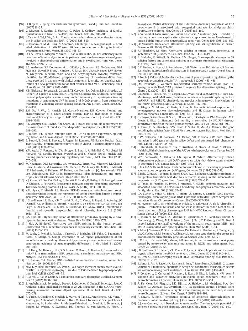
25J. Tazi et al. / Biochimica et Biophysica Acta 1792 (2009) 14–26
[97] H. Birgens, R. Ljung, The thalassaemia syndromes, Scand. J. Clin. Lab. Invest. 67(2007) 11–25.
[98] C. Maayan, E. Kaplan, S. Shachar, O. Peleg, S. Godfrey, Incidence of familialdysautonomia in Israel 1977–1981, Clin. Genet. 32 (1987) 106–108.
[99] I. Carmel, S. Tal, I. Vig, G. Ast, Comparative analysis detects dependencies amongthe 5′ splice-site positions, RNA 10 (2004) 828–840.
[100] C. Ibrahim el, M.M. Hims, N. Shomron, C.B. Burge, S.A. Slaugenhaupt, R. Reed,Weak definition of IKBKAP exon 20 leads to aberrant splicing in familialdysautonomia, Hum. Mutat. 28 (2007) 41–53.
[101] D. Cheishvili, C. Maayan, Y. Smith, G. Ast, A. Razin, IKAP/hELP1 deficiency in thecerebrum of familial dysautonomia patients results in down regulation of genesinvolved in oligodendrocyte differentiation and inmyelination, Hum.Mol. Genet.16 (2007) 2097–2104.
[102] B.S. Andresen, S.F. Dobrowolski, L. O'Reilly, J. Muenzer, S.E. McCandless, D.M.Frazier, S. Udvari, P. Bross, I. Knudsen, R. Banas, D.H. Chace, P. Engel, E.W. Naylor,N. Gregersen, Medium-chain acyl-CoA dehydrogenase (MCAD) mutationsidentified by MS/MS-based prospective screening of newborns differ fromthose observed in patients with clinical symptoms: identification and character-ization of a new, prevalent mutation that results in mild MCAD deficiency, Am. J.Hum. Genet. 68 (2001) 1408–1418.
[103] K.B. Nielsen, S. Sorensen, L. Cartegni, T.J. Corydon, T.K. Doktor, L.D. Schroeder, L.S.Reinert, O. Elpeleg, A.R. Krainer, N. Gregersen, J. Kjems, B.S. Andresen, Seeminglyneutral polymorphic variants may confer immunity to splicing-inactivatingmutations: a synonymous SNP in exon 5 of MCAD protects from deleteriousmutations in a flanking exonic splicing enhancer, Am. J. Hum. Genet. 80 (2007)416–432.
[104] S.H. Ou, F. Wu, D. Harrich, L.F. Garcia-Martinez, R.B. Gaynor, Cloning andcharacterization of a novel cellular protein, TDP-43, that binds to humanimmunodeficiency virus type 1 TAR DNA sequence motifs, J. Virol. 69 (1995)3584–3596.
[105] K.K. Acharya, C.K. Govind, A.N. Shore, M.H. Stoler, P.P. Reddi, cis-requirement forthe maintenance of round spermatid-specific transcription, Dev. Biol. 295 (2006)781–790.
[106] E. Buratti, F.E. Baralle, Multiple roles of TDP-43 in gene expression, splicingregulation, and human disease, Front. Biosci. 13 (2008) 867–878.
[107] E. Buratti, T. Dork, E. Zuccato, F. Pagani, M. Romano, F.E. Baralle, Nuclear factorTDP-43 and SR proteins promote in vitro and in vivo CFTR exon 9 skipping, EMBOJ. 20 (2001) 1774–1784.
[108] Y.M. Ayala, S. Pantano, A. D'Ambrogio, E. Buratti, A. Brindisi, C. Marchetti, M.Romano, F.E. Baralle, Human, Drosophila, and C. elegans TDP43: nucleic acidbinding properties and splicing regulatory function, J. Mol. Biol. 348 (2005)575–588.
[109] M. Neumann, D.M. Sampathu, L.K. Kwong, A.C. Truax, M.C. Micsenyi, T.T. Chou, J.Bruce, T. Schuck, M. Grossman, C.M. Clark, L.F. McCluskey, B.L. Miller, E. Masliah,I.R. Mackenzie, H. Feldman, W. Feiden, H.A. Kretzschmar, J.Q. Trojanowski, V.M.Lee, Ubiquitinated TDP-43 in frontotemporal lobar degeneration and amyo-trophic lateral sclerosis, Science 314 (2006) 130–133.
[110] Y.J. Zhang, Y.F. Xu, C.A. Dickey, E. Buratti, F. Baralle, R. Bailey, S. Pickering-Brown,D. Dickson, L. Petrucelli, Progranulin mediates caspase-dependent cleavage ofTAR DNA binding protein-43, J. Neurosci. 27 (2007) 10530–10534.
[111] Y.M. Ayala, T. Misteli, F.E. Baralle, TDP-43 regulates retinoblastoma proteinphosphorylation through the repression of cyclin-dependent kinase 6 expres-sion, Proc. Natl. Acad. Sci. U. S. A. 105 (2008) 3785–3789.
[112] J. Sreedharan, I.P. Blair, V.B. Tripathi, X. Hu, C. Vance, B. Rogelj, S. Ackerley, J.C.Durnall, K.L. Williams, E. Buratti, F. Baralle, J. de Belleroche, J.D. Mitchell, P.N.Leigh, A. Al-Chalabi, C.C. Miller, G. Nicholson, C.E. Shaw, TDP-43 mutations infamilial and sporadic amyotrophic lateral sclerosis, Science 319 (2008)1668–1672.
[113] G.S. Huh, R.O. Hynes, Regulation of alternative pre-mRNA splicing by a novelrepeated hexanucleotide element, Genes Dev. 8 (1994) 1561–1574.
[114] J. Hui, A. Bindereif, Alternative pre-mRNA splicing in the human system:unexpected role of repetitive sequences as regulatory elements, Biol. Chem. 386(2005) 1265–1271.
[115] M. Laule, C. Meisel, I. Prauka, I. Cascorbi, U. Malzahn, S.B. Felix, G. Baumann, I.Roots, K. Stangl, V. Stangl, Interaction of CA repeat polymorphism of theendothelial nitric oxide synthase and hyperhomocysteinemia in acute coronarysyndromes: evidence of gender-specific differences, J. Mol. Med. 81 (2003)305–309.
[116] L.H. Hung, M. Heiner, J. Hui, S. Schreiner, V. Benes, A. Bindereif, Diverse roles ofhnRNP L in mammalian mRNA processing: a combined microarray and RNAianalysis, RNA 14 (2008) 284–296.
[117] L.P. Ranum, T.A. Cooper, RNA-mediated neuromuscular disorders, Annu. Rev.Neurosci. 29 (2006) 259–277.
[118] N.M. Kuyumcu-Martinez, G.S. Wang, T.A. Cooper, Increased steady-state levels ofCUGBP1 in myotonic dystrophy 1 are due to PKC-mediated hyperphosphoryla-tion, Mol. Cell 28 (2007) 68–78.
[119] R. Sorek, G. Ast, D. Graur, Alu-containing exons are alternatively spliced, GenomeRes. 12 (2002) 1060–1067.
[120] B. Knebelmann, L. Forestier, L. Drouot, S. Quinones, C. Chuet, F. Benessy, J. Saus, C.Antignac, Splice-mediated insertion of an Alu sequence in the COL4A3 mRNAcausing autosomal recessive Alport syndrome, Hum. Mol. Genet. 4 (1995)675–679.
[121] R. Varon, R. Gooding, C. Steglich, L. Marns, H. Tang, D. Angelicheva, K.K. Yong, P.Ambrugger, A. Reinhold, B. Morar, F. Baas, M. Kwa, I. Tournev, V. Guerguelcheva, I.Kremensky, H. Lochmuller, A. Mullner-Eidenbock, L. Merlini, L. Neumann, J.Burger, M. Walter, K. Swoboda, P.K. Thomas, A. von Moers, N. Risch, L.
Kalaydjieva, Partial deficiency of the C-terminal-domain phosphatase of RNApolymerase II is associated with congenital cataracts facial dysmorphismneuropathy syndrome, Nat. Genet. 35 (2003) 185–189.
[122] R. Vervoort, R. Gitzelmann, W. Lissens, I. Liebaers, A mutation (IVS8+0.6kbdelTC)creating a new donor splice site activates a cryptic exon in an Alu-element inintron8 of the human beta-glucuronidase gene,Hum. Genet.103 (1998) 686–693.
[123] J.P. Venables, Unbalanced alternative splicing and its significance in cancer,Bioessays 28 (2006) 378–386.
[124] R.I. Skotheim, M. Nees, Alternative splicing in cancer: noise, functional, orsystematic? Int. J. Biochem. Cell. Biol. 39 (2007) 1432–1449.
[125] E. Stickeler, F. Kittrell, D. Medina, S.M. Berget, Stage-specific changes in SRsplicing factors and alternative splicing in mammary tumorigenesis, Oncogene18 (1999) 3574–3582.
[126] D.C. Fischer, K. Noack, I.B. Runnebaum, D.O. Watermann, D.G. Kieback, S. Stamm,E. Stickeler, Expression of splicing factors in human ovarian cancer, Oncol. Rep.11(2004) 1085–1090.
[127] P. Forch, J. Valcarcel, Molecular mechanisms of gene expression regulation by theapoptosis-promoting protein TIA-1, Apoptosis 6 (2001) 463–468.
[128] J.M. Izquierdo, J. Valcarcel, Fas-activated serine/threonine kinase (FAST K)synergizes with TIA-1/TIAR proteins to regulate Fas alternative splicing, J. Biol.Chem. 282 (2007) 1539–1543.
[129] L.K. Zerbe, I. Pino, R. Pio, P.F. Cosper, L.D. Dwyer-Nield, A.M. Meyer, J.D. Port, L.M.Montuenga, A.M. Malkinson, Relative amounts of antagonistic splicing factors,hnRNP A1 and ASF/SF2, change during neoplastic lung growth: implications forpre-mRNA processing, Mol. Carcinog. 41 (2004) 187–196.
[130] C. Ghigna, M. Moroni, C. Porta, S. Riva, G. Biamonti, Altered expression ofheterogeneous nuclear ribonucleoproteins and SR factors in human colonadenocarcinomas, Cancer Res. 58 (1998) 5818–5824.
[131] C. Ghigna, S. Giordano, H. Shen, F. Benvenuto, F. Castiglioni, P.M. Comoglio, M.R.Green, S. Riva, G. Biamonti, Cell motility is controlled by SF2/ASF throughalternative splicing of the Ron protooncogene, Mol. Cell 20 (2005) 881–890.
[132] R. Karni, E. de Stanchina, S.W. Lowe, R. Sinha, D. Mu, A.R. Krainer, The geneencoding the splicing factor SF2/ASF is a proto-oncogene, Nat. Struct. Mol. Biol. 14(2007) 185–193.
[133] D.W. Neklason, C.H. Solomon, A.L. Dalton, S.K. Kuwada, R.W. Burt, Intron 4mutation in APC gene results in splice defect and attenuated FAP phenotype,Fam. Cancer 3 (2004) 35–40.
[134] H. Kurahashi, K. Takami, T. Oue, T. Kusafuka, A. Okada, A. Tawa, S. Okada, I.Nishisho, Biallelic inactivation of the APC gene in hepatoblastoma, Cancer Res. 55(1995) 5007–5011.
[135] W.S. Samowitz, A. Thliveris, L.N. Spirio, R. White, Alternatively splicedadenomatous polyposis coli (APC) gene transcripts that delete exons mutatedin attenuated APC, Cancer Res. 55 (1995) 3732–3734.
[136] L. Xia, K.A. St Denis, B. Bapat, Evidence for a novel exon in the coding region of theadenomatous polyposis coli (APC) gene, Genomics 28 (1995) 589–591.
[137] S. Bala, C. Kraus, J. Wijnen, P. Meera Khan, W.G. Ballhausen, Multiple products inthe protein truncation test due to alternative splicing in the adenomatouspolyposis coli (APC) gene, Hum. Genet. 98 (1996) 528–533.
[138] Q. Tanko, B. Franklin, H. Lynch, J. Knezetic, A hMLH1 genomic mutation andassociated novel mRNA defects in a hereditary non-polyposis colorectal cancerfamily, Mutat. Res. 503 (2002) 37–42.
[139] L.A. Clarke, I. Veiga, G. Isidro, P. Jordan, J.S. Ramos, S. Castedo, M.G. Boavida,Pathological exon skipping in an HNPCC proband with MLH1 splice acceptor sitemutation, Genes Chromosomes Cancer 29 (2000) 367–370.
[140] M. Nystrom-Lahti, M. Holmberg, P. Fidalgo, R. Salovaara, A. de la Chapelle, J.Jiricny, P. Peltomaki, Missense and nonsense mutations in codon 659 of MLH1cause aberrant splicing of messenger RNA in HNPCC kindreds, Genes Chromo-somes Cancer 26 (1999) 372–375.
[141] I. Tournier, M. Vezain, A. Martins, F. Charbonnier, S. Baert-Desurmont, S.Olschwang, Q. Wang, M.P. Buisine, J. Soret, J. Tazi, T. Frébourg and M. Tosi, Alarge fraction of unclassified variants of the mismatch repair genes MLH1 andMSH2 is associated with splicing defects., Hum Mut. (2008) 1–13.
[142] Y. Miki, J. Swensen, D. Shattuck-Eidens, P.A. Futreal, K. Harshman, S. Tavtigian, Q.Liu, C. Cochran, L.M. Bennett, W. Ding, et al., A strong candidate for the breast andovarian cancer susceptibility gene BRCA1, Science 266 (1994) 66–71.
[143] H.X. Liu, L. Cartegni, M.Q. Zhang, A.R. Krainer, A mechanism for exon skippingcaused by nonsense or missense mutations in BRCA1 and other genes, Nat.Genet. 27 (2001) 55–58.
[144] J.D. Hoffman, S.E. Hallam, V.L. Venne, E. Lyon, K. Ward, Implications of a novelcryptic splice site in the BRCA1 gene, Am. J. Med. Genet. 80 (1998) 140–144.
[145] T.I. Orban, E. Olah, Emerging roles of BRCA1 alternative splicing, Mol. Pathol. 56(2003) 191–197.
[146] E. Serra, E. Ars, A. Ravella, A. Sanchez, S. Puig, T. Rosenbaum, X. Estivill, C. Lazaro,Somatic NF1mutational spectrum in benign neurofibromas:mRNA splice defectsare common among point mutations, Hum. Genet. 108 (2001) 416–429.
[147] P. Colapietro, C. Gervasini, F. Natacci, L. Rossi, P. Riva, L. Larizza, NF1 exon 7skipping and sequence alterations in exonic splice enhancers (ESEs) in aneurofibromatosis 1 patient, Hum. Genet. 113 (2003) 551–554.
[148] A. De Klein, P.H. Riegman, E.K. Bijlsma, A. Heldoorn, M. Muijtjens, M.A. denBakker, C.J. Avezaat, E.C. Zwarthoff, A G→A transition creates a branch pointsequence and activation of a cryptic exon, resulting in the hereditary disorderneurofibromatosis 2, Hum. Mol. Genet. 7 (1998) 393–398.
[149] P. Sazani, R. Kole, Therapeutic potential of antisense oligonucleotides asmodulators of alternative splicing, J. Clin. Invest. 112 (2003) 481–486.
[150] G.J. van Ommen, J. van Deutekom, A. Aartsma-Rus, The therapeutic potential ofantisense-mediated exon skipping, Curr. Opin. Mol. Ther. 10 (2008) 140–149.
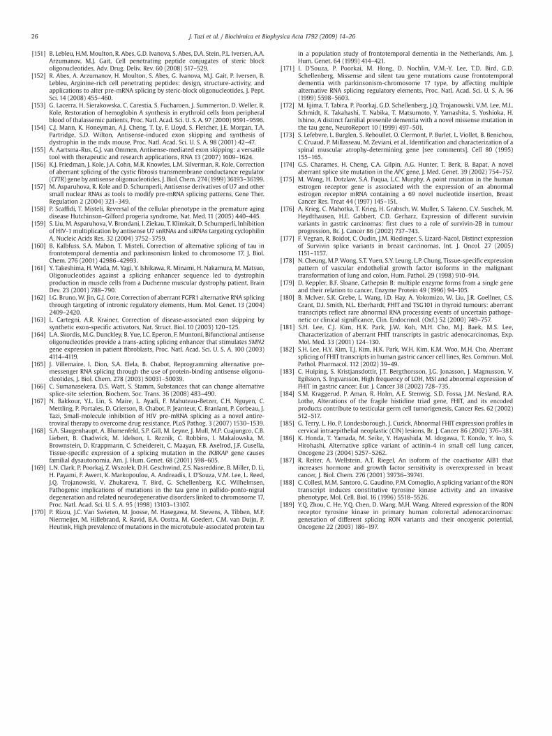
26 J. Tazi et al. / Biochimica et Biophysica Acta 1792 (2009) 14–26
[151] B. Lebleu, H.M. Moulton, R. Abes, G.D. Ivanova, S. Abes, D.A. Stein, P.L. Iversen, A.A.Arzumanov, M.J. Gait, Cell penetrating peptide conjugates of steric blockoligonucleotides, Adv. Drug. Deliv. Rev. 60 (2008) 517–529.
[152] R. Abes, A. Arzumanov, H. Moulton, S. Abes, G. Ivanova, M.J. Gait, P. Iversen, B.Lebleu, Arginine-rich cell penetrating peptides: design, structure-activity, andapplications to alter pre-mRNA splicing by steric-block oligonucleotides, J. Pept.Sci. 14 (2008) 455–460.
[153] G. Lacerra, H. Sierakowska, C. Carestia, S. Fucharoen, J. Summerton, D. Weller, R.Kole, Restoration of hemoglobin A synthesis in erythroid cells from peripheralblood of thalassemic patients, Proc. Natl. Acad. Sci. U. S. A. 97 (2000) 9591–9596.
[154] C.J. Mann, K. Honeyman, A.J. Cheng, T. Ly, F. Lloyd, S. Fletcher, J.E. Morgan, T.A.Partridge, S.D. Wilton, Antisense-induced exon skipping and synthesis ofdystrophin in the mdx mouse, Proc. Natl. Acad. Sci. U. S. A. 98 (2001) 42–47.
[155] A. Aartsma-Rus, G.J. van Ommen, Antisense-mediated exon skipping: a versatiletool with therapeutic and research applications, RNA 13 (2007) 1609–1624.
[156] K.J. Friedman, J. Kole, J.A. Cohn, M.R. Knowles, L.M. Silverman, R. Kole, Correctionof aberrant splicing of the cystic fibrosis transmembrane conductance regulator(CFTR) gene by antisense oligonucleotides, J. Biol. Chem. 274 (1999) 36193–36199.
[157] M. Asparuhova, R. Kole and D. Schumperli, Antisense derivatives of U7 and othersmall nuclear RNAs as tools to modify pre-mRNA splicing patterns, Gene Ther.Regulation 2 (2004) 321–349.
[158] P. Scaffidi, T. Misteli, Reversal of the cellular phenotype in the premature agingdisease Hutchinson–Gilford progeria syndrome, Nat. Med. 11 (2005) 440–445.
[159] S. Liu, M. Asparuhova, V. Brondani, I. Ziekau, T. Klimkait, D. Schumperli, Inhibitionof HIV-1 multiplication by antisense U7 snRNAs and siRNAs targeting cyclophilinA, Nucleic Acids Res. 32 (2004) 3752–3759.
[160] B. Kalbfuss, S.A. Mabon, T. Misteli, Correction of alternative splicing of tau infrontotemporal dementia and parkinsonism linked to chromosome 17, J. Biol.Chem. 276 (2001) 42986–42993.
[161] Y. Takeshima, H. Wada, M. Yagi, Y. Ishikawa, R. Minami, H. Nakamura, M. Matsuo,Oligonucleotides against a splicing enhancer sequence led to dystrophinproduction in muscle cells from a Duchenne muscular dystrophy patient, BrainDev. 23 (2001) 788–790.
[162] I.G. Bruno, W. Jin, G.J. Cote, Correction of aberrant FGFR1 alternative RNA splicingthrough targeting of intronic regulatory elements, Hum. Mol. Genet. 13 (2004)2409–2420.
[163] L. Cartegni, A.R. Krainer, Correction of disease-associated exon skipping bysynthetic exon-specific activators, Nat. Struct. Biol. 10 (2003) 120–125.
[164] L.A. Skordis, M.G. Dunckley, B. Yue, I.C. Eperon, F. Muntoni, Bifunctional antisenseoligonucleotides provide a trans-acting splicing enhancer that stimulates SMN2gene expression in patient fibroblasts, Proc. Natl. Acad. Sci. U. S. A. 100 (2003)4114–4119.
[165] J. Villemaire, I. Dion, S.A. Elela, B. Chabot, Reprogramming alternative pre-messenger RNA splicing through the use of protein-binding antisense oligonu-cleotides, J. Biol. Chem. 278 (2003) 50031–50039.
[166] C. Sumanasekera, D.S. Watt, S. Stamm, Substances that can change alternativesplice-site selection, Biochem. Soc. Trans. 36 (2008) 483–490.
[167] N. Bakkour, Y.L. Lin, S. Maire, L. Ayadi, F. Mahuteau-Betzer, C.H. Nguyen, C.Mettling, P. Portales, D. Grierson, B. Chabot, P. Jeanteur, C. Branlant, P. Corbeau, J.Tazi, Small-molecule inhibition of HIV pre-mRNA splicing as a novel antire-troviral therapy to overcome drug resistance, PLoS Pathog. 3 (2007) 1530–1539.
[168] S.A. Slaugenhaupt, A. Blumenfeld, S.P. Gill, M. Leyne, J. Mull, M.P. Cuajungco, C.B.Liebert, B. Chadwick, M. Idelson, L. Reznik, C. Robbins, I. Makalowska, M.Brownstein, D. Krappmann, C. Scheidereit, C. Maayan, F.B. Axelrod, J.F. Gusella,Tissue-specific expression of a splicing mutation in the IKBKAP gene causesfamilial dysautonomia, Am. J. Hum. Genet. 68 (2001) 598–605.
[169] L.N. Clark, P. Poorkaj, Z. Wszolek, D.H. Geschwind, Z.S. Nasreddine, B. Miller, D. Li,H. Payami, F. Awert, K. Markopoulou, A. Andreadis, I. D'Souza, V.M. Lee, L. Reed,J.Q. Trojanowski, V. Zhukareva, T. Bird, G. Schellenberg, K.C. Wilhelmsen,Pathogenic implications of mutations in the tau gene in pallido-ponto-nigraldegeneration and related neurodegenerative disorders linked to chromosome 17,Proc. Natl. Acad. Sci. U. S. A. 95 (1998) 13103–13107.
[170] P. Rizzu, J.C. Van Swieten, M. Joosse, M. Hasegawa, M. Stevens, A. Tibben, M.F.Niermeijer, M. Hillebrand, R. Ravid, B.A. Oostra, M. Goedert, C.M. van Duijn, P.Heutink, High prevalence of mutations in the microtubule-associated protein tau
in a population study of frontotemporal dementia in the Netherlands, Am. J.Hum. Genet. 64 (1999) 414–421.
[171] I. D'Souza, P. Poorkai, M. Hong, D. Nochlin, V.M.-Y. Lee, T.D. Bird, G.D.Schellenberg, Missense and silent tau gene mutations cause frontotemporaldementia with parkinsonism-chromosome 17 type, by affecting multiplealternative RNA splicing regulatory elements, Proc. Natl. Acad. Sci. U. S. A. 96(1999) 5598–5603.
[172] M. Iijima, T. Tabira, P. Poorkaj, G.D. Schellenberg, J.Q. Trojanowski, V.M. Lee, M.L.Schmidt, K. Takahashi, T. Nabika, T. Matsumoto, Y. Yamashita, S. Yoshioka, H.Ishino, A distinct familial presenile dementia with a novel missense mutation inthe tau gene, NeuroReport 10 (1999) 497–501.
[173] S. Lefebvre, L. Burglen, S. Reboullet, O. Clermont, P. Burlet, L. Viollet, B. Benichou,C. Cruaud, P. Millasseau, M. Zeviani, et al., Identification and characterization of aspinal muscular atrophy-determining gene [see comments], Cell 80 (1995)155–165.
[174] G.S. Charames, H. Cheng, C.A. Gilpin, A.G. Hunter, T. Berk, B. Bapat, A novelaberrant splice site mutation in the APC gene, J. Med. Genet. 39 (2002) 754–757.
[175] M. Wang, H. Dotzlaw, S.A. Fuqua, L.C. Murphy, A point mutation in the humanestrogen receptor gene is associated with the expression of an abnormalestrogen receptor mRNA containing a 69 novel nucleotide insertion, BreastCancer Res. Treat 44 (1997) 145–151.
[176] A. Krieg, C. Mahotka, T. Krieg, H. Grabsch, W. Muller, S. Takeno, C.V. Suschek, M.Heydthausen, H.E. Gabbert, C.D. Gerharz, Expression of different survivinvariants in gastric carcinomas: first clues to a role of survivin-2B in tumourprogression, Br. J. Cancer 86 (2002) 737–743.
[177] F. Vegran, R. Boidot, C. Oudin, J.M. Riedinger, S. Lizard-Nacol, Distinct expressionof Survivin splice variants in breast carcinomas, Int. J. Oncol. 27 (2005)1151–1157.
[178] N. Cheung, M.P.Wong, S.T. Yuen, S.Y. Leung, L.P. Chung, Tissue-specific expressionpattern of vascular endothelial growth factor isoforms in the malignanttransformation of lung and colon, Hum. Pathol. 29 (1998) 910–914.
[179] D. Keppler, B.F. Sloane, Cathepsin B: multiple enzyme forms from a single geneand their relation to cancer, Enzyme Protein 49 (1996) 94–105.
[180] B. McIver, S.K. Grebe, L. Wang, I.D. Hay, A. Yokomizo, W. Liu, J.R. Goellner, C.S.Grant, D.I. Smith, N.L. Eberhardt, FHIT and TSG101 in thyroid tumours: aberranttranscripts reflect rare abnormal RNA processing events of uncertain pathoge-netic or clinical significance, Clin. Endocrinol. (Oxf.) 52 (2000) 749–757.
[181] S.H. Lee, C.J. Kim, H.K. Park, J.W. Koh, M.H. Cho, M.J. Baek, M.S. Lee,Characterization of aberrant FHIT transcripts in gastric adenocarcinomas, Exp.Mol. Med. 33 (2001) 124–130.
[182] S.H. Lee, H.Y. Kim, T.J. Kim, H.K. Park, W.H. Kim, K.M. Woo, M.H. Cho, Aberrantsplicing of FHIT transcripts in human gastric cancer cell lines, Res. Commun. Mol.Pathol. Pharmacol. 112 (2002) 39–49.
[183] C. Huiping, S. Kristjansdottir, J.T. Bergthorsson, J.G. Jonasson, J. Magnusson, V.Egilsson, S. Ingvarsson, High frequency of LOH, MSI and abnormal expression ofFHIT in gastric cancer, Eur. J. Cancer 38 (2002) 728–735.
[184] S.M. Kraggerud, P. Aman, R. Holm, A.E. Stenwig, S.D. Fossa, J.M. Nesland, R.A.Lothe, Alterations of the fragile histidine triad gene, FHIT, and its encodedproducts contribute to testicular germ cell tumorigenesis, Cancer Res. 62 (2002)512–517.
[185] G. Terry, L. Ho, P. Londesborough, J. Cuzick, Abnormal FHIT expression profiles incervical intraepithelial neoplastic (CIN) lesions, Br. J. Cancer 86 (2002) 376–381.
[186] K. Honda, T. Yamada, M. Seike, Y. Hayashida, M. Idogawa, T. Kondo, Y. Ino, S.Hirohashi, Alternative splice variant of actinin-4 in small cell lung cancer,Oncogene 23 (2004) 5257–5262.
[187] R. Reiter, A. Wellstein, A.T. Riegel, An isoform of the coactivator AIB1 thatincreases hormone and growth factor sensitivity is overexpressed in breastcancer, J. Biol. Chem. 276 (2001) 39736–39741.
[188] C. Collesi, M.M. Santoro, G. Gaudino, P.M. Comoglio, A splicing variant of the RONtranscript induces constitutive tyrosine kinase activity and an invasivephenotype, Mol. Cell. Biol. 16 (1996) 5518–5526.
[189] Y.Q. Zhou, C. He, Y.Q. Chen, D. Wang, M.H. Wang, Altered expression of the RONreceptor tyrosine kinase in primary human colorectal adenocarcinomas:generation of different splicing RON variants and their oncogenic potential,Oncogene 22 (2003) 186–197.


![Biochimica et Biophysica Acta - immed.org considerations/09.07.2017 updates/Membrane... · G.L. Nicolson, M.E. Ash / Biochimica et Biophysica Acta 1859 (2017) 1704–1724 1705 [8]](https://img.pdfslide.net/doc/110x75/5c684f1e09d3f2f5638b5509/biochimica-et-biophysica-acta-immed-considerations09072017-updatesmembrane.jpg)