Embed Size (px)
Citation preview
Biochimica et Biophysica Acta 1853 (2015) 1035–1045
Contents lists available at ScienceDirect
Biochimica et Biophysica Acta
j ourna l homepage: www.e lsev ie r .com/ locate /bbamcr
A conserved domain important for association of eukaryotic J-proteinco-chaperones Jjj1 and Zuo1 with the ribosome
Lindsey A. Kaschner a,b, Ruchika Sharma b, Om Kumar Shrestha b, Alison E. Meyer b,1, Elizabeth A. Craig b,⁎a Graduate Program in Cellular and Molecular Biology, University of Wisconsin–Madison, USAb Department of Biochemistry, University of Wisconsin–Madison, USA
⁎ Corresponding author at: 433 Babcock Drive, Departmof Wisconsin–Madison, Madison, WI 53706, USA. Tel.: +1
E-mail address: [email protected] (E.A. Craig).1 Present address: Blood Research Institute, 8733 Wate
WI 53226, USA.
http://dx.doi.org/10.1016/j.bbamcr.2015.01.0140167-4889/© 2015 Elsevier B.V. All rights reserved.
a b s t r a c t
a r t i c l e i n f oArticle history:Received 29 August 2014Received in revised form 20 January 2015Accepted 22 January 2015Available online 30 January 2015
Keywords:J-proteinRibosome biogenesisHsp70Molecular chaperoneRibosome association
J-proteins, obligate co-chaperones, provide specialization for Hsp70 function in a variety of cellular processes.Two of the 13 J-proteins of the yeast cytosol/nucleus, Zuo1 and Jjj1, are associated with 60S ribosomal subunits.Abundant Zuo1 facilitates folding of nascent polypeptides; Jjj1, of much lower abundance, functions in ribosomebiogenesis. However, overexpression of Jjj1 substantially rescues growth defects of cells lacking Zuo1.We analyzed a region held in common by Zuo1 and Jjj1, outside the signature J-domain found in all J-proteins.This shared “zuotin homology domain” (ZHD) is important for ribosome association of both proteins. An N-terminal segment of Jjj1, containing the J-domain and ZHD, is ribosome-associated and, like full-length Jjj1, iscompetent to rescue both the cold- and cation-sensitivity of Δzuo1. However, this fragment, when expressedat normal levels, cannot rescue the cytosolic ribosome biogenesis defect of Δjjj1. Our results are consistentwith a model in which the primary functions of Zuo1 and Jjj1 occur in the cytosol. In addition, our data suggestthat Zuo1 and Jjj1 bind overlapping sites on ribosomes due to an interaction via their common ZHDs, but Jjj1binds primarily to pre-60S particles and Zuo1 to mature subunits. We hypothesize that ZUO1 and JJJ1, whichare conserved throughout eukaryotes, arose from an ancient duplication of a progenitor J-protein gene thatencoded the ZHD ribosome-binding region; subsequently, specialized roles and additional ribosome interactionsites evolved.
© 2015 Elsevier B.V. All rights reserved.
1. Introduction
All Hsp70-based molecular chaperone machineries use the samefundamental biochemical mechanism of action, cycles of interactionwith client proteins driven by ATP binding and hydrolysis, to functionin many essential cellular processes [1,2]. These processes range fromolding of nascent chains as they emerge from ribosomes to driving pro-tein translocation across membranes, protecting cells from heat stress,facilitating biogenesis of Fe/S clusters, and remodeling protein:proteincomplexes [3,4]. Much of the capacity for such functional versatilityis due to the fact that Hsp70s interact with an array of J-protein co-chaperones [5]. For nucleus of the budding/nucleus of the buddingyeast Saccharomyces cerevisiae contains 13 J-proteins. All members ofthe J-protein superfamily possess a ~70 residue J-domain that bindsHsp70 and is responsible for the stimulation of Hsp70's ATPase activity,an obligatory step for stabilizing Hsp70's interaction with a client
ent of Biochemistry, University608 263 7105.
rtown Plank Road, Milwaukee,
protein. However, outside their J-domains, J-proteins vary widely in se-quence and structure [3]. These diverse regions often interact with clientproteins, targeting them toHsp70, or localize the J-protein to a particularsite of action.
Eukaryotes contain two ribosome-associated J-proteins, called Zuo1and Jjj1 in yeast (DNAJC2 and DNAJC21, respectively, in human cells).Both associate with the large ribosomal subunit [6–8]. Both have well-established roles: Zuo1 in chaperoning nascent chains and Jjj1 in a latestep of subunitmaturation, removing biogenesis factors. Zuo1 is presenton approximately 1 of every 3 ribosomes [9,10], Jjj1 is present at onlyabout 1 per 1000 ribosomes [10]. Cells lacking Zuo1 are slow-growing,particularly at low temperatures and hypersensitive to cations [6,11,12], general defects likely reflecting the myriad of clients whose denovo folding requires ribosome-associated chaperones. As expected,loss of the ribosome-associated Hsp70:J-protein machinery results inaggregation of many newly-made polypeptides [13,14]. Cells lackingJjj1 are slow-growing and cold-sensitive, and exhibit hallmarks of inef-ficient 60S-maturation, such as decreased levels of 60S subunits and ac-cumulation of aberrant polysomes [7,15].
Jjj1's role in ribosome biogenesis is an example of involvementof Hsp70/J-protein chaperone machinery in remodeling proteincomplexes. A fewof themany factors involved in 60S subunit biogenesistransit with pre-ribosomal particles to the cytosol [16]. These shuttling
1036 L.A. Kaschner et al. / Biochimica et Biophysica Acta 1853 (2015) 1035–1045
factors must be removed and recycled back to the nucleus. Jjj1 is re-quired for removal of one such shuttling factor, Arx1 [7,15,17].In doing so, Jjj1 partners not only with Hsp70, but also with another60S-biogenesis factor, Rei1. In wild-type cells, Arx1 is largely associatedwith nuclear pre-60S particles due to efficient removal from cytosolic60S particles and recycling to the nucleus. In the absence of Jjj1,however, Arx1 accumulates in the cytosol.
Consistent with their different roles, many regions outside the J-domain are quite disparate [6,8,17–20]. In Zuo1, an N-terminal regionis required for interaction with its heterodimeric partner Ssz1, apositively-charged rRNA-binding region is required for stable interac-tion with ribosomes, and the extreme C-terminus forms a helicalbundle that may regulate ribosome association. On the other hand, theC-terminus of Jjj1 is comprised of a largely charged region flanked byC2H2 zinc fingers, which facilitates binding to Rei1. Moreover, in fungiJjj1 and Zuo1 function with different Hsp70 partners, Jjj1 with thegeneral Ssa class of Hsp70s, Zuo1 with the fungal-specific ribosome-associated Ssb Hsp70 [7,21].
However, despite strong evidence that these two ribosome-associated J-proteins carry out distinct functions consistent with thesesequence differences, there are intriguing hints of functional overlap.Overexpression of the relatively low-abundance Jjj1 can partially rescuethe cold sensitivity and cation hyper-sensitivity of Δzuo1 [7,22]. Herewe report on our analysis of a second region of high similarity betweenZuo1 and Jjj1, in addition to the J-domain, the ~80 residue zuotinhomology domain (ZHD) [7,18]. The ZHD is important for ribosome as-sociation of both proteins, suggesting that these proteins have overlap-ping ribosome-binding sites. The partial rescue of Δzuo1 phenotypes byoverexpression of Jjj1 does not require its region specialized inribosome biogenesis, suggesting that the tethering of a J-domain toan appropriate site on the 60S subunit may be sufficient for basalZuo1-like activity.
2. Materials and methods
2.1. Yeast strains, plasmids and growth conditions
All yeast strains used in this study are isogenic with DS10, with thegenotype his3-11, 15 leu2-3, 112 lys1 lys2 trp1Δ ura3-52. Deletion strainshave been published as follows: Δzuo1::HIS [6], Δjjj1::TRP [7],Δarx1::KanMX [7], Δjjj1::TRP Δarx1::KanMX [7], Δjjj1::TRP ARX1-GFP::HIS+ [7]. A list of yeast plasmids used in this study is shown inSuppl. Table 1; all plasmids used are centromeric plasmids based onthe pRS plasmid series [23,24]. Substitution of codons in ZUO1 and JJJ1and deletion of codons in JJJ1wasdone byQuikChange PCRmutagenesis(Stratagene). Strainswere grown in richmedium (YPD) orminimalme-dium as previously described [12]. For growth assays, approximatelyequal concentrations of cells were spotted ontominimal mediumplatesfrom 10-fold serial dilutions. Plates with paromomycin (250 μg/ml)were incubated for 3 days at 30 °C; plates without paromomycin wereincubated for 3 days at 23 °C or for 2 days at 30 °C.
2.2. Preparation of yeast extracts and analysis of ribosome association
For comparison of total protein levels, yeast cell extracts wereprepared as follows. Δzuo1 or Δjjj1 cells containing the indicatedplasmids were grown at 30 °C in minimal medium to an OD600 of0.4–0.5. The equivalent of 0.4 OD units of cells was harvested by centri-fugation then resuspended in 50 μl of water. 50 μl of 0.2 M NaOH wasadded, incubated for 5 min at room temperature, then pelleted andresuspended in 50 μl of SDS sample buffer and boiled for 5 min [25].Equivalent amounts of extract were subjected to SDS-PAGE and immu-noblot analysis. Quantitation of band intensity was done using ImageJsoftware [26].
Lysate preparation and sucrose gradient centrifugation for polysomeanalysis were as follows. Δzuo1 or Δjjj1 cells containing the indicated
plasmids were grown at 30 °C, unless otherwise noted, to an OD600
between 0.5 and 0.7 in 50 ml of selective minimal medium, treatedwith 100 μg/ml of cycloheximide and harvested by centrifugation at4 °C. Cells were then washed with 10 ml of ice-cold buffer I (20 mMTris–HCl (pH 7.5), 50mMKCl and 5mMMgCl2); pelleted by centrifuga-tion at 4 °C; and resuspended in 0.75ml of ice-cold buffer I plus 1.5mMpepstatin; cOmplete, Mini, EDTA-free Protease Inhibitor Cocktail(Roche) and 20 units of Recombinant RNasin Ribonuclease Inhibitor(Promega). 300 μl of ice-cold glass beads was added to the cell suspen-sion, and lysates were prepared by vortexing at 4 °C six times for 30 swith 30 s of cooling in between. Lysates were clarified by centrifugationat 14,000 rpm for 10min. To fractionate polysomes, 10OD260 units of ly-sate were applied to the top of a 4 ml 5–50% sucrose gradient in buffer Iand centrifuged for 80 min at 45,000 rpm at 4 °C in a SW50.1 Ti rotor(Beckman). Gradients were monitored for absorbance at 254 nm todetect ribosomal subunits, monosomes and polysomes, and 0.4 mlfractions were collected. Proteins were precipitated by incubationwith 86% acetone overnight at −20 °C before SDS-PAGE and immuno-blot analysis.
For RNase-treatment, lysates were prepared as above, then250 μg/ml RNaseA was added to lysates on ice, incubated at 16° for10 min, returned to ice and immediately applied to cold gradientsand centrifuged, as above.
2.3. Antibodies and immunoblotting
To generate antibodies specific to the N-terminus of Jjj1, Jjj11–304with a C-terminal 6x His tag was expressed from the pET3a vector(Novagen) in an Escherichia coli strain, BL21 dnaK− dnaJ−, and purified.The Jjj11–304 protein was used as an immunogen to generate anti-Jjj1polyclonal antibodies in rabbits (Harlan). Anti-Rpl3 was a gift from JonWarner (Albert Einstein College of Medicine, Bronx, NY). Anti-Jjj1specific to the C-terminus (raised against Jjj1304–590), anti-Zuo1 andanti-Ssc1 were produced as reported [6,7,27]. Immunoblot detectionwas done using Amersham ECL HRP-Linked Secondary Antibodies (GEHealthcare) and Western Lightning Plus ECL substrate (PerkinElmer).
2.4. Microscopy
JJJ1-encoding plasmids as indicated, as well as plasmid encodingRFP-Pus1, were transformed into Δjjj1 or Δjjj1 ARX1-GFP. Overnightcultures grown in minimal medium at 30 °C were diluted into freshminimal medium to an OD600 of 0.2 and cultured at 30 °C or 23 °C asindicated to an OD600 between 0.5 and 0.7, then washed with ddH2Oprior to imaging. Fluorescence was visualized using a Zeiss AxioImager.M2 epi-fluorescence microscope with a 100× oil immersionobjective lens, and images were captured with an AxioCam MRmcamera controlled with AxioVision software (Carl Zeiss Microscopy,LLC, Thornwood, NY).
2.5. Protein expression and purification
DNAencoding residues 166–303 of Zuo1were amplified by PCR, andcloned into a modified pET-28a vector that contains an N-terminal His6tag, thioredoxin (TRX) tag and a tobacco etch virus (rTEV) proteasecleavage site. Two arginine mutations (RR247,251AA) were introducedinto wild type ZUO1166–303 via QuikChange PCR mutagenesis (Strata-gene). 15N-labeled protein expression was conducted in Rosetta 2(DE3) pLysS E. coli cells using 1 l of auto-induction medium [28,29].Cells were resuspended in lysis buffer (50 mM Tris pH 7.5, 250 mMNaCl, 10 mM imidazole) with cOmplete, EDTA-free Protease InhibitorCocktail (Roche), then lysed by French Press. Lysates were clarifiedby centrifugation. Tagged Zuo1166–303 was purified using benchtop nickel column chromatography and size-exclusion chromatogra-phy. Tags were removed by proteolysis with recombinant rTEVprotease, for 14–18 h at 4 °C. Tagged protein, cleaved His6/TRX
1037L.A. Kaschner et al. / Biochimica et Biophysica Acta 1853 (2015) 1035–1045
tag and His-tagged rTEV were separated from Zuo1166–303 bybench top nickel chromatography. Protein was used immediately orstored at−80 °C.
2.6. NMR spectroscopy and data processing
All 2D 15N–1H Heteronuclear Single-Quantum Correlation (HSQC)spectra were recorded at 4 °C using a Varian 600 MHz spectrometer.NMR data were processed using NMRPipe [30] and analyzed usingSparky [31]. All NMR experiments were conducted in 25 mM sodiumphosphate (pH 7.5), 200 mM NaCl, 5 mM dithiothreitol and 7% D2O.
3. Results
3.1. Two conserved ZHD region residues are important for function of bothJjj1 and Zuo1
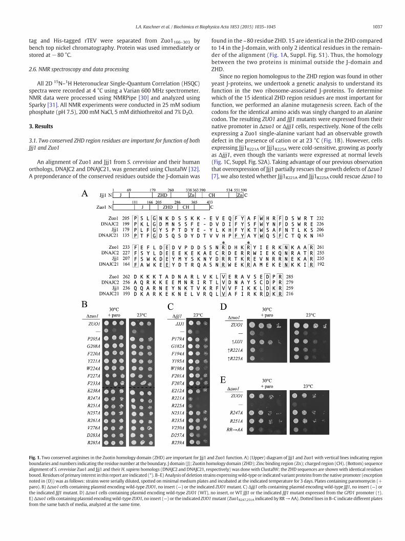
An alignment of Zuo1 and Jjj1 from S. cerevisiae and their humanorthologs, DNAJC2 and DNAJC21, was generated using ClustalW [32].A preponderance of the conserved residues outside the J-domain was
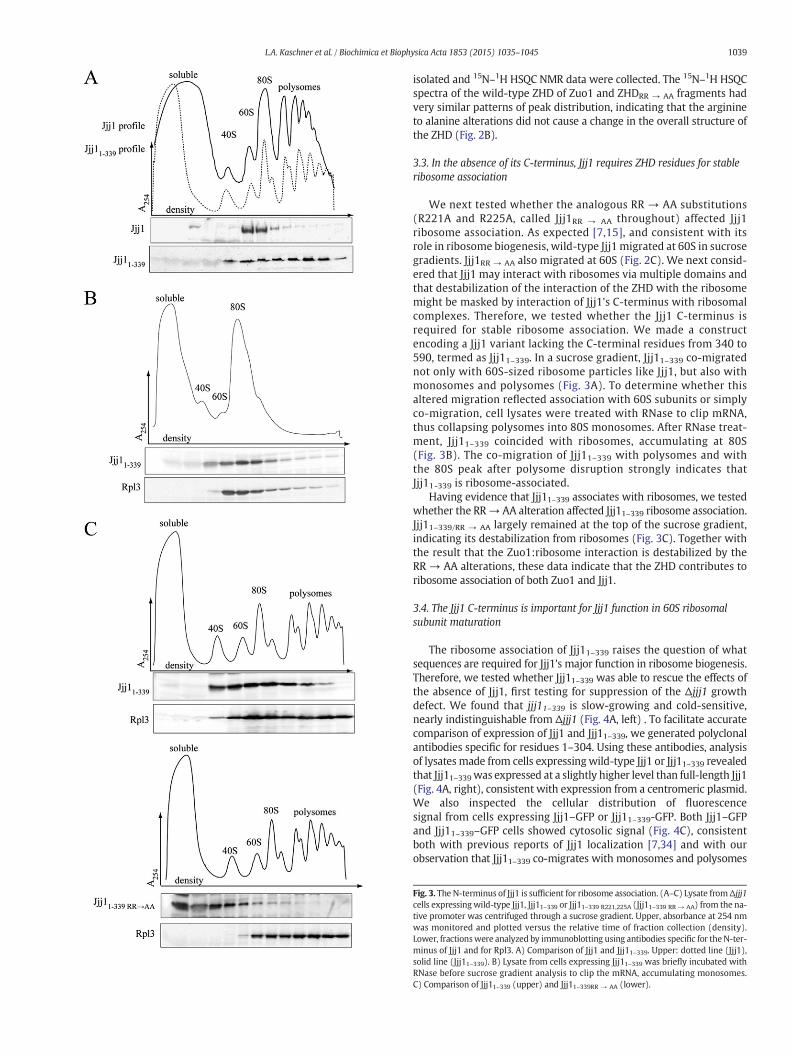
Fig. 1. Two conserved arginines in the Zuotin homology domain (ZHD) are important for Jjj1 anboundaries and numbers indicating the residue number at the boundary. J domain (J); Zuotin hoalignment of S. cerevisiae Zuo1 and Jjj1 and theirH. sapiens homologs (DNAJC2 and DNAJC21, reboxed. Residues of primary interest in this report are indicated (*). B–E) Analysis of deletion stranoted in (D)) was as follows: strains were serially diluted, spotted on minimal medium plates aparo). B) Δzuo1 cells containing plasmid encoding wild-type ZUO1, no insert (—) or the indicatthe indicated JJJ1 mutant. D) Δzuo1 cells containing plasmid encoding wild-type ZUO1 (WT),E)Δzuo1 cells containing plasmid encodingwild-type ZUO1, no insert (—) or the indicated ZUO1from the same batch of media, analyzed at the same time.
found in the ~80 residue ZHD. 15 are identical in the ZHD comparedto 14 in the J-domain, with only 2 identical residues in the remain-der of the alignment (Fig. 1A, Suppl. Fig. S1). Thus, the homologybetween the two proteins is minimal outside the J-domain andZHD.
Since no region homologous to the ZHD region was found in otheryeast J-proteins, we undertook a genetic analysis to understand itsfunction in the two ribosome-associated J-proteins. To determinewhich of the 15 identical ZHD region residues are most important forfunction, we performed an alanine mutagenesis screen. Each of thecodons for the identical amino acids was singly changed to an alaninecodon. The resulting ZUO1 and JJJ1 mutants were expressed from theirnative promoter in Δzuo1 or Δjjj1 cells, respectively. None of the cellsexpressing a Zuo1 single-alanine variant had an observable growthdefect in the presence of cation or at 23 °C (Fig. 1B). However, cellsexpressing Jjj1R221A or Jjj1R225A were cold-sensitive, growing as poorlyas Δjjj1, even though the variants were expressed at normal levels(Fig. 1C, Suppl. Fig. S2A). Taking advantage of our previous observationthat overexpression of Jjj1 partially rescues the growth defects ofΔzuo1[7], we also tested whether Jjj1R221A and Jjj1R225A could rescue Δzuo1 to
d Zuo1 function. A) (Upper) diagram of Jjj1 and Zuo1 with vertical lines indicating regionmology domain (ZHD); Zinc binding region (Zn); charged region (CH). (Bottom) sequencespectively) was donewith ClustalW; the ZHD sequences are shownwith identical residuesins expressingwild-type or indicated variant proteins from the native promoter (exceptionnd incubated at the indicated temperature for 3 days. Plates containing paromomycin (+ed ZUO1mutant. C) Δjjj1 cells containing plasmid encoding wild-type JJJ1, no insert (—) orno insert, or WT JJJ1 or the indicated JJJ1 mutant expressed from the GPD1 promoter (↑).mutant (Zuo1R247,251A indicated by RR→AA). Dotted lines in B–C indicate different plates
1038 L.A. Kaschner et al. / Biochimica et Biophysica Acta 1853 (2015) 1035–1045
the same extent. We placed JJJ1, jjj1R221A and jjj1R225A under the strongGPD1 promoter. Though all the three proteins were expressed atvery similar levels, only wild-type Jjj1 rescued the cation-sensitivity ofΔzuo1 cells (Fig. 1D, Suppl. Fig. S2B). Since a low level of Zuo1 (b5%the level in wild-type cells) is sufficient to support growth indistin-guishable from that of cells with normal Zuo1 levels [33], we consideredthat deleterious effects of the single-alanine substitutions in Zuo1mightbe masked. To test more stringently whether the analogous argininesthat are important for Jjj1 function are also important for Zuo1 function,we constructed a double substitution mutant, changing the codonsfor both arginine 247 and arginine 251 of ZUO1 to alanine (calledzuo1RR → AA throughout). zuo1RR → AA was both cation- and cold-sensitive (Fig. 1E), though Zuo1RR → AA was expressed at a similarlevel to wild-type Zuo1 protein (Suppl. Fig. S2C). Together these resultssuggest that the analogous residues of the ZHD, R247 and R251 in Zuo1,and R221 and R225 in Jjj1, are functionally important.
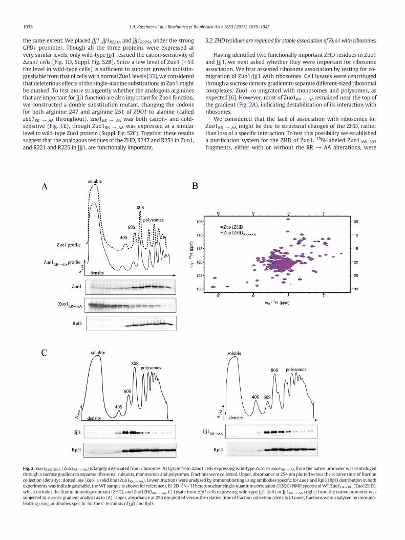
Fig. 2. Zuo1R247,251A (Zuo1RR → AA) is largely dissociated from ribosomes. A) Lysate from Δzuo1through a sucrose gradient to separate ribosomal subunits, monosomes and polysomes. Fractiocollection (density); dotted line (Zuo1), solid line (Zuo1RR → AA). Lower, fractionswere analyzedexperiments was indistinguishable, theWT sample is shown for reference). B) 2D 15N–1H heterwhich includes the Zuotin homology domain (ZHD), and Zuo1ZHDRR → AA. C) Lysate from Δjjjsubjected to sucrose gradient analysis as in (A). Upper, absorbance at 254 nm plotted versus thblotting using antibodies specific for the C-terminus of Jjj1 and Rpl3.
3.2. ZHD residues are required for stable association of Zuo1with ribosomes
Having identified two functionally important ZHD residues in Zuo1and Jjj1, we next asked whether they were important for ribosomeassociation. We first assessed ribosome association by testing for co-migration of Zuo1/Jjj1 with ribosomes. Cell lysates were centrifugedthrough a sucrose density gradient to separate different-sized ribosomalcomplexes. Zuo1 co-migrated with monosomes and polysomes, asexpected [6]. However, most of Zuo1RR → AA remained near the top ofthe gradient (Fig. 2A), indicating destabilization of its interaction withribosomes.
We considered that the lack of association with ribosomes forZuo1RR → AA might be due to structural changes of the ZHD, ratherthan loss of a specific interaction. To test this possibility we establisheda purification system for the ZHD of Zuo1. 15N-labeled Zuo1166–303fragments, either with or without the RR → AA alterations, were
cells expressing wild-type Zuo1 or Zuo1RR → AA from the native promoter was centrifugednswere collected. Upper, absorbance at 254 nm plotted versus the relative time of fractionby immunoblotting using antibodies specific for Zuo1 and Rpl3 (Rpl3 distribution in bothonuclear single-quantum correlation (HSQC) NMR spectra ofWT Zuo1166–303 (Zuo1ZHD),1 cells expressing wild-type Jjj1 (left) or Jjj1RR → AA (right) from the native promoter wase relative time of fraction collection (density). Lower, fractions were analyzed by immuno-
1039L.A. Kaschner et al. / Biochimica et Biophysica Acta 1853 (2015) 1035–1045
isolated and 15N–1H HSQC NMR data were collected. The 15N–1H HSQCspectra of the wild-type ZHD of Zuo1 and ZHDRR → AA fragments hadvery similar patterns of peak distribution, indicating that the arginineto alanine alterations did not cause a change in the overall structure ofthe ZHD (Fig. 2B).
3.3. In the absence of its C-terminus, Jjj1 requires ZHD residues for stableribosome association
We next tested whether the analogous RR → AA substitutions(R221A and R225A, called Jjj1RR → AA throughout) affected Jjj1ribosome association. As expected [7,15], and consistent with itsrole in ribosome biogenesis, wild-type Jjj1 migrated at 60S in sucrosegradients. Jjj1RR → AA also migrated at 60S (Fig. 2C). We next consid-ered that Jjj1 may interact with ribosomes via multiple domains andthat destabilization of the interaction of the ZHD with the ribosomemight be masked by interaction of Jjj1's C-terminus with ribosomalcomplexes. Therefore, we tested whether the Jjj1 C-terminus isrequired for stable ribosome association. We made a constructencoding a Jjj1 variant lacking the C-terminal residues from 340 to590, termed as Jjj11–339. In a sucrose gradient, Jjj11–339 co-migratednot only with 60S-sized ribosome particles like Jjj1, but also withmonosomes and polysomes (Fig. 3A). To determine whether thisaltered migration reflected association with 60S subunits or simplyco-migration, cell lysates were treated with RNase to clip mRNA,thus collapsing polysomes into 80S monosomes. After RNase treat-ment, Jjj11–339 coincided with ribosomes, accumulating at 80S(Fig. 3B). The co-migration of Jjj11–339 with polysomes and withthe 80S peak after polysome disruption strongly indicates thatJjj11-339 is ribosome-associated.
Having evidence that Jjj11–339 associates with ribosomes, we testedwhether the RR→ AA alteration affected Jjj11–339 ribosome association.Jjj11–339/RR → AA largely remained at the top of the sucrose gradient,indicating its destabilization from ribosomes (Fig. 3C). Together withthe result that the Zuo1:ribosome interaction is destabilized by theRR → AA alterations, these data indicate that the ZHD contributes toribosome association of both Zuo1 and Jjj1.
3.4. The Jjj1 C-terminus is important for Jjj1 function in 60S ribosomalsubunit maturation
The ribosome association of Jjj11–339 raises the question of whatsequences are required for Jjj1's major function in ribosome biogenesis.Therefore, we tested whether Jjj11–339 was able to rescue the effects ofthe absence of Jjj1, first testing for suppression of the Δjjj1 growthdefect. We found that jjj11–339 is slow-growing and cold-sensitive,nearly indistinguishable from Δjjj1 (Fig. 4A, left) . To facilitate accuratecomparison of expression of Jjj1 and Jjj11–339, we generated polyclonalantibodies specific for residues 1–304. Using these antibodies, analysisof lysatesmade from cells expressingwild-type Jjj1 or Jjj11–339 revealedthat Jjj11–339was expressed at a slightly higher level than full-length Jjj1(Fig. 4A, right), consistent with expression from a centromeric plasmid.We also inspected the cellular distribution of fluorescencesignal from cells expressing Jjj1–GFP or Jjj11–339-GFP. Both Jjj1–GFPand Jjj11–339–GFP cells showed cytosolic signal (Fig. 4C), consistentboth with previous reports of Jjj1 localization [7,34] and with ourobservation that Jjj11–339 co-migrates with monosomes and polysomes
Fig. 3. The N-terminus of Jjj1 is sufficient for ribosome association. (A–C) Lysate fromΔjjj1cells expressingwild-type Jjj1, Jjj11–339 or Jjj11–339 R221,225A (Jjj11–339 RR → AA) from the na-tive promoter was centrifuged through a sucrose gradient. Upper, absorbance at 254 nmwas monitored and plotted versus the relative time of fraction collection (density).Lower, fractionswere analyzed by immunoblotting using antibodies specific for theN-ter-minus of Jjj1 and for Rpl3. A) Comparison of Jjj1 and Jjj11–339. Upper: dotted line (Jjj1),solid line (Jjj11–339). B) Lysate from cells expressing Jjj11–339 was briefly incubated withRNase before sucrose gradient analysis to clip the mRNA, accumulating monosomes.C) Comparison of Jjj11–339 (upper) and Jjj11–339RR → AA (lower).
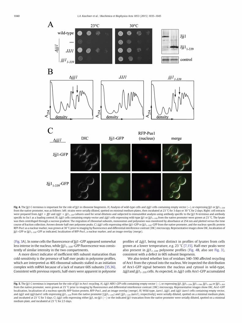
Fig. 4. The Jjj1 C-terminus is important for the role of Jjj1 in ribosome biogenesis. A) Analysis of wild-type cells and Δjjj1 cells containing empty vector (—), or expressing Jjj1 or Jjj11–339from the native promoter, was as follows: left: strains were serially diluted, spotted onminimal medium plates, then incubated at 23 °C for 3 days or 30 °C for 2 days. Right: cell extractswere prepared from Δjjj1+ JJJ1 and Δjjj1+ jjj11–339 cultures used for serial dilutions and subjected to immunoblot analysis using antibody specific to the Jjj1 N-terminus and antibodyspecific to Ssc1 as a loading control. B) Δjjj1 cells containing empty vector and Δjjj1 cells expressing wild-type Jjj1 or Jjj11–339 from the native promoter were grown at 23 °C. The lysatewas then centrifuged through a sucrose gradient. The migration of ribosomal subunits, monosomes and polysomes was monitored by absorbance at 254 nm and plotted versus the timecourse of fraction collection. Arrows denote half-mer polysome peaks. C)Δjjj1 cells expressing either Jjj1–GFP or Jjj11–339–GFP from the native promoter, and the nucleus-specific proteinRFP-Pus1 as a nuclearmarker, was grown at 30 °C prior to imaging byfluorescence anddifferential interference contrast (DIC)microscopy. Representative images showDIC, localization ofJjj1–GFP or Jjj11–339–GFP as indicated, localization of RFP-Pus1, a nuclear marker, and an image overlay (merge).
1040 L.A. Kaschner et al. / Biochimica et Biophysica Acta 1853 (2015) 1035–1045
(Fig. 3A). In some cells the fluorescence of Jjj1–GFP appeared somewhatless intense in the nucleus, while Jjj11–339–GFP fluorescence was consis-tently of similar intensity in the two compartments.
A more direct indicator of inefficient 60S subunit maturation thancold-sensitivity is the presence of half-mer peaks in polysome profiles,which are interpreted as 40S ribosomal subunits stalled in an initiationcomplex with mRNA because of a lack of mature 60S subunits [35,36].Consistent with previous reports, half-mers were apparent in polysome
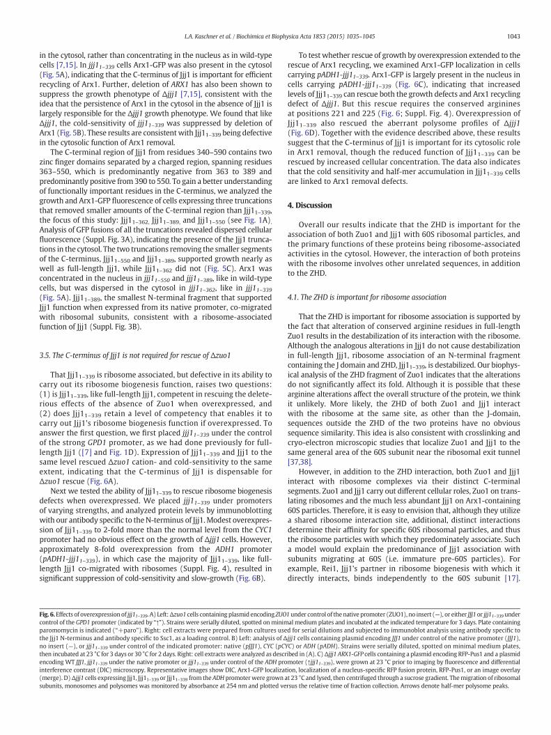
Fig. 5. The Jjj1 C-terminus is important for the role of Jjj1 in Arx1 recycling. A) Δjjj1 ARX1-GFP cfrom the native promoter, were grown at 23 °C prior to imaging by fluorescence and differenlocalization, localization of a nucleus-specific RFP fusion protein, RFP-Pus1, and an image ovand Δjjj1 and Δjjj1Δarx1 cells expressing jjj11–339 from the native promoter [(jjj11–339) and (jand incubated at 23 °C for 3 days. C) Δjjj1 cells expressing either Jjj1, no Jjj1 (−) or the indicamedium plate, and incubated at 23 °C for 2.5 days.
profiles of Δjjj1, being most distinct in profiles of lysates from cellsgrown at a lower temperature, e.g. 23 °C [7,15]. Half-mer peaks werealso present in jjj11–339 polysome profiles (Fig. 4B, also see Fig. 3),consistent with a defect in 60S subunit biogenesis.
We also tested whether loss of residues 340–590 affected recyclingof Arx1 from the cytosol into the nucleus. We inspected the distributionof Arx1-GFP signal between the nucleus and cytosol in wild-type,Δjjj1and jjj11–339 cells. As expected, in Δjjj1 cells Arx1-GFP accumulated
ells containing empty vector (—), or expressing Jjj1, Jjj11–339, Jjj11–550, Jjj11–389 or Jjj11–362tial interference contrast (DIC) microscopy. Representative images show DIC, Arx1-GFPerlay (merge). B) Wild-type, Δarx1, Δjjj1, and Δjjj1 Δarx1 cells containing empty vector,jj11–339 Δarx1), respectively], were serially diluted, spotted on a minimal medium plate,ted Jjj1 truncation from the native promoter were serially diluted, spotted on a minimal
1043L.A. Kaschner et al. / Biochimica et Biophysica Acta 1853 (2015) 1035–1045
in the cytosol, rather than concentrating in the nucleus as in wild-typecells [7,15]. In jjj11–339 cells Arx1-GFP was also present in the cytosol(Fig. 5A), indicating that the C-terminus of Jjj1 is important for efficientrecycling of Arx1. Further, deletion of ARX1 has also been shown tosuppress the growth phenotype of Δjjj1 [7,15], consistent with theidea that the persistence of Arx1 in the cytosol in the absence of Jjj1 islargely responsible for the Δjjj1 growth phenotype. We found that likeΔjjj1, the cold-sensitivity of jjj11–339 was suppressed by deletion ofArx1 (Fig. 5B). These results are consistentwith Jjj11–339 being defectivein the cytosolic function of Arx1 removal.
The C-terminal region of Jjj1 from residues 340–590 contains twozinc finger domains separated by a charged region, spanning residues363–550, which is predominantly negative from 363 to 389 andpredominantly positive from 390 to 550. To gain a better understandingof functionally important residues in the C-terminus, we analyzed thegrowth and Arx1-GFP fluorescence of cells expressing three truncationsthat removed smaller amounts of the C-terminal region than Jjj11–339,the focus of this study: Jjj11–362, Jjj11–389, and Jjj11–550 (see Fig. 1A).Analysis of GFP fusions of all the truncations revealed dispersed cellularfluorescence (Suppl. Fig. 3A), indicating the presence of the Jjj1 trunca-tions in the cytosol. The two truncations removing the smaller segmentsof the C-terminus, Jjj11–550 and Jjj11–389, supported growth nearly aswell as full-length Jjj1, while Jjj11–362 did not (Fig. 5C). Arx1 wasconcentrated in the nucleus in jjj11–550 and jjj11–389, like in wild-typecells, but was dispersed in the cytosol in jjj11–362, like in jjj11–339(Fig. 5A). Jjj11–389, the smallest N-terminal fragment that supportedJjj1 function when expressed from its native promoter, co-migratedwith ribosomal subunits, consistent with a ribosome-associatedfunction of Jjj1 (Suppl. Fig. 3B).
3.5. The C-terminus of Jjj1 is not required for rescue of Δzuo1
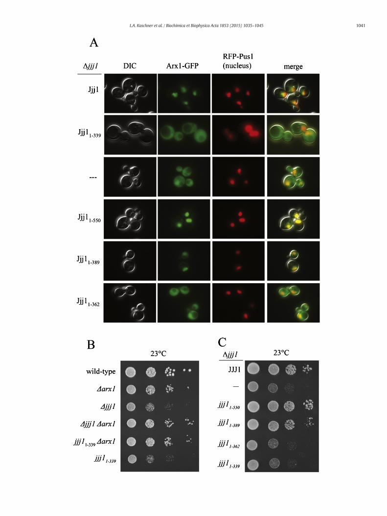
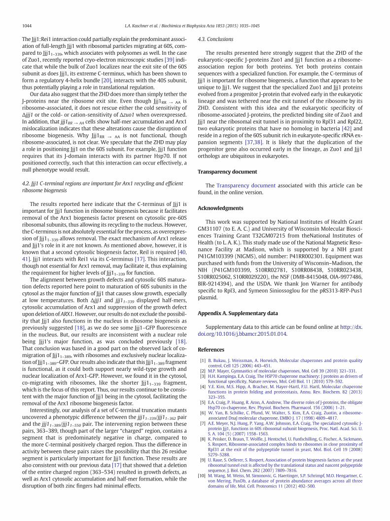
That Jjj11–339 is ribosome associated, but defective in its ability tocarry out its ribosome biogenesis function, raises two questions:(1) is Jjj11–339, like full-length Jjj1, competent in rescuing the delete-rious effects of the absence of Zuo1 when overexpressed, and(2) does Jjj11–339 retain a level of competency that enables it tocarry out Jjj1's ribosome biogenesis function if overexpressed. Toanswer the first question, we first placed jjj11–339 under the controlof the strong GPD1 promoter, as we had done previously for full-length Jjj1 ([7] and Fig. 1D). Expression of Jjj11–339 and Jjj1 to thesame level rescued Δzuo1 cation- and cold-sensitivity to the sameextent, indicating that the C-terminus of Jjj1 is dispensable forΔzuo1 rescue (Fig. 6A).
Next we tested the ability of Jjj11–339 to rescue ribosome biogenesisdefects when overexpressed. We placed jjj11–339 under promotersof varying strengths, and analyzed protein levels by immunoblottingwith our antibody specific to theN-terminus of Jjj1.Modest overexpres-sion of Jjj11–339 to 2-fold more than the normal level from the CYC1promoter had no obvious effect on the growth of Δjjj1 cells. However,approximately 8-fold overexpression from the ADH1 promoter(pADH1-jjj11–339), in which case the majority of Jjj11–339, like full-length Jjj1 co-migrated with ribosomes (Suppl. Fig. 4), resulted insignificant suppression of cold-sensitivity and slow-growth (Fig. 6B).
Fig. 6.Effects of overexpression of jjj11–339. A) Left:Δzuo1 cells containing plasmid encoding ZUOcontrol of the GPD1 promoter (indicated by “↑”). Strains were serially diluted, spotted on minimparomomycin is indicated (“+paro”). Right: cell extracts were prepared from cultures usethe Jjj1 N-terminus and antibody specific to Ssc1, as a loading control. B) Left: analysis of Δno insert (—), or jjj11–339 under control of the indicated promoter: native (pJJJ1), CYC (pCYthen incubated at 23 °C for 3 days or 30 °C for 2 days. Right: cell extracts were analyzed as descrencoding WT JJJ1, jjj11–339 under the native promoter or jjj11–339 under control of the ADH prointerference contrast (DIC) microscopy. Representative images show DIC, Arx1-GFP localizat(merge). D)Δjjj1 cells expressing Jjj1, Jjj11–339 or Jjj11–339 from the ADH promoterwere grown asubunits, monosomes and polysomes was monitored by absorbance at 254 nm and plotted ve
To test whether rescue of growth by overexpression extended to therescue of Arx1 recycling, we examined Arx1-GFP localization in cellscarrying pADH1-jjj11–339. Arx1-GFP is largely present in the nucleus incells carrying pADH1-jjj11–339 (Fig. 6C), indicating that increasedlevels of Jjj11–339 can rescue both the growth defects and Arx1 recyclingdefect of Δjjj1. But this rescue requires the conserved argininesat positions 221 and 225 (Fig. 6; Suppl. Fig. 4). Overexpression ofJjj11–339 also rescued the aberrant polysome profiles of Δjjj1(Fig. 6D). Together with the evidence described above, these resultssuggest that the C-terminus of Jjj1 is important for its cytosolic rolein Arx1 removal, though the reduced function of Jjj11–339 can berescued by increased cellular concentration. The data also indicatesthat the cold sensitivity and half-mer accumulation in Jjj11–339 cellsare linked to Arx1 removal defects.
4. Discussion
Overall our results indicate that the ZHD is important for theassociation of both Zuo1 and Jjj1 with 60S ribosomal particles, andthe primary functions of these proteins being ribosome-associatedactivities in the cytosol. However, the interaction of both proteinswith the ribosome involves other unrelated sequences, in additionto the ZHD.
4.1. The ZHD is important for ribosome association
That the ZHD is important for ribosome association is supported bythe fact that alteration of conserved arginine residues in full-lengthZuo1 results in the destabilization of its interaction with the ribosome.Although the analogous alterations in Jjj1 do not cause destabilizationin full-length Jjj1, ribosome association of an N-terminal fragmentcontaining the J domain and ZHD, Jjj11–339, is destabilized. Our biophys-ical analysis of the ZHD fragment of Zuo1 indicates that the alterationsdo not significantly affect its fold. Although it is possible that thesearginine alterations affect the overall structure of the protein, we thinkit unlikely. More likely, the ZHD of both Zuo1 and Jjj1 interactwith the ribosome at the same site, as other than the J-domain,sequences outside the ZHD of the two proteins have no obvioussequence similarity. This idea is also consistent with crosslinking andcryo-electron microscopic studies that localize Zuo1 and Jjj1 to thesame general area of the 60S subunit near the ribosomal exit tunnel[37,38].
However, in addition to the ZHD interaction, both Zuo1 and Jjj1interact with ribosome complexes via their distinct C-terminalsegments. Zuo1 and Jjj1 carry out different cellular roles, Zuo1 on trans-lating ribosomes and the much less abundant Jjj1 on Arx1-containing60S particles. Therefore, it is easy to envision that, although they utilizea shared ribosome interaction site, additional, distinct interactionsdetermine their affinity for specific 60S ribosomal particles, and thusthe ribosome particles with which they predominately associate. Sucha model would explain the predominance of Jjj1 association withsubunits migrating at 60S (i.e. immature pre-60S particles). Forexample, Rei1, Jjj1's partner in ribosome biogenesis with which itdirectly interacts, binds independently to the 60S subunit [17].
1under control of thenative promoter (ZUO1), no insert (—), or either JJJ1or jjj11–339underal medium plates and incubated at the indicated temperature for 3 days. Plate containingd for serial dilutions and subjected to immunoblot analysis using antibody specific tojjj1 cells containing plasmid encoding JJJ1 under control of the native promoter (JJJ1),C) or ADH (pADH). Strains were serially diluted, spotted on minimal medium plates,ibed in (A). C)Δjjj1 ARX1-GFP cells containing a plasmid encoding RFP-Pus1 and a plasmidmoter (↑jjj11–339), were grown at 23 °C prior to imaging by fluorescence and differentialion, localization of a nucleus-specific RFP fusion protein, RFP-Pus1, or an image overlayt 23 °C and lysed, then centrifuged through a sucrose gradient. Themigration of ribosomalrsus the relative time of fraction collection. Arrows denote half-mer polysome peaks.
1044 L.A. Kaschner et al. / Biochimica et Biophysica Acta 1853 (2015) 1035–1045
The Jjj1:Rei1 interaction could partially explain the predominant associ-ation of full-length Jjj1 with ribosomal particles migrating at 60S, com-pared to Jjj11–339, which associates with polysomes as well. In the caseof Zuo1, recently reported cryo-electron microscopic studies [39] indi-cate that while the bulk of Zuo1 localizes near the exit site of the 60Ssubunit as does Jjj1, its extreme C-terminus, which has been shown toform a regulatory 4-helix bundle [20], interacts with the 40S subunit,thus potentially playing a role in translational regulation.
Our data also suggest that the ZHDdoesmore than simply tether theJ-proteins near the ribosome exit site. Even though Jjj1RR → AA isribosome-associated, it does not rescue either the cold sensitivity ofΔjjj1 or the cold- or cation-sensitivity of Δzuo1 when overexpressed.In addition, that jjj1RR → AA cells show half-mer accumulation and Arx1mislocalization indicates that these alterations cause the disruption ofribosome biogenesis. Why Jjj1RR → AA is not functional, thoughribosome-associated, is not clear. We speculate that the ZHD may playa role in positioning Jjj1 on the 60S subunit. For example, Jjj1 functionrequires that its J-domain interacts with its partner Hsp70. If notpositioned correctly, such that this interaction can occur effectively, anull phenotype would result.
4.2. Jjj1 C-terminal regions are important for Arx1 recycling and efficientribosome biogenesis
The results reported here indicate that the C-terminus of Jjj1 isimportant for Jjj1 function in ribosome biogenesis because it facilitatesremoval of the Arx1 biogenesis factor present on cytosolic pre-60Sribosomal subunits, thus allowing its recycling to the nucleus. However,the C-terminus is not absolutely essential for the process, as overexpres-sion of Jjj11–339 allows removal. The exact mechanism of Arx1 releaseand Jjj1's role in it are not known. As mentioned above, however, it isknown that a second cytosolic biogenesis factor, Reil is required [40,41]. Jjj1 interacts with Rei1 via its C-terminus [17]. This interaction,though not essential for Arx1 removal, may facilitate it, thus explainingthe requirement for higher levels of Jjj11–339 for function.
The alignment between growth defects and cytosolic 60S matura-tion defects reported here point to maturation of 60S subunits in thecytosol as the major function of Jjj1 that causes slow growth, especiallyat low temperatures. Both Δjjj1 and jjj11–339 displayed half-mers,cytosolic accumulation of Arx1 and suppression of the growth defectupon deletion of ARX1. However, our results do not exclude the possibil-ity that Jjj1 also functions in the nucleus in ribosome biogenesis aspreviously suggested [18], as we do see some Jjj1–GFP fluorescencein the nucleus. But, our results are inconsistent with a nuclear rolebeing Jjj1's major function, as was concluded previously [18].That conclusion was based in a good part on the observed lack of co-migration of Jjj11–389, with ribosomes and exclusively nuclear localiza-tion of Jjj11–389–GFP. Our results also indicate that this Jjj11–389 fragmentis functional, as it could both support nearly wild-type growth andnuclear localization of Arx1-GFP. However, we found it in the cytosol,co-migrating with ribosomes, like the shorter Jjj11–339 fragment,which is the focus of this report. Thus, our results continue to be consis-tent with the major function of Jjj1 being in the cytosol, facilitating theremoval of the Arx1 ribosome biogenesis factor.
Interestingly, our analysis of a set of C-terminal truncation mutantsuncovered a phenotypic difference between the jjj11–339/jjj11–362 pairand the jjj11–389/jjj11–550 pair. The intervening region between thesepairs, 363–389, though part of the larger “charged” region, contains asegment that is predominately negative in charge, compared tothe more C-terminal positively charged region. Thus the difference inactivity between these pairs raises the possibility that this 26 residuesegment is particularly important for Jjj1 function. These results arealso consistent with our previous data [17] that showed that a deletionof the entire charged region (363–534) resulted in growth defects, aswell as Arx1 cytosolic accumulation and half-mer formation, while thedisruption of both zinc fingers had minimal effects.
4.3. Conclusions
The results presented here strongly suggest that the ZHD of theeukaryotic-specific J-proteins Zuo1 and Jjj1 function as a ribosome-association region for both proteins. Yet both proteins containsequences with a specialized function. For example, the C-terminus ofJjj1 is important for ribosome biogenesis, a function that appears to beunique to Jjj1. We suggest that the specialized Zuo1 and Jjj1 proteinsevolved from a progenitor J-protein that evolved early in the eukaryoticlineage and was tethered near the exit tunnel of the ribosome by itsZHD. Consistent with this idea and the eukaryotic specificity ofribosome-associated J-proteins, the predicted binding site of Zuo1 andJjj1 near the ribosomal exit tunnel is in proximity to Rpl31 and Rpl22,two eukaryotic proteins that have no homolog in bacteria [42] andreside in a region of the 60S subunit rich in eukaryote-specific rRNA ex-pansion segments [37,38]. It is likely that the duplication of theprogenitor gene also occurred early in the lineage, as Zuo1 and Jjj1orthologs are ubiquitous in eukaryotes.
Transparency document
The Transparency document associated with this article can befound, in the online version.
Acknowledgments
This work was supported by National Institutes of Health GrantGM31107 (to E. A. C.) and University of Wisconsin Molecular Biosci-ences Training Grant T32GM07215 from theNational Institutes ofHealth (to L. A. K.). This study made use of the National Magnetic Reso-nance Facility at Madison, which is supported by a NIH grantP41GM103399 (NIGMS), old number: P41RR002301. Equipment waspurchased with funds from the University of Wisconsin–Madison, theNIH (P41GM103399, S10RR02781, S10RR08438, S10RR023438,S10RR025062, S10RR029220), the NSF (DMB-8415048, OIA-9977486,BIR-9214394), and the USDA. We thank Jon Warner for antibodyspecific to Rpl3, and Symeon Siniossoglou for the pRS313-RFP-Pus1plasmid.
Appendix A. Supplementary data
Supplementary data to this article can be found online at http://dx.doi.org/10.1016/j.bbamcr.2015.01.014.
References
[1] B. Bukau, J. Weissman, A. Horwich, Molecular chaperones and protein qualitycontrol, Cell 125 (2006) 443–451.
[2] M.P. Mayer, Gymnastics of molecular chaperones, Mol. Cell 39 (2010) 321–331.[3] H.H. Kampinga, E.A. Craig, The HSP70 chaperone machinery: J proteins as drivers of
functional specificity, Nature reviews, Mol. Cell Biol. 11 (2010) 579–592.[4] Y.E. Kim, M.S. Hipp, A. Bracher, M. Hayer-Hartl, F.U. Hartl, Molecular chaperone
functions in protein folding and proteostasis, Annu. Rev. Biochem. 82 (2013)323–355.
[5] E.A. Craig, P. Huang, R. Aron, A. Andrew, The diverse roles of J-proteins, the obligateHsp70 co-chaperone, Rev. Physiol. Biochem. Pharmacol. 156 (2006) 1–21.
[6] W. Yan, B. Schilke, C. Pfund, W. Walter, S. Kim, E.A. Craig, Zuotin, a ribosome-associated DnaJ molecular chaperone, EMBO J. 17 (1998) 4809–4817.
[7] A.E. Meyer, N.J. Hung, P. Yang, A.W. Johnson, E.A. Craig, The specialized cytosolic J-protein Jjj1, functions in 60S ribosomal subunit biogenesis, Proc. Natl. Acad. Sci. U.S. A. 104 (5) (2007) 1558–1563.
[8] K. Peisker, D. Braun, T. Wolfle, J. Hentschel, U. Funfschilling, G. Fischer, A. Sickmann,S. Rospert, Ribosome-associated complex binds to ribosomes in close proximity ofRpl31 at the exit of the polypeptide tunnel in yeast, Mol. Biol. Cell 19 (2008)5279–5288.
[9] U. Raue, S. Oellerer, S. Rospert, Association of protein biogenesis factors at the yeastribosomal tunnel exit is affected by the translational status and nascent polypeptidesequence, J. Biol. Chem. 282 (2007) 7809–7816.
[10] M. Wang, M. Weiss, M. Simonovic, G. Haertinger, S.P. Schrimpf, M.O. Hengartner, C.von Mering, PaxDb, a database of protein abundance averages across all threedomains of life, Mol. Cell. Proteomics 11 (2012) 492–500.
1045L.A. Kaschner et al. / Biochimica et Biophysica Acta 1853 (2015) 1035–1045
[11] M. Gautschi, H. Lilie, U. Funfschilling, A. Mun, S. Ross, T. Lithgow, P. Rucknagel, S.Rospert, RAC, a stable ribosome-associated complex in yeast formed by the DnaK–DnaJ homologs Ssz1p and zuotin, Proc. Natl. Acad. Sci. U. S. A. 98 (2001) 3762–3767.
[12] S. Kim, E. Craig, Broad sensitivity of Saccharomyces cerevisiae lacking ribosome-associated chaperone Ssb or Zuo1 to cations, including aminoglycosides, Eukaryot.Cell 4 (2005) 82–89.
[13] A. Koplin, S. Preissler, Y. Ilina, M. Koch, A. Scior, M. Erhardt, E. Deuerling, A dualfunction for chaperones SSB-RAC and the NAC nascent polypeptide-associatedcomplex on ribosomes, J. Cell Biol. 189 (2010) 57–68.
[14] F. Willmund, M. del Alamo, S. Pechmann, T. Chen, V. Albanese, E.B. Dammer, J. Peng,J. Frydman, The cotranslational function of ribosome-associated Hsp70 in eukaryoticprotein homeostasis, Cell 152 (2013) 196–209.
[15] E. Demoinet, A. Jacquier, G. Lutfalla, M. Fromont-Racine, The Hsp40 chaperone Jjj1 isrequired for the nucleo-cytoplasmic recycling of preribosomal factors in Saccharo-myces cerevisiae, RNA 13 (2007) 1570–1581.
[16] V.G. Panse, A.W. Johnson, Maturation of eukaryotic ribosomes: acquisition offunctionality, Trends Biochem. Sci. 35 (2010) 260–266.
[17] A.E. Meyer, L.A. Hoover, E.A. Craig, The cytosolic J-protein, Jjj1, and Rei1 function inthe removal of the pre-60 S subunit factor Arx1, J. Biol. Chem. 285 (2010) 961–968.
[18] V. Albanese, S. Reissmann, J. Frydman, A ribosome-anchored chaperone networkthat facilitates eukaryotic ribosome biogenesis, J. Cell Biol. 189 (2010) 69–81.
[19] J. Fiaux, J. Horst, A. Scior, S. Preissler, A. Koplin, B. Bukau, E. Deuerling, Structuralanalysis of the ribosome-associated complex (RAC) reveals an unusual Hsp70/Hsp40 interaction, J. Biol. Chem. 285 (2010) 3227–3234.
[20] J.K. Ducett, F.C. Peterson, L.A. Hoover, A.J. Prunuske, B.F. Volkman, E.A. Craig,Unfolding of the C-terminal domain of the J-protein Zuo1 releases autoinhibitionand activates Pdr1-dependent transcription, J. Mol. Biol. 425 (2013) 19–31.
[21] P. Huang, M. Gautschi, W. Walter, S. Rospert, E.A. Craig, The Hsp70 Ssz1 modulatesthe function of the ribosome-associated J-protein Zuo1, Nat. Struct. Mol. Biol. 12(2005) 497–504.
[22] C. Sahi, E.A. Craig, Network of general and specialty J protein chaperones of the yeastcytosol, Proc. Natl. Acad. Sci. U. S. A. 104 (2007) 7163–7168.
[23] R.S. Sikorski, P. Hieter, A system of shuttle vectors and yeast host strains designedfor efficient manipulation of DNA in Saccharomyces cerevisiae, Genetics 122 (1989)19–27.
[24] D. Mumberg, R. Muller, M. Funk, Yeast vectors for the controlled expression ofheterologous proteins in different genetic backgrounds, Gene 156 (1995) 119–122.
[25] V.V. Kushnirov, Rapid and reliable protein extraction from yeast, Yeast 16 (2000)857–860.
[26] C.A. Schneider, W.S. Rasband, K.W. Eliceiri, NIH Image to ImageJ: 25 years of imageanalysis, Nat. Methods 9 (2012) 671–675.
[27] Q. Liu, P. D'Silva, W. Walter, J. Marszalek, E.A. Craig, Regulated cycling of mitochon-drial Hsp70 at the protein import channel, Science 300 (2003) 139–141.
[28] R.C. Tyler, H.K. Sreenath, S. Singh, D.J. Aceti, C.A. Bingman, J.L. Markley, B.G. Fox, Auto-induction medium for the production of [U-15 N]- and [U-13C, U-15 N]-labeled
proteins for NMR screening and structure determination, Protein Expr. Purif. 40(2005) 268–278.
[29] B.G. Fox, P.G. Blommel, Autoinduction of protein expression, Curr. Protoc. ProteinSci. 56 (2009) 5.23.1–5.23.18.
[30] F. Delaglio, S. Grzesiek, G.W. Vuister, G. Zhu, J. Pfeifer, A. Bax, NMRPipe: a multidi-mensional spectral processing system based on UNIX pipes, J. Biomol. NMR 6(1995) 277–293.
[31] W. Lee, W.M. Westler, A. Bahrami, H.R. Eghbalnia, J.L. Markley, PINE-SPARKY:graphical interface for evaluating automated probabilistic peak assignments in pro-tein NMR spectroscopy, Bioinformatics 25 (2009) 2085–2087.
[32] J.D. Thompson, D.G. Higgins, T.J. Gibson, CLUSTAL W: improving the sensitivity ofprogressive multiple sequence alignment through sequence weighting, position-specific gap penalties and weight matrix choice, Nucleic Acids Res. 22 (1994)4673–4680.
[33] H. Hundley, H. Eisenman, W. Walter, T. Evans, Y. Hotokezaka, M. Wiedmann, E.Craig, The in vivo function of the ribosome-associated Hsp70, Ssz1, does not requireits putative peptide-binding domain, Proc. Natl. Acad. Sci. U. S. A. 99 (2002)4203–4208.
[34] W.K. Huh, J.V. Falvo, L.C. Gerke, A.S. Carroll, R.W. Howson, J.S. Weissman, E.K. O'Shea,Global analysis of protein localization in budding yeast, Nature 425 (2003) 686–691.
[35] T.L. Helser, R.A. Baan, A.E. Dahlberg, Characterization of a 40S ribosomal subunitcomplex in polyribosomes of Saccharomyces cerevisiae treated with cycloheximide,Mol. Cell. Biol. 1 (1981) 51–57.
[36] M.O. Rotenberg, M. Moritz, J.L. Woolford Jr., Depletion of Saccharomyces cerevisiaeribosomal protein L16 causes a decrease in 60S ribosomal subunits and formationof half-mer polyribosomes, Genes Dev. 2 (1988) 160–172.
[37] B.J. Greber, D. Boehringer, C. Montellese, N. Ban, Cryo-EM structures of Arx1 andmaturation factors Rei1 and Jjj1 bound to the 60S ribosomal subunit, Nat. Struct.Mol. Biol. 19 (2012) 1228–1233.
[38] C. Leidig, G. Bange, J. Kopp, S. Amlacher, A. Aravind, S. Wickles, G. Witte, E. Hurt, R.Beckmann, I. Sinning, Structural characterization of a eukaryotic chaperone—theribosome-associated complex, Nat. Struct. Mol. Biol. 20 (2013) 23–28.
[39] Y. Zhang, C. Ma, Y. Yuan, J. Zhu, N. Li, C. Chen, S. Wu, L. Yu, J. Lei, N. Gao, Structuralbasis for interaction of a cotranslational chaperone with the eukaryotic ribosome,Nat. Struct. Mol. Biol. 21 (2014) 1042–1046.
[40] N.J. Hung, A.W. Johnson, Nuclear recycling of the pre-60S ribosomal subunit-associated factor Arx1 depends on Rei1 in Saccharomyces cerevisiae, Mol. Cell. Biol.26 (2006) 3718–3727.
[41] A. Lebreton, C. Saveanu, L. Decourty, J.C. Rain, A. Jacquier, M. Fromont-Racine, Afunctional network involved in the recycling of nucleocytoplasmic pre-60S factors,J. Cell Biol. 173 (2006) 349–360.
[42] L. Jenner, S. Melnikov, N. Garreau de Loubresse, A. Ben-Shem, M. Iskakova, A.Urzhumtsev, A. Meskauskas, J. Dinman, G. Yusupova, M. Yusupov, Crystal structureof the 80S yeast ribosome, Curr. Opin. Struct. Biol. 22 (2012) 759–767.


![Biochimica et Biophysica Acta - immed.org considerations/09.07.2017 updates/Membrane... · G.L. Nicolson, M.E. Ash / Biochimica et Biophysica Acta 1859 (2017) 1704–1724 1705 [8]](https://img.pdfslide.net/doc/110x75/5c684f1e09d3f2f5638b5509/biochimica-et-biophysica-acta-immed-considerations09072017-updatesmembrane.jpg)