Embed Size (px)
Citation preview
lable at ScienceDirect
Biologicals 50 (2017) 27e34
Contents lists avai
Biologicals
journal homepage: www.elsevier .com/locate/bio logicals
Biophysical characterization and structure of the Fab fragment fromthe NIST reference antibody, RM 8671
Ioannis Karageorgos a, b, Elyssia S. Gallagher a, b, 1, Connor Galvin a, b,D. Travis Gallagher a, b, **, Jeffrey W. Hudgens a, b, *
a Biomolecular Measurement Division, National Institute of Standards & Technology, Rockville, MD 20850, USAb Institute for Bioscience and Biotechnology Research, 9600 Gudelsky Drive, Rockville, MD 20850, USA
a r t i c l e i n f o
Article history:Received 28 April 2017Received in revised form13 September 2017Accepted 18 September 2017Available online 29 September 2017
Keywords:AntibodyCrystal structurePhysical chemical characterizationIgG1kNISTmAbReference material
* Corresponding author. Biomolecular Measuremenof Standards & Technology, Rockville, MD 20850, USA** Corresponding author.Biomolecular Measuremenof Standards & Technology, Rockville, MD 20850, USA
E-mail addresses: [email protected]@baylor.edu (E.S. Gallagher), [email protected] (J.W. Hudgens).
1 Current address: Department of Chemistry and BioOne Bear Place #97348, Waco, TX, 76798.
https://doi.org/10.1016/j.biologicals.2017.09.0051045-1056/Published by Elsevier Ltd on behalf of Inte
a b s t r a c t
Monoclonal antibody pharmaceuticals are the fastest-growing class of therapeutics, with a wide range ofclinical applications. To assure their safety, these protein drugs must demonstrate highly consistentpurity and stability. Key to these objectives is higher order structure measurements validated by cali-bration to reference materials. We describe preparation, characterization, and crystal structure of the Fabfragment prepared from the NIST Reference Antibody RM 8671 (NISTmAb). NISTmAb is a humanizedIgG1k antibody, produced in murine cell culture and purified by standard biopharmaceutical productionmethods, developed at the National Institute of Standards and Technology (NIST) to serve as a referencematerial. The Fab fragment was derived from NISTmAb through papain cleavage followed by protein Abased purification. The purified Fab fragment was characterized by SDS-PAGE, capillary gel electropho-resis, multi-angle light scattering, size exclusion chromatography, mass spectrometry, and x-ray crys-tallography. The crystal structure at 0.2 nm resolution includes four independent Fab molecules withcomplete light chains and heavy chains through Cys 223, enabling assessment of conformational vari-ability and providing a well-characterized reference structure for research and engineering applications.This nonproprietary, publically available reference material of known higher-order structure can supportmetrology in biopharmaceutical applications, and it is a suitable platform for validation of molecularmodeling studies.
Published by Elsevier Ltd on behalf of International Alliance for Biological Standardization.
1. Introduction
The number of FDA-approved monoclonal antibody (mAb)pharmaceuticals has increased exponentially since 2000, doublingevery four years, and now numbers about 60. Global sales ofmonoclonal antibody pharmaceuticals in 2016 were $105B,comprising greater than ten metric tons of material [1,2]. The mAbdrugs demonstrate a trend toward smarter, more specific, andhence, safer therapeutics. Their production also reflects a
t Division, National Institute.t Division, National Institute.(I. Karageorgos), Elyssia_
[email protected] (D.T. Gallagher),
chemistry, Baylor University,
rnational Alliance for Biological St
significant investment since mAbs are among the greatest molec-ular weight, complex therapeutics ever produced. Part of the pricefor their etiologic specificity is that their production, requiringcultured mammalian cells, is highly elaborate, and the proteinsthemselves are relatively heterogeneous and sensitive to theirphysical environment [3]. Since their clinical efficacy depends ontheir biophysical properties, in particular the higher order struc-ture, it is necessary to measure those properties repeatedly andconsistently. Therefore, a well-characterized mAb reference mate-rial that can facilitate development of improved analytical tools andcomputational simulations, particularly for characterization ofhigher order structure, will help assure the continued improve-ment of mAb biotherapeutics.
Reference materials reside at the heart of monoclonal antibodyproduction to assure consistent product properties and quality overthe lifetime of the therapeutic protein. The National Institute ofStandards and Technology (NIST) has introduced a IgG1k antibodyreference material, NISTmAb (RM 8671) [4,5], as part of a program
andardization.
I. Karageorgos et al. / Biologicals 50 (2017) 27e3428
to develop fundamental materials and data in support of the bio-pharmaceutical industry. To support the utility of this referencematerial, it was characterized by over twenty-four analyticalmethods and the results from these studies are presented in threevolumes [6e8]. Those volumes focus on the mAb and do notinclude crystal structures for NISTmAb or its Fab fragment.
Here we report the preparation, biophysical characterization,and x-ray crystal structure of the Fab fragment derived fromNISTmAb material RM 8671. Mass spectrometry indicates thatheavy chain cleavage by papain occurred after His 227. The crystalstructure is generally representative of the Protein Data Bank'srepertoire of about 500 human and humanized Fab structures [9].However, the structure is more complete (in particular, includingthe entire light chain and all 5 disulfides per Fab) and includes fourindependent Fab fragments, thus providing information on struc-tural variability. The structure reported here is of the Fab fragmentin its ligand-free (apo) state, enabling comparison with the previ-ously published Fab complex (holo) structure [10] and inferenceson complementarity determining region (CDR) mobility. Thestructure provides reproducible reference data to support com-parisons over time and across Fab-related projects in research andmolecular engineering.
2. Materials and methods
2.1. Preparation of Fab fragment
Fab fragment production by cleavage of mAbs using papain,followed by protein A has become routine, such that several com-mercial kits are available, using agarose-immobilized forms of thepeptidase (extracted from papaya fruit latex) and protein A (fromStaph. aureus, recombinantly expressed in E. coli) [11]. Papain'sbroad specificity enables it to cleave all 4 subclasses of human IgGin the hinge region, despite their different hinge sequences.Cleavage efficiencies correlate with hinge lengths (IgG3 >>IgG1 > IgG2 ~ IgG4) and are generally enhanced by reducing agent,suggesting that accessibility of the hinge to the 23 kDa enzyme isthe key to cleavage [12]. For this study NISTmAb, produced inmurine cell culture, was treated by the components of the PierceFab Preparation kit (Model 44985, Thermo Scientific, Waltham,MA) to produce the Fab fragment. This method uses immobilizedpapain for cleavage and immobilized protein A to isolate the Fabfragment. For a small-scale preparation, 4 mg of NISTmAb in 0.5 mLof 20 mmol/L L-histidine, pH 6.0, were pre-equilibrated in cleavagebuffer using a Zeba spin desalting column and then incubated with0.25 mL (50% slurry) preequilibrated immobilized papain undermixing for 5 h at 37 �C. The digest was separated from papain bycentrifugation (5000 g for 1 min). Papain was washed with 0.5 mLsodium phosphate buffered saline (PBS) solution (pH 7.4) forcomplete recovery of the digest. The digest was then applied to aPBS-equilibrated NAb column (protein A agarose) and incubated atroom temperature for 10 min with agitation to capture the Fc andundigested NISTmAb. Fab fragment was then collected as the su-pernatant after centrifugation at 1000g for 1 min. Half of the Fabfragment was dialyzed in 10 mmol/L disodium phosphate,10 mmol/L monosodium phosphate, 150 mmol/L sodium chloride,pH 7.4, and the other half in 100 mmol/L ammonium acetate, pH6.0. For preparations of Fab from NISTmAb the typical yield is 55%.
2.2. Size exclusion chromatography
200 mL 100 mmol/L filtered Fab protein was applied to a GEHealthcare Superdex 75 10/300 GL column (GE Healthcare Bio-Sciences, Pittsburgh, PA), which was previously calibrated andequilibrated on an €AKTAPurifier system (Amersham Pharmacia
Biotechnology, Amersham, UK). (Note: In this text all references tofilters refer to a 0.22 mm nominal pore size.) The dialyzed Fabsamples, with 10 mmol/L disodium phosphate, 10 mmol/L mono-sodium phosphate, 150 mmol/L sodium chloride, pH 7.4, and100 mmol/L ammonium acetate, pH 6.0, were monitored at280 nm, and the elution volume was recorded for each peak.
2.3. SDS-PAGE
Protein samples and molecular mass markers (Bio-Rad Labora-tories, Inc., Hercules, CA) were resolved on a 15% SDS-PAGE gel inreducing (incubated at 70 �C for 5 min in Laemni buffer (Sigma-Aldrich, Inc., St. Louis, MO) containing 5% b-mercaptoethanol (v/v))and nonreducing (absence of b-mercaptoethanol) conditions. Gelswere stained using Coomassie blue (Model 1610786, Bio-Rad,Hercules, CA) stain.
2.4. Capillary gel electrophoresis (CGE)
200 mg filtered Fabwas mixed with 100 mL of SDS- sample buffer(100 mmol/L Tris e HCl, pH 9.0, 1% SDS) and 4 mL of a 10 kDa in-ternal standard. Half of the sample has been treated with 5 mL b-mercaptoethanol to reduce disulfide bondswhile the other half wasused to conduct CGE in non-reduced conditions. Samples werecentrifuged at 300 g for 1 min and heated at 70 �C for 10 min. A PA800 Plus Pharmaceutical Analysis System (PA 80 Plus, Sciex) withPDA detection at 220 nm was used to analyze Fab in reduced andnon-reduced conditions. For each separation cycle, the capillarywas first preconditioned with 0.1 mmol/L NaOH, 0.1 mmol/L HCl,deionizedwater, and SDS gel buffer. Prior to use, all gel buffers weredegassed for 2 min under vacuum. Samples were electrokineticallyintroduced by applying voltage at �5 kV for 20 s. Electrophoresiswas performed at constant voltage with applied field strengthof �497 V/cm with a capillary thermostatted to 25 �C, using recir-culating liquid coolant.
2.5. Mass spectrometric analysis
To determine the molecular weight of the Fab fragment, analiquot of intact desalted and filtered Fab was analyzed by an Agi-lent Infinity II UHPLC coupled with an Agilent 6545 (electrospray)Q-TOF mass spectrometer (Agilent Technologies, Santa Clara, CA). ABio-Spin 6 column (Bio-Rad, Hercules, CA) with 50 mmol/Lammonium bicarbonate buffer, pH 7.4, was used for desalting Fab.The Fab (3 mg) was injected onto an Agilent PLRP-S Column(1 mm � 50 mm, 100 nm pore size, 5 mm particle size). The mobilephase comprised Solvent A consisting of 0.1% formic acid in water(v/v) and Solvent B consisting of 0.1% formic acid in acetonitrile (v/v). After desalting by flowing a mixture of 80% Solvent A and 20%Solvent B through the column for 2 min at 0.4 mL/min, the samplewas eluted from the column as the mobile phase was changed via alinear gradient to 20% Solvent A and 80% Solvent B over 18 min. Thecolumnwas held at a constant temperature of 60 �C. The Q-TOFwasoperated in 2 GHz Extended Mass Range (500e5000 m/z) mode atan acquisition rate of 1 spectrum/s. The 1221.990,637m/z ion of theHP-1221 calibration standard (part #G1982-85001, Agilent Tech-nologies, Santa Clara, CA) was used as a reference mass throughoutthe run. Deconvolution of the resulting spectrum was performedusing BioConfirm 8.0, using the maximum entropy algorithm. Thecombined uncertainty of this mass measurement is ±5 Da.
2.6. Multi angle light scattering
200 mL of 100 mmol/L filtered Fab protein was injected into aShodex protein KW-804 column (Showa Denko America, Inc., New
I. Karageorgos et al. / Biologicals 50 (2017) 27e34 29
York, NY) that was coupled to an Agilent 1200 HPLC system (AgilentTechnologies, Santa Clara, CA), comprising a degasser, an isotropicpump, an autosampler and a variable wavelength UV detector. Therunning buffer was 20 mmol/L monosodium phosphate, 100 mmol/L sodium chloride, pH 7.3, with a flow rate 0.5 mL/min. A multi-angle light scattering detector (MALS), model miniDawn Treos(Wyatt Technology Corporation, Santa Barbara, CA), operating at awavelength of 658 nm with an Optilab rEX detector (Wyatt Tech-nology Corporation, Santa Barbara, CA), was used for molar massdetermination. Astra® software version 5.3.4.14 (Wyatt TechnologyCorporation, Santa Barbara, CA) was used to handle signals from thedetectors (MALS, differential refractive index (dRI), and UV) tocompute the proteinMw and concentration values using a refractiveindex increment dn/dc value of 0.185 mL/g. MALS and dRI in-struments were operated at 25 �C.
2.7. Crystallography
The Fab fragment was crystallized by vapor diffusion and itsstructure was determined by molecular replacement methods asdescribed in Ref. [13]. Crystal growth, twinning and pseudosym-metry caused some complications, but the final structure refinedwell and has been deposited in the Protein Data Bank (PDB)(Ref. #9) as 5K8A. The final structure has Rfree ¼ 0.24; other sta-tistics are in the PDB and in Ref. [13].
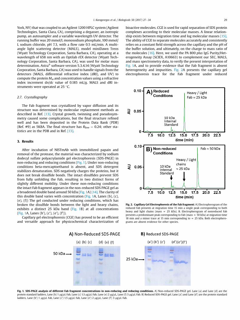
Fig. 2. Capillary Gel Electrophoresis of the Fab fragment: A) Electropherogram of thereduced Fab presents at migration time 15 min a single peak corresponding to bothheavy and light chains (mass z 25 kDa). B. Electropherogram of nonreduced Fabpresents a predominant peak corresponding to Fab (mass z 50 kDa) at migration time18 min and a minor trace at 15 min corresponding to z 25 kDa. Both electrophero-grams are absent evidence for other species.
3. Results
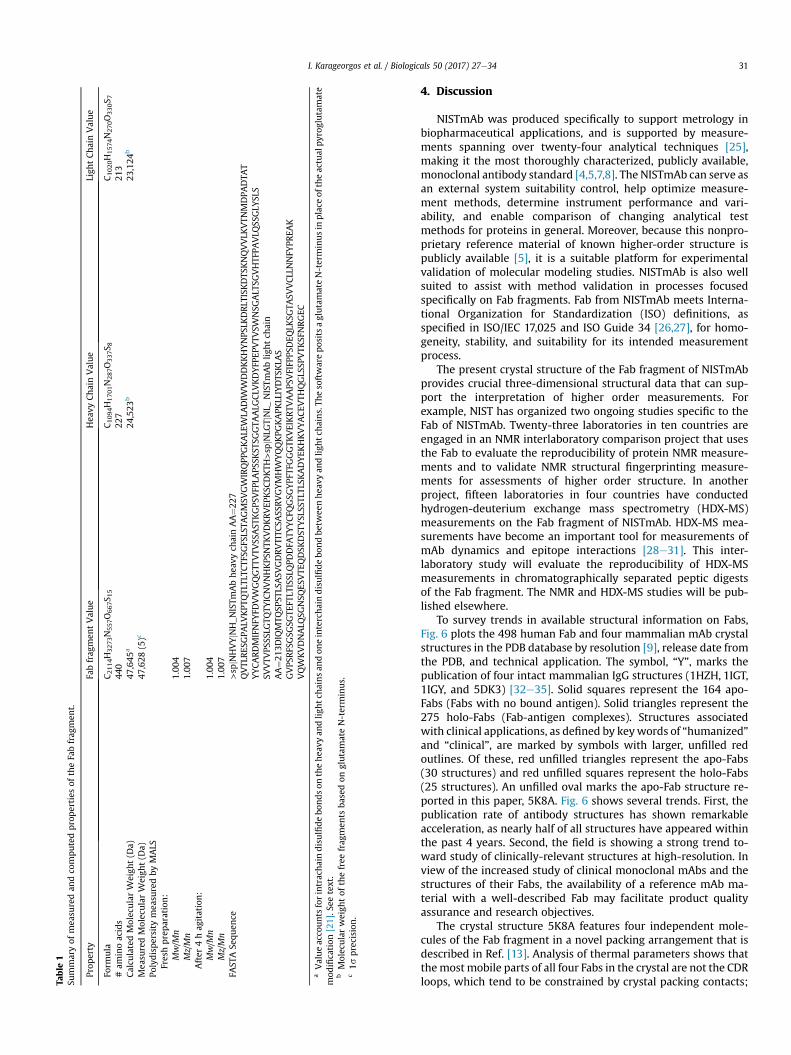
After incubation of NISTmAb with immobilized papain andremoval of the protease, the material was characterized by sodiumdodecyl sulfate polyacrylamide gel electrophoresis (SDS-PAGE) innon-reducing and reducing conditions (Fig. 1). Under non-reducingconditions beta-mercaptoethanol is absent, and SDS detergentstabilizes denaturation. SDS negatively charges the proteins, but itdoes not break disulfide bonds. The intact disulfides prevent SDSfrom fully unfolding the Fab, resulting in two distinct forms ofslightly different mobility. Under these non-reducing conditionsthe intact Fab fragment appears in the non-reduced SDS PAGE gel asa broadened double band around 50 kDa (Fig.1A) [14]. The clarity ofthis double band varies with concentration (Fig. 1A, Lanes (b), (c),(e), (f)) The gel conducted under reducing conditions, which hasbroken the disulfide bonds between the light and heavy chains,exhibits a distinct 25 kDa band (Fig. 1B) at all concentrations(Fig. 1A, Lanes (b’), (c’), (e’), (f’)).
Capillary gel electrophoresis (CGE) has proved to be an efficientand versatile approach for physicochemical characterization of
Fig. 1. SDS-PAGE analysis of different Fab fragment concentrations in non-reducing anprotein standard ladders, Lane (b) 1 mg/mL Fab, Lane (c) 1.5 mg/mL Fab, Lane (e) 2 mg/mL, Lane (fladders, Lane (b’) 1 mg/mL Fab, Lane (c’) 1.5 mg/mL Fab, Lane (e’) 2 mg/mL, Lane (f’) 3 mg/mL Fa
bioactive molecules. CGE is used for rapid separation of SDS proteincomplexes according to their molecular masses. A linear relation-ship exists between migration time and log molecular masses [15].The ability of CGE to separatemolecules accurately and consistentlyrelies on a constant field strength across the capillary and the pH ofthe buffer solution, and ultimately, on the charge to mass ratio ofthe molecules [16]. Here, we used the PA 800 plus IgG Purity/Het-erogeneity Assay (SCIEX, A10663) to complement our SEC, MALS,and mass spectrometry data, to verify the present interpretation ofFig. 1A, and to provide evidence that the Fab fragment is absentheterogeneity and impurities. Fig. 2A presents the capillary gelelectrophoresis trace for the Fab fragment under reduced
d reducing conditions. A) Non-reduced SDS-PAGE gel. Lane (a) and Lane (d) are the) 3 mg/mL Fab. B) Reduced SDS-PAGE gel. Lane (a’) and Lane (d’) are the protein standardb.

I. Karageorgos et al. / Biologicals 50 (2017) 27e3430
conditions. The trace shows only one peak at 15.0 min elution time,corresponding toz25 kDa, which are the molecular weights of theheavy and light chains of the Fab. The minor peak around 12 minelution time is the 10 kDa internal standard. Similar profile is ob-tained for CGE of Fab fragment under non-reduced conditions(Fig. 2B). This trace exhibits one major peak at 18.0 min elutiontime, corresponding toz50 kDa, which is themass of the intact Fabfragment, as this sample is not reduced. Fig. 2B exhibits a smalltrace at 15 min elution, indicating that a small fraction of Fabfragment has been reduced by the presence of SDS. Under bothreduced and non-reduced conditions, peaks at lower or highermolecular weight are not observed, indicating the absence of im-purities and aggregates.
Size exclusion chromatography then yielded Fab protein of pu-rity better than 99% (Fig. 3A). Electrospray quadrupole time-of-flight (ESI-Q-TOF) of the size exclusion chromatography (SEC)eluent exhibited a series of [M þ nH]nþ, (n ¼ 1, 2, 3 …) ion peaks(not shown). Deconvolution of these peaks reduced to a synthetic[M] neutral mass spectrum of Fab fragment, evincing a species of47,628 (±5) Da [17]. The calculated mass is 47,627 Da; hence, themass spectrum is in accord with the calculated mass. Computationof the calculated mass requires a series of adjustments for proteinmodifications. Popular protein mass calculators yield the mass,47,655 Da, by assuming the presence of a glutamate at the N-ter-minus and four intrachain and one interchain disulfide bonds.However, NISTmAb has a pyroglutamate (pGlu) at the N-terminus
Fig. 3. SEC analysis. A) Lower trace: Size exclusion chromatograph (SEC) of freshlyprepared Fab fragment (detector set at labs ¼ 280 nm) that displays a single eluent,corresponding to a z50 kDa product. Upper trace: SEC of the Fab fragment aftershaking for 4 h at 23 �C on the platform of a vortex mixer (See text.). B) The decon-voluted electrospray quadrupole time-of-flight (ESI-Q-TOF) mass spectrum fromeffluent at the signal maximum of the SEC chromatography trace. Thedeconvolutedpeak at 47,628 (±5)m/z, indicates a measured molecular weight for the Fab fragment of4762 (±5) g/mol.
of the heavy chain, which is a frequently observed cyclization of thegene-encoded N-terminal glutamine [18e20]. Accounting for thepyroglutamate (�18 Da) and five disulfides (four intrachain and onebetween chains, thus, losing ten hydrogens per Fab (�10 Da)[7,21,22]. Hence, the observed and predicted mass of the Fab frag-ment are in accord and confirm that papain cleaves NISTmAb afterCDKTH227. Table 1 summarizes these properties of the Fab fragmentof NISTmAb. The combined uncertainty of each measurement re-ported in this work is one standard deviation (1s).
Multi-angle light scattering measurements (MALS) determinedthe polydispersity values, Mw/Mn ¼ 1.004 and Mz/Mn ¼ 1.007(Table 1), whereMn, Mw, andMz are molecular weights determinedby a number average, a mass average, and as the third moment ofmass, respectively. These polydispersity values indicate that the Fabsolution is substantially free (over 99%) of aggregates or oligomers.Together the SEC, ESI-Q-TOF, and MALS data imply an upper boundon all macromolecular impurities including aggregates and oligo-mers that is well below one half percent.
MALS and SEC measurements indicate that the Fab fragment isresistant to aggregation under stresses typical of refrigerated stor-age or shipping on ice. For example, to determine the feasibility forshipping samples, a 1.5 mL Eppendorf tubewas filled with 500 mL ofFab solution (10 mmol/L disodium phosphate, 10 mmol/L mono-sodium phosphate,150mmol/L sodium chloride, pH 7.4,100 mmol/LFab, affixed sideways on a vortex platform (Vortex Genie 2,Bohemia, NY, USA), and agitated at 3200 repetitions per minute for4 h at 23 �C. The SEC chromatography trace (Fig. 3A, upper trace)andMALS results (Table 1) were unchanged. Similar measurementsindicated that Fab solutions remain unchanged when storedat �20 �C for eight months and that buffered, pH 7.4 solutionsprepared from lyophilized Fab are also the same (data not shown).
The crystal structure (Protein Data Base (PDB) file 5K8A) [9]contains four independent copies of the Fab fragment, each con-taining the full light chain (213 aa) and heavy chain residues 1 to223. In each Fab fragment the light chain and observed heavy chaincarboxyl termini (i.e., for heavy chain, Cys 223) form a disulfidebond, the only covalent bond between light and heavy chains in theIgG1 subclass (Fig. 4). Most reported Fab structures omit this di-sulfide bond because it is relatively mobile, and thus, unobservedby diffraction. The mass spectrum of the crystallized Fab materialindicates that heavy chain residues 224 to 227 are present in thecrystal, but these residues are not observed. In the structure, thefour Fab heterodimers are designated by chain identifiers LH, MV,AB, and EF. The four molecules are related by pseudo symmetry andhave generally similar conformations except at crystal contacts[13]. Five cis-prolines per Fab are observed at the normal locations.Pairwise root-mean-square deviations over their alpha carbonatoms range from 0.07 nm to 0.12 nm, and the four elbow angles(relating Fv to constant domains), are 140�, 143�, 143�, and 145�,respectively [23]. The surface areas buried in the light-heavyinterface are 19.24 nm2, 18.43 nm2, 18.68 nm2, and 19.16 nm2,respectively [24]. The four Fabs have different packing contacts anddifferent surface conformations, including a few apparent contact-induced perturbations of the CDR loop main chains with magni-tudes of z0.1 nm (Fig. 5). Main chain conformations vary signifi-cantly only for three residues; for two of these (light chain 29 andheavy 222) crystal contacts impinge; the other (heavy chain 140) isbetween glycines in an external loop. Over the 436 residuescomprising each Fab fragment (each observed 4 times), 83 havesome side chain rotamer variation among the four molecules. Mostof these are surface locations where crystal contacts within 0.5 nmoffer a reasonable explanation. The Fab molecule with the fewestCDR contacts, LH, is listed first in PDB file 5K8A and is used forrendering the structure below.
Table
1Su
mmaryof
mea
suredan
dco
mputedproperties
oftheFabfrag
men
t.
Prop
erty
Fabfrag
men
tValue
Hea
vyChainValue
Ligh
tChainValue
Form
ula
C 2114H3273N557O667S 1
5C 1
094H1701N287O337S 8
C 1020H1574N270O330S 7
#am
inoacids
440
227
213
Calcu
latedMolecularW
eigh
t(D
a)47
,645
a24
,523
b23
,124
b
Mea
suredMolecularW
eigh
t(D
a)47
,628
(5)c
Polydispersity
mea
suredby
MALS
Freshpreparation:
Mw/M
n1.00
4Mz/Mn
1.00
7After
4hag
itation:
Mw/M
n1.00
4Mz/Mn
1.00
7FA
STASe
quen
ce>sp
jNHVYjNH_N
ISTm
Abhea
vych
ainAA¼2
27QVTL
RES
GPA
LVKPT
QTL
TLTC
TFSG
FSLS
TAGMSV
GW
IRQPP
GKALE
WLA
DIW
WDDKKHYNPS
LKDRLT
ISKDTS
KNQVVLK
VTN
MDPA
DTA
TYYCARDMIFNFY
FDVW
GQGTT
VTV
SSAST
KGPS
VFP
LAPS
SKST
SGGTA
ALG
CLV
KDYFP
EPVTV
SWNSG
ALT
SGVHTF
PAVLQ
SSGLY
SLS
SVVTV
PSSS
LGTQ
TYICNVNHKPS
NTK
VDKRVEP
KSC
DKTH
>sp
jNLG
TjNL_
NISTm
Ablig
htch
ain
AA¼2
13DIQ
MTQ
SPST
LSASV
GDRVTITC
SASS
RVGYMHW
YQQKPG
KAPK
LLIYDTS
KLA
SGVPS
RFS
GSG
SGTE
FTLT
ISSL
QPD
DFA
TYYCFQ
GSG
YPF
TFGGGTK
VEIKRTV
AAPS
VFIFP
PSDEQ
LKSG
TASV
VCLLNNFY
PREA
KVQW
KVDNALQ
SGNSQ
ESVTE
QDSK
DST
YSL
SSTL
TLSK
ADYEK
HKVYACEV
THQGLS
SPVTK
SFNRGEC
aValueacco
unts
forintrachaindisulfidebo
ndson
thehea
vyan
dlig
htc
hainsan
don
einterchaindisulfidebo
ndbe
twee
nhea
vyan
dlig
htc
hains.Th
esoftwarepositsaglutamateN-terminusin
place
oftheactual
pyrog
lutamate
mod
ification
[21].S
eetext.
bMolecularweigh
tof
thefree
frag
men
tsba
sedon
glutamateN-terminus.
c1s
precision
.
I. Karageorgos et al. / Biologicals 50 (2017) 27e34 31
4. Discussion
NISTmAb was produced specifically to support metrology inbiopharmaceutical applications, and is supported by measure-ments spanning over twenty-four analytical techniques [25],making it the most thoroughly characterized, publicly available,monoclonal antibody standard [4,5,7,8]. The NISTmAb can serve asan external system suitability control, help optimize measure-ment methods, determine instrument performance and vari-ability, and enable comparison of changing analytical testmethods for proteins in general. Moreover, because this nonpro-prietary reference material of known higher-order structure ispublicly available [5], it is a suitable platform for experimentalvalidation of molecular modeling studies. NISTmAb is also wellsuited to assist with method validation in processes focusedspecifically on Fab fragments. Fab from NISTmAb meets Interna-tional Organization for Standardization (ISO) definitions, asspecified in ISO/IEC 17,025 and ISO Guide 34 [26,27], for homo-geneity, stability, and suitability for its intended measurementprocess.
The present crystal structure of the Fab fragment of NISTmAbprovides crucial three-dimensional structural data that can sup-port the interpretation of higher order measurements. Forexample, NIST has organized two ongoing studies specific to theFab of NISTmAb. Twenty-three laboratories in ten countries areengaged in an NMR interlaboratory comparison project that usesthe Fab to evaluate the reproducibility of protein NMR measure-ments and to validate NMR structural fingerprinting measure-ments for assessments of higher order structure. In anotherproject, fifteen laboratories in four countries have conductedhydrogen-deuterium exchange mass spectrometry (HDX-MS)measurements on the Fab fragment of NISTmAb. HDX-MS mea-surements have become an important tool for measurements ofmAb dynamics and epitope interactions [28e31]. This inter-laboratory study will evaluate the reproducibility of HDX-MSmeasurements in chromatographically separated peptic digestsof the Fab fragment. The NMR and HDX-MS studies will be pub-lished elsewhere.
To survey trends in available structural information on Fabs,Fig. 6 plots the 498 human Fab and four mammalian mAb crystalstructures in the PDB database by resolution [9], release date fromthe PDB, and technical application. The symbol, “Y”, marks thepublication of four intact mammalian IgG structures (1HZH, 1IGT,1IGY, and 5DK3) [32e35]. Solid squares represent the 164 apo-Fabs (Fabs with no bound antigen). Solid triangles represent the275 holo-Fabs (Fab-antigen complexes). Structures associatedwith clinical applications, as defined by keywords of “humanized”and “clinical”, are marked by symbols with larger, unfilled redoutlines. Of these, red unfilled triangles represent the apo-Fabs(30 structures) and red unfilled squares represent the holo-Fabs(25 structures). An unfilled oval marks the apo-Fab structure re-ported in this paper, 5K8A. Fig. 6 shows several trends. First, thepublication rate of antibody structures has shown remarkableacceleration, as nearly half of all structures have appeared withinthe past 4 years. Second, the field is showing a strong trend to-ward study of clinically-relevant structures at high-resolution. Inview of the increased study of clinical monoclonal mAbs and thestructures of their Fabs, the availability of a reference mAb ma-terial with a well-described Fab may facilitate product qualityassurance and research objectives.
The crystal structure 5K8A features four independent mole-cules of the Fab fragment in a novel packing arrangement that isdescribed in Ref. [13]. Analysis of thermal parameters shows thatthemostmobile parts of all four Fabs in the crystal are not the CDRloops, which tend to be constrained by crystal packing contacts;
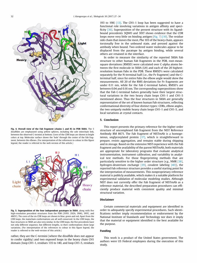
Fig. 4. Overall view of the Fab fragment (chains L and H) in PDB 5K8A. The 5disulfides are emphasized using yellow spheres, including the sole interchain link,between the observed C-termini at bottom. Cores of the CDR loops are shown in brightcolors at top. Molecular surface shows the 'hole' through the center of the Fab frag-ment, between the elbows. (For interpretation of the references to colour in this figurelegend, the reader is referred to the web version of this article.)
Fig. 5. Superposition of the four independent paratopes in 5K8A, along with fivehigh-resolution precedent structures from the PDB (2VXV, 2XZA, 3N9G, 3NTC, and4M5Y). The cores of the six CDR loops are shown in blue, green and red. Apart from theCDR loops, the mainchain conformations are all well conserved. In the CDR loops, thefour structures in 5K8A are also very similar. In the CDR loops, the five precedents havenot only different sequence, but different lengths, so their conformations show largevariations. (For interpretation of the references to colour in this figure legend, thereader is referred to the web version of this article.)
I. Karageorgos et al. / Biologicals 50 (2017) 27e3432
rather, they are the C-termini (where the disulfide does not appearto confer rigidity) and two exposed loops in the heavy chain CH1domain (loop CH1-1, residues 135 to 140, and loop CH1-5, residues
193 to 198) [13]. The CH1-1 loop has been suggested to have afunctional role involving variations in antigen affinity and speci-ficity [36]. Superposition of the present structure with its ligand-bound precedents 3QWO and 3IXT shows evidence that the CDRloops move very little on binding antigen (Fig. 7) [10]. The residueside chain that moves themost, Phe 103 of the heavy chain, appearstorsionally free in the unbound state, and pressed against theantibody when bound. Two ordered water molecules appear to bedisplaced from the paratope by antigen binding, while severalothers are retained in the interface.
In order to measure the similarity of the reported 5K8A Fabstructure to other human Fab fragments in the PDB, root-mean-square deviations (RMSD) were calculated over C-alpha atoms be-tween the first molecule in 5K8A (LH) and each of the 20 highest-resolution human Fabs in the PDB. These RMSD's were calculatedseparately for the N-terminal half (i.e., the Fv fragment) and the C-terminal half, since for entire Fabs the elbow angle would skew themeasurements. All 20 of the RMS deviations for Fv fragments areunder 0.11 nm, while for the Fab C-terminal halves, RMSD's arebetween 0.04 and 0.16 nm. The corresponding superpositions showthat the Fab C-terminal halves generally have their largest struc-tural variations in the two heavy chain loops CH1-1 and CH1-5mentioned above. Thus the four structures in 5K8A are generallyrepresentative of the set of known human Fab structures, reflectingconformational diversity of four distinct types: CDRs, elbow angles,the two uniquely mobile heavy chain loops CH1-1 and CH1-5, andlocal variations at crystal contacts.
5. Conclusion
This report presents the primary reference for the higher orderstructure of uncomplexed Fab fragment from the NIST ReferenceAntibody RM 8671. The Fab fragment of NISTmAb is a homoge-neous, unglycosylated protein [37], which is straightforward toprepare, resists aggregation, and remains stable during shippingand in storage. Based on the extensive NISTexperiencewith the Fabfragment and the availability of the parent NISTmAb, bothmaterialsare appropriate for laboratory programs that evaluate analyticalinstrumentation, instrument calibration, and variability in analyt-ical test methods. For those fingerprinting methods that areparticularly sensitive to the higher order structure (e.g., NMR [38],hydrogen-deuterium exchange [39], covalent labeling [40]), thereported Fab reference structure provides a useful starting point forthe interpretation of measurements. This nonproprietary referencematerial is publicly available, whichmakes it a suitable platform forexperimental validation of molecular modeling studies. AlthoughNIST does not currently offer the Fab fragment of NISTmAb as areference material, the described preparation procedures can effi-ciently produce material with consistent quality and minimalstructural variation.
Disclaimer
Certain commercial materials and equipment are identified inorder to adequately specify experimental procedures. Such identi-fications neither imply recommendation or endorsement by theNational Institute of Standards and Technology nor does it implythat the material or equipment identified is the best available forthe purpose.
Funding
This work is a product of the United States government. Theauthors were US Federal employees during the execution of thisstudy.
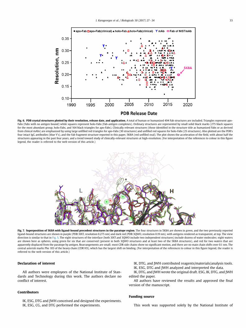
Fig. 6. PDB crystal structures plotted by their resolution, release date, and application. A total of human or humanized 494 Fab structures are included. Triangles represent apo-Fabs (Fabs with no antigen bound) while squares represent holo-Fabs (Fab-antigen complexes). Ordinary structures are represented by small solid black marks (275 black squaresfor the most abundant group, holo-Fabs, and 164 black triangles for apo-Fabs). Clinically relevant structures (those identified in the structure title as humanized Fabs or as derivedfrom clinical mAbs) are emphasized by using large unfilled red triangles for apo-Fabs (30 structures) and unfilled red squares for holo-Fabs (25 structures). Also plotted are the PDB'sfour intact IgG antibodies (blue Y's), and the Fab fragment structure reported in this paper, 5K8A (red unfilled oval). The plot shows the acceleration of the field, with about half thestructures appearing in the past four years, and a trend toward study of clinically-relevant structures at high-resolution. (For interpretation of the references to colour in this figurelegend, the reader is referred to the web version of this article.)
Fig. 7. Superposition of 5K8A with ligand-bound precedent structures in the paratope region. The four structures in 5K8A are shown in green, and the two previously reportedligand-bound structures are shown in purple (PDB:3IXT, resolution 0.275 nm) and dark red (PDB:3QWO, resolution 0.19 nm), with antigens rendered as transparent, at top. The viewdirection is similar to that in Fig. 5. The eight structures of the interface (both 3IXT and 3QWO include two independent structures) include dozens of water molecules; eight watersare shown here as spheres, using green for six that are conserved (present in both 3QWO structures and at least two of the 5K8A structures), and red for two waters that areapparently displaced from the paratope by antigen. Rearrangements are small; most CDR side chains show no significant motion, and there are no main chain shifts over 0.1 nm. Thecentral asterisk marks Phe 103 of the heavy chain (CDR H3), which has the largest shift on binding. (For interpretation of the references to colour in this figure legend, the reader isreferred to the web version of this article.)
I. Karageorgos et al. / Biologicals 50 (2017) 27e34 33
Declaration of interest
All authors were employees of the National Institute of Stan-dards and Technology during this work. The authors declare noconflict of interest.
Contributors
IK, ESG, DTG and JWH conceived and designed the experiments.IK, ESG, CG, and DTG performed the experiments.
IK, DTG, and JWH contributed reagents/materials/analysis tools.IK, ESG, DTG and JWH analyzed and interpreted the data.IK, DTG, and JWHwrote the original draft. ESG, IK, DTG, and JWH
edited the paper.All authors have reviewed the results and approved the final
version of the manuscript.
Funding source
This work was supported solely by the National Institute of
I. Karageorgos et al. / Biologicals 50 (2017) 27e3434
Standards and Technology, a nonregulatory agency of the Depart-ment of Commerce, United States of America Federal government.
Acknowledgments
I.K. and E.S.G. acknowledge the support of National Academy ofScience's National Research Council postdoctoral fellowships. C.G.acknowledges support from a NIST Summer UndergraduateResearch Fellowship. We thank Dr. L. Arbogast for guidance on earlyFab preparations, Dr. J. E. Schiel for help with the ESI-Q-TOF mea-surements and for technical discussions and Drs. R. Zangmeister, D.Ripple and J. Marino for careful reading and suggestions on themanuscript.
Appendix A. Supplementary data
Supplementary data related to this article can be found athttps://doi.org/10.1016/j.biologicals.2017.09.005.
References
[1] Ecker DM, Jones SD, Levine HL. The therapeutic monoclonal antibody market.mAbs 2015;7:9e14.
[2] Antibodies Market worth US$ 341 Bn by 2026. [accessed 13 OCT 2016]. http://www.medgadget.com/2016/10/antibodies-market-worth-us-341-bn-by-2026.html.
[3] Liu H, Gaza-Bulseco G, Faldu D, Chumsae C, Sun J. Heterogeneity of mono-clonal antibodies. J Pharm Sci;97:2426e2447.
[4] RM 8671-NISTmAb, Humanized IgG1k Monoclonal Antibody. Gaithersburg,MD: National Institute of Standards and Technology, [accessed 12 SEP 2016].https://www-s.nist.gov/srmors/view_detail.cfm?srm¼8671.
[5] NIST Monoclonal Antibody Reference Material 8671. Gaithersburg, MD,[accessed 30 MAR 2017]. https://www.nist.gov/programs-projects/nist-monoclonal-antibody-reference-material-8671.
[6] Schiel JE, Darryl LD, Oleg VB. State-of-the-Art and emerging Technologies fortherapeutic monoclonal antibody characterization volume 1. Monoclonalantibody therapeutics: structure, function, and regulatory space. In: ACSsymposium series. American Chemical Society; 2014.
[7] Schiel JE, Darryl LD, Oleg VB. State-of-the-Art and emerging Technologies fortherapeutic monoclonal antibody characterization volume 2. Biopharmaceu-tical characterization: the NISTmAb case study. In: ACS symposium series.American Chemical Society; 2015.
[8] Schiel JE, Darryl LD, Oleg VB. State-of-the-Art and emerging Technologies fortherapeutic monoclonal antibody characterization volume 3. Defining thenext generation of analytical and biophysical techniques. In: ACS symposiumseries. American Chemical Society; 2015.
[9] Berman HM, Henrick K, Nakamura H. Announcing the worldwide protein DataBank. Nat Struct Biol 2003;10:980.
[10] McLellan JS, Correia BE, Chen M, Yang Y, Graham BS, Schief WR, et al. Designand characterization of epitope-scaffold immunogens that present themotavizumab epitope from respiratory syncytial virus. J Mol Biol 2011;409:853e66.
[11] Kaufmann B, Vogt MR, Goudsmit J, Holdaway HA, Aksyuk AA, Chipman PR,et al. Neutralization of West Nile virus by cross-linking of its surface proteinswith Fab fragments of the human monoclonal antibody CR4354. PNAS2010;107:18950e5.
[12] Michaelsen TE. Fragmentation and conformational changes of IgG subclasses.In: Shakib F, editor. The human IgG subclasses UK. Pergamon Press; 1990.p. 31.
[13] DT G IK, JW H CG. Crystal packing geometry and pseudosymmetry in thestructure of the Fab fragment from the NIST reference antibody, RM 8671 Datain Brief. 2017. submitted for publication.
[14] Instructions: Pierce Fab Preparation Kit 44985. Rockford, lL USA: PierceBiotechnology, Thermo Fisher Scientific Inc.; 2013. www.thermoscientific.com/pierce.
[15] Lausch R, Scheper T, Reif OW, Schl€osser J, Fleischer J, Freitag R. Rapid capillarygel electrophoresis of proteins. J Chromatogr 1993;654:190e5.
[16] Sekhon BS. An overview of capillary electrophoresis: pharmaceutical, bio-pharmaceutical and biotechnology applications. J Pharm Educ Res 2011;2:2.
[17] Mann M, Meng CK, Fenn JB. Interpreting mass spectra of multiply chargedions. Anal Chem 1989;61:1702e8.
[18] Yu L, Vizel A, Huff MB, Young M, Remmele Jr RL, He B. Investigation of N-terminal glutamate cyclization of recombinant monoclonal antibody informulation development. J Pharm Biomed Anal 2006;42:455e63.
[19] Liu YD, Goetze AM, Bass RB, Flynn GC. N-terminal glutamate to pyroglutamateconversion in vivo for human IgG2 antibodies. J Biol Chem 2011;286:11211e7.
[20] Formolo T, Ly M, Levy M, Kilpatrick L, Lute S, Phinney K, et al. Determinationof the NISTmAb primary structure. In: Schiel JE, Davis DL, Borisov OV, editors.State-of-the-Art and emerging Technologies for therapeutic monoclonalantibody characterization volume 2. Biopharmaceutical characterization: theNISTmAb case study. American Chemical Society; 2015. p. 1e62.
[21] Kilpatrick E. NIST Mass and Fragment Calculator Software, ver. 1.3. Gai-thersburg, MD: NIST. https://www.nist.gov/services-resources/software/nist-mass-and-fragment-calculator-software.
[22] Kilpatrick EL, Liao W-L, Camara JE, Turko IV, Bunk DM. Expression and char-acterization of 15N-labeled human C-reactive protein in Escherichia coli andPichia pastoris for use in isotope-dilution mass spectrometry. Protein ExprPurif 2012;85:94e9.
[23] Stanfield RL, Zemla A, Wilson IA, Rupp B. Antibody elbow angles are influ-enced by their light chain class. J Mol Biol 2006;357:1566e74.
[24] Krissinel E, Henrick K. Inference of macromolecular assemblies from crystal-line state. J Mol Biol 2007;372:774e97.
[25] Schiel JE, Tarlov MJ, Phinney KW, Borisov OV, Davis DL. A global partnershipadvancing biopharmaceutical development: summary and future perspec-tives. In: Schiel JE, Davis DL, Borisov OV, editors. State-of-the-Art andemerging Technologies for therapeutic monoclonal antibody characterizationvolume 3. Defining the next generation of analytical and biophysical tech-niques. American Chemical Society; 2015. p. 415e31.
[26] Panteghini M, Forest JC. Standardization in laboratory medicine: New chal-lenges. Clin Chim Acta 2005;355:1e12.
[27] Argiriadi MA, Xiang T, Wu C, Ghayur T, Borhani DW. Unusual water-mediatedantigenic recognition of the proinflammatory cytokine Interleukin-18. J BiolChem 2009;284:24478e89.
[28] Berkowitz SA, Engen JR, Mazzeo JR, Jones GB. Analytical tools for character-izing biopharmaceuticals and the implications for biosimilars. Nat Rev DrugDiscov 2012;11:527e40.
[29] Houde D, Berkowitz SA. Conformational comparability of factor IX-Fc Fusionprotein, factor IX, and purified Fc fragment in the absence and presence ofcalcium. J Pharm Sci 2012;101:1688e700.
[30] Malito E, Faleri A, Lo Surdo P, Veggi D, Maruggi G, Grassi E, et al. Defining aprotective epitope on factor H binding protein, a key meningococcal virulencefactor and vaccine antigen. PNAS 2013;110:3304e9.
[31] Pirrone GF, Iacob RE, Engen JR. Applications of hydrogen/deuterium exchangeMS from 2012 to 2014. Anal Chem 2015;87:99e118.
[32] Scapin G, Yang X, Prosise WW, McCoy M, Reichert P, Johnston JM, et al.Structure of full-length human anti-PD1 therapeutic IgG4 antibody pem-brolizumab. Nat Struct Mol Biol 2015;22:953e8.
[33] Harris LJ, Larson SB, Hasel KW, McPherson A. Refined structure of an intactIgG2a monoclonal antibody. Biochemistry 1997;36:1581e97.
[34] Harris LJ, Skaletsky E, McPherson A. Crystallographic structure of an intactIgG1 monoclonal antibody. J Mol Biol 1998;275:861e72.
[35] Saphire EO, Parren PWHI, Pantophlet R, Zwick MB, Morris GM, Rudd PM, et al.Crystal structure of a neutralizing human IgG against HIV-1: a template forvaccine design. Science 2001;293:1155.
[36] Sela-Culang I, Alon S, Ofran Y. A systematic comparison of free and boundantibodies reveals binding-related conformational changes. J Immunol2012;189:4890e9.
[37] Prien JM, St€ockmann H, Albrecht S, Martin SM, Varatta M, Furtado M, et al.Orthogonal Technologies for NISTmAb N-Glycan structure elucidation andquantitation. In: Schiel JE, Davis DL, Borisov OV, editors. State-of-the-Art andemerging Technologies for therapeutic monoclonal antibody characterizationvolume 2. Biopharmaceutical characterization: the NISTmAb case study.American Chemical Society; 2015. p. 185e235.
[38] Arbogast LW, Brinson RG, Marino JP. Mapping monoclonal antibody structureby 2D C-13 NMR at natural abundance. Anal Chem 2015;87:3556e61.
[39] Marino JP, Brinson RG, Hudgens JW, Ladner JE, Gallagher DT, Gallagher ES,et al. Emerging Technologies to assess the higher order structure of mono-clonal antibodies. In: Schiel JE, Davis DL, Borisov OV, editors. State-of-the-Artand emerging Technologies for therapeutic monoclonal antibody character-ization volume 3. Defining the next generation of analytical and biophysicaltechniques. American Chemical Society; 2015. p. 17e43.
[40] Kaur P, Kiselar J, Shi W, Yang S, Chance MR. Covalent labeling techniques forcharacterizing higher order structure of monoclonal antibodies. In: Schiel JE,Davis DL, Borisov OV, editors. State-of-the-Art and emerging Technologies fortherapeutic monoclonal antibody characterization volume 3. Defining thenext generation of analytical and biophysical techniques. American ChemicalSociety; 2015. p. 45e73.