Embed Size (px)
Citation preview
PICTORIAL REVIEW
Branchial cleft anomalies: a pictorial review of embryologicaldevelopment and spectrum of imaging findings
Ashok Adams1 & Kshitij Mankad2& Curtis Offiah1
& Lucy Childs1
Received: 22 August 2015 /Revised: 25 November 2015 /Accepted: 26 November 2015 /Published online: 10 December 2015# The Author(s) 2015. This article is published with open access at Springerlink.com
AbstractThe branchial arches are the embryological precursors of theface, neck and pharynx. Anomalies of the branchial arches arethe second most common congenital lesions of the head andneck in children, with second branchial arch anomalies by farthe most common. Clinically, these congenital anomalies maypresent as cysts, sinus tracts, fistulae or cartilaginous remnantswith typical clinical and radiological findings. We review thenormal embryological development of the branchial archesand the anatomical structures of the head and neck that derivefrom each arch. The typical clinical and radiological appear-ances of both common and uncommon branchial arch abnor-malities are discussed with an emphasis on branchial cleftanomalies.
Key points• Anomalies of the branchial arches usually present as cysts,sinuses or fistulae.
• Second branchial arch anomalies account for approximately95 % of cases.
• There are no pathognomonic imaging features so diagnosisdepends on a high index of suspicion and knowledge oftypical locations.
• Persistent cysts, fistulae or recurrent localised infection maybe due to branchial arch anomalies.
• Surgical excision of the cyst or tract is the most commoncurative option.
Keywords Branchial arch . Branchial cleft . Pharyngealapparatus . Pharyngeal pouch . Branchial cleft cyst
Introduction
The branchial arches represent the embryological precursorsof the face, neck and pharynx. Anomalies of the branchialarches are the second most common congenital lesions ofthe head and neck in children [1]. They may present as cysts,sinus tracts, fistulae or cartilaginous remnants and presentwith typical clinical and radiological patterns dependent onwhich arch is involved. The embryological development ofthe branchial arches is reviewed and the radiological appear-ance of branchial arch abnormalities is presented, primarilyfocusing on branchial cleft anomalies.
Embryological development of the branchial arches
The branchial arches develop between the fourth and seventhweek of gestation and form the embryological precursors ofthe ear and muscles, blood vessels, bones, cartilage and mu-cosal lining of the face, neck and pharynx (Fig. 1). In total, sixpairs of branchial arches form on either side of the pharyngealforegut in cranio-caudal succession. The fifth pharyngeal archis usually only rudimentary, or never forms, so ultimately onlyfive arches formulate adult structures [1, 2]. The fifth archdoes not contribute to anatomical structures in humans.Schematically, the sixth arch is often represented as part ofthe fourth arch due to its small size.
* Ashok [email protected]
1 Department of Radiology, Royal London Hospital, Barts HealthNHS Trust, London, UK
2 Department of Radiology, Great Ormond Street Hospital, GreatOrmond Street, London, UK
Insights Imaging (2016) 7:69–76DOI 10.1007/s13244-015-0454-5
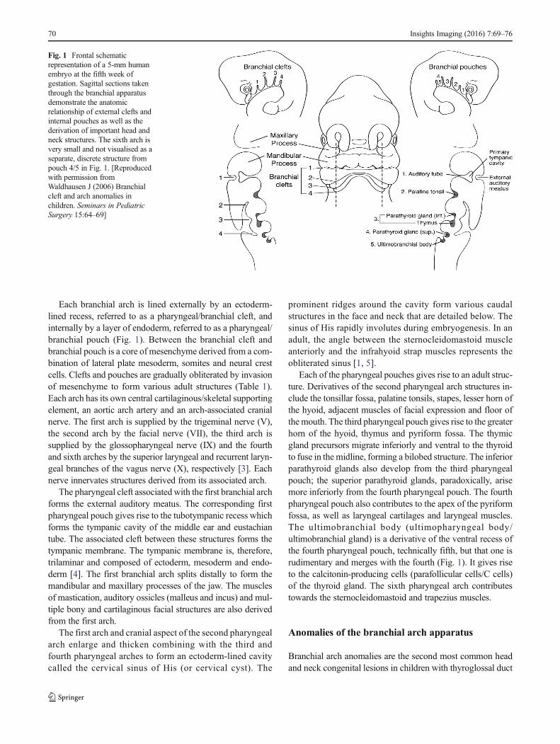
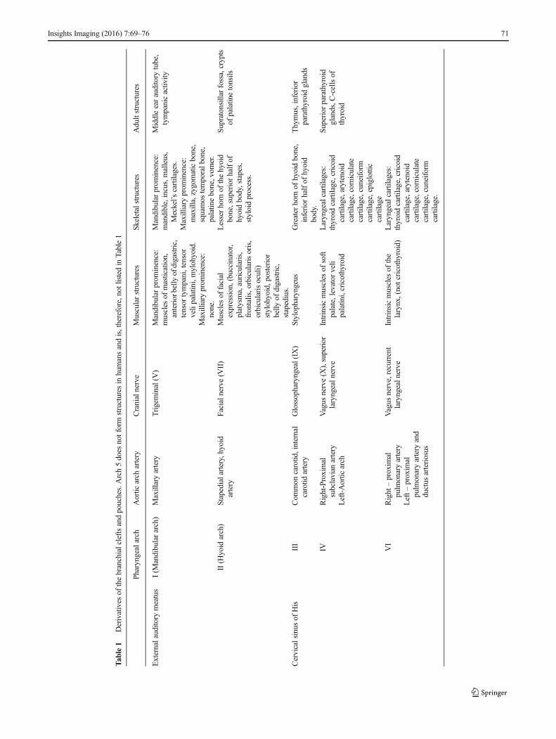
Each branchial arch is lined externally by an ectoderm-lined recess, referred to as a pharyngeal/branchial cleft, andinternally by a layer of endoderm, referred to as a pharyngeal/branchial pouch (Fig. 1). Between the branchial cleft andbranchial pouch is a core of mesenchyme derived from a com-bination of lateral plate mesoderm, somites and neural crestcells. Clefts and pouches are gradually obliterated by invasionof mesenchyme to form various adult structures (Table 1).Each arch has its own central cartilaginous/skeletal supportingelement, an aortic arch artery and an arch-associated cranialnerve. The first arch is supplied by the trigeminal nerve (V),the second arch by the facial nerve (VII), the third arch issupplied by the glossopharyngeal nerve (IX) and the fourthand sixth arches by the superior laryngeal and recurrent laryn-geal branches of the vagus nerve (X), respectively [3]. Eachnerve innervates structures derived from its associated arch.
The pharyngeal cleft associated with the first branchial archforms the external auditory meatus. The corresponding firstpharyngeal pouch gives rise to the tubotympanic recess whichforms the tympanic cavity of the middle ear and eustachiantube. The associated cleft between these structures forms thetympanic membrane. The tympanic membrane is, therefore,trilaminar and composed of ectoderm, mesoderm and endo-derm [4]. The first branchial arch splits distally to form themandibular and maxillary processes of the jaw. The musclesof mastication, auditory ossicles (malleus and incus) and mul-tiple bony and cartilaginous facial structures are also derivedfrom the first arch.
The first arch and cranial aspect of the second pharyngealarch enlarge and thicken combining with the third andfourth pharyngeal arches to form an ectoderm-lined cavitycalled the cervical sinus of His (or cervical cyst). The
prominent ridges around the cavity form various caudalstructures in the face and neck that are detailed below. Thesinus of His rapidly involutes during embryogenesis. In anadult, the angle between the sternocleidomastoid muscleanteriorly and the infrahyoid strap muscles represents theobliterated sinus [1, 5].
Each of the pharyngeal pouches gives rise to an adult struc-ture. Derivatives of the second pharyngeal arch structures in-clude the tonsillar fossa, palatine tonsils, stapes, lesser horn ofthe hyoid, adjacent muscles of facial expression and floor ofthe mouth. The third pharyngeal pouch gives rise to the greaterhorn of the hyoid, thymus and pyriform fossa. The thymicgland precursors migrate inferiorly and ventral to the thyroidto fuse in the midline, forming a bilobed structure. The inferiorparathyroid glands also develop from the third pharyngealpouch; the superior parathyroid glands, paradoxically, arisemore inferiorly from the fourth pharyngeal pouch. The fourthpharyngeal pouch also contributes to the apex of the pyriformfossa, as well as laryngeal cartilages and laryngeal muscles.The ultimobranchial body (ultimopharyngeal body/ultimobranchial gland) is a derivative of the ventral recess ofthe fourth pharyngeal pouch, technically fifth, but that one isrudimentary and merges with the fourth (Fig. 1). It gives riseto the calcitonin-producing cells (parafollicular cells/C cells)of the thyroid gland. The sixth pharyngeal arch contributestowards the sternocleidomastoid and trapezius muscles.
Anomalies of the branchial arch apparatus
Branchial arch anomalies are the second most common headand neck congenital lesions in children with thyroglossal duct
Fig. 1 Frontal schematicrepresentation of a 5-mm humanembryo at the fifth week ofgestation. Sagittal sections takenthrough the branchial apparatusdemonstrate the anatomicrelationship of external clefts andinternal pouches as well as thederivation of important head andneck structures. The sixth arch isvery small and not visualised as aseparate, discrete structure frompouch 4/5 in Fig. 1. [Reproducedwith permission fromWaldhausen J (2006) Branchialcleft and arch anomalies inchildren. Seminars in PediatricSurgery 15:64–69]
70 Insights Imaging (2016) 7:69–76
Tab
le1
Derivatives
ofthebranchialcleftsandpouches.Arch5does
notform
structures
inhumansandis,therefore,not
listedin
Table1
Pharyngealarch
Aortic
arch
artery
Cranialnerve
Muscularstructures
Skeletalstructures
Adultstructures
Externalaudito
rymeatus
I(M
andibulararch)
Maxillaryartery
Trigeminal(V
)Mandibularprom
inence:
muscles
ofmastication,
anteriorbelly
ofdigastric,
tensor
tympani,tensor
velipalatin
i,mylohyoid.
Maxilliary
prom
inence:
none.
Mandibularprom
inence:
mandible,incus,malleus,
Meckel’s
cartilages.
Maxilliary
prom
inence:
maxilla,zygomaticbone,
squamos
temporalb
one,
palatin
ebone,vom
er.
Middleearauditory
tube,
tympanicactiv
ity
II(H
yoid
arch)
Stapedialartery,hyoid
artery
Facialn
erve
(VII)
Muscles
offacial
expression,(buccinator,
platysma,auricularis,
frontalis,orbicularisoris,
orbicularisoculi)
stylohyoid,posterior
belly
ofdigastric,
stapedius.
Lesserhorn
ofthehyoid
bone,superiorhalfof
hyoidbody,stapes,
styloidprocess.
Supratonsillarfossa,crypts
ofpalatin
etonsils
Cervicalsinus
ofHis
III
Com
mon
carotid
,internal
carotid
artery
Glossopharyngeal(IX
)Stylopharyngeus
Greater
horn
ofhyoidbone,
inferior
halfof
hyoid
body.
Thymus,inferior
parathyroidglands
IVRight-Proximal
subclavian
artery
Left-Aortic
arch
Vagus
nerve(X
),superior
laryngealn
erve
Intrinsicmuscles
ofsoft
palate,levator
veli
palatin
i,cricothyroid
Laryngealcartilages:
thyroidcartilage,cricoid
cartilage,arytenoid
cartilage,corniculate
cartilage,cuneiform
cartilage,epiglottic
cartilage
Superior
parathyroid
glands,C
-cellsof
thyroid
VI
Right
–proxim
alpulm
onaryartery
Left–
proxim
alpulm
onaryartery
and
ductus
arteriosus
Vagus
nerve,recurrent
laryngealn
erve
Intrinsicmuscles
ofthe
larynx,(notcricothyroid)
Laryngealcartilages:
thyroidcartilage,cricoid
cartilage,arytenoid
cartilage,corniculate
cartilage,cuneiform
cartilage.
Insights Imaging (2016) 7:69–76 71
remnants being the most common. They represent approxi-mately 20 % of cervical masses, hence branchial arch anom-alies are considered in the differential diagnosis of head andneck masses in children [6]. Second branchial arch anomaliesare the most common and account for approximately 95 % ofcases. First branchial arch anomalies account for 1–4 % caseswith third and fourth branchial arch anomalies being extreme-ly rare [6]. Branchial arch anomalies may present as cysts,fistulas, sinuses or cartilaginous remnants. Branchial cyststypically present in older children/young adults, whereas fis-tulas typically present in infants/young children. Branchialarch anomalies may be bilateral in 2–3 % of cases where theyare often familial [7]. A sinus is a blind ending tract and in thecontext of a branchial arch anomaly, may connect with eitherthe skin (branchial cleft sinus) or with the pharynx (branchialpouch sinus). A fistula is a communication between twoepithelialised surfaces and with regard to branchial archanomalies, requires communication between a persistentpouch and cleft. If no communication occurs with the innermucosa or outer skin, then the trapped branchial arch remnantforms a cyst. There are a number of theories proposed toaccount for the formation of these anomalies, the most widelyaccepted theory being that they result from incomplete oblit-eration of the branchial apparatus, primarily the cleft [8]. Inthe case of sinuses and fistulae, the pharyngeal membrane andpouch are also implicated.
Second branchial cleft anomalies
Second branchial cleft anomalies most commonly present ascysts followed by sinuses and fistulae [9]. Most are presentwithin the submandibular space but they can occur anywherealong the course of the second branchial arch tract whichextends from the skin overlying the supraclavicular fossa, be-tween the internal and external carotid arteries, to enter thepharynx at the level of the tonsillar fossa [2]. They have pre-viously been classified into four different sub-types by Baileyin 1929 [10]:
Type I – Most superficial and lies along the anterior sur-face of sternocleidomastoid deep to the platysma, but notin contact with the carotid sheath.Type II – Most common type where the branchial cleftcyst lies anterior to the sternocleidomastoid muscle, pos-terior to the submandibular gland, adjacent and lateral tothe carotid sheath.Type III – Extends medially between the bifurcation ofthe internal and external carotid arteries, lateral to thepharyngeal wall.Type IV- Lies deep to the carotid sheath within the pha-ryngeal mucosal space and opens into the pharynx.
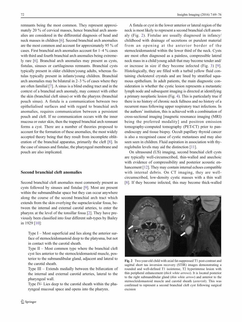
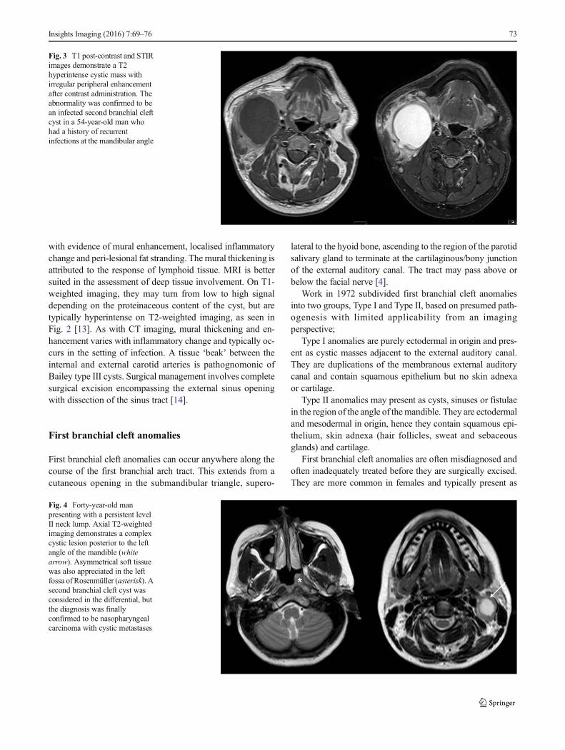
A fistula or cyst in the lower anterior or lateral region of theneck is most likely to represent a second branchial cleft anom-aly (Fig. 2). Fistulae are usually diagnosed in infancy/childhood with drainage of secretions or purulent materialf rom an open ing a t the an t e r io r borde r o f thesternocleidomastoid within the lower third of the neck. Cystsare most often diagnosed as a painless, compressible lateralneck mass in a child/young adult that may become tender and/or increase in size if they become infected (Fig. 3) [9].Histologically, they are filled with a turbid yellow fluid con-taining cholesterol crystals and are lined by stratified squa-mous epithelium. In adult patients, the main diagnostic con-sideration is whether the cystic lesion represents a metastaticlymph node and subsequent imaging is directed at identifyinga primary neoplastic lesion (Fig. 4). This is particularly true ifthere is no history of chronic neck fullness and no history of arecurrent mass following upper respiratory tract infections. Inthe authors’ institution, this is achieved with a combination ofcross-sectional imaging [magnetic resonance imaging (MRI)being the preferred modality] and positron emissiontomography-computed tomography (PET-CT) prior to pan-endoscopy and tissue biopsy. Occult papillary thyroid canceris also a recognised cause of cystic metastases and may alsoseen seen in children. Fluid aspiration in association with thy-roglobulin levels may aid the distinction [11].
On ultrasound (US) imaging, second branchial cleft cystsare typically well-circumscribed, thin-walled and anechoicwith evidence of compressibility and posterior acoustic en-hancement [12]. They may contain internal echoes compatiblewith internal debris. On CT imaging, they are well-circumscribed, low-density cystic masses with a thin wall[8]. If they become infected, this may become thick-walled
Fig. 2 Two-year-old child with axial fat-suppressed T1 post-contrast andsagittal short tau inversion recovery (STIR) images demonstrating arounded and well-defined T1 isointense, T2 hyperintense lesion withthin peripheral enhancement (thick white arrows). It is located posteriorto the right submandibular gland (thin white arrow) and anterior to thesternocleidomastoid muscle and carotid sheath (asterisk). This wasconfirmed to represent a second branchial cleft cyst following surgicalexcision
72 Insights Imaging (2016) 7:69–76
with evidence of mural enhancement, localised inflammatorychange and peri-lesional fat stranding. Themural thickening isattributed to the response of lymphoid tissue. MRI is bettersuited in the assessment of deep tissue involvement. On T1-weighted imaging, they may turn from low to high signaldepending on the proteinaceous content of the cyst, but aretypically hyperintense on T2-weighted imaging, as seen inFig. 2 [13]. As with CT imaging, mural thickening and en-hancement varies with inflammatory change and typically oc-curs in the setting of infection. A tissue ‘beak’ between theinternal and external carotid arteries is pathognomonic ofBailey type III cysts. Surgical management involves completesurgical excision encompassing the external sinus openingwith dissection of the sinus tract [14].
First branchial cleft anomalies
First branchial cleft anomalies can occur anywhere along thecourse of the first branchial arch tract. This extends from acutaneous opening in the submandibular triangle, supero-
lateral to the hyoid bone, ascending to the region of the parotidsalivary gland to terminate at the cartilaginous/bony junctionof the external auditory canal. The tract may pass above orbelow the facial nerve [4].
Work in 1972 subdivided first branchial cleft anomaliesinto two groups, Type I and Type II, based on presumed path-ogenesis with limited applicability from an imagingperspective;
Type I anomalies are purely ectodermal in origin and pres-ent as cystic masses adjacent to the external auditory canal.They are duplications of the membranous external auditorycanal and contain squamous epithelium but no skin adnexaor cartilage.
Type II anomalies may present as cysts, sinuses or fistulaein the region of the angle of themandible. They are ectodermaland mesodermal in origin, hence they contain squamous epi-thelium, skin adnexa (hair follicles, sweat and sebaceousglands) and cartilage.
First branchial cleft anomalies are often misdiagnosed andoften inadequately treated before they are surgically excised.They are more common in females and typically present as
Fig. 4 Forty-year-old manpresenting with a persistent levelII neck lump. Axial T2-weightedimaging demonstrates a complexcystic lesion posterior to the leftangle of the mandible (whitearrow). Asymmetrical soft tissuewas also appreciated in the leftfossa of Rosenmüller (asterisk). Asecond branchial cleft cyst wasconsidered in the differential, butthe diagnosis was finallyconfirmed to be nasopharyngealcarcinoma with cystic metastases
Fig. 3 T1 post-contrast and STIRimages demonstrate a T2hyperintense cystic mass withirregular peripheral enhancementafter contrast administration. Theabnormality was confirmed to bean infected second branchial cleftcyst in a 54-year-old man whohad a history of recurrentinfections at the mandibular angle
Insights Imaging (2016) 7:69–76 73
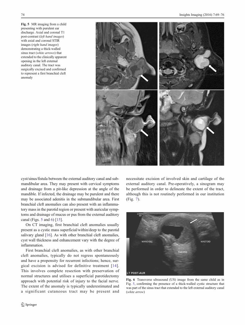
cyst/sinus/fistula between the external auditory canal and sub-mandibular area. They may present with cervical symptomsand drainage from a pit-like depression at the angle of themandible. If infected, the drainage may be purulent and theremay be associated adenitis in the submandibular area. Firstbranchial cleft anomalies can also present with an inflamma-tory mass in the parotid region or present with auricular symp-toms and drainage of mucus or pus from the external auditorycanal (Figs. 5 and 6) [15].
On CT imaging, first branchial cleft anomalies usuallypresent as a cystic mass superficial/within/deep to the parotidsalivary gland [16]. As with other branchial cleft anomalies,cyst wall thickness and enhancement vary with the degree ofinflammation.
First branchial cleft anomalies, as with other branchialcleft anomalies, typically do not regress spontaneouslyand have a propensity for recurrent infections; hence, sur-gical excision is advised for definitive treatment [14].This involves complete resection with preservation ofnormal structures and utilises a superficial parotidectomyapproach with potential risk of injury to the facial nerve.The extent of the anomaly is typically underestimated anda significant cutaneous tract may be present and
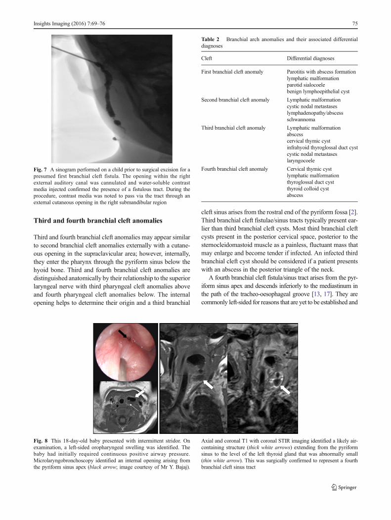
necessitate excision of involved skin and cartilage of theexternal auditory canal. Pre-operatively, a sinogram maybe performed in order to delineate the extent of the tract,although this is not routinely performed in our institution(Fig. 7).
Fig. 6 Transverse ultrasound (US) image from the same child as inFig. 5, confirming the presence of a thick-walled cystic structure thatwas part of the sinus tract that extended to the left external auditory canal(white arrow)
Fig. 5 MR imaging from a childpresenting with purulent eardischarge. Axial and coronal T1post-contrast (left hand images)with axial and coronal STIRimages (right hand images)demonstrating a thick-walledsinus tract (white arrows) thatextended to the clinically apparentopening in the left externalauditory canal. The tract wassurgically excised and confirmedto represent a first branchial cleftanomaly
74 Insights Imaging (2016) 7:69–76
Third and fourth branchial cleft anomalies
Third and fourth branchial cleft anomalies may appear similarto second branchial cleft anomalies externally with a cutane-ous opening in the supraclavicular area; however, internally,they enter the pharynx through the pyriform sinus below thehyoid bone. Third and fourth branchial cleft anomalies aredistinguished anatomically by their relationship to the superiorlaryngeal nerve with third pharyngeal cleft anomalies aboveand fourth pharyngeal cleft anomalies below. The internalopening helps to determine their origin and a third branchial
cleft sinus arises from the rostral end of the pyriform fossa [2].Third branchial cleft fistulae/sinus tracts typically present ear-lier than third branchial cleft cysts. Most third branchial cleftcysts present in the posterior cervical space, posterior to thesternocleidomastoid muscle as a painless, fluctuant mass thatmay enlarge and become tender if infected. An infected thirdbranchial cleft cyst should be considered if a patient presentswith an abscess in the posterior triangle of the neck.
A fourth branchial cleft fistula/sinus tract arises from the pyr-iform sinus apex and descends inferiorly to the mediastinum inthe path of the tracheo-oesophageal groove [13, 17]. They arecommonly left-sided for reasons that are yet to be established and
Table 2 Branchial arch anomalies and their associated differentialdiagnoses
Cleft Differential diagnoses
First branchial cleft anomaly Parotitis with abscess formationlymphatic malformationparotid sialocoelebenign lymphoepithelial cyst
Second branchial cleft anomaly Lymphatic malformationcystic nodal metastaseslymphadenopathy/abscessschwannoma
Third branchial cleft anomaly Lymphatic malformationabscesscervical thymic cystinfrahyoid thyroglossal duct cystcystic nodal metastaseslaryngocoele
Fourth branchial cleft anomaly Cervical thymic cystlymphatic malformationthyroglossal duct cystthyroid colloid cystabscess
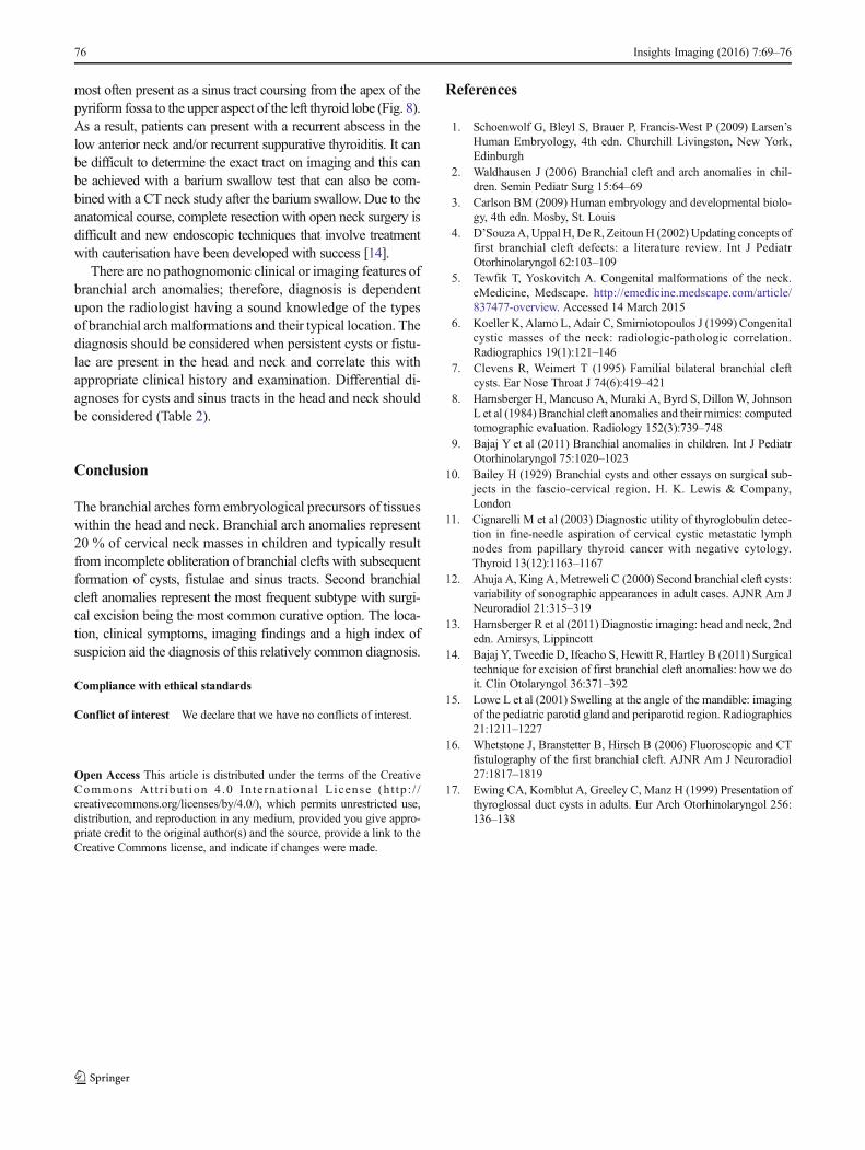
Fig. 8 This 18-day-old baby presented with intermittent stridor. Onexamination, a left-sided oropharyngeal swelling was identified. Thebaby had initially required continuous positive airway pressure.Microlaryngobronchoscopy identified an internal opening arising fromthe pyriform sinus apex (black arrow; image courtesy of Mr Y. Bajaj).
Axial and coronal T1 with coronal STIR imaging identified a likely air-containing structure (thick white arrows) extending from the pyriformsinus to the level of the left thyroid gland that was abnormally small(thin white arrow). This was surgically confirmed to represent a fourthbranchial cleft sinus tract
Fig. 7 A sinogram performed on a child prior to surgical excision for apresumed first branchial cleft fistula. The opening within the rightexternal auditory canal was cannulated and water-soluble contrastmedia injected confirmed the presence of a fistulous tract. During theprocedure, contrast media was noted to pass via the tract through anexternal cutaneous opening in the right submandibular region
Insights Imaging (2016) 7:69–76 75
most often present as a sinus tract coursing from the apex of thepyriform fossa to the upper aspect of the left thyroid lobe (Fig. 8).As a result, patients can present with a recurrent abscess in thelow anterior neck and/or recurrent suppurative thyroiditis. It canbe difficult to determine the exact tract on imaging and this canbe achieved with a barium swallow test that can also be com-bined with a CT neck study after the barium swallow. Due to theanatomical course, complete resection with open neck surgery isdifficult and new endoscopic techniques that involve treatmentwith cauterisation have been developed with success [14].
There are no pathognomonic clinical or imaging features ofbranchial arch anomalies; therefore, diagnosis is dependentupon the radiologist having a sound knowledge of the typesof branchial archmalformations and their typical location. Thediagnosis should be considered when persistent cysts or fistu-lae are present in the head and neck and correlate this withappropriate clinical history and examination. Differential di-agnoses for cysts and sinus tracts in the head and neck shouldbe considered (Table 2).
Conclusion
The branchial arches form embryological precursors of tissueswithin the head and neck. Branchial arch anomalies represent20 % of cervical neck masses in children and typically resultfrom incomplete obliteration of branchial clefts with subsequentformation of cysts, fistulae and sinus tracts. Second branchialcleft anomalies represent the most frequent subtype with surgi-cal excision being the most common curative option. The loca-tion, clinical symptoms, imaging findings and a high index ofsuspicion aid the diagnosis of this relatively common diagnosis.
Compliance with ethical standards
Conflict of interest We declare that we have no conflicts of interest.
Open Access This article is distributed under the terms of the CreativeCommons At t r ibut ion 4 .0 In te rna t ional License (h t tp : / /creativecommons.org/licenses/by/4.0/), which permits unrestricted use,distribution, and reproduction in any medium, provided you give appro-priate credit to the original author(s) and the source, provide a link to theCreative Commons license, and indicate if changes were made.
References
1. Schoenwolf G, Bleyl S, Brauer P, Francis-West P (2009) Larsen’sHuman Embryology, 4th edn. Churchill Livingston, New York,Edinburgh
2. Waldhausen J (2006) Branchial cleft and arch anomalies in chil-dren. Semin Pediatr Surg 15:64–69
3. Carlson BM (2009) Human embryology and developmental biolo-gy, 4th edn. Mosby, St. Louis
4. D’SouzaA,Uppal H, DeR, Zeitoun H (2002) Updating concepts offirst branchial cleft defects: a literature review. Int J PediatrOtorhinolaryngol 62:103–109
5. Tewfik T, Yoskovitch A. Congenital malformations of the neck.eMedicine, Medscape. http://emedicine.medscape.com/article/837477-overview. Accessed 14 March 2015
6. Koeller K, Alamo L, Adair C, Smirniotopoulos J (1999) Congenitalcystic masses of the neck: radiologic-pathologic correlation.Radiographics 19(1):121–146
7. Clevens R, Weimert T (1995) Familial bilateral branchial cleftcysts. Ear Nose Throat J 74(6):419–421
8. Harnsberger H, Mancuso A, Muraki A, Byrd S, Dillon W, JohnsonL et al (1984) Branchial cleft anomalies and their mimics: computedtomographic evaluation. Radiology 152(3):739–748
9. Bajaj Y et al (2011) Branchial anomalies in children. Int J PediatrOtorhinolaryngol 75:1020–1023
10. Bailey H (1929) Branchial cysts and other essays on surgical sub-jects in the fascio-cervical region. H. K. Lewis & Company,London
11. Cignarelli M et al (2003) Diagnostic utility of thyroglobulin detec-tion in fine-needle aspiration of cervical cystic metastatic lymphnodes from papillary thyroid cancer with negative cytology.Thyroid 13(12):1163–1167
12. Ahuja A, King A, Metreweli C (2000) Second branchial cleft cysts:variability of sonographic appearances in adult cases. AJNR Am JNeuroradiol 21:315–319
13. Harnsberger R et al (2011) Diagnostic imaging: head and neck, 2ndedn. Amirsys, Lippincott
14. Bajaj Y, Tweedie D, Ifeacho S, Hewitt R, Hartley B (2011) Surgicaltechnique for excision of first branchial cleft anomalies: how we doit. Clin Otolaryngol 36:371–392
15. Lowe L et al (2001) Swelling at the angle of the mandible: imagingof the pediatric parotid gland and periparotid region. Radiographics21:1211–1227
16. Whetstone J, Branstetter B, Hirsch B (2006) Fluoroscopic and CTfistulography of the first branchial cleft. AJNR Am J Neuroradiol27:1817–1819
17. Ewing CA, Kornblut A, Greeley C, Manz H (1999) Presentation ofthyroglossal duct cysts in adults. Eur Arch Otorhinolaryngol 256:136–138
76 Insights Imaging (2016) 7:69–76