Embed Size (px)
Citation preview
Bull. Fac. Agrie., Ilniv. Cairo, 45 (1 994): 91 9-932.
RESTRICTION ENDONUCLEASE ANALYSIS AND DIAGNOSIS OF THE GRANULOSIS VIRUS ISOLATED FROM SPODOPTERA
LZTTORALIS BOISD. IN WEST AFRICA AND MULTIPLIED IN EGYPT.
(Received: 23. 6. 1994 )
BY S. Abol-Ela, G. Fediere, A.. Nour El-Din, O. Khamiss and M.Salah
Entomovirology Laboratory, Faculty of Agriculture-ORSTOM, Cairo University, Egypt
ABSTRACT
A Granulosis virus isolated from Spodoptera liftoralis Boisd. (Lep. Noctuidae) in Cote d'Ivoire more than 15 years ago was multiplied in Egypt on the insect host and characterized. The size of the DNA of this Baculovirus, that we name SlGV, was estimated to be 108 Kilobases and restriction patterns have been drawn up for 15 endonucleases. Viral diagnosis methods, inmunoenzymatic test. ELISA and genomic probe, have been established and tried out for epidemiological studies of the disease. The virus was compared to other GVs of Sesamia eretica, Phthorimaea operculella, Cydia pomonella, Chilo infuseatellus and Cryptophlebia leucotreta using the same technique.
Key words: Granulosis virus, Restriction endonuclease. Spodoptera littoralis,
INTRODUCTION
A Granulosis Virus (GV) was isolated from the Egyptian cotton leaf worm Spodoptera littoralis Boisd. (Lepidoptera: Noctuidae) more than 15 years ago in Côte d'Ivoire This species is an economically important polyphagous pest attacking cotton, ;regetables, rice, maize and tobacco in many tropical and subtropical regions of Africa. The Baculovirus was purified for bioassay and for production using mass rearing of this pest on an artificial diet paillon, 1983). A strain of this pathogenic GV that we name SlGV
-919-
ORSTOM Fonds Documentaire
(isolate 1 from Bouake in Cote d'Ivoire) is now currently multiplied in Egypt on larvae collected from the field and reared on semi-artificial diet
The present work is considered as a part of a laboratory program concerning the inter-relationship between the different viruses isolated from S.littoralis and their host. The objective of the characterisation of this virus is to detect the appearence of other or local GVs infecting the same host regarding the geographical distribution of this pest, as well as the possibility of including this baculovirus in the application strategy. It is important to mention that two small RNA viruses were recentlly isolated from naturally infected larvae. and the genetic recombination among S litioralis NPVs is actually under study.
In the present investigation, we report the first DNA characterization using restriction endonuclease (REN) and viral diagnosis of the SlGV. Actually, work is going on to find an endemic SlGV strain in Egypt which could be more adapted for the Egyptian agro-ecosystem.
MATERTALS AND METHODS
Vírus isolate
The granules were obtained from natural infections of S. littoralis larvae collected from cotton fields. This virus was isolated in Bouake in Côte d'Ivoire and purified by Dr. Pierre Monsarrat and M.Fransois Baillon (ORSTOM). This isolate was propagated in the Egyptian S. littoralis larvae and the viral suspension is available at the Entomovirology Laboratory, Faculty of Agriculture, Cairo University.
Granules purification
The infected larvae were homogenized in T.S. buEer (50 mM Tris, 2 mM SDS, pH 7.8). The homogenized viral suspension was filtered and clarified, then a series of successive sedimentations on continuous sugar gradient 30/70% (p/p) using ultracentrifugation at 45 O00 g for 30 minutes was conducted. The highly purifíed viral granules were checked by spectrophotometer DU-70 through 450 nm wavelenght. The viral suspension was stocked in Tris buffer (Tris 50 mM, pH 7.8) under - 20 OC
-921-
Electron microscopy
Purified viral suspension was negatively stained with 2% (WhV) uranyl acetate, pH 7.4.
DNA extraction
The extraction of DNA from the purified GV capsules was carried out using the procedures of Fediere et al., (1993). The DNA was released from purified virus particles by a treatment with 0.05 m Na2C03, then lysed with sarkosyl and proteinase K at 37 OC within 2 hours. The exclusion of the protein from the DNA suspension was conducted through 3- phenolic extractions. The DNA was precipitated by addition of 2 volumes of absolute ethanol in presence of sodium acetate (0.3M final ) for 14 hours at -20 OC. After rapid centrifugation, the pellet was washed in 70% ethanol and incubated in T.E. buffer. The concentration of DNA was finally measured according to its optic density through 260 nm wave lengh.
Restriction enzyme digestion and gel electrophoresis
Viral DNA (lug) was digested in a final volume of 20 ul, for 4 h at 37 Co, with restriction endonucleases using the conditions recommended by the suppliers (Bohreinger). Electrophoresis was carried out using 1% agarose gel in Tris-EDTA-Phosphate buffer (TEP) (90 mM Tris-Phosphate, 20 mM EDTA, pH 8.0) containing 0.5 ug/ml ethidium bromide. Electrophoresis was carried out at 50 V for 2 h. The gel was visualised and photographed on a W transilluminator. The size of the DNA fragments was estimated by comparison with fragments of lambda DNA digested with Hind III (marker II), lambda DNA digested with both Eco RI and Hind III (marker III) and SPPl DNA digested with Eco RI (marker WI).
Nucleic probe and hybridization
The digoxigenin-labelled SlGV DNA probe was applied according to the protocol recommended by the suppliers (Boerhinger). The same protocol was applied for the hybridization "dot blot" technique in order to determine the probe title. The Southern blot method (Southern, 1975) was applied to venfy the presence of the total fragments of the genome and for detecting the sequence homology between the SlGV DNA and that of Sesamia cretìca GV (ScGV) (Fediere et al., 1993). This insect is a noctoid pest of maize in Eastern Africa and the most important borer in Egypt.
c
-922-
ELISA test
For detecting the Baculoviral protein, a specific rabbit serum was prepared by injecting the total protein of granules virions. The indirect method of ELISA test "Enzyme Linked Immun0 Sorbent Assay" using the alkaline phosphatase was conducted according to Kelly et al., (1978).
RESULTS AND DISCUSSION
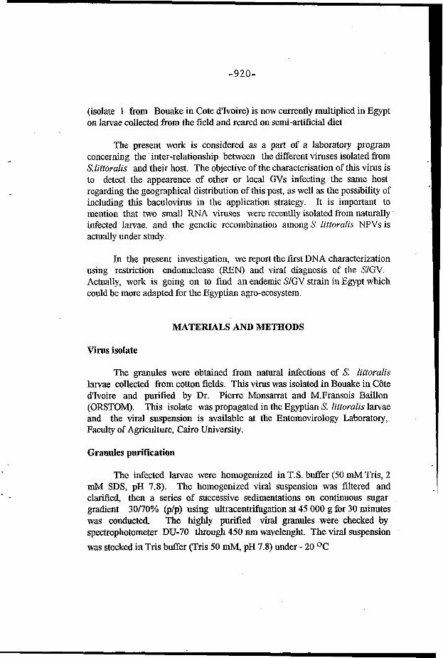
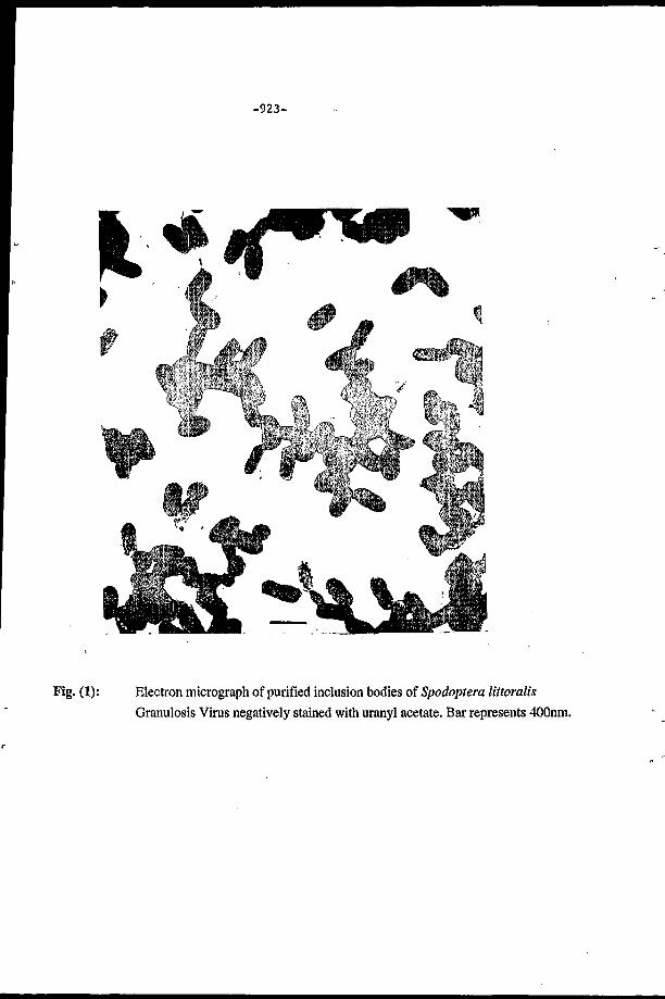
The electron microscopic examination of purified viral suspension revealed the presence of ovoid-shape granules measuring 350-370 x 170-190 nm (Fig. 1) the fine observation of the granule structure indicated the viral particle envelope as well as its nucleocapsid.
The purified DNA of SlGV was digested by 15 endonucleases of current use. No restriction sites were observed when the genome was digested with Hpa I, Not I, Sma I and Sph I, while only two restriction sites were detected by Stu I giving two fragments of 7.2 and 98 Kilobases (Kb).
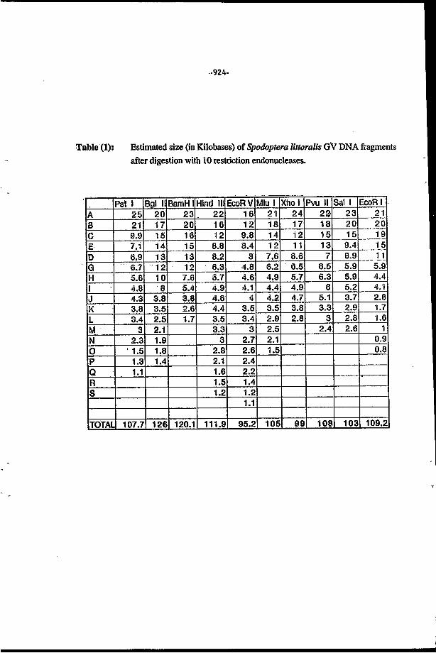
However, the digestion by the endonucleases Bam HI, Bgl II, Eco RI, Eco RV, Hind III, Mlu I, Pst I, Pvu II, Sal I andXho I revealeddifferent electrophoretic profiles composed of 11, 15, 14, 19, 18, 14, 16, 12, 12 and 11 fxagments, respectively, in which the sizes are shown in Table (1).
The molecular weight of the genome was estimated by addition of the size of all fragments in each electophoretic profile. The mean of the DNA molecular weight was about 108 Kb.
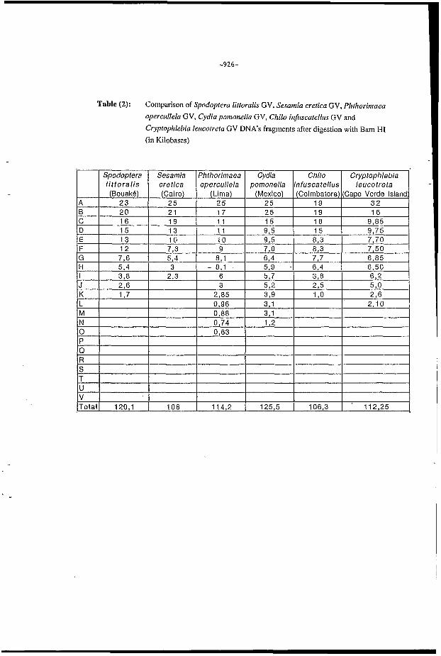
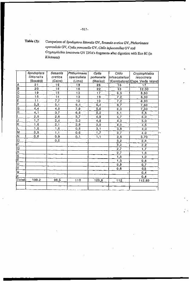
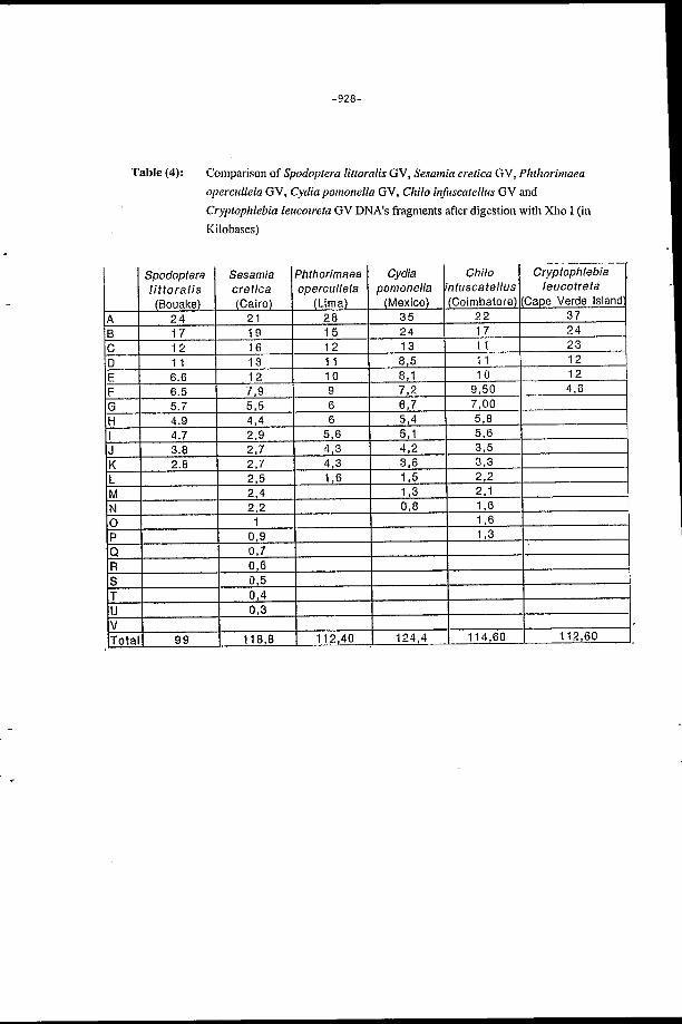
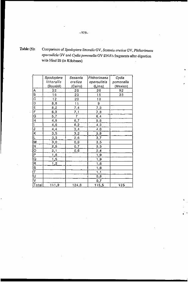
Using the equivalent endonucleases for genome digestion, the electrophoretic profile of SlGV was not identical with those of other GV DNAs already characterized from the following species: Sesamia eretica, strain of Cairo, Egypt, (Fediere et al., 1993), Chilo infuscatellzis, strain of Coimbatore, India, (Easwaramoorthy and Cory, 1990), Phthorimaea operculella, strain of Lima, Peru, (Vickers et al., 1991), Ctyptophlebia leucofreta, strain of Cape Verde Island, Guinea, (Jehle ef al., 1992) and Cydiapomonella, strain of Mexico, Mexico, (Crook et al., 1985) (Tables 2,3,4 and 5).
A total nucleic probe labeling with Digoxigenin was prepared. The capacity of this probe for detecting the viral DNA was tested using the dot-blot technique, the deposit of 2ul was capable for detecting 5 pg of DNA. The above mentioned method was used for detecting the homology between SlGV DNA and ScGV DNA. The latter insect is a noctoid pest of maize in Eastern Africa and the most important borer in Egypt. Nosignofrecognitionwas found showing the specifcity of the probe. The experiments of hybridization
-923-
Fig. (1): Electron micrograph of purified inclusion bodies of Spodopteru littoralis Granulosis Virus negatively stained with uranyl acetate. Bar represents 400nm.
F
-924-
Table (1): Estimated size (in Kilobases) of Spodoptera lirturulis GV DNA feagments after digestion with 10 restriction endonucleases.
-925-
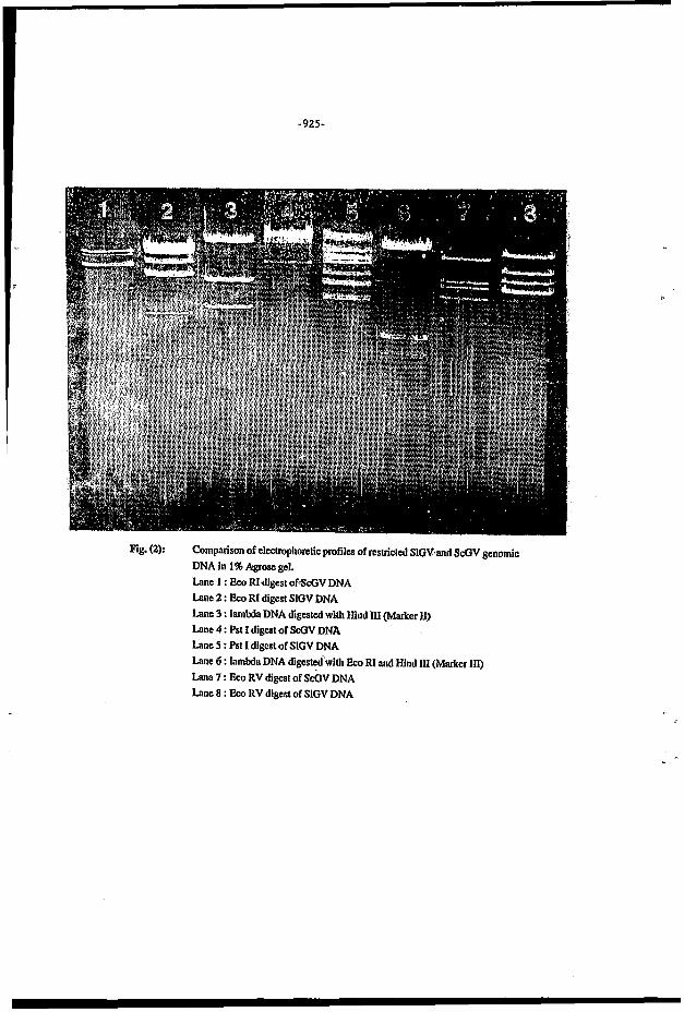
Fig. (2): Comparison of electmphontic profiles of mtricled SlGV and ScGV genomic DNA in 1% Agrose gel. Lane 1 : Eco RI digest ofScGV DNA Lane 2 : Eco RI digest SlGV DNA Lane 3 : lambda DNA digested with Hind III (Marker Il) Lane 4 : Pst I digest of ScGV DNA Lane 5 : Pst I digest of SlGV DNA Lane 6 : lambda DNA digested with Eco RI and Hind III (Marker III) Lane 7 : Eco RV digest of S&V DNA Lane 8 : Eco RV digest of SlGV DNA
c
-926-
Table (2): Comparison of Spodoptera littoralis GV, Sesanzia eretica GV, Pkthoritiiaea opcreitlleln GV, Cydia potiionella GV, Chilo infrtscatellus GV and CryptophleDia leiieotreta GV DNA's fragments after digestion with Bam HI (in Kilobases)
-927-
Table (3): Comparison of Spodoptera littoralis GV, Sesaniia crefica GV, Phthoriniaca opercidlcla GV, Cydia poinonella GV, Chilo itijùscatellus GV and Ciypfophlebia leucotreta GV DNA's fragments after digestion with Eco RI (in Kilobases)
Spodopfera Sesamia Phthorimaea Cydia Chilo Crypfophlebia littoralis cretica opercol/ela pomonella 'nfuscatellus leocofrefa
-928-
Table (4): Comparison of Simioptera littoralis GV, Sesainiu eretica GV, Phthor-iniaca opreullela GV, Cydia yonionella GV, Chilo infitscatellits GV and Cryi)fophlebia leucotreta GV DNA's fragments after digestion with Xho I (in Kilobases)
I I I l 114,60 I 112,60 . Totall 99 I 11833 I 112,110 I 124,4 I
-929-
U
Total V
Table (5): Comparison of Spodoptera littoralis GV, Sesarrlia cretica GV, Phthorirnaea opercullela GV and Cydia poinoilella GV DNA's fragments after digestion with Hind III (in Kilobases)
0,8 0,7
111,9 124,8 115,5 125
I ' Spodoptera Sesamìa Phthorimaea Cydia l i t toral is eretica opercullela pomonella
.7
-930-
after obtaining the electrophoretic profiles of restricted SlGV and ScGV genomic DNA by Eco EU, Pst I and Eco V (Fig. 2), by Southern blot technique. did not permit the recognition of all the electrophoretic fragments by the probe which confirm its specificity.
An antiserum titered as U1200 was prepared using all dissolved proteins (the granulin and the capsid proteins). By applying the ELISA test with the alkaline phosphatase indirect method, I ng of the dissolved proteins was detected. An equal concentration of Sc GV viral proteins was less intensively visible using the same test for detection.
The partial homology between the two types of virus detected by ELISA test was due to the presence of the same sequence coding for the protein of high degree of conservation (this result was confirmed by ELISA test, while the electrophoretic profiles of the two viruses were completely different).
The two viral diagnostic tools, which were prepared and titered in the present study, represent certain importance for the study of Spodoptera littoralis viral epidemiology. Such a study is highly required for the determination of the viral existence among natural pest populations, as well as its persistence. These observations are needed for managing the biological control strategy.
ACKNOWLEDGEMENT
The authors express their sincere gratitude to Dr. Pierre Monsarrat and M.Franc-ois Baillon, from ORSTOM Institute in Coote d'Ivoire for supplying the strain of Granulosis Virus.
REFERENCES
Baillon, F., (1983): Contribution d lètude des virus entomopathogènes utilisès en culture cotonnière en Côte d'Ivoire. ORSTOM ed. 72p.
Crook, N.E., Spencer, RA., Payne, C. andLeisy, D.J., (1985): Variation in Cydiu pomonella Granulosis Virus isolates and physical maps of the DNA from three variants. J. Gen. Virol., 66: 2423-2430.
Easwaramoorthy, S.and Cory J.S., (1998): Characterization of the DNA of Granulosis Viruses isolated from two closely related moths, Chilo infirscatellus and C. suceariphagus indicus. Arch. Virol., 110: 113- 119.
-931-
Fedibre, G., Taha, A.A., Abol-Ela, S, Lery, X., Zeddam, J.L., Veyrunes, J.C., and Giannotti, J., (1993): A new Granulosis Virus isolated from Sesaïnia cretica Led. (Lepidoptera: Noctuidae) in Nort-East Africa: DNA characterization and viral diagnosis. C.R. Acad. Sci. Paris, 3 16: 1350-134.
Jehle, A.J., Abckhaus, H., Fritsch, E. and Huber, J., (1992): Physical map of the Cryptophlebia leucotreta granulosis vírus genome and its relationship to the genome of Cydia pomonella granulosisvirus. J. Gen. Virol., 73: 1621-1626.
Kelly, D.C., Edwards, M.L., Evans, H.F. and Robertson, J.S., (1978): The use of the enzyme linked immunosorbent assay to detect a nuclear polyhedrosis virus in Heliothis armigera larvae. J. Gen. Virol., 40: 465- 469.
Southern, E.M., (1975): Detection of specific sequences among DNA fragments separated by gel electrophoresis. J. Mol. Biol., 98: 503-517.
Vickers, J.M., Cory, J.S. and Entwistle, P.F., (1991): DNA characterization of eight geographic isolates of Granulosis Virus from the potato tuber moth (Phthorimaea operculella) (Lepidoptera, Gelechiidae). J. Invertebr. Pathol., 57:334-342.

U L L E T I . F FACULTY ISSN:0526 - 8613
VOL. 4%
N 0 . 4
QCTQBER
I994
OF AGRICULTURE
UNIVERSITY OF CAIRO
GIZA, EGYPT
- ORSTOR": F0nds Qocumentaire