Embed Size (px)
Citation preview
Cardiovascular Unit PPT
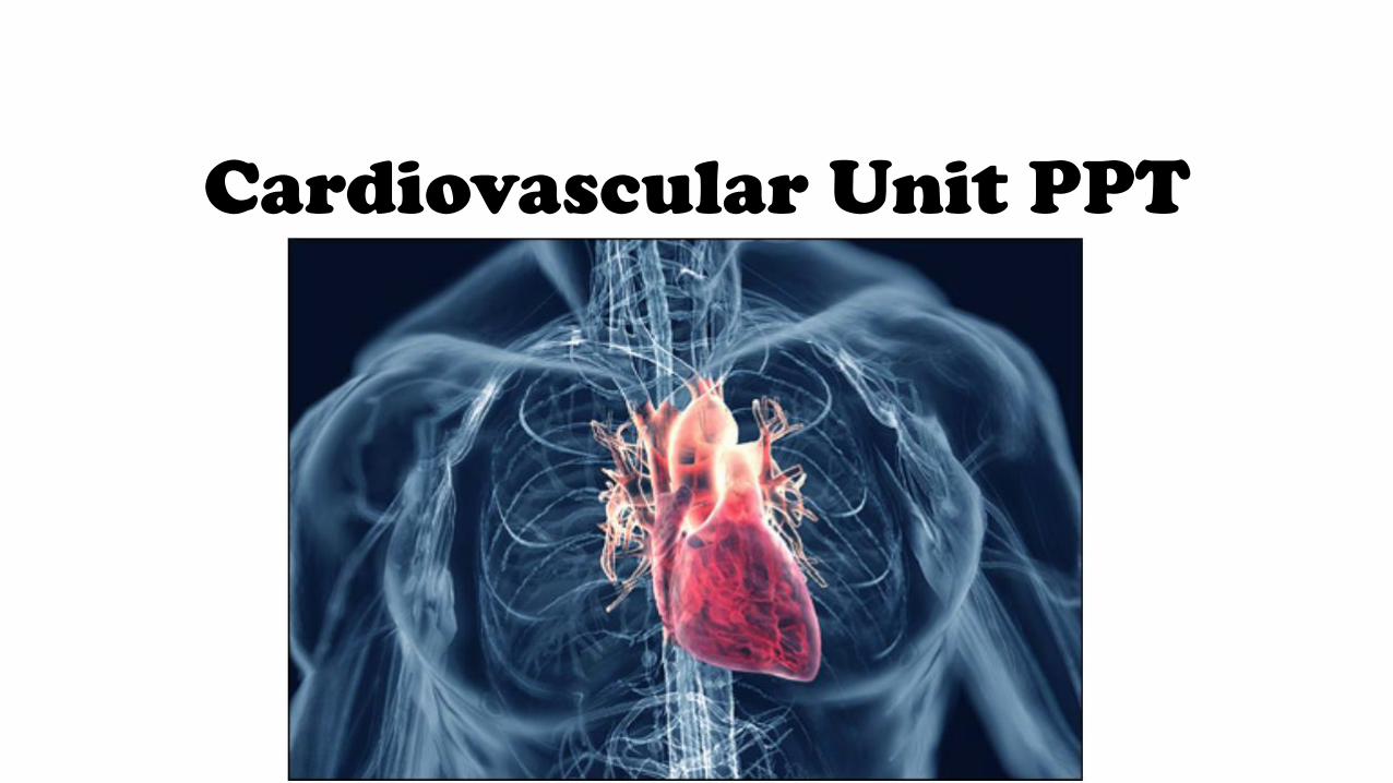
Muscle Memory
Muscle Name Location Movement
Trapezius
Levator Scapulae
Rhomboid Major
Rhomboid Minor
Partner Flashcards with heart model
Supplies you will need:• Colored utensils for flashcards
• 1 flashcard sheet
• 1 heart model (between the partners)
• 1 WET erase marker (can on black shelving)
You will need to sit with your partner
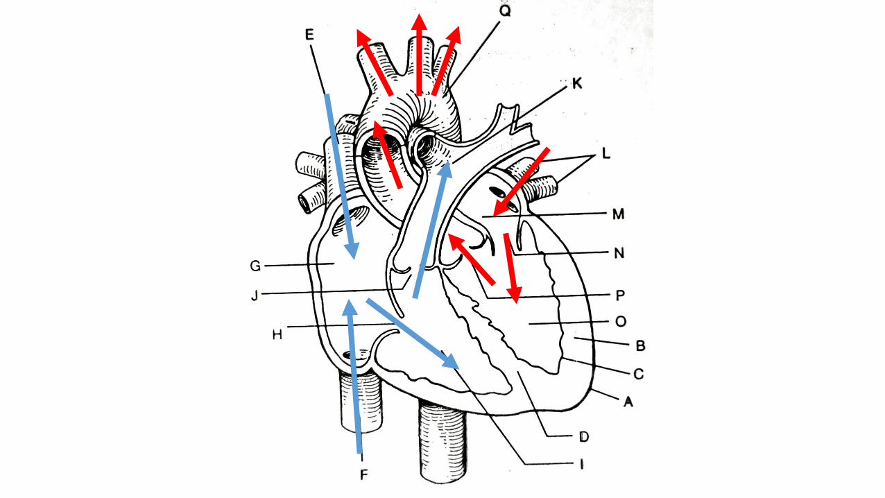
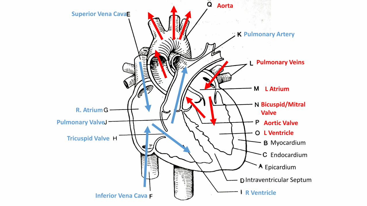
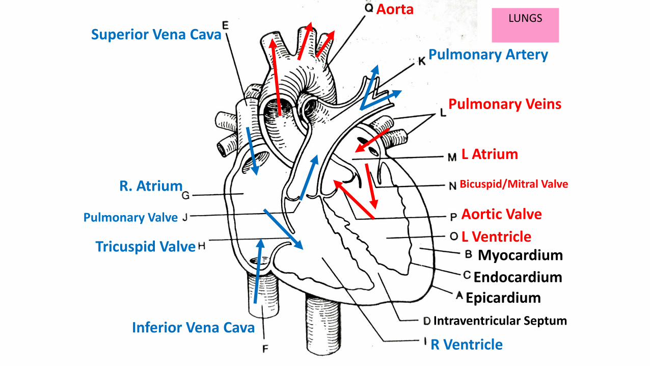
Superior Vena Cava
Aortic Valve
Bicuspid/Mitral Valve
L Atrium
Pulmonary Veins
Pulmonary Artery
Aorta
Epicardium
Endocardium
Myocardium
L Ventricle
Inferior Vena Cava R Ventricle
Intraventricular Septum
Tricuspid Valve
R. Atrium
Pulmonary Valve
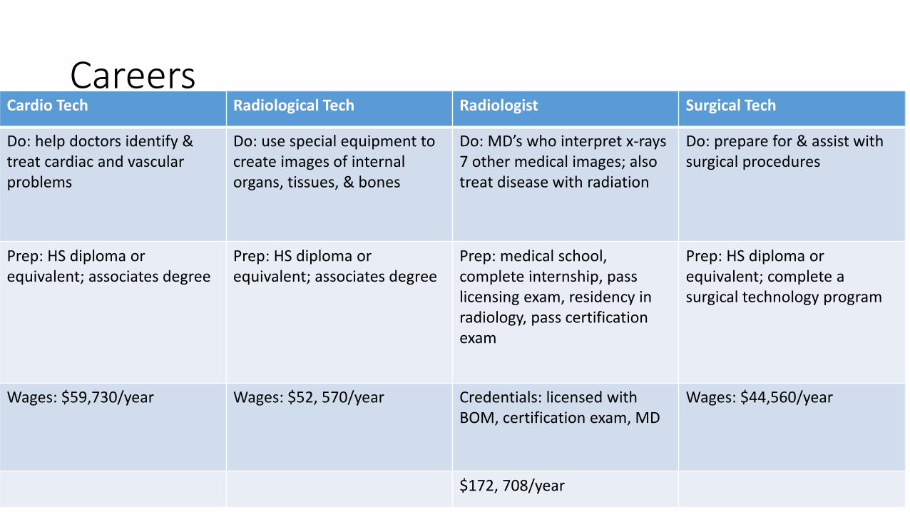
CareersCardio Tech Radiological Tech Radiologist Surgical Tech
Do: help doctors identify & treat cardiac and vascular problems
Do: use special equipment to create images of internal organs, tissues, & bones
Do: MD’s who interpret x-rays 7 other medical images; also treat disease with radiation
Do: prepare for & assist with surgical procedures
Prep: HS diploma or equivalent; associates degree
Prep: HS diploma or equivalent; associates degree
Prep: medical school, complete internship, pass licensing exam, residency in radiology, pass certification exam
Prep: HS diploma or equivalent; complete a surgical technology program
Wages: $59,730/year Wages: $52, 570/year Credentials: licensed with BOM, certification exam, MD
Wages: $44,560/year
$172, 708/year
Review Heart Anatomy
•Quizlet.live
Video segment: Watch the first time then take notes
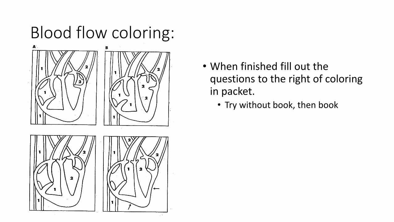
Blood flow coloring:
• When finished fill out the questions to the right of coloring in packet. • Try without book, then book
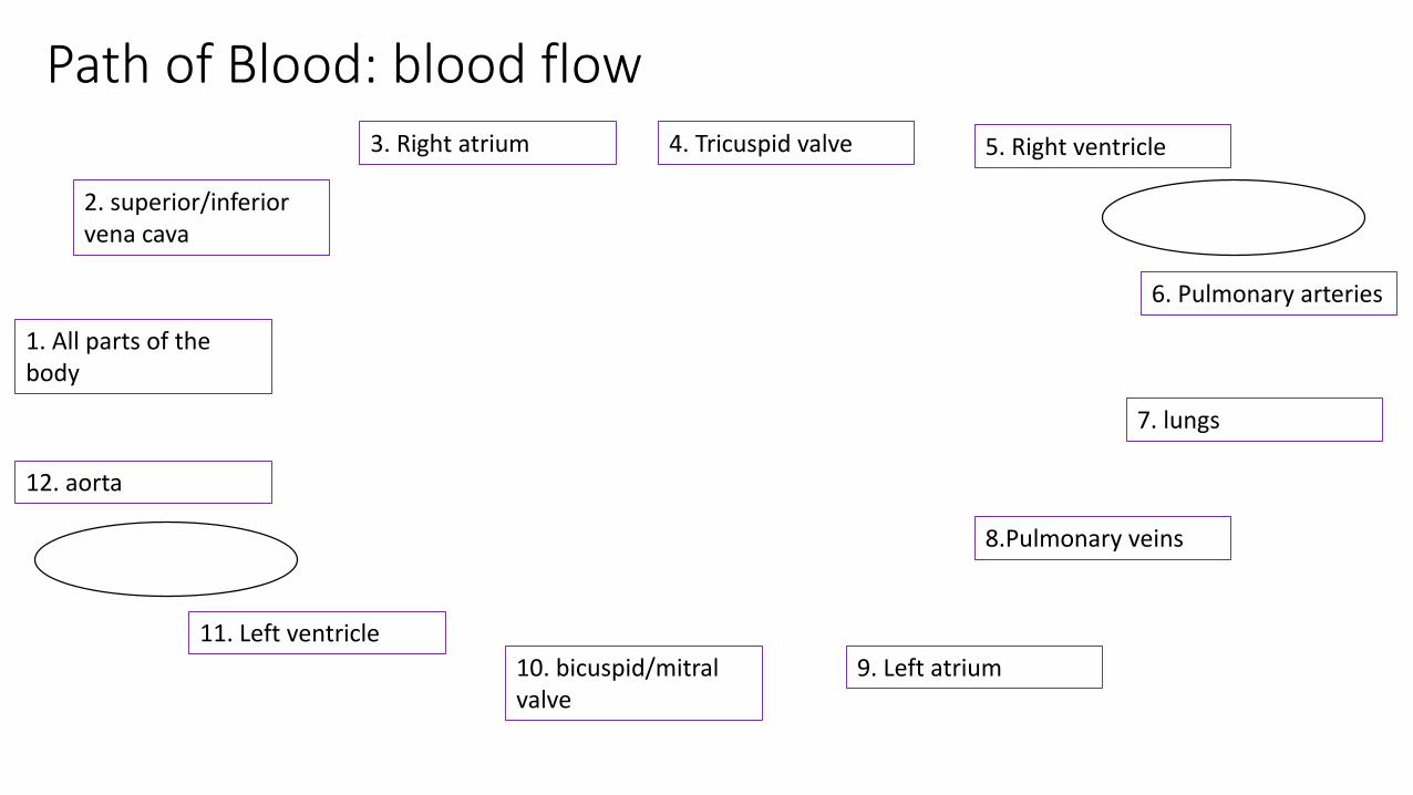
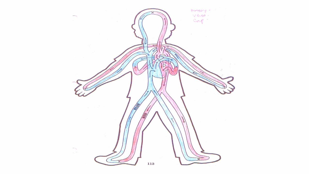
Path of Blood: blood flow
2. superior/inferior vena cava
1. All parts of the body
3. Right atrium
8.Pulmonary veins
12. aorta
4. Tricuspid valve
11. Left ventricle
10. bicuspid/mitral valve
9. Left atrium
7. lungs
6. Pulmonary arteries
5. Right ventricle
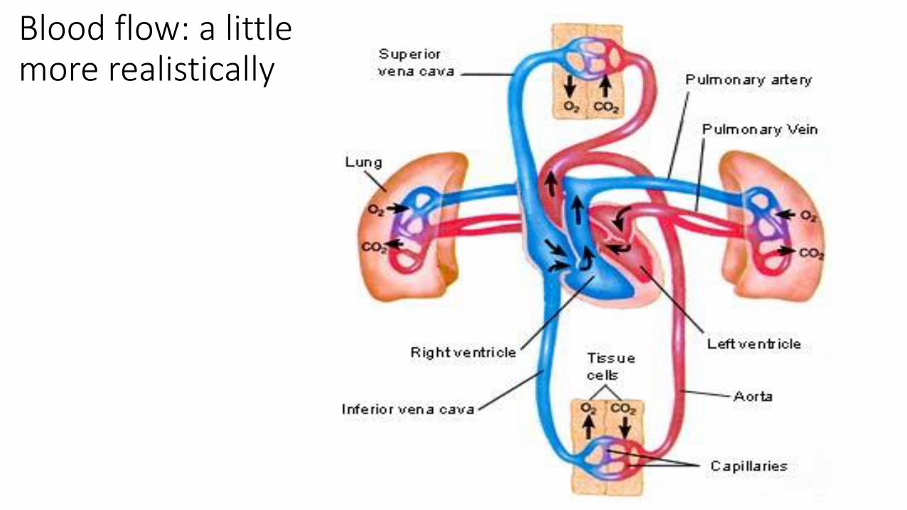
Blood flow: a little more realistically
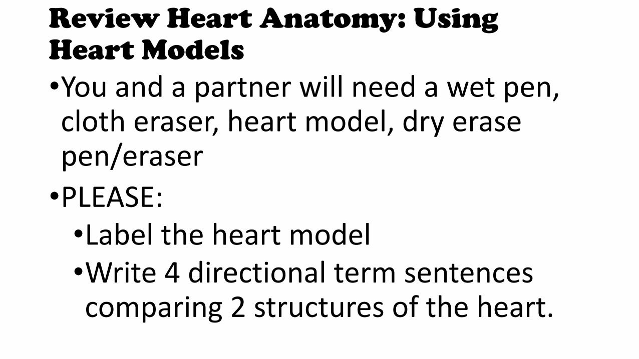
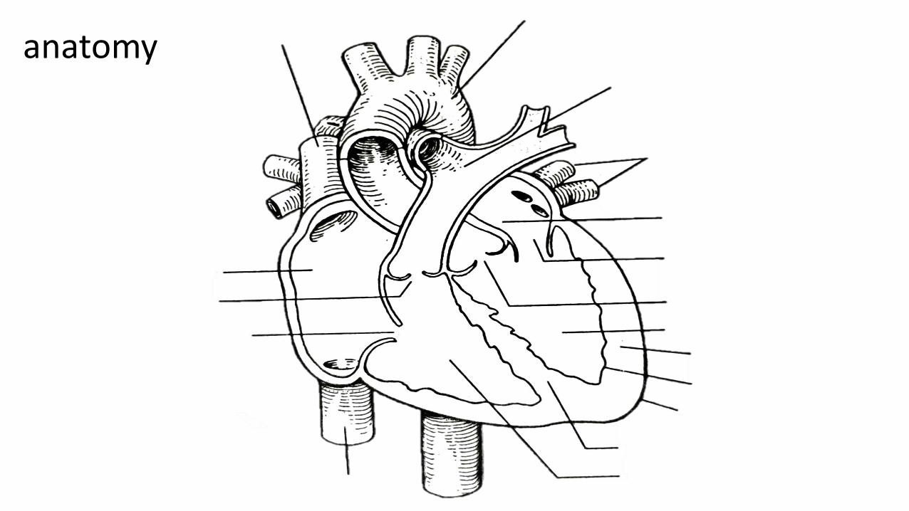
Review Heart Anatomy: Using Heart Models
•You and a partner will need a wet pen, cloth eraser, heart model, dry erase pen/eraser
•PLEASE:•Label the heart model•Write 4 directional term sentences comparing 2 structures of the heart.
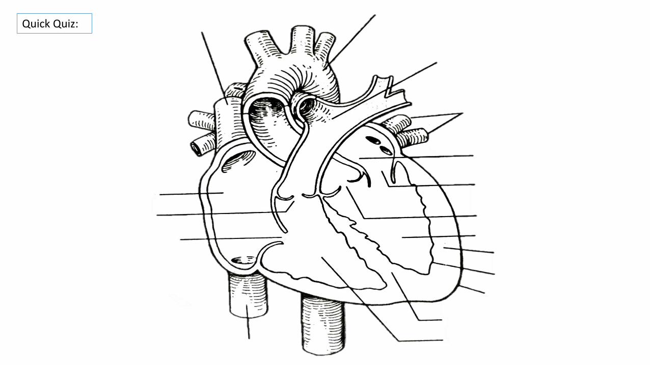
Quick Quiz:
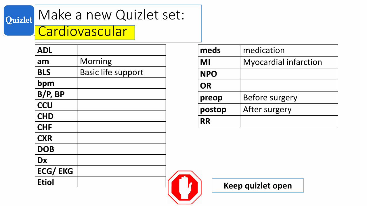
Make a new Quizlet set: CardiovascularADLam MorningBLS Basic life supportbpm B/P, BPCCUCHDCHFCXRDOBDxECG/ EKGEtiol
meds medication
MI Myocardial infarction
NPO
OR
preop Before surgery
postop After surgery
RR
Keep quizlet open

Shiny desk: Medical abbreviations practice
1. Take a family history, date of birth, weight before examination.
______________________________________________________
2. Record all vital signs, blood pressure, temperature and pulse three times a day
______________________________________________________
3. Take chest xray, electrocardiogram before surgery
______________________________________________________
4. Move patient to recovery room with wheelchair and give them bathroom privileges.
______________________________________________________
Take FH, DOB, wt before exam
Record VS, BP T, P tid
Take CXR, ECG/EKG preop
Move pt RR c w/c BRP
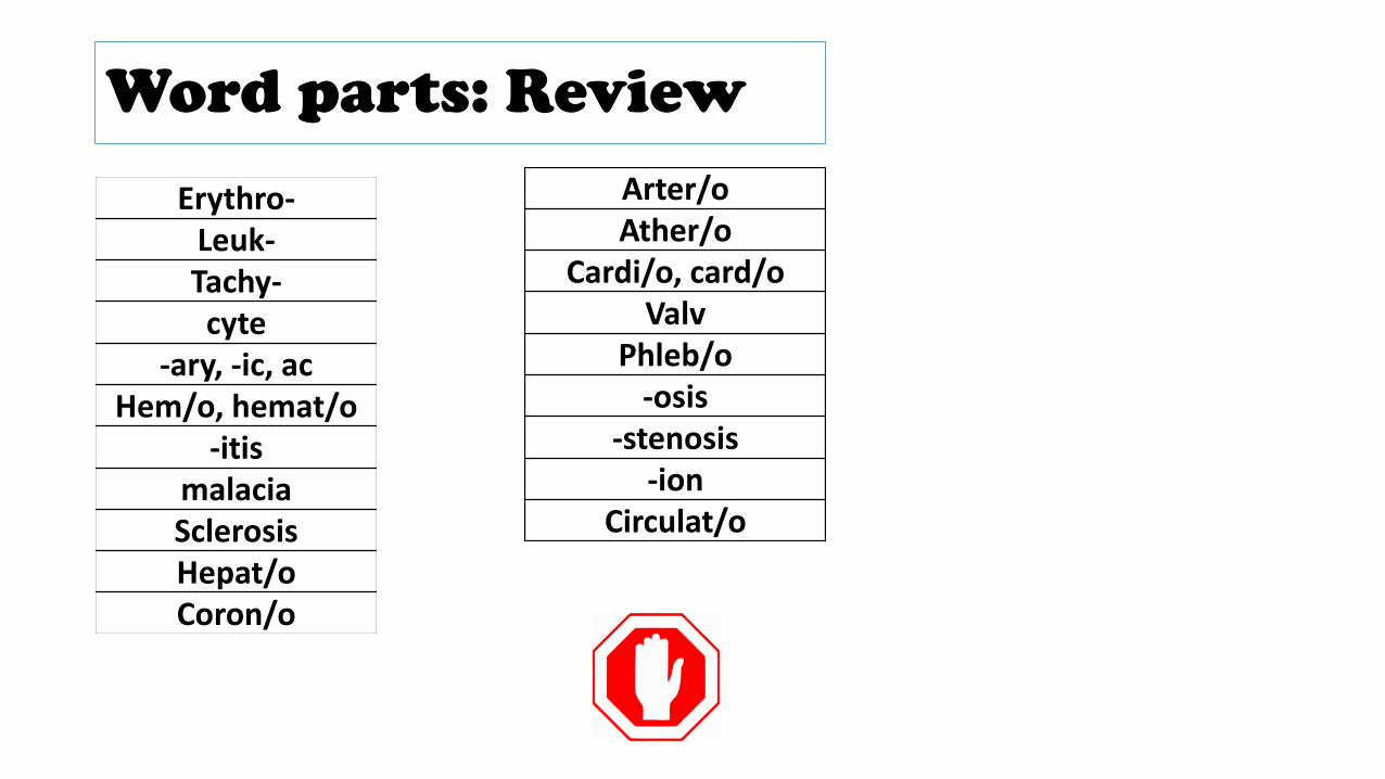
Word parts: Review
Erythro-Leuk-Tachy-cyte
-ary, -ic, acHem/o, hemat/o
-itismalaciaSclerosisHepat/oCoron/o
Arter/oAther/o
Cardi/o, card/oValv
Phleb/o-osis
-stenosis-ion
Circulat/o
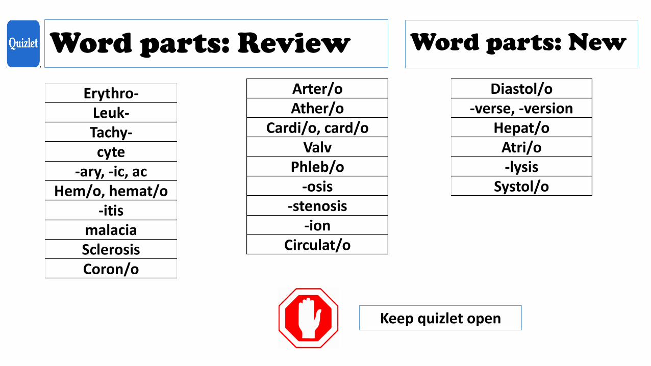
Word parts: Review
Erythro-Leuk-Tachy-cyte
-ary, -ic, acHem/o, hemat/o
-itismalaciaSclerosisCoron/o
Arter/oAther/o
Cardi/o, card/oValv
Phleb/o-osis
-stenosis-ion
Circulat/o
Word parts: New
Diastol/o-verse, -version
Hepat/oAtri/o-lysis
Systol/o
Keep quizlet open
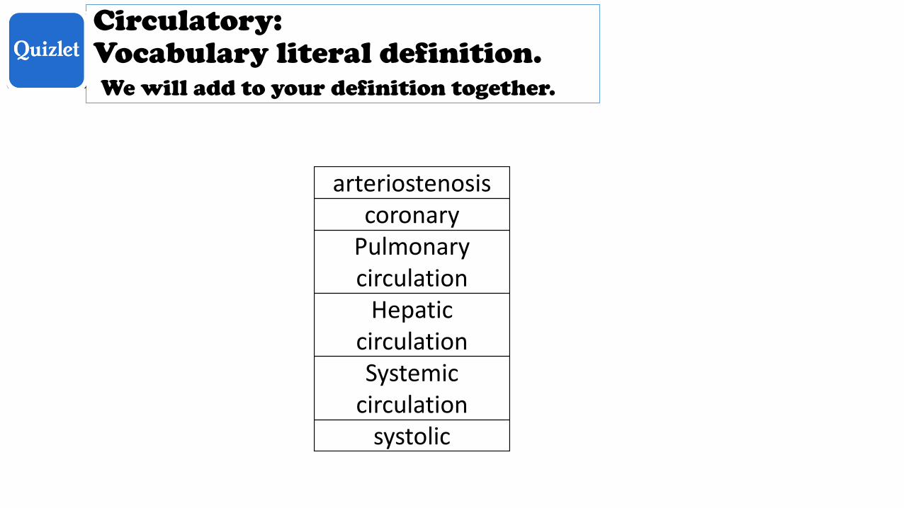
Circulatory: Vocabulary literal definition. We will add to your definition together.
arteriostenosiscoronary
Pulmonary circulation
Hepatic circulationSystemic
circulationsystolic
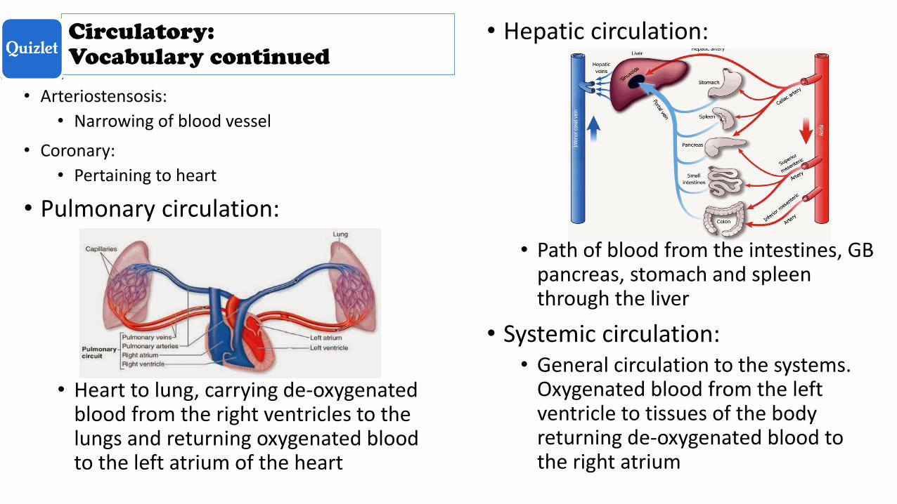
Circulatory:
Vocabulary continued
• Arteriostensosis:
• Narrowing of blood vessel
• Coronary:
• Pertaining to heart
• Pulmonary circulation:
• Heart to lung, carrying de-oxygenated blood from the right ventricles to the lungs and returning oxygenated blood to the left atrium of the heart
• Hepatic circulation:
• Path of blood from the intestines, GB pancreas, stomach and spleen through the liver
• Systemic circulation: • General circulation to the systems.
Oxygenated blood from the left ventricle to tissues of the body returning de-oxygenated blood to the right atrium
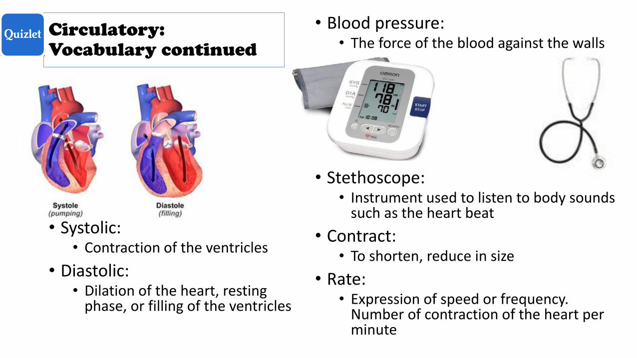
• Systolic: • Contraction of the ventricles
• Diastolic:• Dilation of the heart, resting
phase, or filling of the ventricles
• Blood pressure: • The force of the blood against the walls
• Stethoscope: • Instrument used to listen to body sounds
such as the heart beat
• Contract: • To shorten, reduce in size
• Rate: • Expression of speed or frequency.
Number of contraction of the heart per minute
Circulatory:
Vocabulary continued
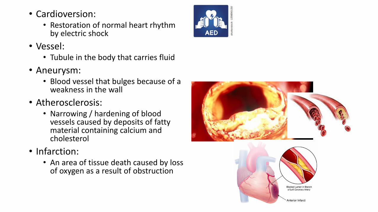
• Cardioversion: • Restoration of normal heart rhythm
by electric shock
• Vessel: • Tubule in the body that carries fluid
• Aneurysm: • Blood vessel that bulges because of a
weakness in the wall
• Atherosclerosis:• Narrowing / hardening of blood
vessels caused by deposits of fatty material containing calcium and cholesterol
• Infarction: • An area of tissue death caused by loss
of oxygen as a result of obstruction
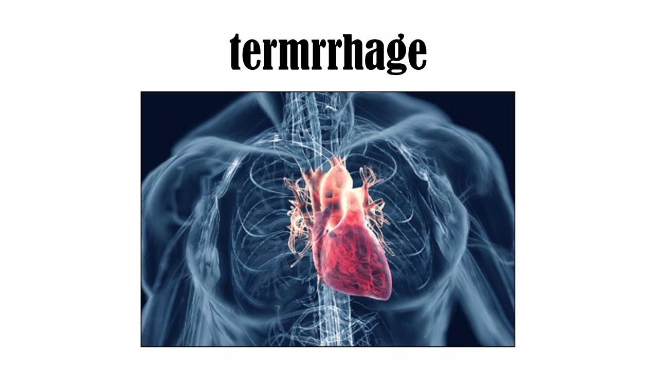
termrrhage
Practice quiz: Forms
Video segment: watch the first time, then take notes
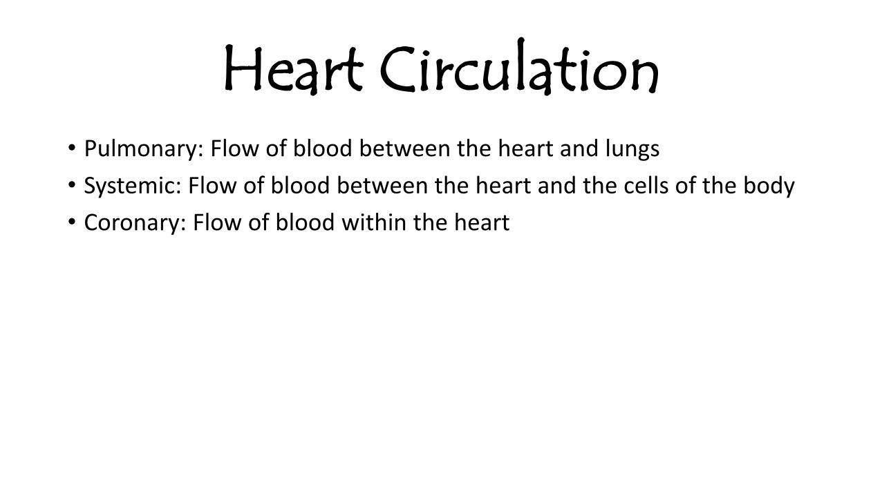
Heart Circulation• Pulmonary: Flow of blood between the heart and lungs
• Systemic: Flow of blood between the heart and the cells of the body
• Coronary: Flow of blood within the heart
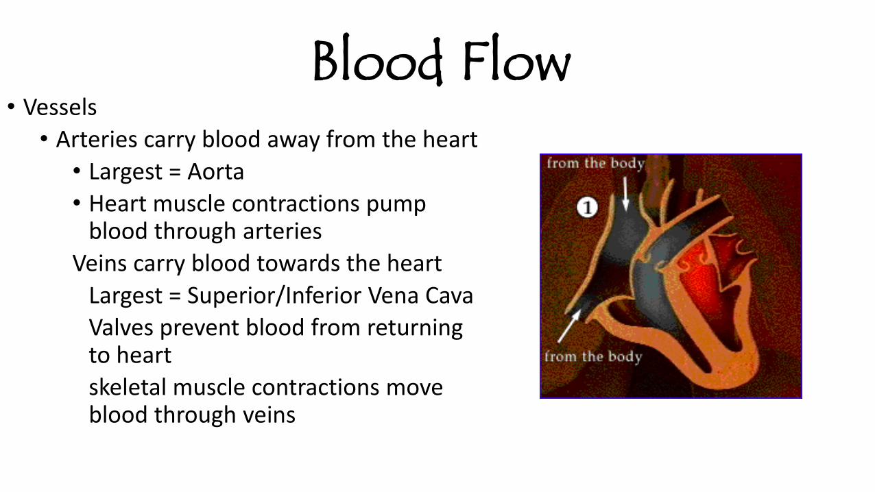
Blood Flow• Vessels
• Arteries carry blood away from the heart
• Largest = Aorta
• Heart muscle contractions pump blood through arteries
Veins carry blood towards the heart
Largest = Superior/Inferior Vena Cava
Valves prevent blood from returning to heart
skeletal muscle contractions move blood through veins
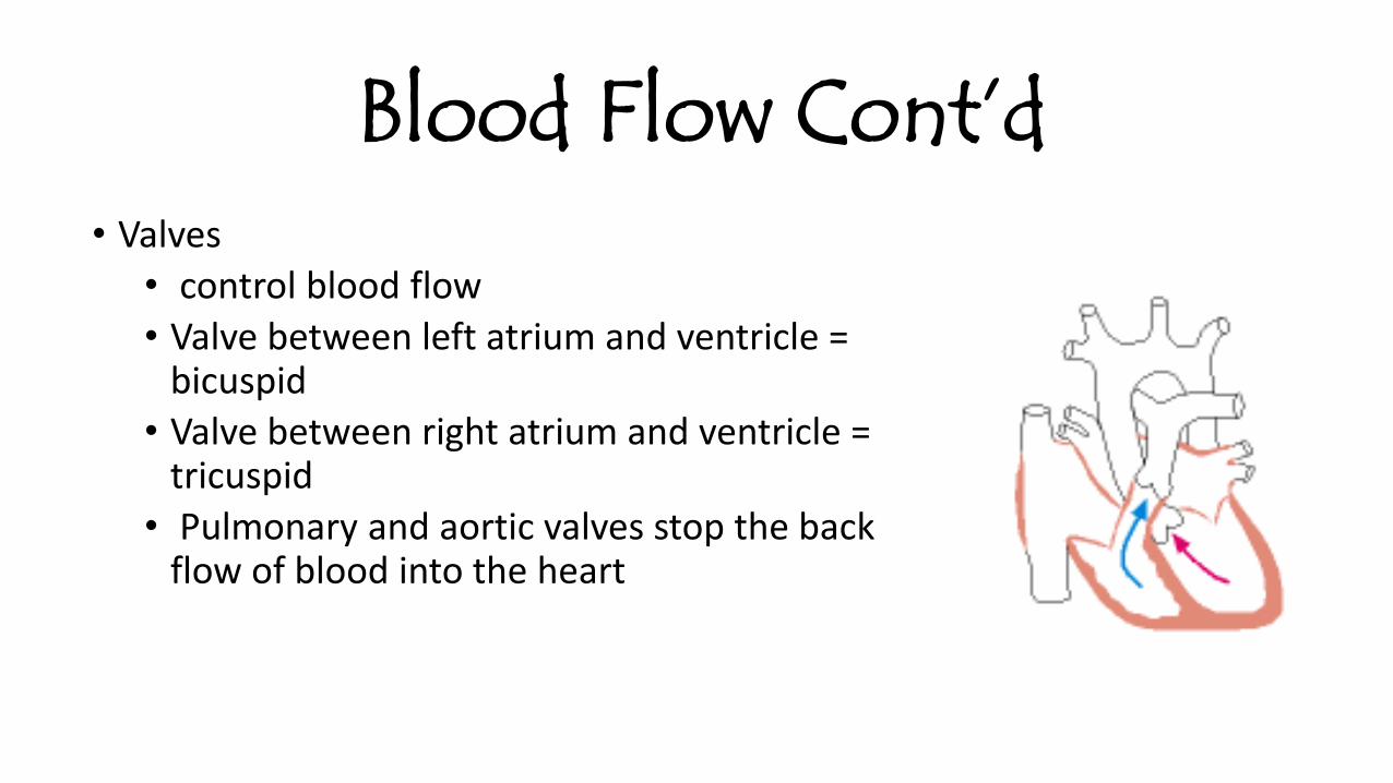
Blood Flow Cont’d• Valves
• control blood flow
• Valve between left atrium and ventricle = bicuspid
• Valve between right atrium and ventricle = tricuspid
• Pulmonary and aortic valves stop the back flow of blood into the heart
W.A. follow up: in lab group1. Correct answers
2. Looking at # 10 &11: The ventricles push blood out of the heart. If they are not working perfectly :a) What is that persons body not receiving
b) What would the long term affects be
c) How could you treat it
3. On the back of W.A. list and define the directional terms
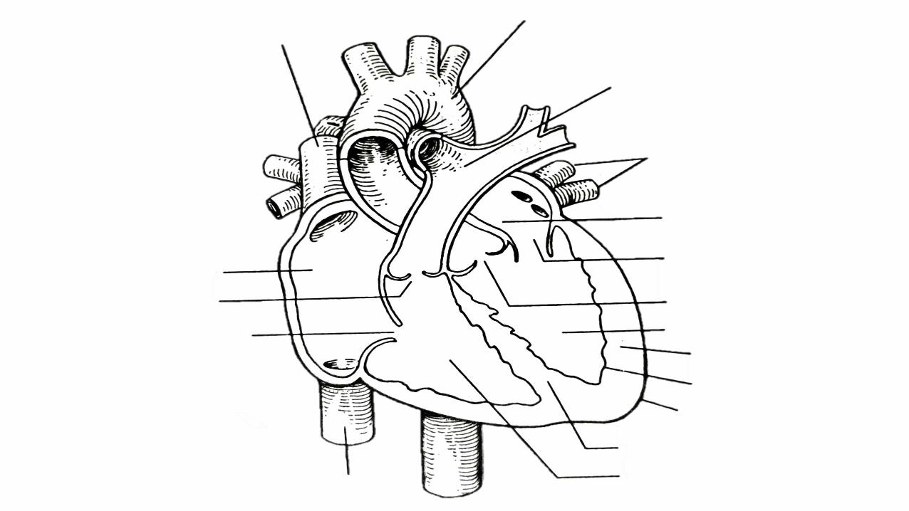
4. On the heart diagram label the anatomical structures that are on the worksheet. There should be 7+ (actually label the heart diagram)
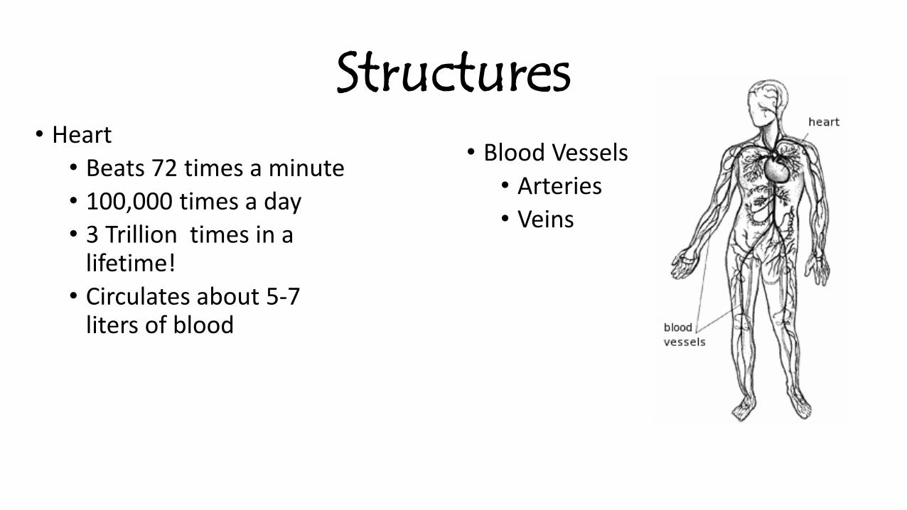
Structures• Heart
• Beats 72 times a minute
• 100,000 times a day
• 3 Trillion times in a lifetime!
• Circulates about 5-7 liters of blood
• Blood Vessels
• Arteries
• Veins
Functions• Transport nutrients and oxygen
• Transport waste to kidneys
• Distribute hormones and antibodies
• Help control body temperature and maintain homeostasis
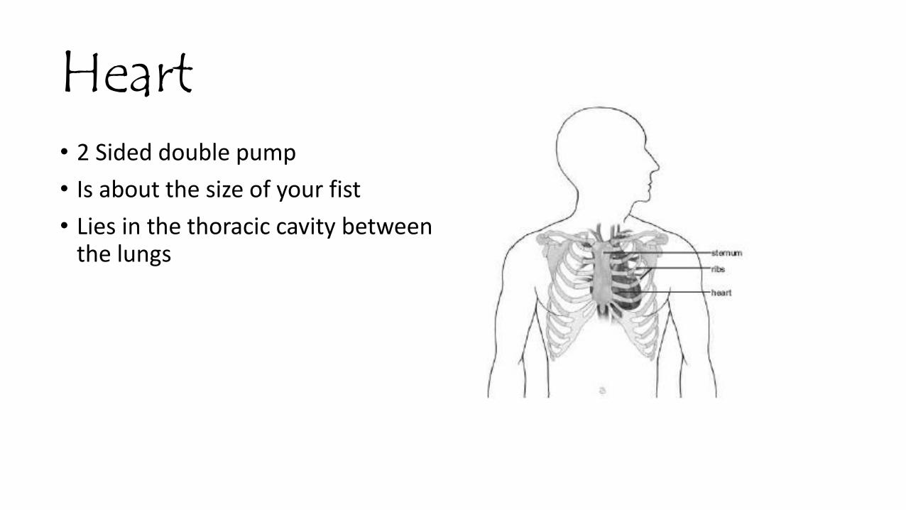
Heart• 2 Sided double pump
• Is about the size of your fist
• Lies in the thoracic cavity between the lungs
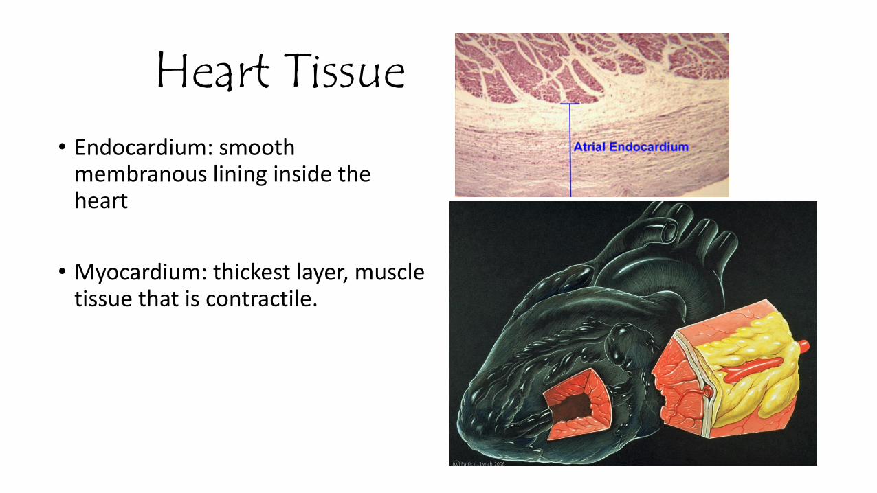
Heart Tissue• Endocardium: smooth
membranous lining inside the heart
• Myocardium: thickest layer, muscle tissue that is contractile.
Heart Tissue Cont’d
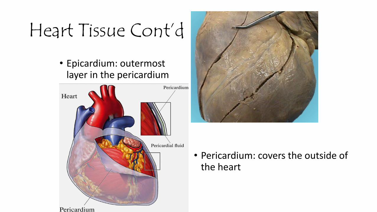
• Epicardium: outermost layer in the pericardium
• Pericardium: covers the outside of the heart
Parts of the Heart• Divided into right and left sides
• 2 chambers in each side, for a total of 4 chambers
• Atrium: top, where blood enters
• Ventricles: bottom, where blood leaves
• Left and right sides separated by a partition called a septum

Review

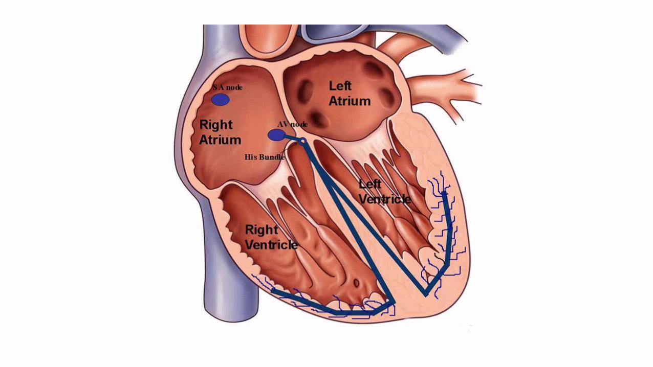
Cardiac Conduction System• Electrical Impulses produce a wave
that can be recorded on the ECG
• Consists of• Sinoatrial (SA) node
• Atrioventriclular (AV) node
• Bundle of His (AV Bundle)
• Bundle Branches
• Purkinje Fibers (network)
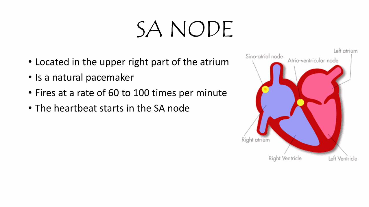
SA NODE• Located in the upper right part of the atrium
• Is a natural pacemaker
• Fires at a rate of 60 to 100 times per minute
• The heartbeat starts in the SA node
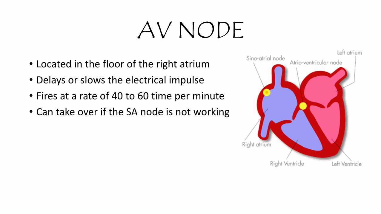
AV NODE• Located in the floor of the right atrium
• Delays or slows the electrical impulse
• Fires at a rate of 40 to 60 time per minute
• Can take over if the SA node is not working
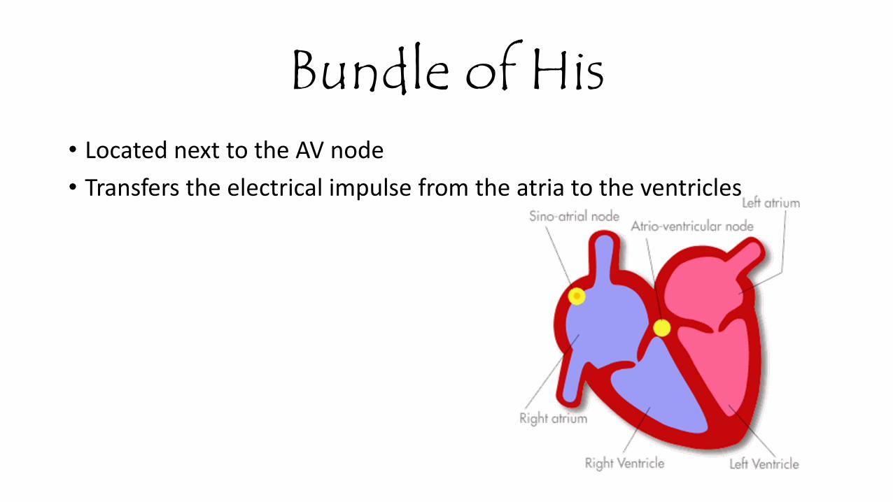
Bundle of His• Located next to the AV node
• Transfers the electrical impulse from the atria to the ventricles
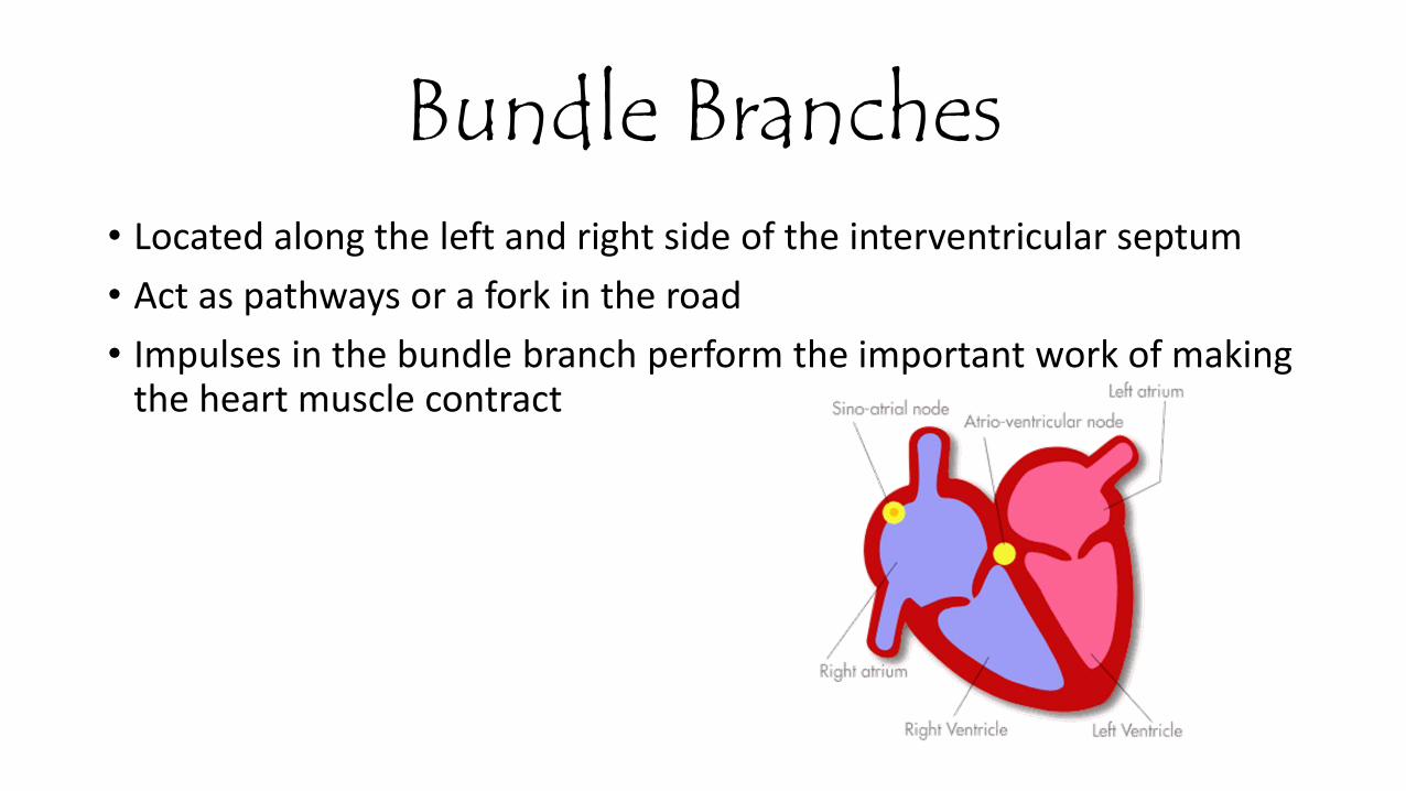
Bundle Branches• Located along the left and right side of the interventricular septum
• Act as pathways or a fork in the road
• Impulses in the bundle branch perform the important work of making the heart muscle contract
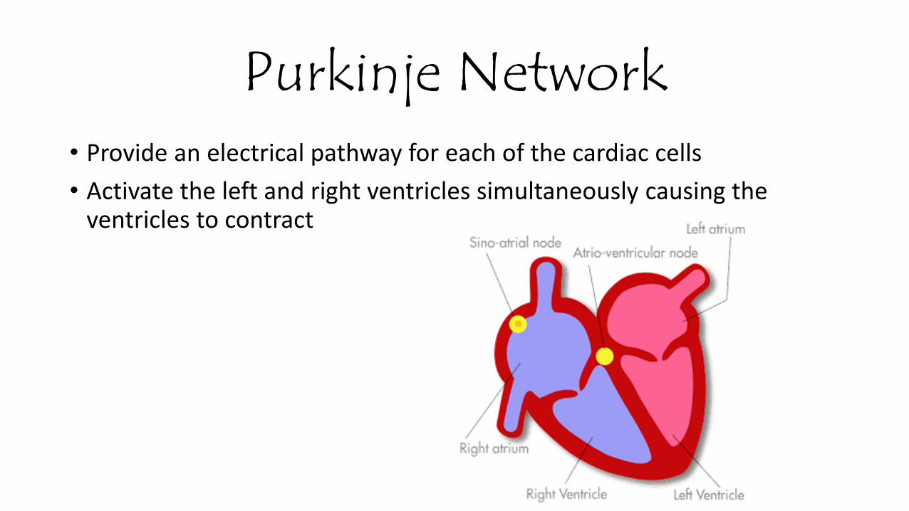
Purkinje Network• Provide an electrical pathway for each of the cardiac cells
• Activate the left and right ventricles simultaneously causing the ventricles to contract
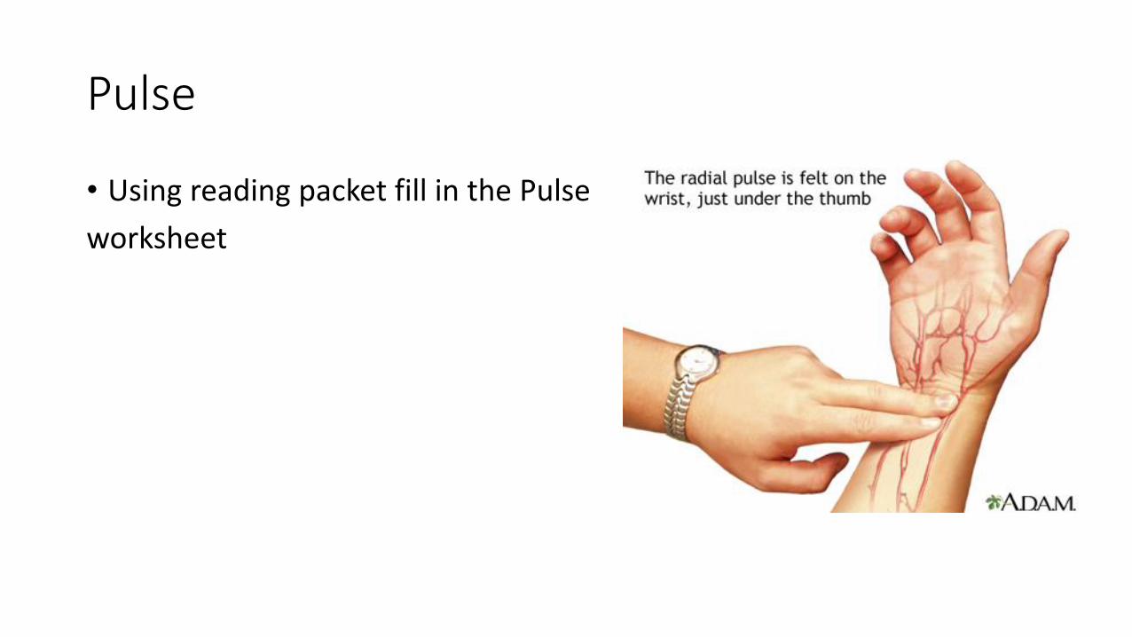
Pulse
• Using reading packet fill in the Pulse
worksheet
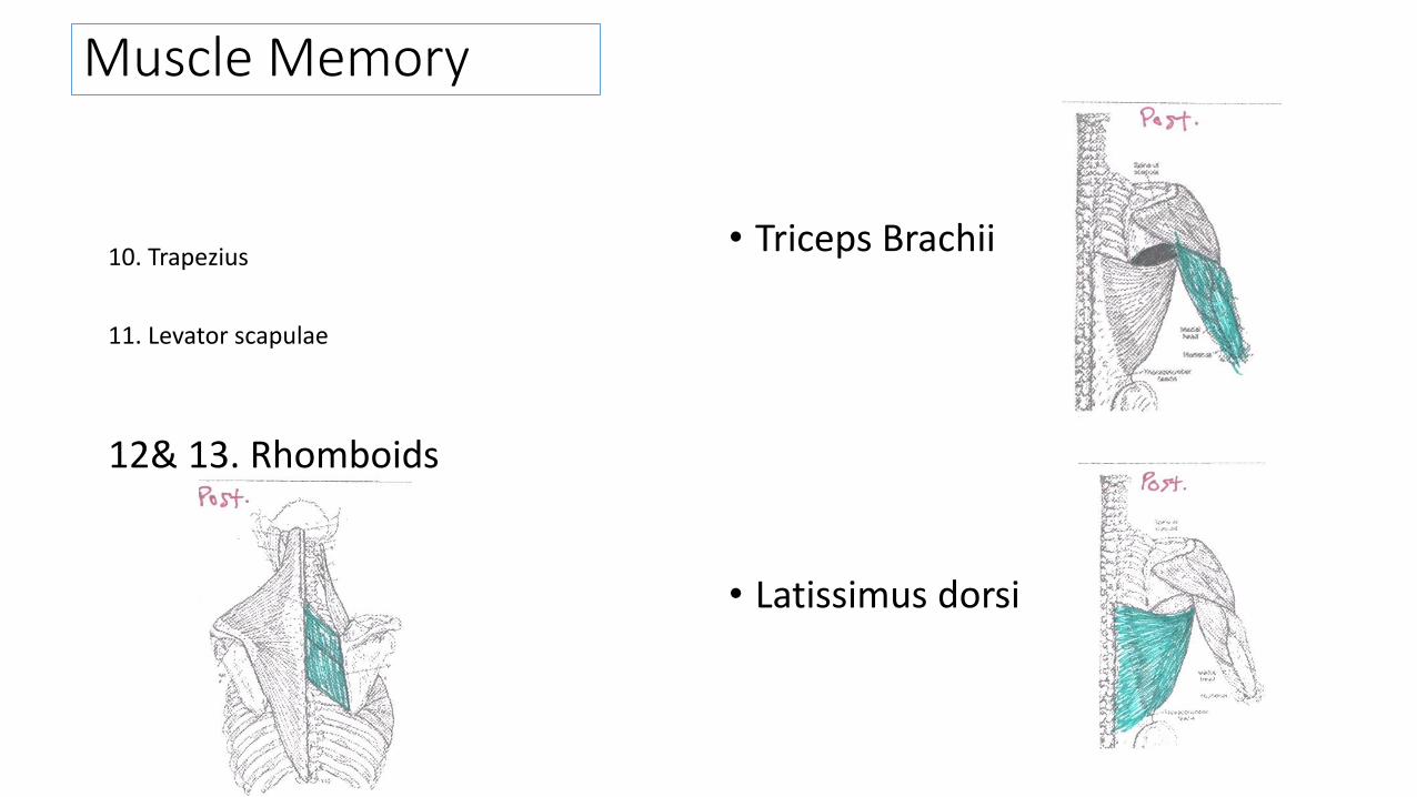
Muscle Memory
10. Trapezius
11. Levator scapulae
12& 13. Rhomboids
• Triceps Brachii
• Latissimus dorsi
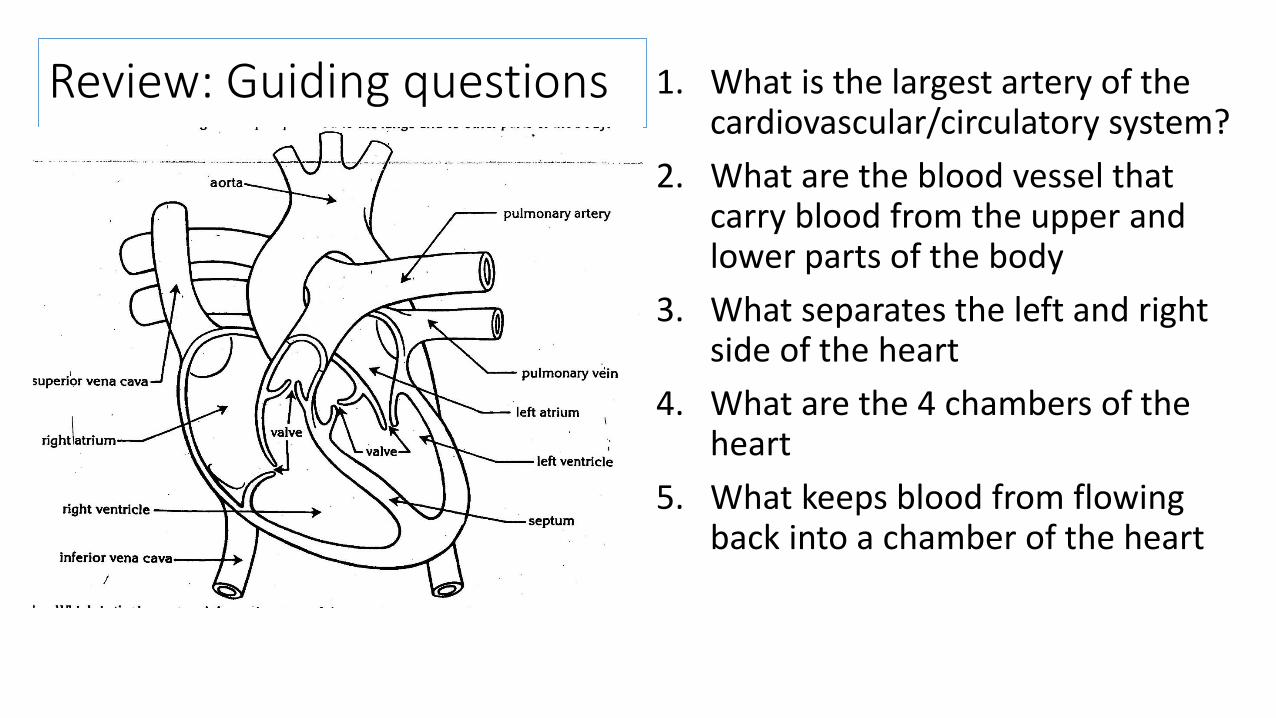
Review: Guiding questions 1. What is the largest artery of the cardiovascular/circulatory system?
2. What are the blood vessel that carry blood from the upper and lower parts of the body
3. What separates the left and right side of the heart
4. What are the 4 chambers of the heart
5. What keeps blood from flowing back into a chamber of the heart

Reivew:
• Quizlet.live
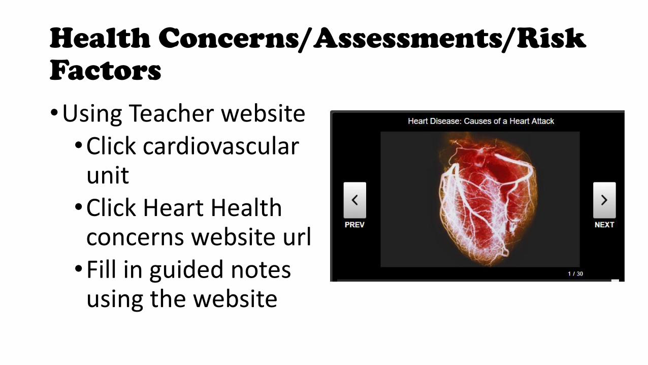
Health Concerns/Assessments/Risk Factors
•Using Teacher website•Click cardiovascular unit•Click Heart Health concerns website url•Fill in guided notes using the website
Unit Practice quiz: forms
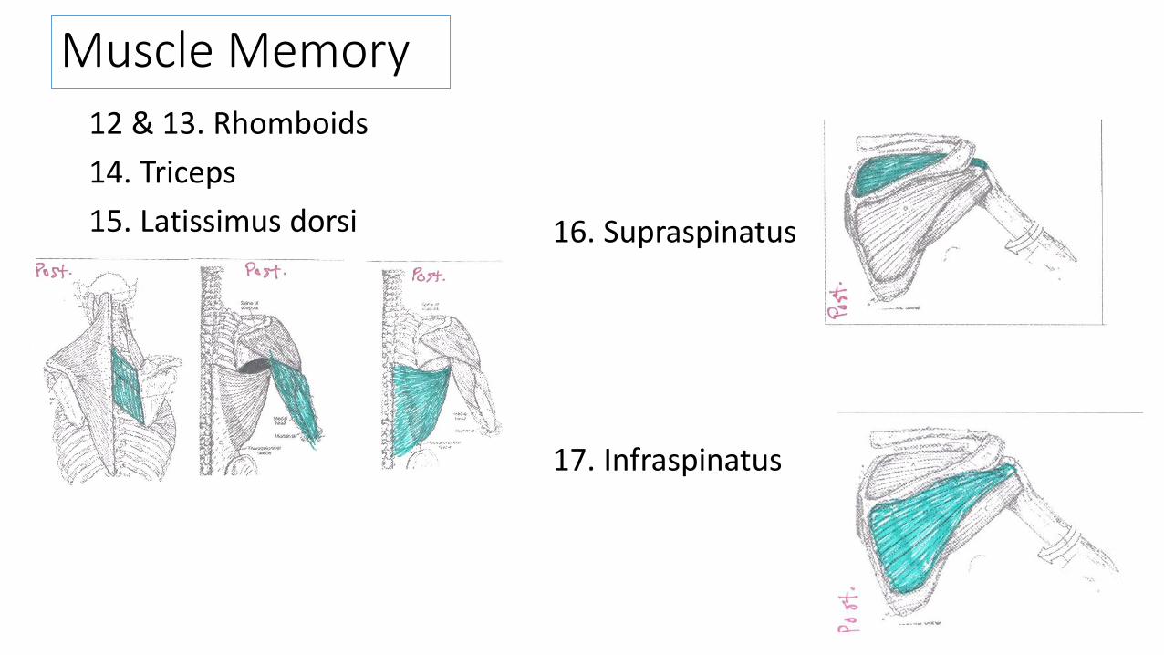
Muscle Memory
12 & 13. Rhomboids
14. Triceps
15. Latissimus dorsi 16. Supraspinatus
17. Infraspinatus
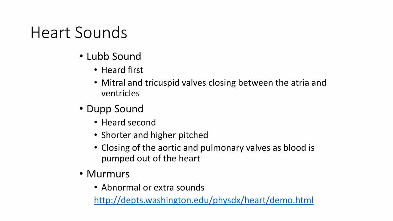
Heart Sounds• Lubb Sound
• Heard first
• Mitral and tricuspid valves closing between the atria and ventricles
• Dupp Sound• Heard second
• Shorter and higher pitched
• Closing of the aortic and pulmonary valves as blood is pumped out of the heart
• Murmurs• Abnormal or extra sounds
http://depts.washington.edu/physdx/heart/demo.html
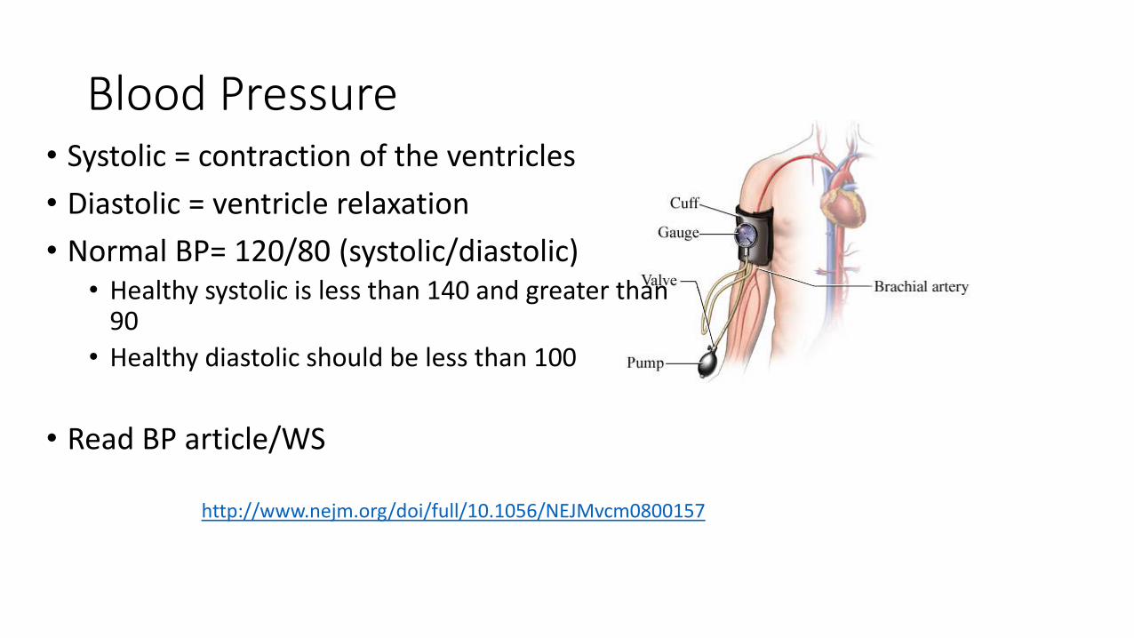
Blood Pressure• Systolic = contraction of the ventricles
• Diastolic = ventricle relaxation
• Normal BP= 120/80 (systolic/diastolic)• Healthy systolic is less than 140 and greater than
90
• Healthy diastolic should be less than 100
• Read BP article/WS
http://www.nejm.org/doi/full/10.1056/NEJMvcm0800157
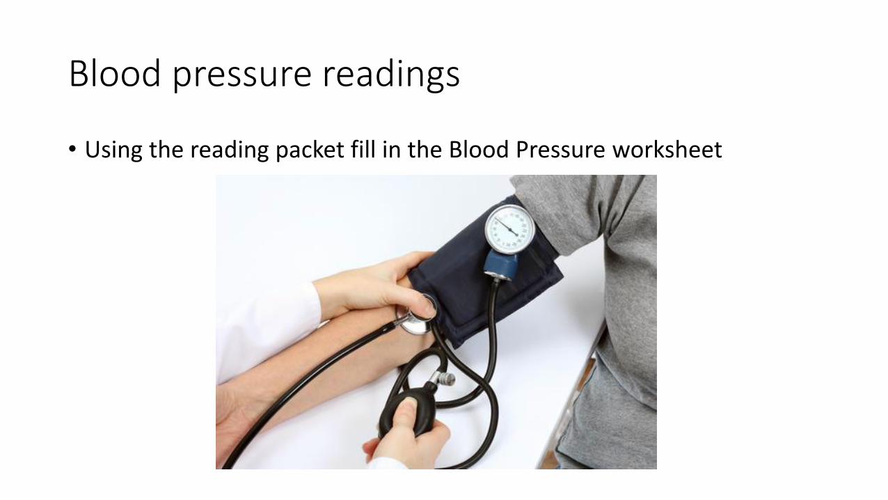
Blood pressure readings
• Using the reading packet fill in the Blood Pressure worksheet
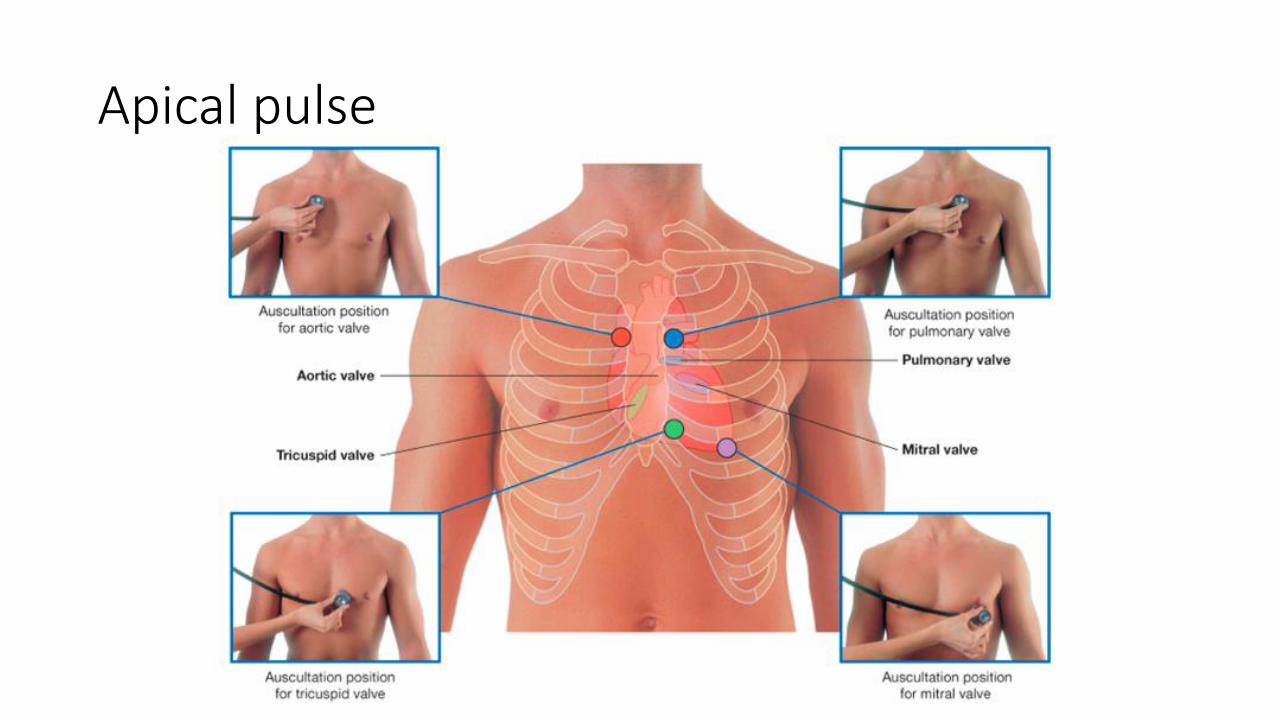
Apical pulse
Practice quizzes
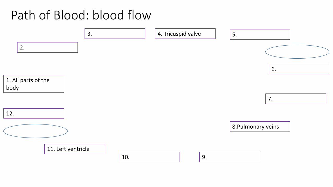
Path of Blood: blood flow
2.
1. All parts of the body
3.
8.Pulmonary veins
12.
4. Tricuspid valve
11. Left ventricle
10. 9.
7.
6.
5.
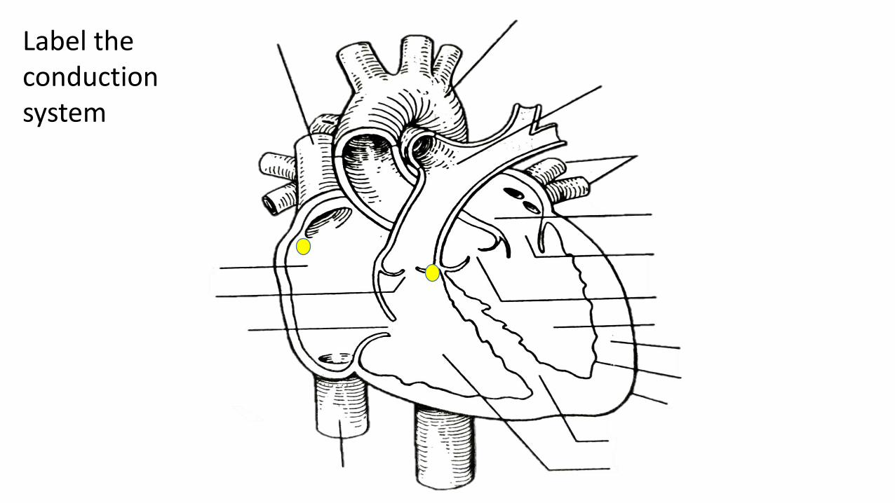
Label the conduction system
Superior Vena Cava
Aortic Valve
Bicuspid/Mitral Valve
L Atrium
Pulmonary Veins
Pulmonary Artery
Aorta
Epicardium
Endocardium
MyocardiumL Ventricle
Inferior Vena CavaR Ventricle
Intraventricular Septum
Tricuspid Valve
R. Atrium
Pulmonary Valve
LUNGS
anatomy
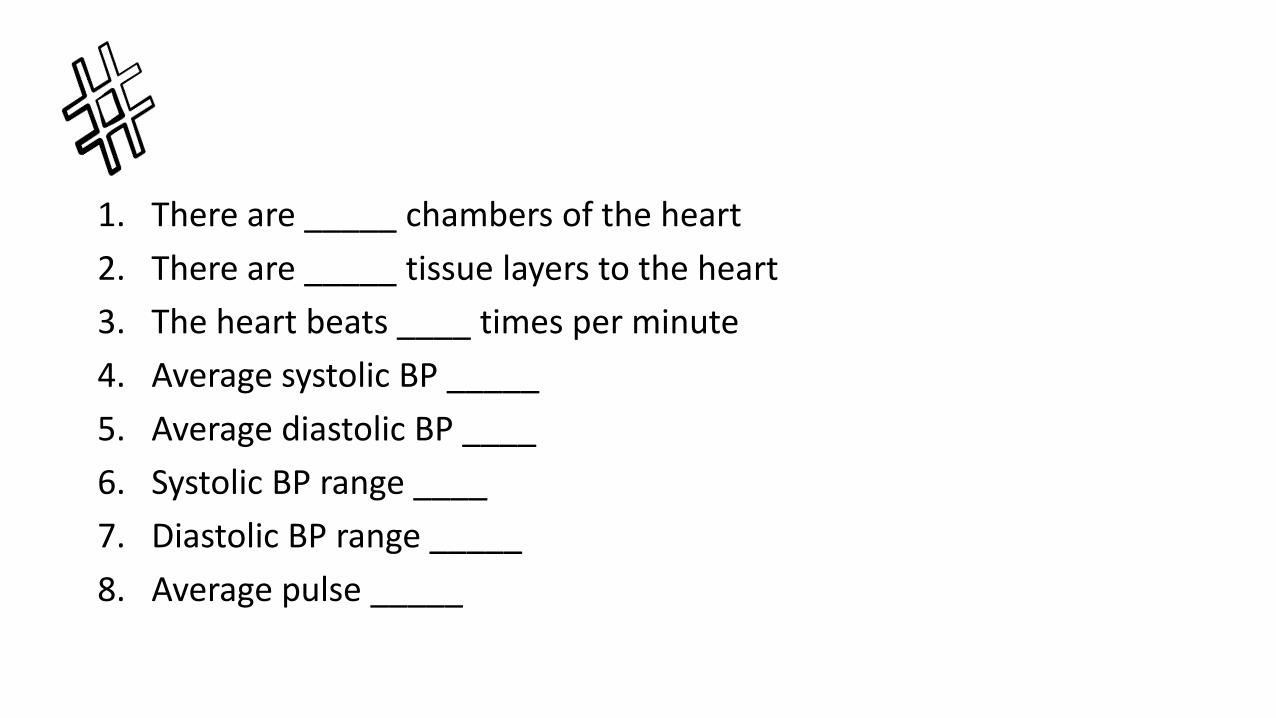
1. There are _____ chambers of the heart
2. There are _____ tissue layers to the heart
3. The heart beats ____ times per minute
4. Average systolic BP _____
5. Average diastolic BP ____
6. Systolic BP range ____
7. Diastolic BP range _____
8. Average pulse _____


![Sistema Cardiovascular Fisiot [1216].ppt](https://img.pdfslide.net/doc/110x75/577c78ab1a28abe05490a128/sistema-cardiovascular-fisiot-1216ppt.jpg)