Embed Size (px)
Citation preview
CASE REPORT Open Access
Calcaneal osteomyelitis presenting with acutetarsal tunnel syndrome: a case reportDavinder PS Baghla1*, Sajid Shariff2, Raman Dega2
Abstract
Introduction: Cases of acute tarsal tunnel syndrome are rare. To the best of our knowledge, we describe the onlyreported case of acute posterior tibial nerve compression resulting from adjacent haemotogenous pyogeniccalcaneal osteomyelitis.
Case presentation: A previously healthy 38-year-old Caucasian woman developed symptoms of acute tarsaltunnel syndrome in her right foot over a six-day period. No antecedent trauma or systemic symptoms were noted.Magnetic resonance imaging and bone scan imaging, followed by surgical decompression and bone biopsyconfirmed a diagnosis of Staphylococcus aureus calcaneal osteomyelitis. Her pain and paraesthesia disappeared afterthe operation, while her inflammatory markers normalised during a 12-week course of antibiotics. After four yearsshe has remained asymptomatic without any indication of recurrence.
Conclusion: This case is not just unique in describing osteomyelitis as a cause of tarsal tunnel syndrome, becausehaemotogenous calcaneal osteomyelitis is in itself a rare pathology. We recommend considering infection as adifferential diagnosis in patients presenting with acute tarsal tunnel syndrome.
IntroductionThe tarsal tunnel is a fibro-osseous space bounded bythe flexor retinaculum, posterior talus and the calcaneus,as well as the anterior medial malleolar artery. Variousanatomical compartmental divisions have been described[1]. The tarsal tunnel houses the tendons of tibialis pos-terior, flexor digitorum longus and flexor hallucislongus, as well as the posterior tibial nerve, artery andvein.Tarsal tunnel syndrome is an entrapment neuropathy
of one or all branches of the posterior tibial nerve (lat-eral plantar, medial plantar and medial calcaneal nerves)and was first described independently by Keck and Lamin 1962 [2,3]. Various aetiologies for tarsal tunnel syn-drome have been previously described, but here wedescribe its first known case of unique association withosteomyelitis.
Case presentationWe present the case of a previously healthy 38-year-oldCaucasian woman who presented to our hospital’s emer-gency department with a six-day history of severe sharp
and burning right heel and foot pain with inability tobear weight. She had no antecedent trauma or systemicsymptoms. The pain was referred distally along themedial and lateral plantar aspect of her foot into thetoes, with exacerbation at night and with ambulation.A physical examination revealed a warm localised
swelling around her medial malleolus with no overlyingerythema. Her ankle movements were normal but hersubtalar joint movement was painful and restricted. Aneurological examination confirmed altered sensationover the plantar surface of her foot and toes. Tinel’ssign was also noted to be absent along the course of herposterior tibial nerve.Our patient’s inflammatory markers were raised (white
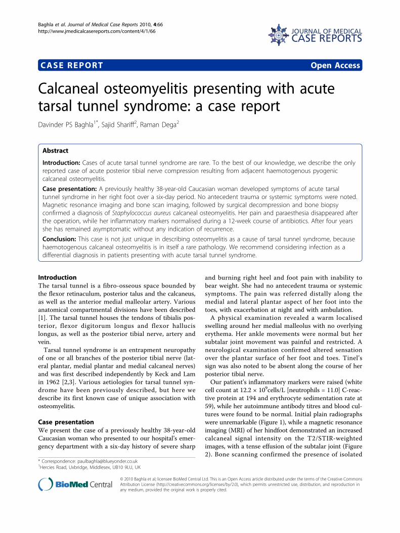
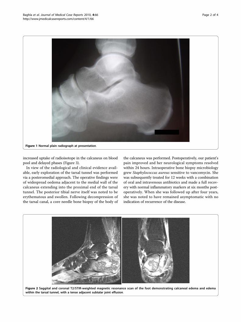
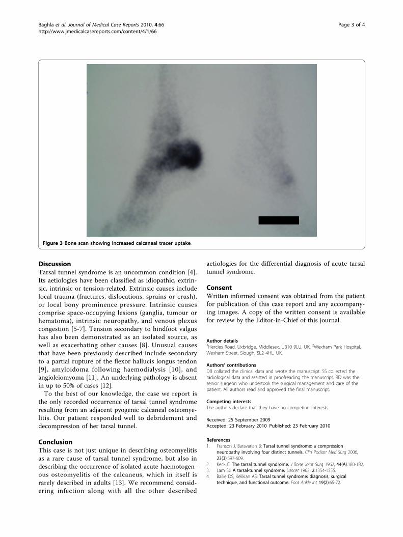
cell count at 12.2 × 10⁹cells/L [neutrophils = 11.0] C-reac-tive protein at 194 and erythrocyte sedimentation rate at59), while her autoimmune antibody titres and blood cul-tures were found to be normal. Initial plain radiographswere unremarkable (Figure 1), while a magnetic resonanceimaging (MRI) of her hindfoot demonstrated an increasedcalcaneal signal intensity on the T2/STIR-weightedimages, with a tense effusion of the subtalar joint (Figure2). Bone scanning confirmed the presence of isolated
* Correspondence: [email protected] Road, Uxbridge, Middlesex, UB10 9LU, UK
Baghla et al. Journal of Medical Case Reports 2010, 4:66http://www.jmedicalcasereports.com/content/4/1/66 JOURNAL OF MEDICAL
CASE REPORTS
© 2010 Baghla et al; licensee BioMed Central Ltd. This is an Open Access article distributed under the terms of the Creative CommonsAttribution License (http://creativecommons.org/licenses/by/2.0), which permits unrestricted use, distribution, and reproduction inany medium, provided the original work is properly cited.
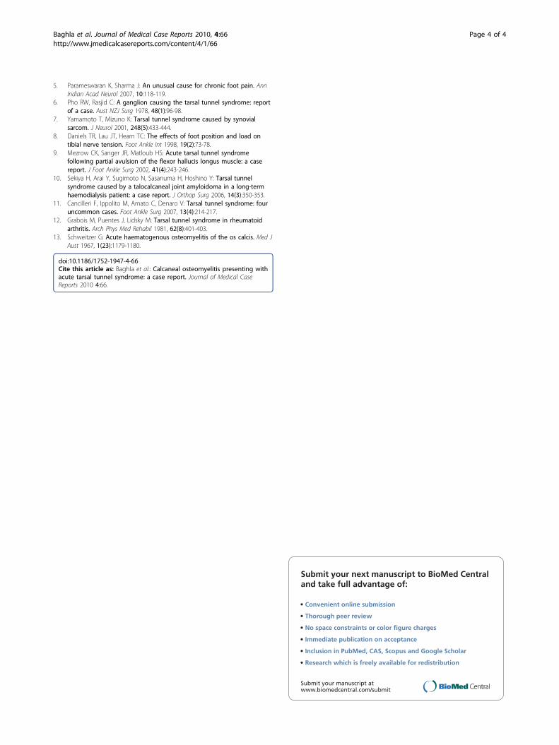
increased uptake of radioisotope in the calcaneus on bloodpool and delayed phases (Figure 3).In view of the radiological and clinical evidence avail-
able, early exploration of the tarsal tunnel was performedvia a posteromedial approach. The operative findings wereof widespread oedema adjacent to the medial wall of thecalcaneus extending into the proximal end of the tarsaltunnel. The posterior tibial nerve itself was noted to beerythematous and swollen. Following decompression ofthe tarsal canal, a core needle bone biopsy of the body of
the calcaneus was performed. Postoperatively, our patient’spain improved and her neurological symptoms resolvedwithin 24 hours. Intraoperative bone biopsy microbiologygrew Staphylococcus aureus sensitive to vancomycin. Shewas subsequently treated for 12 weeks with a combinationof oral and intravenous antibiotics and made a full recov-ery with normal inflammatory markers at six months post-operatively. When she was followed up after four years,she was noted to have remained asymptomatic with noindication of recurrence of the disease.
Figure 1 Normal plain radiograph at presentation.
Figure 2 Saggital and coronal T2/STIR-weighted magnetic resonance scan of the foot demonstrating calcaneal edema and edemawithin the tarsal tunnel, with a tense adjacent subtalar joint effusion.
Baghla et al. Journal of Medical Case Reports 2010, 4:66http://www.jmedicalcasereports.com/content/4/1/66
Page 2 of 4
DiscussionTarsal tunnel syndrome is an uncommon condition [4].Its aetiologies have been classified as idiopathic, extrin-sic, intrinsic or tension-related. Extrinsic causes includelocal trauma (fractures, dislocations, sprains or crush),or local bony prominence pressure. Intrinsic causescomprise space-occupying lesions (ganglia, tumour orhematoma), intrinsic neuropathy, and venous plexuscongestion [5-7]. Tension secondary to hindfoot valgushas also been demonstrated as an isolated source, aswell as exacerbating other causes [8]. Unusual causesthat have been previously described include secondaryto a partial rupture of the flexor hallucis longus tendon[9], amyloidoma following haemodialysis [10], andangioleiomyoma [11]. An underlying pathology is absentin up to 50% of cases [12].To the best of our knowledge, the case we report is
the only recorded occurrence of tarsal tunnel syndromeresulting from an adjacent pyogenic calcaneal osteomye-litis. Our patient responded well to debridement anddecompression of her tarsal tunnel.
ConclusionThis case is not just unique in describing osteomyelitisas a rare cause of tarsal tunnel syndrome, but also indescribing the occurrence of isolated acute haemotogen-ous osteomyelitis of the calcaneus, which in itself israrely described in adults [13]. We recommend consid-ering infection along with all the other described
aetiologies for the differential diagnosis of acute tarsaltunnel syndrome.
ConsentWritten informed consent was obtained from the patientfor publication of this case report and any accompany-ing images. A copy of the written consent is availablefor review by the Editor-in-Chief of this journal.
Author details1Hercies Road, Uxbridge, Middlesex, UB10 9LU, UK. 2Wexham Park Hospital,Wexham Street, Slough, SL2 4HL, UK.
Authors’ contributionsDB collated the clinical data and wrote the manuscript. SS collected theradiological data and assisted in proofreading the manuscript. RD was thesenior surgeon who undertook the surgical management and care of thepatient. All authors read and approved the final manuscript.
Competing interestsThe authors declare that they have no competing interests.
Received: 25 September 2009Accepted: 23 February 2010 Published: 23 February 2010
References1. Franson J, Baravarian B: Tarsal tunnel syndrome: a compression
neuropathy involving four distinct tunnels. Clin Podiatr Med Surg 2006,23(3):597-609.
2. Keck C: The tarsal tunnel syndrome. J Bone Joint Surg 1962, 44(A):180-182.3. Lam SJ: A tarsal-tunnel syndrome. Lancet 1962, 2:1354-1355.4. Bailie DS, Kelikian AS: Tarsal tunnel syndrome: diagnosis, surgical
technique, and functional outcome. Foot Ankle Int 19(2):65-72.
Figure 3 Bone scan showing increased calcaneal tracer uptake.
Baghla et al. Journal of Medical Case Reports 2010, 4:66http://www.jmedicalcasereports.com/content/4/1/66
Page 3 of 4
5. Parameswaran K, Sharma J: An unusual cause for chronic foot pain. AnnIndian Acad Neurol 2007, 10:118-119.
6. Pho RW, Rasjid C: A ganglion causing the tarsal tunnel syndrome: reportof a case. Aust NZJ Surg 1978, 48(1):96-98.
7. Yamamoto T, Mizuno K: Tarsal tunnel syndrome caused by synovialsarcom. J Neurol 2001, 248(5):433-444.
8. Daniels TR, Lau JT, Hearn TC: The effects of foot position and load ontibial nerve tension. Foot Ankle Int 1998, 19(2):73-78.
9. Mezrow CK, Sanger JR, Matloub HS: Acute tarsal tunnel syndromefollowing partial avulsion of the flexor hallucis longus muscle: a casereport. J Foot Ankle Surg 2002, 41(4):243-246.
10. Sekiya H, Arai Y, Sugimoto N, Sasanuma H, Hoshino Y: Tarsal tunnelsyndrome caused by a talocalcaneal joint amyloidoma in a long-termhaemodialysis patient: a case report. J Orthop Surg 2006, 14(3):350-353.
11. Cancilleri F, Ippolito M, Amato C, Denaro V: Tarsal tunnel syndrome: fouruncommon cases. Foot Ankle Surg 2007, 13(4):214-217.
12. Grabois M, Puentes J, Lidsky M: Tarsal tunnel syndrome in rheumatoidarthritis. Arch Phys Med Rehabil 1981, 62(8):401-403.
13. Schweitzer G: Acute haematogenous osteomyelitis of the os calcis. Med JAust 1967, 1(23):1179-1180.
doi:10.1186/1752-1947-4-66Cite this article as: Baghla et al.: Calcaneal osteomyelitis presenting withacute tarsal tunnel syndrome: a case report. Journal of Medical CaseReports 2010 4:66.
Submit your next manuscript to BioMed Centraland take full advantage of:
• Convenient online submission
• Thorough peer review
• No space constraints or color figure charges
• Immediate publication on acceptance
• Inclusion in PubMed, CAS, Scopus and Google Scholar
• Research which is freely available for redistribution
Submit your manuscript at www.biomedcentral.com/submit
Baghla et al. Journal of Medical Case Reports 2010, 4:66http://www.jmedicalcasereports.com/content/4/1/66
Page 4 of 4