Embed Size (px)
Citation preview
Cauda equina syndrome: the state of
the evidence in 2019
Munchi S ChokseyMD (Cantab) FRCS (Surgical neurology)
Consultant neurological and spinal surgeon
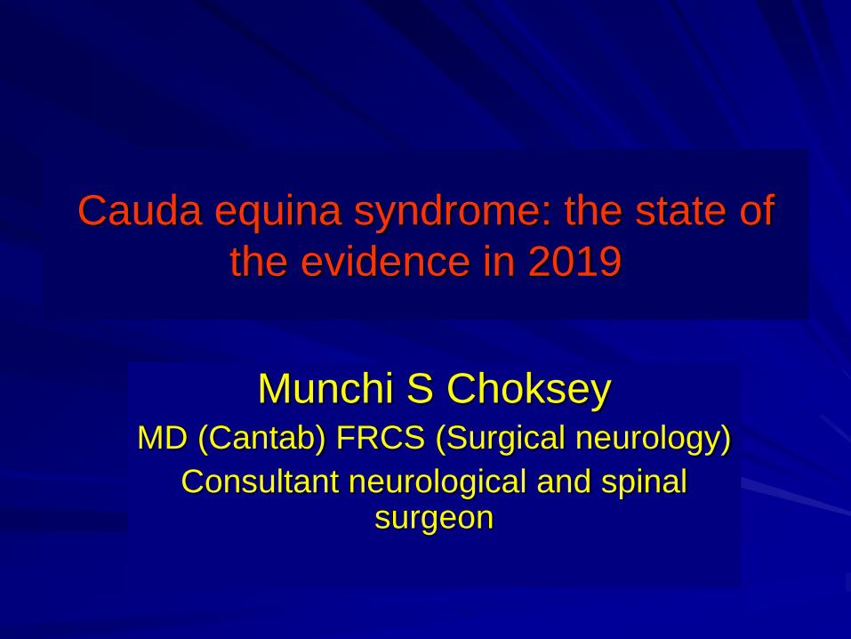
Spinal neurosurgery
35 years of experience
6000 + lumbar spinal
1200 + neck operations
Thoracic spines
Degenerative disease
Fractures,
Tumours
Infections
Paediatrics
Busy legal practice

CES - objectives
Anatomy and physiology
Pathogenesis – why things go wrong
Definitions - varied
Clinical presentation
Natural history
Investigation, MRI and its limitations
Action - current guidelines from BASS and SBNS
Which operation ?
Outcomes - expected and actual

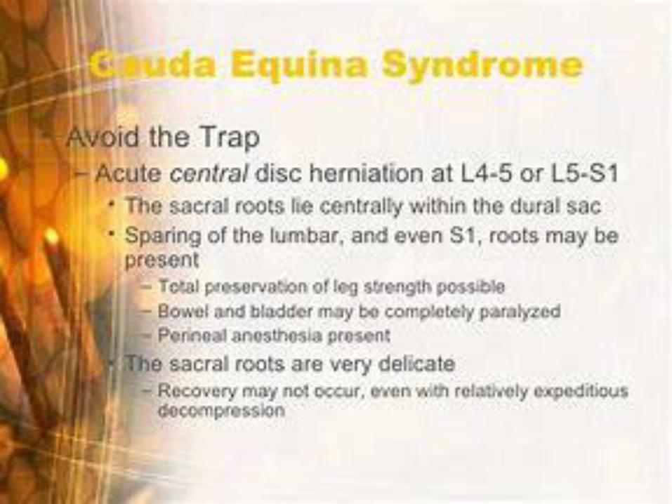
Acute CES: genuine problem or “crying wolf”
Back pain is very common
Over 7 million “sufferers” in UK at any one time
Over 100 million working days lost p.a
Spot the genuine case
Clinically difficult – but then if it was easy
everyone would do it:
5 years in medical school – hs to be worth
something


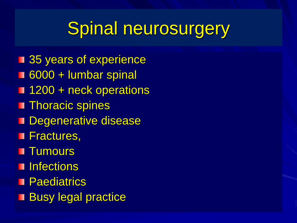
Lumbar spine anatomy


Cauda equina blood vessels
Run longitudinally.
No anastomoses across roots
Loss of a segment of blood
supply – no recovery

Ischaemia
But: death is not the mere stoppage of a machine; it is also
total ruin of the supposed machinery. Similarly-and this is
a lesson which I wish to emphasize as strongly as I can -
partial anoxaemia means not a mere slowing down of life,
but progressive and perhaps irreparable damage to living
structure.
J S Haldane
A Lecture on the Symptoms, Causes, and Prevention of
Anoxaemia (Insufficient Supply of Oxygen to the Tissues),
and the Value of Oxygen in its Treatment
Br Med J 1919; 2:65 (Published 19 July 1919)
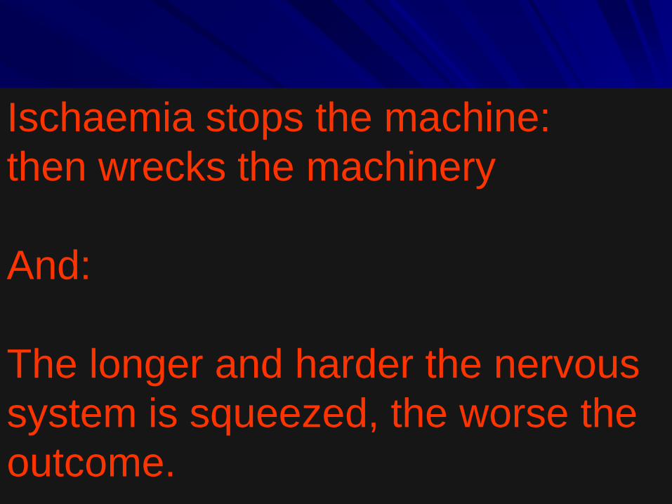
Ischaemia stops the machine:
then wrecks the machinery
And:
The longer and harder the nervous
system is squeezed, the worse the
outcome.

Pathophysiology
Takahashi et al
Compression not the sole cause
Combination of chemical irritation and
compression
Local high concentration of inflammatory
mediators
Vascular damage, thrombosis and
infarction

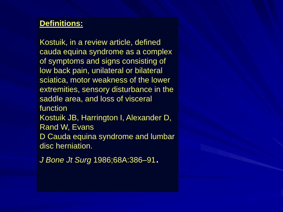
Definitions:
Kostuik, in a review article, defined
cauda equina syndrome as a complex
of symptoms and signs consisting of
low back pain, unilateral or bilateral
sciatica, motor weakness of the lower
extremities, sensory disturbance in the
saddle area, and loss of visceral
function
Kostuik JB, Harrington I, Alexander D,
Rand W, Evans
D Cauda equina syndrome and lumbar
disc herniation.
J Bone Jt Surg 1986;68A:386–91.
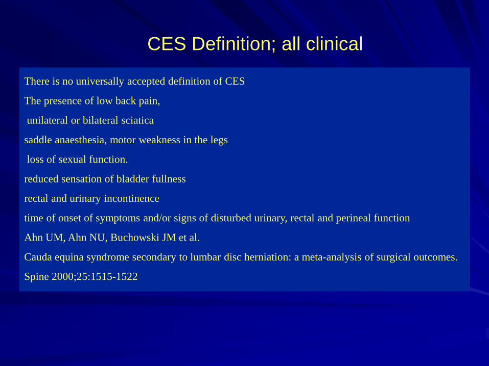
CES Definition; all clinical
There is no universally accepted definition of CES
The presence of low back pain,
unilateral or bilateral sciatica
saddle anaesthesia, motor weakness in the legs
loss of sexual function.
reduced sensation of bladder fullness
rectal and urinary incontinence
time of onset of symptoms and/or signs of disturbed urinary, rectal and perineal function
Ahn UM, Ahn NU, Buchowski JM et al.
Cauda equina syndrome secondary to lumbar disc herniation: a meta-analysis of surgical outcomes.
Spine 2000;25:1515-1522
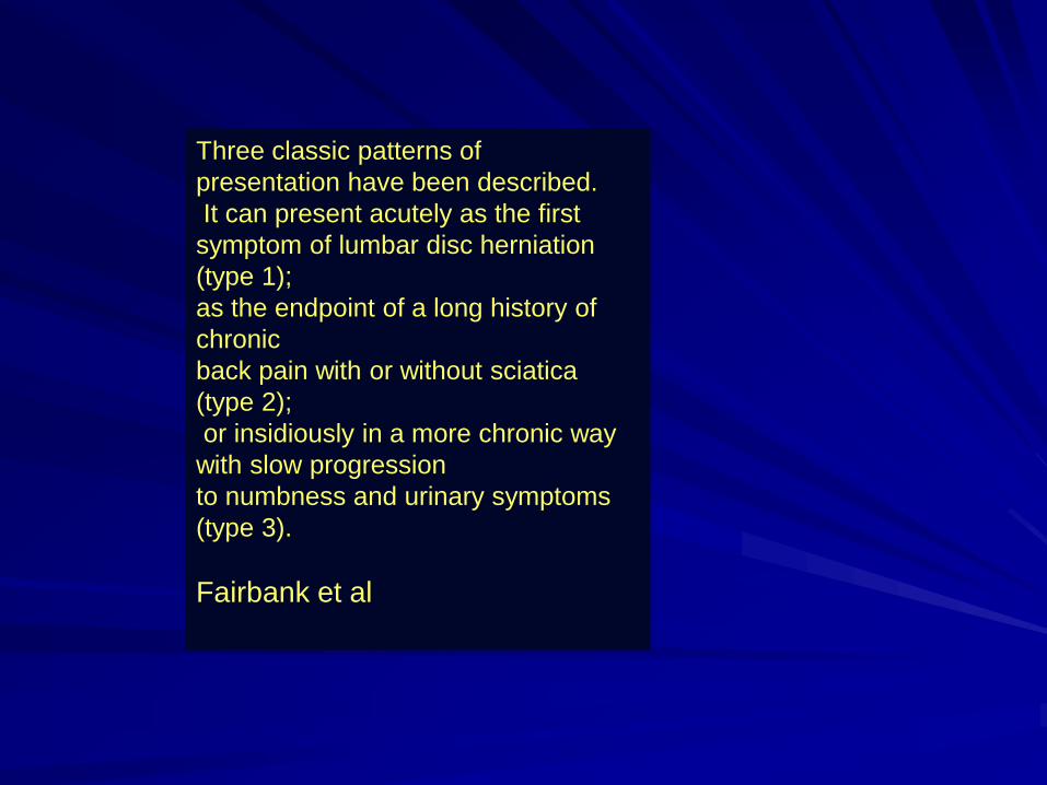
Three classic patterns of
presentation have been described.
It can present acutely as the first
symptom of lumbar disc herniation
(type 1);
as the endpoint of a long history of
chronic
back pain with or without sciatica
(type 2);
or insidiously in a more chronic way
with slow progression
to numbness and urinary symptoms
(type 3).
Fairbank et al
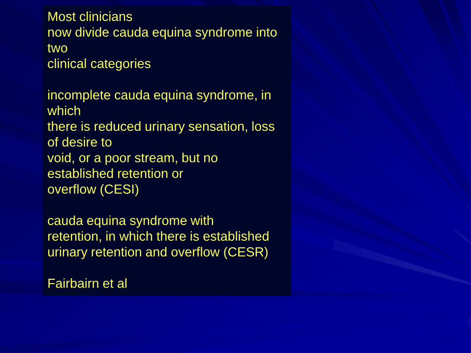
Most clinicians
now divide cauda equina syndrome into
two
clinical categories
incomplete cauda equina syndrome, in
which
there is reduced urinary sensation, loss
of desire to
void, or a poor stream, but no
established retention or
overflow (CESI)
cauda equina syndrome with
retention, in which there is established
urinary retention and overflow (CESR)
Fairbairn et al

3 categories (all clinical)
Suspected or threatened (CESS)
Incomplete (CESI)
Complete (CESR)
Todd (2015)
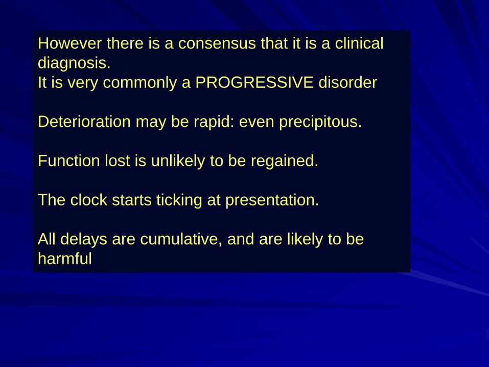
However there is a consensus that it is a clinical
diagnosis.
It is very commonly a PROGRESSIVE disorder
Deterioration may be rapid: even precipitous.
Function lost is unlikely to be regained.
The clock starts ticking at presentation.
All delays are cumulative, and are likely to be
harmful
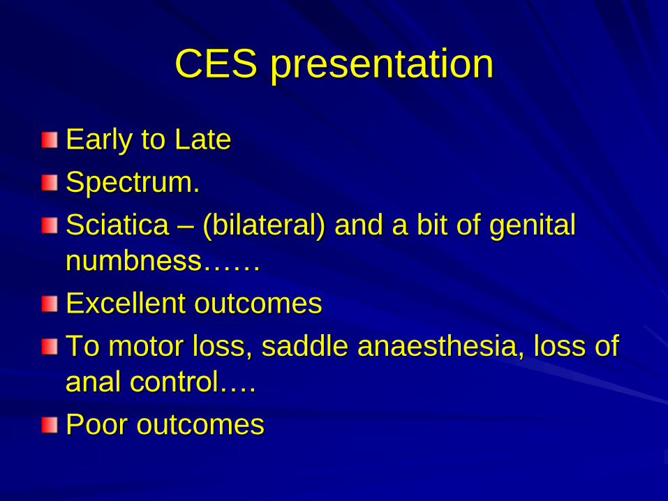
CES presentation
Early to Late
Spectrum.
Sciatica – (bilateral) and a bit of genital
numbness……
Excellent outcomes
To motor loss, saddle anaesthesia, loss of
anal control….
Poor outcomes
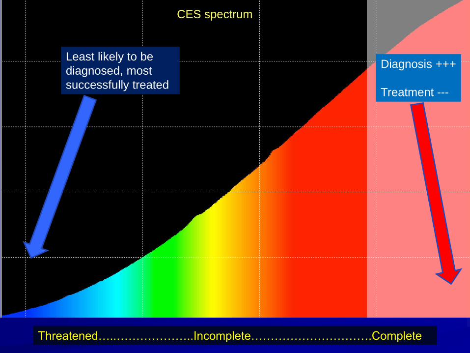
Least likely to be
diagnosed, most
successfully treated
Diagnosis +++
Treatment ---
CES spectrum
Threatened…..………………..Incomplete………………………….Complete
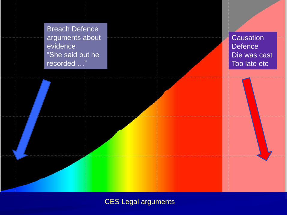
Breach Defence
arguments about
evidence
“She said but he
recorded …”
Causation
Defence
Die was cast
Too late etc
CES Legal arguments
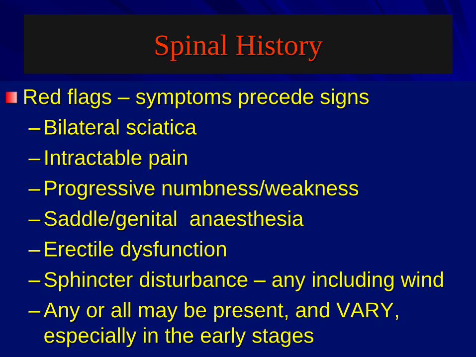
Spinal History
Red flags – symptoms precede signs
– Bilateral sciatica
– Intractable pain
– Progressive numbness/weakness
– Saddle/genital anaesthesia
– Erectile dysfunction
– Sphincter disturbance – any including wind
– Any or all may be present, and VARY,
especially in the early stages
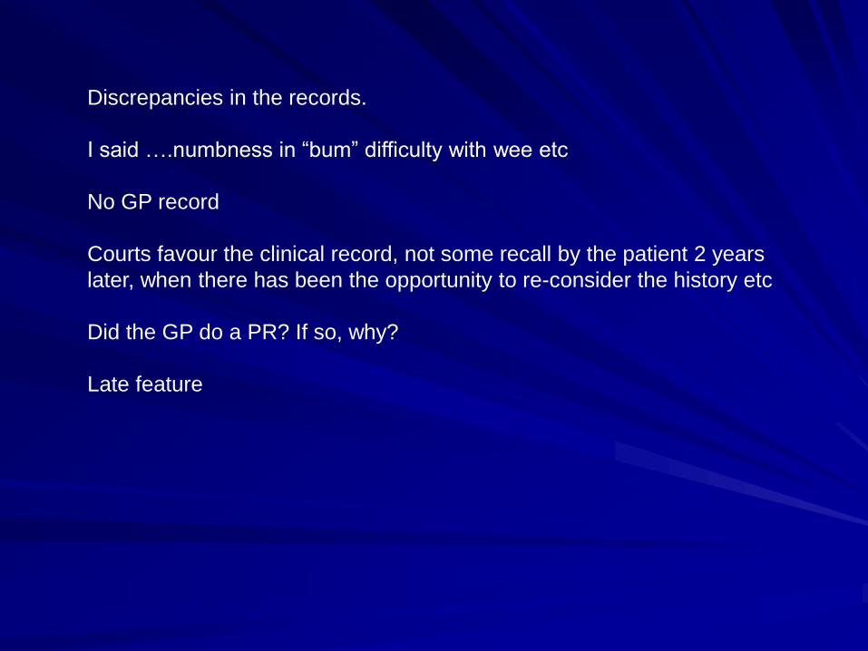
Discrepancies in the records.
I said ….numbness in “bum” difficulty with wee etc
No GP record
Courts favour the clinical record, not some recall by the patient 2 years
later, when there has been the opportunity to re-consider the history etc
Did the GP do a PR? If so, why?
Late feature

Acute spinal pain: CES and infection
Worrying symptoms
Pain
– constant
– Getting inexorably worse
– Night pain – waking from sleep (mass)
– Extreme reluctance to move (infection/
instability)
– “unwell” patient: gut feeling
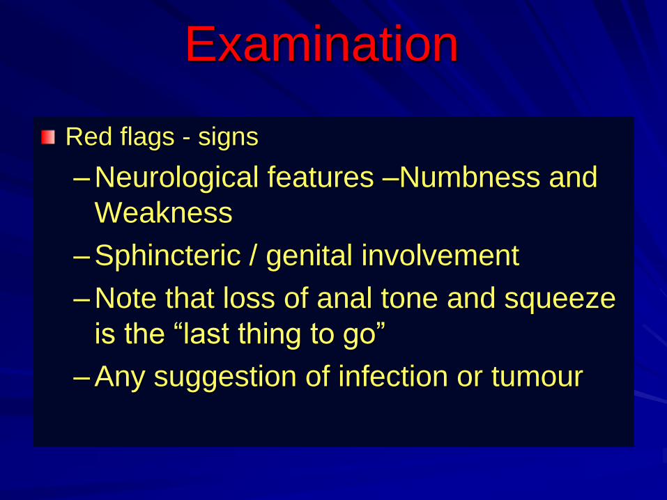
Examination
Red flags - signs
– Neurological features –Numbness and
Weakness
– Sphincteric / genital involvement
– Note that loss of anal tone and squeeze
is the “last thing to go”
– Any suggestion of infection or tumour

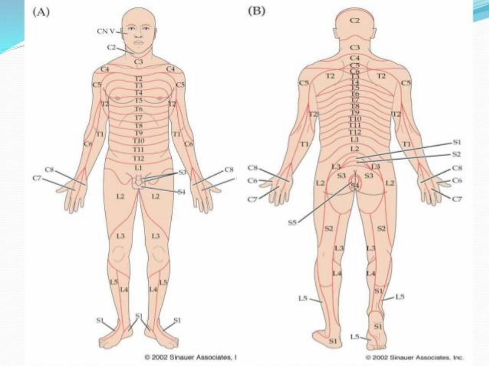
MYOTOMES
Hip flexion L2 and L3
Hip extension L 4 and L5
Knee Extension L3 and L4
Knee flexion L5 and S1
Ankle dorsiflexion (L4) mainly L5
Ankle Plantar Flexion S1 (poss.S2)
Perianal corrugator cutis ani – S 2 3 4 “wink reflex”
Anal tone and squeeze S 2 3 4
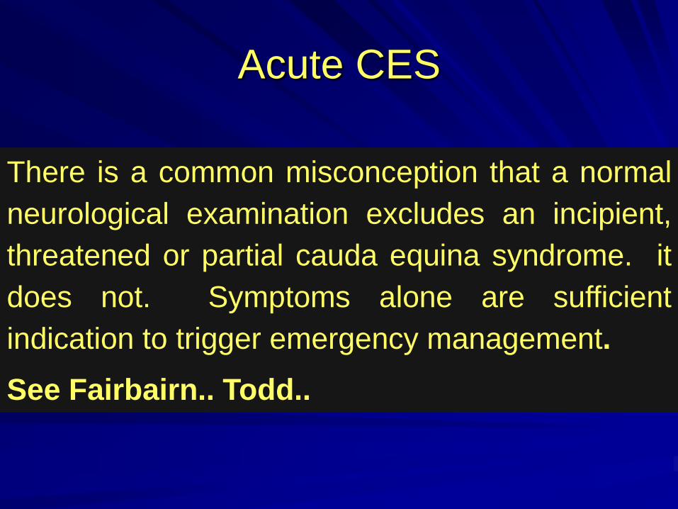
Acute CES
There is a common misconception that a normal
neurological examination excludes an incipient,
threatened or partial cauda equina syndrome. it
does not. Symptoms alone are sufficient
indication to trigger emergency management.
See Fairbairn.. Todd..
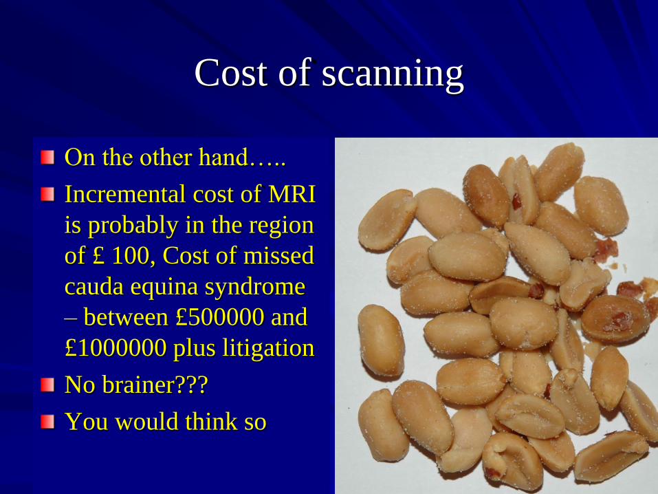
Cost of scanning
On the other hand…..
Incremental cost of MRI
is probably in the region
of £ 100, Cost of missed
cauda equina syndrome
– between £500000 and
£1000000 plus litigation
No brainer???
You would think so

Massive L4/5 disc
prolapse.
No CSF visible around
cauda equina
Surgery mandatory
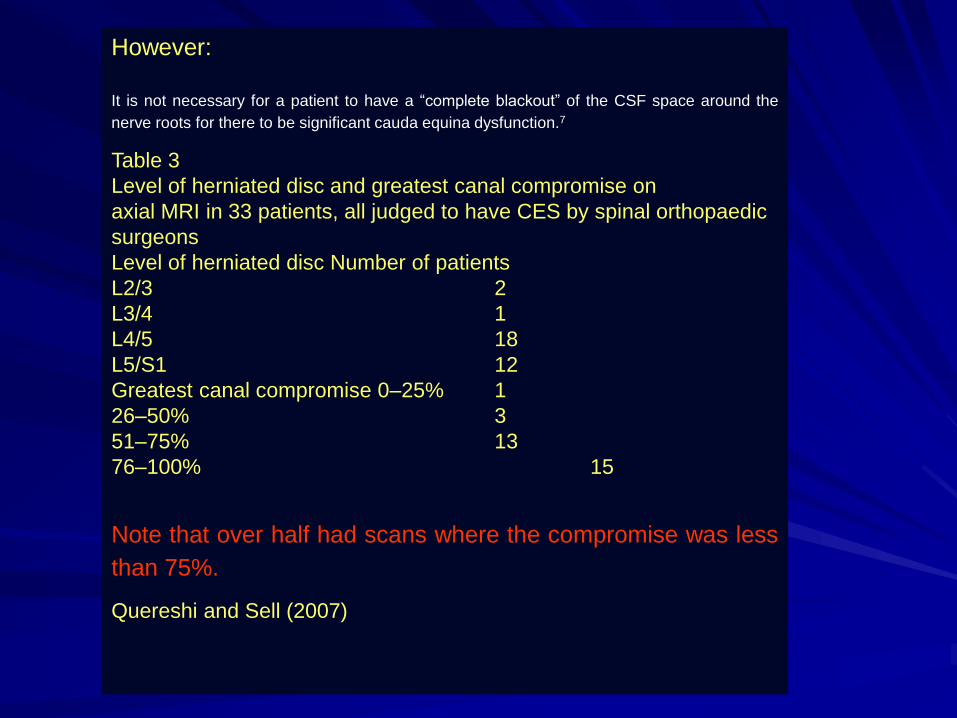
However:
It is not necessary for a patient to have a “complete blackout” of the CSF space around the
nerve roots for there to be significant cauda equina dysfunction.7
Table 3
Level of herniated disc and greatest canal compromise on
axial MRI in 33 patients, all judged to have CES by spinal orthopaedic
surgeons
Level of herniated disc Number of patients
L2/3 2
L3/4 1
L4/5 18
L5/S1 12
Greatest canal compromise 0–25% 1
26–50% 3
51–75% 13
76–100% 15
Note that over half had scans where the compromise was less
than 75%.
Quereshi and Sell (2007)
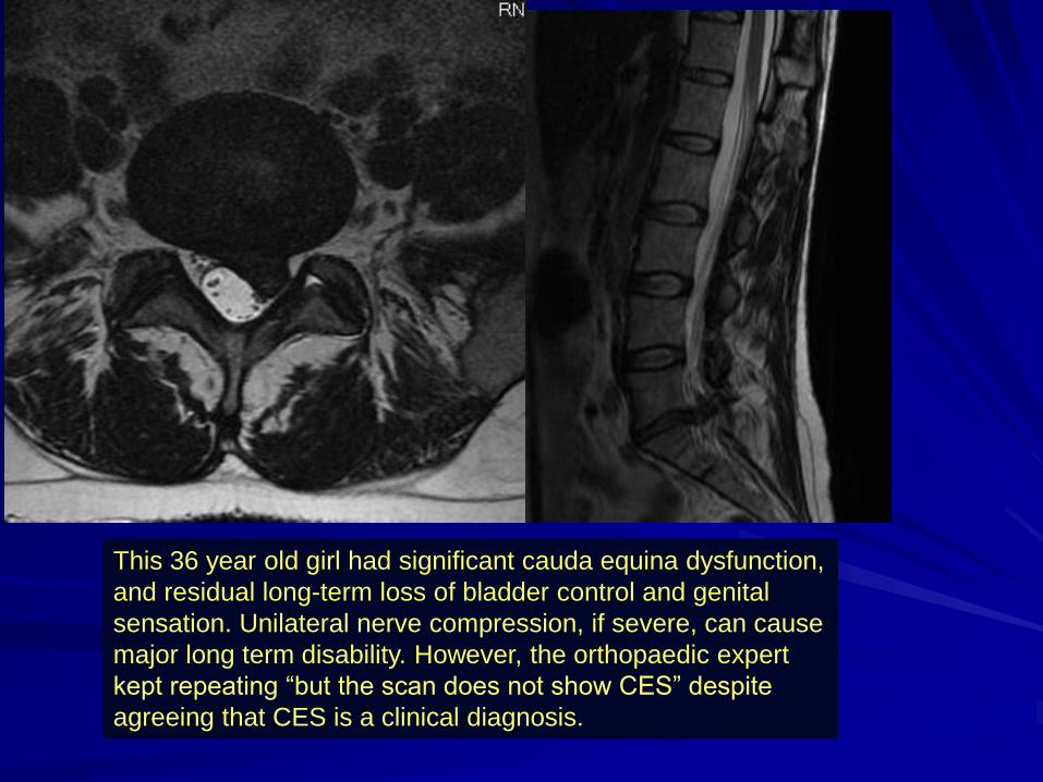
This 36 year old girl had significant cauda equina dysfunction,
and residual long-term loss of bladder control and genital
sensation. Unilateral nerve compression, if severe, can cause
major long term disability. However, the orthopaedic expert
kept repeating “but the scan does not show CES” despite
agreeing that CES is a clinical diagnosis.

23 year old man with bilateral sciatica – delays and intra-tumoural haemorrhage
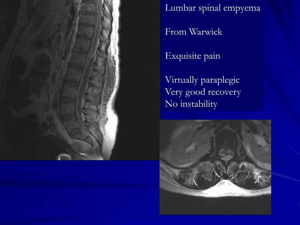
Lumbar spinal empyema
From Warwick
Exquisite pain
Virtually paraplegic
Very good recovery
No instability
Triage scheme
– Spinal pain (SP) alone – GP unless red flag
– SP + Tingling – OPD
– SP + T + N needs scan soon
– SP + T + N +W – take over, urgent scan
– SP + cauda equina features – admit, treat
immediately
– Record exactly what you need in the way
of scan, theatre and urgency
Acute CES

Cauda equina syndrome “6 Ss”
my guidance to my juniors
Starve
Scan
Steroids - possibly
Summon a surgeon
Surgery
Six hours
This way YOU keep out of Court
If no theatre, anaesthetist, ODP ..Management problem
Cf. recent spinal cord injury case…
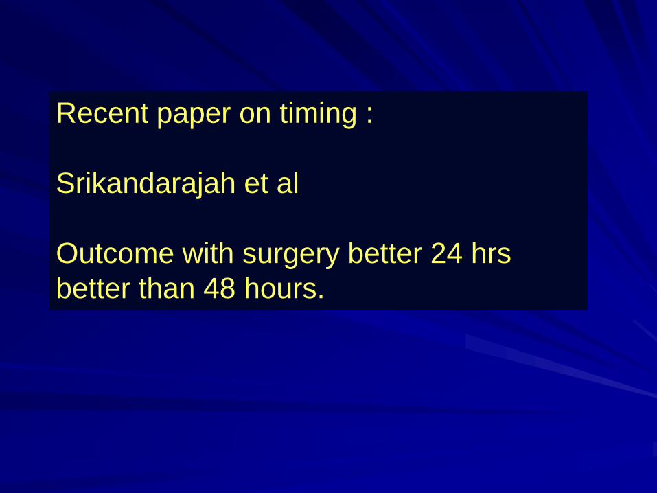
Recent paper on timing :
Srikandarajah et al
Outcome with surgery better 24 hrs
better than 48 hours.
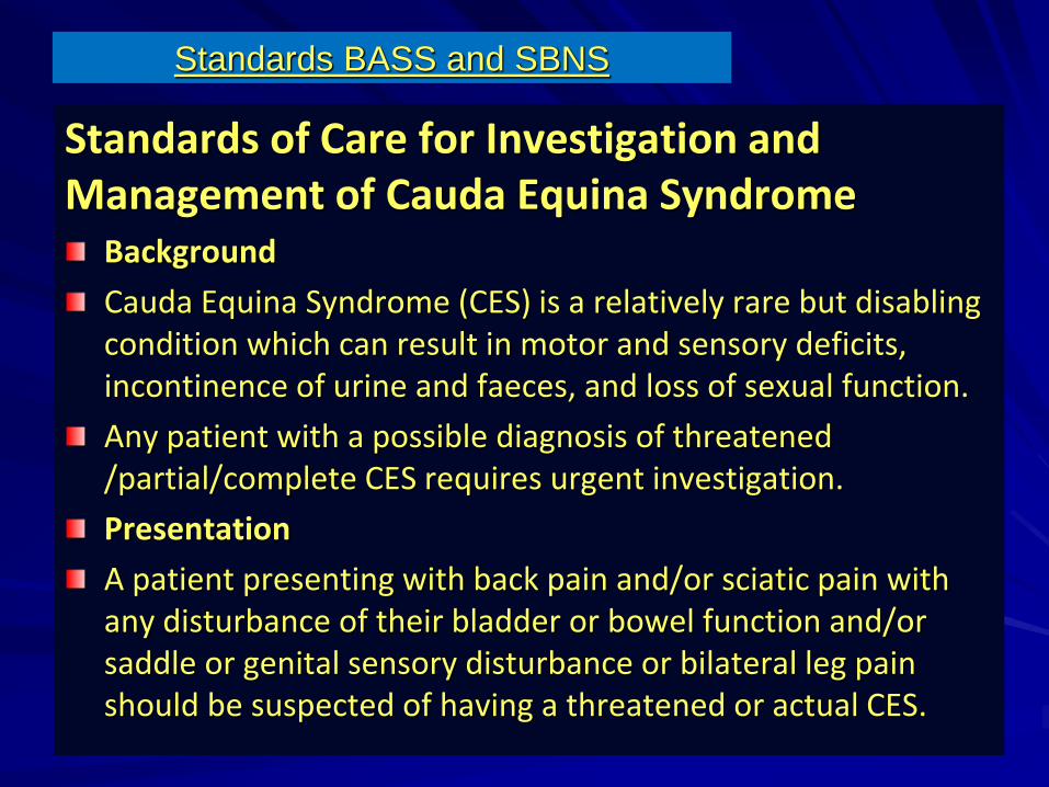
Standards BASS and SBNS
Standards of Care for Investigation and Management of Cauda Equina Syndrome
Background
Cauda Equina Syndrome (CES) is a relatively rare but disabling condition which can result in motor and sensory deficits, incontinence of urine and faeces, and loss of sexual function.
Any patient with a possible diagnosis of threatened /partial/complete CES requires urgent investigation.
Presentation
A patient presenting with back pain and/or sciatic pain with any disturbance of their bladder or bowel function and/or saddle or genital sensory disturbance or bilateral leg pain should be suspected of having a threatened or actual CES.
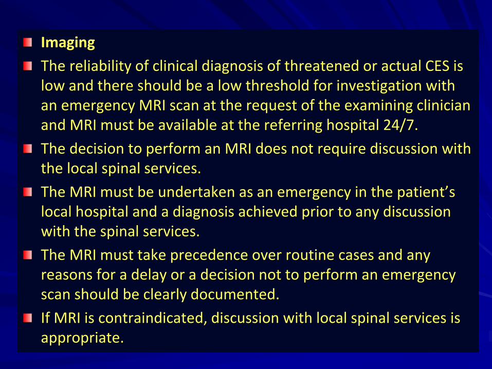
Imaging
The reliability of clinical diagnosis of threatened or actual CES is low and there should be a low threshold for investigation with an emergency MRI scan at the request of the examining clinician and MRI must be available at the referring hospital 24/7.
The decision to perform an MRI does not require discussion with the local spinal services.
The MRI must be undertaken as an emergency in the patient’s local hospital and a diagnosis achieved prior to any discussion with the spinal services.
The MRI must take precedence over routine cases and any reasons for a delay or a decision not to perform an emergency scan should be clearly documented.
If MRI is contraindicated, discussion with local spinal services is appropriate.
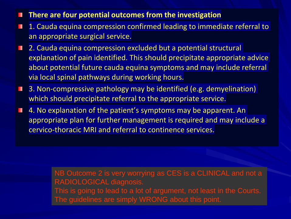
There are four potential outcomes from the investigation
1. Cauda equina compression confirmed leading to immediate referral to an appropriate surgical service.
2. Cauda equina compression excluded but a potential structural explanation of pain identified. This should precipitate appropriate advice about potential future cauda equina symptoms and may include referral via local spinal pathways during working hours.
3. Non-compressive pathology may be identified (e.g. demyelination) which should precipitate referral to the appropriate service.
4. No explanation of the patient’s symptoms may be apparent. An appropriate plan for further management is required and may include a cervico-thoracic MRI and referral to continence services.
NB Outcome 2 is very worrying as CES is a CLINICAL and not a
RADIOLOGICAL diagnosis.
This is going to lead to a lot of argument, not least in the Courts.
The guidelines are simply WRONG about this point.
Surgery
Nothing is to be gained by delaying surgery and should be undertaken at the earliest opportunity, considering the duration and clinical course of symptoms and signs, and the potential for increased morbidity while operating in the night. We do not consider that there is anything in the literature that justifies contravention of this principle and reasons for any delay in surgery should be clearly documented.
Ambiguous? Do we expect surgery at night or
not? Why delay at all?
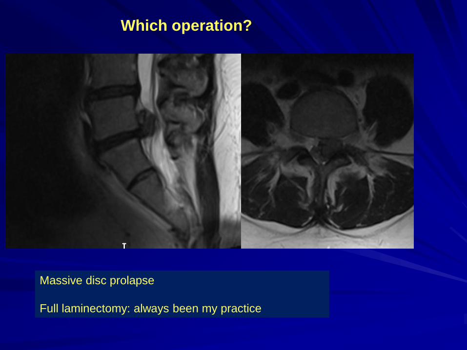
Which operation?
Massive disc prolapse
Full laminectomy: always been my practice
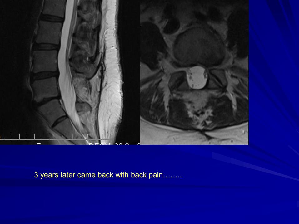
3 years later came back with back pain……..
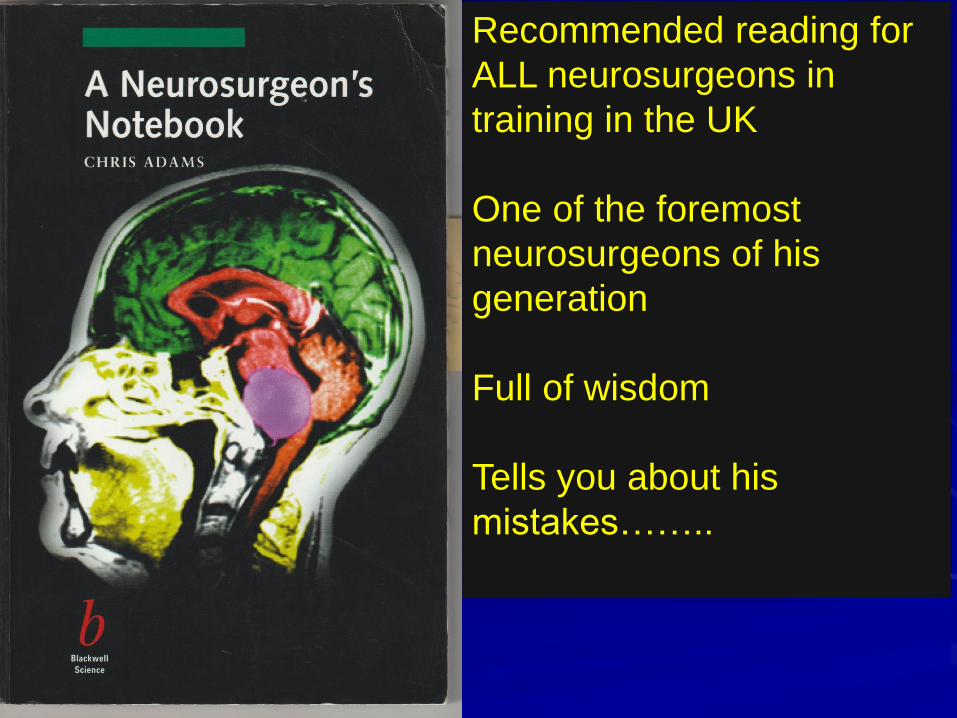
Recommended reading for
ALL neurosurgeons in
training in the UK
One of the foremost
neurosurgeons of his
generation
Full of wisdom
Tells you about his
mistakes……..

From Adams: A neurosurgeons Notebook

“There is no place in the management of this
condition for the micro discectomy approach that is
routine for the radicular pain syndrome as sciatica
due to disc herniation. The traditional mid line
approach with bilateral exposure of laminae and
complete laminectomy should be performed to
immediately decompress the cauda equina prior to
manipulation the thecal sac in an attempt to
remove the disc fragments.”(H Bridwell et al., 1997
page 1558)
There is a modern tendency to disregard these
warnings, and to carry out surgery through a small
exposure. This is a very poor approach – and in my
view negligent: particularly if there is a dural tear.

Outcome.
This is a devastating condition.
Neuropathic pain more likely – lecture in itself
Dreadful effect from impotence; anhedonia/loss of all sexual function
Incontinence of urine, flatus and faeces
Employment difficult/ impossible
Degree of recovery at 2 years determined by:
Deficit at the time of surgery: what goes into theatre should come out of
theatre
Evidence of subsequent recovery.
Material vs absolute contribution to poor result.
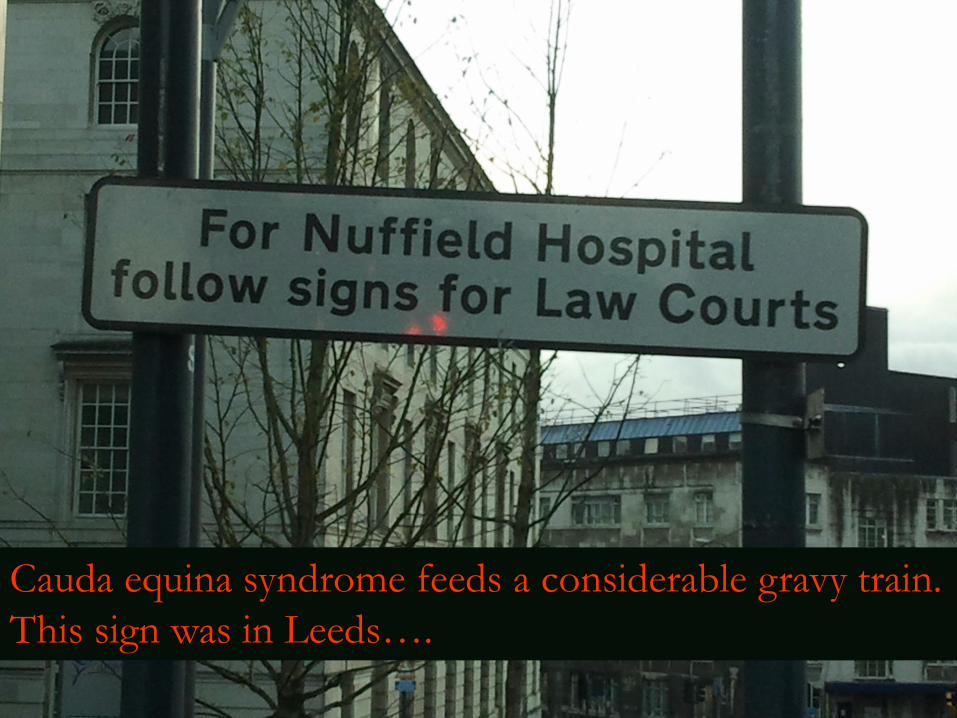
Cauda equina syndrome feeds a considerable gravy train.
This sign was in Leeds….

Question Time?
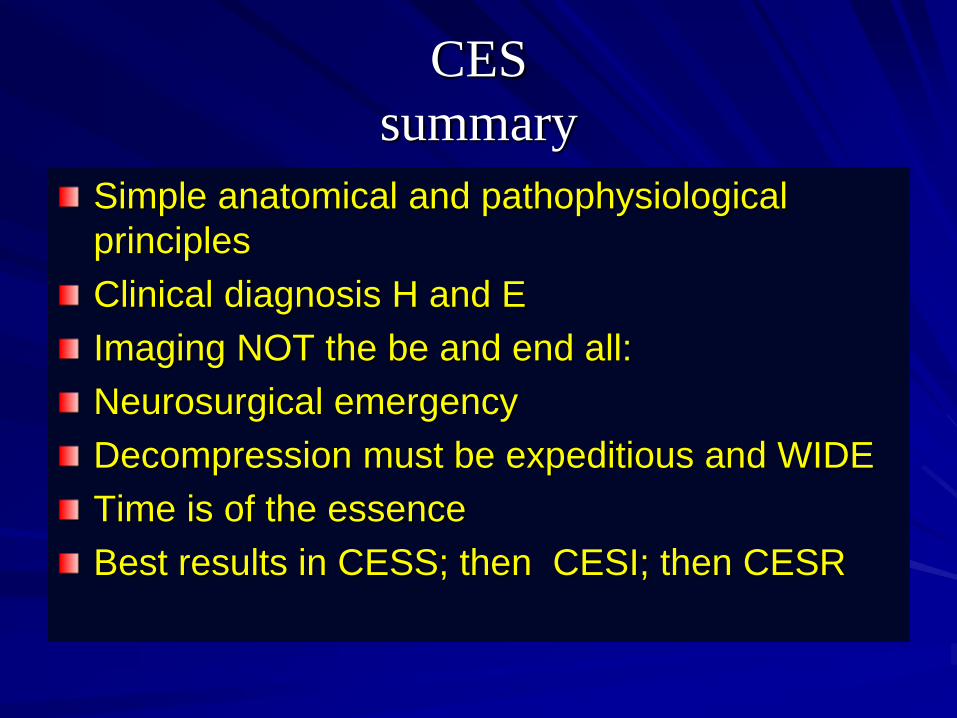
CES
summary
Simple anatomical and pathophysiological
principles
Clinical diagnosis H and E
Imaging NOT the be and end all:
Neurosurgical emergency
Decompression must be expeditious and WIDE
Time is of the essence
Best results in CESS; then CESI; then CESR