Embed Size (px)
Citation preview

Cellular/Molecular
Localization of the Endocannabinoid-Degrading EnzymeFatty Acid Amide Hydrolase in Rat Dorsal Root GanglionCells and Its Regulation after Peripheral Nerve Injury
Isobel J. Lever,1 Michelle Robinson,3 Mario Cibelli,2 Cleoper Paule,1 Peter Santha,1 Louis Yee,1 Stephen P. Hunt,3
Benjamin F. Cravatt,4 Maurice R. Elphick,5 Istvan Nagy,1 and Andrew S. C. Rice1
1Pain Research Group and 2Department of Anaesthetics, Pain Medicine and Intensive Care, Faculty of Medicine, Imperial College London, Chelsea andWestminster Hospital Campus, London SW10 9NH, United Kingdom, 3Department of Anatomy and Developmental Biology, University College London,London WC1E 6BT, United Kingdom, 4The Skaggs Institute for Chemical Biology and Departments of Cell Biology and Chemistry, The Scripps ResearchInstitute, La Jolla, California 92037, and 5School of Biological and Chemical Sciences, Queen Mary, University of London, London E1 4NS, United Kingdom
Fatty acid amide hydrolase (FAAH) is a degradative enzyme for a group of endogenous signaling lipids that includes anandamide (AEA).AEA acts as an endocannabinoid and an endovanilloid by activating cannabinoid and vanilloid type 1 transient receptor potential(TRPV1) receptors, respectively, on dorsal root ganglion (DRG) sensory neurons. Inhibition of FAAH activity increases AEA concentra-tions in nervous tissue and reduces sensory hypersensitivity in animal pain models. Using immunohistochemistry, Western blotting, andreverse transcription-PCR, we demonstrate the location of the FAAH in adult rat DRG, sciatic nerve, and spinal cord. In naive rats, FAAHimmunoreactivity localized to the soma of 32.7 � 0.8% of neurons in L4 and L5 DRG. These were small-sized (mean soma area, 395.96 �5.6 �m 2) and predominantly colabeled with peripherin and isolectin B4 markers of unmyelinated C-fiber neurons; 68% colabeled withantibodies to TRPV1 (marker of nociceptive DRG neurons), and �2% colabeled with NF200 (marker of large myelinated neurons).FAAH-IR was also present in small, NF200-negative cultured rat DRG neurons. Incubation of these cultures with the FAAH inhibitorURB597 increased AEA-evoked cobalt uptake in a capsazepine-sensitive manner. After sciatic nerve axotomy, there was a rightward shiftin the cell-size distribution of FAAH-immunoreactive (IR) DRG neurons ipsilateral to injury: FAAH immunoreactivity was detected inlarger-sized cells that colabeled with NF200. An ipsilateral versus contralateral increase in both the size and proportion of FAAH-IR DRGoccurred after spinal nerve transection injury but not after chronic inflammation of the rat hindpaw 2 d after injection of completeFreund’s adjuvant. This study reveals the location of FAAH in neural tissue involved in peripheral nociceptive transmission.
IntroductionThe endogenous ligands for cannabinoid receptors (endocan-nabinoids) are rapidly synthesized inside activated neurons (DiMarzo et al., 1994; Piomelli et al., 1998). Tissue concentrations ofendocannabinoids increase locally after injury (Calignano et al.,1998; Baker et al., 2001; Dinis et al., 2004) or ischemia (Muthianet al., 2004) and are elevated in paw skin and in the periaqueduc-tal gray (PAG) brain region after Formalin injury, providing ev-idence of activity-dependent endocannabinoid production innociceptive pathways (Calignano et al., 1998; Walker et al., 1999).
When injected into rat skin after inflammation or nerve in-jury, the endocannabinoid anandamide (AEA) mediates analge-sic effects via the activation of peripheral cannabinoid receptors(CBRs) (Calignano et al., 1998; Richardson et al., 1998; Guindonand Beaulieu, 2006). AEA signaling at CBRs is relatively transient
because of the short half-life of AEA in tissue (Di Marzo et al.,1994). AEA is inactivated by cellular reuptake, followed by hy-drolysis or oxidative degradation (Deutsch and Chin 1993; Kozakand Marnett, 2002). Hydrolysis is catalyzed by a membrane-bound serine hydrolase enzyme, fatty acid amide hydrolase(FAAH) (Cravatt et al., 1996; Deutsch et al., 2002).
FAAH activity regulates AEA levels in tissue because inhibi-tion of the enzyme or disruption of the faah gene in knock-outmice (FAAH�/�), both cause substantial increases in AEA con-centrations measured in rodent brain and spinal cord (Cravatt etal., 2001; Lichtman et al., 2004a; Hohmann et al., 2005). FAAHinactivation can reduce pain-related behavior by increasing AEAsignaling at CBRs (Cravatt et al., 2001, 2004), reducing hyperre-flexic responses after inflammatory (Lichtman et al., 2004b; Holtet al., 2005; Jayamanne et al., 2006) or neuropathic injury (Changet al., 2006; Jhaveri et al., 2006; Russo et al., 2007), and enhancingstress-induced analgesia (Hohmann et al., 2005).
Extracellular AEA can activate CBRs in vitro, inhibiting theactivity of small, C-fiber dorsal root ganglion (DRG) neurons ofthe type that transmit nociceptive input to the CNS (Richardsonet al., 1998; Ross et al., 2001; Ahluwalia et al., 2003a). Theseneurons express vanilloid type 1 transient receptor potential
Received Aug. 26, 2008; revised Nov. 4, 2008; accepted Jan. 14, 2009.This work was supported by the Wellcome Trust (London Pain Consortium).Correspondence should be addressed to Prof. Andrew S. C. Rice, Pain Research Group, Department of Anaesthet-
ics, Pain Medicine and Intensive Care, Imperial College London, Chelsea and Westminster Hospital Campus, 369Fulham Road, London SW10 9NH, UK. E-mail: [email protected].
DOI:10.1523/JNEUROSCI.4071-08.2009Copyright © 2009 Society for Neuroscience 0270-6474/09/293766-15$15.00/0
3766 • The Journal of Neuroscience, March 25, 2009 • 29(12):3766 –3780
(TRPV1) receptors that are activated by capsaicin and noxiousheat and are critical to the development of inflammatory thermalhyperalgesia (Caterina et al., 1997, 2000). AEA production hasbeen measured in capsaicin-sensitive DRG neurons and can ex-cite DRG neurons intracellularly by activation of TRPV1 recep-tors (Ahluwalia et al., 2003a; van der Stelt et al., 2005).
Neurons that produce AEA are also likely to contain FAAH(van der Stelt et al., 2005; Millns et al., 2006). In rodent CNS, thisstatus is supported by the proximity of FAAH-immunoreactive(IR) neurons to AEA-responsive CBRs (Egertova et al., 1998,2003; Gulyas et al., 2004). FAAH expression is reported in areasconcerned with nociceptive transmission (PAG, thalamus, andthe spinal cord) (Tsou et al., 1998). However, nothing is knownabout the cellular distribution of FAAH in primary sensory neu-rons. This study examines the expression of FAAH immunoreac-tivity in the DRG and spinal cord and investigates changes asso-ciated with nerve injury and inflammatory states.
Materials and MethodsAdult male Wistar rats of 250 –300 g were used for all experiments, whichwere conducted according to United Kingdom Home Office regulations.
Sciatic nerve surgery. Rats were anesthetized by inhalation of 1–2%isoflurane (Abbott Laboratories) in O2 and N2O, and one of the follow-ing aseptic procedures was applied to the left sciatic nerve. For axotomy,the nerve was exposed at midthigh level, tightly ligated with a 4.0 suture,and then sectioned. For spinal nerve transection (SNT), the L5 spinalnerve was exposed, tightly ligated, and sectioned 2– 4 mm distal from thesuture. Sham surgery was performed by exposing the appropriate part ofthe nerve in the absence of a ligation and sectioning injury. The timepoints at which the tissue was harvested after nerve injury ranged from 3to 7 d after surgery. Sensory testing was conducted on SNT-injured ani-mals to determine the establishment of hypersensitivity to mechanicaland cold stimulation. Animals were placed in Plexiglas boxes with 0.8-cm-diameter mesh flooring and allowed to acclimatize for 15 min oruntil exploratory behavior ceased. For testing mechanical hypersensitiv-ity, an electronic von Frey device (Moller et al., 1998), with a probe tiparea of 0.5 mm 2 (Somedic Sales), was applied manually to the midplantarsurface of the hindpaw at a rate of 8 –15 g/s. The paw-withdrawal thresh-old was defined as the average force in grams that evoked an activelimb-withdrawal response over five applications; at least 3 min elapsedbetween each test. Withdrawal to a cold stimulus was assessed using theacetone drop application technique modified from the work of Carlton etal. (1994). Sampling was performed by the application of a single bubbleof acetone to the midplantar surface of each hindpaw from the tip of a 1ml syringe. A positive response was recorded if the animal withdrew thepaw after acetone application. The paw was sampled five times, and amean was calculated. Mean � SEM paw-withdrawal responses for me-chanical and cooling stimuli, measured ipsilateral and contralateral toinjury, were calculated for animals in each group on each testing daybefore and after surgery. Statistical comparisons were made betweenwithdrawal responses (on different testing days or between different an-imal groups on the same testing day) using a one-way ANOVA, followedby the appropriate post hoc multiple comparison procedure. This waseither a Tukey’s or Dunn’s test, or a Kruskal–Wallis one-way ANOVA onranks using the Student–Newman–Keuls method.
Complete Freund’s adjuvant-induced inflammation. Chronic inflam-mation of left rat hindpaws was induced by a 50 �l injection of 50%complete Freund’s adjuvant (CFA) (Sigma-Aldrich). As described previ-ously, the development of thermal hypersensitivity was tested on theipsilateral and contralateral side to CFA inflammation, 2 d after injectionusing the plantar test (Hargreaves et al., 1988). Animals were placed inPlexiglas boxes, and, after the acclimatization period, timed paw-withdrawal response latencies were measured in response to an infraredbeam (thermal stimulus) (Ugo Basile) positioned under the plantar sur-face of the paw. The mean � SEM of three withdrawal responses wascalculated per paw, and the data were analyzed as described for responsesto mechanical and cold stimulation.
Immunohistochemistry. Rats were anesthetized with sodium pentobar-bital (60 mg/kg) and perfused through the ascending aorta with 100 ml of0.9% saline and then 300 ml of 4% paraformaldehyde in 0.1 M phosphatebuffer (PB), pH 7.4. Lumbar (L4 and L5) DRG were located by tracing thelumbar dorsal roots back to the sciatic nerve. Dissected tissue was post-fixed for 1–2 h at 4°C, cryoprotected in 15–30% sucrose in 0.1 M PB for12 h at 4°C, and embedded in mounting medium. Cryosections were cutand thaw mounted onto Superfrost slides (VWR) at the following thick-nesses: coronal brain, 30 �m; transverse spinal cord, 20 �m; sciatic nerve,10 �m; and DRG, 10 �m. Fresh-frozen tissue from FAAH�/� orFAAH�/� mice (Ledent et al., 1999) (obtained from a colony maintainedby Prof. David Baker, Institute of Neurology, UCL, London, UK) werecryosectioned and fixed in acetone. For paraffin sections, postfixed tissuewas embedded in paraffin wax and cut at 5 �m thickness on a vibraslice.Dried and dewaxed sections were processed identically for immunohis-tochemistry. Slides were incubated with PBST (0.1 M phosphate buffer,0.1% sodium azide, 0.3% Triton X-100, and 0.3 M NaCl) containing 10%normal donkey serum for 1 h at room temperature. A rabbit primaryantibody raised to a truncated form of rat FAAH protein, �TM FAAHcomprising amino acids 38 –579 as described previously (Patricelli et al.,1998; Cravatt et al., 2001) was diluted 1:2000 in PBST containing one ofthe following costaining primary antibodies: mouse anti neuronal-specific nuclear protein (NeuN) at 1:2000 (Millipore Bioscience ResearchReagents), sheep anti-calcitonin gene-related peptide (CGRP) at 1:2000(Affinity BioReagents), mouse anti-NF200 (N52) at 1:1 to 1:2000(Sigma-Aldrich), or goat anti-peripherin at 1:100 (Santa Cruz Biotech-nology). Slides were incubated with antibody mixtures for 48 h at roomtemperature and processed for signal amplification using avidin– biotin(Vector Laboratories) and tyramide reagents [tyramide signal amplifica-tion (TSA)] (PerkinElmer Life and Analytical Sciences). This was fol-lowed by a 2 h incubation with fluorescent 5-(4,6-dichlorotriazinyl)-aminofluorescein or rhodamine-conjugated streptavidin antibodies(1:200; The Jackson Laboratory) together with an appropriatefluorophore-conjugated secondary antibody raised against the costain-ing primary antibody. Fluorescein-labeled Griffonia simplicifolia isolec-tin B4 (IB4) (Sigma-Aldrich) was incubated at 10 �g/ml for 1 h duringthe final incubation step for FAAH staining. Slides were mounted inVectashield medium containing a 4�,6�-diamidino-2-phenylindole(DAPI) nuclear counterstain (Vector Laboratories). Costaining of FAAHwith other rabbit antibodies [anti-TRPV1 at 1:1000 (Affinity BioRe-agents), anti-S100 at 1:1200 (Dako), and anti-activating transcriptionfactor 3 (ATF3) (Santa Cruz Biotechnology)] was performed subsequentto the TSA procedure for the FAAH antibody, as described previously(Michael et al., 1997). Costaining of spinal cord tissue with type 1 can-nabinoid receptor (CB1) antibodies was performed with TSA, followedby incubation of FAAH antibodies at 1:200. The rabbit CB1 antibodyused was raised to amino acids 461– 473 of the C-terminal domain of therat CB1 receptor [affinity purified from antiserum 2824.4(2) (Egertovaand Elphick, 2000)] and incubated at 1:10 for 64 h at room temperature.Controls for FAAH immunohistochemistry included the following: (1)omission of the primary antibody, (2) preabsorption of the FAAH anti-body at 1:1000 with the immunizing peptide at 10 �g/ml for 1 h at roomtemperature followed by tissue incubation with the preabsorbed anti-body, (3) testing the antibody on tissue from FAAH�/� mice (Ledent etal., 1999), and (4) use of an alternative rabbit polyclonal antibody raisedagainst a 17 aa immunogen from the N terminus of human FAAH pro-tein (immunogen 100% conserved in rat) (Millipore Bioscience ResearchReagents).
Image analysis and quantification of immunofluorescent DRG cells. Im-munofluorescent images were visualized using a Leica DMR Fluores-cence microscope and captured on a Hamamatsu CCD camera usingQWIN image processing software (Leica). Quantification of the imagesfrom immunopositive DRG cells was semiautomated using software-based measurement of fluorescence intensity. The standard thresholdgrayscale intensity for detection of FAAH-immunoreactive cells was setat 130 –255 arbitrary units (a.u.) and applied uniformly to all DRG im-ages captured under identical illumination and exposure conditions. Atleast 100 DRG cells were randomly sampled per animal from serial sec-tions at a distance of �10 sections apart from each other. Outlines of
Lever et al. • FAAH Expression in DRG J. Neurosci., March 25, 2009 • 29(12):3766 –3780 • 3767

DRG cell profiles containing a DAPI- or NeuN-labeled nucleus weredrawn over each image to produce an overlay of the nucleated cell pro-files. Exclusion of non-nucleated, immunopositive cell profiles from thesize analysis was designed to prevent inaccurate measurements ofelliptical-shaped DRG neuronal soma measured in cross section (Po-trebic et al., 2003). A version of the nucleated cell profile overlay wasmodified using threshold intensity measurements to represent the pop-ulation of immunopositive nucleated profiles. Both overlays were used tocount and make area and fluorescence intensity measurements of allnucleated DRG cell profiles, as well as immunopositive cells within thisgroup. Analysis of DRG images from injured and uninjured rats wasperformed by an investigator who was blinded to the treatment groups.Measurements were expressed as mean � SEM for DRG sections sam-pled from each animal and for animals in each experimental group. Thepercentage of NeuN-IR DRG neurons colabeled with another markerwere calculated for each animal and expressed as a mean � SEM perexperimental group. Statistical comparisons were made between differ-ent experimental groups using Student’s t tests or one-way ANOVAs,together with the appropriate post hoc multiple comparison procedure.
Western blotting. Frozen tissue samples were thawed to 4°C and ho-mogenized in lysis buffer: 50 mM Tris, pH 8.0, 100 mM NaCl, 10% glyc-erol, 1% Triton X-100, 5 mM EDTA, 1 mM PMSF, 10 �g/ml antipain, 10�g/ml leupeptin, 1 mM sodium vanadate, 1 �g/ml pepstatin A (all fromSigma-Aldrich). Lysates were rotated for 1 h at 4°C and then centrifuged(10 min at 12,000 � g). Supernatants were collected and the total proteinconcentration determined using a BCA kit (Pierce via Perbio Science).Samples (normalized for protein concentration) were denatured in theappropriate PAGE loading buffer at 95°C for 5 min (or 70°C for 10 minfor Bis-Tris gels). Protein extracts were separated using one of two elec-trophoresis systems: 10% Tris-HCl polyacrylamide gels (Bio-Rad) orNuPAGE Novex 4 –12% Bis-Tris gels (Invitrogen) before transfer to ni-trocellulose membranes. After blocking in a 6% solution of driedskimmed milk in TBST (Tris-HCl-buffered saline at pH 8.0 containing0.05% Tween 20), blots were probed with �TM FAAH primary antibody(1:200 in block) at 4°C overnight. After detection using peroxidase-conjugated secondary antibody and chemiluminescence, membraneswere stripped and reprobed with anti-glyceraldehyde-3-phosphate dehy-drogenase (GAPDH) antibody (1:5000 in TBST at 4°C overnight; Ab-cam). Densitometric analysis of gel bands was performed using QWINimage analysis software (Leica). Briefly, images of the bands were cap-tured under standard light conditions, and the optical densities of pixelssampled from images of each gel band were measured using a standard-sized area selection tool. Average pixel measurements derived from threeseparate gels were compared between different experimental groups us-ing a one-way ANOVA and post hoc Tukey’s test.
Reverse transcriptase-PCR. Reverse transcriptase (RT)-PCR was per-formed using samples from two L4 DRG per rat, 30 mg of lumbarspinal cord or 30 mg of hippocampal tissue. RNA was extracted ac-cording to the method of Akiba et al. (2004) using an RNeasy mini kit(Qiagen). Using a mastercycler (Eppendorf), reverse transcription pro-ceeded at 42°C for 50 min, followed by a 15 min inactivation step at 70°C.A duplicate set of RNA samples (with the transcriptase enzyme omitted)was included in this reaction as a control for genomic DNA contamina-tion in the PCR step. Both sample sets were run simultaneously for PCRamplification. PCR primers were designed across FAAH exon bound-aries to prevent the amplification of genomic DNA (forward, 5�-GCCCTT CAG AGA GCA GCT CT-3� designed across exons 7 and 8, inter-vening intron 1159 bp; reverse, 5�-CTT TTC AGC TGA CCG AGGAC-3� across exons 11 and 12, intervening intron 265 bp). Databaseanalysis with a basic local alignment search tool BLASTN search of Gen-Bank was performed to assess and rule out potential hybridization withrat cDNA sequences other than FAAH. After 30 s denaturation at 96°C,the PCR reaction proceeded for 30 cycles of 1 min at 58°C for annealingand 3 min at 72°C for extension. PCR products were run on 2% agarosegels using standard protocols. The predicted size of the amplicon was 394bp.
Dissociated DRG culture. DRG neurons were isolated from adultSprague Dawley rats according to previously described methods (SinghTahim et al., 2005). Briefly, coverslips bearing cultured cells were main-
tained at 37°C for �2 d. After three washes in PBS solution at 37°C,immunohistochemistry procedures were then performed at room tem-perature. Coverslips were fixed in 4% paraformaldehyde for 30 min,followed by PBS washes, incubation with PBST containing 10% normaldonkey serum for 30 min, and then incubation with �TM FAAH primaryantibody at 1:400 for 12 h. Costaining primary antibodies were eitherNeuN at 1:1000 or NF200 at 1:10,000. Secondary antibodies raisedagainst rabbit and mouse IgG conjugated to cyanine 3 or FITC fluoro-phores (The Jackson Laboratory) were incubated for 1 h at 1:300 in PBSTcontaining 4% normal donkey serum. Coverslips were slide mountedusing Vectashield.
Cobalt uptake. TRPV1-mediated activation of cultured DRG neuronswas assessed by cobalt uptake, as described previously (Sathianathan etal., 2003). Coverslips were washed for 2 min in buffer solution (in mM:57.5 NaCl, 5 KCl, 2 MgCl2, 10 HEPES, 12 glucose, and 139 sucrose, pH7.4) and then preincubated for 5 min in buffer containing one of thefollowing: the FAAH enzyme inhibitor URB597 (cyclohexylcarbamicacid 3-carbamoyl biphenyl-3-yl ester) (Mor et al., 2004; Fegley et al.,2005); buffer solution only (control); SR141716 [N-(piperidin-1-yl)-5-(4-chlorophenyl)-1-(2,4-dichlorophenyl)-4-methyl-3-pyrazole carbox-amide] (CB1 antagonist); URB597 � SR141716; or URB597 and theTRPV1 antagonist capsazepine (Table 1). Cells were then incubated inone of the buffer solutions containing 5 mM CoCl2 (cobalt-uptake solu-tion) as listed in Table 1.
Cobalt taken up by the neurons through activated nonselective cat-ionic channels (Nagy et al., 1993) was precipitated by adding a 2.5%mercaptoethanol solution in buffer for 1 min. Cells were fixed in 70%ethanol. The cobalt precipitate (Co-ppt) was visualized by light micros-copy. After background subtraction, images of labeled and nonlabeledcells were captured in grayscale, and their average optical intensity wasmeasured within defined cell areas using QWIN image analysis software.Frequency distributions of optical intensity data (representing Co-pptlevels in �100 cells after each active or vehicle treatment) were fitted toGaussian curves to determine the intensity threshold for Co-ppt-positivecells, as described previously (Sathianathan et al., 2003). Negative andpositive control experiments were performed for each culture, in whichcontrol buffer or 10 �M capsaicin was added to the cobalt-containingbuffer, respectively. Optical intensities from cells in the two types ofexperiment were fitted to Gaussian curves. The negative experimentsrepresented two cell subpopulations, whereas three separate subpopula-tions were defined in the positive experiment. Mean optical intensityvalues for cells in the third subpopulation were higher than those of thetwo other subpopulations in both experiments and represented labeledcells. The cutoff point between the labeled and nonlabeled cells was de-termined as the mean value minus the 95% confidence interval for thethird Gaussian curve in positive experiments. This optical intensity valuewas used to establish the relative number of labeled cells in all coverslipsfrom the same culture. Data were presented as mean � SEM. Differencesin the relative number of labeled cells produced by various treatments
Table 1. Details of the preincubation and cobalt uptake buffers used in cobaltuptake experiments
Preincubation buffer solutions Cobalt uptake buffer solutions
Reagent Concentration (�M) Reagent Concentration (�M)
URB597 (FAAH inhibitor) 1 Capsaicin 1SR141716 1 AEA 1URB597 � 1 URB597 � 1
SR1 1 AEA 1URB597 � 1 URB597 � 1
Capsazepine 10 CAPZ 10(TRPV1 antagonist) SR141716 1
Buffer (control) SR141716 � 1AEA 1
SR141716 � 1URB597 1
A list of reagents and their concentrations when added to preincubation buffer solution or cobalt uptake buffersolution. Cultured DRG cells mounted on coverslips were transferred to one of the preincubation solutions for 5 minbefore transfer into a cobalt uptake buffer solution containing CoCl2 for 5 min.
3768 • J. Neurosci., March 25, 2009 • 29(12):3766 –3780 Lever et al. • FAAH Expression in DRG
were analyzed by one-way ANOVA, with significance established by theFisher’s protected least significant difference (PLSD) test. Differenceswere regarded as significant at p � 0.05.
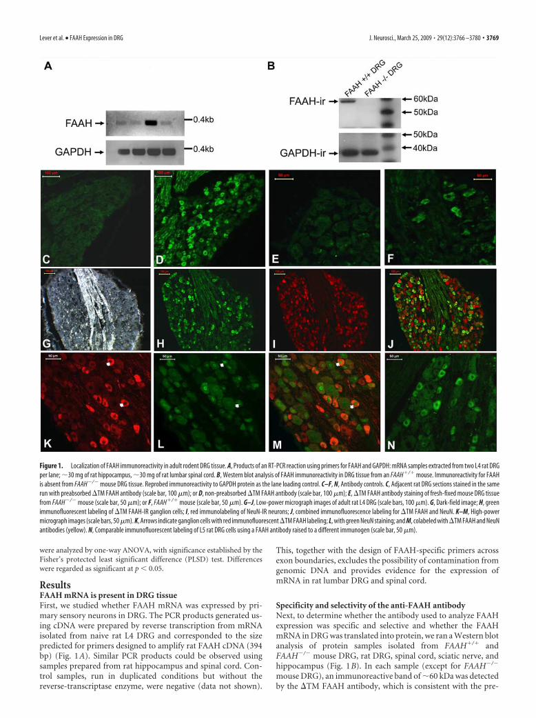
ResultsFAAH mRNA is present in DRG tissueFirst, we studied whether FAAH mRNA was expressed by pri-mary sensory neurons in DRG. The PCR products generated us-ing cDNA were prepared by reverse transcription from mRNAisolated from naive rat L4 DRG and corresponded to the sizepredicted for primers designed to amplify rat FAAH cDNA (394bp) (Fig. 1A). Similar PCR products could be observed usingsamples prepared from rat hippocampus and spinal cord. Con-trol samples, run in duplicated conditions but without thereverse-transcriptase enzyme, were negative (data not shown).
This, together with the design of FAAH-specific primers acrossexon boundaries, excludes the possibility of contamination fromgenomic DNA and provides evidence for the expression ofmRNA in rat lumbar DRG and spinal cord.
Specificity and selectivity of the anti-FAAH antibodyNext, to determine whether the antibody used to analyze FAAHexpression was specific and selective and whether the FAAHmRNA in DRG was translated into protein, we ran a Western blotanalysis of protein samples isolated from FAAH�/� andFAAH�/� mouse DRG, rat DRG, spinal cord, sciatic nerve, andhippocampus (Fig. 1B). In each sample (except for FAAH�/�
mouse DRG), an immunoreactive band of 60 kDa was detectedby the �TM FAAH antibody, which is consistent with the pre-
Figure 1. Localization of FAAH immunoreactivity in adult rodent DRG tissue. A, Products of an RT-PCR reaction using primers for FAAH and GAPDH: mRNA samples extracted from two L4 rat DRGper lane; 30 mg of rat hippocampus, 30 mg of rat lumbar spinal cord. B, Western blot analysis of FAAH immunoreactivity in DRG tissue from an FAAH�/� mouse. Immunoreactivity for FAAHis absent from FAAH�/� mouse DRG tissue. Reprobed immunoreactivity to GAPDH protein as the lane loading control. C–F, N, Antibody controls. C, Adjacent rat DRG sections stained in the samerun with preabsorbed �TM FAAH antibody (scale bar, 100 �m); or D, non-preabsorbed �TM FAAH antibody (scale bar, 100 �m); E, �TM FAAH antibody staining of fresh-fixed mouse DRG tissuefrom FAAH�/� mouse (scale bar, 50 �m); or F, FAAH�/� mouse (scale bar, 50 �m). G–J, Low-power micrograph images of adult rat L4 DRG (scale bars, 100 �m). G, Dark-field image; H, greenimmunofluorescent labeling of �TM FAAH-IR ganglion cells; I, red immunolabeling of NeuN-IR neurons; J, combined immunofluorescence labeling for �TM FAAH and NeuN. K–M, High-powermicrograph images (scale bars, 50 �m). K, Arrows indicate ganglion cells with red immunofluorescent �TM FAAH labeling; L, with green NeuN staining; and M, colabeled with �TM FAAH and NeuNantibodies (yellow). N, Comparable immunofluorescent labeling of L5 rat DRG cells using a FAAH antibody raised to a different immunogen (scale bar, 50 �m).
Lever et al. • FAAH Expression in DRG J. Neurosci., March 25, 2009 • 29(12):3766 –3780 • 3769

dicted molecular mass of rat FAAH protein, 63 kDa (Cravatt etal., 1996), and comparable with the size of FAAH-IR proteinisolated from other neuronal tissues (Cravatt et al., 2001; Eger-tova et al., 2003). The same antibody, when used for immunohis-tochemistry, produced staining in a subpopulation of cells insections of DRG taken from FAAH�/� mice (Fig. 1F). However,the staining was absent in DRG sections from FAAH�/� mice(Fig. 1E). DRG obtained from rats also contained a subpopula-tion of FAAH-IR cells (Fig. 1G–J,K–M). Colabeling of the sec-tions with the NeuN marker of neuronal nuclei indicated thatthese FAAH-IR cells were primary sensory neurons (Fig. 1 J,M).Preabsorption of the primary antibody resulted in a lack of FAAHimmunoreactivity in rat DRG tissue (Fig. 1C) when comparedwith the staining produced using the non-preabsorbed antibodyon a serial section from the same DRG (Fig. 1D). A similar stain-ing pattern was observed in rat lumbar DRG tissue stained withan alternative FAAH antibody raised against a different area ofthe FAAH protein (Fig. 1N). These findings confirm the specific-ity of the FAAH antibody, as demonstrated previously for othertypes of neuronal tissue (Cravatt et al., 2001; Egertova et al.,2003), and suggest that FAAH mRNA is translated into protein ina subpopulation of primary sensory neurons in both mouse andrat DRG.
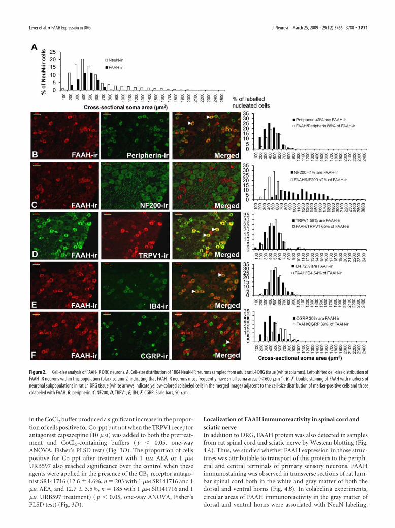
Localization of FAAH immunoreactivity in smallDRG neuronsFAAH immunoreactivity was found in the cell soma of DRGneurons colabeled with NeuN (Fig. 1 J,M). Of the 1804 NeuN-IRDRG cells captured for image analysis (n 6), 591 were FAAH-IR. On average, 32.7 � 0.8% (mean � SEM) of NeuN-labeledcells sampled from each animal were FAAH-IR. The cell-size dis-tribution of FAAH-IR and NeuN-IR neuron populations from L4DRG revealed that most nucleated FAAH-IR neurons were smallin size (Fig. 2A) compared with the total population of NeuN-IRneurons. The mean � SEM area of FAAH-IR cells was 396.0 �5.6 �m 2. This was significantly smaller than the average size ofNeuN-IR cells, 511.9 � 8.4 �m 2 ( p � 0.05, Kruskal–Wallis one-way ANOVA).
Distribution of FAAH among DRG neuron subpopulationsThe location of FAAH immunoreactivity on small DRG neuronswas confirmed by costaining studies with markers associated withsmall or large DRG cell populations (Fig. 2B–F). Consideringonly those neuronal profiles with a DAPI-stained nucleus, FAAHimmunoreactivity demonstrated a high degree of colocalizationwith peripherin (neurofilament light), a marker found predom-inantly in small DRG neurons with unmyelinated C-fiber axons(Goldstein et al., 1991). On average, 85.6 � 7.2% of FAAH-IRcells counted (n 180) costained with peripherin antibodies and48.6 � 5.7% of peripherin-labeled cells (n 334) stained withFAAH (Fig. 2B). In contrast, �2% of FAAH-IR neurons (n 131) were colabeled with the NF200 antibody (Fig. 2C) recogniz-ing the 200 kDa heavy chain neurofilament protein found inmedium- to large-sized cell bodies subtending myelinated axons(Lawson and Waddell, 1991).
The population of DRG neurons with unmyelinated C-fiberscan be further subdivided into cells that bind the lectin IB4 andthose containing the neuropeptide CGRP (Snider and McMa-hon, 1998). FAAH immunoreactivity was predominantly foundin the IB4-labeled subpopulation, as a larger proportion ofFAAH-IR DRG cells colabeled with IB4 (72.0 � 4.2%; n 131)(Fig. 2E). A smaller proportion of DRG neurons were colabeledwith FAAH and CGRP antibodies (39.4 � 13.1%; n 76), and
only 30.1 � 10.9% of CGRP-IR cells counted (n 244) hadFAAH labeling (Fig. 2F). Measurement of the average intensity ofCGRP staining in FAAH-IR soma (74.4 � 3.0 a.u.) showed thatthis was significantly lower than in CGRP-IR cells without FAAHstaining (94.6 � 2.0 a.u.; p � 0.001, t test). This indicates that apopulation of weakly CGRP stained neurons were FAAH-IR.
A large proportion of FAAH-IR neurons (64.5 � 3.2%; n 124) costained for TRPV1 (Fig. 2D), the receptor conferring cap-saicin sensitivity on small nociceptive sensory neurons (Caterinaet al., 1997). Analysis of costaining data produced using FAAHand TRPV1 antibodies indicates that approximately half ofTRPV1-labeled cells do not have FAAH staining. Given that ap-proximately half of the TRPV1-expressing cells are IB4 positive(Michael et al., 1999), and that FAAH was expressed mainly byIB4-labeled neurons, it is likely that the majority of neurons ex-pressing TRPV1 and FAAH should also be IB4 positive.
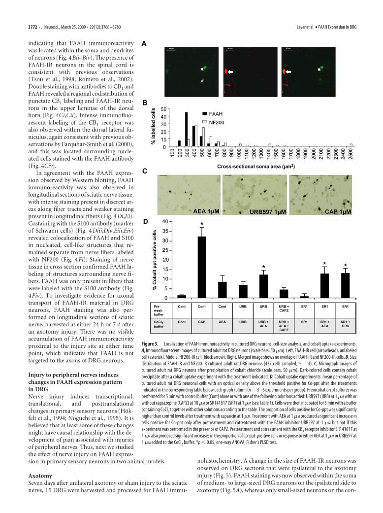
Culturing does not induce major changes in FAAH expressionin primary sensory neuronsCultured adult rat DRG cells were costained with antibodies toFAAH and NF200 (Fig. 3A). On average, 26.3 � 1.4% of n 437DRG cells were FAAH labeled. The NF200 antibody labeled18.3 � 1.5% of cells. In agreement with staining in DRG tissuesections, colabeling of FAAH with the NF200 large-cell markerwas rare in cultured DRG cells (1.5 � 1.2%). In addition, theaverage area of FAAH-IR cells (302.1 � 16.9 �m 2; n 109) wassignificantly smaller than NF200-labeled cells (650.9 � 87.6�m 2; n 68; p � 0.006, Student’s t test); cell-size distributionsare shown in Figure 3B. The distribution of FAAH immunoreac-tivity within the small cell population of cultured rat DRG neu-rons is consistent with the pattern of labeling in tissue sections ofrat DRG and with the costaining of FAAH immunoreactivity witha marker of small cells in cultured rat trigeminal neurons (Price etal., 2005). These data suggest that culturing does not induce ma-jor transcriptional or translational changes in FAAH expression.
FAAH is functional in primary sensory neuronsNext, we studied the effect the FAAH inhibitor URB597 onTRPV1-mediated responses in cultured primary sensory neuronsafter 2 d in culture. TRPV1 is a nonselective cationic channel,which, in addition to Na�, K�, and Ca 2�, is also permeable toCo 2� ions (Sathianathan et al., 2003; Singh Tahim et al.,2005). AEA is an endogenous agonist of the TRPV1 ion chan-nel (Zygmunt et al., 1999), and incubation of cultured DRGneurons with AEA induces cobalt uptake in these cells (SinghTahim et al., 2005). We assessed cobalt accumulation in cul-tured primary sensory neurons in a series of conditions, in-cluding the application of URB597, an inhibitor of FAAH thathas been shown to reduce FAAH activity in cultured rat CNStissue (Hohmann et al., 2005).
In control experiments, incubation of DRG neurons with cap-saicin (1 �M) for 5 min in the presence of CoCl2 induced Co 2�
influx in a mean � SEM of 32.1 � 5.4% of cells (n 234). Theproportion of Co-ppt-positive cells was significantly higher thanafter incubating the cells with the CoCl2-containing buffer with-out capsaicin (1.3 � 0.4%; n 256; p � 0.05, one-way ANOVA,followed by Fisher’s PLSD test) (Fig. 3D). Incubation in CoCl2buffer containing either AEA (1 �M) or the FAAH inhibitorURB597 (1 �M) also induced Co 2� influx (Fig. 3C). The propor-tion of Co-ppt-positive cells after AEA or URB597 treatment was8.3 � 3.8%, n 175 and 8.6 � 2.2%, n 189, respectively (notsignificantly different from the control). Preincubation and co-incubation of cells with URB597 (1 �M) added together with AEA
3770 • J. Neurosci., March 25, 2009 • 29(12):3766 –3780 Lever et al. • FAAH Expression in DRG
in the CoCl2 buffer produced a significant increase in the propor-tion of cells positive for Co-ppt but not when the TRPV1 receptorantagonist capsazepine (10 �M) was added to both the pretreat-ment and CoCl2-containing buffers ( p � 0.05, one-wayANOVA, Fisher’s PLSD test) (Fig. 3D). The proportion of cellspositive for Co-ppt after treatment with 1 �M AEA or 1 �M
URB597 also reached significance over the control when theseagents were applied in the presence of the CB1 receptor antago-nist SR141716 (12.6 � 4.6%, n 203 with 1 �M SR141716 and 1�M AEA, and 12.7 � 3.5%, n 185 with 1 �M SR141716 and 1�M URB597 treatment) ( p � 0.05, one-way ANOVA, Fisher’sPLSD test) (Fig. 3D).
Localization of FAAH immunoreactivity in spinal cord andsciatic nerveIn addition to DRG, FAAH protein was also detected in samplesfrom rat spinal cord and sciatic nerve by Western blotting (Fig.4A). Thus, we studied whether FAAH expression in those struc-tures was attributable to transport of this protein to the periph-eral and central terminals of primary sensory neurons. FAAHimmunostaining was observed in transverse sections of rat lum-bar spinal cord both in the white and gray matter of both thedorsal and ventral horns (Fig. 4B). In colabeling experiments,circular areas of FAAH immunoreactivity in the gray matter ofdorsal and ventral horns were associated with NeuN labeling,
Figure 2. Cell-size analysis of FAAH-IR DRG neurons. A, Cell-size distribution of 1804 NeuN-IR neurons sampled from adult rat L4 DRG tissue (white columns). Left-shifted cell-size distribution ofFAAH-IR neurons within this population (black columns) indicating that FAAH-IR neurons most frequently have small soma areas (�600 �m 2). B–F, Double staining of FAAH with markers ofneuronal subpopulations in rat L4 DRG tissue (white arrows indicate yellow-colored colabeled cells in the merged image) adjacent to the cell-size distribution of marker-positive cells and thosecolabeled with FAAH: B, peripherin; C, NF200; D, TRPV1; E, IB4; F, CGRP. Scale bars, 50 �m.
Lever et al. • FAAH Expression in DRG J. Neurosci., March 25, 2009 • 29(12):3766 –3780 • 3771
indicating that FAAH immunoreactivitywas located within the soma and dendritesof neurons (Fig. 4Bii–Biv). The presence ofFAAH-IR neurons in the spinal cord isconsistent with previous observations(Tsou et al., 1998; Romero et al., 2002).Double staining with antibodies to CB1 andFAAH revealed a regional codistribution ofpunctate CB1 labeling and FAAH-IR neu-rons in the upper laminae of the dorsalhorn (Fig. 4Ci,Cii). Intense immunofluo-rescent labeling of the CB1 receptor wasalso observed within the dorsal lateral fu-niculus, again consistent with previous ob-servations by Farquhar-Smith et al. (2000),and this was located surrounding nucle-ated cells stained with the FAAH antibody(Fig. 4Civ).
In agreement with the FAAH expres-sion observed by Western blotting, FAAHimmunoreactivity was also observed inlongitudinal sections of sciatic nerve tissue,with intense staining present in discreet ar-eas along fiber tracts and weaker stainingpresent in longitudinal fibers (Fig. 4Di,Ei).Costaining with the S100 antibody (markerof Schwann cells) (Fig. 4Diii,Div,Eiii,Eiv)revealed colocalization of FAAH and S100in nucleated, cell-like structures that re-mained separate from nerve fibers labeledwith NF200 (Fig. 4Fi). Staining of nervetissue in cross section confirmed FAAH la-beling of structures surrounding nerve fi-bers. FAAH was only present in fibers thatwere labeled with the S100 antibody (Fig.4Fiv). To investigate evidence for axonaltransport of FAAH-IR material in DRGneurons, FAAH staining was also per-formed on longitudinal sections of sciaticnerve, harvested at either 24 h or 7 d afteran axotomy injury. There was no visibleaccumulation of FAAH immunoreactivityproximal to the injury site at either timepoint, which indicates that FAAH is nottargeted to the axons of DRG neurons.
Injury to peripheral nerves induceschanges in FAAH expression patternin DRGNerve injury induces transcriptional,translational, and posttranslationalchanges in primary sensory neurons (Hok-felt et al., 1994; Noguchi et al., 1995). It isbelieved that at least some of these changesmight have causal relationship with the de-velopment of pain associated with injuriesof peripheral nerves. Thus, next we studiedthe effect of nerve injury on FAAH expres-sion in primary sensory neurons in two animal models.
AxotomySeven days after unilateral axotomy or sham injury to the sciaticnerve, L5 DRG were harvested and processed for FAAH immu-
nohistochemistry. A change in the size of FAAH-IR neurons wasobserved on DRG sections that were ipsilateral to the axotomyinjury (Fig. 5). FAAH staining was now observed within the somaof medium- to large-sized DRG neurons on the ipsilateral side toaxotomy (Fig. 5A), whereas only small-sized neurons on the con-
Figure 3. Localization of FAAH immunoreactivity in cultured DRG neurons, cell-size analysis, and cobalt uptake experiments.A, Immunofluorescent images of cultured adult rat DRG neurons (scale bars, 50 �m). Left, FAAH-IR cell (arrowhead), unlabeledcell (asterisk). Middle, NF200-IR cell (block arrow). Right, Merged image shows no overlap of FAAH-IR and NF200-IR cells. B, Sizedistribution of FAAH-IR and NF200-IR cultured adult rat DRG neurons (437 cells sampled, n 4). C, Micrograph images ofcultured adult rat DRG neurons after precipitation of cobalt chloride (scale bars, 50 �m). Dark-colored cells contain cobaltprecipitate after a cobalt uptake experiment with the treatment indicated. D, Cobalt uptake experiments: mean percentage ofcultured adult rat DRG neuronal cells with an optical density above the threshold positive for Co-ppt after the treatmentsindicated in the corresponding table below each graph column (n 3– 6 experiments per group). Preincubation of cultures wasperformed for 5 min with control buffer (Cont) alone or with one of the following solutions added: URB597 (URB) at 1 �M with orwithout capsazepine (CAPZ) at 10 �M or SR141617 (SR1) at 1 �M (see Table 1). Cells were then incubated for 5 min with a buffercontaining CoCl2 together with other solutions according to the table. The proportion of cells positive for Co-ppt was significantlyhigher than control levels after treatment with capsaicin at 1 �M. Treatment with AEA at 1 �M produced a significant increase incells positive for Co-ppt only after pretreatment and cotreatment with the FAAH inhibitor URB597 at 1 �M but not if thisexperiment was performed in the presence of CAPZ. Pretreatment and cotreatment with the CB1 receptor inhibitor SR141617 at1 �M also produced significant increases in the proportion of Co-ppt-positive cells in response to either AEA at 1 �M or URB597 at1 �M added to the CoCl2 buffer. *p � 0.05, one-way ANOVA, Fisher’s PLSD test.
3772 • J. Neurosci., March 25, 2009 • 29(12):3766 –3780 Lever et al. • FAAH Expression in DRG
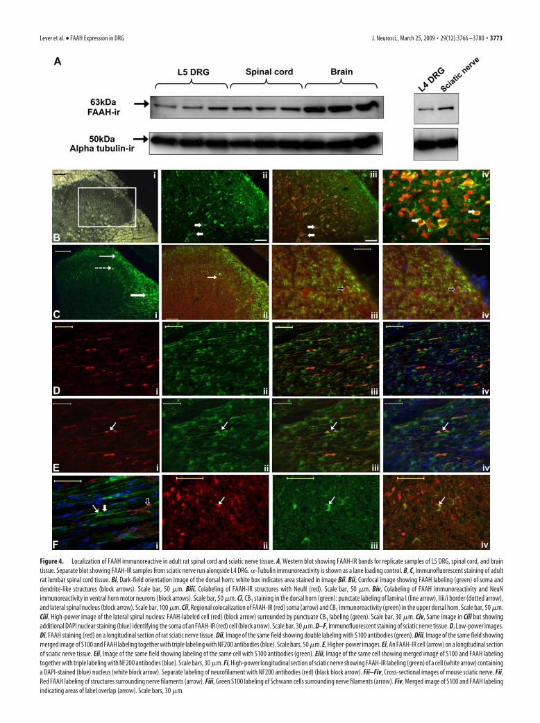
Figure 4. Localization of FAAH immunoreactive in adult rat spinal cord and sciatic nerve tissue. A, Western blot showing FAAH-IR bands for replicate samples of L5 DRG, spinal cord, and braintissue. Separate blot showing FAAH-IR samples from sciatic nerve run alongside L4 DRG. �-Tubulin immunoreactivity is shown as a lane loading control. B, C, Immunofluorescent staining of adultrat lumbar spinal cord tissue. Bi, Dark-field orientation image of the dorsal horn: white box indicates area stained in image Bii. Bii, Confocal image showing FAAH labeling (green) of soma anddendrite-like structures (block arrows). Scale bar, 50 �m. Biii, Colabeling of FAAH-IR structures with NeuN (red). Scale bar, 50 �m. Biv, Colabeling of FAAH immunoreactivity and NeuNimmunoreactivity in ventral horn motor neurons (block arrows). Scale bar, 50 �m. Ci, CB1 staining in the dorsal horn (green): punctate labeling of lamina I (line arrow), IIii/i border (dotted arrow),and lateral spinal nucleus (block arrow). Scale bar, 100 �m. Cii, Regional colocalization of FAAH-IR (red) soma (arrow) and CB1 immunoreactivity (green) in the upper dorsal horn. Scale bar, 50 �m.Ciii, High-power image of the lateral spinal nucleus: FAAH-labeled cell (red) (block arrow) surrounded by punctuate CB1 labeling (green). Scale bar, 30 �m. Civ, Same image in Ciii but showingadditional DAPI nuclear staining (blue) identifying the soma of an FAAH-IR (red) cell (block arrow). Scale bar, 30 �m. D–F, Immunofluorescent staining of sciatic nerve tissue. D, Low-power images.Di, FAAH staining (red) on a longitudinal section of rat sciatic nerve tissue. Dii, Image of the same field showing double labeling with S100 antibodies (green). Diii, Image of the same field showingmerged image of S100 and FAAH labeling together with triple labeling with NF200 antibodies (blue). Scale bars, 50 �m. E, Higher-power images. Ei, An FAAH-IR cell (arrow) on a longitudinal sectionof sciatic nerve tissue. Eii, Image of the same field showing labeling of the same cell with S100 antibodies (green). Eiii, Image of the same cell showing merged image of S100 and FAAH labelingtogether with triple labeling with NF200 antibodies (blue). Scale bars, 30 �m. Fi, High-power longitudinal section of sciatic nerve showing FAAH-IR labeling (green) of a cell (white arrow) containinga DAPI-stained (blue) nucleus (white block arrow). Separate labeling of neurofilament with NF200 antibodies (red) (black block arrow). Fii–Fiv, Cross-sectional images of mouse sciatic nerve. Fii,Red FAAH labeling of structures surrounding nerve filaments (arrow). Fiii, Green S100 labeling of Schwann cells surrounding nerve filaments (arrow). Fiv, Merged image of S100 and FAAH labelingindicating areas of label overlap (arrow). Scale bars, 30 �m.
Lever et al. • FAAH Expression in DRG J. Neurosci., March 25, 2009 • 29(12):3766 –3780 • 3773

tralateral side were FAAH-IR (Fig. 5B), consistent with the pat-tern of staining found in lumbar DRG from naive animals (Fig.1K). This difference in the size of FAAH-IR neurons on the ipsi-lateral compared with the contralateral side to injury was notobserved in DRG tissue from sham-operated animals (Fig. 5C).Cell-size analysis of DRG soma derived from the ipsilateral andcontralateral sides of axotomized or sham-operated rats 7 d after
surgery and naive rats (Fig. 5D–F) confirmed a rightward shift inthe size of FAAH-IR neurons on the injured side of axotomizedrats compared with those on the contralateral side (Fig. 5D). Themean area of the FAAH-IR DRG neurons was significantly in-creased on the side ipsilateral to an axotomy injury (603.3 � 38.4�m 2; n 6; 330 cells) compared with FAAH-IR neurons fromthe uninjured side (440.5 � 20.7 �m 2; n 6; 448 cells; p � 0.05
Figure 5. Larger-sized DRG neurons are FAAH-IR after axotomy injury to the sciatic nerve. A, B, I–K, Immunofluorescent labeling of L5 DRG tissue 7 d after an axotomy injury to the sciatic nerve(scale bars, 50 �m). A, DRG tissue ipsilateral to the injury. Left, Large-sized FAAH-IR neurons (arrows). Middle, Small-sized TPRV1-IR neurons (block arrows). Right, Merged image; large-sizedFAAH-IR neurons (arrows) are not TRPV1 labeled. B, Same animal as in A, DRG tissue contralateral to the injury. Left, Small-sized FAAH-IR neuron (arrow). Middle, Small TRPV1-IR neuron (blockarrow). Right, Colabeling of TRPV1 and FAAH in a small neuron (arrow). C, L5 DRG tissue ipsilateral to a sham axotomy injury. Left, Small-sized FAAH-IR neuron (arrow). Middle, Small TRPV1-IRneuron (block arrow). Right, Small neuron colabeled with TRPV1 and FAAH immunoreactivity (arrow). D, Cell-size distribution of FAAH-IR L5 DRG neurons on the ipsilateral and contralateral side toa 7 d axotomy injury: rightward shift in the size of FAAH-IR neurons on the ipsilateral side to injury (total of 1656 NeuN-labeled cells sampled, n 6). E, Cell-size distribution of FAAH-IR L5 DRGneurons on the ipsilateral and contralateral side to a 7 d sham axotomy injury: comparable cell-size distribution of FAAH-IR neurons on the ipsilateral and contralateral sides to sham injury (total of952 NeuN-labeled cells sampled, n 4). F, Cell-size distribution of FAAH-IR L5 DRG neurons from naive animals (total of 1241 NeuN-labeled cells sampled, n 4). G, Mean cell-area plot for FAAH-IRneurons from axotomized (n 6) or sham axotomized (n 4) and naive (n 4) animals at 7 d postoperation (PO). PO7, FAAH-IR neurons on the ipsilateral side to injury are larger than on thecontralateral side or the ipsilateral side of sham-injured or naive animals. *p � 0.05, ANOVA with post hoc Tukey’s test. Area plots for ipsilateral and contralateral FAAH-IR neurons from axotomizedanimals (n 3) 3 d after operation and naive animals (n 4). PO3, FAAH-IR neurons on the ipsilateral side are larger than the contralateral side or naive animals. #p � 0.05, Student’s t test. H,Plot of the number of L5 DRG cells measured in D in each size category on ipsilateral and contralateral sides to 7 d axotomy injury. I, DRG tissue ipsilateral to axotomy. Left, Large FAAH-IR neuron(arrow). Middle, NF200 labeling of a large-sized neuron (block arrow). Right, Merged image showing FAAH and NF200 colabeling in a large-sized neuron (arrow). Scale bars, 50 �m. J, Same animalas in I, contralateral to axotomy. Left, Small FAAH-IR neuron (arrow). Middle, NF200 labeling of large-sized neuron (block arrow). Right, FAAH (arrow) and NF200 labeling (block arrow) are notcolocalized in the merged image. Scale bars, 50 �m. K, DRG tissue ipsilateral to axotomy. Left, Large FAAH-IR neuron (arrow). Middle, ATF3 labeling in a large-sized neuron (block arrow). Right, ATF3labeling within a large-sized FAAH-IR neuron (arrow). Scale bars, 50 �m.
3774 • J. Neurosci., March 25, 2009 • 29(12):3766 –3780 Lever et al. • FAAH Expression in DRG

ANOVA, post hoc Tukey’s test) and those on the ipsilateral side tosham-operated animals (Fig. 5G). This change could not be ac-counted for by differences in the number of NeuN-labeled neu-rons sampled from ipsilateral (total of n 808 cells) and con-tralateral (total of n 848 cells) DRG in each size category (Fig.5H) and did not occur in DRG profiles from the injured anduninjured sides of sham-operated or naive animals (Fig. 5E–G).The increase in the average size of FAAH-IR neurons ipsilateralversus contralateral to injury was also significant when DRG tis-sue was harvested 3 d after axotomy surgery (Fig. 5G). The pro-portion of FAAH-IR neurons on the ipsilateral side to injury was42.1 � 5.7% (n 808 NeuN-IR cells) compared with the con-tralateral side (35.7 � 3.2%; n 848 NeuN-IR cells), althoughthis difference was not statistically significant. The proportion ofFAAH-IR cells on the contralateral side was close to that found innaive animals (32.8 � 0.9%; n 1241 NeuN-IR cells).
The change in the distribution pattern of FAAH immunore-activity was confirmed by costaining of large FAAH-IR cells withanti-NF200 antibodies ipsilateral, but not contralateral, to anaxotomy injury (Fig. 5 I, J). Costaining the sections with the anti-FAAH and anti-TRPV1 antibodies was also performed on bothipsilateral and contralateral DRG sections after axotomy (Fig.5A,B). In agreement with previous findings (Michael and Priest-ley, 1999), these experiments showed that the proportion ofTRPV1-IR DRG neurons ipsilateral to axotomy injury (14.5 �1.9%; n 386) was reduced compared with the proportion ofthese cells on the contralateral side (38.4 � 3.7%; n 409).Consistent with the changes in FAAH and TRPV1 expression, therelative number of cells coexpressing FAAH and TRPV1 was alsoaltered. Whereas 60.7 � 10.6% (n 131) of the FAAH-IR cellswere colabeled with TRPV1 on the contralateral side, only 8.5 �3.9% (n 164) of the FAAH-labeled cells costained for TRPV1on the ipsilateral side. The difference in the proportion of the cellsbetween the two sides was significant ( p � 0.0001). Costainingwas also performed using the anti-FAAH antibody and an anti-body raised against ATF3 (Fig. 5K), which identifies primarysensory neurons under sustained cellular stress. There were noATF3-immunolabeled cells on the contralateral side, but the ma-jority (74.9 � 3.5; n 282) of FAAH-IR neurons ipsilateral toaxotomy injury contained ATF3 labeling, including large-sizedneurons of �600 �m 2.
Spinal nerve transectionIn agreement with previously published data, after the ligationand transection of a lumbar spinal nerve (SNT injury), animalsdeveloped reflex hypersensitivity to mechanical and cold stimuli,assessed 7 d after surgery. The mean � SEM force in grams re-quired for reflex withdrawal of injured hindpaws (26.9 � 4.0 g)was significantly lower when compared with the preinjury pawbaseline (52.9 � 2.3 g) ( p � 0.001) and with the responses ofuninjured contralateral paws (48.8 � 1.7 g) ( p � 0.001) or of theipsilateral paws of sham-operated animals (45.4 � 2.5 g) ( p �0.01; ANOVA, Tukey’s tests; n 4 per group). In sham-operatedanimals, no differences were found between mean withdrawalresponses at baseline (48.5 � 2.1 and 49.8 � 2.1 g) and 7 d afterinjury (45.4 � 2.5 and 50.0 � 2.6 g) for ipsilateral and contralat-eral paws, respectively, and there was no difference in baselineresponses (50.6 � 2.5 g) compared with 7 d after injury on thecontralateral side of SNT animals ( p � 0.05; ANOVA, Tukey’stest). SNT-injured paws all responded with 100% frequency to acold acetone stimulus, a significantly higher response rate com-pared with the opposite uninjured paws (mean response rate,5.0 � 5.8%), the ipsilateral paws of sham-operated animals
(10.0 � 6.7%), and ipsilateral pre-SNT injury baseline (0%) ( p �0.001, Kruskal–Wallis ANOVA, Student–Newman–Keuls; n 4per group). Using this test, there was no difference in the re-sponses of sham-operated animals on the ipsilateral side versusthe contralateral side to injury (5.0 � 5.8%) or from respectivepreinjury baselines (0 and 5.0 � 5.8%).
As with DRG tissue from axotomized animals, larger-sizedneurons ipsilateral to the SNT injury stained positively for FAAHimmunoreactivity (Fig. 6A), whereas those on the contralateralside (Fig. 6B) and the ipsilateral side of sham-operated animalsdid not (Fig. 6C). Cell-size analysis confirmed a pronouncedrightward shift in the size distribution of ipsilateral FAAH-IRneurons (Fig. 6D). On average, FAAH-IR cells were significantlylarger ipsilaterally (658.1 � 12.1 �m 2; n 219) than those on thecontralateral side (402.4 � 12.4 �m 2; n 156) or on the ipsilat-eral or contralateral side of sham-operated animals (414.5 � 10.7�m 2, n 215 and 429.2 � 12.4, n 224 for ipsilateral andcontralateral sides, respectively) (Fig. 6E). SNT injury increasedthe proportion of FAAH-IR cells (50.6 � 3.7%; n 442 NeuN-IRcells) when compared with the contralateral side (34.5 � 5.8%;n 449 NeuN-IR cells) or the ipsilateral side of sham-operatedanimals (33.8 � 3.1%; n 630) ( p � 0.05, ANOVA, Tukey’stest), whereas the proportion of FAAH-IR neurons on the con-tralateral side of SNT animals was not significantly different fromthat found in naive animals (32.7 � 0.8%; n 1402 NeuN-IRcells). There was a concomitant reduction in the proportion ofTRPV1-labeled cells on the injured side (1.6 � 1.9%; n 4)compared with the uninjured side (46.1 � 1.3%; n 162) (Fig.6A,B). This ipsilateral decrease was more extensive comparedwith axotomized tissue ( p � 0.05, ANOVA, Tukey’s test).
Western blot analysis of protein extracts from SNT-injuredand contralateral DRG confirmed that FAAH immunoreactivitywas upregulated on the ipsilateral side of the injury (Fig. 6F).Densiometric analysis of the FAAH-IR bands showed higher op-tical density values with ipsilateral DRG samples compared withthose on the uninjured, contralateral side. There was no differ-ence in the optical density values measured for GAPDH-IR bands(Fig. 6G).
Inflammation of peripheral tissues does not induce changesin FAAH expression patternSimilarly to peripheral nerve injury, inflammation of the periph-eral tissues also induces plastic changes to cell markers in primarysensory neurons (Calza et al., 1998; Ji et al., 2002). Thus, we alsostudied whether inflammation of peripheral tissues induces anychanges in the FAAH expression pattern. CFA injection inducedinflammation of the hindpaw skin and produced concomitantreflex hypersensitivity to thermal and mechanical stimuli, mea-sured by behavioral testing before and after injection. With-drawal latencies to thermal stimulation were significantly shorterfor the inflamed hindpaw compared with preinjury baseline lev-els and were also shorter when compared with the uninjuredcontralateral paw ( p � 0.05, one-way ANOVA, Tukey’s test)(Fig. 7A). Similarly, the threshold for paw-withdrawal responsesto mechanical stimulation were also significantly reduced ipsilat-erally at 1 and 2 d after CFA injection when compared with pre-injury baseline levels and the responses of the contralateral pawsat these time points ( p � 0.01, one-way ANOVA, Tukey’s test)(Fig. 7B). Ipsilateral and contralateral L5 DRG from CFA-treatedanimals (n 4) were processed for FAAH immunohistochemis-try. Analysis of the cell-size distribution of FAAH-IR soma (Fig.7C) revealed no inflammation-related changes ipsilateral to CFAinjection. There was no difference in the mean � SEM soma size
Lever et al. • FAAH Expression in DRG J. Neurosci., March 25, 2009 • 29(12):3766 –3780 • 3775

of FAAH-IR DRG neurons on the inflamed side compared withthe contralateral side or DRG neurons from naive animals ( p 0.885, one-way ANOVA). There was also no change in the aver-age proportion of FAAH-IR DRG neurons ipsilateral (27.2 �3.1%; n 467 nucleated cells) versus contralateral (29.2 � 1.7%;n 446 nucleated cells) to the inflammation ( p 0.5921, Stu-dent’s t test).
DiscussionTo better understand how AEA might modulate peripheral noci-ceptive transmission, it is important to map the location of FAAHin tissue relevant to this function. This study investigated FAAHexpression in DRG sensory neurons, peripheral nerve tissue, andspinal cord neurons.
RT-PCR experiments provided evidence for the presence ofFAAH mRNA in adult rat DRG and spinal cord. FAAH proteinwas detected in rat DRG, spinal cord, and peripheral nerve tissueusing Western blotting. The size of the immunoreactive bandswas comparable with those obtained from brain tissue homoge-nate (Egertova et al., 1998; Cravatt et al., 2001; Ortega-Gutierrez
et al., 2004) and cells transfected with FAAH cDNA (Patricelli etal., 1998).
FAAH protein-specific antibodies labeled approximately one-third of neuronal soma in rat DRG tissue and dissociated culture.Cell-size measurements and the absence of costaining withNF200 antibodies indicated that FAAH-IR DRG neurons weresmall, of the kind subtending unmyelinated C-fiber axons (Law-son and Waddell, 1991) and colocalizing with small cell markers:peripherin, IB4, and TRPV1 (Goldstein et al., 1991; Michael andPriestley, 1999; Liu et al., 2004). Many FAAH-IR neurons(64.5%) costained for TRPV1, consistent with FAAH and TRPV1costaining observed in cultured rat trigeminal ganglia neurons(Price et al., 2005) and hippocampal and cerebellar mouse brainneurons (Cristino et al., 2008).
Colocalization of FAAH and TRPV1 in DRG soma is sup-ported by functional evidence from cobalt uptake experiments.Pretreatment of DRG cultures with the FAAH enzyme inhibitorURB597 increased the proportion of cells positive for Co-pptafter AEA application. AEA- and URB597-dependent cobalt up-
Figure 6. Larger-sized DRG neurons are FAAH-IR after SNT. Immunolabeling of lumbar DRG tissue from rats receiving a spinal nerve transection injury. Left, FAAH labeling. Middle, TRPV1 labeling.Right, Neurons with TRPV1 and FAAH labeling (scale, bars 50 �m). A, Images of DRG tissue on the ipsilateral side to SNT injury. Left, A large-sized FAAH-IR neuron (arrow). Middle, No TRPV1-labeledcells visible after TRPV1 staining. Right, FAAH-IR large-sized neuron (arrow); no cells colabeled with FAAH and TRPV1. B, Images of DRG tissue on the contralateral side to SNT injury of the sameanimal as in A. Left, Small FAAH-IR neuron (arrow). Middle, Small TRPV1-IR neuron (block arrow). Right, Neuron colabeled with TRPV1 and FAAH (arrow) immunoreactivity. C, Images of lumbar DRGtissue from the ipsilateral side of a sham-operated SNT animal. Left, Small FAAH-IR neuron (arrow). Middle, Small TRPV1-IR neuron (block arrow). Right, Colabeled TRPV1-IR and FAAH-IR neuron(arrow). D, Cell-size distribution of FAAH-IR neurons ipsilateral and contralateral to an SNT injury. E, Mean cell area plot for FAAH-IR neurons on the ipsilateral and contralateral sides of SNT-injuredanimals (n 4) and sham-operated animals (n 4). The size of FAAH-IR DRG neurons is greater ipsilateral to an SNT injury compared with the contralateral side and the ipsilateral and contralateralsides of sham-operated animals (*p � 0.001, one-way ANOVA, post hoc Tukey’s test). F, Western blot of lumbar DRG tissue samples ipsilateral and contralateral to SNT injury. Each sample containedtissue from two DRG (total of n 6). G, Optical density measurements (mean � SEM) of FAAH-IR and GAPDH-IR Western blot bands from lumbar DRG samples ipsilateral and contralateral to spinalnerve transection injury (average of 3 separate gels). Mean optical density measurements for Western blot bands of FAAH immunoreactivity were higher ipsilateral to the injury compared with thecontralateral side (*p � 0.05, one-way ANOVA, Tukey’s test); there was no ipsilateral versus contralateral difference in the optical densities of GAPDH-IR bands.
3776 • J. Neurosci., March 25, 2009 • 29(12):3766 –3780 Lever et al. • FAAH Expression in DRG

take was blocked by pretreatment with the TRPV1 receptor an-tagonist capsazepine. These results suggest that FAAH inhibitionraises the concentration of AEA inside DRG cells, which increasesTRPV1 activation and the influx of cobalt ions.
AEA can activate DRG TRPV1 receptors when applied to theintracellular side of the plasma membrane, with greater potencythan on the extracellular side (Hwang et al., 2000; Evans et al.,2004). Considering its lower affinity for TRPV1 than CB1 recep-tors (Ahluwalia et al., 2003a,b; Ross, 2003), short half-life, andlipophilic nature, AEA is likely to bind to TRPV1 receptors closeto its site of synthesis inside the same neuron. This has beendemonstrated by the measurement of TRPV1-mediated currents
activated from within individual DRG cells by intracellular AEAproduced in response to rising [Ca 2�]i (triggered by depletion ofintracellular calcium stores) (van der Stelt et al., 2005) or capsa-icin (Millns et al., 2006), consistent with the enhancing effects ofURB597- on TRPV1-dependent cobalt uptake in this study. To-gether, these data provide functional evidence for the coexistenceof FAAH, AEA, and TRPV1 in DRG neurons.
The proportion of DRG cells positive for Co-ppt after AEA orURB597 treatment was significantly higher when AEA orURB597 were applied with the CB1 receptor antagonistSR141716. This is consistent with studies demonstrating that an-tagonism of DRG CB1 receptors increases the potency of AEA atTRPV1 receptors (Ahluwalia et al., 2003a). At low concentra-tions, exogenous AEA can reduce TRPV1-evoked excitatory re-sponses via the activation of CB1 receptors coupled to inhibitorysignaling systems (Richardson et al., 1998; Ellington et al., 2002;Ahluwalia et al., 2003b; Evans et al., 2004).
Given the neurochemical phenotype of FAAH-IR neurons,coexpression of CB1 receptors is unlikely. This is because, undernormal uninflamed conditions (with low peripheral NGF levels),the expression of CB1 receptors on sensory neurons from DRG ortrigeminal ganglia is predominantly on large-sized NF200-positive cells (Hohmann and Herkenham, 1999; Bridges et al.,2003; Price et al., 2003; Amaya et al., 2006). Consequently, extra-cellular AEA produced from FAAH-IR DRG neurons might beexpected to signal at CB1 receptors on separate cells. FAAHalso hydrolyzes palmitoylethanolamine (PEA), another en-dogenous lipid implicated in the modulation of pain responses(Calignano et al., 1998; Jaggar et al., 1998; LoVerme et al.,2005) and a putative ligand for the peroxisome proliferator-activated receptor-�, located on mouse DRG neurons(LoVerme et al., 2006). PEA could compete with AEA forhydrolysis by FAAH and produce entourage effects at TRPV1receptors (Smart et al., 2002).
FAAH immunoreactivity was detected in the soma of dorsaland ventral horn neurons and those in the dorsal lateral funiculusof spinal cord tissue, matching previous observations (Tsou et al.,1998; Romero et al., 2002). The intracellular location in neuronalsoma and dendritic-like processes is consistent with staining ob-served in brain (Egertova et al., 2003), confirmed at the ultra-structural level (Gulyas et al., 2004). FAAH labeling of white mat-ter areas is consistent with detection of FAAH mRNA (Thomas etal., 1997) and FAAH-IR cells with oligodendrocyte-type mor-phology in mouse brain (Egertova et al., 2003). Moreover, thecolabeling of nucleated FAAH-IR cells with S100 antibodies insciatic nerve tissue in this study provides evidence that FAAH isexpressed in the oligodendrocyte/Schwann cell population la-beled by S100 (Palacios et al., 2004).
Axotomy of the sciatic nerve trunk or a spinal nerve branchboth produced an increase in the size of ipsilateral FAAH-IRDRG neurons. This, together with costaining with NF200, sug-gests the induction of FAAH expression in large-sized DRG neu-rons in response to axonal injury. The neurochemical phenotypesof sensory neuron DRG soma undergo profound changes in re-sponse to transection of their peripheral axons. Larger-sized neu-rons can adopt the phenotypic characteristics of smaller cells(Hudson et al., 2001), such as de novo expression of neuropep-tides (Hokfelt et al., 1994; Noguchi et al., 1995). These changesare more pronounced when the injury is made closer to the cellbody by transecting the spinal nerve (Shortland et al., 1997).Sciatic nerve axotomy reduced TRPV1 labeling in the DRG, asreported previously (Michael and Priestley, 1999) (although thisreduction was greater after spinal nerve transection), but the pro-
Figure 7. CFA inflammation. Behavior and cell-size analysis of FAAH-IR DRG neurons. A, At2 d after CFA injection, paw-withdrawal latencies to a heat stimulus (mean � SEM), measuredusing the plantar test in CFA-inflamed rat hindpaws, were shorter than baseline withdrawallatencies and those for the noninflamed contralateral paws (*p � 0.05, one-way ANOVA,Tukey’s test). B, Paw-withdrawal latencies to a mechanical stimulus (mean � SEM), measuredusing an electronic von Frey device in CFA-inflamed rat hindpaws, were lower compared withpreinjury baseline values and those of noninflamed contralateral paws tested at 1 and 2 d afterCFA intraplantar (i.pl.) injection (*p � 0.01, one-way ANOVA, Tukey’s test). C, Cell-size distri-bution of FAAH-IR DRG neurons ipsilateral and contralateral to a CFA inflammation of the rathindpaw, 2 d after CFA injection. There was no difference in the mean � SEM soma size ofFAAH-IR DRG neurons on the inflamed side (n 127) compared with the contralateral side(n 131) or DRG neurons from naive animals (n 143) ( p 0.885, one-way ANOVA).
Lever et al. • FAAH Expression in DRG J. Neurosci., March 25, 2009 • 29(12):3766 –3780 • 3777
portion of FAAH-labeled cells was not significantly increasedipsilateral to an axotomy but only after spinal nerve injury.Changes to the size and number of FAAH-IR neurons were morepronounced after injury to the L5 spinal nerve of the sciatic nervethan after peripheral axotomy. SNT injury also produced hyper-sensivity to cold and mechanical stimuli applied to the ipsilateralhindpaws receiving sciatic nerve innervation. These behavioralchanges are synonymous with neuropathic pain behavior in rats(Kim and Chung, 1992; Bridges et al., 2001). De novo expressionof FAAH on larger NF200-IR neurons after nerve injury mayincrease colocalization with CB1 receptor-expressing DRG neu-rons (Bridges et al., 2003). Although evidence indicates that over-all CB1 receptor expression in the DRG is downregulated afternerve injury (Costigan et al., 2002; Zhang et al., 2007), it is pos-sible that, after axotomy, FAAH activity in larger neurons directlyregulates AEA signaling at CB1 receptors on the same DRG cell.Signaling of AEA may predominate at CB1 receptors in theseconditions, attributable to the downregulation of TRPV1 recep-tors after injury.
ATF3 labeling in DRG neurons occurs after both types ofsciatic nerve injury and is an indicator of neurons in a stress statecaused by axonal damage (Tsujino et al., 2000). Increased cellularAEA production is similarly associated with cell damage and con-current elevations in intracellular calcium, such as those occur-ring as a result of an excitotoxic or demyelination injury (Baker etal., 2003; Pryce et al., 2003). The presence of ATF3 in most DRGneurons containing FAAH after nerve injury makes it is plausiblethat the increase in FAAH protein occurs in response to elevatedconcentrations of AEA induced by axonal damage to the thesecells.
A 2 d CFA-induced inflammation of the hindpaw producedbehavioral hypersensitivity to mechanical stimuli in commonwith SNT animals but did not change the size distribution or thenumber FAAH-IR DRG neurons. This difference could be relatedto the differential modulation of TRPV1 and CB1 receptor ex-pression after paw inflammation. In contrast to axotomy injury,CFA-induced inflammation induces de novo expression of CB1 insmall neurons, including 67% of the TPRV1-labeled population(Amaya et al., 2006). The number and responsiveness of smallTRPV1- and IB4-labeled DRG neurons also increases 2 d afterCFA inflammation (Breese et al., 2005). NGF levels (elevated ininflamed tissues) are correlated with the degree of CB1 andTRPV1 colocalization in cultured DRG neurons (Ahluwalia et al.,2002). AEA overproduction in inflammatory conditions, such asbladder cystitis, has been suggested to mediate prohyperalgesiceffects via TRPV1 receptors, in a manner that is potentiated byFAAH inhibitors (Dinis et al., 2004). Whether AEA has a netexcitatory or inhibitory effect on DRG neurons can be influencedby several factors, the nature of which affects whether AEA signalspredominantly at TRPV1 or CB1 receptors (Ahluwalia et al.,2003a; Ross, 2003). Consequently, it is plausible that FAAH ex-pression would be differentially regulated in response to specificinjury-related changes to its target receptors to control AEA sig-naling appropriately.
In this study, both anatomical and functional evidence wasused to localize the FAAH enzyme, implicated in controlling cel-lular AEA concentrations, to the soma of peripheral sensory neu-rons. Its distribution within the neuronal subset with nociceptivefunctions is modulated by nerve injury. This may be indicative ofa role for the endocannabinoid and endovanilloid signalingproperties of AEA in modulating peripheral nociceptivetransmission.
ReferencesAhluwalia J, Urban L, Bevan S, Capogna M, Nagy I (2002) Cannabinoid 1
receptors are expressed by nerve growth factor- and glial cell-derivedneurotrophic factor-responsive primary sensory neurones. Neuroscience110:747–753.
Ahluwalia J, Yaqoob M, Urban L, Bevan S, Nagy I (2003a) Activation ofcapsaicin-sensitive primary sensory neurones induces anandamide pro-duction and release. J Neurochem 84:585–591.
Ahluwalia J, Urban L, Bevan S, Nagy I (2003b) Anandamide regulates neu-ropeptide release from capsaicin-sensitive primary sensory neurones byactivating both the cannabinoid 1 receptor and the vannilloid receptor 1in vitro. Eur J Neurosci 17:2611–2618.
Akiba Y, Kato S, Katsube K, Nakamura M, Takeuchi K, Ishii H, Hibi T (2004)Transient receptor potential vanilloid subfamily 1 expressed in pancreaticislet beta cells modulates insulin secretion in rats. Biochem Biophys ResCommun 321:219 –225.
Amaya F, Shimosato G, Kawasaki Y, Hashimoto S, Tanaka Y, Ji RR, Tanaka M(2006) Induction of CB1 cannabinoid receptor by inflammation in pri-mary afferent neurons facilitates antihyperalgesic effect of peripheral CB1agonist. Pain 124:175–183.
Baker D, Pryce G, Croxford JL, Brown P, Pertwee RG, Makriyannis A,Khanolkar A, Layward L, Fezza F, Bisogno T, Di Marzo V (2001) Endo-cannabinoids control spasticity in a multiple sclerosis model. FASEB J15:300 –302.
Baker D, Pryce G, Giovannoni G, Thompson AJ (2003) The therapeuticpotential of cannabis. Lancet Neurol 2:291–298.
Breese NM, George AC, Pauers LE, Stucky CL (2005) Peripheral inflamma-tion selectively increases TRPV1 function in IB4-positive sensory neuronsfrom adult mouse. Pain 115:37– 49.
Bridges D, Ahmad K, Rice AS (2001) The synthetic cannabinoidWIN55,212-2 attenuates hyperalgesia and allodynia in a rat model ofneuropathic pain. Br J Pharmacol 133:586 –594.
Bridges D, Rice AS, Egertova M, Elphick MR, Winter J, Michael GJ (2003)Localisation of cannabinoid receptor 1 in rat dorsal root ganglion using insitu hybridisation and immunohistochemistry. Neuroscience119:803– 812.
Calignano A, La Rana G, Giuffrida A, Piomelli D (1998) Control of paininitiation by endogenous cannabinoids. Nature 394:277–281.
Calza L, Pozza M, Zanni M, Manzini CU, Manzini E, Hokfelt T (1998) Pep-tide plasticity in primary sensory neurons and spinal cord duringadjuvant-induced arthritis in the rat: an immunocytochemical and in situhybridization study. Neuroscience 82:575–589.
Carlton SM, Lekan HA, Kim SH, Chung JM (1994) Behavioral manifesta-tions of an experimental model for peripheral neuropathy produced byspinal nerve ligation in the primate. Pain 56:155–166.
Caterina MJ, Schumacher MA, Tominaga M, Rosen TA, Levine JD, Julius D(1997) The capsaicin receptor: a heat-activated ion channel in the painpathway. Nature 389:816 – 824.
Caterina MJ, Leffler A, Malmberg AB, Martin WJ, Trafton J, Petersen-ZeitzKR, Koltzenburg M, Basbaum AI, Julius D (2000) Impaired nociceptionand pain sensation in mice lacking the capsaicin receptor. Science288:306 –313.
Chang L, Luo L, Palmer JA, Sutton S, Wilson SJ, Barbier AJ, Breitenbucher JG,Chaplan SR, Webb M (2006) Inhibition of fatty acid amide hydrolaseproduces analgesia by multiple mechanisms. Br J Pharmacol148:102–113.
Costigan M, Befort K, Karchewski L, Griffin RS, D’Urso D, Allchorne A,Sitarski J, Mannion JW, Pratt RE, Woolf CJ (2002) Replicate high-density rat genome oligonucleotide microarrays reveal hundreds of reg-ulated genes in the dorsal root ganglion after peripheral nerve injury.BMC Neurosci 3:16.
Cravatt BF, Giang DK, Mayfield SP, Boger DL, Lerner RA, Gilula NB (1996)Molecular characterization of an enzyme that degrades neuromodulatoryfatty-acid amides. Nature 384:83– 87.
Cravatt BF, Demarest K, Patricelli MP, Bracey MH, Giang DK, Martin BR,Lichtman AH (2001) Supersensitivity to anandamide and enhanced en-dogenous cannabinoid signaling in mice lacking fatty acid amide hydro-lase. Proc Natl Acad Sci U S A 98:9371–9376.
Cravatt BF, Saghatelian A, Hawkins EG, Clement AB, Bracey MH, LichtmanAH (2004) Functional disassociation of the central and peripheral fattyacid amide signaling systems. Proc Natl Acad Sci U S A 101:10821–10826.
Cristino L, Starowicz K, De Petrocellis L, Morishita J, Ueda N, Guglielmotti V,
3778 • J. Neurosci., March 25, 2009 • 29(12):3766 –3780 Lever et al. • FAAH Expression in DRG

Di Marzo V (2008) Immunohistochemical localization of anabolic andcatabolic enzymes for anandamide and other putative endovanilloids inthe hippocampus and cerebellar cortex of the mouse brain. Neuroscience151:955–968.
Deutsch DG, Chin SA (1993) Enzymatic synthesis and degradation of anan-damide, a cannabinoid receptor agonist. Biochem Pharmacol46:791–796.
Deutsch DG, Ueda N, Yamamoto S (2002) The fatty acid amide hydrolase(FAAH). The fatty acid amide hydrolase (FAAH). Prostaglandins LeukotEssent Fatty Acids 66:201–210.
Di Marzo V, Fontana A, Cadas H, Schinelli S, Cimino G, Schwartz JC, Pio-melli D (1994) Formation and inactivation of endogenous cannabinoidanandamide in central neurons. Nature 372:686 – 691.
Dinis P, Charrua A, Avelino A, Yaqoob M, Bevan S, Nagy I, Cruz F (2004)Anandamide-evoked activation of vanilloid receptor 1 contributes to thedevelopment of bladder hyperreflexia and nociceptive transmission tospinal dorsal horn neurons in cystitis. J Neurosci 24:11253–11263.
Egertova M, Elphick MR (2000) Localisation of cannabinoid receptors inthe rat brain using antibodies to the intracellular C-terminal tail of CB1.J Comp Neurol 422:159 –171.
Egertova M, Giang DK, Cravatt BF, Elphick MR (1998) A new perspectiveon cannabinoid signalling: complementary localization of fatty acidamide hydrolase and CB1 receptor in rat brain. Proc Biol Sci265:2081–2085.
Egertova M, Cravatt BF, Elphick MR (2003) Comparative analysis of fattyacid amide hydrolase and CB(1) cannabinoid receptor expression in themouse brain: evidence of a widespread role for fatty acid amide hydrolasein regulation of endocannabinoid signaling. Neuroscience 119:481– 496.
Ellington HC, Cotter MA, Cameron NE, Ross RA (2002) The effect of can-nabinoids on capsaicin-evoked calcitonin gene-related peptide (CGRP)release from the isolated paw skin of diabetic and non-diabetic rats. Neu-ropharmacology 42:966 –975.
Evans RM, Scott RH, Ross RA (2004) Multiple actions of anandamide onneonatal rat cultured sensory neurones. Br J Pharmacol 141:1223–1233.
Farquhar-Smith WP, Egertova M, Bradbury EJ, McMahon SB, Rice AS, El-phick MR (2000) Cannabinoid CB1 receptor expression in rat spinalcord. Mol Cell Neurosci 15:510 –521.
Fegley D, Gaetani S, Duranti A, Tontini A, Mor M, Tarzia G, Piomelli D(2005) Characterization of the fatty acid amide hydrolase inhibitor cy-clohexly carbamic acid 3�-carbamoyl-biphenyl-3-yl ester (URB597): ef-fects on anandamide and oleoylethanolamide deactivation. J PharmacolExp Ther 313:352–358.
Goldstein ME, House SB, Gainer H (1991) NF-L and peripherin immuno-reactivities define distinct classes of rat sensory ganglion cells. J NeurosciRes 30:92–104.
Guindon J, Beaulieu P (2006) Antihyperalgesic effects of local injections ofanandamide, ibuprofen, rofecoxib and their combinations in a model ofneuropathic pain. Neuropharmacology 50:814 – 823.
Gulyas AI, Cravatt BF, Bracey MH, Dinh TP, Piomelli D, Boscia F, Freund TF(2004) Segregation of two endocannabinoid-hydrolysing enzymes intopre- and postsynaptic compartments in the rat hippocampus, cerebellumand amygdala. Eur J Neurosci 20:441– 458.
Hargreaves K, Dubner R, Brown F, Flores C, Joris J (1988) A new and sen-sitive method for measuring thermal nociception in cutaneous hyperal-gesia. Pain 32:77– 88.
Hohmann AG, Herkenham M (1999) Localization of central cannabinoidCB1 receptor messenger RNA in neuronal subpopulations of rat dorsalroot ganglia: a double-label in situ hybridization study. Neuroscience90:923–931.
Hohmann AG, Suplita RL, Bolton NM, Neely MH, Fegley D, Mangieri R,Krey JF, Walker JM, Holmes PV, Crystal JD, Duranti A, Tontini M, MorM, Tarzia G, Piomelli D (2005) An endocannabinoid mechanism forstress-induced analgesia. Nature 435:1108 –1112.
Hokfelt T, Zhang X, Wiesenfeld-Hallin Z (1994) Messenger plasticity inprimary sensory neurons following axotomy and its functional implica-tions. Trends Neurosci 17:22–30.
Holt S, Comelli F, Costa B, Fowler CJ (2005) Inhibitors of fatty acid amidehyrdolase reduce carrageenan-induce hind paw inflammation inpentobarbital-treated mice: comparison with indomethacin and possibleinvolvement of cannabinoid receptors. Br J Pharmacol 146:467– 476.
Hudson LJ, Bevan S, Wotherspoon G, Gentry C, Fox A, Winter J (2001) VR1
protein expression increases in undamaged DRG neurons after partialnerve injury. Eur J Neurosci 13:2105–2114.
Hwang SW, Cho H, Kwak J, Lee SY, Kang CJ, Jung J, Cho S, Min KH, Suh YG,Kim D, Oh U (2000) Direct activation of capsaicin receptors by prod-ucts of lipoxygenase: endogenous capsaicin-like substances. Proc NatlAcad Sci U S A 97:6155– 6160.
Jaggar SI, Hasnie FS, Sellaturay S, Rice ASC (1998) The anti-hyperalgesicactions of the cannabinoid anandamide and the putative CB2 agonistpalmitoylethanolamide in models of visceral and somatic inflammatorypain. Pain 76:189 –199.
Jayamanne A, Greenwood R, Mitchell VA, Aslan S, Piomelli D, Vaughan CW(2006) Actions of the FAAH inhibitor URB597 in neuropathic andchronic inflammatory pain models. Br J Pharmacol 147:281–288.
Jhaveri MD, Richardson D, Kendall DA, Barrett DA, Chapman V (2006)Analgesic effects of fatty acid amide hydrolase inhibition in a rat model ofneuropathic pain. J Neurosci 26:13318 –13327.
Ji RR, Samad TA, Jin SX, Schmoll R, Woolf CJ (2002) p38 MAPK activationby NGF in primary sensory neurons after inflammation increases TRPV1levels and maintains heat hyperalgesia. Neuron 36:57– 68.
Kim SH, Chung JM (1992) An experimental model for peripheral neurop-athy produced by segmental spinal nerve ligation in the rat. Pain50:355–363.
Kozak KR, Marnett LJ (2002) Oxidative metabolism of endocannabinoids.Prostaglandins Leukot Essent Fatty Acids 66:211–220.
Lawson SN, Waddell PJ (1991) Soma neurofilament immunoreactivity isrelated to cell size and fibre conduction velocity in rat primary sensoryneurons. J Physiol 435:41– 63.
Ledent C, Valverde O, Cossu G, Petitet F, Aubert JF, Beslot F, Bohme GA,Imperato A, Pedrazzini T, Roques BP, Vassart G, Fratta W, Parmentier M(1999) Unresponsiveness to cannabinoids and reduced addictive effectsof opiates in CB1 receptor knockout mice. Science 283:401– 404.
Lichtman AH, Shelton CC, Advani T, Cravatt BF (2004a) Mice lacking fattyacid amide hydrolase exhibit a cannabinoid receptor-mediated pheno-typic hypoalgesia. Pain 109:319 –327.
Lichtman AH, Leung D, Shelton CC, Saghatelian A, Hardouin C, Boger DL,Cravatt BF (2004b) Reversible inhibitors of fatty acid amide hydrolasethat promote analgesia: evidence for an unprecedented combination ofpotency and selectivity. J Pharmacol Exp Ther 311:441– 448.
Liu M, Willmott NJ, Michael GJ, Priestley JV (2004) Differential pH andcapsaicin responses of Griffonia simplicifolia IB4 (IB4)-positive and IB4-negative small sensory neurons. Neuroscience 127:659 – 672.
LoVerme J, La Rana G, Russo R, Calignano A, Piomelli D (2005) The searchfor the palmitoylethanolamide receptor. Life Sci 77:1685–1698.
LoVerme J, Russo R, La Rana G, Fu J, Farthing J, Mattace-Raso G, Meli R,Hohmann A, Calignano A, Piomelli D (2006) Rapid broad-spectrumanalgesia through activation of peroxisome proliferator-activatedreceptor-alpha. J Pharmacol Exp Ther 319:1051–1061.
Michael GJ, Priestley JV (1999) Differential expression of the mRNA for thevanilloid receptor subtype 1 in cells of the adult rat dorsal root and nodoseganglia and its downregulation by axotomy. J Neurosci 19:1844 –1854.
Michael GJ, Averill S, Nitkunan A, Rattray M, Bennett DL, Yan Q, Priestley JV(1997) Nerve growth factor treatment increases brain-derived neurotro-phic factor selectively in TrkA-expressing dorsal root ganglion cells and intheir central terminations within the spinal cord. J Neurosci17:8476 – 8490.
Millns PJ, Chimenti M, Ali N, Ryland E, de Lago E, Fernandez-Ruiz J, Chap-man V, Kendall DA (2006) Effects of inhibition of fatty acid amide hy-drolase vs the anandamide membrane transporter on TRPV1-mediatedcalcium responses in adult DRG neurons; the role of CB receptors. EurJ Neurosci 24:3489 –3495.
Moller KA, Johansson B, Berge OG (1998) Assessing mechanical allodyniain the rat paw with a new electronic algometer. J Neurosci Methods84:41– 47.
Mor M, Rivara S, Lodola A, Plazzi PV, Tarzia G, Duranti A, Tontini A,Piersanti G, Kathuria S, Piomelli D (2004) Cyclohexylcarbamic acid 3�-or 4� substituted biphenyl-3-yl esters as fatty acid amide hydrolase inhib-itors: synthesis, quantitative structure-activity relationships and molecu-lar modeling studies. J Med Chem 47:4998 –5008.
Muthian S, Rademacher DJ, Roelke CT, Gross GJ, Hillard CJ (2004) Anan-damide content is increased and CB1 cannabinoid receptor blockade isprotective during transient, focal cerebral ischemia. Neuroscience129:743–750.
Lever et al. • FAAH Expression in DRG J. Neurosci., March 25, 2009 • 29(12):3766 –3780 • 3779

Nagy I, Pabla R, Matesz C, Dray A, Woolf CJ, Urban L (1993) Cobalt uptakeenables identification of capsaicin- and bradykinin-sensitive subpopula-tions of rat dorsal root ganglion cells in vitro. Neuroscience 56:241–246.
Noguchi K, Kawai Y, Fukuoka T, Senba E, Miki K (1995) Substance P in-duced by peripheral nerve injury in primary afferent sensory neurons andits effect on dorsal column nucleus neurons. J Neurosci 15:7633–7643.
Ortega-Gutierrez S, Hawkins EG, Viso A, Lopez-Rodríguez ML, Cravatt BF(2004) Comparison of anandamide transport in FAAH wild-type andknockout neurons: evidence for contributions by both FAAH and theCB1 receptor to anandamide uptake. Biochemistry 43:8184 – 8190.
Palacios G, Muro A, Verdu E, Pumarola M, Vela JM (2004) Immunohisto-chemical localization of the sigma1 receptor in Schwann cells of rat sciaticnerve. Brain Res 1007:65–70.
Patricelli MP, Lashuel HA, Giang DK, Kelly JW, Cravatt BF (1998) Compar-ative characterization of a wild type and transmembrane domain-deletedfatty acid amide hydrolase: identification of the transmembrane domainas a site for oligomerization. Biochemistry 37:15177–15187.
Piomelli D, Beltramo M, Giuffrida A, Stella N (1998) Endogenous cannabi-noid signalling. Neurobiol Dis 5:462– 473.
Potrebic S, Ahn AH, Skinner K, Fields HL, Basbaum AI (2003) Peptidergicnociceptors of both trigeminal and dorsal root ganglia express serotonin1D receptors: implications for the selective antimigraine action oftriptans. J Neurosci 23:10988 –10997.
Price TJ, Helesic G, Parghi D, Hargreaves KM, Flores CM (2003) The neu-ronal distribution of cannabinoid receptor type 1 in the trigeminal gan-glion of the rat. Neuroscience 120:155–162.
Price TJ, Patwardhan AM, Flores CM, Hargreaves KM (2005) A role for theanandamide membrane transporter in TRPV1-mediated neurosecretionfrom trigeminal sensory neurons. Neuropharmacology 49:25–39.
Pryce G, Ahmed Z, Hankey DJ, Jackson SJ, Croxford JL, Pocock JM, Ledent C,Petzold A, Thompson AJ, Giovannoni G, Cuzner ML, Baker D (2003)Cannabinoids inhibit neurodegeneration in models of multiple sclerosis.Brain 126:2191–2202.
Richardson JD, Kilo S, Hargreaves KM (1998) Cannabinoids reduce hyper-algesia and inflammation via interaction with peripheral CB1 receptors.Pain 75:111–119.
Romero J, Hillard CJ, Calero M, Rabano A (2002) Fatty acid amide hydro-lase localisation in the human central nervous system: an immunohisto-chemical study. Mol Brain Res 100:85–93.
Ross RA (2003) Anandamide and vanilloid TRPV1 receptors. Br J Pharma-col 140:790 – 801.
Ross RA, Coutts AA, McFarlane SM, Anavi-Goffer S, Irving AJ, Pertwee RG,MacEwan DJ, Scott RH (2001) Actions of cannabinoid receptor ligandson rat cultured sensory neurones: implications for antinociception. Neu-ropharmacology 40:221–232.
Russo R, Loverme J, La Rana G, Compton TR, Parrott J, Duranti A, Tontini A,
Mor M, Tarzia G, Calignano A, Piomelli D (2007) The fatty acid amidehydrolase inhibitor URB597 (cyclohexylcarbamic acid 3�-carbamoylbiphenyl-3-yl ester) reduces neuropathic pain after oral ad-ministration in mice. J Pharmacol Exp Ther 322:236 –242.
Sathianathan V, Avelino A, Charrua A, Santha P, Matesz K, Cruz F, Nagy I(2003) Insulin induces cobalt uptake in a subpopulation of rat culturedprimary sensory neurones. Eur J Neurosci 18:2477–2486.
Shortland P, Kinman E, Molander C (1997) Sprouting of A-fibre primaryafferents into lamina II in two rat models of neuropathic pain. Eur J Pain1:215–227.
Singh Tahim A, Santha P, Nagy I (2005) Inflammatory mediators convertanandamide into a potent activator of the vanilloid type 1 transient recep-tor potential receptor in nociceptive primary sensory neurons. Neuro-science 136:539 –548.
Smart D, Jonsson KO, Vandevoorde S, Lambert DM, Fowler CJ (2002) “En-tourage” effects of N-acyl ethanolamines at human vanilloid receptors.Comparison of effects upon anandamide-induced vanilloid receptor ac-tivation and upon anandamide metabolism. Br J Pharmacol136:452– 458.
Snider WD, McMahon SB (1998) Tackling pain at the source: new ideasabout nociceptors. Neuron 20:629 – 632.
Thomas EA, Cravatt BF, Danielson PE, Gilula NB, Sutcliffe JG (1997) Fattyacid amide hydrolase, the degradative enzyme for anandamide and ole-amide, has selective distribution in neurons within the rat central nervoussystem. J Neurosci Res 50:1047–1052.
Tsou K, Nogueron MI, Muthian S, Sanudo-Pena MC, Hillard CJ, DeutschDG, Walker JM (1998) Fatty acid amide hydrolase is located preferen-tially in large neurons in the rat central nervous system as revealed byimmunohistochemisty. Neurosci Lett 254:137–140.
Tsujino H, Kondo E, Fukuoka T, Dai Y, Tokunaga A, Miki K, Yonenobu K,Ochi T, Noguchi K (2000) Activating transcription factor 3 (ATF3) in-duction by axotomy in sensory and motorneurones: a novel neuronalmarker of nerve injury. Mol Cell Neurosci 15:170 –182.
van der Stelt M, Trevisani M, Vellani V, De Petrocellis L, Schiano Moriello A,Campi B, McNaughton P, Geppetti P, Di Marzo V (2005) Anandamideacts as an intracellular messenger amplifying Ca 2� influx via TRPV1channels. EMBO J 24:3026 –3037.
Walker JM, Huang SM, Strangman NM, Tsou K, Sanudo-Pena MC (1999)Pain modulation by release of the endogenous cannabinoid anandamide.Proc Natl Acad Sci U S A 96:12198 –12203.
Zhang F, Hong S, Stone V, Smith PJ (2007) Expression of cannabinoid CB1
receptors in models of diabetic neuropathy. J Pharmacol Exp Ther323:508 –515.
Zygmunt PM, Petersson J, Andersson DA, Chuang H, Sørgård M, Di MarzoV, Julius D, Hogestatt ED (1999) Vanilloid receptors on sensory nervesmediate the vasodilator action of anandamide. Nature 400:452– 457.
3780 • J. Neurosci., March 25, 2009 • 29(12):3766 –3780 Lever et al. • FAAH Expression in DRG