Embed Size (px)
Citation preview
Cellular/Molecular
The Interface between Membrane-Spanning and CytosolicDomains in Ca2�-Dependent K� Channels Is Involved in �Subunit Modulation of Gating
Xiaohui Sun, Jingyi Shi, Kelli Delaloye, Xiao Yang, Huanghe Yang, Guohui Zhang, and Jianmin CuiDepartment of Biomedical Engineering, Cardiac Bioelectricity and Arrhythmia Center, Center for the Investigation of Membrane Excitability Disorders,Washington University, St. Louis, Missouri 63130
Large-conductance, voltage-, and Ca 2�-dependent K � (BK) channels are broadly expressed in various tissues to modulate neuronalactivity, smooth muscle contraction, and secretion. BK channel activation depends on the interactions among the voltage sensing domain(VSD), the cytosolic domain (CTD), and the pore gate domain (PGD) of the Slo1 �-subunit, and is further regulated by accessory �subunits (�1–�4). How � subunits fine-tune BK channel activation is critical to understand the tissue-specific functions of BK channels.Multiple sites in both Slo1 and the � subunits have been identified to contribute to the interaction between Slo1 and the � subunits.However, it is unclear whether and how the interdomain interactions among the VSD, CTD, and PGD are altered by the � subunits to affectchannel activation. Here we show that human �1 and �2 subunits alter interactions between bound Mg 2� and gating charge R213 anddisrupt the disulfide bond formation at the VSD–CTD interface of mouse Slo1, indicating that the � subunits alter the VSD–CTD interface.Reciprocally, mutations in the Slo1 that alter the VSD–CTD interaction can specifically change the effects of the �1 subunit on the Ca 2�
activation and of the �2 subunit on the voltage activation. Together, our data suggest a novel mechanism by which the � subunitsmodulated BK channel activation such that a � subunit may interact with the VSD or the CTD and alter the VSD–CTD interface of the Slo1,which enables the � subunit to have effects broadly on both voltage and Ca 2�-dependent activation.
IntroductionVoltage- and Ca 2�-activated K � (BK) channels are composedof pore-forming � subunits encoded by the slo1 gene andaccessory � and � subunits. Various � (�1–�4) or � subunitsmodify channel properties differently (McManus et al., 1995;Xia et al., 1999; Brenner et al., 2000; Yan and Aldrich, 2010,2012), providing a major mechanism for the diverse tissue-specific phenotypes. In brain, the �4 subunit is most abundantbut other � subunits are also found (Brenner et al., 2000;Poulsen et al., 2009). The association of �4 subunits withFragile X mental retardation protein modulates neurotrans-mission in hippocampal CA3 pyramidal neurons (Deng et al.,2013). A mutation of �3 is associated with human epilepsy(Lorenz et al., 2007). � Subunit function is fundamental forthe molecular basis of BK channel-related physiological andpathophysiologic processes.
The Slo1 subunit contains three structural domains: the cytosolicdomain (CTD), which contains Ca2� binding sites; the voltage sens-ing domain (VSD), which comprises the transmembrane segmentsS1–S4; and the pore gate domain (PGD), which comprises S5–S6(Fig. 1). Interactions among these domains are important for thePGD to open in response to physiological stimulations of membranepotential, intracellular Ca2� and Mg2� (Lee and Cui, 2010). The �subunits share a similar membrane topology, with two transmem-brane segments (TM1 and TM2), an extracellular loop, and the in-tracellular N-terminus and C-terminus. A BK channel contains fourSlo1 subunits and up to four � subunits (Wang et al., 2002). TM1and TM2 are packed at the mouth of the cleft between VSDs of twoadjacent Slo1 subunits, with TM1 close to S1 of one VSD and TM2close to S0 of adjacent VSD (Liu et al., 2010). The location of �subunits allows their interaction with the VSD, PGD, and CTD ofSlo1, which may alter or constrain the interactions among thesedomains. However, such alteration and the consequence on BKchannel activation have not been known or explored.
We began this study by investigating the effects of the �1 and�2 subunits on Mg 2�-dependent activation of BK channels. Pre-vious studies showed that Mg 2� binds in between the VSD andCTD interface of Slo1 and interacts with gating charge R213 in S4to activate the channel (Yang et al., 2007, 2008a; Fig. 1). In thisstudy, we found that the �1 and �2 subunits reduce Mg 2� sensi-tivity and alter the Mg 2� binding site and the interaction betweenthe bound Mg 2� with R213. Subsequently, we studied how mu-tations that change the VSD–CTD interface (Yang et al., 2013)affect the function of both � subunits. The results suggest that the
Received Feb. 10, 2013; revised May 21, 2013; accepted May 28, 2013.Author contributions: X.S., H.Y., and J.C. designed research; X.S., J.S., K.D., X.Y., and G.Z. performed research; X.S.
and G.Z. analyzed data; X.S., H.Y., and J.C. wrote the paper.This work was supported by National Institutes of Health Grants R01-HL70393 and R01-NS060706 (to J.C.). J.C. is
the Professor of Biomedical Engineering on the Spencer T. Olin Endowment. We thank Dr. Jimin Ding for helpfulsuggestions on the statistics of data analyses. The mSlo1 clone and h�2 clone were kindly provided by Drs. LawrenceSalkoff and Chris Lingle (Washington University, St. Louis, MO) and by Dr. Robert Brenner (University of Texas HealthScience Center at San Antonio, San Antonio, TX), respectively.
The authors declare no competing financial interests.Correspondence should be addressed to Jianmin Cui, Department of Biomedical Engineering, Washington Uni-
versity in St. Louis, 290C Whitaker Hall, One Brookings Drive, St. Louis, MO 63130. E-mail: [email protected]:10.1523/JNEUROSCI.0620-13.2013
Copyright © 2013 the authors 0270-6474/13/3311253-09$15.00/0
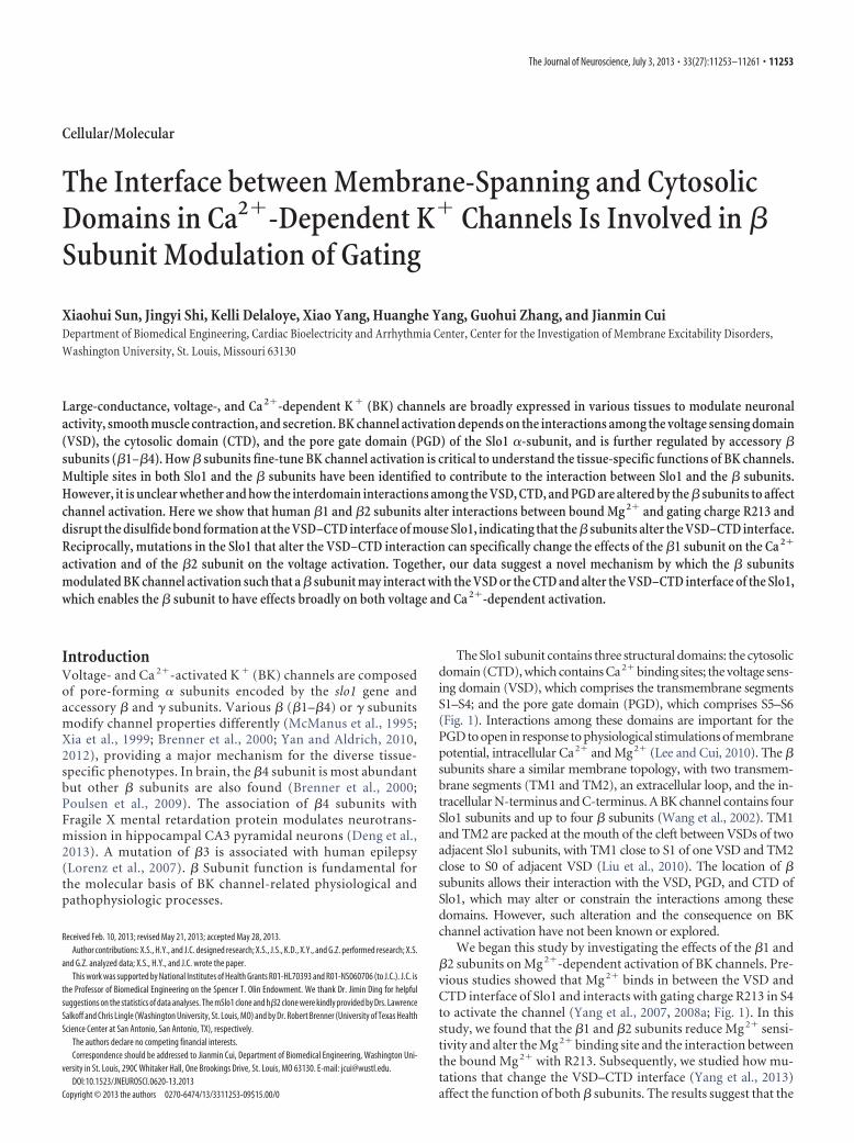
The Journal of Neuroscience, July 3, 2013 • 33(27):11253–11261 • 11253
association of � subunits alters the VSD–CTD interface and theVSD–CTD interface alteration in turn interferes with the actionof both � subunits, providing a novel perspective on how � sub-units modify BK channel activation.
Materials and MethodsMutagenesis and expression. All point mutations were made from thembr5 splice variant of mouse Slo1 (mSlo1, GenBank accession numberL16912; Butler et al., 1993) by using overlap-extension PCR (Shi et al.,2002) with Pfu polymerase (Stratagene). Human �1 and �2 (KCNMB1and KCNMB2; GenBank accession numbers U25138 and AF209747)cDNAs were subcloned into pcDNA3.1(�). The �2 with N terminus-deleted (�2ND) subunit was created by removing amino acids frompositions 2–20. The PCR-amplified regions for all constructs were veri-fied by sequencing. cRNA was transcribed in vitro using T3 polymerase(Ambion) for all Slo1 constructs and T7 polymerase (Ambion) for �1and �2ND subunits. We injected 0.05–50 ng of Slo1 cRNA or a mixtureof 5–20 ng of Slo1 and 15– 60 ng of � subunit cRNAs (the ratio of theamount of Slo1 to the amount of � is 1:3 to ensure the channel is satu-rated with � subunit) into Xenopus laevis oocytes (stage IV–V) 3– 6 dbefore experiment (female Xenopus laevis were used here). Oocytes wereincubated in ND96 solution (in mM: 96 NaCl, 2 KCl, 1.8 CaCl2, 1 MgCl2,5 HEPES, pH 7.6) at 18°C.
Electrophysiology. Macroscopic and single-channel currents were re-corded from excised inside-out patches formed with borosilicate pipettesof 0.6 –1.0 M� resistance. Data were acquired by using an Axopatch200-B patch-clamp amplifier (Molecular Devices) and Pulse acquisitionsoftware (HEKA Electronik). The current signals were low-pass filteredat 10 kHz with the four-pole Bessel filter built in the amplifier and digi-tized at 20 �s intervals. For most of the macroscopic current recordings,capacity and leak currents were subtracted using a P/5 protocol with aholding potential of �120 mV. However, with currents recorded in thepresence of either �1 or �2ND at 100 �M [Ca 2�], no leak subtraction wasconducted. The pipette solution contained the following (in mM): 140potassium methanesulfonic acid (KMeSO3), 20 HEPES, 2 KCl, and 2MgCl2, pH 7.2. The basal internal solution contained the following (inmM): 140 KMeSO3, 20 HEPES, 2 KCl, 1 EGTA, 22 mg/l 18-crown-6-tetracarboylic acid (Sigma-Aldrich), pH 7.2. CaCl2 standard solution wasadded to obtain the desired free [Ca 2�], which was measured by a Ca 2�-sensitive electrode (Thermo Electron). The nominal 0 �M [Ca 2�] solu-tion contained 5 mM EGTA with no added Ca 2�, and the free [Ca 2�] iscalculated to be �0.5 nM. The 2-(trimethylammonium)ethyl methane-thiosulfonate bromide (MTSET) and dithiothreitol (DTT) working so-lutions were diluted from the stock to the final concentration of 0.2 and10 mM, respectively, by using the nominal 0 �M [Ca 2�] solution. TheMTSET solution was freshly prepared right before each perfusion sinceits lifetime is �10 min. DTT, MTSET, or DTT-plus-MTSET treatmentswere performed by perfusing excised patch with 10 mM DTT for 5 min,
with 200 �M MTSET for 3 min, or with 10 mM DTT for 5 min before 3min 200 �M MTSET perfusion, respectively.
Gating currents were recorded in the inside-out configuration as well.The pipette solution contained the following (in mM): 127 tetraethylam-monium (TEA), 125 HMeSO3, 2 HCl, 2 MgCl2, and 20 HEPES, pH 7.2.The internal solution contains (in mM) the following: 141 N-ethyl-D-glucamine, 135 HMeSO3, 6 HCl, 20 HEPES, and 5 EGTA, pH 7.2. Toprevent the saturation of fast capacitive transients, voltage commandswere filtered at 20 kHz with an eight-pole Bessel filter (Frequency De-vices; Horrigan and Aldrich, 1999). Data were sampled at 100 kHz withan 18-bit analog-to-digital converter (ITC-18, Instrutech) and filtered at10 kHz with an internal filter of Axopatch (Molecular Devices). Capaci-tive transients and leak currents were subtracted using a P/5 protocolwith a holding potential of �120 mV. All experiments were conducted atroom temperature (22–24°C).
Data analyses. Relative conductance was determined by measuringtail current amplitudes at negative voltages as indicated for the wild-type (WT) and mutant Slo1 channels with and without � subunits.The gating charge movements were determined by integrating thearea under the rising phase and single exponential fits to the decayingphase of on gating current at various voltages. The G–V relationshipor the charge–voltage (Q–V ) relations of the WT and mutant chan-nels with and without � subunits were fitted with the Boltzmannequation as follows in Equation 1:
G
Gmax�
1
1 � exp� �zF�V � V1⁄2�
RT �or in Equation 2:
Q
Qmax�
1
1 � exp� �zF�V � V1⁄2�
RT �In Equation 1, G/Gmax is the ratio of conductance to maximum conduc-tance, z is the number of equivalent charges, V is membrane potential,V1/2 is the voltage at which the channel is 50% activated, F is the faradayconstant, R is gas constant, and T is the absolute temperature. In Equa-tion 2, Q/Qmax is the ratio of gating charge to maximum gating charge, zis the gating charge associated with voltage sensor movement, V1/2 is thevoltage for half of the gating charge movements at the closed conforma-tion of the channel, and the other parameters have the same meaning asin Equation 1. Free energy change (�G) were calculated as �(z * V1/2)(Cui and Aldrich, 2000). Mg 2� sensitivity refers to 10 mM Mg 2�-induced G–V relationship shift. Ca 2� sensitivity refers to 100 �M Ca 2�-induced G–V relationship shift.
For limiting slope measurement, the open probability of single chan-nels was analyzed with Igor Pro software (WaveMetrics) and plotted atrelative membrane potentials to construct a PO–V relation, which wasthen fitted to the HCA (Horrigan, Cui, and Aldrich) model as the follow-ing Equation 3 (Horrigan et al., 1999):
Po �1
1 �
exp��zLFV
RT �Lo �1 � exp�zJF�V � Vhc�
RT �1 � exp�zJF�V � Vho�
RT ��4
where zL is the charge associated with gate opening when all the voltagesensors are at their resting state; zJ is the charge associated with voltagesensor movements; L0 is the intrinsic open probability at 0 mV while allthe voltage sensors are at their resting state; and Vhc and Vho are thevoltages for half of the voltage sensors to be at their activation state at theclosed and the open conformations of the gate, respectively.
Curve fittings were performed using the Levenberg–Marquardt al-gorithm in Igor Pro software (WaveMetrics) for nonlinear least-squares fits. The means of the data were obtained by averaging from 3
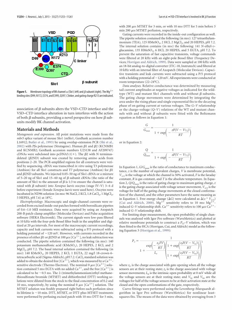
Figure 1. Membrane topology of BK channel � (Slo1; left) and � subunit (right). The Mg 2�
binding sites (D99, N172, E374, and E399), Q397, C linker, and gating charge R213 are indicated.
11254 • J. Neurosci., July 3, 2013 • 33(27):11253–11261 Sun et al. • VSD-CTD Interface Is Involved in BK � Function
to 16 patches and error bars represent SEM. Statistics were performedusing GraphPad Prism5 software (GraphPad Software) and SPSS soft-ware (IBM). Unpaired Student’s t test and multiple ANOVA wereperformed and a p-value of �0.05 is considered significant.
Results�1 and �2ND subunits reduce Mg 2�
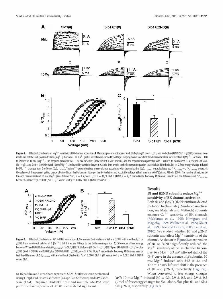
sensitivity of BK channel activationBoth �1 and �2ND (�2 N terminus-deletedmutation to eliminate �2-induced inactiva-tion; see Materials and Methods) subunitsenhance Ca2� sensitivity of BK channels(McManus et al., 1995; Nimigean andMagleby, 1999; Wallner et al., 1999; Xia etal., 1999; Orio and Latorre, 2005; Lee et al.,2010). We studied whether �1 and �2NDsubunits also affect Mg2� sensitivity of thechannel. As shown in Figure 2, coexpressionof �1 or �2ND significantly reduced theMg2� sensitivity of the BK channel. In con-trast to a 64.4 3.3 mV leftward shift of theG–V curve in the absence of � subunits, 10mM Mg2� induced only 34.3 2.4 and37.2 1.5 mV leftward shift in the presenceof �1 and �2ND, respectively (Fig. 2B).When converted to free energy changes
(�G) 10 mM Mg2� induced 5.0 0.3, 2.9 0.5, and 2.9 0.5kJ/mol of free energy changes for Slo1 alone, Slo1 plus �1, and Slo1plus �2ND, respectively (Fig. 2C).
Figure 2. Effects of � subunits on Mg 2� sensitivity of BK channel activation. A, Macroscopic current traces of Slo1, Slo1-plus-�1 (Slo1��1), and Slo1-plus-�2ND (Slo1��2ND) channels frominside-out patches in 0 (top) and 10 mM [Mg 2�] (bottom). The [Ca 2�] is 0. Currents were elicited by voltages ranging from 0 to 250 mV for 20 ms with 10 mV increments at 0 [Mg 2�], or from �100to 250 mV at 10 mM [Mg 2�]. The prepulse potential was �80 mV for 20 ms (only the last 0.5 ms shown), and the repolarization potential was �80 mV. B, Normalized G–V relations of Slo1,Slo1��1, and Slo1��2ND in 0 and 10 mM [Mg 2�], indicated by symbols shown in A. Solid lines are fits to the Boltzmann equation (Materials and Methods, Eq. 1). C, Free energy change inducedby [Mg 2�] changes from 0 to 10 mM (�G0--10 Mg). The Mg 2�-dependent free energy change associated with channel gating (�G0--10 Mg) was calculated as z * V1/2 0 Mg � z*V1/2 10 Mg, where z isthe valence of the apparent gating charge obtained from the Boltzmann fitting of the G–V relation and V1/2 is the voltage at half maximum G–V (Cui and Aldrich, 2000). The number of patches (n)for each channel in 0 and 10 mM [Mg 2�] is as follows: Slo1, n 9, 4; Slo1��1, n 16, 9; Slo1��2ND, n 6, 7, respectively. Two-way ANOVA was used to test the differences of �G0--10 Mg
between channels: *p 0.013, Slo1��1 versus Slo1; p 0.006, Slo1��2ND versus Slo1.
Figure 3. Effects of � subunits on R213–R397 interaction. A, Normalized G–V relations of WT and Q397R with or without �1 or�2ND from inside-out patches at 0 [Ca 2�]. Solid lines are fittings to the Boltzmann equation. B, Differences of free energybetween WT and Q397R channels (�GWT-Q397R). For Slo1, Q397R, Slo1 plus �1 (Slo1��1), Q397R plus �1 (Q397R��1), Slo plus�2ND (Slo1��2ND), and Q397R plus �2ND (Q397R��2ND), n 9, 5, 16, 14, 6, 7, respectively. Two-way ANOVA was used totest the differences of �GWT-Q397R with and without � subunits: *p � 0.0001, Slo1��1 versus Slo1; p 0.082, Slo1��2NDversus Slo1.
Sun et al. • VSD-CTD Interface Is Involved in BK � Function J. Neurosci., July 3, 2013 • 33(27):11253–11261 • 11255
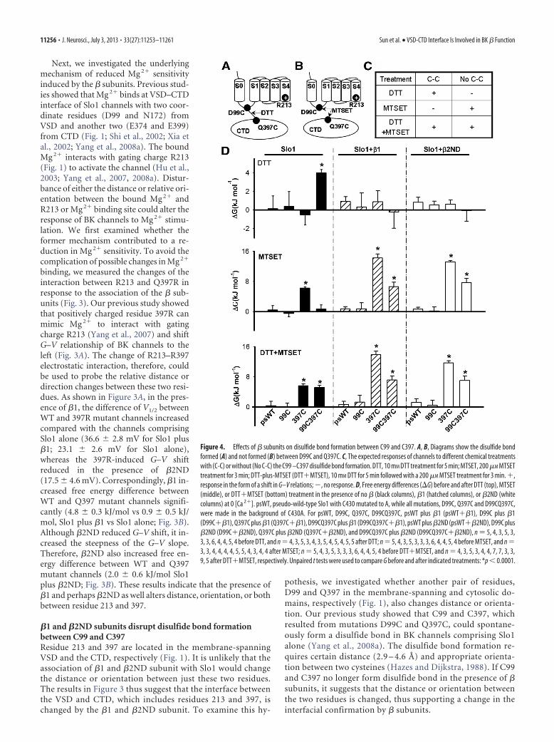
Next, we investigated the underlyingmechanism of reduced Mg 2� sensitivityinduced by the � subunits. Previous stud-ies showed that Mg 2� binds at VSD–CTDinterface of Slo1 channels with two coor-dinate residues (D99 and N172) fromVSD and another two (E374 and E399)from CTD (Fig. 1; Shi et al., 2002; Xia etal., 2002; Yang et al., 2008a). The boundMg 2� interacts with gating charge R213(Fig. 1) to activate the channel (Hu et al.,2003; Yang et al., 2007, 2008a). Distur-bance of either the distance or relative ori-entation between the bound Mg 2� andR213 or Mg 2� binding site could alter theresponse of BK channels to Mg 2� stimu-lation. We first examined whether theformer mechanism contributed to a re-duction in Mg 2� sensitivity. To avoid thecomplication of possible changes in Mg 2�
binding, we measured the changes of theinteraction between R213 and Q397R inresponse to the association of the � sub-units (Fig. 3). Our previous study showedthat positively charged residue 397R canmimic Mg 2� to interact with gatingcharge R213 (Yang et al., 2007) and shiftG–V relationship of BK channels to theleft (Fig. 3A). The change of R213–R397electrostatic interaction, therefore, couldbe used to probe the relative distance ordirection changes between these two resi-dues. As shown in Figure 3A, in the pres-ence of �1, the difference of V1/2 betweenWT and 397R mutant channels increasedcompared with the channels comprisingSlo1 alone (36.6 2.8 mV for Slo1 plus�1; 23.1 2.6 mV for Slo1 alone),whereas the 397R-induced G–V shiftreduced in the presence of �2ND(17.5 4.6 mV). Correspondingly, �1 in-creased free energy difference betweenWT and Q397 mutant channels signifi-cantly (4.8 0.3 kJ/mol vs 0.9 0.5 kJ/mol, Slo1 plus �1 vs Slo1 alone; Fig. 3B).Although �2ND reduced G–V shift, it in-creased the steepness of the G–V slope.Therefore, �2ND also increased free en-ergy difference between WT and Q397mutant channels (2.0 0.6 kJ/mol Slo1plus �2ND; Fig. 3B). These results indicate that the presence of�1 and perhaps �2ND as well alters distance, orientation, or bothbetween residue 213 and 397.
�1 and �2ND subunits disrupt disulfide bond formationbetween C99 and C397Residue 213 and 397 are located in the membrane-spanningVSD and the CTD, respectively (Fig. 1). It is unlikely that theassociation of �1 and �2ND subunit with Slo1 would changethe distance or orientation between just these two residues.The results in Figure 3 thus suggest that the interface betweenthe VSD and CTD, which includes residues 213 and 397, ischanged by the �1 and �2ND subunit. To examine this hy-
pothesis, we investigated whether another pair of residues,D99 and Q397 in the membrane-spanning and cytosolic do-mains, respectively (Fig. 1), also changes distance or orienta-tion. Our previous study showed that C99 and C397, whichresulted from mutations D99C and Q397C, could spontane-ously form a disulfide bond in BK channels comprising Slo1alone (Yang et al., 2008a). The disulfide bond formation re-quires certain distance (2.9 – 4.6 Å) and appropriate orienta-tion between two cysteines (Hazes and Dijkstra, 1988). If C99and C397 no longer form disulfide bond in the presence of �subunits, it suggests that the distance or orientation betweenthe two residues is changed, thus supporting a change in theinterfacial confirmation by � subunits.
Figure 4. Effects of � subunits on disulfide bond formation between C99 and C397. A, B, Diagrams show the disulfide bondformed (A) and not formed (B) between D99C and Q397C. C, The expected responses of channels to different chemical treatmentswith (C-C) or without (No C-C) the C99 –C397 disulfide bond formation. DTT, 10 mM DTT treatment for 5 min; MTSET, 200 �M MTSETtreatment for 3 min; DTT-plus-MTSET (DTT�MTSET), 10 mM DTT for 5 min followed with a 200 �M MTSET treatment for 3 min. �,response in the form of a shift in G–V relations; �, no response. D, Free energy differences (�G) before and after DTT (top), MTSET(middle), or DTT�MTSET (bottom) treatment in the presence of no � (black columns), �1 (hatched columns), or �2ND (whitecolumns) at 0 [Ca 2�]. psWT, pseudo-wild-type Slo1 with C430 mutated to A, while all mutations, D99C, Q397C and D99CQ397C,were made in the background of C430A. For psWT, D99C, Q397C, D99CQ397C, psWT plus �1 (psWT��1), D99C plus �1(D99C��1), Q397C plus �1 (Q397C��1), D99CQ397C plus �1 (D99CQ397C��1), psWT plus �2ND (psWT��2ND), D99C plus�2ND (D99C��2ND), Q397C plus �2ND (Q397C��2ND), and D99CQ397C plus �2ND (D99CQ397C��2ND), n 5, 4, 3, 5, 3,3, 3, 6, 4, 4, 5, 4 before DTT, and n 4, 3, 5, 3, 4, 3, 5, 4, 5, 4, 5, 5 after DTT; n 5, 4, 3, 5, 3, 3, 3, 6, 4, 4, 5, 4 before MTSET, and n 3, 3, 4, 4, 4, 4, 5, 5, 4, 3, 4, 4 after MTSET; n 5, 4, 3, 5, 3, 3, 3, 6, 4, 4, 5, 4 before DTT�MTSET, and n 4, 3, 5, 3, 4, 4, 7, 7, 3, 3,9, 5 after DTT�MTSET, respectively. Unpaired t tests were used to compare G before and after indicated treatments: *p � 0.0001.
11256 • J. Neurosci., July 3, 2013 • 33(27):11253–11261 Sun et al. • VSD-CTD Interface Is Involved in BK � Function
DTT and MTSET reagents were used to detect the formationof the disulfide bond. DTT is a reducing agent that can breakdisulfide bond into single cysteines (Fig. 4A), whereas MTSETcan covalently modify single cysteines and introduce a positivecharge into cysteine (Fig. 4B). C430A was used as pseudo-WT(psWT) here since C430 in native Slo1 channel can be modifiedby MTSET and affect channel activation (Zhang and Horrigan,2005). Our previous studies showed that if C99 and C397 form adisulfide bond, DTT treatment affects channel activation andshifts the G–V curve to the left, whereas MTSET has no effect onchannel gating since the disulfide bond protects C99 and C397from MTSET modification; however, if treating channels withDTT first to break the disulfide bond, both C397 and C99 will beavailable for MTSET modification and the modification of C397shifts G–V relation to more negative voltages (Yang et al., 2008a).On the other hand, if C99 and C397 does not form disulfide bond,DTT should not affect channel gating, whereas MTSET wouldmodify channel activation with or without DTT pretreatment(Fig. 4C).
We repeated the previous experiments on channels compris-ing Slo1 alone and obtained results as expected for a disulfide
bond being formed between C99 andC397 (Fig. 4C,D, left, black columns).DTT treatment (Fig. 4D, top left) shiftedG–V relation to left, corresponding to afree energy change of 4.0 0.4 kJ/mol,only in the double mutation D99CQ397C(disulfide bond formed) but not in threecontrol channels, psWT, D99C, or Q397C(no disulfide bond formed). On the otherhand, MTSET treatment (Fig. 4D, middleleft) shifted G–V to more negative volt-ages, corresponding to a free energychange of 6.2 0.3 kJ/mol in only Q397C,but not in D99CQ397C. After the pre-treatment of DTT, however, MTSETtreatment (Fig. 4D, bottom left) shiftedG–V of both Q397C and D99CQ397C tomore negative voltages.
Contrast to the results on channelscomprising Slo1 alone (Fig. 4D, left, blackcolumns), the results on channels with thecoexpression of �1 (Fig. 4D, middle,hatched columns) or �2ND (Fig. 4D,right, white columns) are consistent withno disulfide bond being formed be-tween C99 and C397 (Fig. 4C). DTTtreatment had no effect on D99CQ397Cor any of the control channels (Fig. 4 D,top middle and right), while MTSETtreatment shifted G–V of both Q397Cand D99CQ397C to more negative volt-ages, corresponding to significant freeenergy changes (Fig. 4 D, middle middleand right). Pretreatment of DTT did notaffect the results of MTSET treatment(Fig. 4 D, bottom middle and right).These results indicate that the associa-tion of the �1 and �2ND subunits dis-rupted the formation of the disulfidebond between C99 and C397, supportingthe idea that the � subunits altered the VS-D–CTD interface. Considering that D99 is
one of the Mg2� binding residues and the Mg2� binding site isformed between the VSD–CTD domains, these results suggest thatMg2� binding could be perturbed by � subunits.
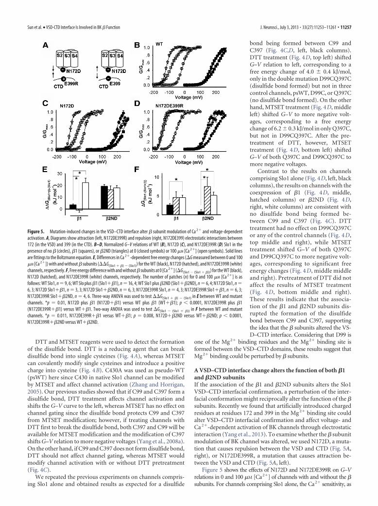
A VSD–CTD interface change alters the function of both �1and �2ND subunitsIf the association of the �1 and �2ND subunits alters the Slo1VSD–CTD interfacial confirmation, a perturbation of the inter-facial conformation might reciprocally alter the function of the �subunits. Recently we found that artificially introduced chargedresidues at residues 172 and 399 in the Mg 2� binding site couldalter VSD–CTD interfacial confirmation and affect voltage- andCa 2�-dependent activation of BK channels through electrostaticinteraction (Yang et al., 2013). To examine whether the � subunitmodulation of BK channel was altered, we used N172D, a muta-tion that causes repulsion between the VSD and CTD (Fig. 5A,right), or N172DE399R, a mutation that causes attraction be-tween the VSD and CTD (Fig. 5A, left).
Figure 5 shows the effects of N172D and N172DE399R on G–Vrelations in 0 and 100 �M [Ca2�] of channels with and without the �subunits. For channels comprising Slo1 alone, the Ca2� sensitivity, as
Figure 5. Mutation-induced changes in the VSD–CTD interface alter � subunit modulation of Ca 2� and voltage-dependentactivation. A, Diagrams show attraction (left, N172DE399R) and repulsion (right, N172DE399) electrostatic interactions between172 (in the VSD) and 399 (in the CTD). B–D, Normalized G–V relations of WT (B), N172D (C), and N172DE399R (D) Slo1 in thepresence of no � (circles), �1 (squares), or �2ND (triangles) at 0 (closed symbols) or 100 �M [Ca 2�] (open symbols). Solid linesare fittings to the Boltzmann equation. E, Differences in Ca 2�-dependent free energy changes (�G measured between 0 and 100�M [Ca 2�]) with and without � subunits (��G(Slo1 � �) � (Slo1)) for the WT (black), N172D (hatched), and N172DE399R (white)channels, respectively. F, Free energy difference with and without � subunits at 0 [Ca 2�] (�G(Slo1 � (Slo1 � �))) for the WT (black),N172D (hatched), and N172DE399R (white) channels, respectively. The number of patches (n) for 0 and 100 �M [Ca 2�] is asfollows: WT Slo1, n 9, 6; WT Slo plus �1 (Slo1��1), n 16, 4; WT Slo1 plus �2ND (Slo1��2ND), n 6, 4; N172D Slo1, n 6, 3; N172D Slo1��1, n 3, 3; N172D Slo1��2ND, n 6, 3; N172DE399R Slo1, n 4, 3; N172DE399R Slo1��1, n 6, 3;N172DE399R Slo1��2ND, n 4, 6. Three-way ANOVA was used to test ��G(Slo1 � �) � (Slo1) in E between WT and mutantchannels. *p 0.01, N172D plus �1 (N172D��1) versus WT plus �1 (WT��1); p � 0.0001, N172DE399R plus �1(N172DE399R��1) versus WT��1. Two-way ANOVA was used to test �G(Slo1 � (Slo1 � �)) in F between WT and mutantchannels. *p 0.011, N172DE399R��1 versus WT��1; p 0.008, N172D��2ND versus WT��2ND; p � 0.0001,N172DE399R��2ND versus WT��2ND.
Sun et al. • VSD-CTD Interface Is Involved in BK � Function J. Neurosci., July 3, 2013 • 33(27):11253–11261 • 11257
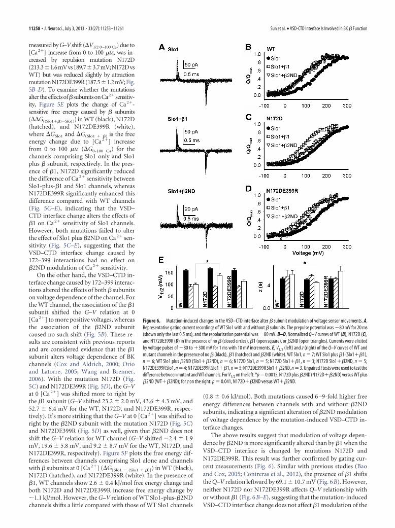
measured by G–V shift (�V1/2 0–100 Ca) due to[Ca2�] increase from 0 to 100 �M, was in-creased by repulsion mutation N172D(213.31.6mVvs189.73.7mV;N172DvsWT) but was reduced slightly by attractionmutationN172DE399R(187.51.2mV;Fig.5B–D). To examine whether the mutationsaltertheeffectsof�subunitsonCa2� sensitiv-ity, Figure 5E plots the change of Ca2�-sensitive free energy caused by � subunits(��G((Slo1��)�Slo1)) in WT (black), N172D(hatched), and N172DE399R (white),where �GSlo1 and �G(Slo1 � �) is the freeenergy change due to [Ca 2�] increasefrom 0 to 100 �M (�G0-100 Ca) for thechannels comprising Slo1 only and Slo1plus � subunit, respectively. In the pres-ence of �1, N172D significantly reducedthe difference of Ca 2� sensitivity betweenSlo1-plus-�1 and Slo1 channels, whereasN172DE399R significantly enhanced thisdifference compared with WT channels(Fig. 5C–E), indicating that the VSD–CTD interface change alters the effects of�1 on Ca 2� sensitivity of Slo1 channels.However, both mutations failed to alterthe effect of Slo1 plus �2ND on Ca 2� sen-sitivity (Fig. 5C–E), suggesting that theVSD–CTD interface change caused by172–399 interactions had no effect on�2ND modulation of Ca 2� sensitivity.
On the other hand, the VSD–CTD in-terface change caused by 172–399 interac-tions altered the effects of both � subunitson voltage dependence of the channel, Forthe WT channel, the association of the �1subunit shifted the G–V relation at 0[Ca 2�] to more positive voltages, whereasthe association of the �2ND subunitcaused no such shift (Fig. 5B). These re-sults are consistent with previous reportsand are considered evidence that the �1subunit alters voltage dependence of BKchannels (Cox and Aldrich, 2000; Orioand Latorre, 2005; Wang and Brenner,2006). With the mutation N172D (Fig.5C) and N172DE399R (Fig. 5D), the G–Vat 0 [Ca 2�] was shifted more to right bythe �1 subunit (G–V shifted 23.2 2.0 mV, 43.6 4.3 mV, and52.7 6.4 mV for the WT, N172D, and N172DE399R, respec-tively). It’s more striking that the G–V at 0 [Ca 2�] was shifted toright by the �2ND subunit with the mutation N172D (Fig. 5C)and N172DE399R (Fig. 5D) as well, given that �2ND does notshift the G–V relation for WT channel (G–V shifted �2.4 1.9mV, 19.6 5.8 mV, and 9.2 8.7 mV for the WT, N172D, andN172DE399R, respectively). Figure 5F plots the free energy dif-ferences between channels comprising Slo1 alone and channelswith � subunits at 0 [Ca 2�] (�G[Slo1 � (Slo1 � �)]) in WT (black),N172D (hatched), and N172DE399R (white). In the presence of�1, WT channels show 2.6 0.4 kJ/mol free energy change andboth N172D and N172DE399R increase free energy change by�1.1 kJ/mol. However, the G–V relation of WT Slo1-plus-�2NDchannels shifts a little compared with those of WT Slo1 channels
(0.8 0.6 kJ/mol). Both mutations caused 6 –9-fold higher freeenergy differences between channels with and without �2NDsubunits, indicating a significant alteration of �2ND modulationof voltage dependence by the mutation-induced VSD–CTD in-terface changes.
The above results suggest that modulation of voltage depen-dence by �2ND is more significantly altered than by �1 when theVSD–CTD interface is changed by mutations N172D andN172DE399R. This result was further confirmed by gating cur-rent measurements (Fig. 6). Similar with previous studies (Baoand Cox, 2005; Contreras et al., 2012), the presence of �1 shiftsthe Q–V relation leftward by 69.1 10.7 mV (Fig. 6B). However,neither N172D nor N172DE399R affects Q–V relationship withor without �1 (Fig. 6B–E), suggesting that the mutation-inducedVSD–CTD interface change does not affect �1 modulation of the
Figure 6. Mutation-induced changes in the VSD–CTD interface alter � subunit modulation of voltage sensor movements. A,Representative gating current recordings of WT Slo1 with and without � subunits. The prepulse potential was �80 mV for 20 ms(shown only the last 0.5 ms), and the repolarization potential was �80 mV. B–D, Normalized Q–V curves of WT (B), N172D (C),and N172DE399R (D) in the presence of no � (closed circles), �1 (open square), or �2ND (open triangles). Currents were elicitedby voltage pulses of �80 to �300 mV for 1 ms with 10 mV increments. E, V1/2 (left) and z (right) of the Q–V curves of WT andmutant channels in the presence of no � (black), �1 (hatched) and �2ND (white). WT Slo1, n 7; WT Slo1 plus �1 (Slo1��1),n 6; WT Slo1 plus �2ND (Slo1��2ND), n 6; N172D Slo1, n 5; N172D Slo1��1, n 3; N172D Slo1��2ND, n 5;N172DE399R Slo1, n4; N172DE399R Slo1��1, n5; N172DE399R Slo1��2ND, n3. Unpaired t tests were used to test thedifference between mutant and WT channels. For V1/2 on the left: *p0.0015, N172D plus �2ND (N172D��2ND) versus WT plus�2ND (WT��2ND); for z on the right: p 0.041, N172D��2ND versus WT��2ND.
11258 • J. Neurosci., July 3, 2013 • 33(27):11253–11261 Sun et al. • VSD-CTD Interface Is Involved in BK � Function
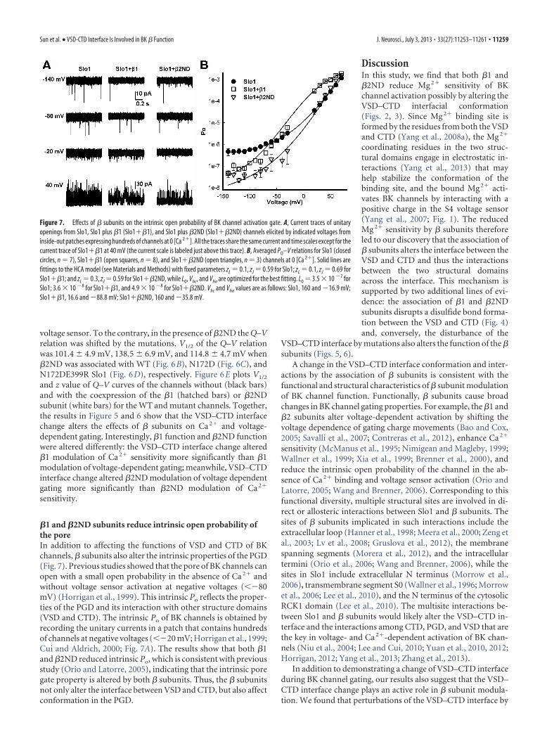
voltage sensor. To the contrary, in the presence of �2ND the Q–Vrelation was shifted by the mutations. V1/2 of the Q–V relationwas 101.4 4.9 mV, 138.5 6.9 mV, and 114.8 4.7 mV when�2ND was associated with WT (Fig. 6B), N172D (Fig. 6C), andN172DE399R Slo1 (Fig. 6D), respectively. Figure 6E plots V1/2
and z value of Q–V curves of the channels without (black bars)and with the coexpression of the �1 (hatched bars) or �2NDsubunit (white bars) for the WT and mutant channels. Together,the results in Figure 5 and 6 show that the VSD–CTD interfacechange alters the effects of � subunits on Ca 2� and voltage-dependent gating. Interestingly, �1 function and �2ND functionwere altered differently: the VSD–CTD interface change altered�1 modulation of Ca 2� sensitivity more significantly than �1modulation of voltage-dependent gating; meanwhile, VSD–CTDinterface change altered �2ND modulation of voltage dependentgating more significantly than �2ND modulation of Ca 2�
sensitivity.
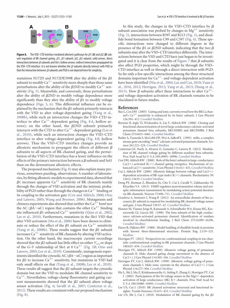
�1 and �2ND subunits reduce intrinsic open probability ofthe poreIn addition to affecting the functions of VSD and CTD of BKchannels, � subunits also alter the intrinsic properties of the PGD(Fig. 7). Previous studies showed that the pore of BK channels canopen with a small open probability in the absence of Ca 2� andwithout voltage sensor activation at negative voltages (��80mV) (Horrigan et al., 1999). This intrinsic Po reflects the proper-ties of the PGD and its interaction with other structure domains(VSD and CTD). The intrinsic Po of BK channels is obtained byrecording the unitary currents in a patch that contains hundredsof channels at negative voltages (��20 mV; Horrigan et al., 1999;Cui and Aldrich, 2000; Fig. 7A). The results show that both �1and �2ND reduced intrinsic Po, which is consistent with previousstudy (Orio and Latorre, 2005), indicating that the intrinsic poregate property is altered by both � subunits. Thus, the � subunitsnot only alter the interface between VSD and CTD, but also affectconformation in the PGD.
DiscussionIn this study, we find that both �1 and�2ND reduce Mg 2� sensitivity of BKchannel activation possibly by altering theVSD–CTD interfacial conformation(Figs. 2, 3). Since Mg 2� binding site isformed by the residues from both the VSDand CTD (Yang et al., 2008a), the Mg 2�
coordinating residues in the two struc-tural domains engage in electrostatic in-teractions (Yang et al., 2013) that mayhelp stabilize the conformation of thebinding site, and the bound Mg 2� acti-vates BK channels by interacting with apositive charge in the S4 voltage sensor(Yang et al., 2007; Fig. 1). The reducedMg 2� sensitivity by � subunits thereforeled to our discovery that the association of� subunits alters the interface between theVSD and CTD and thus the interactionsbetween the two structural domainsacross the interface. This mechanism issupported by two additional lines of evi-dence: the association of �1 and �2NDsubunits disrupts a disulfide bond forma-tion between the VSD and CTD (Fig. 4)and, conversely, the disturbance of the
VSD–CTD interface by mutations also alters the function of the �subunits (Figs. 5, 6).
A change in the VSD–CTD interface conformation and inter-actions by the association of � subunits is consistent with thefunctional and structural characteristics of � subunit modulationof BK channel function. Functionally, � subunits cause broadchanges in BK channel gating properties. For example, the �1 and�2 subunits alter voltage-dependent activation by shifting thevoltage dependence of gating charge movements (Bao and Cox,2005; Savalli et al., 2007; Contreras et al., 2012), enhance Ca 2�
sensitivity (McManus et al., 1995; Nimigean and Magleby, 1999;Wallner et al., 1999; Xia et al., 1999; Brenner et al., 2000), andreduce the intrinsic open probability of the channel in the ab-sence of Ca 2� binding and voltage sensor activation (Orio andLatorre, 2005; Wang and Brenner, 2006). Corresponding to thisfunctional diversity, multiple structural sites are involved in di-rect or allosteric interactions between Slo1 and � subunits. Thesites of � subunits implicated in such interactions include theextracellular loop (Hanner et al., 1998; Meera et al., 2000; Zeng etal., 2003; Lv et al., 2008; Gruslova et al., 2012), the membranespanning segments (Morera et al., 2012), and the intracellulartermini (Orio et al., 2006; Wang and Brenner, 2006), while thesites in Slo1 include extracellular N terminus (Morrow et al.,2006), transmembrane segment S0 (Wallner et al., 1996; Morrowet al., 2006; Lee et al., 2010), and the N terminus of the cytosolicRCK1 domain (Lee et al., 2010). The multisite interactions be-tween Slo1 and � subunits would likely alter the VSD–CTD in-terface and the interactions among CTD, PGD, and VSD that arethe key in voltage- and Ca 2�-dependent activation of BK chan-nels (Niu et al., 2004; Lee and Cui, 2010; Yuan et al., 2010, 2012;Horrigan, 2012; Yang et al., 2013; Zhang et al., 2013).
In addition to demonstrating a change of VSD–CTD interfaceduring BK channel gating, our results also suggest that the VSD–CTD interface change plays an active role in � subunit modula-tion. We found that perturbations of the VSD–CTD interface by
Figure 7. Effects of � subunits on the intrinsic open probability of BK channel activation gate. A, Current traces of unitaryopenings from Slo1, Slo1 plus �1 (Slo1��1), and Slo1 plus �2ND (Slo1��2ND) channels elicited by indicated voltages frominside-out patches expressing hundreds of channels at 0 [Ca 2�]. All the traces share the same current and time scales except for thecurrent trace of Slo1��1 at 40 mV (the current scale is labeled just above this trace). B, Averaged PO–V relations for Slo1 (closedcircles, n 7), Slo1��1 (open squares, n 8), and Slo1��2ND (open triangles, n 3) channels at 0 [Ca 2�]. Solid lines arefittings to the HCA model (see Materials and Methods) with fixed parameters zL 0.1, zJ 0.59 for Slo1; zL 0.1, zJ 0.69 forSlo1��1; and zL 0.3, zJ 0.59 for Slo1��2ND, while L0, Vhc, and Vho are optimized for the best fitting. L0 3.5 � 10 �7 forSlo1; 3.6 � 10 �8 for Slo1��1, and 4.9 � 10 �8 for Slo1��2ND. Vhc and Vho values are as follows: Slo1, 160 and �16.9 mV;Slo1��1, 16.6 and �88.8 mV; Slo1��2ND, 160 and �35.8 mV.
Sun et al. • VSD-CTD Interface Is Involved in BK � Function J. Neurosci., July 3, 2013 • 33(27):11253–11261 • 11259
mutations N172D and N172DE399R alter the ability of the �1subunit to modify Ca 2� sensitivity more sharply than those sameperturbations alter the ability of the �2ND to modify Ca 2� sen-sitivity (Fig. 5). Meanwhile, and conversely, those perturbationsalter the ability of �2ND to modify voltage dependence moresignificantly than they alter the ability of �1 to modify voltagedependence (Figs. 5, 6). This differential influence can be ex-plained by the mechanism that the �1 subunit primarily interactswith the VSD to alter voltage-dependent gating (Yang et al.,2008b), while such an interaction changes the VSD–CTD in-terface to alter Ca 2�-dependent gating (Fig. 8 A, hollow ar-rows); on the other hand, the �2ND subunit primarilyinteracts with the CTD to alter Ca 2�-dependent gating (Lee etal., 2010), while such an interaction changes the VSD–CTDinterface to alter voltage-dependent gating (Fig. 8 B, hollowarrows). Thus the VSD–CTD interface changes provide anallosteric mechanism to propagate the effects of different �subunits to all aspects of BK channel gating, and the pertur-bation of the VSD–CTD interface has a lesser influence on theeffects of the primary interaction between a � subunit and Sol1than on the downstream allosteric effects.
The proposed mechanism (Fig. 8) may also explain many pre-vious, sometimes puzzling, observations. A number of laborato-ries, by fitting allosteric models to experimental data, showed that�1 increases apparent Ca 2� sensitivity of BK channels mainlythrough the changes of VSD activation and the intrinsic proba-bility of PGD rather than through the changes in Ca 2� binding orits coupling to the activation gate (Cox and Aldrich, 2000; Orioand Latorre, 2005; Wang and Brenner, 2006). Mutagenesis andchimera experiments also showed that neither the Ca 2� bowl northe AC (�A��C) region that contains the other Ca 2� bindingsite influenced �1-enhanced Ca 2� sensitivity (Qian et al., 2002;Lee et al., 2010). Furthermore, mutations in the Slo1 VSD thatalter VSD activation (Ma et al., 2006) have been shown to elimi-nate or reduce the effect of the �1 subunit on Ca 2� sensitivity(Yang et al., 2008b). These results suggest that the �1 subunitincreases Ca 2� sensitivity of BK channels by altering VSD activa-tion. On the other hand, the macroscopic current recordingsshowed that the �2 subunit has little effect on either V1/2 or slopeof the G–V relationship of Slo1 at 0 Ca 2� (Fig. 5B; Orio andLatorre, 2005; Lee et al., 2010). Consistently, mutagenesis exper-iments identified the cytosolic AC (�A��C) region as importantfor �2 to increase Ca 2� sensitivity, but mutations in VSD hadonly small effects on that (Yang et al., 2008b; Lee et al., 2010).These results all suggest that the �2 subunit targets the cytosolicdomain but not the VSD to modulate BK channel sensitivity toCa 2�. Nevertheless, voltage-clamp fluorometry and gating cur-rent measurements showed that the �2 subunit alters voltagesensor activation (Fig. 6; Savalli et al., 2007; Contreras et al.,2012). These results are consistent with our proposed mechanism(Fig. 8).
In this study, the changes in the VSD–CTD interface by �subunit association was probed by changes in Mg 2� sensitivity(Fig. 2), interactions between R397 and R213 (Fig. 3), and disul-fide bond formation between C99 and C397 (Fig. 4). These spe-cific interactions were changed to different degrees in thepresence of the �1 or �2ND subunit, indicating that the two �subunits may alter the VSD–CTD interface differently. The inter-actions between the VSD and CTD have just begun to be investi-gated and it is clear from the results of Figure 7 that � subunitsalso affect PGD properties, which might be through the VSD–CTD interface as well or through a direct interaction with PGD.So far only a few specific interactions among the three structuraldomains important for Ca 2�- and voltage-dependent activationhave been identified (Niu et al., 2004; Lee and Cui, 2010; Yuan etal., 2010, 2012; Horrigan, 2012; Yang et al., 2013; Zhang et al.,2013). How � subunits affect these interactions to alter Ca 2�-and voltage-dependent activation of BK channels remains to beelucidated in future studies.
ReferencesBao L, Cox DH (2005) Gating and ionic currents reveal how the BKCa chan-
nel’s Ca 2� sensitivity is enhanced by its beta1 subunit. J Gen Physiol126:393– 412. CrossRef Medline
Brenner R, Jegla TJ, Wickenden A, Liu Y, Aldrich RW (2000) Cloning andfunctional characterization of novel large conductance calcium-activatedpotassium channel beta subunits, hKCNMB3 and hKCNMB4. J BiolChem 275:6453– 6461. CrossRef Medline
Butler A, Tsunoda S, McCobb DP, Wei A, Salkoff L (1993) mSlo, a complexmouse gene encoding “maxi” calcium-activated potassium channels. Sci-ence 261:221–224. CrossRef Medline
Contreras GF, Neely A, Alvarez O, Gonzalez C, Latorre R (2012) Modula-tion of BK channel voltage gating by different auxiliary beta subunits.Proc Natl Acad Sci U S A 109:18991–18996. CrossRef Medline
Cox DH, Aldrich RW (2000) Role of the beta1 subunit in large-conductanceCa(2�)-activated K(�) channel gating energetics. Mechanisms of en-hanced Ca(2�) sensitivity. J Gen Physiol 116:411– 432. CrossRef Medline
Cui J, Aldrich RW (2000) Allosteric linkage between voltage and Ca(2�)-dependent activation of BK-type mslo1 K(�) channels. Biochemistry 39:15612–15619. CrossRef Medline
Deng PY, Rotman Z, Blundon JA, Cho Y, Cui J, Cavalli V, Zakharenko SS,Klyachko VA (2013) FMRP regulates neurotransmitter release and syn-aptic information transmission by modulating action potential durationvia BK channels. Neuron 77:696 –711. CrossRef Medline
Gruslova A, Semenov I, Wang B (2012) An extracellular domain of the ac-cessory �1 subunit is required for modulating BK channel voltage sensorand gate. J Gen Physiol 139:57– 67. CrossRef Medline
Hanner M, Vianna-Jorge R, Kamassah A, Schmalhofer WA, Knaus HG, Kac-zorowski GJ, Garcia ML (1998) The beta subunit of the high conduc-tance calcium-activated potassium channel. Identification of residuesinvolved in charybdotoxin binding. J Biol Chem 273:16289 –16296.CrossRef Medline
Hazes B, Dijkstra BW (1988) Model building of disulfide bonds in proteinswith known three-dimensional structure. Protein Eng 2:119 –125.Medline
Horrigan FT (2012) Perspectives on: conformational coupling in ion chan-nels: conformational coupling in BK potassium channels. J Gen Physiol140:625– 634. CrossRef Medline
Horrigan FT, Aldrich RW (1999) Allosteric voltage gating of potassiumchannels II. Mslo channel gating charge movement in the absence ofCa(2�). J Gen Physiol 114:305–336. CrossRef Medline
Horrigan FT, Cui J, Aldrich RW (1999) Allosteric voltage gating of potas-sium channels I. Mslo ionic currents in the absence of Ca(2�). J GenPhysiol 114:277–304. CrossRef Medline
Hu L, Shi J, Ma Z, Krishnamoorthy G, Sieling F, Zhang G, Horrigan FT, CuiJ (2003) Participation of the S4 voltage sensor in the Mg2�-dependentactivation of large conductance (BK) K� channels. Proc Natl Acad SciU S A 100:10488 –10493. CrossRef Medline
Lee US, Cui J (2010) BK channel activation: structural and functional in-sights. Trends Neurosci 33:415– 423. CrossRef Medline
Lee US, Shi J, Cui J (2010) Modulation of BK channel gating by the �2
Figure 8. The VSD–CTD interface mediated allosteric pathways for �1 (A) and �2 (B) sub-unit regulation of BK channel gating. �1, �1 subunit; �2, �2 subunit; solid arrows, directinteractions between � subunits and Slo1; hollow arrows, indirect interactions propagated viathe VSD–CTD interface. It is not known whether the � subunits directly interact with PGD sothat the interaction between � subunits and PGD is not depicted here for simplicity.
11260 • J. Neurosci., July 3, 2013 • 33(27):11253–11261 Sun et al. • VSD-CTD Interface Is Involved in BK � Function
subunit involves both membrane-spanning and cytoplasmic domains ofSlo1. J Neurosci 30:16170 –16179. CrossRef Medline
Liu G, Niu X, Wu RS, Chudasama N, Yao Y, Jin X, Weinberg R, Zakharov SI,Motoike H, Marx SO, Karlin A (2010) Location of modulatory beta sub-units in BK potassium channels. J Gen Physiol 135:449 – 459. CrossRefMedline
Lorenz S, Heils A, Kasper JM, Sander T (2007) Allelic association of a trun-cation mutation of the KCNMB3 gene with idiopathic generalized epi-lepsy. Am J Med Genet B Neuropsychiatr Genet 144B:10 –13. CrossRefMedline
Lv C, Chen M, Gan G, Wang L, Xu T, Ding J (2008) Four-turn alpha-helicalsegment prevents surface expression of the auxiliary hbeta2 subunit ofBK-type channel. J Biol Chem 283:2709 –2715. Medline
Ma Z, Lou XJ, Horrigan FT (2006) Role of charged residues in the S1–S4voltage sensor of BK channels. J Gen Physiol 127:309 –328. CrossRefMedline
McManus OB, Helms LM, Pallanck L, Ganetzky B, Swanson R, Leonard RJ(1995) Functional role of the beta subunit of high conductance calcium-activated potassium channels. Neuron 14:645– 650. CrossRef Medline
Meera P, Wallner M, Toro L (2000) A neuronal beta subunit (KCNMB4)makes the large conductance, voltage- and Ca2�-activated K� channelresistant to charybdotoxin and iberiotoxin. Proc Natl Acad Sci U S A97:5562–5567. CrossRef Medline
Morera FJ, Alioua A, Kundu P, Salazar M, Gonzalez C, Martinez AD, StefaniE, Toro L, Latorre R (2012) The first transmembrane domain (TM1) ofbeta2-subunit binds to the transmembrane domain S1 of alpha-subunitin BK potassium channels. FEBS Lett 586:2287–2293. CrossRef Medline
Morrow JP, Zakharov SI, Liu G, Yang L, Sok AJ, Marx SO (2006) Definingthe BK channel domains required for beta1-subunit modulation. ProcNatl Acad Sci U S A 103:5096 –5101. CrossRef Medline
Nimigean CM, Magleby KL (1999) The beta subunit increases the Ca2�sensitivity of large conductance Ca2�-activated potassium channels byretaining the gating in the bursting states. J Gen Physiol 113:425– 440.CrossRef Medline
Niu X, Qian X, Magleby KL (2004) Linker-gating ring complex as passivespring and Ca(2�)-dependent machine for a voltage- and Ca(2�)-activated potassium channel. Neuron 42:745–756. CrossRef Medline
Orio P, Latorre R (2005) Differential effects of beta 1 and beta 2 subunits onBK channel activity. J Gen Physiol 125:395– 411. CrossRef Medline
Orio P, Torres Y, Rojas P, Carvacho I, Garcia ML, Toro L, Valverde MA,Latorre R (2006) Structural determinants for functional coupling be-tween the beta and alpha subunits in the Ca2�-activated K� (BK) chan-nel. J Gen Physiol 127:191–204. CrossRef Medline
Poulsen AN, Wulf H, Hay-Schmidt A, Jansen-Olesen I, Olesen J, Klaerke DA(2009) Differential expression of BK channel isoforms and beta-subunitsin rat neuro-vascular tissues. Biochim Biophys Acta 1788:380 –389.CrossRef Medline
Qian X, Nimigean CM, Niu X, Moss BL, Magleby KL (2002) Slo1 tail do-mains, but not the Ca2� bowl, are required for the beta 1 subunit toincrease the apparent Ca2� sensitivity of BK channels. J Gen Physiol120:829 – 843. CrossRef Medline
Savalli N, Kondratiev A, de Quintana SB, Toro L, Olcese R (2007) Modes ofoperation of the BKCa channel beta2 subunit. J Gen Physiol 130:117–131.CrossRef Medline
Shi J, Krishnamoorthy G, Yang Y, Hu L, Chaturvedi N, Harilal D, Qin J, CuiJ (2002) Mechanism of magnesium activation of calcium-activated po-tassium channels. Nature 418:876 – 880. CrossRef Medline
Wallner M, Meera P, Toro L (1996) Determinant for beta-subunit regula-tion in high-conductance voltage-activated and Ca(2�)-sensitive K�channels: an additional transmembrane region at the N terminus. ProcNatl Acad Sci U S A 93:14922–14927. CrossRef Medline
Wallner M, Meera P, Toro L (1999) Molecular basis of fast inactivation involtage and Ca2�-activated K� channels: a transmembrane beta-subunit homolog. Proc Natl Acad Sci U S A 96:4137– 4142. CrossRefMedline
Wang B, Brenner R (2006) An S6 mutation in BK channels reveals beta1subunit effects on intrinsic and voltage-dependent gating. J Gen Physiol128:731–744. CrossRef Medline
Wang YW, Ding JP, Xia XM, Lingle CJ (2002) Consequences of the stoichi-ometry of Slo1 � and auxiliary � subunits on functional properties oflarge-conductance Ca 2�-activated K� channels. J Neurosci 22:1550 –1561. Medline
Xia XM, Ding JP, Lingle CJ (1999) Molecular basis for the inactivation ofCa 2�- and voltage-dependent BK channels in adrenal chromaffin cellsand rat insulinoma tumor cells. J Neurosci 19:5255–5264. Medline
Xia XM, Zeng X, Lingle CJ (2002) Multiple regulatory sites in large-conductance calcium-activated potassium channels. Nature 418:880 –884. CrossRef Medline
Yan J, Aldrich RW (2010) LRRC26 auxiliary protein allows BK channel ac-tivation at resting voltage without calcium. Nature 466:513–516. CrossRefMedline
Yan J, Aldrich RW (2012) BK potassium channel modulation by leucine-rich repeat-containing proteins. Proc Natl Acad Sci U S A 109:7917–7922.CrossRef Medline
Yang H, Hu L, Shi J, Delaloye K, Horrigan FT, Cui J (2007) Mg2� mediatesinteraction between the voltage sensor and cytosolic domain to activateBK channels. Proc Natl Acad Sci U S A 104:18270 –18275. CrossRefMedline
Yang H, Shi J, Zhang G, Yang J, Delaloye K, Cui J (2008a) Activation of Slo1BK channels by Mg2� coordinated between the voltage sensor and RCK1domains. Nat Struct Mol Biol 15:1152–1159. CrossRef Medline
Yang H, Zhang G, Shi J, Lee US, Delaloye K, Cui J (2008b) Subunit-specificeffect of the voltage sensor domain on Ca2� sensitivity of BK channels.Biophys J 94:4678 – 4687. CrossRef Medline
Yang J, Yang H, Sun X, Delaloye K, Yang X, Moller A, Shi J, Cui J (2013)Interaction between residues in the Mg2�-binding site regulates BKchannel activation. J General Physiology 141:217–228. CrossRef Medline
Yuan P, Leonetti MD, Pico AR, Hsiung Y, MacKinnon R (2010) Structure ofthe human BK channel Ca2�-activation apparatus at 3.0 A resolution.Science 329:182–186. CrossRef Medline
Yuan P, Leonetti MD, Hsiung Y, MacKinnon R (2012) Open structure ofthe Ca2� gating ring in the high-conductance Ca2�-activated K� chan-nel. Nature 481:94 –97. CrossRef Medline
Zeng XH, Xia XM, Lingle CJ (2003) Redox-sensitive extracellular gatesformed by auxiliary beta subunits of calcium-activated potassium chan-nels. Nat Struct Biol 10:448 – 454. CrossRef Medline
Zhang G, Horrigan FT (2005) Cysteine modification alters voltage- andCa(2�)-dependent gating of large conductance (BK) potassium chan-nels. J Gen Physiol 125:213–236. CrossRef Medline
Zhang GH, Yang JQ, Yang HH, Shi JY, Yang X, Delaloye K, Moller A, Cui J(2013) Electrostatic interaction between the S4 –S5 linker and the cyto-plasmic end of S6 regulates BK channel gating. Philadelphia: BiophysicsSociety 57th Annual Meeting, 2367-Pos.
Sun et al. • VSD-CTD Interface Is Involved in BK � Function J. Neurosci., July 3, 2013 • 33(27):11253–11261 • 11261