Embed Size (px)
Citation preview

ORIGINAL PAPER
Cerebellum, Language, and Cognition in Autism and SpecificLanguage Impairment
Steven M. Hodge Æ Nikos Makris Æ David N. Kennedy Æ Verne S. Caviness Jr. ÆJames Howard Æ Lauren McGrath Æ Shelly Steele Æ Jean A. Frazier ÆHelen Tager-Flusberg Æ Gordon J. Harris
� Springer Science+Business Media, LLC 2009
Abstract We performed cerebellum segmentation and
parcellation on magnetic resonance images from right-
handed boys, aged 6–13 years, including 22 boys with
autism [16 with language impairment (ALI)], 9 boys with
Specific Language Impairment (SLI), and 11 normal con-
trols. Language-impaired groups had reversed asymmetry
relative to unimpaired groups in posterior-lateral cerebellar
lobule VIIIA (right side larger in unimpaired groups, left
side larger in ALI and SLI), contralateral to previous
findings in inferior frontal cortex language areas. Lobule
VIIA Crus I was smaller in SLI than in ALI. Vermis vol-
ume, particularly anterior I–V, was decreased in language-
impaired groups. Language performance test scores
correlated with lobule VIIIA asymmetry and with anterior
vermis volume. These findings suggest ALI and SLI
subjects show abnormalities in neurodevelopment of
fronto-corticocerebellar circuits that manage motor control
and the processing of language, cognition, working mem-
ory, and attention.
Keywords Autism � Specific language impairment �Cerebellum � Broca’s area � Asymmetry
Introduction
Autism is a neurodevelopmental disorder displaying defi-
cits in social interaction and communication skills, repeti-
tive behaviors, and stereotyped interests (APA 1994).
Language deficits range from absence of functional lan-
guage, to impairments in phonological processing, vocab-
ulary, and higher order syntax and semantics (Rapin 1996;
Tager-Flusberg 2003, 2006; Tager-Flusberg and Caronna
2007; Tager-Flusberg et al. 2005). However, some children
with autism have normal language skills (Tager-Flusberg
and Joseph 2003). Language-impaired children with autism
displayed a similar language profile to non-autistic children
with specific language impairment (SLI) (Bishop 2003;
Kjelgaard and Tager-Flusberg 2001), a disorder of delayed
language development in the absence of other cognitive
impairments. Furthermore, family and genetic linkage
studies have implicated overlap between autism and SLI
(Fisher et al. 2003; Santangelo and Folstein 1999).
Neuroimaging studies in autism and SLI have demon-
strated brain structure and function abnormalities in infe-
rior frontal gyrus (IFG) language-association cortex
(Broca’s area). In typically developing right-handed sub-
jects, Broca’s area regions tend to be larger in the left
hemisphere than in the right (Foundas et al. 1998; Keller
et al. 2007). However, magnetic resonance imaging (MRI)
S. M. Hodge � N. Makris � D. N. Kennedy �V. S. Caviness Jr. � J. Howard
Center for Morphometric Analysis, Massachusetts General
Hospital, Boston, MA, USA
S. M. Hodge � G. J. Harris (&)
Radiology Computer Aided Diagnostics Laboratory,
Massachusetts General Hospital, 25 New Chardon St. Suite
400C, Boston, MA 02114, USA
e-mail: [email protected]
L. McGrath � S. Steele � H. Tager-Flusberg
Lab of Cognitive Neuroscience, Boston University School
of Medicine, Boston, MA, USA
J. A. Frazier
Department of Psychiatry, Harvard Medical School, Boston,
MA, USA
J. A. Frazier
Center for Child and Adolescent Development, Department of
Psychiatry, Cambridge Health Alliance, Cambridge, MA, USA
123
J Autism Dev Disord
DOI 10.1007/s10803-009-0872-7
reports demonstrated reversal from normal IFG structural
asymmetry (larger in the right hemisphere) in right-handed
boys with autism (Herbert et al. 2002), particularly those
with autism and language-impairment (ALI), and in non-
autistic boys with specific language impairment (SLI)
(De Fosse et al. 2004). As early as 1986, researchers
identified reversed language dominance in right-handed
male autistic children in reports using cortical evoked
reponses to language stimuli (Dawson et al. 1986). Func-
tional imaging studies have also demonstrated reversed or
abnormal cerebral blood flow asymmetry in frontal lan-
guage cortex in children with autism (Burroni et al. 2008;
Chiron et al. 1995; Ohnishi et al. 2000), and during per-
formance of a language task in high-functioning adults
with autism (Boddaert et al. 2004; Muller et al. 1999).
High-functioning adults with autism spectrum disorders
(ASD) showed decreased functional MRI (fMRI) activa-
tion in Broca’s area in response to syntactic and semantic
tasks (Harris et al. 2006; Just et al. 2004) or increased right
frontal activation (Takeuchi et al. 2004), further implicat-
ing abnormalities in frontal language areas in autism.
Functional neuroimaging studies in right-handed normal
control subjects have demonstrated contralateral activation
in right posterior lateral cerebellum in concert with left
inferior frontal activation during language tasks (Binder
et al. 1997; Desmond et al. 1997; Harris et al. 2006;
Hubrich-Ungureanu et al. 2002; Jansen et al. 2005; Peter-
sen et al. 1988; Xiang et al. 2003), including phonological
processing (Mathiak et al. 2002). While normal control
subjects displayed lexical-semantic fMRI activation in left
inferior frontal gyrus and also in right posterior lateral
cerebellum, high-functioning adults with ASD had
decreased Broca’s area activation and did not present right
cerebellar activation (Harris et al. 2006).
The cerebellum was traditionally associated primarily
with sensorimotor function and balance. However, cere-
bellum has extensive reciprocal connections with cerebral
cortex and limbic systems (Schmahmann 1996, 2001). The
expanded neocerebellar hemispheres evolved in concert
with cerebral association areas, in particular prefrontal
cortex, facilitating a cerebellar role in language processing
(Leiner et al. 1986). There are robust interconnections
between frontal cortex language regions and contralateral
posterior cerebellar hemispheres (Gebhart et al. 2002;
Roskies et al. 2001; Stoodley and Schmahmann 2009).
These fronto-corticocerebellar circuits could facilitate most
areas of cognitive function, including language, executive
function, working memory, attention, and emotion (Makris
et al. 2005; Schmahmann et al. 2001). Clinical lesion
studies and functional neuroimaging link anterior cerebel-
lum with sensorimotor function, while posterior lateral
cerebellum (lobules VI–VIII) is associated with language,
verbal working memory, cognition, and attention (Stoodley
and Schmahmann 2009; Desmond and Fiez 1998; Leiner
et al. 1991; Levisohn et al. 2000; Neau et al. 2000; Petersen
et al. 1989; Riva and Giorgi 2000; Schmahmann and
Sherman 1998; Harris et al. 2006).
Cerebellar neuropathology has been implicated in aut-
ism, including enlarged IV ventricle, loss of Purkinje cells
in lateral and inferior cerebellar cortex (most severe in
posterior lateral hemispheres), and abnormal or reduced
numbers of neurons in deep cerebellar nuclei (Bauman and
Kemper 1985; Kemper and Bauman 1998; Kemper and
Bauman 2002; Kulesza and Mangunay 2008; Ritvo et al.
1986). Subjects with partial cerebellar agenesis demon-
strate autistic-like behaviors, including stereotypical per-
formance, obsessive rituals, difficulty understanding social
cues, tactile defensiveness, perseveration, disinhibition,
poor working memory, and language deficits such as
problems with verbal fluency, expressive language delay,
impaired prosody, and overgeneralization of past tense
verbs (Bobylova et al. 2007; Chheda et al. 2002;
Schmahmann 2004). Several prior structural imaging
studies implicated cerebellar deficits in autism, although
the neuroimaging literature on this issue is mixed (Stan-
field et al. 2008; Brambilla et al. 2003). While several
studies have demonstrated decreased vermis size in autism
(Courchesne et al. 1994; Courchesne et al. 1988; Ha-
shimoto et al. 1993; Kaufmann et al. 2003), other studies
have not replicated this (Hardan et al. 2001; Kleiman et al.
1992; Piven et al. 1992, 1997), or have related it to dif-
ferences in intelligence (Piven and Arndt 1995; Stanfield
et al. 2008). Thus, there is variability in the structural
imaging literature related to cerebellum and autism
(Courchesne 1999; Piven and Arndt 1995; Rapin 1999).
However, most prior cerebellar imaging studies in autism
have focused on the measurement of the cross-sectional
area of the vermis.
The current study is the first to systematically assess
specific putative cognitive and language-related regions in
the cerebellar hemispheres using cerebellar volumetric
segmentation and parcellation with MRI in groups of
normal control (NC) boys, boys with autism (with and
without language impairment; ALI and ALN, respec-
tively), and in boys with SLI. Since posterior lateral cere-
bellar lobules VI-VIII were identified through lesion and
functional neuroimaging studies as most closely tied to
language, verbal working memory, and cognition (Stoodley
and Schmahmann 2009), our hypotheses were: (1) poster-
ior lateral cerebellar hemisphere lobules VI–VIII would
show reversed asymmetry in ALI and SLI relative to ALN
and NC, contralateral to that previously reported in inferior
frontal language regions; (2) volumetric differences in
lobules VI–VIII would be observed between groups; and
(3) decreased vermis volume would be observed in ALI
and SLI if vermis effects were tied to language deficits, or
J Autism Dev Disord
123
alternatively in ALI and ALN if vermis effects were more
closely tied to autism symptoms.
Methods
Participants
Subjects were identical to those previously described in a
study of volumetric asymmetry in language-association
cerebral cortex (De Fosse et al. 2004), including 22 boys
with autism [16 of whom had language impairment (ALI)
and 6 with normal language (ALN)], 9 boys with specific
language impairment (SLI), and 11 normal control (NC)
boys. This project was approved by the human studies
institutional review board (IRB), and all subjects enrolled
in this study provided signed assent (for minors) and
parental consent.
Subjects included males 6–13 years old (Table 1), with
no significant group-matching differences in age among the
groups, F(3, 38) = 1.3, p = 0.3. All subjects were pre-
dominantly right-handed, as determined by the Edinburgh
Inventory in NC subjects (Oldfield 1971) and by the Dean
Laterality Preference Schedule in all other groups (Dean
1978, 1982). Subjects were excluded if they had neuro-
logical damage or had been diagnosed with Fragile X,
neurofibromatosis, cerebral palsy, tuberous sclerosis,
William’s syndrome or Down’s syndrome. Autism spec-
trum disorders were ruled out in SLI and NC groups. The
NC group had no DSM-IV (Diagnostic and Statistical
Manual of Mental Disorders, version 4) Axis I diagnoses
(APA 1994) based on consensus agreement between the
Schedule for Affective Disorders and Schizophrenia for
School-Age Children—Epidemiologic Version (Orvaschel
and Puig-Antich 1987) and clinical interview with a board-
certified child psychiatrist (JAF). None of the children were
currently on psychotropic medications.
Diagnosis of autism was established via the Autism
Diagnostic Interview—Revised (Lord et al. 1994), Autism
Diagnostic Observation Schedule (Lord et al. 2000), and
examination by an expert clinician to verify that subjects
met DSM-IV criteria for autism (APA 1994). Language
skills were assessed using the Clinical Evaluation of Lan-
guage Fundamentals (CELF, 3rd edition; Semel et al.
1995) and the nonsense word repetition subtest of the
NEPSY (Korkman et al. 1998). Criteria for language
impairment were a CELF score B81 or a score B6 on the
NEPSY nonsense word repetition subtest. These criteria for
specific language impairment (SLI) were selected based on
current research on language impairments in older children
(Tager-Flusberg and Cooper 1999), and are similar to those
used in other research on SLI (e.g., Conti-Ramsden 2003).
Based on these criteria, 16 subjects with autism were
classified with language impairment (ALI) and six subjects
with autism were classified with normal language (ALN).
Subjects with SLI had a history of significant language
delay and had been referred for clinical treatment. Of the
nine subjects in the SLI group, five met the above criteria
for current language impairment. Those subjects with only
a history of language impairment fell within the normal
limits on the language measures at the time of testing,
which is common in older children with SLI (Conti-
Ramsden and Botting 1999). We included these children
because although they no longer met strict criteria for a
current SLI diagnosis, they all had documented histories of
poor performance on language tests and were all currently
still struggling with language-based learning difficulties,
which is a common developmental trajectory for children
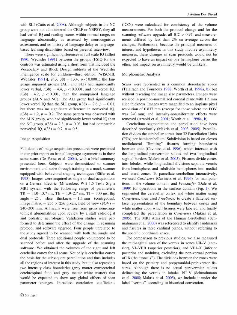
Table 1 Characteristics of the subject groups
NC
(mean ± SD)
n = 11
ALN
(mean ± SD)
n = 6
ALI
(mean ± SD)
n = 16
SLI
(mean ± SD)
n = 9
Overall Post hoc contrasts
F P NC & ALN vs.
ALI & SLI
NC vs.
ALN
SLI vs.
ALI
Age (years) 10.4 ± 2.7 8.3 ± 0.9 9.8 ± 2.1 9.9 ± 2.3 1.3 0.3
Full scale IQa 114.5 ± 11.3 109.3 ± 24.1 78.3 ± 14.7 93.4 ± 15.5 13.4 \0.001 * *
Verbal IQ 115.9 ± 12.4 97.7 ± 19.3 75.1 ± 15.9 92.5 ± 15.6 14.9 \0.001 * * *
Nonverbal IQ 110.6 ± 11 116.3 ± 24.9 87.9 ± 14.3 95.7 ± 14.6 7.2 \0.001 *
Language testing
Nonword repetitionb – 8.5 ± 1.9 6 ± 2.3 8.1 ± 2.9 3.1 0.06
CELFc – 101 ± 12.1 65.5 ± 9.4 85.4 ± 11.2 23.5 \0.001 *
NC Normal control, ALN autism with normal language, ALI autism with language impairment, SLI specific language impairmenta SLI and subjects with autism: Differential Ability Scales (Elliot 1990); control subjects: WISC-III (Wechsler 1991)b Repetition of nonsense words subtest of NEPSY (Korkman et al. 1998)c Clinical Evaluation of Language Fundamentals (Semel et al. 1995)
* p \ 0.05
J Autism Dev Disord
123
with SLI (Catts et al. 2008). Although subjects in the NC
group were not administered the CELF or NEPSY, they all
had verbal IQ and reading scores within normal range, no
language abnormality as assessed by neurocognitive
assessment, and no history of language delay or language-
based learning disabilities based on parental interview.
There were significant differences in full scale IQ (Elliot
1990; Wechsler 1991) between the groups (FSIQ for the
controls was estimated using a short form that included the
Vocabulary and Block Design subtests of the Wechsler
intelligence scale for children—third edition [WISC-III,
Wechsler 1991)], F(3, 38) = 13.4, p \ 0.0001: the lan-
guage impaired groups (ALI and SLI) had significantly
lower verbal, t(38) = 4.4, p \ 0.0001, and nonverbal IQ,
t(38) = 4.2, p \ 0.001, than the unimpaired language
groups (ALN and NC). The ALI group had significantly
lower verbal IQ than the SLI group, t(38) = 2.6, p = 0.01,
but there was no significant difference in nonverbal IQ,
t(38) = 1.2, p = 0.2. The same pattern was observed with
the ALN group, who had significantly lower verbal IQ than
the NC group, t(38) = 2.3, p = 0.03, but had comparable
nonverbal IQ, t(38) = 0.7, p = 0.5.
Image Acquisition
Full details of image acquisition procedures were presented
in our prior report on frontal language asymmetries in these
same scans (De Fosse et al. 2004), with a brief summary
presented here. Subjects were desensitized to scanner
environment and noise through training in a mock scanner
equipped with behavioral shaping techniques (Slifer et al.
1993). Images were acquired as single or dual-acquisitions
on a General Electric (Milwaukee, WI) 1.5 Tesla Signa
MRI system with the following range of parameters:
TR = 11.0–13.7 ms, TE = 1.9–2.7 ms, TI = 300 ms, flip
angle = 25�, slice thickness = 1.5 mm (contiguous),
image matrix = 256 9 256 pixels, field of view (FOV) =
240–300 mm. All scans were free from gross neuroana-
tomical abnormalities upon review by a staff radiologist
and pediatric neurologist. Validation studies were per-
formed to determine the effect of the change in scanning
protocol and software upgrade. Four people unrelated to
the study agreed to be scanned with both the single and
dual protocols. Three additional people volunteered to be
scanned before and after the upgrade of the scanning
software. We obtained the volumes of the right and left
cerebellar cortex for all scans. Not only is cerebellar cortex
the basis for the subsequent parcellation and thus includes
all the regions of interest in this study, but it also represents
two intensity class boundaries (gray matter–extracerebral
cerebrospinal fluid and gray matter–white matter) that
would be expected to show any global effects of scan
parameter changes. Intraclass correlation coefficients
(ICCs) were calculated for consistency of the volume
measurements. For both the protocol change and for the
scanning software upgrade, all ICC [ 0.97, and measure-
ments differed by less than 2% on average across the
changes. Furthermore, because the principal measures of
interest and hypotheses in this study involve asymmetry
measures, these changes in scan protocols would not be
expected to have an impact on one hemisphere versus the
other, and impact on asymmetry would be unlikely.
Morphometric Analysis
Scans were reoriented in a common stereotactic space
(Talairach and Tournoux 1988; Worth et al. 1998a, b), but
without rescaling the image size parameters. Images were
resliced in position-normalized coronal plane with 1.5 mm
slice thickness. Images were magnified to an in-plane pixel
resolution of 0.837 mm (except for those where the FOV
was 240 mm) and intensity-nonuniformity effects were
removed (Arnold et al. 2001; Worth et al. 1998a, b).
Cerebellum segmentation and parcellation have been
described previously (Makris et al. 2003, 2005). Parcella-
tion divides the cerebellar cortex into 32 Parcellation Units
(PUs) per hemicerebellum. Subdivision is based on eleven
mediolateral ‘‘limiting’’ fissures forming boundaries
between units (Caviness et al. 1996), which intersect with
the longitudinal paravermian sulcus and two longitudinal
sagittal borders (Makris et al. 2005). Fissures divide cortex
into lobules, while longitudinal divisions separate vermis
from hemisphere, and subdivide hemispheres into medial
and lateral zones. To parcellate cerebellum interactively,
we used Cardviews (Caviness et al. 1996) for manipula-
tions in the volume domain, and FreeSurfer (Dale et al.
1999) for operations in the surface domain (Fig. 1). We
segmented the cerebellar cortex and white matter using
Cardviews, then used FreeSurfer to create a flattened sur-
face representation of the boundary between cortex and
white matter upon which fissures were labeled, and finally
completed the parcellation in Cardviews (Makris et al.
2005). The MRI Atlas of the Human Cerebellum (Sch-
mahmann et al. 2000) was referenced to identify landmarks
and fissures in three cardinal planes, without referring to
the specific coordinate space.
For comparison to previous studies, we also measured
the mid-sagittal area of the vermis in zones I/II–V (ante-
rior), VI–VIIB (superior posterior), and VIII–X (inferior
posterior and nodulus), excluding the non-vermal portion
of IX (the ‘‘tonsils’’). The divisions between the zones were
based on the primary and prepyramidal-prebiventor fis-
sures. Although there is no actual paravermian sulcus
delineating the vermis in lobules I/II–V (Schmahmann
et al. 2000; Makris et al. 2005), we include it under the
label ‘‘vermis’’ according to historical convention.
J Autism Dev Disord
123
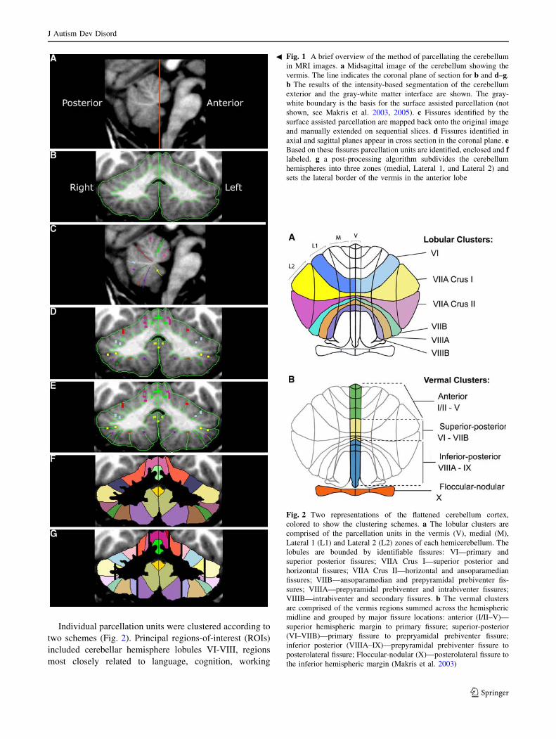
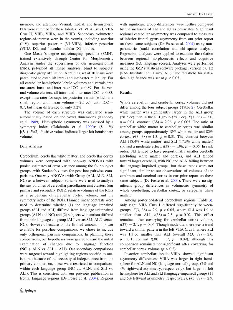
Individual parcellation units were clustered according to
two schemes (Fig. 2). Principal regions-of-interest (ROIs)
included cerebellar hemisphere lobules VI-VIII, regions
most closely related to language, cognition, working
Fig. 2 Two representations of the flattened cerebellum cortex,
colored to show the clustering schemes. a The lobular clusters are
comprised of the parcellation units in the vermis (V), medial (M),
Lateral 1 (L1) and Lateral 2 (L2) zones of each hemicerebellum. The
lobules are bounded by identifiable fissures: VI—primary and
superior posterior fissures; VIIA Crus I—superior posterior and
horizontal fissures; VIIA Crus II—horizontal and ansoparamedian
fissures; VIIB—ansoparamedian and prepyramidal prebiventer fis-
sures; VIIIA—prepyramidal prebiventer and intrabiventer fissures;
VIIIB—intrabiventer and secondary fissures. b The vermal clusters
are comprised of the vermis regions summed across the hemispheric
midline and grouped by major fissure locations: anterior (I/II–V)—
superior hemispheric margin to primary fissure; superior-posterior
(VI–VIIB)—primary fissure to prepryamidal prebiventer fissure;
inferior posterior (VIIIA–IX)—prepyramidal prebiventer fissure to
posterolateral fissure; Floccular-nodular (X)—posterolateral fissure to
the inferior hemispheric margin (Makris et al. 2003)
Fig. 1 A brief overview of the method of parcellating the cerebellum
in MRI images. a Midsagittal image of the cerebellum showing the
vermis. The line indicates the coronal plane of section for b and d–g.
b The results of the intensity-based segmentation of the cerebellum
exterior and the gray-white matter interface are shown. The gray-
white boundary is the basis for the surface assisted parcellation (not
shown, see Makris et al. 2003, 2005). c Fissures identified by the
surface assisted parcellation are mapped back onto the original image
and manually extended on sequential slices. d Fissures identified in
axial and sagittal planes appear in cross section in the coronal plane. eBased on these fissures parcellation units are identified, enclosed and flabeled. g a post-processing algorithm subdivides the cerebellum
hemispheres into three zones (medial, Lateral 1, and Lateral 2) and
sets the lateral border of the vermis in the anterior lobe
b
J Autism Dev Disord
123
memory, and attention. Vermal, medial, and hemispheric
PUs were summed for these lobules: VI, VIIA Crus I, VIIA
Crus II, VIIB, VIIIA, and VIIIB. Secondary volumetric
regions-of-interest were in the vermis, including anterior
(I–V), superior posterior (VI–VIIB), inferior posterior
(VIIIA–IX), and floccular nodular (X) lobules.
One Master’s degree neuroimaging specialist (SMH),
trained extensively through Center for Morphometric
Analysis under the supervision of our neuroanatomist
(NM), peformed all image analyses, blind to subjects’
diagnostic group affiliation. A training set of 10 scans were
parcellated to establish intra- and inter-rater reliability. For
all cerebellar hemispheric lobule volumes and vermis area
measures, intra- and inter-rater ICCs [ 0.89. For the ver-
mal volume clusters, all intra- and inter-rater ICCs [ 0.83,
except intra-rater for superior-posterior vermis (which is a
small region with mean volume = 2.5 cc), with ICC =
0.7, but mean difference of only 3.2%.
The volume of each structure was calculated semi-
automatically based on the voxel dimensions (Kennedy
et al. 1989). Hemispheric asymmetry was assessed by a
symmetry index (Galaburda et al. 1990): (L - R)/
[(L ? R)/2]. Positive values indicate larger left hemisphere
volume.
Data Analysis
Cerebellum, cerebellar white matter, and cerebellar cortex
volumes were compared with one-way ANOVAs with
pooled estimates of error variance among the four subject
groups, with Student’s t-tests for post-hoc pairwise com-
parisons. One-way ANOVAs with Group (ALI, ALN, SLI,
NC) as a between-subjects variable were used to analyze
the raw volumes of cerebellar parcellation unit clusters (our
primary and secondary ROIs), relative volumes of the ROIs
as a percentage of cerebellar cortex volume, and the
symmetry index of the ROIs. Planned linear contrasts were
used to determine whether (1) the language impaired
groups (SLI and ALI) differed from language unimpaired
groups (ALN and NC) and (2) subjects with autism differed
from their language co-group (ALI versus SLI; ALN versus
NC). However, because of the limited amount of power
available for post-hoc comparisons, we chose to include
only orthogonal pairwise comparisons. In planning these
comparisons, our hypotheses were geared toward the initial
examination of changes due to language function
(NC ? ALN vs. SLI ? ALI). Our secondary comparisons
were targeted toward highlighting regions specific to aut-
ism, but because of the necessity of independence from the
primary comparison, these were restricted to comparisons
within each language group (NC vs. ALN, and SLI vs.
ALI). This is consistent with our previous publication in
frontal language regions (De Fosse et al. 2004). Regions
with significant group differences were further compared
by the inclusion of age and IQ as covariates. Significant
regional cerebellar asymmetry was compared to measures
of inferior frontal gyrus asymmetry from our prior report
on these same subjects (De Fosse et al. 2004) using non-
parametric (rank) correlation and chi-square analysis.
Regression analyses were applied to examine the relation
between regional morphometric effects and cognitive
measures (IQ, language scores). Analyses were performed
using the JMP statistical software package, version 5.0.1.2
(SAS Institute Inc., Carey, NC). The threshold for statis-
tical significance was set at p \ 0.05.
Results
Whole cerebellum and cerebellar cortex volumes did not
differ among the four subject groups (Table 2). Cerebellar
white matter was significantly larger in the ALI group
(28.2 cc) than in the SLI group (25.1 cc), F(3, 38) = 3.0,
p = 0.04, contrast t(38) = 2.96, p \ 0.005. The ratio of
cerebellar white matter to cerebellar cortex was similar
among groups (approximately 18% white matter and 82%
cortex, F(3, 38) = 1.3, p = 0.3). The contrast between
ALI (18.4% white matter) and SLI (17.3% white matter)
showed a moderate effect, t(38) = 1.96, p = 0.06. In rank
order, SLI tended to have proportionally smaller cerebelli
(including white matter and cortex), and ALI tended
toward larger cerebelli, with NC and ALN falling between
the language-impaired groups, but these trends were not
significant, similar to our observations of volumes of the
cerebrum and cerebral cortex in our prior report on these
same subjects (De Fosse et al. 2004). There were no sig-
nificant group differences in volumetric symmetry of
whole cerebellum, cerebellar cortex, or cerebellar white
matter.
Among posterior-lateral cerebellum regions (Table 3),
only right VIIA Crus I differed significantly between-
groups, F(3, 38) = 2.9, p \ 0.05, where SLI was 1.9 cc
smaller than ALI, t(38) = 2.5, p = 0.02. This effect
remained after covarying for cerebellar cortex volume,
t(37) = 2.1, p = 0.04. Though moderate, there was a trend
toward a similar pattern in the left VIIA Crus I, where SLI
was 1.3 cc smaller than ALI (overall F(3, 38) = 2.0,
p = 0.1; contrast t(38) = 1.7, p = 0.09), although this
comparison remained non-significant after covarying for
cerebellar cortex volume (p [ 0.2).
Posterior cerebellar lobule VIIIA showed significant
asymmetry differences: VIIIA was larger in right hemi-
sphere for ALN and NC (language-normal) groups (7% and
4% rightward asymmetry, respectively), but larger in left
hemisphere for ALI and SLI (language-impaired) groups (11
and 6% leftward asymmetry, respectively), F(3, 38) = 2.9,
J Autism Dev Disord
123
Table 2 Gross cerebellar volumes
Region NC
(mean ± SD)
n = 11
ALN
(mean ± SD)
n = 6
ALI
(mean ± SD)
n = 16
SLI
(mean ± SD)
n = 9
Overall Post hoc contrasts
F P NC & ALN
vs. ALI & SLI
NC vs.
ALN
SLI vs.
ALI
Raw volume (cc)
Whole cerebellum 149.8 ± 10.8 148.5 ± 9.3 153.5 ± 12.1 145.3 ± 10.1 1.1 0.4
Cerebellar white matter 26.7 ± 2.9 26.5 ± 1.9 28.2 ± 2.8 25.1 ± 1.9 3.0 0.04 *
Cerebellar cortex 123.1 ± 10.0 122.0 ± 8.2 125.3 ± 10.4 120.2 ± 8.8 0.6 0.7
Ratio to whole cerebellum volume (%)
Cerebellar white matter 17.9 ± 1.8 17.9 ± 1.0 18.4 ± 1.4 17.3 ± 0.9 1.3 0.3
Cerebellar cortex 82.1 ± 1.8 82.1 ± 1.0 81.6 ± 1.4 82.7 ± 0.9 1.3 0.3
Symmetry coefficienta
Whole cerebellum 0.4 ± 1.1 -0.9 ± 1.6 0.3 ± 1.9 0.1 ± 1.4 1.0 0.4
Cerebellar white matter 1.2 ± 6.8 0.7 ± 6.3 3.2 ± 11.8 -4.9 ± 15.9 1.0 0.4
Cerebellar cortex 0.2 ± 1.6 -1.3 ± 2.3 -0.4 ± 2.0 1.0 ± 2.7 1.6 0.2
a Positive symmetry coefficient indicates left hemisphere volume is larger than right hemisphere volume. Negative value indicates right
hemisphere volume is larger
NC Normal control, ALN autism with normal language, ALI autism with language impairment, SLI specific language impairment
* p \ 0.05
Table 3 Asymmetries and volumes of the lobular (gyral) cerebellar parcellation units in posterior lateral cerebellum (Lobules VI–VIII)
Region NC
(mean ± SD)
n = 11
ALN
(mean ± SD)
n = 6
ALI
(mean ± SD)
n = 16
SLI
(mean ± SD)
n = 9
Overall Post hoc contrasts
F P NC & ALN
vs. ALI & SLI
NC vs.
ALN
SLI vs.
ALI
Symmetry coefficienta
VI -0.6 ± 14.0 -4.0 ± 3.4 -4.5 ± 12.0 5.5 ± 14.6 1.3 0.3
VIIA Crus I -4.3 ± 6.0 0.4 ± 4.1 -2.7 ± 8.6 1.3 ± 8.8 1.2 0.3
VIIA Crus II -1.5 ± 15.8 -8.4 ± 19.5 -13.6 ± 17.5 -6.1 ± 11.2 1.3 0.3
VIIB 7.1 ± 25.3 10.9 ± 28.8 4.3 ± 24.1 -4.7 ± 17.4 0.6 0.6
VIIIA -4.2 ± 19.6 -6.6 ± 19.5 10.9 ± 11.5 6.3 ± 15.5 2.9 0.05 *
VIIIB 7.8 ± 10.7 12.2 ± 14.8 9.3 ± 12.1 8.2 ± 17.1 0.2 0.9
Right lobular zone volume
VI 9.2 ± 1.5 9.1 ± 1.0 9.6 ± 1.9 8.5 ± 1.2 0.8 0.5
VIIA Crus I 14.1 ± 1.7 13.6 ± 1.3 15.4 ± 1.8 13.5 ± 2.2 2.9 0.05 *
VIIA Crus II 8.4 ± 1.9 8.5 ± 1.5 9.2 ± 2.2 9.0 ± 2.0 0.4 0.7
VIIB 6.5 ± 1.5 6.2 ± 1.3 6.1 ± 1.5 6.2 ± 1.9 0.2 0.9
VIIIA 6.1 ± 1.6 6.5 ± 2.2 5.9 ± 1.4 5.1 ± 0.7 1.3 0.3
VIIIB 4.5 ± 0.8 4.6 ± 1.1 4.7 ± 1.0 4.8 ± 0.7 0.2 0.9
Left lobular zone volume
VI 9.2 ± 2.0 8.7 ± 0.8 9.1 ± 1.7 9.0 ± 1.2 0.1 0.9
VIIA Crus I 13.5 ± 1.5 13.6 ± 1.1 15.0 ± 1.9 13.7 ± 2.4 2.0 0.1
VIIA Crus II 8.2 ± 1.4 7.9 ± 2.0 8.0 ± 1.8 8.5 ± 1.7 0.2 0.9
VIIB 6.9 ± 1.0 6.9 ± 1.5 6.4 ± 2.0 5.9 ± 1.8 0.7 0.5
VIIIA 5.8 ± 1.5 5.9 ± 1.2 6.6 ± 1.8 5.6 ± 1.3 1.1 0.4
VIIIB 4.8 ± 0.7 5.3 ± 1.7 5.1 ± 0.8 5.4 ± 1.6 0.4 0.7
a Positive symmetry coefficient indicates left hemisphere volume is larger than right hemisphere volume. Negative value indicates right
hemisphere volume is larger
NC Normal control, ALN autism with normal language, ALI autism with language impairment, SLI specific language impairment
* p \ 0.05
J Autism Dev Disord
123
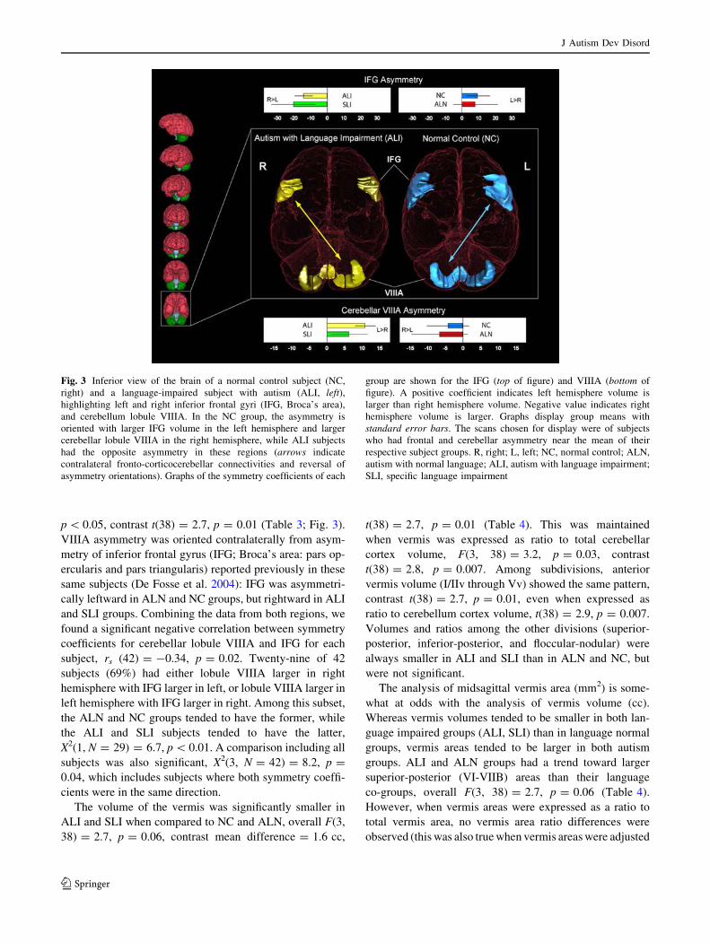
p \ 0.05, contrast t(38) = 2.7, p = 0.01 (Table 3; Fig. 3).
VIIIA asymmetry was oriented contralaterally from asym-
metry of inferior frontal gyrus (IFG; Broca’s area: pars op-
ercularis and pars triangularis) reported previously in these
same subjects (De Fosse et al. 2004): IFG was asymmetri-
cally leftward in ALN and NC groups, but rightward in ALI
and SLI groups. Combining the data from both regions, we
found a significant negative correlation between symmetry
coefficients for cerebellar lobule VIIIA and IFG for each
subject, rs (42) = -0.34, p = 0.02. Twenty-nine of 42
subjects (69%) had either lobule VIIIA larger in right
hemisphere with IFG larger in left, or lobule VIIIA larger in
left hemisphere with IFG larger in right. Among this subset,
the ALN and NC groups tended to have the former, while
the ALI and SLI subjects tended to have the latter,
X2(1, N = 29) = 6.7, p \ 0.01. A comparison including all
subjects was also significant, X2(3, N = 42) = 8.2, p =
0.04, which includes subjects where both symmetry coeffi-
cients were in the same direction.
The volume of the vermis was significantly smaller in
ALI and SLI when compared to NC and ALN, overall F(3,
38) = 2.7, p = 0.06, contrast mean difference = 1.6 cc,
t(38) = 2.7, p = 0.01 (Table 4). This was maintained
when vermis was expressed as ratio to total cerebellar
cortex volume, F(3, 38) = 3.2, p = 0.03, contrast
t(38) = 2.8, p = 0.007. Among subdivisions, anterior
vermis volume (I/IIv through Vv) showed the same pattern,
contrast t(38) = 2.7, p = 0.01, even when expressed as
ratio to cerebellum cortex volume, t(38) = 2.9, p = 0.007.
Volumes and ratios among the other divisions (superior-
posterior, inferior-posterior, and floccular-nodular) were
always smaller in ALI and SLI than in ALN and NC, but
were not significant.
The analysis of midsagittal vermis area (mm2) is some-
what at odds with the analysis of vermis volume (cc).
Whereas vermis volumes tended to be smaller in both lan-
guage impaired groups (ALI, SLI) than in language normal
groups, vermis areas tended to be larger in both autism
groups. ALI and ALN groups had a trend toward larger
superior-posterior (VI-VIIB) areas than their language
co-groups, overall F(3, 38) = 2.7, p = 0.06 (Table 4).
However, when vermis areas were expressed as a ratio to
total vermis area, no vermis area ratio differences were
observed (this was also true when vermis areas were adjusted
Fig. 3 Inferior view of the brain of a normal control subject (NC,
right) and a language-impaired subject with autism (ALI, left),highlighting left and right inferior frontal gyri (IFG, Broca’s area),
and cerebellum lobule VIIIA. In the NC group, the asymmetry is
oriented with larger IFG volume in the left hemisphere and larger
cerebellar lobule VIIIA in the right hemisphere, while ALI subjects
had the opposite asymmetry in these regions (arrows indicate
contralateral fronto-corticocerebellar connectivities and reversal of
asymmetry orientations). Graphs of the symmetry coefficients of each
group are shown for the IFG (top of figure) and VIIIA (bottom of
figure). A positive coefficient indicates left hemisphere volume is
larger than right hemisphere volume. Negative value indicates right
hemisphere volume is larger. Graphs display group means with
standard error bars. The scans chosen for display were of subjects
who had frontal and cerebellar asymmetry near the mean of their
respective subject groups. R, right; L, left; NC, normal control; ALN,
autism with normal language; ALI, autism with language impairment;
SLI, specific language impairment
J Autism Dev Disord
123

Ta
ble
4C
ereb
ella
rv
erm
iscl
ust
erv
olu
mes
and
area
s.V
olu
me
un
its
are
the
sum
of
left
and
rig
ht
hem
isp
her
es
Ver
mis
reg
ion
NC
(mea
n±
SD
)
n=
11
AL
N(m
ean
±S
D)
n=
6
AL
I(m
ean
±S
D)
n=
16
SL
I(m
ean
±S
D)
n=
9
Ov
eral
lP
ost
ho
cco
ntr
asts
FP
NC
&A
LN
vs.
AL
I&
SL
IN
Cv
s.A
LN
SL
Iv
s.A
LI
Raw
vo
lum
e(c
c)
Wh
ole
ver
mis
13
.6±
2.0
13
.5±
1.7
12
.0±
1.7
12
.0±
1.6
2.7
0.0
6*
An
teri
or,
I/II
–V
7.4
±1
.57
.1±
0.8
6.1
±1
.36
.2±
1.0
3.1
0.0
4*
Su
per
ior-
po
ster
ior,
VI–
VII
B2
.7±
0.4
2.9
±0
.52
.7±
0.4
2.6
±0
.40
.50
.7
Infe
rio
r-p
ost
erio
r,V
IIIA
–IX
3.0
±0
.53
.1±
0.6
2.8
±0
.52
.8±
0.5
1.4
0.3
Flo
ccu
lar-
no
du
lar,
X0
.4±
0.1
0.4
±0
.10
.4±
0.1
0.4
±0
.10
.40
.8
Rat
ioto
cere
bel
lar
cort
exv
olu
me
(%)
Wh
ole
ver
mis
11
.0±
1.2
11
.1±
1.2
9.6
±1
.61
0.0
±1
.13
.20
.03
*
An
teri
or,
I/II
–V
6.0
±1
.05
.8±
0.5
4.9
±1
.15
.1±
0.7
3.6
0.0
2*
Su
per
ior-
po
ster
ior,
VI–
VII
B2
.2±
0.3
2.3
±0
.32
.2±
0.4
2.2
±0
.30
.40
.7
Infe
rio
r-p
ost
erio
r,V
IIIA
–IX
2.5
±0
.32
.6±
0.5
2.2
±0
.32
.3±
0.4
2.0
0.1
Flo
ccu
lar-
no
du
lar,
X0
.3±
0.1
0.4
±0
.10
.3±
0.0
50
.4±
0.1
0.7
0.6
Raw
area
(mm
2)
Wh
ole
ver
mis
11
21
.5±
12
0.5
11
86
.5±
15
0.4
11
56
.8±
11
1.5
10
70
.7±
66
.41
.70
.2
An
teri
or,
I/II
–V
47
2.2
±5
8.0
50
3.4
±7
2.1
47
6.2
±5
5.0
44
1.6
±2
0.2
1.7
0.2
Su
per
ior-
po
ster
ior
VI–
VII
B2
76
.9±
35
.33
23
.9±
67
.03
15
.9±
43
.12
85
.0±
34
.62
.70
.06
*
Infe
rio
r-p
ost
erio
r,V
IIIA
–X
37
2.5
±5
4.1
35
9.1
±4
3.5
36
4.7
±5
6.8
34
4.0
±3
5.7
0.
60
.7
Rat
ioto
wh
ole
ver
mis
area
(%)
An
teri
or,
I/II
–V
42
.1±
2.3
42
.4±
1.4
41
.1±
2.2
41
.3±
2.1
0.8
0.5
Su
per
ior-
po
ster
ior
VI–
VII
B2
4.8
±2
.52
7.1
±3
.12
7.4
±3
.72
6.6
±2
.11
.8
0.2
Infe
rio
r-p
ost
erio
r,V
IIIA
-X3
3.1
±2
.53
0.5
±3
.73
1.4
±2
.83
2.1
±2
.41
.40
.3
NC
No
rmal
con
tro
l,A
LN
auti
smw
ith
no
rmal
lan
gu
age,
AL
Iau
tism
wit
hla
ng
uag
eim
pai
rmen
t,S
LI
spec
ific
lan
gu
age
imp
airm
ent
*p\
0.0
5
J Autism Dev Disord
123
for cerebellum volume or overall brain volume). The cor-
relation between each vermis area (mm2) and its respective
volume (cc) showed a high correlation [r(42) = 0.68] for the
superior-posterior and the inferior-posterior regions, but a
lower correlation (r(42) = 0.42) for the anterior vermis. A
comparison of slopes along the autism dimension (ALN &
ALI versus NC & SLI) showed that anterior vermis area was
correlated with volume in the NC & SLI group [r(20) =
0.73, p \ 0.001], but not in the ALN & ALI group
[r(22) = 0.30, p = 0.2], slope comparison T = 2.5, p =
0.02. This suggests that while the ALI and ALN groups may
tend toward larger midsagittal areas, discordantly, language-
impaired groups (ALI, SLI) may have a restricted lateral
extent of the vermis as indicated by the location of the par-
avermian sulcus, leading to smaller volumes.
Further exploratory analyses were done to clarify the
role of age and IQ in the results reported above. When
added as a covariate, neither age nor nonverbal IQ changed
the significance of group contrasts for right lobule VIIA
Crus I, symmetry of lobule VIIIA, total vermis, or anterior
vermis. When verbal IQ [which is significantly correlated
with anterior vermis, r(40) = 0.39, p = 0.01] was covar-
ied, group contrasts for total and anterior vermis were
inconclusive, but there was no change in significant group
contrasts for the volume of right lobule VIIA Crus I or
symmetry of lobule VIIIA. Examining the correlation of
language performance and structural measures further
demonstrated a relationship between posterior-lateral cer-
ebellar asymmetry and language: CELF total language
scores were significantly correlated with the symmetry of
lobule VIIIA, r(27) = -0.43, p = 0.02. That is, higher
scores were related to increased rightward asymmetry. In
addition, anterior vermis volumes were significantly cor-
related with nonverbal IQ [r(40) = 0.39, p = 0.03], the
NEPSY nonsense word repetition score [r(27) = 0.41,
p = 0.03], and a composite measure of vocabulary based
on the Expressive Vocabulary Test (Williams 1997) and
the Peapody Picture Vocabulary Test (Dunn and Dunn
1997; r(27) = 0.38, p = 0.03).
Discussion
In the current study, we investigated cerebellar regional
structural volume and asymmetry in the same subjects as
our previous report on Broca’s area asymmetry (De Fosse
et al. 2004). We applied quantitative analysis of discrete
cerebellar regions to determine whether previously repor-
ted asymmetry patterns in Broca’s area would be reflected
contralaterally in posterior lateral cerebellum. In lobule
VIIIA, we found that right-handed language-normal groups
(ALN and NC) had larger right-sided volume, while the
right-handed language-impaired groups (ALI and SLI) had
larger volume on the left. Furthermore, most subjects with
language impairment also showed asymmetry reversal of
inferior frontal gyrus (IFG). While the majority of normal
control boys had larger left-sided IFG coupled with larger
right-sided cerebellar lobule VIIIA, language-impaired
groups (ALI and SLI) showed the opposite pattern with
larger right-sided IFG and left-sided VIIIA. Language-
normal boys with autism were more similar to controls.
While prior functional neuroimaging studies in normal
controls demonstrated contralateral connectivity and cor-
respondence between left frontal language regions and
right posterior lateral cerebellum (Desmond and Fiez 1998;
Harris et al. 2006; Metter et al. 1987; Roskies et al. 2001),
the current study is the first to identify correspondence
between structural asymmetries in both Broca’s area and
contralateral posterior lateral cerebellum regions. Further-
more, the regional cerebellar asymmetries reported in the
current study were observed in the presence of global
cerebellar symmetry in all subject groups, indicating that
the asymmetry was regionally specific. In contrast to this
localized observation in boys, adult controls displayed
rightward global cerebellar cortex asymmetry, whereas
dyslexic adults had cerebellar symmetry (Rae et al. 2002).
Thus, the development of global cerebellar cortex asym-
metry may occur later during adolescence, as it was not
observed in the NC boys in the current study.
In our prior reports of language-related frontal lobe
regions (De Fosse et al. 2004; Herbert et al. 2002), right-
handed control subjects displayed typical Broca’s area
asymmetry, oriented with larger gray matter volumes in
left hemisphere, while this asymmetry pattern was reversed
in language-impaired subjects with autism and SLI. In
studies of cerebellar asymmetry in developmental dyslexia,
rightward cerebellar asymmetries were reported in normal
controls, with reversal in dyslexic subjects (Leonard et al.
2001; Eckert et al. 2003; Kibby et al. 2008). However,
asymmetry of respective language and cognition-associated
regions of posterior lateral cerebellum has not been pre-
viously reported in subjects with autism or SLI. Posterior
lateral cerebellar regions have robust interconnections with
contralateral frontal cerebral cortex and are integral to
language and cognitive functions, as shown in cerebellar
lesion (Gebhart et al. 2002; Schmahmann 2004) and
functional imaging studies (Stoodley and Schmahmann
2009; Benson et al. 1999; Desmond and Fiez 1998; Harris
et al. 2006; Hubrich-Ungureanu et al. 2002; Jansen et al.
2005; Petersen et al. 1989). Evidence for these fronto-
corticocerebellar connections also includes the develop-
ment of crossed cerebellar diaschisis (hypometabolism in a
cerebellar hemisphere contralateral to a cerebral hemi-
spheric lesion) following lesions in contralateral frontal
language areas (Metter et al. 1987), and reversed cerebellar
diaschisis (hypometabolism in a cerebral hemisphere
J Autism Dev Disord
123
contralateral to a cerebellar hemispheric lesion) leading to
decreased activation in contralateral cerebral cortex fol-
lowing cerebellar lesions (Botez-Marquard and Botez
1997; Schmahmann 1991; Schmahmann and Sherman
1998). Furthermore, posterior lateral cerebellar lesions are
frequently associated with language deficits (Leggio et al.
2000; Pollack et al. 1995), including agrammatism,
decreased verbal fluency, inability to detect one’s own
errors on a verb generation task, dysprosodia, mild anomia,
impaired initiation of language, and retrieval of words and
stories (Silveri et al. 1994; Molinari et al. 1997; Fiez et al.
1992; Schmahmann 2004), particularly with lesions in right
posterior lateral cerebellum (Molinari et al. 1997; Gebhart
et al. 2002). Thus, in association with its direct cortico-
cerebellar connections with left inferior frontal language
regions, right posterior lateral cerebellum has been directly
implicated in language functioning (Gebhart et al. 2002).
The focus of our study was posterior lateral cerebellum,
particularly lobules VI, VII, and VIII, since these regions
have been identified as most closely tied with cognitive
function, language, verbal working memory, and attention
(Stoodley and Schmahmann 2009; Desmond and Fiez
1998; Leiner et al. 1991; Levisohn et al. 2000; Neau et al.
2000; Petersen et al. 1989; Riva and Giorgi 2000; Sch-
mahmann and Sherman 1998). Posterior lateral cerebellum
also displayed the most marked decrease in number of
Purkinje cells in autism (Kemper and Bauman 2002). In
primate studies, lobule VIII is the site of the second sen-
sorimotor homunculus and receives inputs from motor
areas, while area 46 projects to lobule VII/Crus I/II (Kelly
and Strick 2003). However, in human neuroimaging stud-
ies, while prior research has suggested that language is
more closely identified with lobules VI and VIIA Crus I
(Stoodley and Schmahmann 2009; Ackermann et al. 1998;
Fiez and Raichle 1997), VIIIA (as well as VI, VIIB, and
VIIA Crus I) is generally associated with working memory,
with VIIIA particularly identified with motoric rehearsal
and working memory (Desmond et al. 1997). Thus, VIIIA
may play a substantial role in language as well, perhaps
through mediation of verbal working memory (Stoodley
and Schmahmann 2009). Furthermore, Broca’s area is
related to language production, and thus, the relationship
between IFG and VIIIA asymmetry differences in language
impaired groups could be related to a potential motoric role
of VIIIA in language production and/or verbal rehearsal.
Children with autism often show deficits in verbal media-
tion and verbal encoding strategies that foster working
memory (Joseph et al. 2005). Furthermore, we observed a
correlation between VIIIA asymmetry and language scale
scores on the CELF, further supporting a relationship
between language function and VIIIA asymmetry.
While we observed similarities between ALI and SLI in
reversal of VIIIA asymmetry and reduced anterior vermis
volume measures, we also observed specific differences
between these language-impaired groups. Cerebellar white
matter was significantly larger in ALI than in SLI, with
language-normal groups’ (ALN and NC) measures falling
between. Increased cerebellar white matter volume in ALI
may result from overgrowth and lack of pruning, and is
also consistent with earlier reports of increased cerebral
white matter volume in boys with autism (Herbert et al.
2003, 2004). Furthermore, we observed regional volume
differences between ALI and SLI groups in right hemi-
sphere lobule VIIA Crus I, a cerebellar region consistently
associated with language processing, suggesting that, while
there are structural imaging and language similarities
between these disorders, there may be important language-
related developmental differences as well. For example, the
ALI group had significantly worse CELF scores than the
SLI group; thus, the larger VIIA Crus I volume in ALI
relative to SLI in this case is likely an abnormal finding,
despite smaller volumes in right lobule VIIIA being related
to language deficit and lower CELF scores.
To date, cerebellar structural neuroimaging findings in
autism have largely focused on vermis sagittal cross-sec-
tional area, yet results have been mixed among research
groups, with some disagreement of results (Stanfield et al.
2008; Brambilla et al. 2003). We observed differences in
anterior vermis (I/II–V) volumes, with smaller vermis
volume in ALI and SLI groups than in the language normal
groups, and we observed a correlation between anterior
vermis volume and language test scores (verbal IQ,
NEPSY non-word repetition, and vocabulary tests), an
interesting observation given that anterior vermis is tradi-
tionally associated with motor function. However, we did
not observe decreased vermis cross-sectional areas in aut-
ism or SLI, and in fact, vermis area had a trend toward
being larger, particularly in superior-posterior vermis area,
in both autism groups (ALI, ALN). The discrepancy
between vermis volume and area results in the current
study, particularly in anterior vermis, may result from ab-
mormalities in lateral width of the anterior vermis, as
suggested by the low correlation between anterior vermis
area and volume in the autism groups. Thus, to some
extent, our data supports a vermis deficit in autism
observed by some groups (Courchesne et al. 1988, 1994;
Hashimoto et al. 1993; Kaufmann et al. 2003; Rojas et al.
2006), at least in language-impaired subjects. However, we
localized the volumetric deficit primarily in anterior vermis
(I/II-V), whereas several other studies have reported autism
vermis area differences in VI-VII (Stanfield et al. 2008;
Courchesne et al. 1988, 1994; Kaufmann et al. 2003) or
VIII-X (Hashimoto et al. 1993). Our observation of cor-
relation between anterior vermis volumes and IQ measures
is consistent with earlier studies suggesting that the vermis
effect may be IQ related, particularly with respect to verbal
J Autism Dev Disord
123
IQ (Piven et al. 1992, 1995), as demonstrated in a large
meta-analysis of autism MRI studies that demonstrated that
the area of vermis VI–VII showed modifying effects of age
and IQ based on meta-regression analysis (Stanfield et al.
2008). Thus, in relation to the controversy surrounding
vermis size in autism, our findings are consistent with a
vermis effect more closely related to language and IQ
effects than to autism specifically. However, there is reason
to consider that vermis deficits may also be related to
autism. For example, the vermis is thought to be involved
with affective function through interconnections with the
limbic system (Schmahmann 2004; Schmahmann and
Sherman 1998). Cerebellar lesions or tumors in vermis lead
to irritability and autistic-like features (Levisohn et al.
2000; Riva and Giorgi 2000; Schmahmann 2004). Thus,
vermis is considered to be limbic cerebellum (Heath 1977;
Schmahmann 1991, 2000, 2004), and may play a role in
affective aspects of autism.
In the current study, ALI and SLI shared similar
abnormalities in both VIIIA asymmetry and in decreased
anterior vermis volume, while ALN was more similar with
normal controls. This pairing of structural brain abnor-
malities among ALI and SLI subjects [also observed in
Broca’s asymmetry reversal in our prior report (De Fosse
et al. 2004)] suggests these anomalies are more closely
related to language and cognitive deficits than to autism
effects per se. However, SLI and ALI share a similar
genetic predisposition to language and communication
deficits (Fisher et al. 2003; Santangelo and Folstein 1999),
and these shared genotype or phenotypic traits may mani-
fest in abnormal structural brain development. For exam-
ple, family and twin studies found that first degree relatives
of probands with autism more frequently display deficits in
language skills than occur in the general population (Bailey
et al. 1995; Folstein et al. 1999; Fombonne et al. 1997;
Ruser et al. 2007). Furthermore, siblings of children with
SLI have an elevated risk for autism (Tomblin et al. 2003).
Genetic linkage studies in these two developmental disor-
ders point to overlapping regions on chromosome 7q
(Barrett et al. 1999; IMGSAC 1998; O’Brien et al. 2003)
and chromosome 13q (Barrett et al. 1999; Bartlett et al.
2002). In genetic studies of autism, signals on both 7q and
13q increase significantly when linkage studies restrict
their analyses to autism families with clear signs of lan-
guage impairment (Alarcon et al. 2002; Bradford et al.
2001) or language delay (Alarcon et al. 2008). These
findings suggest that genetic abnormalities leading to the
phenotype of developmental language disorders (Fisher
et al. 2003) may overlap with genetic alterations that are
liability factors for autism. Furthermore, decreased vol-
umes combined with reversed asymmetry in these frontal
and cerebellar regions may represent a preexisting marker
of illness susceptibility or endophenotype (Almasy and
Blangero 2001; Breiter and Gasic 2004). Asymmetry pat-
terns are developmental in nature (Giedd et al. 1996; Giedd
2004; Makris et al. 2004), and alterations in such patterns
are genetically influenced (Galloway 1990; Hyatt and Yost
1998; Lowe et al. 1996; Piedra et al. 1998). Genetic tran-
scription factors may lead to atypical asymmetry patterns
that emerge very early in fetal development. For example,
Sun et al. (2005) identified genes that were differentially
expressed in left and right perisylvian regions in cortices
from 14-week-old fetuses. These findings suggest genetic
factors may contribute to development of cerebral asym-
metry, and may influence the atypical asymmetry patterns
reported in autism and SLI.
There were several limitations in the current study. First,
there were relatively small subject groups and modest
effect sizes. Second, we did not perform multiple com-
parison corrections: however, the approach was focused on
hypothesis-driven analyses of a discrete, limited number of
anatomically-specific regions, and the current approach
was specifically selected to expand upon prior observations
in language-related cerebral cortex regions in these groups,
and link asymmetry reversals in cortex to those in contra-
lateral regions of posterior-lateral cerebellum. While the
modest effect and sample sizes, combined with a limited
number of uncorrected multiple comparisons, may detract
from the robustness and impact of the findings, prior
studies of cerebellum differences in autism tend to be
moderate as well. The fact that the current study does
demonstrate significant cerebellar regional asymmetry
reversal and volume differences despite the modest sample
and effect sizes is promising and noteworthy, particularly
since the differences reported in the present and previous
studies may be constrained by the extensive heterogeneity
inherent in autism. Thus, as noted in the current report,
differences were observed between autism language sub-
groups, supporting the need for care in subject selection
and inclusion criteria in the presence of heterogeneous
populations in autism studies. Finally, our study would
benefit from further confirmation in larger subject samples,
and could be expanded to assess girls with autism and
specific language impairment, as well as older and higher-
functioning subjects with autism.
In conclusion, posterior lateral cerebellum is highly
interconnected with frontal cortex, and involved in cogni-
tive function, language, working memory, and attention
(Schmahmann 2004; Schmahmann and Sherman 1998).
Right posterior lateral cerebellum is closely linked with left
frontal language areas, including Broca’s area. We
observed reversed asymmetry in posterior cerebellar lobule
VIIIA in right-handed language-impaired boys, with and
without autism, but not in right-handed language-normal
boys with or without autism. This cerebellar asymmetry
reversal was contralateral to that previously reported in
J Autism Dev Disord
123
Broca’s area in these same subjects. Combined with our
earlier report of reversed asymmetry in Broca’s area, the
current study supports abnormal development of the typical
asymmetry of fronto-corticocerebellar language circuits in
subjects with language-impairment, with or without
autism.
Acknowledgments This work was supported in part by a grant from
NIDCD to Drs. Tager-Flusberg and Harris, U19/PO1 DC 03610,
which is part of the NICHD/NIDCD funded Collaborative Programs
of Excellence in Autism; a grant from the NIMH to Dr. Jean Frazier,
K08 MH01573; and by a Young Investigator Award funded by
NARSAD and a grant from ALSA to Dr. Nikos Makris. The authors
also thank Trinity Urban and Joyce Miller for proofreading comments
on the manuscript.
Appendix
Author Names and Affiliations at the Time of the Study
Steven M. Hodge: Radiology Computer Aided Diagnostics
Laboratory and Center for Morphometric Analysis, Mas-
sachusetts General Hospital, Boston, MA 02129, USA.
Nikos Makris: Center for Morphometric Analysis, Massa-
chusetts General Hospital, Boston, MA 02129, USA. David
N. Kennedy: Center for Morphometric Analysis, Massa-
chusetts General Hospital, Boston, MA 02129, USA. Verne
S. Caviness, Jr.: Center for Morphometric Analysis, Mas-
sachusetts General Hospital, Boston, MA 02129, USA.
James Howard: Center for Morphometric Analysis, Mas-
sachusetts General Hospital, Boston, MA 02129, USA.
Lauren McGrath: Boston University School of Medicine,
Lab of Cognitive Neuroscience, Boston, MA, USA. Shelly
Steele: Boston University School of Medicine, Lab of
Cognitive Neuroscience, Boston, MA, USA. Jean A. Fra-
zier: Department of Psychiatry, Harvard Medical School,
Boston, MA; Center for Child and Adolescent Develop-
ment, Department of Psychiatry, Cambridge Health Alli-
ance, Cambridge, MA, USA. Helen Tager-Flusberg:
Boston University School of Medicine, Lab of Cognitive
Neuroscience, Boston, MA, USA. Gordon J. Harris:
Radiology Computer Aided Diagnostics Laboratory, Mas-
sachusetts General Hospital, Boston, MA 02114, USA.
Changes in Author Affiliations Since the Study Took
Place
Steven M. Hodge—none. Nikos Makris—none. David N.
Kennedy—none. Verne S. Caviness, Jr.—none. James
Howard—Cognitive Neurology and Alzheimer’s Disease
Center, Northwestern University Feinberg School of
Medicine, Chicago, IL,60611. Lauren McGrath—Depart-
ment of Psychology, University of Denver, Denver, CO,
80208. Shelly Steele—Department of Psychology, Uni-
versity of Illinois at Chicago, Chicago, IL, 60612. Jean A.
Frazier—none. Helen Tager-Flusberg—none. Gordon J.
Harris—none.
References
Ackermann, H., Wildgruber, D., Daum, I., & Grodd, W. (1998). Does
the cerebellum contribute to cognitive aspects of speech
production? A functional magnetic resonance imaging (fMRI)
study in humans. Neuroscience Letters, 247, 187–190.
Alarcon, M., Abrahams, B. S., Stone, J. L., Duvall, J. A., Perederiy, J.
V., Bomar, J. M., et al. (2008). Linkage, association, and gene-
expression analyses identify CNTNAP2 as an autism-suscepti-
bility gene. American Journal of Human Genetics, 82, 150–159.
Alarcon, M., Cantor, R. M., Liu, J., Gilliam, T. C., & Geschwind, D.
H. (2002). Evidence for a language quantitative trait locus on
chromosome 7q in multiplex autism families. American Journalof Human Genetics, 70, 60–71.
Almasy, L., & Blangero, J. (2001). Endophenotypes as quantitative
risk factors for psychiatric disease: Rationale and study design.
American Journal of Medical Genetics, 105, 42–44.
APA. (1994). DSM-IV: Diagnostic and statistic manual of mentaldisorders (4th ed.). Washington, DC: American Psychiatric
Press.
Arnold, J. B., Liow, J. S., Schaper, K. A., Stern, J. J., Sled, J. G.,
Shattuck, D. W., et al. (2001). Qualitative and quantitative
evaluation of six algorithms for correcting intensity nonunifor-
mity effects. Neuroimage, 13, 931–943.
Bailey, A., Le Couteur, A., Gottesman, I., Bolton, P., Simonoff, E.,
Yuzda, E., et al. (1995). Autism as a strongly genetic disorder:
Evidence from a British twin study. Psychological Medicine, 25,
63–77.
Barrett, S., Beck, J. C., Bernier, R., Bisson, E., Braun, T. A.,
Casavant, T. L., et al. (1999). An autosomal genomic screen for
autism. Collaborative linkage study of autism. American Journalof Medical Genetics, 88, 609–615.
Bartlett, C. W., Flax, J. F., Logue, M. W., Vieland, V. J., Bassett, A.
S., Tallal, P., et al. (2002). A major susceptibility locus for
specific language impairment is located on 13q21. AmericanJournal of Human Genetics, 71, 45–55.
Bauman, M., & Kemper, T. L. (1985). Histoanatomic observations of
the brain in early infantile autism. Neurology, 35, 866–874.
Benson, R. R., FitzGerald, D. B., LeSueur, L. L., Kennedy, D. N.,
Kwong, K. K., Buchbinder, B. R., et al. (1999). Language
dominance determined by whole brain functional MRI in
patients with brain lesions. Neurology, 52, 798–809.
Binder, J. R., Frost, J. A., Hammeke, T. A., Cox, R. W., Rao, S. M., &
Prieto, T. (1997). Human brain language areas identified by
functional magnetic resonance imaging. Journal of Neurosci-ence, 17, 353–362.
Bishop, D. V. (2003). Autism and specific language impairment:
Categorical distinction or continuum? Novartis FoundationSymposium, 251, 213–226 (discussion 226–234, 281–297).
Bobylova, M. Y., Petrukhin, A. S., Dunaevskaya, G. N., Piliya, S. V.,
& Il’ina, E. S. (2007). Clinical-psychological characteristics of
children with dysgenesis of the cerebellar vermis. Neuroscienceand Behavioral Physiology, 37, 755–759.
Boddaert, N., Chabane, N., Belin, P., Bourgeois, M., Royer, V.,
Barthelemy, C., et al. (2004). Perception of complex sounds in
autism: Abnormal auditory cortical processing in children.
American Journal of Psychiatry, 161, 2117–2120.
J Autism Dev Disord
123
Botez-Marquard, T., & Botez, M. I. (1997). Olivopontocerebellar
atrophy and Friedreich’s ataxia: Neuropsychological conse-
quences of bilateral versus unilateral cerebellar lesions. In R. J.
Bradly, R. A. Harris & P. Jenner (Series Eds.), J. D. Schmah-
mann (Vol. Ed.), International review of neurobiology, Vol. 41.
The cerebellum and cognition (pp. 387–410). San Diego:
Academic Press.
Bradford, Y., Haines, J., Hutcheson, H., Gardiner, M., Braun, T.,
Sheffield, V., et al. (2001). Incorporating language phenotypes
strengthens evidence of linkage to autism. American Journal ofMedical Genetics, 105, 539–547.
Brambilla, P., Hardan, A., di Nemi, S. U., Perez, J., Soares, J. C., &
Barale, F. (2003). Brain anatomy and development in autism:
Review of structural MRI studies. Brain Research Bulletin, 61,
557–569.
Breiter, H. C., & Gasic, G. P. (2004). A general circuitry processing
reward/aversion information and its implications for neuropsy-
chiatric illness. In M. S. Gazzaniga (Ed.), The cognitiveneurosciences (pp. 1043–1065). Cambridge: MIT.
Burroni, L., Orsi, A., Monti, L., Hayek, Y., Rocchi, R., & Vattimo, A.
G. (2008). Regional cerebral blood flow in childhood autism: A
SPET study with SPM evaluation. Nuclear Medicine Commu-nications, 29, 150–156.
Catts, H., Bridges, M., Little, T., & Tomblin, J. B. (2008). Reading
achievement growth in children with language impairments.
Journal of Speech Language and Hearing Research, 51, 1569–
1579.
Caviness, V. S., Jr., Meyer, J. W., Makris, N., & Kennedy, D. N.
(1996). MRI-based topographic parcellation of the human
neocortex: An anatomically specified method with estimate of
reliability. Journal of Cognitive Neuroscience, 8, 566–587.
Chheda, M., Sherman, J., & Schmahmann, J. D. (2002). Neurologic,
psychiatric and cognitive manifestations in cerebellar agenesis.
Neurology, 58, 356.
Chiron, C., Leboyer, M., Leon, F., Jambaque, I., Nuttin, C., & Syrota,
A. (1995). SPECT of the brain in childhood autism: Evidence for
a lack of normal hemispheric asymmetry. DevelopmentalMedicine and Child Neurology, 37, 849–860.
Conti-Ramsden, G. (2003). Processing and linguistic markers in
young children with specific language impairment (sli). Journalof Speech, Language and Hearing Research, 46(5), 1029–1037.
Conti-Ramsden, G., & Botting, N. (1999). Classification of children
with specific language impairment: Longitudinal considerations.
Journal of Speech, Language, and Hearing Research, 42, 1195–
1204.
Courchesne, E. (1999). An MRI study of autism: The cerebellum
revisited. Neurology, 52, 1106–1107.
Courchesne, E., Saitoh, O., Yeung-Courchesne, R., Press, G. A.,
Lincoln, A. J., Haas, R. H., et al. (1994). Abnormality of
cerebellar vermian lobules VI and VII in patients with infantile
autism: Identification of hypoplastic and hyperplastic subgroups
with MR imaging. AJR. American Journal of Roentgenology,162, 123–130.
Courchesne, E., Yeung-Courchesne, R., Press, G. A., Hesselink, J. R.,
& Jernigan, T. L. (1988). Hypoplasia of cerebellar vermal
lobules VI and VII in autism. New England Journal of Medicine,318, 1349–1354.
Dale, A. M., Fischl, B., & Sereno, M. I. (1999). Cortical surface-
based analysis. I. Segmentation and surface reconstruction.
Neuroimage, 9, 179–194.
Dawson, G., Finley, C., Phillips, S., & Galpert, L. (1986).
Hemispheric specialization and the language abilities of autistic
children. Child Development, 57, 1440–1453.
De Fosse, L., Hodge, S. M., Makris, N., Kennedy, D. N., Caviness, V.
S., Jr., McGrath, L., et al. (2004). Language-association cortex
asymmetry in autism and specific language impairment. Annalsof Neurology, 56, 757–766.
Dean, R. S. (1978). Cerebral laterality and reading comprehension.
Neuropsychologia, 16, 633–636.
Dean, R. S. (1982). Assessing patterns of lateral preference. Journalof Clinical Neuropsychology, 4, 124–128.
Desmond, J. E., & Fiez, J. A. (1998). Neuroimaging studies of the
cerebellum: Language, learning and memory. Trends in Cogni-tive Science, 2, 355–362.
Desmond, J. E., Gabrieli, J. D., Wagner, A. D., Ginier, B. L., &
Glover, G. H. (1997). Lobular patterns of cerebellar activation in
verbal working-memory and finger-tapping tasks as revealed by
functional MRI. Journal of Neuroscience, 17, 9675–9685.
Dunn, L. M., & Dunn, L. M. (1997). Peabody picture vocabulary test(3rd ed.). Circle Pines, MN: American Guidance Service.
Eckert, M. A., Leonard, C. M., Richards, T. L., Aylward, E. H.,
Thomson, J., & Berninger, V. W. (2003). Anatomical correlates
of dyslexia: Frontal and cerebellar findings. Brain, 126, 482–
494.
Elliot, C. D. (1990). Differential ability scales. San Antonio, TX: The
Psychological Corporation, Harcourt Brace and Co.
Fiez, J. A., Petersen, S. E., Cheney, M. K., & Raichle, M. E. (1992).
Impaired non-motor learning and error detection associated with
cerebellar damage. A single case study. Brain, 115(Pt 1), 155–
178.
Fiez, J. A., & Raichle, M. E. (1997). Linguistic processing. In R. J.
Bradly, R. A. Harris & P. Jenner (Series Eds.), J. D. Schmah-
mann (Vol. Ed.), International review of neurobiology: Vol. 41.
The cerebellum and cognition (pp. 233–254). San Diego:
Academic Press.
Fisher, S. E., Lai, C. S., & Monaco, A. P. (2003). Deciphering the
genetic basis of speech and language disorders. Annual Reviewof Neuroscience, 26, 57–80.
Folstein, S. E., Santangelo, S. L., Gilman, S. E., Piven, J., Landa, R.,
Lainhart, J., et al. (1999). Predictors of cognitive test patterns in
autism families. Journal of Child Psychology and Psychiatry andAllied Disciplines, 40, 1117–1128.
Fombonne, E., Bolton, P., Prior, J., Jordan, H., & Rutter, M. (1997). A
family study of autism: cognitive patterns and levels in parents
and siblings. Journal of Child Psychology and Psychiatry andAllied Disciplines, 38, 667–683.
Foundas, A. L., Eure, K. F., Luevano, L. F., & Weinberger, D. R.
(1998). MRI asymmetries of Broca’s area: The pars triangularis
and pars opercularis. Brain and Language, 64, 282–296.
Galaburda, A. M., Rosen, G. D., & Sherman, G. F. (1990). Individual
variability in cortical organization: Its relationship to brain
laterality and implications to function. Neuropsychologia, 28,
529–546.
Galloway, J. (1990). Developmental biology. A handle on handed-
ness. Nature, 346, 223–224.
Gebhart, A. L., Petersen, S. E., & Thach, W. T. (2002). Role of the
posterolateral cerebellum in language. Annals of the New YorkAcademy of Sciences, 978, 318–333.
Giedd, J. N. (2004). Structural magnetic resonance imaging of the
adolescent brain. Annals of the New York Academy of Sciences,1021, 77–85.
Giedd, J. N., Vaituzis, A. C., Hamburger, S. D., Lange, N., Rajapakse,
J. C., Kaysen, D., et al. (1996). Quantitative MRI of the temporal
lobe, amygdala, and hippocampus in normal human develop-
ment: ages 4–18 years. Journal of Comparative Neurology, 366,
223–230.
Hardan, A. Y., Minshew, N. J., Harenski, K., & Keshavan, M. S.
(2001). Posterior fossa magnetic resonance imaging in autism.
Journal of the American Academy of Child and AdolescentPsychiatry, 40, 666–672.
J Autism Dev Disord
123

Harris, G. J., Chabris, C. F., Clark, J., Urban, T., Aharon, I., Steele, S.,
et al. (2006). Brain activation during semantic processing in
autism spectrum disorders via functional magnetic resonance
imaging. Brain and Cognition, 61, 54–68.
Hashimoto, T., Tayama, M., Miyazaki, M., Murakawa, K., & Kuroda,
Y. (1993). Brainstem and cerebellar vermis involvement in
autistic children. Journal of Child Neurology, 8, 149–153.
Heath, R. G. (1977). Modulation of emotion with a brain pacemamer.
Treatment for intractable psychiatric illness. Journal of Nervousand Mental Disease, 165, 300–317.
Herbert, M. R., Harris, G. J., Adrien, K. T., Ziegler, D. A., Makris, N.,
Kennedy, D. N., et al. (2002). Abnormal asymmetry in language
association cortex in autism. Annals of Neurology, 52, 588–596.
Herbert, M. R., Ziegler, D. A., Deutsch, C. K., O’Brien, L. M., Lange,
N., Bakardjiev, A., et al. (2003). Dissociations of cerebral cortex,
subcortical and cerebral white matter volumes in autistic boys.
Brain, 126, 1182–1192.
Herbert, M. R., Ziegler, D. A., Makris, N., Filipek, P. A., Kemper, T.
L., Normandin, J. J., et al. (2004). Localization of white matter
volume increase in autism and developmental language disorder.
Annals of Neurology, 55, 530–540.
Hubrich-Ungureanu, P., Kaemmerer, N., Henn, F. A., & Braus, D.
F. (2002). Lateralized organization of the cerebellum in a
silent verbal fluency task: A functional magnetic resonance
imaging study in healthy volunteers. Neuroscience Letters,319, 91–94.
Hyatt, B. A., & Yost, H. J. (1998). The left–right coordinator: the role
of Vg1 in organizing left–right axis formation. Cell, 93, 37–46.
IMGSAC. (1998). A full genome screen for autism with evidence for
linkage to a region on chromosome 7q. International molecular
genetic study of autism consortium. Human Molecular Genetics,7, 571–578.
Jansen, A., Floel, A., Van Randenborgh, J., Konrad, C., Rotte, M.,
Forster, A. F., et al. (2005). Crossed cerebro-cerebellar language
dominance. Human Brain Mapping, 24, 165–172.
Joseph, R. M., Steele, S. D., Meyer, E., & Tager-Flusberg, H. (2005).
Self-ordered pointing in children with autism: Failure to use
verbal mediation in the service of working memory? Neuro-psychologia, 43, 1400–1411.
Just, M. A., Cherkassky, V. L., Keller, T. A., & Minshew, N. J.
(2004). Cortical activation and synchronization during sentence
comprehension in high-functioning autism: evidence of under-
connectivity. Brain, 127, 1811–1821.
Kaufmann, W. E., Cooper, K. L., Mostofsky, S. H., Capone, G. T.,
Kates, W. R., Newschaffer, C. J., et al. (2003). Specificity of
cerebellar vermian abnormalities in autism: A quantitative
magnetic resonance imaging study. Journal of Child Neurology,18, 463–470.
Keller, S. S., Highley, J. R., Garcia-Finana, M., Sluming, V., Rezaie,
R., & Roberts, N. (2007). Sulcal variability, stereological
measurement and asymmetry of Broca’s area on MR images.
Journal of Anatomy, 211, 534–555.
Kelly, R. M., & Strick, P. L. (2003). Cerebellar loops with motor
cortex and prefrontal cortex of a nonhuman primate. J Neurosci,23, 8432–8444.
Kemper, T. L., & Bauman, M. (1998). Neuropathology of infantile
autism. Journal of Neuropathology and Experimental Neurol-ogy, 57, 645–652.
Kemper, T. L., & Bauman, M. L. (2002). Neuropathology of infantile
autism. Molecular Psychiatry, 7(Suppl 2), S12–S13.
Kennedy, D. N., Filipek, P. A., & Caviness, V. S. (1989). Autonomic
segmentation and volumetric calculations in nuclear magnetic
resonance imaging. IEEE Transactions on Medical Imaging, 8,
1–7.
Kibby, M. Y., Fancher, J. B., Markanen, R., & Hynd, G. W. (2008). A
quantitative magnetic resonance imaging analysis of the
cerebellar deficit hypothesis of dyslexia. J Child Neurol, 23,
368–380.
Kjelgaard, M. M., & Tager-Flusberg, H. (2001). An investigation of
language impairment in autism: Implications for genetic sub-
groups. Language and Cognitive Processes, 16, 287–308.
Kleiman, M. D., Neff, S., & Rosman, N. P. (1992). The brain in
infantile autism: Are posterior fossa structures abnormal?
Neurology, 42, 753–760.
Korkman, M., Kirk, U., & Kemp, S. (1998). NEPSY: A developmentalneuropsychological assessment. San Antonio, TX: The Pyscho-
logical Corporation, Harcourt Brace and Co.
Kulesza, R. J., & Mangunay, K. (2008). Morphological features of the
medial superior olive in autism. Brain Research, 1200, 132–137.
Leggio, M. G., Silveri, M. C., Petrosini, L., & Molinari, M. (2000).
Phonological grouping is specifically affected in cerebellar
patients: A verbal fluency study. Journal of Neurology, Neuro-surgery and Psychiatry, 69, 102–106.
Leiner, H. C., Leiner, A. L., & Dow, R. S. (1986). Does the
cerebellum contribute to mental skills? Behavioral Neurosci-ence, 100, 443–454.
Leiner, H. C., Leiner, A. L., & Dow, R. S. (1991). The human
cerebro-cerebellar system: Its computing, cognitive, and lan-
guage skills. Behavioral Brain Research, 44, 113–128.
Leonard, C. M., Eckert, M. A., Lombardino, L. J., Oakland, T.,
Kranzler, J., Mohr, C. M., et al. (2001). Anatomical risk factors
for phonological dyslexia. Cereb Cortex, 11, 148–157.
Levisohn, L., Cronin-Golomb, A., & Schmahmann, J. D. (2000).
Neuropsychological consequences of cerebellar tumour resection
in children: Cerebellar cognitive affective syndrome in a
paediatric population. Brain, 123(Pt 5), 1041–1050.
Lord, C., Risi, S., Lambrecht, L., Cook, E. H., Jr., Leventhal, B. L.,
DiLavore, P. C., et al. (2000). The autism diagnostic observation
schedule-generic: A standard measure of social and communi-
cation deficits associated with the spectrum of autism. Journal ofAutism and Developmental Disorders, 30, 205–223.
Lord, C., Rutter, M., & Le Couteur, A. (1994). Autism diagnostic
interview-revised: A revised version of a diagnostic interview for
caregivers of individuals with possible pervasive developmental
disorders. Journal of Autism and Developmental Disorders, 24,
659–685.
Lowe, L. A., Supp, D. M., Sampath, K., Yokoyama, T., Wright, C. V.,
Potter, S. S., et al. (1996). Conserved left–right asymmetry of
nodal expression and alterations in murine situs inversus. Nature,381, 158–161.
Makris, N., Gasic, G. P., Seidman, L. J., Goldstein, J. M., Gastfriend,
D. R., Elman, I., et al. (2004). Decreased absolute amygdala
volume in cocaine addicts. Neuron, 44, 729–740.
Makris, N., Hodge, S. M., Haselgrove, C., Kennedy, D. N., Dale, A.,
Fischl, B., et al. (2003). Human cerebellum: surface-assisted
cortical parcellation and volumetry with magnetic resonance
imaging. Journal of Cognitive Neuroscience, 15, 584–599.
Makris, N., Schlerf, J. E., Hodge, S. M., Haselgrove, C., Albaugh, M.
D., Seidman, L. J., et al. (2005). MRI-based surface-assisted
parcellation of human cerebellar cortex: An anatomically
specified method with estimate of reliability. Neuroimage, 25,
1146–1160.
Mathiak, K., Hertrich, I., Grodd, W., & Ackermann, H. (2002).
Cerebellum and speech perception: A functional magnetic
resonance imaging study. Journal of Cognitive Neuroscience,14, 902–912.
Metter, E. J., Kempler, D., Jackson, C. A., Hanson, W. R., Riege, W.
H., Camras, L. R., et al. (1987). Cerebellar glucose metabolism
in chronic aphasia. Neurology, 37, 1599–1606.
Molinari, M., Leggio, M. G., & Silveri, M. C. (1997). Verbal fluency
and agrammatism. In R. J. Bradly, R. A. Harris & P. Jenner
(Series Eds.), J. D. Schmahmann (Vol. Ed.), International
J Autism Dev Disord
123
Review of Neurobiology: Vol. 41. The Cerebellum and Cognition(pp. 325–339). San Diego: Academic Press.
Muller, R. A., Behen, M. E., Rothermel, R. D., Chugani, D. C.,
Muzik, O., Mangner, T. J., et al. (1999). Brain mapping of
language and auditory perception in high-functioning autistic
adults: A PET study. Journal of Autism and DevelopmentalDisorders, 29, 19–31.
Neau, J. P., Arroyo-Anllo, E., Bonnaud, V., Ingrand, P., & Gil, R.
(2000). Neuropsychological disturbances in cerebellar infarcts.
Acta Neurologica Scandinavica, 102, 363–370.
O’Brien, E. K., Zhang, X., Nishimura, C., Tomblin, J. B., & Murray,
J. C. (2003). Association of specific language impairment (SLI)
to the region of 7q31. American Journal of Human Genetics, 72,
1536–1543.
Ohnishi, T., Matsuda, H., Hashimoto, T., Kunihiro, T., Nishikawa,
M., Uema, T., et al. (2000). Abnormal regional cerebral blood
flow in childhood autism. Brain, 123(Pt 9), 1838–1844.
Oldfield, R. C. (1971). The assessment and analysis of handedness:
The Edinburgh inventory. Neuropsychologia, 9, 97–113.
Orvaschel, H., & Puig-Antich, J. (1987). Schedule for affectivedisorders and schizophrenia for school-age children-epidemiol-ogic version (K-SADS-E) (4th ed.). Fort Lauderdale, FL: Nova
University, Center for Psychological Studies.
Petersen, S. E., Fox, P. T., Posner, M. I., Mintun, M., & Raichle, M.
E. (1988). Positron emission tomographic studies of the cortical
anatomy of single-word processing. Nature, 331, 585–589.
Petersen, S. E., Fox, P. T., Posner, M. I., Mintun, M., & Raichle, M.
E. (1989). Positron emission tomographic studies of the
processing of single-words. Journal of Cognitive Neuroscience,1, 153–170.
Piedra, M. E., Icardo, J. M., Albajar, M., Rodriguez-Rey, J. C., & Ros,
M. A. (1998). Pitx2 participates in the late phase of the pathway
controlling left–right asymmetry. Cell, 94, 319–324.
Piven, J., & Arndt, S. (1995). The cerebellum and autism. Neurology,45, 398–402.
Piven, J., Arndt, S., Bailey, J., Havercamp, S., Andreasen, N. C., &
Palmer, P. (1995). An MRI study of brain size in autism.
American Journal of Psychiatry, 152, 1145–1149.
Piven, J., Nehme, E., Simon, J., Barta, P., Pearlson, G., & Folstein, S.
E. (1992). Magnetic resonance imaging in autism: Measurement
of the cerebellum, pons, and fourth ventricle. BiologicalPsychiatry, 31, 491–504.
Piven, J., Saliba, K., Bailey, J., & Arndt, S. (1997). An MRI
study of autism: The cerebellum revisited. Neurology, 49,
546–551.
Pollack, I. F., Polinko, P., Albright, A. L., Towbin, R., & Fitz, C.
(1995). Mutism and pseudobulbar symptoms after resection of
posterior fossa tumors in children: Incidence and pathophysiol-
ogy. Neurosurgery, 37, 885–893.
Rae, C., Harasty, J. A., Dzendrowskyj, T. E., Talcott, J. B., Simpson,
J. M., Blamire, A. M., et al. (2002). Cerebellar morphology in
developmental dyslexia. Neuropsychologia, 40, 1285–1292.
Rapin, I. (1999). Autism in search of a home in the brain. Neurology,52, 902–904.
Rapin, I. (Vol. Ed.). (1996). Clinics in developmental medicine:Vol.139. Preschool children with inadequate communication. Lon-
don: Mac Keith Press.
Ritvo, E. R., Freeman, B. J., Scheibel, A. B., Duong, T., Robinson, H.,
Guthrie, D., et al. (1986). Lower Purkinje cell counts in the
cerebella of four autistic subjects: Initial findings of the UCLA-
NSAC autopsy research report. American Journal of Psychiatry,143, 862–866.
Riva, D., & Giorgi, C. (2000). The cerebellum contributes to higher
functions during development: Evidence from a series of
children surgically treated for posterior fossa tumours. Brain,123(Pt 5), 1051–1061.
Rojas, D. C., Peterson, E., Winterrowd, E., Reite, M. L., Rogers, S. J.,
& Tregellas, J. R. (2006). Regional gray matter volumetric
changes in autism associated with social and repetitive behavior
symptoms. BMC Psychiatry, 6, 56.
Roskies, A. L., Fiez, J. A., Balota, D. A., Raichle, M. E., & Petersen,
S. E. (2001). Task-dependent modulation of regions in the left
inferior frontal cortex during semantic processing. Journal ofCognitive Neuroscience, 13, 829–843.
Ruser, T. F., Arin, D., Dowd, M., Putnam, S., Winklosky, B., Rosen-
Sheidley, B., et al. (2007). Communicative competence in
parents of children with autism and parents of children with
specific language impairment. Journal of Autism and Develop-mental Disorders, 37, 1323–1336.
Santangelo, S. L., & Folstein, S. E. (1999). Autism: A genetic
perspective. In H. Tager-Flusberg (Ed.), Neurodevelopmentaldisorders (pp. 431–447). Cambridge, MA: MIT.
Schmahmann, J. D. (1991). An emerging concept. The cerebellar
contribution to higher function. Archives of Neurology, 48,
1178–1187.
Schmahmann, J. D. (1996). From movement to thought: Anatomic
substrates of the cerebellar contribution to cognitive processing.
Human Brain Mapping, 4, 174–198.
Schmahmann, J. D. (2000). The role of the cerebellum in affect and
psychosis. Journal of Neurolinguistics, 13, 189–214.
Schmahmann, J. D. (2001). The cerebrocerebellar system: Anatomic
substrates of the cerebellar contribution to cognition and
emotion. International Review of Psychiatry, 13, 247–260.
Schmahmann, J. D. (2004). Disorders of the cerebellum: Ataxia,
dysmetria of thought, and the cerebellar cognitive affective
syndrome. Journal of Neuropsychiatry and Clinical Neurosci-ences, 16, 367–378.
Schmahmann, J. D., Doyon, J., Toga, A., Petrides, M., & Evans, A.
(2000). MRI atlas of the human cerebellum. San Diego:
Academic Press.
Schmahmann, J. D., & Sherman, J. C. (1998). The cerebellar
cognitive affective syndrome. Brain, 121(Pt 4), 561–579.
Semel, E., Wiig, E. H., & Secord, W. A. (1995). Clinical evaluationof language fundamentals (3rd ed.). San Antonio, TX: The
Psychological Corporation, Harcourt Brace and Co.
Silveri, M. C., Leggio, M. G., & Molinari, M. (1994). The cerebellum
contributes to linguistic production: A case of agrammatic
speech following a right cerebellar lesion. Neurology, 44, 2047–
2050.
Slifer, K. J., Cataldo, M. F., Cataldo, M. D., Llorente, A. M., &
Gerson, A. C. (1993). Behavior analysis of motion control for
pediatric neuroimaging. Journal of Applied Behavior Analysis,26, 469–470.
Stanfield, A. C., McIntosh, A. M., Spencer, M. D., Philip, R., Gaur,
S., & Lawrie, S. M. (2008). Towards a neuroanatomy of autism:
A systematic review and meta-analysis of structural magnetic
resonance imaging studies. European Psychiatry, 23, 289–299.
Stoodley, C. J., & Schmahmann, J. D. (2009). Functional topography
in the human cerebellum: A meta-analysis of neuroimaging
studies. Neuroimage, 44, 489–501.
Sun, T., Patoine, C., Abu-Khalil, A., Visvader, J., Sum, E., Cherry, T.
J., et al. (2005). Early asymmetry of gene transcription in
embryonic human left and right cerebral cortex. Science, 308,
1794–1798.
Tager-Flusberg, H. (2003). Language impairments in children with
complex neurodevelopmental disorders: The case of autism. In
Y. Levy & J. C. Schaeffer (Eds.), Language competence acrosspopulations: Toward a definition of specific language impair-ment (pp. 297–321). Mahwah, NJ: Lawrence Erlbaum
Associates.
Tager-Flusberg, H. (2006). Defining language phenotypes in autism.
Clinical Neuroscience Research, 6, 219–224.
J Autism Dev Disord
123
Tager-Flusberg, H., & Caronna, E. (2007). Language disorders:
Autism and other pervasive developmental disorders. PediatricClinics of North America, 54, 469–481. vi.
Tager-Flusberg, H., & Cooper, J. (1999). Present and future
possibilities for defining a phenotype for specific language
impairment. Journal of Speech, Language, and HearingResearch, 42(5), 1275–1278.
Tager-Flusberg, H., & Joseph, R. M. (2003). Identifying neurocog-
nitive phenotypes in autism. Philosophical Transactions of theRoyal Society of London. Series B: Biological Sciences, 358,
303–314.
Tager-Flusberg, H., Paul, R., & Lord, C. E. (2005). Language and
communication in autism. In F. Volkmar, R. Paul, A. Klin, & D.
J. Cohen (Eds.), Handbook of autism and pervasive develop-mental disorders: Vol. 1. Diagnosis, development, neurobiology,and behavior (3rd ed., pp. 335–364). New York: Wiley.
Takeuchi, M., Harada, M., Matsuzaki, K., Nishitani, H., & Mori, K.
(2004). Difference of signal change by a language task on
autistic patients using functional MRI. The Journal of MedicalInvestigation: JMI, 51, 59–62.
Talairach, J., & Tournoux, P. (1988). Co-planar stereotaxic atlas ofthe human brain. New York: Thieme Medical Publishers, Inc.
Tomblin, J. B., Hafeman, L. L., & O’Brien, M. (2003). Autism and
autism risk in siblings of children with specific language
impairment. International Journal of Language and Communi-cation Disorders, 38, 235–250.
Wechsler, D. (1991). The wechsler intelligence scale for children (3rd
ed.). San Antonio, TX: The Psychological Corporation, Harcourt
Brace and Co.
Williams, K. T. (1997). Expressive vocabulary test. Circle Pines, MN:
American Guidance Service.
Worth, A. J., Makris, N., Meyer, J. W., Caviness, V. S., Jr., &
Kennedy, D. N. (1998a). Semiautomatic segmentation of brain
exterior in magnetic resonance images driven by empirical
procedures and anatomical knowledge. Medical Image Analysis,2, 315–324.
Worth, A. J., Makris, N., Patti, M. R., Goodman, J. M., Hoge, E. A.,
Caviness, V. S., Jr., et al. (1998b). Precise segmentation of the
lateral ventricles and caudate nucleus in MR brain images using
anatomically driven histograms [letter]. IEEE Transactions onMedical Imaging, 17, 303–310.
Xiang, H., Lin, C., Ma, X., Zhang, Z., Bower, J. M., Weng, X., et al.
(2003). Involvement of the cerebellum in semantic discrimina-
tion: An fMRI study. Human Brain Mapping, 18, 208–214.
J Autism Dev Disord
123