Embed Size (px)
Citation preview
DIAGNOSIS AND TREATMENT OFDIAGNOSIS AND TREATMENT OFDIAGNOSIS AND TREATMENT OF DIAGNOSIS AND TREATMENT OF CERVICAL SPONDYLOTIC CERVICAL SPONDYLOTIC MYELOPATHYMYELOPATHY
MODERATORMODERATOR-- DR S DR S SS KALEKALE
PRESENTERPRESENTER-- DR ANAND V KDR ANAND V K
DEPARTMENT OF NEUROSURGERYDEPARTMENT OF NEUROSURGERYAIIMS, NEW DELHIAIIMS, NEW DELHI
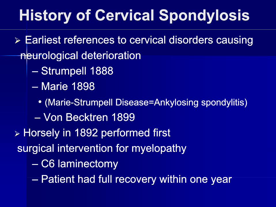
History of Cervical SpondylosisHistory of Cervical SpondylosisEarliest references to cervical disorders causingEarliest references to cervical disorders causing
neurological deteriorationneurological deteriorationeu o og ca de e o a oeu o og ca de e o a o–– StrumpellStrumpell 18881888–– Marie 1898Marie 1898Marie 1898Marie 1898• • (Marie(Marie--StrumpellStrumpell Disease=Disease=AnkylosingAnkylosing spondylitisspondylitis))
–– VonVon BecktrenBecktren 18991899–– Von Von BecktrenBecktren 18991899HorselyHorsely in 1892 performed firstin 1892 performed first
surgical intervention for myelopathysurgical intervention for myelopathysurgical intervention for myelopathy surgical intervention for myelopathy –– C6 laminectomyC6 laminectomy
Patient had full recovery within one yearPatient had full recovery within one year–– Patient had full recovery within one yearPatient had full recovery within one year
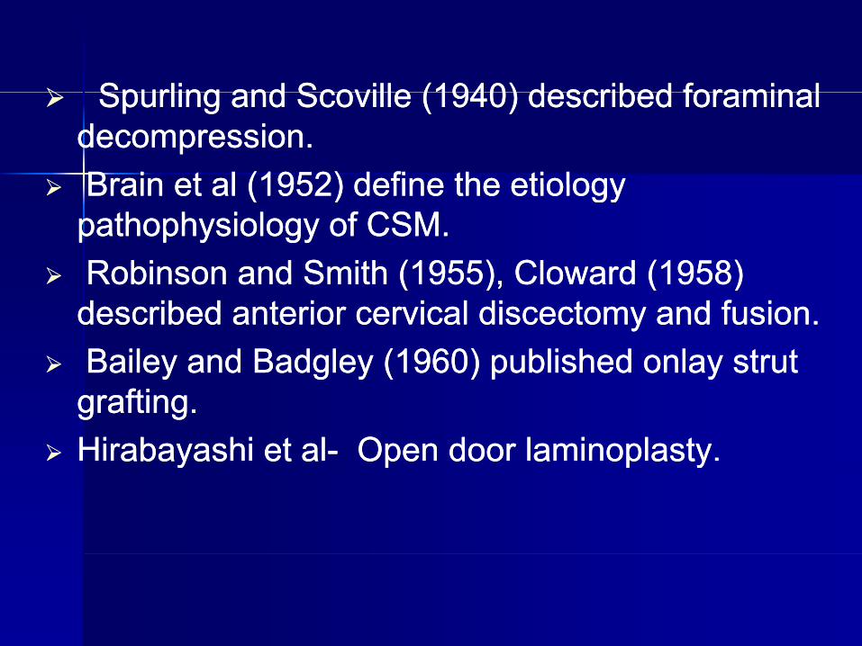
SpurlingSpurling andand ScovilleScoville (1940) described(1940) described foraminalforaminalSpurlingSpurling and and ScovilleScoville (1940) described (1940) described foraminalforaminaldecompression. decompression. Brain et al (1952) define the etiologyBrain et al (1952) define the etiologyBrain et al (1952) define the etiology Brain et al (1952) define the etiology pathophysiologypathophysiology of CSM.of CSM.Robinson and Smith (1955)Robinson and Smith (1955) ClowardCloward (1958)(1958)Robinson and Smith (1955), Robinson and Smith (1955), ClowardCloward (1958) (1958) described anterior cervical discectomy and fusion.described anterior cervical discectomy and fusion.Bailey andBailey and BadgleyBadgley (1960) published(1960) published onlayonlay strutstrutBailey and Bailey and BadgleyBadgley (1960) published (1960) published onlayonlay strut strut grafting.grafting.HirabayashiHirabayashi et alet al-- Open door laminoplasty.Open door laminoplasty.abayasabayas et aet a Ope doo a op astyOpe doo a op asty
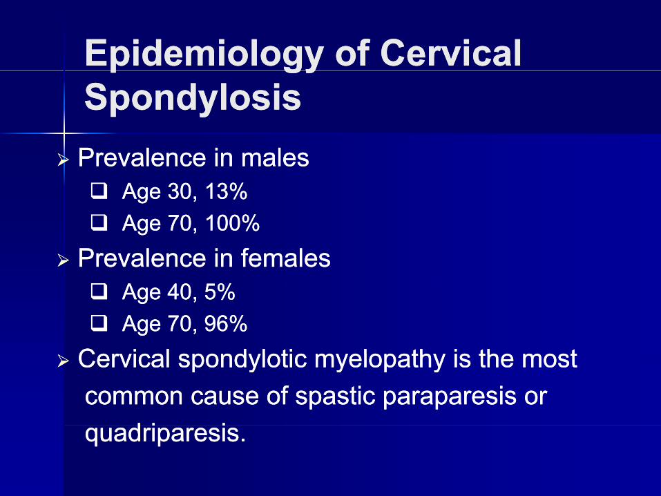
Epidemiology of Cervical Epidemiology of Cervical p gyp gySpondylosisSpondylosisPrevalence in malesPrevalence in males
Age 30, 13%Age 30, 13%Age 70, 100% Age 70, 100%
Prevalence in femalesPrevalence in females%%Age 40, 5%Age 40, 5%
Age 70, 96% Age 70, 96% Cervical spondylotic myelopathy is the mostCervical spondylotic myelopathy is the mostCervical spondylotic myelopathy is the most Cervical spondylotic myelopathy is the most common cause of spastic paraparesis or common cause of spastic paraparesis or
d i id i iquadriparesis. quadriparesis.
Pathophysiology of CervicalPathophysiology of CervicalSpondylosisSpondylosisSpondylosisSpondylosis
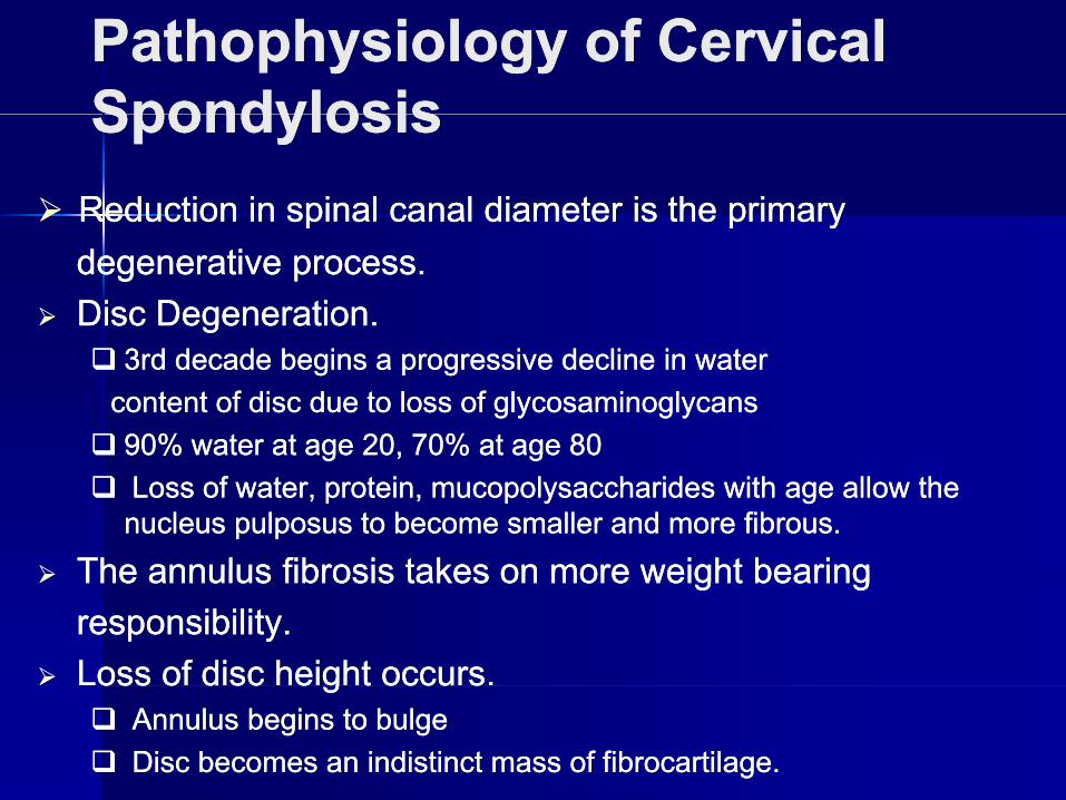
Reduction in spinal canal diameter is the primaryReduction in spinal canal diameter is the primaryp p yp p ydegenerative process.degenerative process.Disc Degeneration.Disc Degeneration.
3rd decade begins a progressive decline in water3rd decade begins a progressive decline in watercontent of disc due to loss of glycosaminoglycanscontent of disc due to loss of glycosaminoglycans90% water at age 20, 70% at age 8090% water at age 20, 70% at age 80g , gg , gLoss of water, protein, mucopolysaccharides with age allow the Loss of water, protein, mucopolysaccharides with age allow the nucleus pulposus to become smaller and more fibrous.nucleus pulposus to become smaller and more fibrous.
The annulus fibrosis takes on more weight bearingThe annulus fibrosis takes on more weight bearingThe annulus fibrosis takes on more weight bearingThe annulus fibrosis takes on more weight bearingresponsibility.responsibility.Loss of disc height occurs.Loss of disc height occurs.oss o d sc e g t occu soss o d sc e g t occu s
Annulus begins to bulgeAnnulus begins to bulgeDisc becomes an indistinct mass of fibrocartilage.Disc becomes an indistinct mass of fibrocartilage.
Pathophysiology of CervicalPathophysiology of CervicalSpondylosisSpondylosis
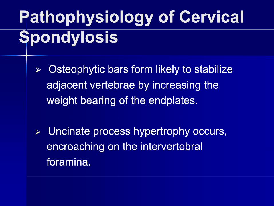
Osteophytic bars form likely to stabilizeOsteophytic bars form likely to stabilizeadjacent vertebrae by increasing theadjacent vertebrae by increasing theadjacent vertebrae by increasing theadjacent vertebrae by increasing theweight bearing of the endplates.weight bearing of the endplates.
Uncinate process hypertrophy occurs,Uncinate process hypertrophy occurs,encroaching on the intervertebralencroaching on the intervertebralencroaching on the intervertebralencroaching on the intervertebralforamina.foramina.
Pathophysiology of CervicalPathophysiology of CervicalS d l iS d l iSpondylosisSpondylosis
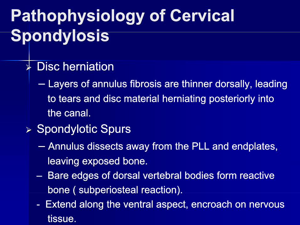
Disc herniationDisc herniationDisc herniationDisc herniation–– Layers of annulus fibrosis are thinner dorsally, leadingLayers of annulus fibrosis are thinner dorsally, leading
t t d di t i l h i ti t i l i tt t d di t i l h i ti t i l i tto tears and disc material herniating posteriorly intoto tears and disc material herniating posteriorly intothe canal.the canal.
Spondylotic SpursSpondylotic SpursSpondylotic SpursSpondylotic Spurs–– Annulus dissects away from the PLL and endplates, Annulus dissects away from the PLL and endplates,
leaving exposed boneleaving exposed boneleaving exposed bone.leaving exposed bone.–– Bare edges of dorsal vertebral bodies form reactiveBare edges of dorsal vertebral bodies form reactive
bone ( subperiosteal reaction).bone ( subperiosteal reaction).bone ( subperiosteal reaction).bone ( subperiosteal reaction).-- Extend along the ventral aspect, encroach on nervousExtend along the ventral aspect, encroach on nervous
tissue.tissue.
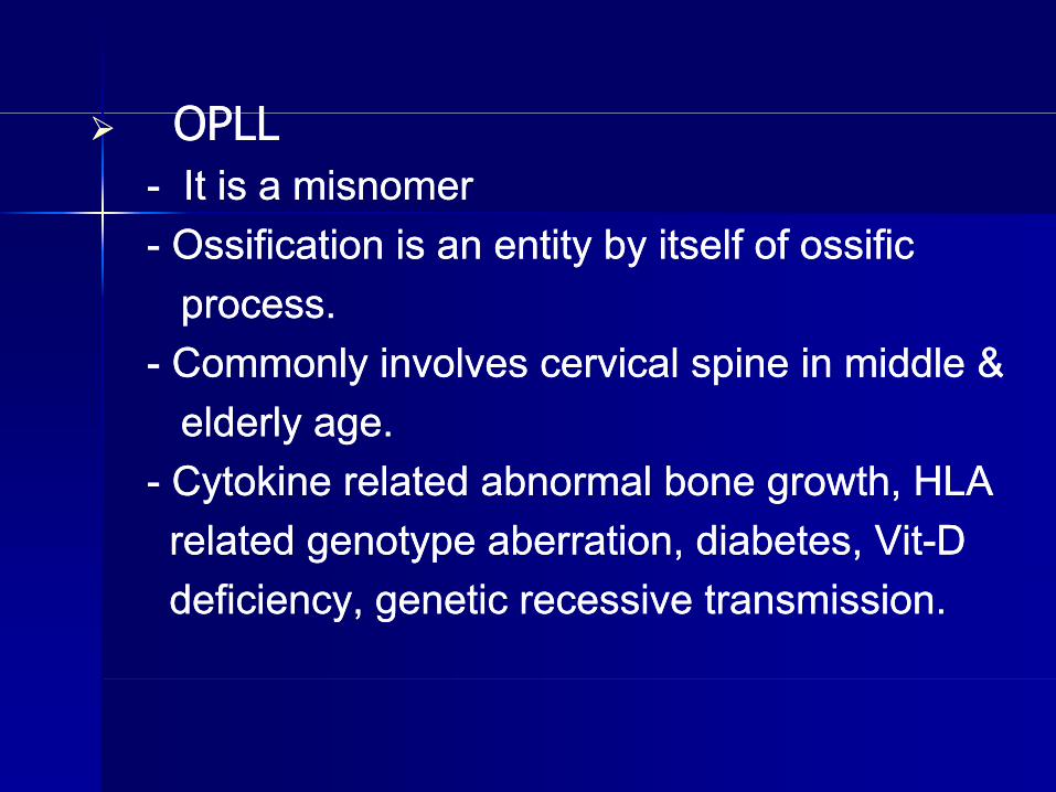
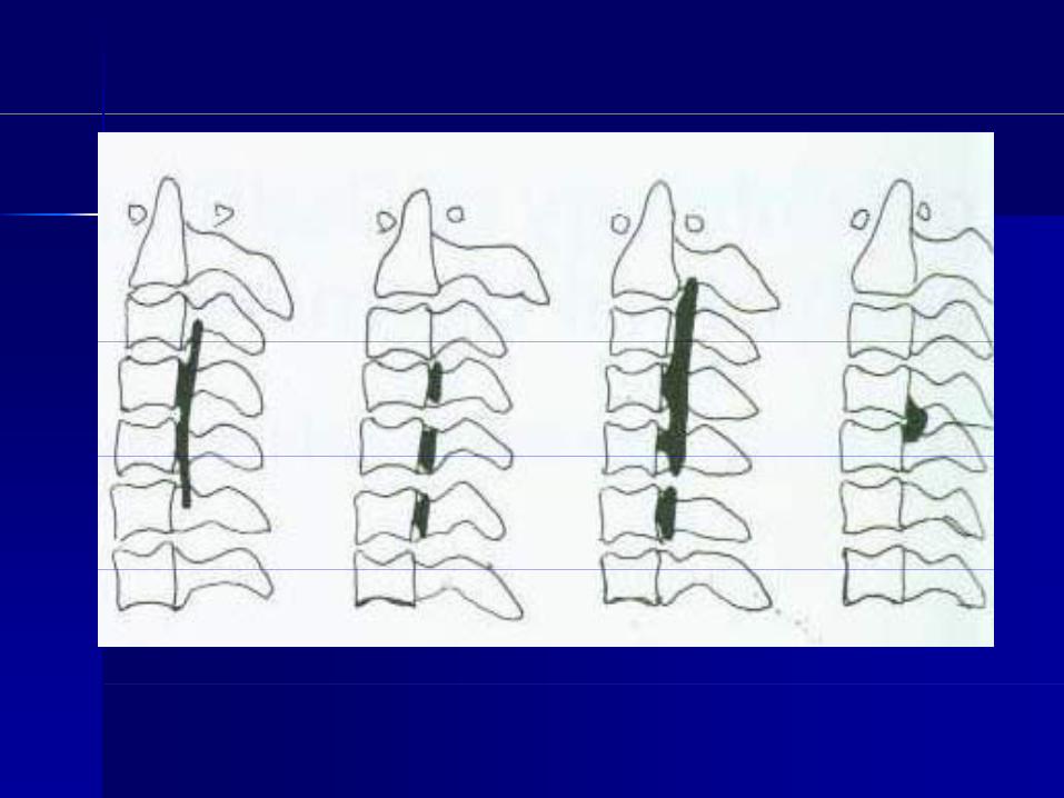
OPLLOPLLOPLLOPLL-- It is a misnomerIt is a misnomer-- Ossification is an entity by itself of ossificOssification is an entity by itself of ossific
process.process.-- Commonly involves cervical spine in middle &Commonly involves cervical spine in middle &
elderly age.elderly age.-- Cytokine related abnormal bone growth, Cytokine related abnormal bone growth, HLAHLArelated genotype aberration, diabetes, related genotype aberration, diabetes, VitVit--DDdeficiency, genetic recessive transmission.deficiency, genetic recessive transmission.
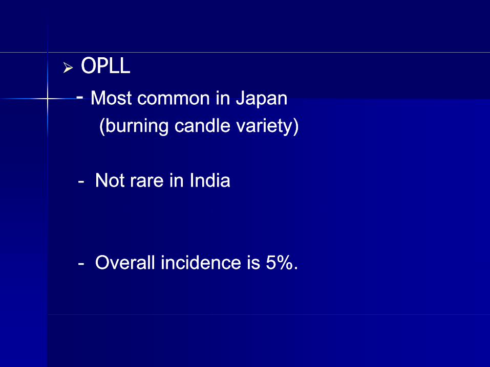
OPLLOPLL-- Most common in JapanMost common in Japanpp
(burning candle variety)(burning candle variety)
-- Not rare in IndiaNot rare in India
Overall incidence is 5%Overall incidence is 5%-- Overall incidence is 5%.Overall incidence is 5%.
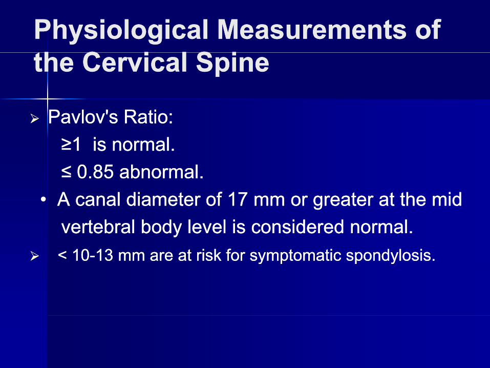
Physiological Measurements of Physiological Measurements of th C i l S ith C i l S ithe Cervical Spinethe Cervical Spine
Pavlov's Ratio:Pavlov's Ratio:≥1 is normal.≥1 is normal.≤ 0.85 abnormal.≤ 0.85 abnormal.
• A canal diameter of 17 mm or greater at the mid• A canal diameter of 17 mm or greater at the midvertebral body level is considered normal.vertebral body level is considered normal.< 10< 10--13 mm are at risk for symptomatic 13 mm are at risk for symptomatic spondylosisspondylosis..y py p p yp y
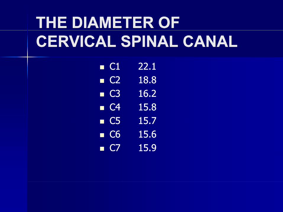
THE DIAMETER OFTHE DIAMETER OFCERVICAL SPINAL CANALCERVICAL SPINAL CANAL
C1 22.1C1 22.1C2 18.8C2 18.8C3 16 2C3 16 2C3 16.2C3 16.2C4 15.8C4 15.8C5 15 7C5 15 7C5 15.7C5 15.7C6 15.6C6 15.6C7 15.9C7 15.9
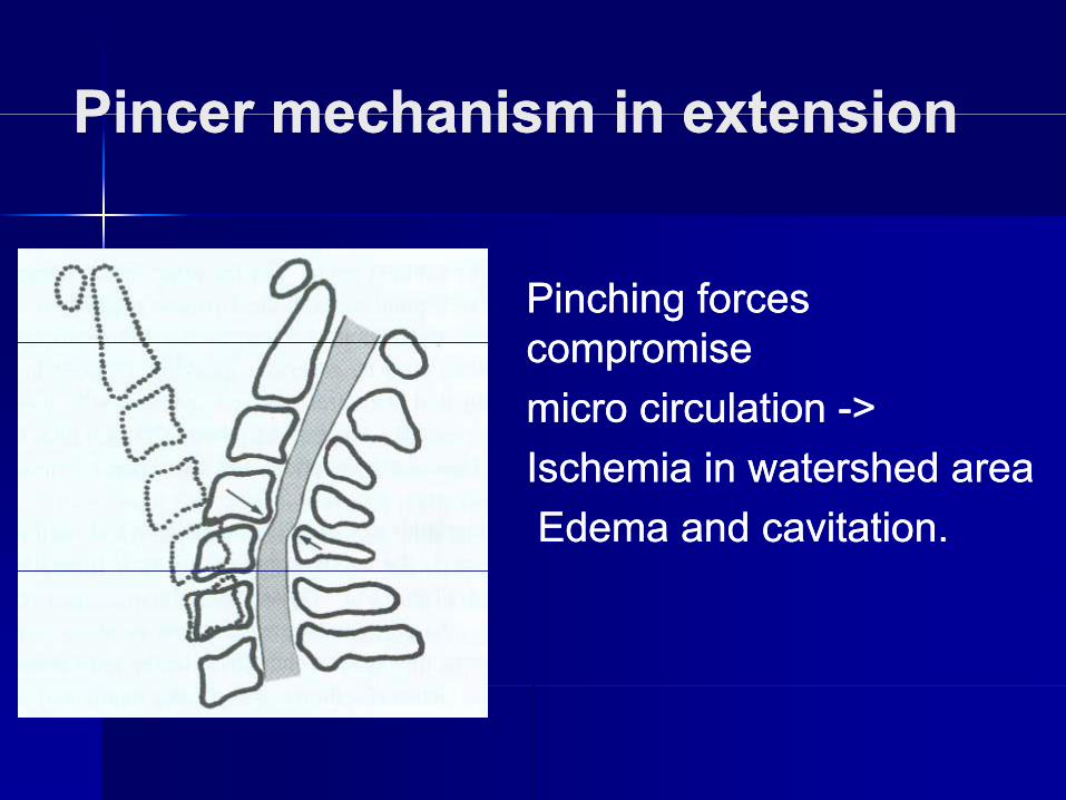
Pincer mechanism in extensionPincer mechanism in extensionPincer mechanism in extensionPincer mechanism in extension
Pinching forces Pinching forces compromisecompromisecompromise compromise micro circulation micro circulation -->>I h i i t h dI h i i t h dIschemia in watershed area Ischemia in watershed area Edema and Edema and cavitationcavitation..
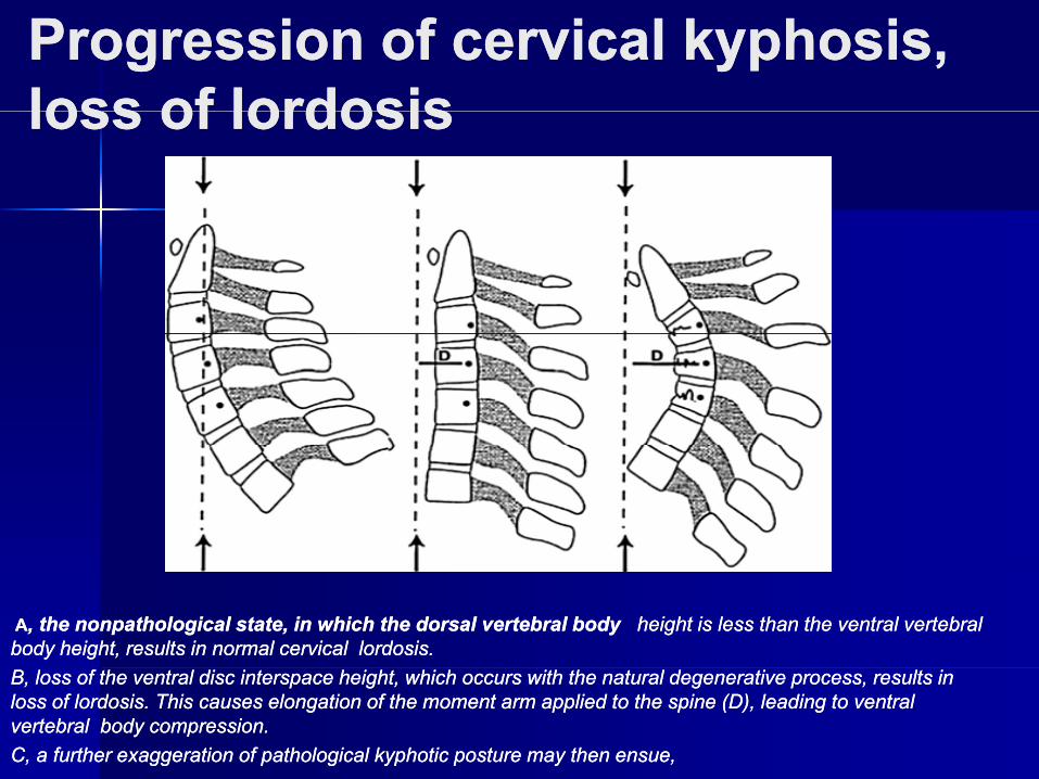
Progression of cervical kyphosis, Progression of cervical kyphosis, loss of lordosisloss of lordosisloss of lordosisloss of lordosis
AA, the , the nonpathologicalnonpathological state, in which the dorsal vertebral body state, in which the dorsal vertebral body height is less than the ventral vertebral height is less than the ventral vertebral body height, results in normal cervical lordosis. body height, results in normal cervical lordosis. B, loss of the ventral disc B, loss of the ventral disc interspaceinterspace height, which occurs with the natural degenerative process, results in height, which occurs with the natural degenerative process, results in loss of lordosis. This causes elongation of the moment arm applied to the spine (D), leading to ventral loss of lordosis. This causes elongation of the moment arm applied to the spine (D), leading to ventral vertebral body compression. vertebral body compression. C, a further exaggeration of pathological C, a further exaggeration of pathological kyphotickyphotic posture may then ensue, posture may then ensue,
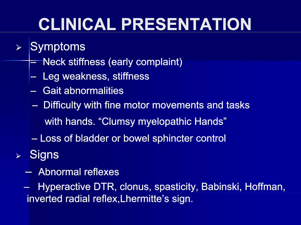
CLINICAL PRESENTATIONCLINICAL PRESENTATIONSymptomsSymptoms–– Neck stiffness (early complaint)Neck stiffness (early complaint)Neck stiffness (early complaint)Neck stiffness (early complaint)–– Leg weakness, stiffnessLeg weakness, stiffness–– Gait abnormalitiesGait abnormalities–– Difficulty with fine motor movements and tasksDifficulty with fine motor movements and tasks
with hands. “Clumsy with hands. “Clumsy myelopathicmyelopathic Hands”Hands”
–– Loss of bladder or bowel sphincter controlLoss of bladder or bowel sphincter control
SignsSignsgg–– Abnormal reflexesAbnormal reflexes–– Hyperactive DTR, clonus, spasticity, Babinski, Hoffman, Hyperactive DTR, clonus, spasticity, Babinski, Hoffman, inverted radial inverted radial reflex,Lhermitte’sreflex,Lhermitte’s sign.sign.
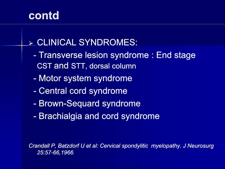
contdcontd
CLINICAL SYNDROMES:CLINICAL SYNDROMES:-- Transverse lesion syndrome : End stage Transverse lesion syndrome : End stage CST CST and and STT, dorsal column STT, dorsal column
-- Motor system syndromeMotor system syndrome-- Central cord syndrome Central cord syndrome -- BrownBrown--SequardSequard syndromesyndrome-- BrachialgiaBrachialgia and cord syndromeand cord syndromegg yy
Crandall PCrandall P BatzdorfBatzdorf U et al: CervicalU et al: Cervical spondyliticspondylitic myelopathy Jmyelopathy J NeurosurgNeurosurgCrandall P, Crandall P, BatzdorfBatzdorf U et al: Cervical U et al: Cervical spondyliticspondylitic myelopathy. J myelopathy. J NeurosurgNeurosurg25:5725:57--66,196666,1966..
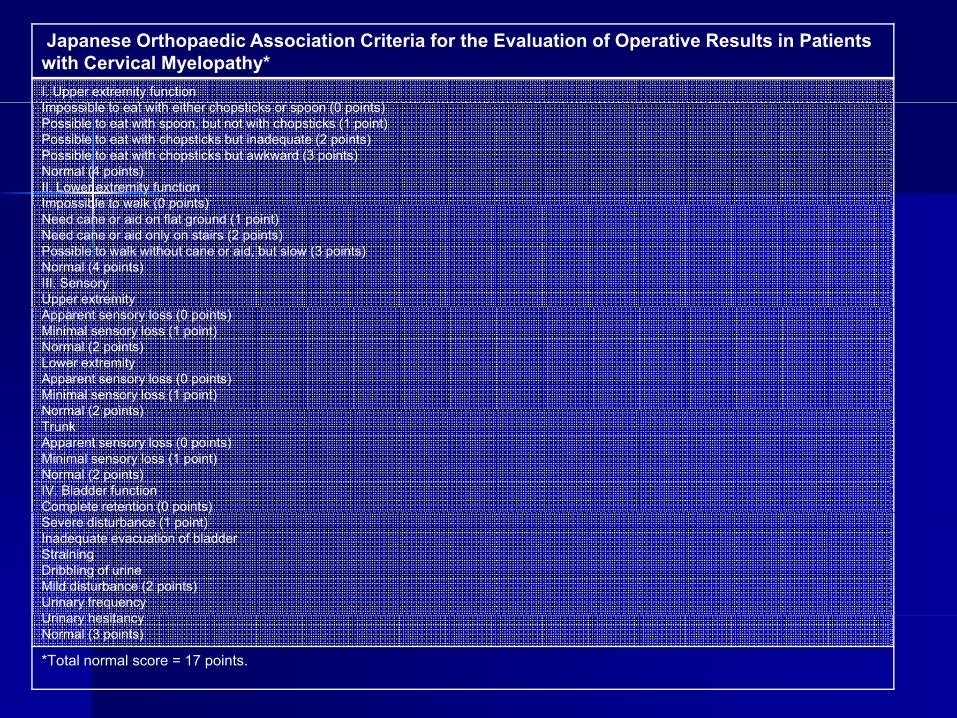
Japanese Orthopaedic Association Criteria for the Evaluation of Operative Results in Patients with Cervical Myelopathy*I. Upper extremity functionI ibl i h i h h i k (0 i )Impossible to eat with either chopsticks or spoon (0 points)Possible to eat with spoon, but not with chopsticks (1 point)Possible to eat with chopsticks but inadequate (2 points)Possible to eat with chopsticks but awkward (3 points)Normal (4 points)II. Lower extremity functionImpossible to walk (0 points)Impossible to walk (0 points)Need cane or aid on flat ground (1 point)Need cane or aid only on stairs (2 points)Possible to walk without cane or aid, but slow (3 points)Normal (4 points)III. SensoryUpper extremityApparent sensory loss (0 points)Minimal sensory loss (1 point)Normal (2 points)Lower extremityApparent sensory loss (0 points)Minimal sensory loss (1 point)Normal (2 points)Normal (2 points)TrunkApparent sensory loss (0 points)Minimal sensory loss (1 point)Normal (2 points)IV. Bladder functionComplete retention (0 points)p ( p )Severe disturbance (1 point)Inadequate evacuation of bladderStrainingDribbling of urineMild disturbance (2 points)Urinary frequencyU i h itUrinary hesitancyNormal (3 points)
*Total normal score = 17 points.
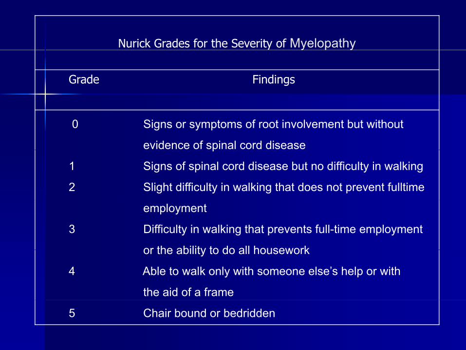
Nurick Grades for the Severity of Myelopathyy p y
Grade Findings
0 Signs or symptoms of root involvement but without
evidence of spinal cord diseasep
1 Signs of spinal cord disease but no difficulty in walking
2 Slight difficulty in walking that does not prevent fulltime
employment
3 Difficulty in walking that prevents full-time employment
or the ability to do all houseworkor the ability to do all housework
4 Able to walk only with someone else’s help or with
the aid of a frame
5 Chair bound or bedridden
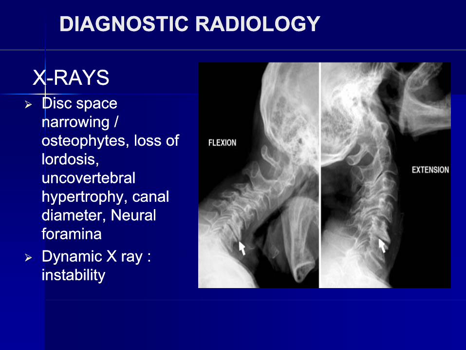
DIAGNOSTIC RADIOLOGYDIAGNOSTIC RADIOLOGY
XX--RAYSRAYSDiDiDisc space Disc space narrowing / narrowing / osteophytes, loss of osteophytes, loss of lordosis, lordosis, uncovertebraluncovertebralhypertrophy canalhypertrophy canalhypertrophy, canal hypertrophy, canal diameter, Neural diameter, Neural foramina foramina Dynamic X ray : Dynamic X ray : instability instability
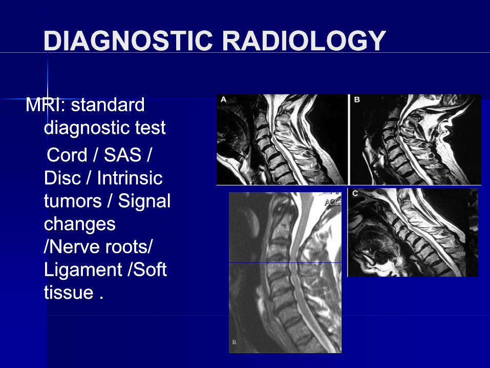
DIAGNOSTIC RADIOLOGYDIAGNOSTIC RADIOLOGY
MRI: standardMRI: standardMRI: standard MRI: standard diagnostic testdiagnostic testCord / SAS /Cord / SAS /Cord / SAS / Cord / SAS / Disc / Intrinsic Disc / Intrinsic tumors / Signal tumors / Signal ggchanges changes /Nerve roots/ /Nerve roots/ Li t /S ftLi t /S ftLigament /Soft Ligament /Soft tissue .tissue .
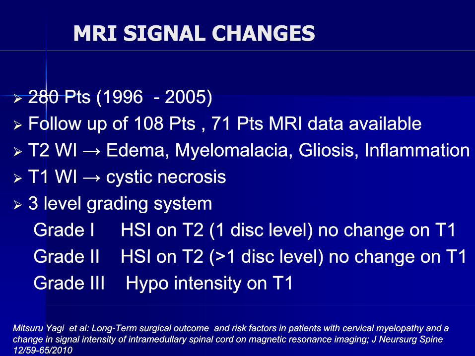
MRI SIGNAL CHANGESMRI SIGNAL CHANGES
280 Pts (1996280 Pts (1996 -- 2005)2005)280 Pts (1996 280 Pts (1996 2005) 2005) Follow up of 108 Pts , 71 Pts MRI data availableFollow up of 108 Pts , 71 Pts MRI data availableT2 WI → EdemaT2 WI → Edema MyelomalaciaMyelomalacia Gliosis InflammationGliosis InflammationT2 WI → Edema, T2 WI → Edema, MyelomalaciaMyelomalacia, Gliosis, Inflammation, Gliosis, InflammationT1 WI → cystic necrosisT1 WI → cystic necrosis3 level grading system3 level grading system3 level grading system3 level grading systemGrade I HSI on T2 (1 disc level) no change on T1Grade I HSI on T2 (1 disc level) no change on T1Grade II HSI on T2 (>1 disc level) no change on T1Grade II HSI on T2 (>1 disc level) no change on T1Grade II HSI on T2 (>1 disc level) no change on T1Grade II HSI on T2 (>1 disc level) no change on T1Grade III Hypo intensity on T1Grade III Hypo intensity on T1
Mitsuru Mitsuru YagiYagi et al: Longet al: Long--Term surgical outcome and risk factors in patients with cervical myelopathy and a Term surgical outcome and risk factors in patients with cervical myelopathy and a change in signal intensity of change in signal intensity of intramedullaryintramedullary spinal cord on magnetic resonance imaging; J spinal cord on magnetic resonance imaging; J NeursurgNeursurg Spine Spine 12/5912/59--65/201065/2010
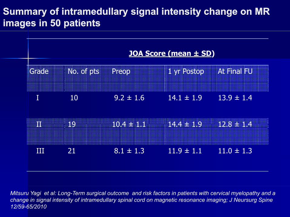
Summary of Summary of intramedullaryintramedullary signal intensity change on MR signal intensity change on MR images in 50 patientsimages in 50 patients
JOA Score (mean ± SD)
Grade No. of pts Preop 1 yr Postop At Final FU
I 10 9.2 ± 1.6 14.1 ± 1.9 13.9 ± 1.4
II 19 10 4 ± 1 1 14 4 ± 1 9 12 8 ± 1 4II 19 10.4 ± 1.1 14.4 ± 1.9 12.8 ± 1.4
III 21 8.1 ± 1.3 11.9 ± 1.1 11.0 ± 1.3
Mitsuru Yagi et al: Long-Term surgical outcome and risk factors in patients with cervical myelopathy and a change in signal intensity of intramedullary spinal cord on magnetic resonance imaging; J Neursurg Spine 12/59-65/2010
ContdContd……
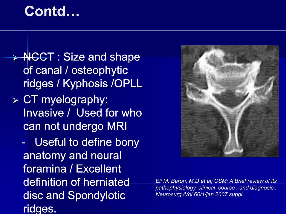
NCCT : Size and shape NCCT : Size and shape ppof canal / osteophytic of canal / osteophytic ridges / Kyphosis /OPLLridges / Kyphosis /OPLLCT myelography: CT myelography: Invasive / Used for who Invasive / Used for who
t d MRIt d MRIcan not undergo MRI can not undergo MRI -- Useful to define bony Useful to define bony
t d lt d lanatomy and neural anatomy and neural foramina / Excellent foramina / Excellent definition of herniateddefinition of herniated Eli M. Baron, M.D et al; CSM: A Brief review of its definition of herniated definition of herniated disc and Spondylotic disc and Spondylotic ridges.ridges.
, ;pathophysiology, clinical course , and diagnosis . Neurosurg /Vol 60/1/jan 2007 suppl
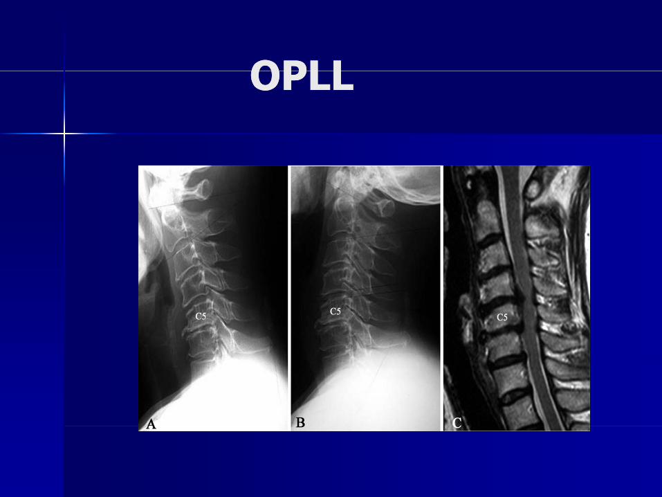
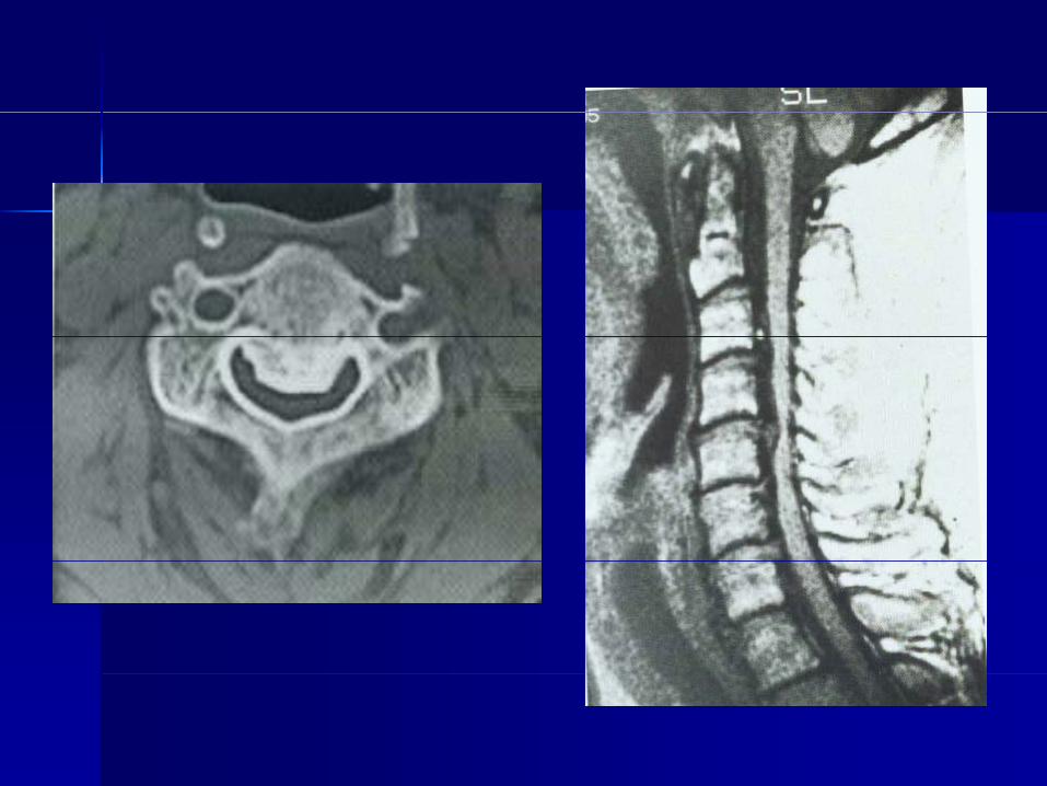
OPLLOPLLOPLLOPLL
TREATMENTTREATMENT
NONNON OPERATIVEOPERATIVENON NON –– OPERATIVEOPERATIVE
OPERATIVEOPERATIVEOPERATIVEOPERATIVE
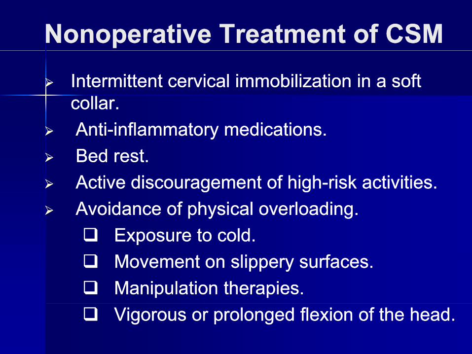
NonoperativeNonoperative Treatment of CSMTreatment of CSMIntermittent cervical immobilization in a soft Intermittent cervical immobilization in a soft collarcollarcollar.collar.AntiAnti--inflammatory medications.inflammatory medications.B d tB d tBed rest.Bed rest.Active discouragement of highActive discouragement of high--risk activities.risk activities.A id f h i l l diA id f h i l l diAvoidance of physical overloading.Avoidance of physical overloading.
Exposure to cold.Exposure to cold.Movement on slippery surfaces.Movement on slippery surfaces.Manipulation therapies.Manipulation therapies.Vigorous or prolonged flexion of the head.Vigorous or prolonged flexion of the head.
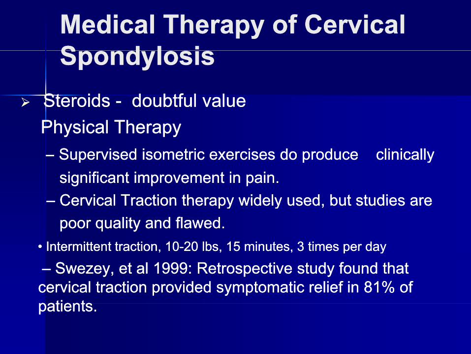
Medical Therapy of Cervical Medical Therapy of Cervical S d l iS d l iSpondylosisSpondylosis
SteroidsSteroids doubtful valuedoubtful valueSteroids Steroids -- doubtful valuedoubtful valuePhysical TherapyPhysical Therapy
Supervised isometric exercises do produce clinicallySupervised isometric exercises do produce clinically–– Supervised isometric exercises do produce clinicallySupervised isometric exercises do produce clinicallysignificant improvement in pain.significant improvement in pain.
–– Cervical Traction therapy widely used but studies areCervical Traction therapy widely used but studies areCervical Traction therapy widely used, but studies areCervical Traction therapy widely used, but studies arepoor quality and flawed.poor quality and flawed.
• Intermittent traction, 10• Intermittent traction, 10--20 lbs, 15 minutes, 3 times per day20 lbs, 15 minutes, 3 times per dayp yp y
–– SwezeySwezey, et al 1999: Retrospective study found that , et al 1999: Retrospective study found that cervical traction provided symptomatic relief in 81% of cervical traction provided symptomatic relief in 81% of patientspatientspatients.patients.
Choosing the Operative ProcedureChoosing the Operative ProcedureChoosing the Operative ProcedureChoosing the Operative Procedure
Sagittal alignmentSagittal alignment
Extent of disease Extent of disease
Location of abnormalityLocation of abnormality
Previous operationsPrevious operationspp
Indications for Operative Indications for Operative Treatment of Cervical MyelopathyTreatment of Cervical Myelopathy
Progressive clinical Progressive clinical myelopathymyelopathy with evidence of with evidence of spinal stenosis.spinal stenosis.pp
Progression of a neurological deficit.Progression of a neurological deficit.g gg g
The failure of neurological findings to improveThe failure of neurological findings to improveThe failure of neurological findings to improve The failure of neurological findings to improve with nonwith non--operative treatment (> 12 wks). operative treatment (> 12 wks).
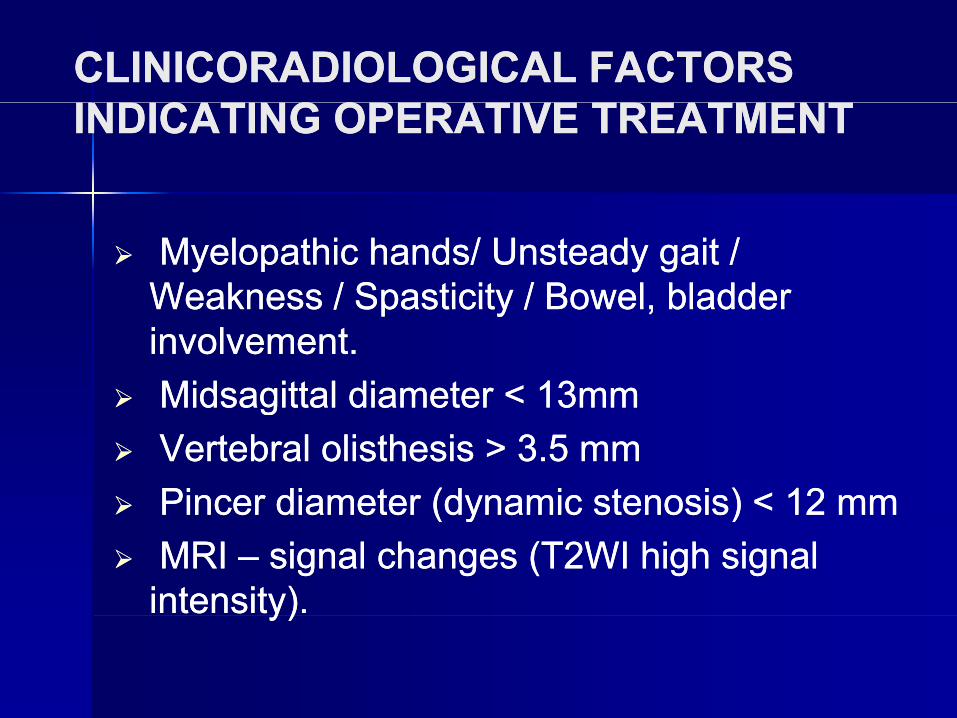
CLINICORADIOLOGICAL FACTORS CLINICORADIOLOGICAL FACTORS C G OC G OINDICATING OPERATIVE TREATMENTINDICATING OPERATIVE TREATMENT
Myelopathic hands/ Unsteady gait / Myelopathic hands/ Unsteady gait / Weakness / Spasticity / Bowel, bladderWeakness / Spasticity / Bowel, bladderWeakness / Spasticity / Bowel, bladder Weakness / Spasticity / Bowel, bladder involvement.involvement.Midsagittal diameter < 13mm Midsagittal diameter < 13mm ggVertebral Vertebral olisthesisolisthesis > 3.5 mm> 3.5 mmPincer diameter (dynamic stenosis) < 12 mmPincer diameter (dynamic stenosis) < 12 mmPincer diameter (dynamic stenosis) 12 mmPincer diameter (dynamic stenosis) 12 mmMRI MRI –– signal changes (T2WI high signal signal changes (T2WI high signal intensity).intensity).y)y)
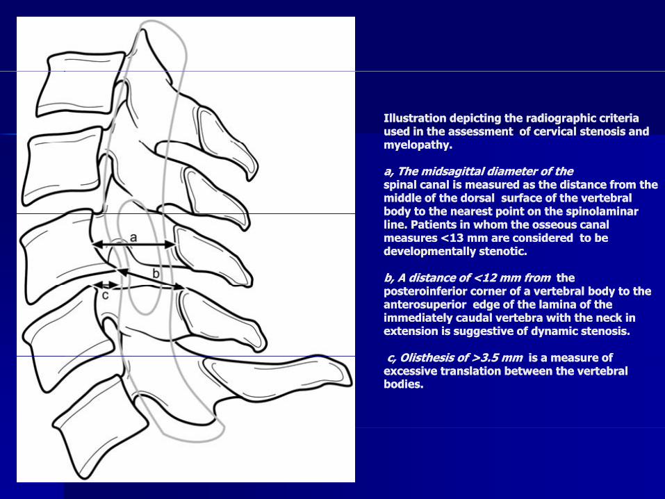
Illustration depicting the radiographic criteria Illustration depicting the radiographic criteria used in the assessment of cervical stenosis and used in the assessment of cervical stenosis and myelopathymyelopathymyelopathy. myelopathy.
a, The a, The midsagittalmidsagittal diameter of thediameter of thespinal canal is measured as the distance from the spinal canal is measured as the distance from the middle of the dorsal surface of the vertebral middle of the dorsal surface of the vertebral body to the nearest point on the spinolaminarbody to the nearest point on the spinolaminarbody to the nearest point on the spinolaminar body to the nearest point on the spinolaminar line. Patients in whom the osseous canal line. Patients in whom the osseous canal measures <13 mm are considered to be measures <13 mm are considered to be developmentally stenotic. developmentally stenotic.
b, A distance of <12 mm from b, A distance of <12 mm from the the posteroinferior corner of a vertebral body to the posteroinferior corner of a vertebral body to the anterosuperior edge of the lamina of the anterosuperior edge of the lamina of the immediately caudal vertebra with the neck in immediately caudal vertebra with the neck in extension is suggestive of dynamic stenosis. extension is suggestive of dynamic stenosis.
c Olisthesis of >3 5 mmc Olisthesis of >3 5 mm is a measure ofis a measure ofc, Olisthesis of >3.5 mm c, Olisthesis of >3.5 mm is a measure of is a measure of excessive translation between the vertebral excessive translation between the vertebral bodies.bodies.
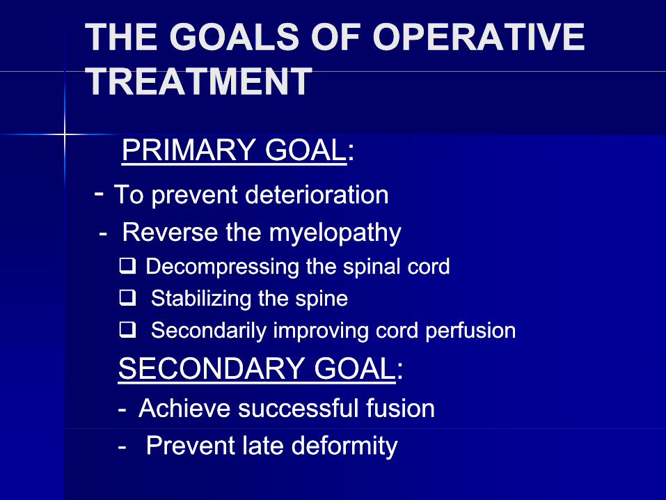
THE GOALS OF OPERATIVE THE GOALS OF OPERATIVE TREATMENTTREATMENTTREATMENTTREATMENT
PRIMARY GOALPRIMARY GOALPRIMARY GOALPRIMARY GOAL::-- To prevent deteriorationTo prevent deterioration-- Reverse the myelopathyReverse the myelopathy
Decompressing the spinal cordDecompressing the spinal cordStabilizing the spine Stabilizing the spine Secondarily improving cord perfusionSecondarily improving cord perfusion
SECONDARY GOALSECONDARY GOAL::-- Achieve successful fusionAchieve successful fusion-- Prevent late deformityPrevent late deformity
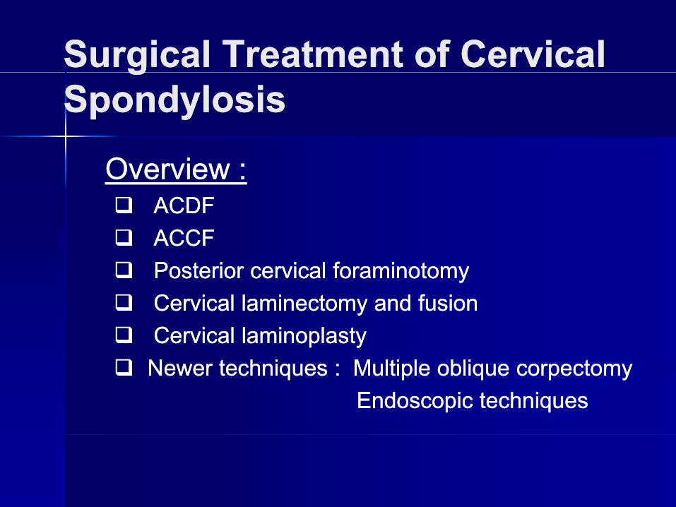
Surgical Treatment of CervicalSurgical Treatment of CervicalggSpondylosisSpondylosis
Overview :Overview :ACDFACDFACCFACCFPosterior cervical Posterior cervical foraminotomyforaminotomyCervical laminectomy and fusionCervical laminectomy and fusionCervical laminoplastyCervical laminoplastyN t h i M lti l bliN t h i M lti l bli ttNewer techniques : Multiple oblique Newer techniques : Multiple oblique corpectomycorpectomy
Endoscopic techniquesEndoscopic techniques
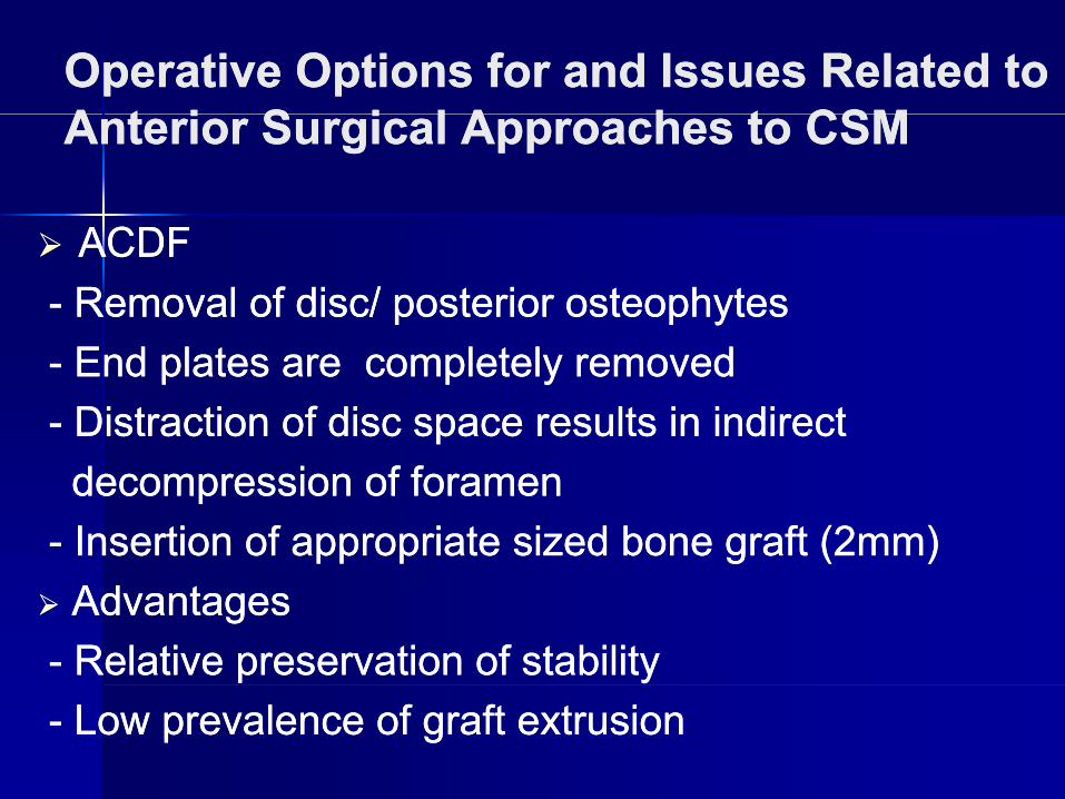
Operative Options for and Issues Related to Operative Options for and Issues Related to A t i S i l A h t CSMA t i S i l A h t CSMAnterior Surgical Approaches to CSMAnterior Surgical Approaches to CSM
ACDFACDFACDF ACDF -- Removal of disc/ posterior osteophytes Removal of disc/ posterior osteophytes -- End plates are completely removedEnd plates are completely removed-- Distraction of disc space results in indirectDistraction of disc space results in indirectdecompression of foramendecompression of foramen
-- Insertion of appropriate sized bone graft (2mm)Insertion of appropriate sized bone graft (2mm)AdvantagesAdvantages
-- Relative preservation of stabilityRelative preservation of stability-- Low prevalence of graft extrusion Low prevalence of graft extrusion
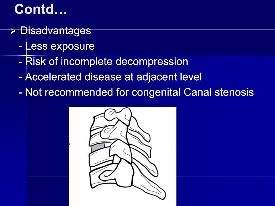
ContdContd……DisadvantagesDisadvantagesDisadvantagesDisadvantages-- Less exposure Less exposure
Ri k f i l t d iRi k f i l t d i-- Risk of incomplete decompressionRisk of incomplete decompression-- Accelerated disease at adjacent levelAccelerated disease at adjacent level
N d d f i l C l iN d d f i l C l i-- Not recommended for congenital Canal stenosisNot recommended for congenital Canal stenosis
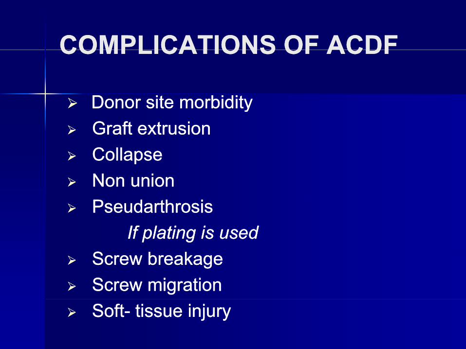
COMPLICATIONS OF ACDFCOMPLICATIONS OF ACDFCOMPLICATIONS OF ACDFCOMPLICATIONS OF ACDF
Donor site morbidityDonor site morbidityDonor site morbidity Donor site morbidity Graft extrusion Graft extrusion C llC llCollapse Collapse Non union Non union PseudarthrosisPseudarthrosis
If plating is used If plating is used Screw breakageScrew breakageScrew migration Screw migration SoftSoft-- tissue injurytissue injury
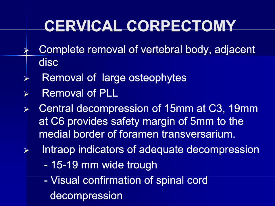
CERVICAL CORPECTOMYCERVICAL CORPECTOMYCERVICAL CORPECTOMYCERVICAL CORPECTOMYComplete removal of vertebral body, adjacent Complete removal of vertebral body, adjacent dididisc disc Removal of large osteophytesRemoval of large osteophytesRemoval of PLLRemoval of PLLCentral decompression of 15mm at C3, 19mm Central decompression of 15mm at C3, 19mm
t C6 id f t i f 5 t tht C6 id f t i f 5 t that C6 provides safety margin of 5mm to the at C6 provides safety margin of 5mm to the medial border of foramen transversarium.medial border of foramen transversarium.I tI t i di t f d t d ii di t f d t d iIntraopIntraop indicators of adequate decompressionindicators of adequate decompression-- 1515--19 mm wide trough 19 mm wide trough
Vi l fi ti f i l dVi l fi ti f i l d-- Visual confirmation of spinal cordVisual confirmation of spinal corddecompressiondecompression
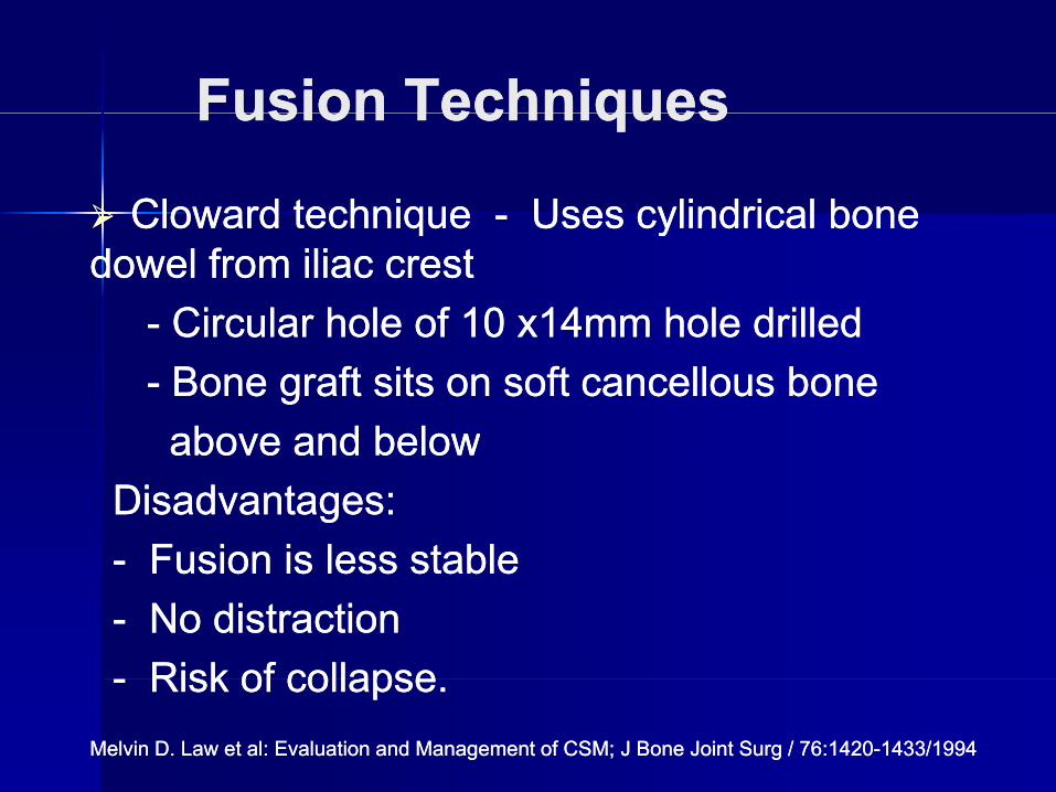
Fusion TechniquesFusion TechniquesFusion TechniquesFusion Techniques
ClowardCloward techniquetechnique -- Uses cylindrical boneUses cylindrical boneClowardCloward technique technique Uses cylindrical bone Uses cylindrical bone dowel from iliac crestdowel from iliac crest
-- Circular hole of 10 x14mm hole drilled Circular hole of 10 x14mm hole drilled -- Bone graft sits on soft Bone graft sits on soft cancellouscancellous boneboneabove and belowabove and belowabove and belowabove and below
Disadvantages: Disadvantages: -- Fusion is less stableFusion is less stableFusion is less stableFusion is less stable-- No distractionNo distraction
Risk of collapseRisk of collapse-- Risk of collapse.Risk of collapse.Melvin D. Law et al: Evaluation and Management of CSM; J Bone Joint Melvin D. Law et al: Evaluation and Management of CSM; J Bone Joint SurgSurg / 76:1420/ 76:1420--1433/1994 1433/1994
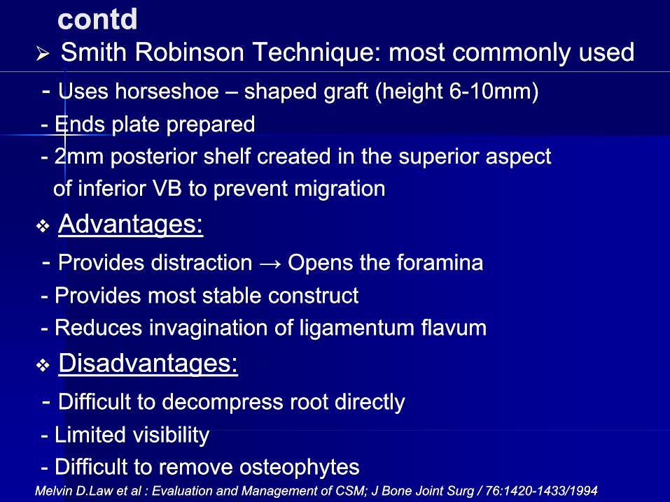
contdcontdSmith Robinson Technique: most commonly usedSmith Robinson Technique: most commonly used
-- Uses horseshoe Uses horseshoe –– shaped graft (height 6shaped graft (height 6--10mm)10mm)-- Ends plate prepared Ends plate prepared -- 2mm posterior shelf created in the superior aspect 2mm posterior shelf created in the superior aspect of inferior VB to prevent migration of inferior VB to prevent migration Ad tAd tAdvantages:Advantages:
-- Provides distraction → Opens the foramina Provides distraction → Opens the foramina -- Provides most stable construct Provides most stable construct -- Reduces invagination of Reduces invagination of ligamentumligamentum flavumflavum
Disadvantages:Disadvantages:Disadvantages:Disadvantages:-- Difficult to decompress root directly Difficult to decompress root directly
Li it d i ibilitLi it d i ibilit-- Limited visibilityLimited visibility-- Difficult to remove osteophytes Difficult to remove osteophytes
Melvin Melvin D.LawD.Law et al : Evaluation and Management of CSM; J Bone Joint et al : Evaluation and Management of CSM; J Bone Joint SurgSurg / 76:1420/ 76:1420--1433/1994 1433/1994
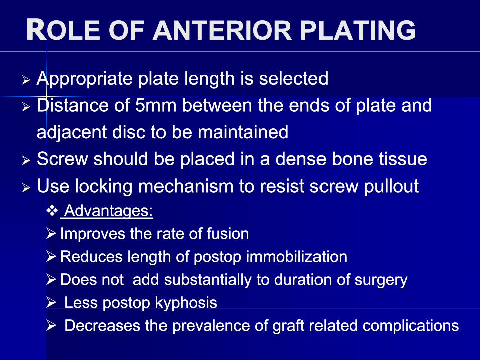
RROLE OF ANTERIOR PLATINGOLE OF ANTERIOR PLATINGAppropriate plate length is selectedAppropriate plate length is selectedDistance of 5mm between the ends of plate andDistance of 5mm between the ends of plate andDistance of 5mm between the ends of plate andDistance of 5mm between the ends of plate andadjacent disc to be maintained adjacent disc to be maintained S h ld b l d i d b tiS h ld b l d i d b tiScrew should be placed in a dense bone tissueScrew should be placed in a dense bone tissueUse locking mechanism to resist screw pulloutUse locking mechanism to resist screw pullout
Ad tAd tAdvantages:Advantages:Improves the rate of fusionImproves the rate of fusionReduces length of postop immobilizationReduces length of postop immobilizationReduces length of postop immobilization Reduces length of postop immobilization Does not add substantially to duration of surgeryDoes not add substantially to duration of surgeryLess postop kyphosisLess postop kyphosisp p ypp p ypDecreases the prevalence of graft related complications Decreases the prevalence of graft related complications
contdcontd……contdcontd……
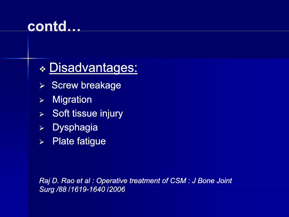
Disadvantages:Disadvantages:Screw breakage Screw breakage MigrationMigrationSoft tissue injurySoft tissue injuryD h iD h iDysphagiaDysphagiaPlate fatiguePlate fatigue
Raj D. Raj D. RaoRao et al : Operative treatment of CSM : J Bone Joint et al : Operative treatment of CSM : J Bone Joint SurgSurg /88/88 //16191619--16401640 //2006 2006
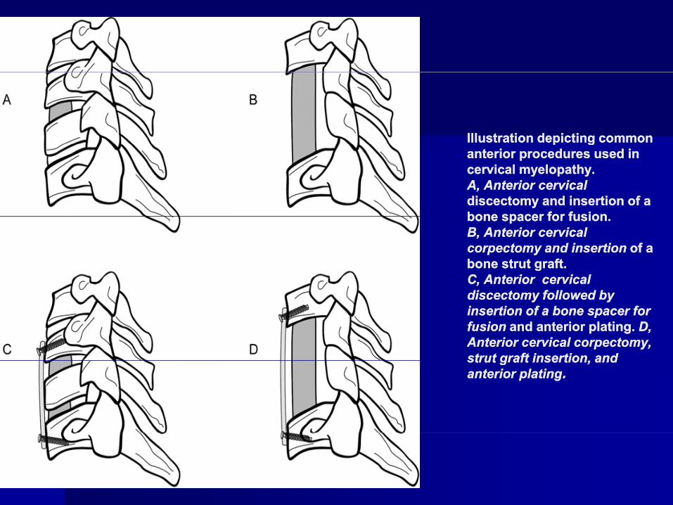
Illustration depicting common Illustration depicting common p gp ganterior procedures used in anterior procedures used in cervical myelopathy. cervical myelopathy. A, Anterior cervicalA, Anterior cervicaldiscectomy and insertion of a discectomy and insertion of a bone spacer for fusionbone spacer for fusionbone spacer for fusion. bone spacer for fusion. B, Anterior cervical B, Anterior cervical corpectomy and insertion corpectomy and insertion of a of a bone strut graft. bone strut graft. C, Anterior cervical C, Anterior cervical discectomy followed by discectomy followed by insertion of a bone spacer for insertion of a bone spacer for fusion fusion and anterior plating. and anterior plating. D, D, Anterior cervical corpectomy, Anterior cervical corpectomy, strut graft insertion andstrut graft insertion andstrut graft insertion, and strut graft insertion, and anterior platinganterior plating..
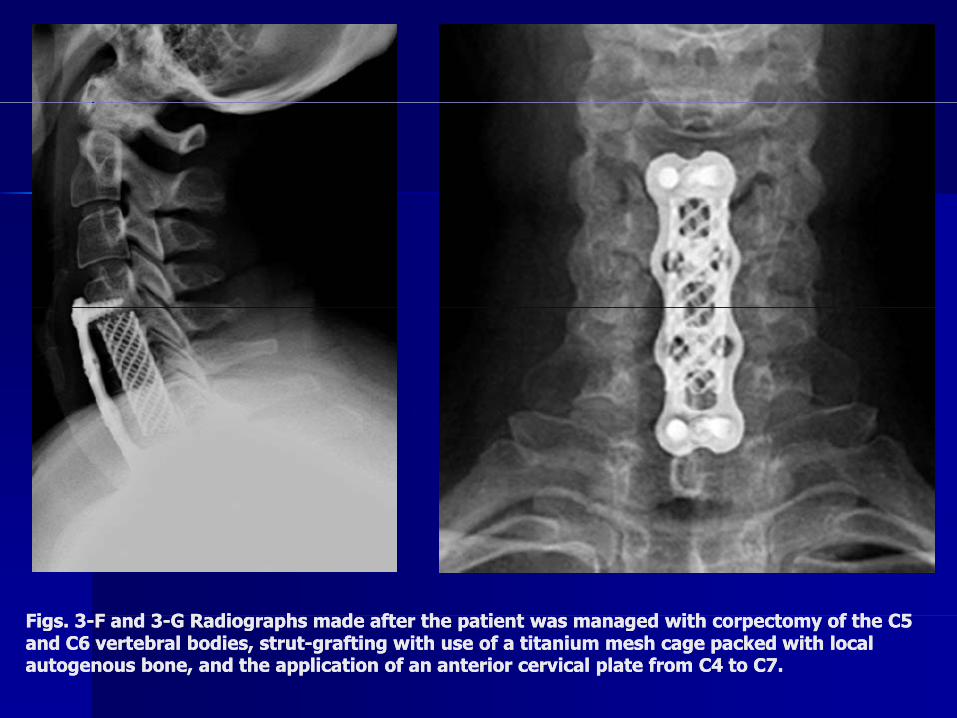
Fi 3Fi 3 F d 3F d 3 G R di h d ft th ti t d ith t f th C5G R di h d ft th ti t d ith t f th C5Figs. 3Figs. 3--F and 3F and 3--G Radiographs made after the patient was managed with corpectomy of the C5 G Radiographs made after the patient was managed with corpectomy of the C5 and C6 vertebral bodies, strutand C6 vertebral bodies, strut--grafting with use of a titanium mesh cage packed with local grafting with use of a titanium mesh cage packed with local autogenous bone, and the application of an anterior cervical plate from C4 to C7. autogenous bone, and the application of an anterior cervical plate from C4 to C7.
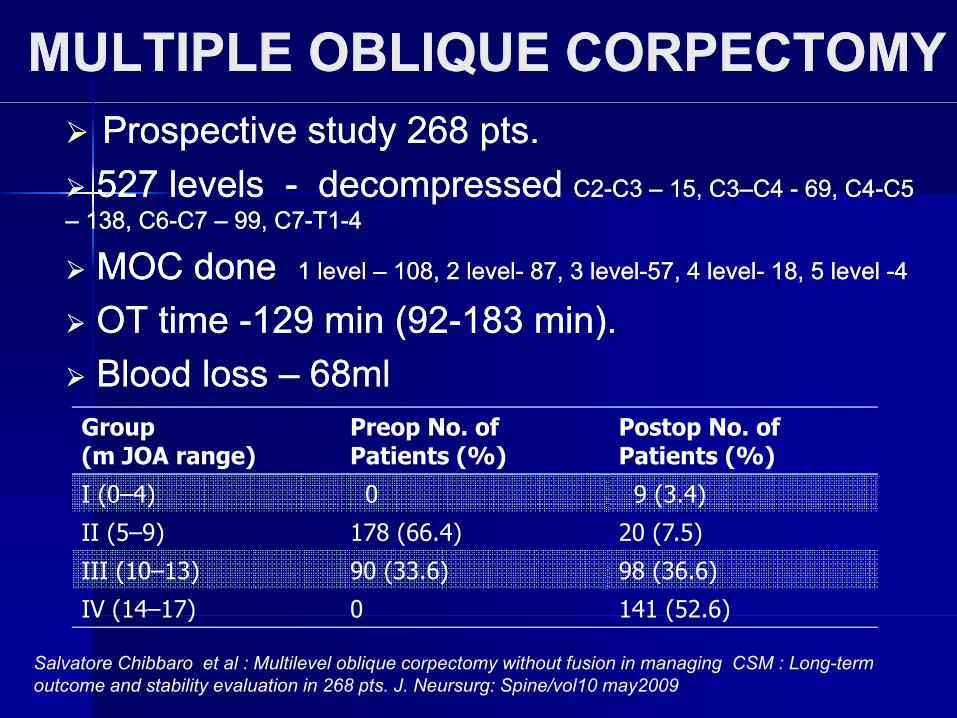
MULTIPLE OBLIQUE CORPECTOMYMULTIPLE OBLIQUE CORPECTOMYProspective study 268 pts.Prospective study 268 pts.527 levels 527 levels -- decompressed decompressed C2C2--C3 C3 –– 15, C315, C3––C4 C4 -- 69, C469, C4--C5 C5
–– 138, C6138, C6--C7 C7 –– 99, C799, C7--T1T1--44
MOC done MOC done 1 level 1 level –– 108, 2 level108, 2 level-- 87, 3 level87, 3 level--57, 4 level57, 4 level-- 18, 5 level 18, 5 level --44
OT tiOT ti 129 i (92129 i (92 183 i )183 i )OT time OT time --129 min (92129 min (92--183 min).183 min).Blood loss Blood loss –– 68ml68ml
Group(m JOA range)
Preop No. ofPatients (%)
Postop No. ofPatients (%)
I (0–4) 0 9 (3.4)
II (5–9) 178 (66.4) 20 (7.5)
III (10–13) 90 (33.6) 98 (36.6)
IV (14–17) 0 141 (52.6)IV (14 17) 0 141 (52.6)
Salvatore Chibbaro et al : Multilevel oblique corpectomy without fusion in managing CSM : Long-term outcome and stability evaluation in 268 pts. J. Neursurg: Spine/vol10 may2009
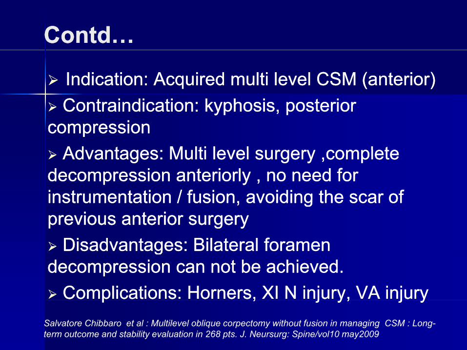
ContdContd… …
Indication: Acquired multi level CSM (anterior)Indication: Acquired multi level CSM (anterior)C t i di ti k h i t iC t i di ti k h i t iContraindication: kyphosis, posterior Contraindication: kyphosis, posterior
compressioncompressionAd antages M lti le el s rger completeAd antages M lti le el s rger completeAdvantages: Multi level surgery ,complete Advantages: Multi level surgery ,complete
decompression anteriorly , no need for decompression anteriorly , no need for instrumentation / fusion avoiding the scar ofinstrumentation / fusion avoiding the scar ofinstrumentation / fusion, avoiding the scar of instrumentation / fusion, avoiding the scar of previous anterior surgeryprevious anterior surgery
Disadvantages: Bilateral foramenDisadvantages: Bilateral foramenDisadvantages: Bilateral foramen Disadvantages: Bilateral foramen decompression can not be achieved.decompression can not be achieved.
Complications: Complications: HornersHorners, XI N injury, VA injury, XI N injury, VA injurypp , j y, j y, j y, j ySalvatore Chibbaro et al : Multilevel oblique corpectomy without fusion in managing CSM : Long-term outcome and stability evaluation in 268 pts. J. Neursurg: Spine/vol10 may2009
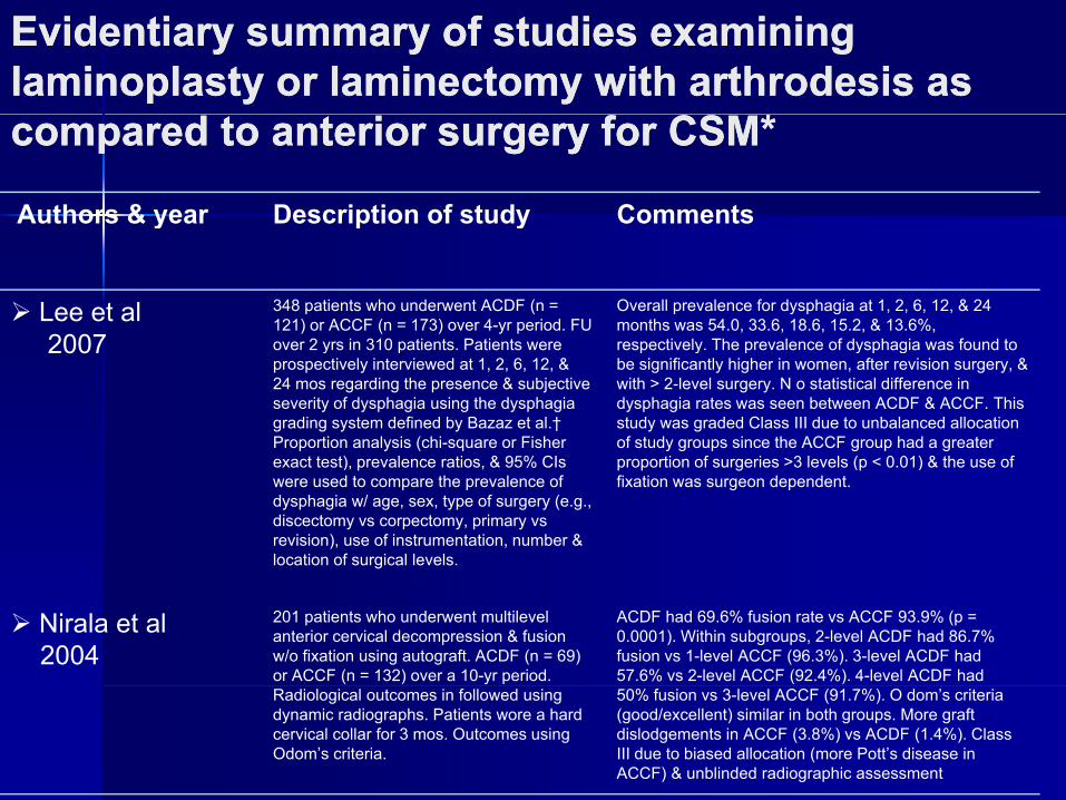
Evidentiary summary of studies examining Evidentiary summary of studies examining laminoplasty or laminectomy with laminoplasty or laminectomy with arthrodesisarthrodesis as as compared to anterior surgery for CSM*compared to anterior surgery for CSM*
Authors & year Description of study Commentsy p y
Lee et al2007
348 patients who underwent ACDF (n = 121) or ACCF (n = 173) over 4-yr period. FU over 2 yrs in 310 patients Patients were
Overall prevalence for dysphagia at 1, 2, 6, 12, & 24 months was 54.0, 33.6, 18.6, 15.2, & 13.6%, respectively The prevalence of dysphagia was found to2007 over 2 yrs in 310 patients. Patients were
prospectively interviewed at 1, 2, 6, 12, &24 mos regarding the presence & subjective severity of dysphagia using the dysphagiagrading system defined by Bazaz et al.† Proportion analysis (chi-square or Fisher
t t t) l ti & 95% CI
respectively. The prevalence of dysphagia was found to be significantly higher in women, after revision surgery, & with > 2-level surgery. N o statistical difference in dysphagia rates was seen between ACDF & ACCF. This study was graded Class III due to unbalanced allocation of study groups since the ACCF group had a greater
ti f i 3 l l ( 0 01) & th fexact test), prevalence ratios, & 95% CIs were used to compare the prevalence of dysphagia w/ age, sex, type of surgery (e.g., discectomy vs corpectomy, primary vsrevision), use of instrumentation, number &location of surgical levels.
proportion of surgeries >3 levels (p < 0.01) & the use of fixation was surgeon dependent.
Nirala et al2004
201 patients who underwent multilevel anterior cervical decompression & fusion w/o fixation using autograft. ACDF (n = 69) or ACCF (n = 132) over a 10-yr period.
ACDF had 69.6% fusion rate vs ACCF 93.9% (p = 0.0001). Within subgroups, 2-level ACDF had 86.7% fusion vs 1-level ACCF (96.3%). 3-level ACDF had 57.6% vs 2-level ACCF (92.4%). 4-level ACDF had
Radiological outcomes in followed using dynamic radiographs. Patients wore a hard cervical collar for 3 mos. Outcomes using Odom’s criteria.
50% fusion vs 3-level ACCF (91.7%). O dom’s criteria (good/excellent) similar in both groups. More graft dislodgements in ACCF (3.8%) vs ACDF (1.4%). Class III due to biased allocation (more Pott’s disease in ACCF) & unblinded radiographic assessment
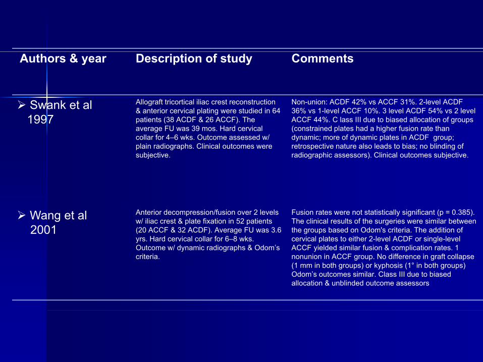
Authors & year Description of study Comments
Swank et al Allograft tricortical iliac crest reconstruction Non-union: ACDF 42% vs ACCF 31% 2-level ACDFSwank et al1997
Allograft tricortical iliac crest reconstruction & anterior cervical plating were studied in 64 patients (38 ACDF & 26 ACCF). The average FU was 39 mos. Hard cervicalcollar for 4–6 wks. Outcome assessed w/ plain radiographs. Clinical outcomes were subjective
Non union: ACDF 42% vs ACCF 31%. 2 level ACDF 36% vs 1-level ACCF 10%. 3 level ACDF 54% vs 2 level ACCF 44%. C lass III due to biased allocation of groups (constrained plates had a higher fusion rate than dynamic; more of dynamic plates in ACDF group; retrospective nature also leads to bias; no blinding of radiographic assessors) Clinical outcomes subjectivesubjective. radiographic assessors). Clinical outcomes subjective.
Wang et al2001
Anterior decompression/fusion over 2 levels w/ iliac crest & plate fixation in 52 patients (20 ACCF & 32 ACDF). Average FU was 3.6 yrs. Hard cervical collar for 6–8 wks. Outcome w/ dynamic radiographs & Odom’s criteria.
Fusion rates were not statistically significant (p = 0.385). The clinical results of the surgeries were similar between the groups based on Odom's criteria. The addition of cervical plates to either 2-level ACDF or single-level ACCF yielded similar fusion & complication rates. 1nonunion in ACCF group. No difference in graft collapsecriteria. nonunion in ACCF group. No difference in graft collapse (1 mm in both groups) or kyphosis (1° in both groups) Odom’s outcomes similar. Class III due to biased allocation & unblinded outcome assessors
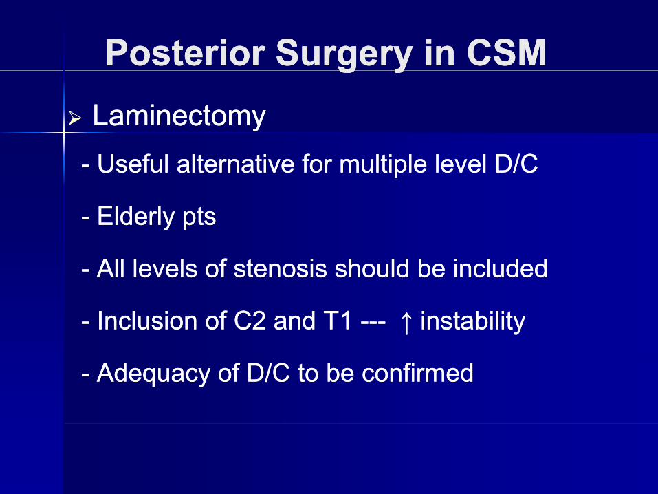
Posterior Surgery in CSMPosterior Surgery in CSMg yg yLaminectomyLaminectomy
-- Useful alternative for multiple level D/C Useful alternative for multiple level D/C
Elderly ptsElderly pts-- Elderly ptsElderly pts
-- All levels of stenosis should be includedAll levels of stenosis should be included
-- Inclusion of C2 and T1 Inclusion of C2 and T1 ------ ↑ instability↑ instability
-- Adequacy of D/C to be confirmed Adequacy of D/C to be confirmed
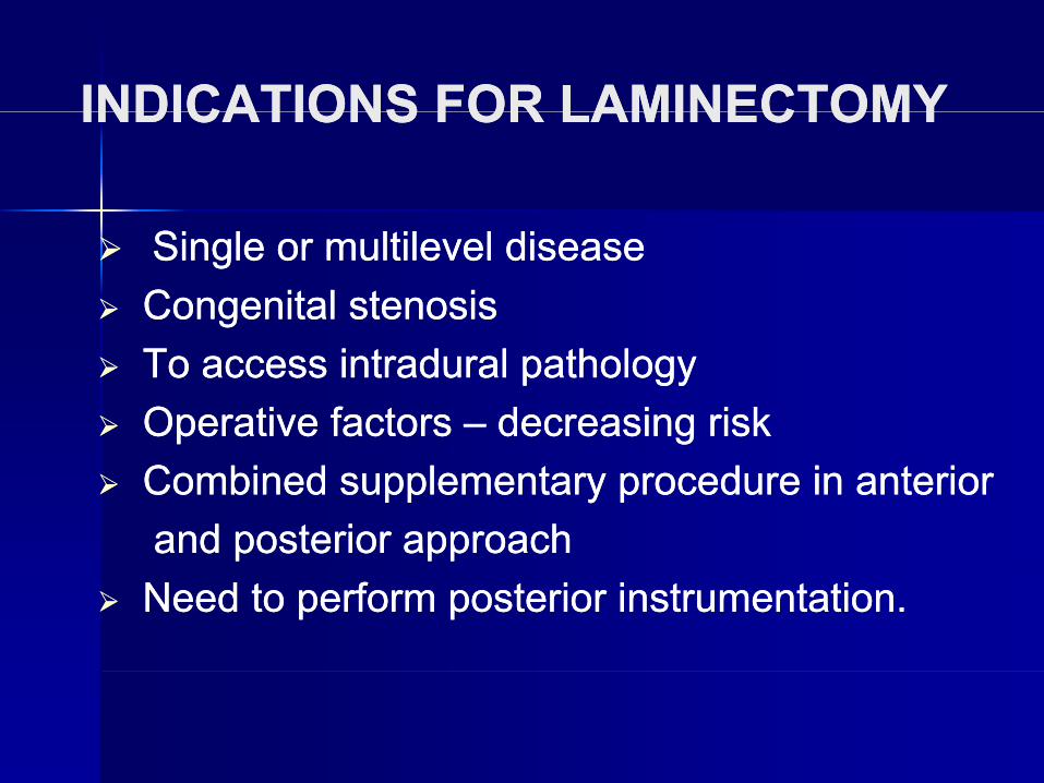
INDICATIONS FOR LAMINECTOMYINDICATIONS FOR LAMINECTOMYINDICATIONS FOR LAMINECTOMYINDICATIONS FOR LAMINECTOMY
Single or multilevel diseaseSingle or multilevel diseaseCongenital stenosisCongenital stenosisTo access intradural pathologyTo access intradural pathologyOperative factors Operative factors –– decreasing riskdecreasing riskCombined supplementary procedure in anterior Combined supplementary procedure in anterior and posterior approachand posterior approachp ppp ppNeed to perform posterior instrumentation.Need to perform posterior instrumentation.
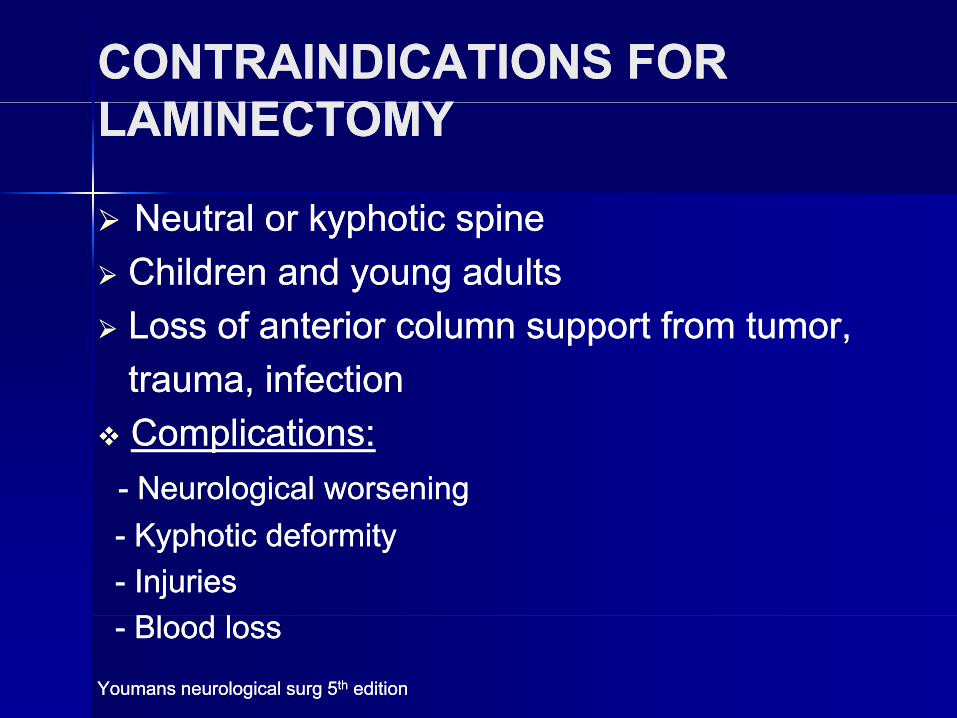
CONTRAINDICATIONS FOR CONTRAINDICATIONS FOR LAMINECTOMYLAMINECTOMYLAMINECTOMYLAMINECTOMY
N t lN t l k h tik h ti iiNeutral or Neutral or kyphotickyphotic spinespineChildren and young adultsChildren and young adultsLoss of anterior column support from tumor, Loss of anterior column support from tumor, trauma, infectiontrauma, infectionComplications:Complications:
-- Neurological worsening Neurological worsening -- KyphoticKyphotic deformitydeformity-- InjuriesInjuries-- Blood lossBlood loss
YoumansYoumans neurological neurological surgsurg 55thth editionedition
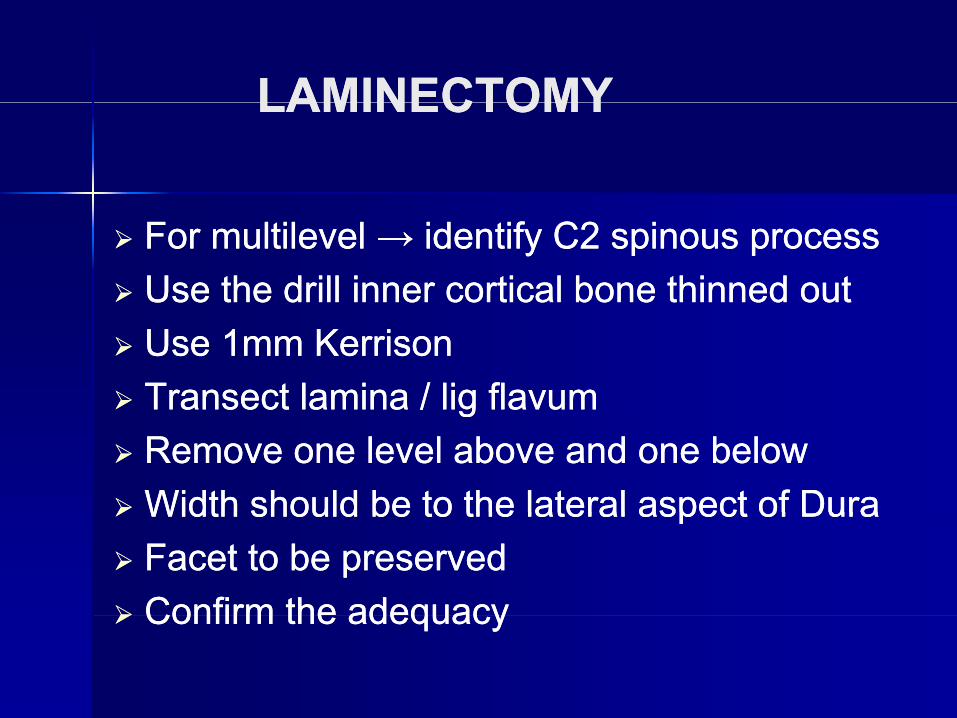
LAMINECTOMYLAMINECTOMYLAMINECTOMYLAMINECTOMY
For multilevel → identify C2 spinous processFor multilevel → identify C2 spinous processUse the drill inner cortical bone thinned outUse the drill inner cortical bone thinned outUse 1mm Use 1mm KerrisonKerrisonTransect lamina / Transect lamina / liglig flavumflavumggRemove one level above and one belowRemove one level above and one belowWidth should be to the lateral aspect of DuraWidth should be to the lateral aspect of DuraWidth should be to the lateral aspect of DuraWidth should be to the lateral aspect of DuraFacet to be preservedFacet to be preservedConfirm the adequacyConfirm the adequacyConfirm the adequacy Confirm the adequacy
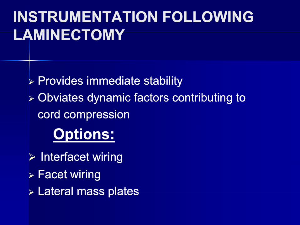
INSTRUMENTATION FOLLOWING INSTRUMENTATION FOLLOWING LAMINECTOMYLAMINECTOMYLAMINECTOMYLAMINECTOMY
Provides immediate stabilityProvides immediate stabilityObviates dynamic factors contributing toObviates dynamic factors contributing toObviates dynamic factors contributing to Obviates dynamic factors contributing to cord compressioncord compression
O iO iOptions:Options:InterfacetInterfacet wiringwiringInterfacetInterfacet wiring wiring Facet wiringFacet wiringLateral mass platesLateral mass platesLateral mass plates Lateral mass plates
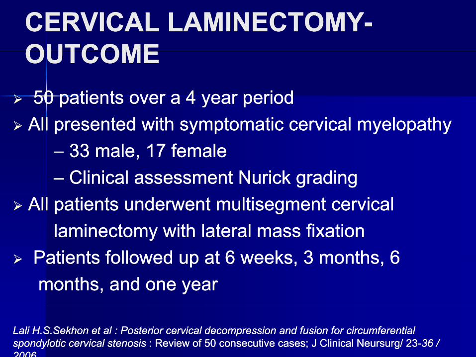
CERVICAL LAMINECTOMYCERVICAL LAMINECTOMY--OUTCOMEOUTCOMEOUTCOMEOUTCOME50 patients over a 4 year period50 patients over a 4 year period50 patients over a 4 year period50 patients over a 4 year period
All presented with symptomatic cervical myelopathyAll presented with symptomatic cervical myelopathy–– 33 male 17 female33 male 17 female33 male, 17 female33 male, 17 female–– Clinical assessment Clinical assessment NurickNurick gradinggrading
All patients underwentAll patients underwent multisegmentmultisegment cervicalcervicalAll patients underwent All patients underwent multisegmentmultisegment cervicalcervicallaminectomy with lateral mass fixationlaminectomy with lateral mass fixation
Patients followed up at 6 weeks 3 months 6Patients followed up at 6 weeks 3 months 6Patients followed up at 6 weeks, 3 months, 6 Patients followed up at 6 weeks, 3 months, 6 months, and one year months, and one year
LaliLali H.S.SekhonH.S.Sekhon et al : Posterior cervical decompression and fusion for circumferential et al : Posterior cervical decompression and fusion for circumferential spondyloticspondylotic cervical stenosiscervical stenosis : Review of 50 consecutive cases; J Clinical : Review of 50 consecutive cases; J Clinical NeursurgNeursurg/ 23/ 23--36 / 36 / 20062006
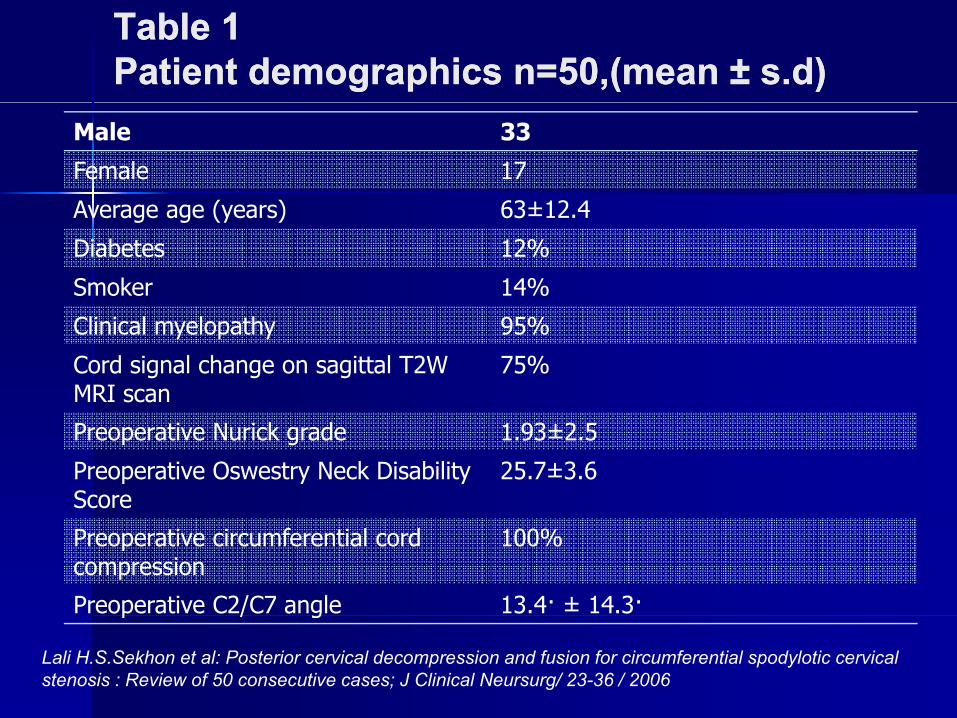
Table 1Table 1Patient demographics n=50,(mean Patient demographics n=50,(mean ±± s.ds.d))
Male 33
Female 17
Average age (years) 63±12 4Average age (years) 63±12.4
Diabetes 12%
Smoker 14%
Clinical myelopathy 95%
Cord signal change on sagittal T2W MRI scan
75%
Preoperative Nurick grade 1.93±2.5
Preoperative Oswestry Neck Disability Score
25.7±3.6
Preoperative circumferential cord compression
100%
Preoperative C2/C7 angle 13.4· ± 14.3·p / g
Lali H.S.Sekhon et al: Posterior cervical decompression and fusion for circumferential spodylotic cervical stenosis : Review of 50 consecutive cases; J Clinical Neursurg/ 23-36 / 2006
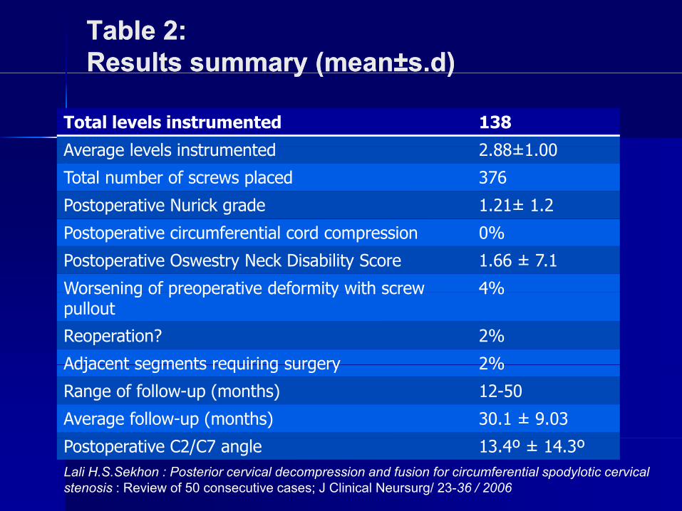
Table 2:Table 2:Results summary (Results summary (meanmean±±s.ds.d))y (y ( ))
Total levels instrumented 138
A l l i t t d 2 88±1 00Average levels instrumented 2.88±1.00
Total number of screws placed 376
Postoperative Nurick grade 1.21± 1.2
Postoperative circumferential cord compression 0%
Postoperative Oswestry Neck Disability Score 1.66 ± 7.1
Worsening of preoperative deformity with screw 4%Worsening of preoperative deformity with screw pullout
4%
Reoperation? 2%
Adjacent segments requiring surgery 2%Adjacent segments requiring surgery 2%
Range of follow-up (months) 12-50
Average follow-up (months) 30.1 ± 9.03
Postoperative C2/C7 angle 13.4º ± 14.3ºLali H.S.Sekhon : Posterior cervical decompression and fusion for circumferential spodylotic cervical stenosis : Review of 50 consecutive cases; J Clinical Neursurg/ 23-36 / 2006
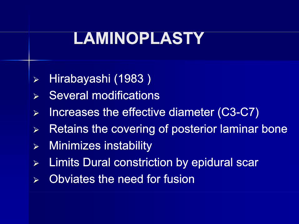
O SO SLAMINOPLASTYLAMINOPLASTY
HirabayashiHirabayashi (1983 ) (1983 ) Several modificationsSeveral modificationsSeveral modificationsSeveral modificationsIncreases the effective diameter (C3Increases the effective diameter (C3--C7)C7)Retains the covering of posterior laminar boneRetains the covering of posterior laminar boneRetains the covering of posterior laminar boneRetains the covering of posterior laminar boneMinimizes instabilityMinimizes instabilityLi it D l t i ti b id lLi it D l t i ti b id lLimits Dural constriction by epidural scarLimits Dural constriction by epidural scarObviates the need for fusionObviates the need for fusion
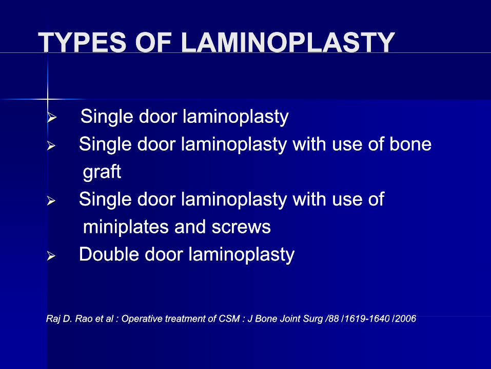
TYPES OF LAMINOPLASTYTYPES OF LAMINOPLASTY
Single door laminoplastySingle door laminoplastySingle door laminoplasty with use of boneSingle door laminoplasty with use of bonegraftgraftSingle door laminoplasty with use ofSingle door laminoplasty with use ofminiplates and screwsminiplates and screwsDouble door laminoplastyDouble door laminoplastyp yp y
R j DR j D RR t l O ti t t t f CSM J B J i tt l O ti t t t f CSM J B J i t SS /88/88 //16191619 16401640 //20062006Raj D. Raj D. RaoRao et al : Operative treatment of CSM : J Bone Joint et al : Operative treatment of CSM : J Bone Joint SurgSurg /88/88 //16191619--16401640 //2006 2006
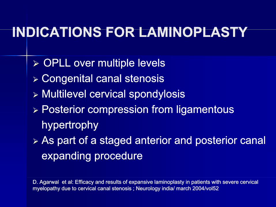
INDICATIONS FOR LAMINOPLASTYINDICATIONS FOR LAMINOPLASTYINDICATIONS FOR LAMINOPLASTYINDICATIONS FOR LAMINOPLASTY
OPLL lti l l lOPLL lti l l lOPLL over multiple levelsOPLL over multiple levelsCongenital canal stenosisCongenital canal stenosisMultilevel cervical Multilevel cervical spondylosisspondylosisPosterior compression from Posterior compression from ligamentousligamentoushypertrophyhypertrophyAs part of a staged anterior and posterior canalAs part of a staged anterior and posterior canalexpanding procedureexpanding procedure
D. Agarwal et al: Efficacy and results of expansive laminoplasty in patients with severe cervical D. Agarwal et al: Efficacy and results of expansive laminoplasty in patients with severe cervical myelopathy due to cervical canal stenosis ; Neurology myelopathy due to cervical canal stenosis ; Neurology indiaindia/ march 2004/vol52/ march 2004/vol52
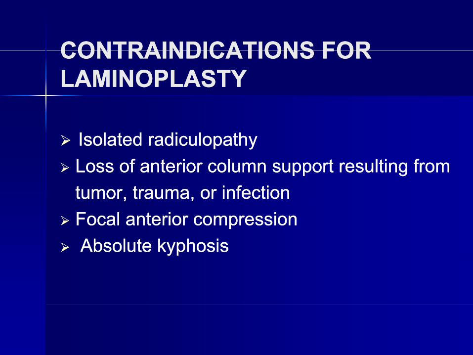
CONTRAINDICATIONS FORCONTRAINDICATIONS FORCONTRAINDICATIONS FOR CONTRAINDICATIONS FOR LAMINOPLASTYLAMINOPLASTY
Isolated radiculopathyIsolated radiculopathyLoss of anterior column support resulting fromLoss of anterior column support resulting fromtumor, trauma, or infectiontumor, trauma, or infectionFocal anterior compressionFocal anterior compressionAbsolute kyphosisAbsolute kyphosisypyp
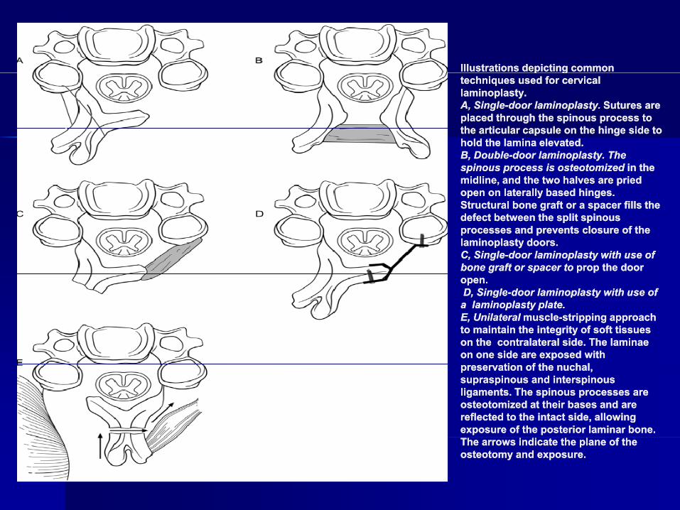
Illustrations depicting common Illustrations depicting common p gp gtechniques used for cervical techniques used for cervical laminoplasty. laminoplasty. A, SingleA, Single--door laminoplasty. door laminoplasty. Sutures are Sutures are placed through the spinous process to placed through the spinous process to the the articulararticular capsule on the hinge side to capsule on the hinge side to hold the lamina elevated.hold the lamina elevated.hold the lamina elevated. hold the lamina elevated. B, DoubleB, Double--door laminoplasty. The door laminoplasty. The spinous process is spinous process is osteotomizedosteotomized in the in the midline, and the two halves are pried midline, and the two halves are pried open on laterally based hinges. open on laterally based hinges. Structural bone graft or a spacer fills the Structural bone graft or a spacer fills the defect between the split spinousdefect between the split spinousdefect between the split spinous defect between the split spinous processes and prevents closure of the processes and prevents closure of the laminoplasty doors. laminoplasty doors. C, SingleC, Single--door laminoplasty with use of door laminoplasty with use of bone graft or spacer to bone graft or spacer to prop the door prop the door open.open.D SingleD Single door laminoplasty with use ofdoor laminoplasty with use ofD, SingleD, Single--door laminoplasty with use of door laminoplasty with use of a laminoplasty plate. a laminoplasty plate. E, Unilateral E, Unilateral musclemuscle--stripping approach stripping approach to maintain the integrity of soft tissues to maintain the integrity of soft tissues on the on the contralateralcontralateral side. The side. The laminaelaminaeon one side are exposed with on one side are exposed with
ti f thti f th h lh lpreservation of the preservation of the nuchalnuchal, , supraspinoussupraspinous and and interspinousinterspinousligaments. The spinous processes are ligaments. The spinous processes are osteotomizedosteotomized at their bases and are at their bases and are reflected to the intact side, allowing reflected to the intact side, allowing exposure of the posterior laminar bone. exposure of the posterior laminar bone. The arrows indicate the plane of the The arrows indicate the plane of the osteotomyosteotomy and exposure.and exposure.
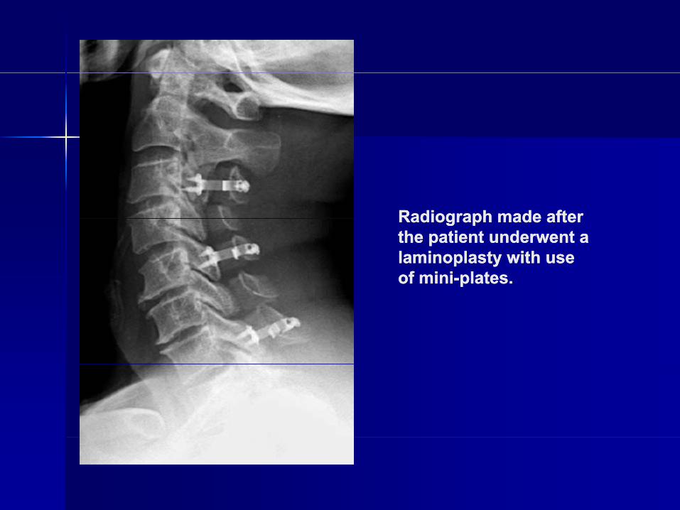
Radiograph made afterRadiograph made afterRadiograph made after Radiograph made after the patient underwent a the patient underwent a laminoplasty with use laminoplasty with use of miniof mini--plates. plates.
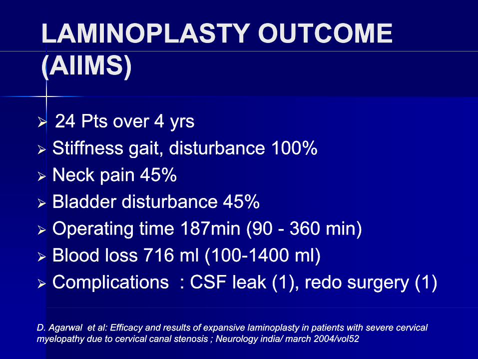
LAMINOPLASTY OUTCOME LAMINOPLASTY OUTCOME (AIIMS)(AIIMS)
24 Pts over 4 yrs24 Pts over 4 yrsStiffness gait, disturbance 100%Stiffness gait, disturbance 100%Stiffness gait, disturbance 100%Stiffness gait, disturbance 100%Neck pain 45%Neck pain 45%Bladder disturbance 45%Bladder disturbance 45%Bladder disturbance 45%Bladder disturbance 45%Operating time 187min (90 Operating time 187min (90 -- 360 min)360 min)Blood loss 716 ml (100Blood loss 716 ml (100--1400 ml)1400 ml)Blood loss 716 ml (100Blood loss 716 ml (100--1400 ml)1400 ml)Complications : CSF leak (1), redo surgery (1)Complications : CSF leak (1), redo surgery (1)
D. Agarwal et al: Efficacy and results of expansive laminoplasty in patients with severe cervical D. Agarwal et al: Efficacy and results of expansive laminoplasty in patients with severe cervical myelopathy due to cervical canal stenosis ; Neurology myelopathy due to cervical canal stenosis ; Neurology indiaindia/ march 2004/vol52/ march 2004/vol52
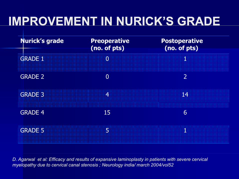
IMPROVEMENT IN NURICK’S GRADEIMPROVEMENT IN NURICK’S GRADENurick’s grade Preoperative
(no. of pts)Postoperative(no. of pts)( p ) ( p )
GRADE 1 0 1
GRADE 2 0 2GRADE 2 0 2
GRADE 3 4 14
GRADE 4 15 6
GRADE 5 5 1
D. Agarwal et al: Efficacy and results of expansive laminoplasty in patients with severe cervical myelopathy due to cervical canal stenosis ; Neurology india/ march 2004/vol52
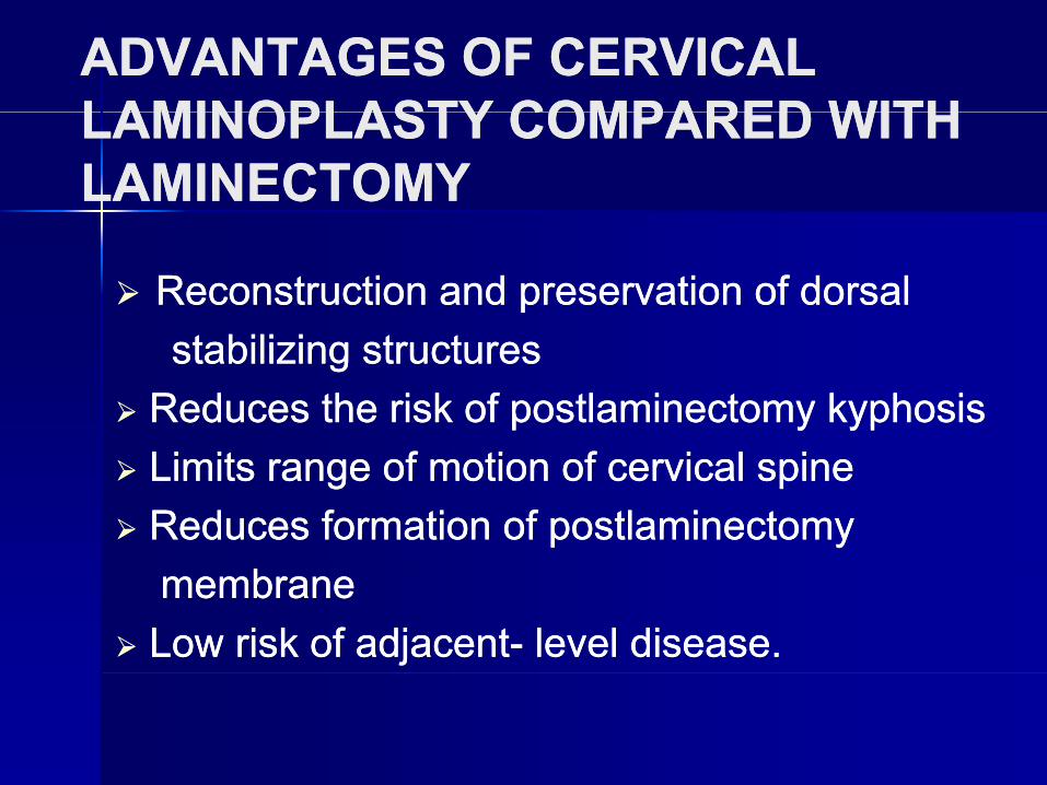
ADVANTAGES OF CERVICAL ADVANTAGES OF CERVICAL LAMINOPLASTY COMPARED WITHLAMINOPLASTY COMPARED WITHLAMINOPLASTY COMPARED WITH LAMINOPLASTY COMPARED WITH LAMINECTOMYLAMINECTOMY
Reconstruction and preservation of dorsal Reconstruction and preservation of dorsal t bili i t tt bili i t tstabilizing structuresstabilizing structures
Reduces the risk of Reduces the risk of postlaminectomypostlaminectomy kyphosiskyphosisLimits range of motion of cervical spineLimits range of motion of cervical spineReduces formation of Reduces formation of postlaminectomypostlaminectomymembranemembraneLow risk of adjacentLow risk of adjacent-- level disease. level disease.
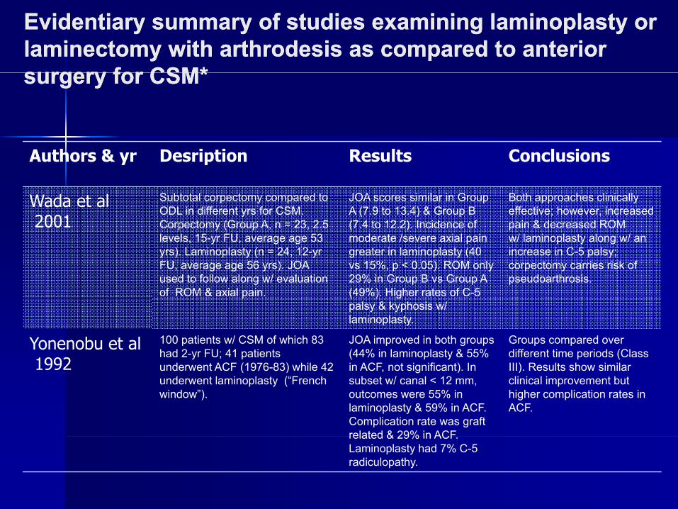
Evidentiary summary of studies examining laminoplasty or Evidentiary summary of studies examining laminoplasty or laminectomy with laminectomy with arthrodesisarthrodesis as compared to anterior as compared to anterior surgery for CSM*surgery for CSM*surgery for CSM*surgery for CSM*
Authors & yr Desription Results Conclusions
Wada et al2001
Subtotal corpectomy compared to ODL in different yrs for CSM.
JOA scores similar in Group A (7.9 to 13.4) & Group B
Both approaches clinically effective; however, increased
2001 Corpectomy (Group A, n = 23, 2.5 levels, 15-yr FU, average age 53 yrs). Laminoplasty (n = 24, 12-yr FU, average age 56 yrs). JOA used to follow along w/ evaluation of ROM & axial pain
(7.4 to 12.2). Incidence of moderate /severe axial pain greater in laminoplasty (40 vs 15%, p < 0.05). ROM only 29% in Group B vs Group A (49%) Higher rates of C 5
pain & decreased ROMw/ laminoplasty along w/ an increase in C-5 palsy; corpectomy carries risk ofpseudoarthrosis.
of ROM & axial pain. (49%). Higher rates of C-5 palsy & kyphosis w/ laminoplasty.
Yonenobu et al1992
100 patients w/ CSM of which 83 had 2-yr FU; 41 patients
d t ACF (1976 83) hil 42
JOA improved in both groups (44% in laminoplasty & 55% i ACF t i ifi t) I
Groups compared over different time periods (Class III) R lt h i il1992 underwent ACF (1976-83) while 42
underwent laminoplasty (“French window”).
in ACF, not significant). In subset w/ canal < 12 mm, outcomes were 55% in laminoplasty & 59% in ACF. Complication rate was graft related & 29% in ACF
III). Results show similarclinical improvement but higher complication rates in ACF.
related & 29% in ACF. Laminoplasty had 7% C-5 radiculopathy.
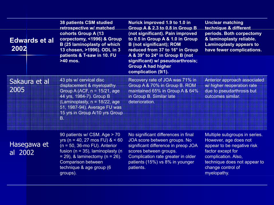
38 patients CSM studied retrospective w/ matched
Nurick improved 1.9 to 1.0 in Group A & 2.3 to 0.8 in Group B
Unclear matching technique & different
Edwards et al2002
retrospective w/ matched cohorts Group A (13 corpectomy, <1996) & Group B (25 laminoplasty of which 13 chosen, >1996). ODL in 3 patients & T-saw in 10. FU
Group A & 2.3 to 0.8 in Group B (not significant). Pain improved to 0.5 in Group A & 1.0 in Group B (not significant); ROM reduced from 37 to 16° in Group A & 39° to 24° in Group B (not
technique & different periods. Both corpectomy& laminoplasty reliable. Laminoplasty appears to have fewer complications.
>40 mos. significant) w/ pseudoarthrosis; Group A had higher complication (9/1).
Sakaura et al2005
43 pts w/ cervical disc displacement & myelopathy.
Recovery rate of JOA was 71% in Group A & 70% in Group B. ROM
Anterior approach associated w/ higher reoperation rate
2005y y
Group A (ACF, n = 15/21, age 44 yrs, 1984-7). Group B (Laminoplasty, n = 18/22, age 51, 1987-94). Average FU was 15 yrs in Group A/10 yrs Group B
maintained 65% in Group A & 64% in Group B. Similar late deterioration.
gdue to pseudarthrosis but outcomes similar.
B.
Hasegawa et90 patients w/ CSM. Age > 70 yrs (n = 40, 27 mos FU) & < 60 (n = 50 36-mo FU) Anterior
No significant differences in final JOA score between groups. No significant difference in preop JOA
Multiple subgroups in series. However, age does not appear to be negative riskHasegawa et
al 2002(n 50, 36 mo FU). Anterior fusion (n = 35), laminoplasty (n = 29), & laminectomy (n = 26). Comparison between technique & age group (6 groups).
significant difference in preop JOA scores between groups. Complication rate greater in older patients (15%) vs 8% in younger patients.
appear to be negative risk factor except for complication. Also,technique does not appear to change control of myelopathy.
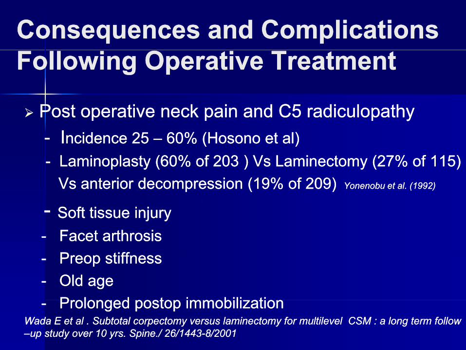
Consequences and ComplicationsConsequences and ComplicationsFollowing Operative TreatmentFollowing Operative Treatment
Post operative neck pain and C5 radiculopathyPost operative neck pain and C5 radiculopathy-- IIncidence 25 ncidence 25 –– 60% (60% (HosonoHosono et al)et al)-- Laminoplasty (60% of 203 ) Vs Laminectomy (27% of 115)Laminoplasty (60% of 203 ) Vs Laminectomy (27% of 115)
Vs anterior decompression (19% of 209) Vs anterior decompression (19% of 209) YonenobuYonenobu et al. (1992)et al. (1992)
-- Soft tissue injurySoft tissue injury-- Facet Facet arthrosisarthrosis-- PreopPreop stiffnessstiffness-- Old ageOld age
P l d t i bili tiP l d t i bili ti-- Prolonged postop immobilization Prolonged postop immobilization Wada E et al . Subtotal Wada E et al . Subtotal corpectomycorpectomy versus laminectomy for multilevel CSM : a long term follow versus laminectomy for multilevel CSM : a long term follow ––up study over 10 yrs. Spine./ 26/1443up study over 10 yrs. Spine./ 26/1443--8/20018/2001

Consequences and ComplicationsConsequences and ComplicationsFollowing Operative TreatmentFollowing Operative TreatmentFollowing Operative TreatmentFollowing Operative Treatment
Postop stiffness :Postop stiffness :pp-- InterlaminarInterlaminar or facet fusion on hinge side or facet fusion on hinge side
Postop stability:Postop stability:-- Incidence of instability 21% for laminectomyIncidence of instability 21% for laminectomyIncidence of instability 21% for laminectomyIncidence of instability 21% for laminectomy-- Relatively rare for laminoplastyRelatively rare for laminoplasty
Adjacent segment degeneration Adjacent segment degeneration C5C5 C6 and C6C6 and C6 C7 most vulnerableC7 most vulnerable-- C5 C5 –– C6 and C6 C6 and C6 –– C7 most vulnerableC7 most vulnerable
-- 3% each yr (3% each yr (HilibrandHilibrand et al)et al)
NEUROLOGICAL COMPLICATIONSNEUROLOGICAL COMPLICATIONSNEUROLOGICAL COMPLICATIONSNEUROLOGICAL COMPLICATIONS
RadiculopathyRadiculopathyRadiculopathyRadiculopathyPermanent myelopathyPermanent myelopathyRecurrent laryngeal nerve palsyRecurrent laryngeal nerve palsyHornersHorners syndromesyndromeyyDysphagiaDysphagiaEsophageal injuriesEsophageal injuriesEsophageal injuriesEsophageal injuriesVertebral artery injuriesVertebral artery injuriesInjuries to tracheaInjuries to trachea
OverviewOverviewOverview Overview Surgery indicated for most pts with clinicallySurgery indicated for most pts with clinicallySurgery indicated for most pts with clinically Surgery indicated for most pts with clinically
evident CSMevident CSMRisk benefit ratio to be assessed in pts with Risk benefit ratio to be assessed in pts with pp
early disease early disease Main objective of Main objective of SxSx is to decompress is to decompress jj pp
adequately and to maintain stabilityadequately and to maintain stabilityType of Type of SxSx depends upon location ,extent of depends upon location ,extent of
pathology and also the alignment , pathology and also the alignment , dimensions of spinal cord.dimensions of spinal cord.
Improvement being higher in young pts, Improvement being higher in young pts, early disease.early disease.