Embed Size (px)
Citation preview
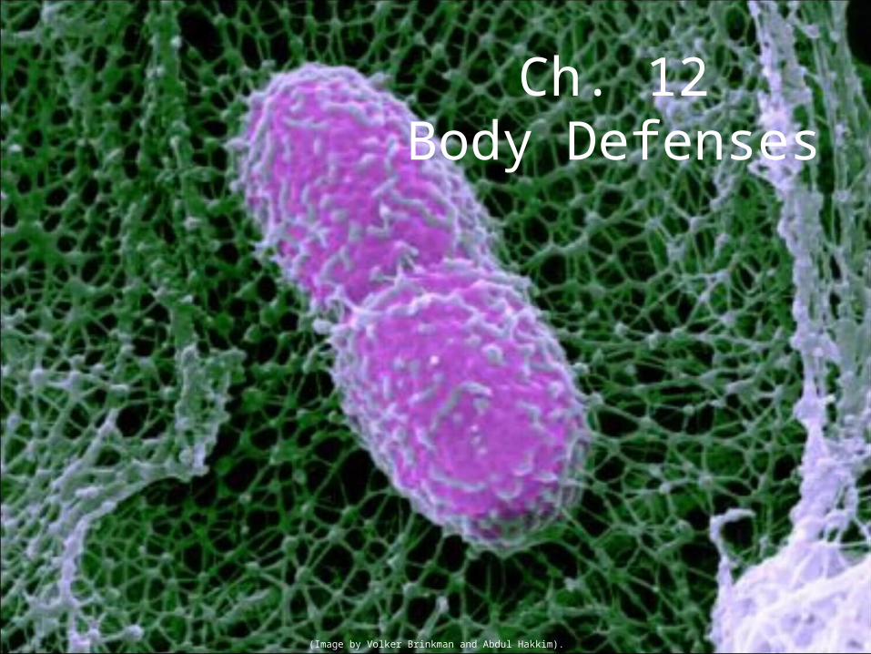
Ch. 12Body Defenses
(Image by Volker Brinkman and Abdul Hakkim).
Outline
•Immune system
•Non-specific response
•Specific response
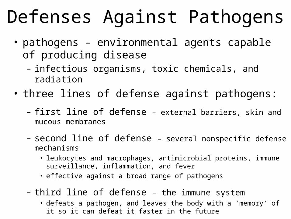
Defenses Against Pathogens• pathogens – environmental agents capable of producing
disease– infectious organisms, toxic chemicals, and radiation
• three lines of defense against pathogens:
– first line of defense – external barriers, skin and mucous membranes
– second line of defense – several nonspecific defense mechanisms• leukocytes and macrophages, antimicrobial proteins, immune surveillance,
inflammation, and fever• effective against a broad range of pathogens
– third line of defense – the immune system• defeats a pathogen, and leaves the body with a ‘memory’ of it so it can defeat it
faster in the future
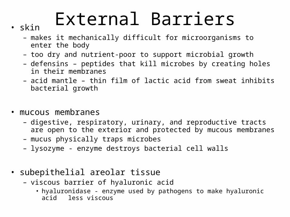
External Barriers• skin
– makes it mechanically difficult for microorganisms to enter the body– too dry and nutrient-poor to support microbial growth– defensins – peptides that kill microbes by creating holes in their
membranes– acid mantle – thin film of lactic acid from sweat inhibits bacterial growth
• mucous membranes– digestive, respiratory, urinary, and reproductive tracts are open to the
exterior and protected by mucous membranes– mucus physically traps microbes– lysozyme - enzyme destroys bacterial cell walls
• subepithelial areolar tissue– viscous barrier of hyaluronic acid
• hyaluronidase - enzyme used by pathogens to make hyaluronic acid less viscous
Leukocytes and Macrophages• phagocytes – phagocytic cells with a voracious
appetite for foreign matter
• five types of leukocytes– neutrophils– eosinophils– basophils– monocytes– lymphocytes
Neutrophils• wander in connective tissue killing bacteria
– phagocytosis and digestion– produces a cloud of bactericidal chemicals– NETs
• create a killing zone– degranulation
• lysosomes discharge into tissue fluid– respiratory burst – neutrophils rapidly absorb oxygen
• toxic chemicals are created (O2
.-, H2O2, HClO)
– kill more bacteria with toxic chemicals than phagocytosis
Eosinophils• Found mucous membranes
• Defend mainly against parasites, allergens• kill tapeworms and roundworms by producing
superoxide, hydrogen peroxide, and toxic proteins
• promote action of basophils and mast cells
• phagocytize antigen-antibody complexes
• limit action of histamine and other inflammatory chemicals
Basophils• secrete chemicals that aid mobility and action of WBC
other leukocytes
– leukotrienes – activate and attract neutrophils and eosinophils
– histamine – a vasodilator which increases blood flow• speeds delivery of leukocytes to the area
– heparin – inhibits the formation of clots• would impede leukocyte mobility
• mast cells also secrete these substances– type of connective tissue cell very similar to basophils
Monocytes• monocytes - emigrate from blood into the connective
tissue and transform into macrophages
• macrophage system – all the body’s avidly phagocytic cells, except leukocytes– wandering macrophages – actively seeking pathogens
• widely distributed in loose connective tissue
– fixed macrophages – phagocytize only pathogens that come to them
• microglia – in central nervous system• alveolar macrophages – in lungs• hepatic macrophages – in liver
Antimicrobial Proteins• proteins that inhibit microbial reproduction
and provide short-term, nonspecific resistance to pathogenic bacteria and viruses
• two families of antimicrobial proteins:
– interferons
– complement system
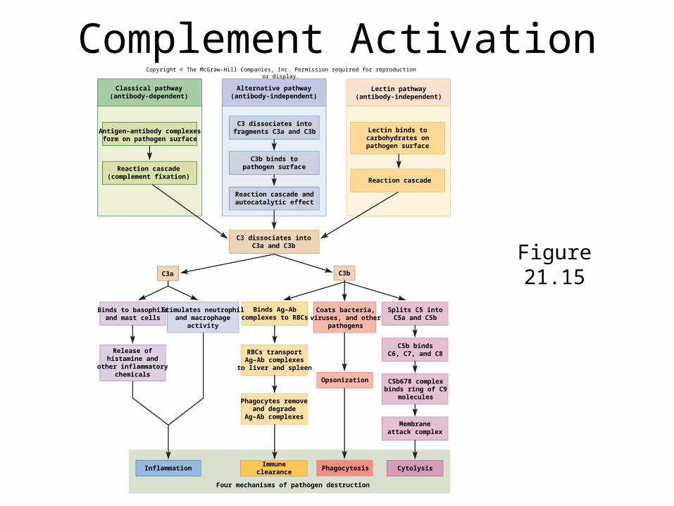
Complement System• complement system – a group of 30 or more globular proteins
that make powerful contributions to both nonspecific resistance and specific immunity– activated complement brings about four methods of pathogen
destruction• inflammation• immune clearance• phagocytosis• cytolysis
– three routes of complement activation• classical pathway• alternative pathway• lectin pathway
Complement System• classical pathway
– requires antibody molecule to get started– thus part of specific immunity– antibody binds to antigen on surface of the pathogenic organism
• forms antigen-antibody (Ag-Ab) complex– changes the antibody’s shape
• exposing a pair of complement-binding sites• binding of complement (C1) sets off a reaction cascade called complement fixation
– results in a chain of complement proteins attaching to the antibody
• alternative pathway– nonspecific, do not require antibody– C3 breaks down in the blood to C3a and C3b
• C3b binds directly to targets such as human tumor cells, viruses, bacteria, and yeasts• triggers cascade reaction with autocatalytic effect where more C3 is formed
• lectin pathway– lectins – plasma proteins that bind to carbohydrates
• bind to certain sugars of a microbial cell surface• sets off another cascade of C3 production
21-13
Complement Activation
Figure 21.15C3a C3b
Four mechanisms of pathogen destruction
Inflammation Cytolysis
Opsonization
Phagocytosis
Reaction cascade
Classical pathway(antibody-dependent)
Antigen–antibody complexesform on pathogen surface
Reaction cascade(complement fixation)
Alternative pathway(antibody-independent)
C3 dissociates intofragments C3a and C3b
C3b binds topathogen surface
Reaction cascade andautocatalytic effect
C3 dissociates intoC3a and C3b
Lectin pathway(antibody-independent)
Lectin binds tocarbohydrates onpathogen surface
Splits C5 intoC5a and C5b
Coats bacteria,viruses, and other
pathogens
Binds Ag–Abcomplexes to RBCs
Stimulates neutrophiland macrophage
activity
Binds to basophilsand mast cells
Release ofhistamine and
other inflammatorychemicals
RBCs transportAg–Ab complexesto liver and spleen
C5b bindsC6, C7, and C8
C5b678 complexbinds ring of C9
moleculesPhagocytes remove
and degradeAg–Ab complexes
Membraneattack complex
Immuneclearance
Copyright © The McGraw-Hill Companies, Inc. Permission required for reproduction or display.
21-14
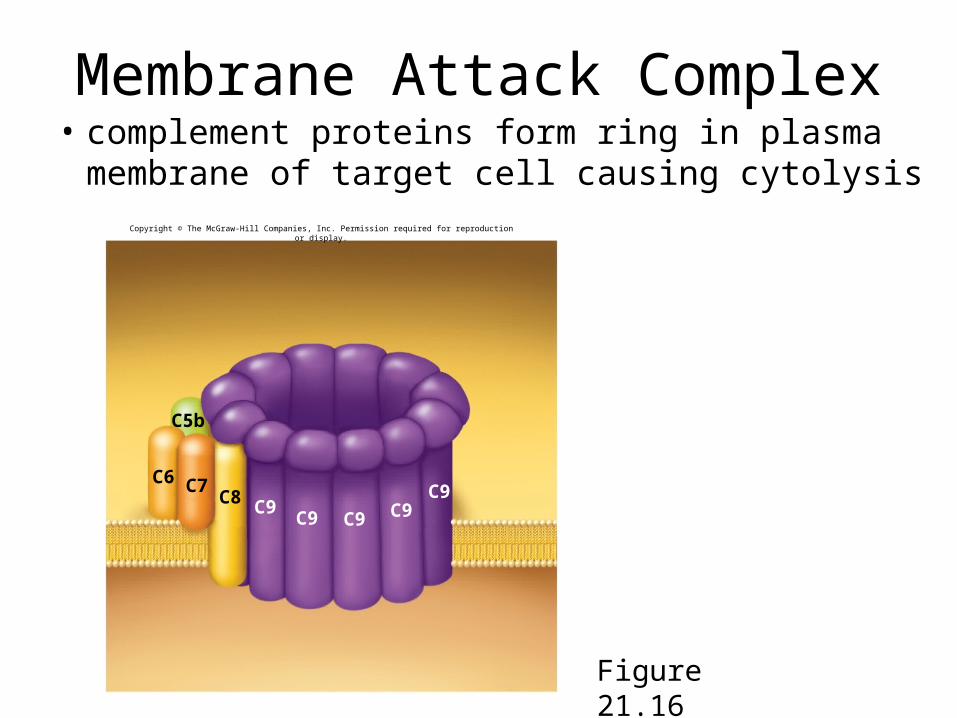
Membrane Attack Complex• complement proteins form ring in plasma
membrane of target cell causing cytolysis
Figure 21.16
C5b
C6 C7C8
C9 C9C9
C9C9
Copyright © The McGraw-Hill Companies, Inc. Permission required for reproduction or display.
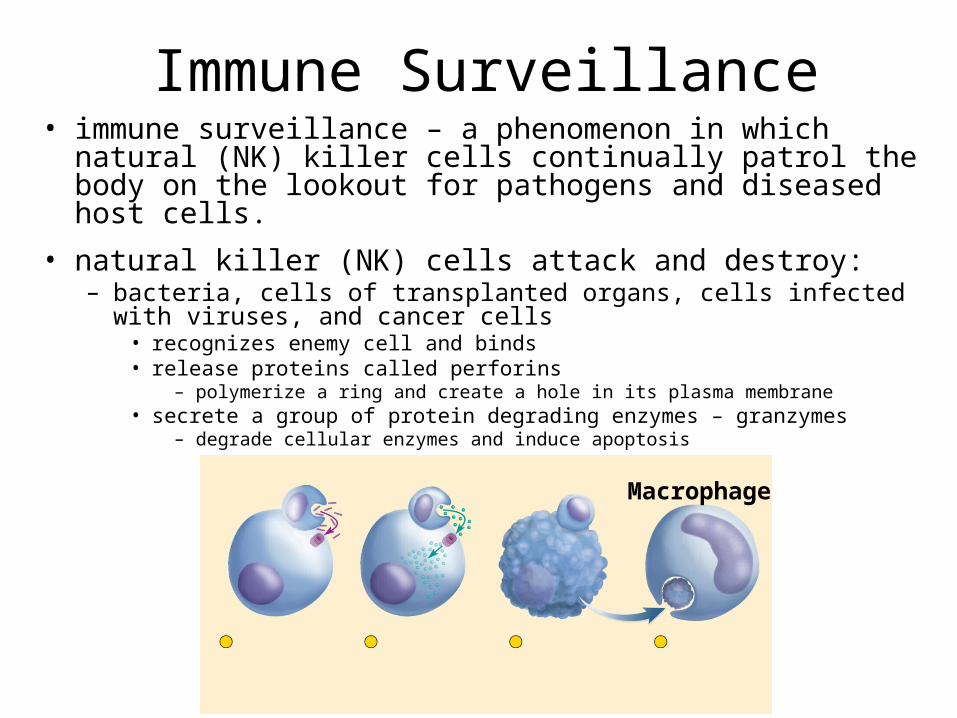
Immune Surveillance• immune surveillance – a phenomenon in which natural (NK)
killer cells continually patrol the body on the lookout for pathogens and diseased host cells.
• natural killer (NK) cells attack and destroy: – bacteria, cells of transplanted organs, cells infected with viruses, and
cancer cells• recognizes enemy cell and binds• release proteins called perforins
– polymerize a ring and create a hole in its plasma membrane• secrete a group of protein degrading enzymes – granzymes
– degrade cellular enzymes and induce apoptosis
Macrophage
Inflammation• inflammation – local defensive response to
tissue injury of any kind, including trauma and infection
• general purposes of inflammation– limit spread of pathogens, then destroys them– remove debris from damaged tissue– initiate tissue repair
• four cardinal signs of inflammation- redness - swelling - heat - pain
Inflammation• suffix -itis denotes inflammation of specific
organs: arthritis, pancreatitis, dermatitis
• cytokines – class of chemicals that regulate inflammation and immunity– secreted mainly by leukocytes
– alter the physiology or behavior of receiving cell
– act at short range, neighboring cells (paracrines) or the same cell that secretes them (autocrines)
– include interferon, interleukins, tumor necrosis factor, chemotactic factors, and others
Processes of Inflammation• three major processes of inflammation
– mobilization of body defenses• Hyperemia• Vasodilation
– containment and destruction of pathogens• Fibrinogen• Heparin• Neutrophils attracted by chemotaxis
– tissue cleanup and repair• Monocytes arrive in 8-12 hours• Edema• Platelet-derived growth factor
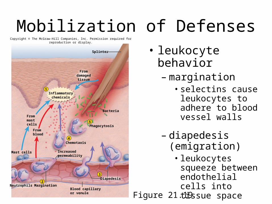
Mobilization of Defenses• leukocyte behavior
– margination• selectins cause
leukocytes to adhere to blood vessel walls
– diapedesis (emigration)
• leukocytes squeeze between endothelial cells into tissue space
Figure 21.19
Splinter
Phagocytosis
1
5
Chemotaxis4
2
Mast cells
Margination
Diapedesis3
Neutrophils
Bacteria
Fromdamaged
tissue
Inflammatorychemicals
Frommastcells
Fromblood
Increasedpermeability
Blood capillaryor venule
Copyright © The McGraw-Hill Companies, Inc. Permission required for reproduction or display.
Specific Immunity• immune system – composed of a large population of widely distributed cells
that recognize foreign substances and act to neutralize or destroy them
• two characteristics distinguish immunity from nonspecific resistance– specificity – immunity directed against a particular pathogen– memory – when re-exposed to the same pathogen, the body reacts so quickly
that there is no noticeable illness
• two types of immunity– cellular (cell-mediated) immunity: (T cells)
• lymphocytes directly attack and destroy foreign cells or diseased host cells• rids the body of pathogens that reside inside human cells, where they are inaccessible
to antibodies• kills cells that harbor them
– humoral (antibody-mediated) immunity: (B cells)• mediated by antibodies that do not directly destroy a pathogen• indirect attack where antibodies assault the pathogen• can only work against the extracellular stage of infectious microorganisms
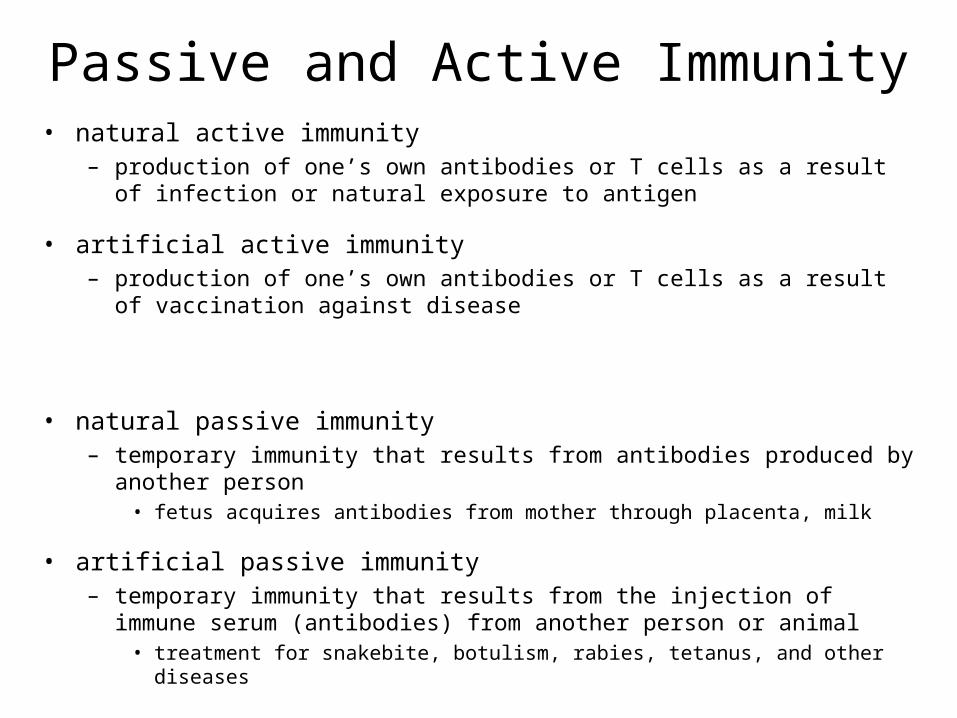
Passive and Active Immunity• natural active immunity
– production of one’s own antibodies or T cells as a result of infection or natural exposure to antigen
• artificial active immunity– production of one’s own antibodies or T cells as a result of vaccination against
disease
• natural passive immunity – temporary immunity that results from antibodies produced by another person
• fetus acquires antibodies from mother through placenta, milk
• artificial passive immunity– temporary immunity that results from the injection of immune serum (antibodies)
from another person or animal• treatment for snakebite, botulism, rabies, tetanus, and other diseases
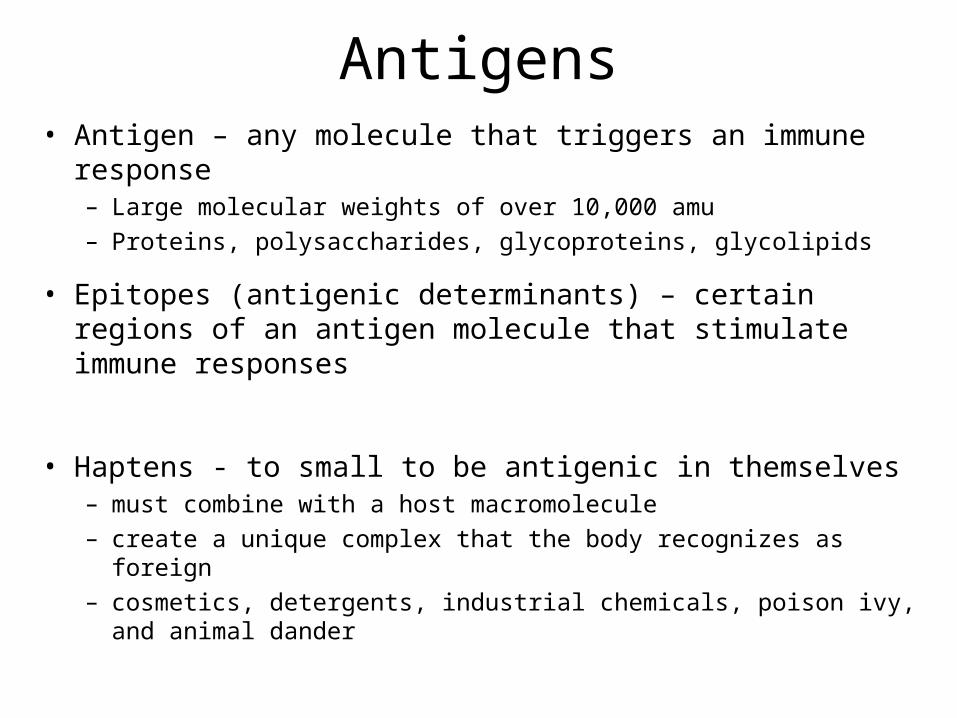
Antigens• Antigen – any molecule that triggers an immune response
– Large molecular weights of over 10,000 amu– Proteins, polysaccharides, glycoproteins, glycolipids
• Epitopes (antigenic determinants) – certain regions of an antigen molecule that stimulate immune responses
• Haptens - to small to be antigenic in themselves– must combine with a host macromolecule– create a unique complex that the body recognizes as foreign– cosmetics, detergents, industrial chemicals, poison ivy, and animal
dander
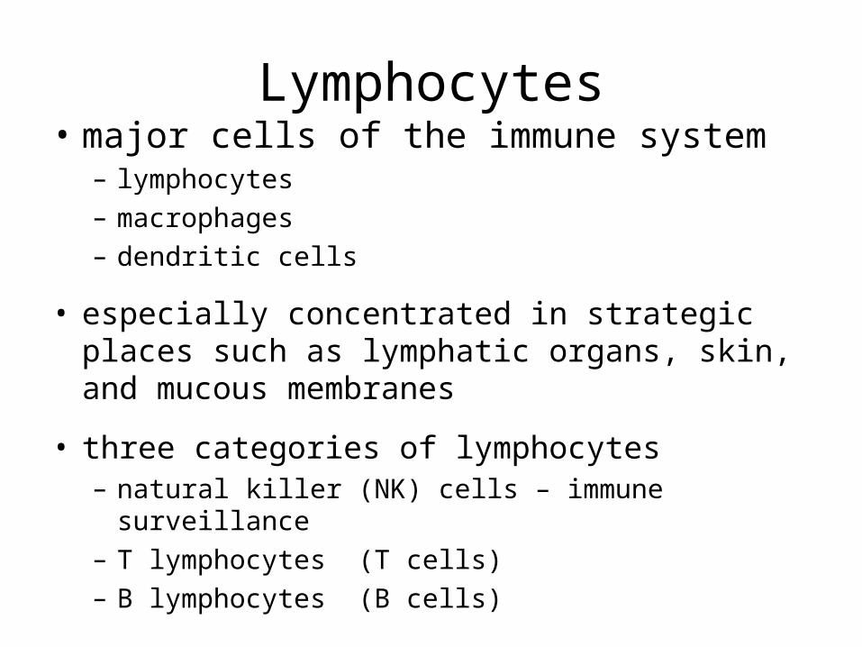
Lymphocytes• major cells of the immune system
– lymphocytes– macrophages– dendritic cells
• especially concentrated in strategic places such as lymphatic organs, skin, and mucous membranes
• three categories of lymphocytes– natural killer (NK) cells – immune surveillance– T lymphocytes (T cells)– B lymphocytes (B cells)
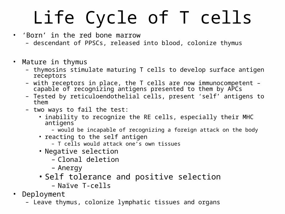
Life Cycle of T cells• ‘Born’ in the red bone marrow
– descendant of PPSCs, released into blood, colonize thymus
• Mature in thymus– thymosins stimulate maturing T cells to develop surface antigen receptors– with receptors in place, the T cells are now immunocompetent – capable of
recognizing antigens presented to them by APCs– Tested by reticuloendothelial cells, present ‘self’ antigens to them– two ways to fail the test:
• inability to recognize the RE cells, especially their MHC antigens– would be incapable of recognizing a foreign attack on the body
• reacting to the self antigen– T cells would attack one’s own tissues
• Negative selection– Clonal deletion– Anergy
• Self tolerance and positive selection– Naïve T-cells
• Deployment– Leave thymus, colonize lymphatic tissues and organs
B Lymphocytes (B cells)• site of development
– group fetal stem cells remain in bone marrow– develop into B cells
• B cell selection– B cells that react to self antigens undergo either anergy or
clonal deletion same as T cell selection
• self-tolerant B cells synthesize antigen surface receptors, divide rapidly, produce immunocompetent clones
• leave bone marrow and colonize same lymphatic tissues and organs as T cells
Antigen-Presenting Cells (APCs)• T cells can not recognize antigens on their own
• Antigen-presenting cells (APCs) are required to help– dendritic cells, macrophages, reticular cells, and B cells function as APCs
• Function of APCs depends on major histocompatibility complex (MHC) proteins– act as cell ‘identification tags’ that label every cell of your body as belonging
to you– structurally unique for each individual, except for identical twins
• Antigen processing– APC encounters antigen– internalizes it by endocytosis and digests– displays epitopes in grooves of the MHC protein
• Antigen presenting– Wander T cell detects an APC with a nonself-antigen, immune attack initiated– Communicate via interleukins
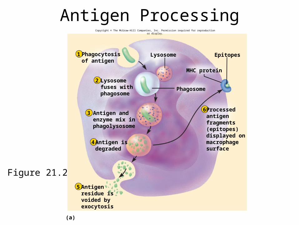
Antigen Processing
Figure 21.21a
Phagosome
Epitopes
MHC protein
(a)
2
3
4
5
6
1 Phagocytosisof antigen
Lysosome
Lysosomefuses withphagosome
Antigen andenzyme mix inphagolysosome
Antigen isdegraded
Antigenresidue isvoided byexocytosis
Processedantigenfragments(epitopes)displayed onmacrophagesurface
Copyright © The McGraw-Hill Companies, Inc. Permission required for reproduction or display.
Cellular Immunity
• cellular (cell-mediated) immunity – a form of specific defense in which the T lymphocytes directly attack and destroy diseased or foreign cells, and the immune system remembers the antigens and prevents them from causing disease in the future
• both cellular and humoral immunity occur in three stages:
– recognition– attack– memory
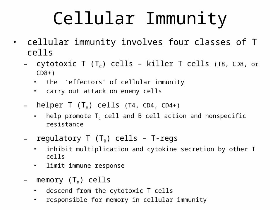
Cellular Immunity• cellular immunity involves four classes of T cells
– cytotoxic T (TC) cells – killer T cells (T8, CD8, or CD8+)
• the ‘effectors’ of cellular immunity• carry out attack on enemy cells
– helper T (TH) cells (T4, CD4, CD4+)
• help promote TC cell and B cell action and nonspecific resistance
– regulatory T (TR) cells – T-regs
• inhibit multiplication and cytokine secretion by other T cells• limit immune response
– memory (TM) cells • descend from the cytotoxic T cells• responsible for memory in cellular immunity
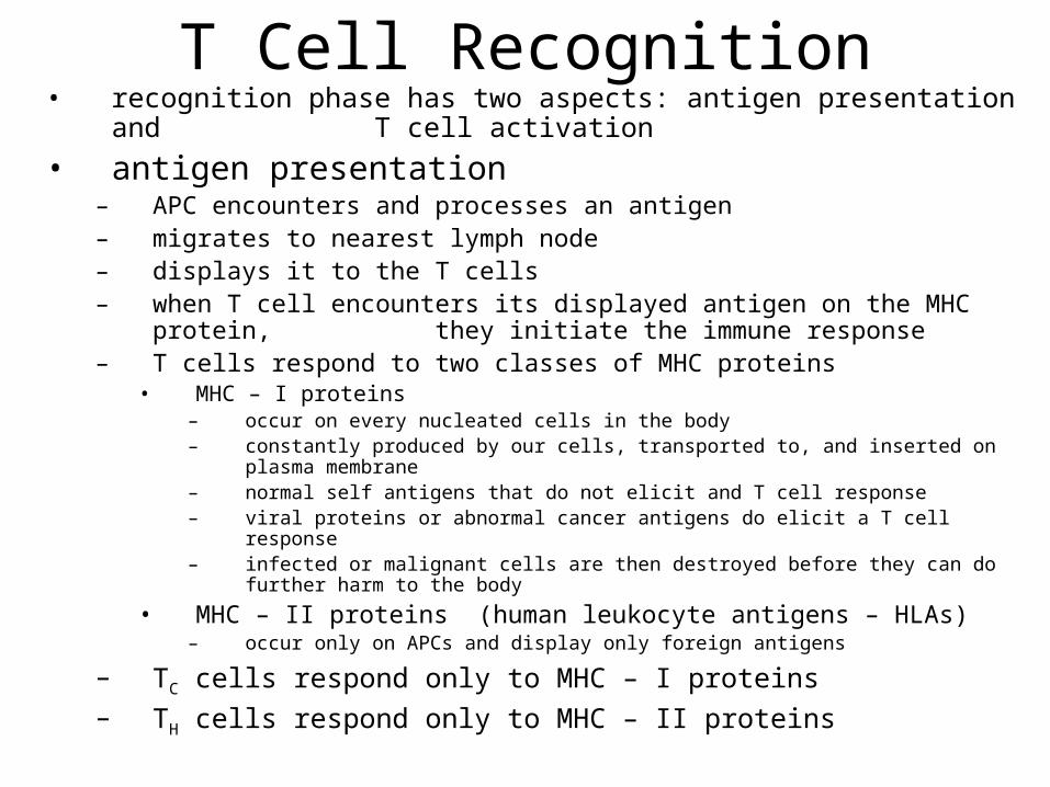
T Cell Recognition• recognition phase has two aspects: antigen presentation and T cell
activation• antigen presentation
– APC encounters and processes an antigen– migrates to nearest lymph node– displays it to the T cells– when T cell encounters its displayed antigen on the MHC protein, they
initiate the immune response– T cells respond to two classes of MHC proteins
• MHC – I proteins– occur on every nucleated cells in the body– constantly produced by our cells, transported to, and inserted on plasma membrane– normal self antigens that do not elicit and T cell response– viral proteins or abnormal cancer antigens do elicit a T cell response– infected or malignant cells are then destroyed before they can do further harm to the body
• MHC – II proteins (human leukocyte antigens – HLAs)– occur only on APCs and display only foreign antigens
– TC cells respond only to MHC – I proteins– TH cells respond only to MHC – II proteins
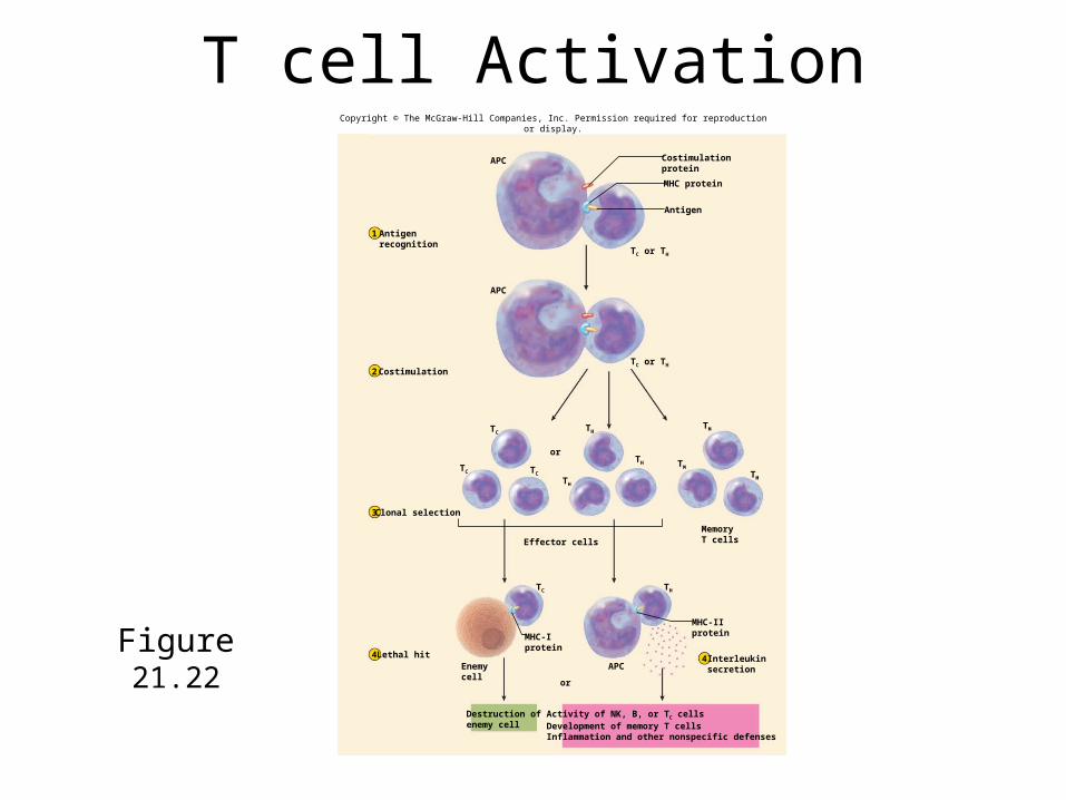
T cell Activation
Figure 21.22
Costimulation
Clonal selection
MHC protein
Antigen
APC
APC
TC or TH
TC TC
TC
TC
TH
TH
TM
TM
TMTH
TH
or
Effector cells
APC4
Activity of NK, B, or TC cellsDevelopment of memory T cellsInflammation and other nonspecific defenses
or
1
2
3
Lethal hit4
Costimulationprotein
TC or TH
MemoryT cells
MHC-IIprotein
Interleukinsecretion
Destruction ofenemy cell
Enemycell
MHC-Iprotein
Antigenrecognition
Copyright © The McGraw-Hill Companies, Inc. Permission required for reproduction or display.
21-32
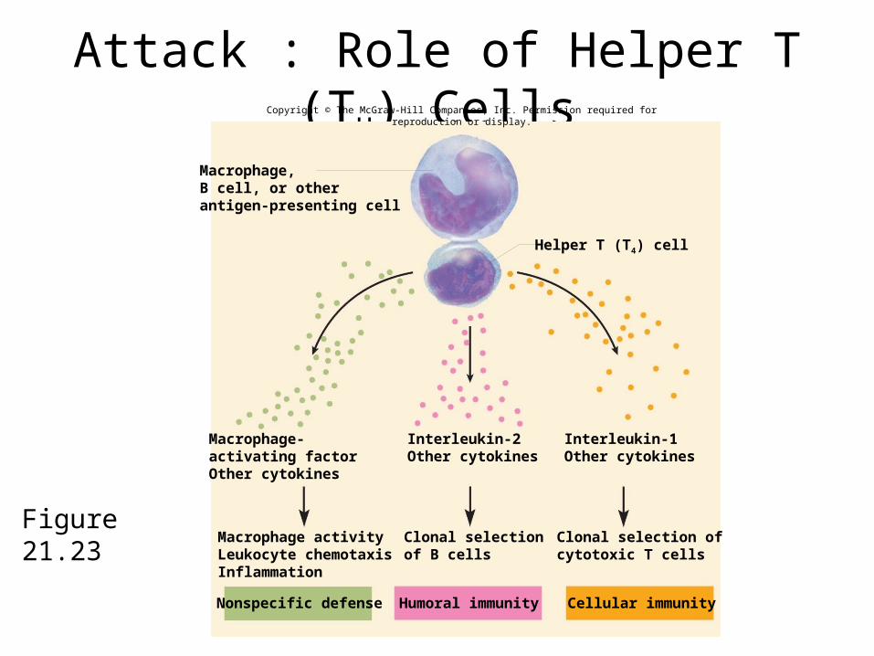
Attack : Role of Helper T (TH) Cells
Figure 21.23
Macrophage,B cell, or otherantigen-presenting cell
Macrophage-activating factorOther cytokines
Macrophage activityLeukocyte chemotaxisInflammation
Interleukin-1Other cytokines
Interleukin-2Other cytokines
Clonal selection ofcytotoxic T cells
Clonal selectionof B cells
Humoral immunity Cellular immunityNonspecific defense
Helper T (T4) cell
Copyright © The McGraw-Hill Companies, Inc. Permission required for reproduction or display.
Attack : Cytotoxic T (TC) Cells• cytotoxic T (TC) cell are the only T cells directly attack
other cells• when TC cell recognizes a complex of antigen and
MHC – I protein on a diseased or foreign cell it ‘docks’ on that cell
– delivers a lethal hit of toxic chemicals• perforin and granzymes – kill cells in the same manner as NK
cells• interferons – inhibit viral replication
– recruit and activate macrophages• tumor necrosis factor (TNF) – aids in macrophage activation and
kills cancer cells– goes off in search of another enemy cell while the
chemicals do their work
21-34
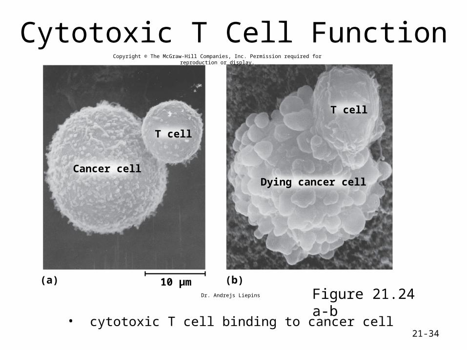
Cytotoxic T Cell Function
• cytotoxic T cell binding to cancer cell
Figure 21.24 a-b(a) (b)
Cancer cell
T cell
T cell
Dying cancer cell
10 µm
Copyright © The McGraw-Hill Companies, Inc. Permission required for reproduction or display.
Dr. Andrejs Liepins
Memory• immune memory follows primary response• following clonal selection, some TC and TH cells become
memory cells– long-lived – more numerous than naïve T cells– fewer steps to be activated, so they respond more rapidly
• T cell recall response– upon re-exposure to same pathogen later in life, memory
cells launch a quick attack so that no noticeable illness occurs
– the person is immune to the disease
Humoral Immunity• humoral immunity is a more indirect method of
defense than cellular immunity
• B lymphocytes of humoral immunity produce antibodies that bind to antigens and tag them for destruction by other means– cellular immunity attacks the enemy cells directly
• works in three stages like cellular immunity– recognition– attack– memory
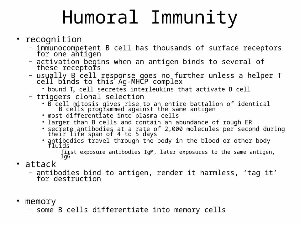
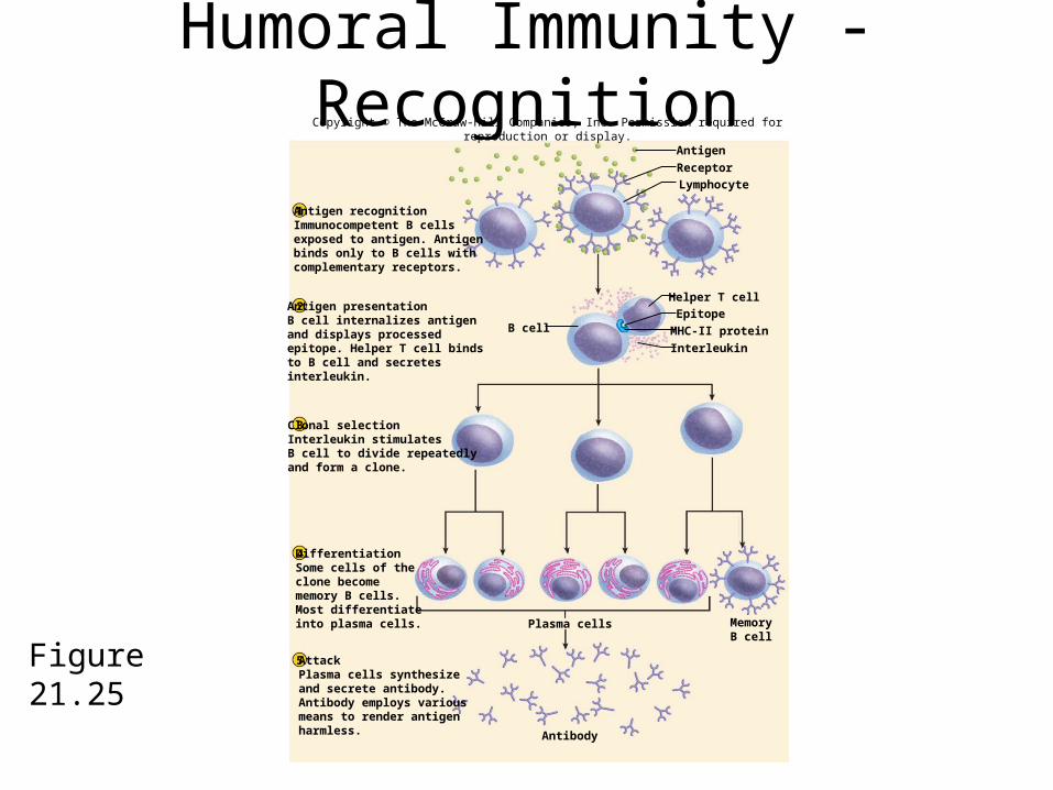
Humoral Immunity• recognition
– immunocompetent B cell has thousands of surface receptors for one antigen
– activation begins when an antigen binds to several of these receptors– usually B cell response goes no further unless a helper T cell binds to this
Ag-MHCP complex• bound TH cell secretes interleukins that activate B cell
– triggers clonal selection• B cell mitosis gives rise to an entire battalion of identical B cells programmed
against the same antigen• most differentiate into plasma cells• larger than B cells and contain an abundance of rough ER• secrete antibodies at a rate of 2,000 molecules per second during their life span
of 4 to 5 days• antibodies travel through the body in the blood or other body fluids
– first exposure antibodies IgM, later exposures to the same antigen, IgG• attack
– antibodies bind to antigen, render it harmless, ‘tag it’ for destruction
• memory– some B cells differentiate into memory cells
Humoral Immunity - Recognition
Figure 21.25Plasma cells
Antibody
AntigenReceptor
Helper T cellEpitopeMHC-II proteinInterleukin
B cell
1
2
3
4
5
Antigen recognitionImmunocompetent B cellsexposed to antigen. Antigenbinds only to B cells withcomplementary receptors.
Antigen presentationB cell internalizes antigenand displays processedepitope. Helper T cell bindsto B cell and secretesinterleukin.
Clonal selectionInterleukin stimulatesB cell to divide repeatedlyand form a clone.
DifferentiationSome cells of theclone becomememory B cells.Most differentiateinto plasma cells.
AttackPlasma cells synthesizeand secrete antibody.Antibody employs variousmeans to render antigenharmless.
Lymphocyte
MemoryB cell
Copyright © The McGraw-Hill Companies, Inc. Permission required for reproduction or display.
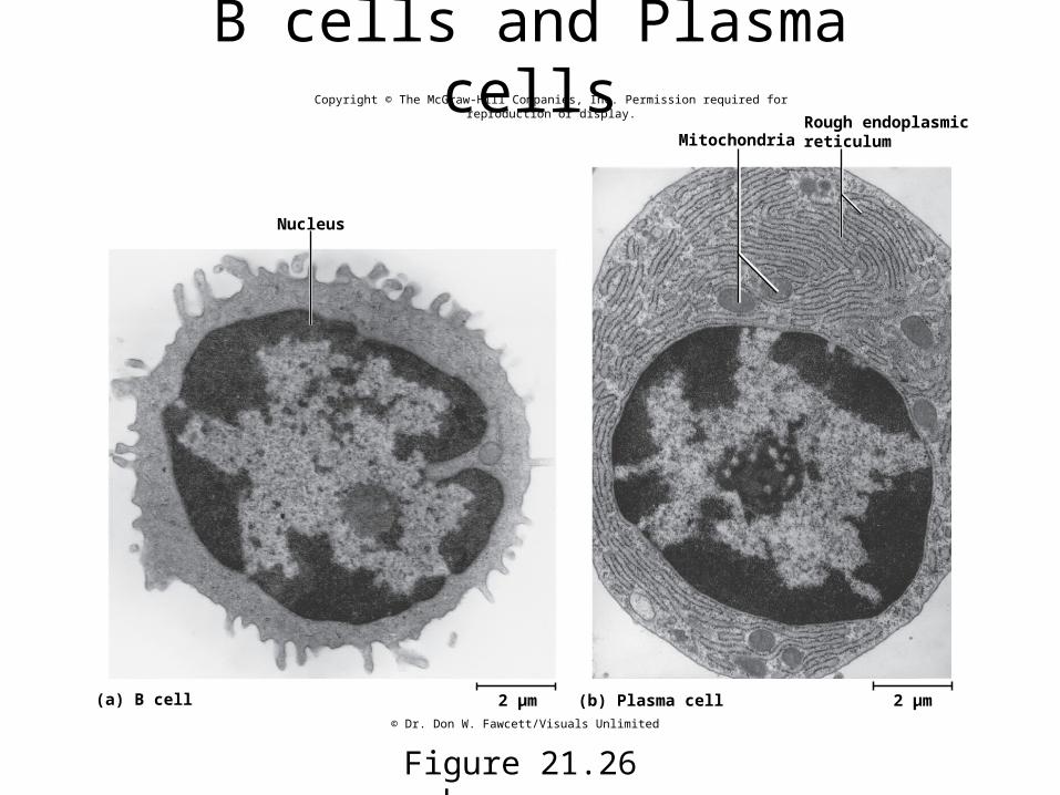
B cells and Plasma cells
Figure 21.26 a-b
Mitochondria
Nucleus
(a) B cell (b) Plasma cell 2 µm
Rough endoplasmicreticulum
2 µm
Copyright © The McGraw-Hill Companies, Inc. Permission required for reproduction or display.
© Dr. Don W. Fawcett/Visuals Unlimited

Antibodies• immunoglobulin (Ig) – an antibody is a defensive gamma globulin
found in the blood plasma, tissue fluids, body secretions, and some leukocyte membranes
• antibody monomer – the basic structural unit of an antibody
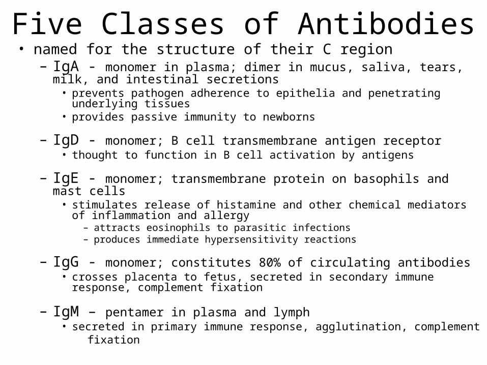
Five Classes of Antibodies• named for the structure of their C region
– IgA - monomer in plasma; dimer in mucus, saliva, tears, milk, and intestinal secretions
• prevents pathogen adherence to epithelia and penetrating underlying tissues• provides passive immunity to newborns
– IgD - monomer; B cell transmembrane antigen receptor• thought to function in B cell activation by antigens
– IgE - monomer; transmembrane protein on basophils and mast cells• stimulates release of histamine and other chemical mediators of inflammation
and allergy – attracts eosinophils to parasitic infections– produces immediate hypersensitivity reactions
– IgG - monomer; constitutes 80% of circulating antibodies• crosses placenta to fetus, secreted in secondary immune response, complement
fixation
– IgM – pentamer in plasma and lymph• secreted in primary immune response, agglutination, complement fixation
Humoral Immunity - Attack• neutralization
– antibodies mask pathogenic region of antigen
• complement fixation– antigen binds to IgM or IgG, antibody changes shape, initiates
complement binding which leads to inflammation, phagocytosis, immune clearance, or cytolysis
– primary defense against foreign cells, bacteria, and mismatched RBCs
• agglutination– antibody has 2-10 binding sites; binds to multiple enemy cells
immobilizing them from spreading
• precipitation– antibody binds antigen molecules (not cells); creates antigen-antibody
complex that precipitates, phagocytized by eosinophils
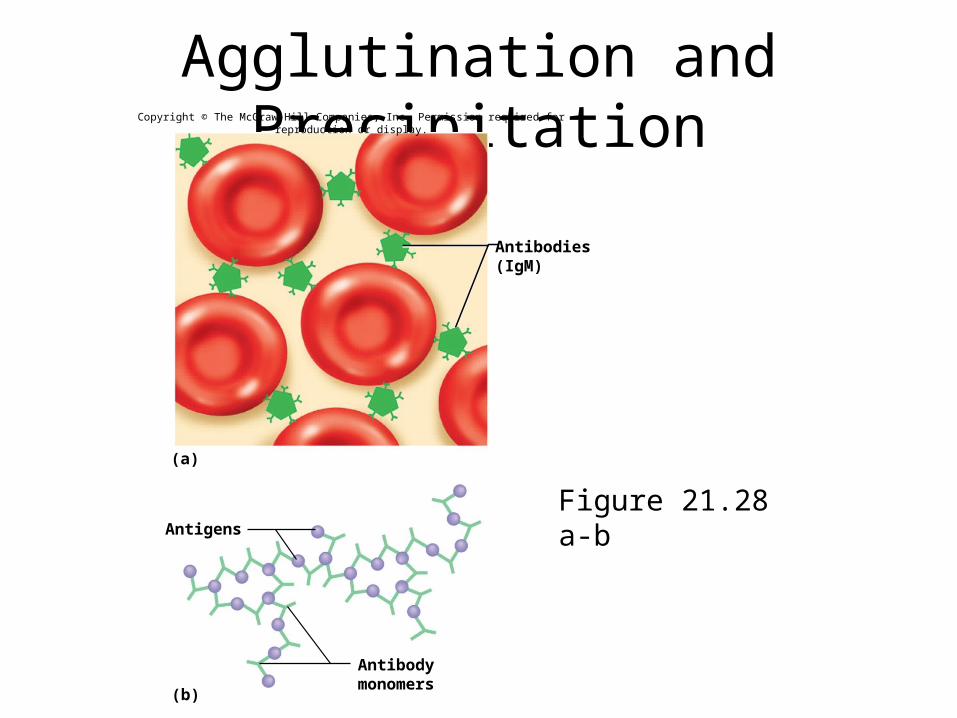
Agglutination and Precipitation
Figure 21.28 a-bAntigens
(a)
(b)
Antibodies(IgM)
Antibodymonomers
Copyright © The McGraw-Hill Companies, Inc. Permission required for reproduction or display.
Humoral Immunity - Memory• primary immune response – immune reaction brought
about by the first exposure to an antigen– appearance of protective antibodies delayed for 3 to 6 days
while naïve B cells multiply and differentiate into plasma cells– as plasma cells produce antibodies, the antibody titer (level in
the blood plasma) rises• IgM appears first, peaks in about 10 days, soon declines• IgG levels rise as IgM declines, but IgG titer drops to a low level within
a month– primary response leaves one with an immune memory of the
antigen• during clonal selection, some of the clone becomes memory B cells• found mainly in germinal centers of the lymph nodes• mount a very quick secondary (anamnestic) response
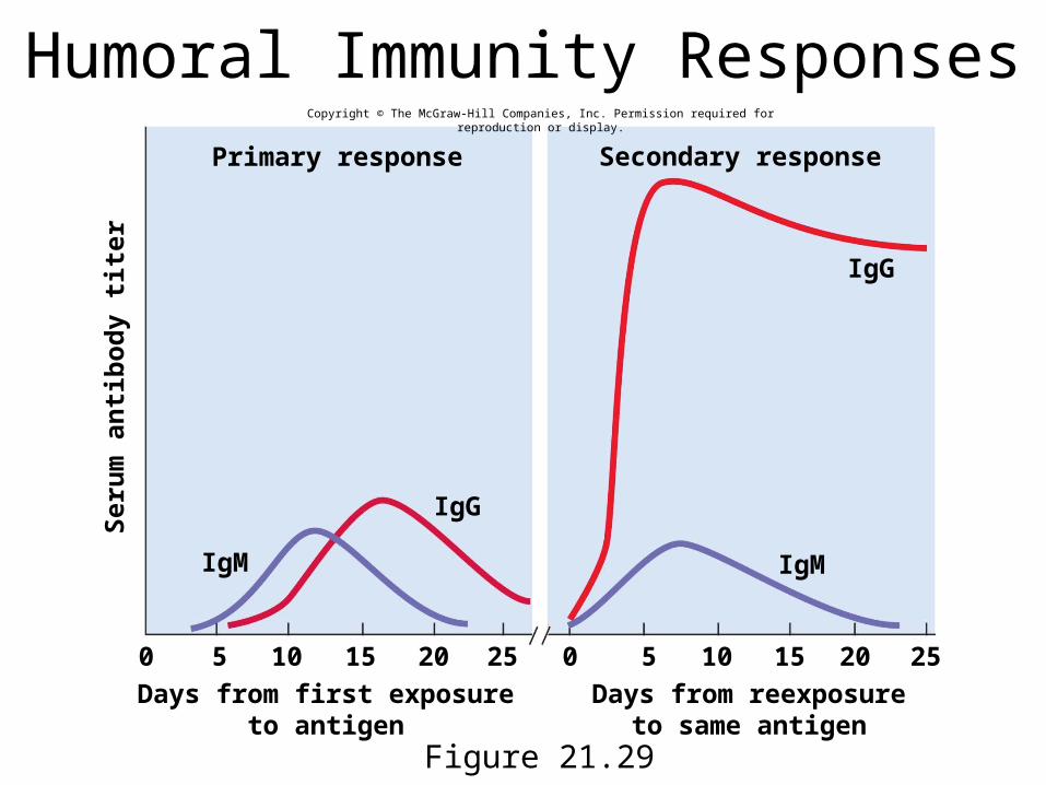
Humoral Immunity Responses
Figure 21.29
Seru
m a
ntibo
dy ti
ter
Primary response
IgM IgM
IgG
IgG
Secondary response
0 0 5 510 1015 1520 2025 25Days from first exposure
to antigenDays from reexposure
to same antigen
Copyright © The McGraw-Hill Companies, Inc. Permission required for reproduction or display.
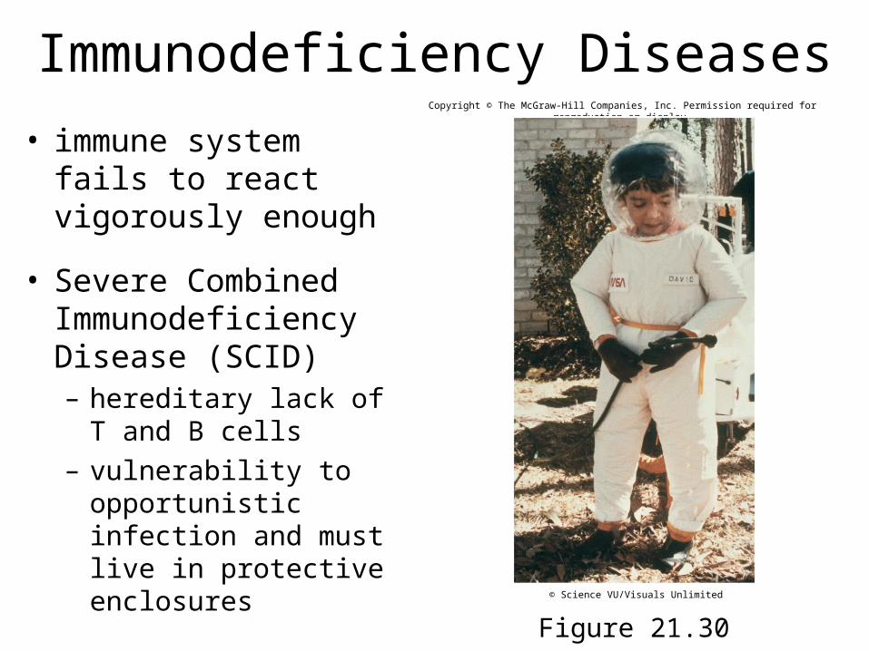
Immunodeficiency Diseases• immune system fails to
react vigorously enough
• Severe Combined Immunodeficiency Disease (SCID)– hereditary lack of T and B
cells– vulnerability to
opportunistic infection and must live in protective enclosures
Figure 21.30
Copyright © The McGraw-Hill Companies, Inc. Permission required for reproduction or display.
© Science VU/Visuals Unlimited
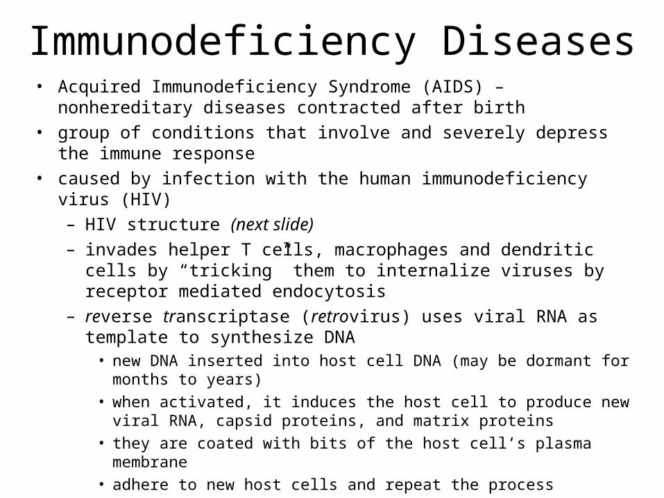
• Acquired Immunodeficiency Syndrome (AIDS) – nonhereditary diseases contracted after birth
• group of conditions that involve and severely depress the immune response• caused by infection with the human immunodeficiency virus (HIV)
– HIV structure (next slide)– invades helper T cells, macrophages and dendritic cells by “tricking”
them to internalize viruses by receptor mediated endocytosis– reverse transcriptase (retrovirus) uses viral RNA as template to
synthesize DNA• new DNA inserted into host cell DNA (may be dormant for months to years)• when activated, it induces the host cell to produce new viral RNA, capsid
proteins, and matrix proteins• they are coated with bits of the host cell’s plasma membrane• adhere to new host cells and repeat the process
Immunodeficiency Diseases
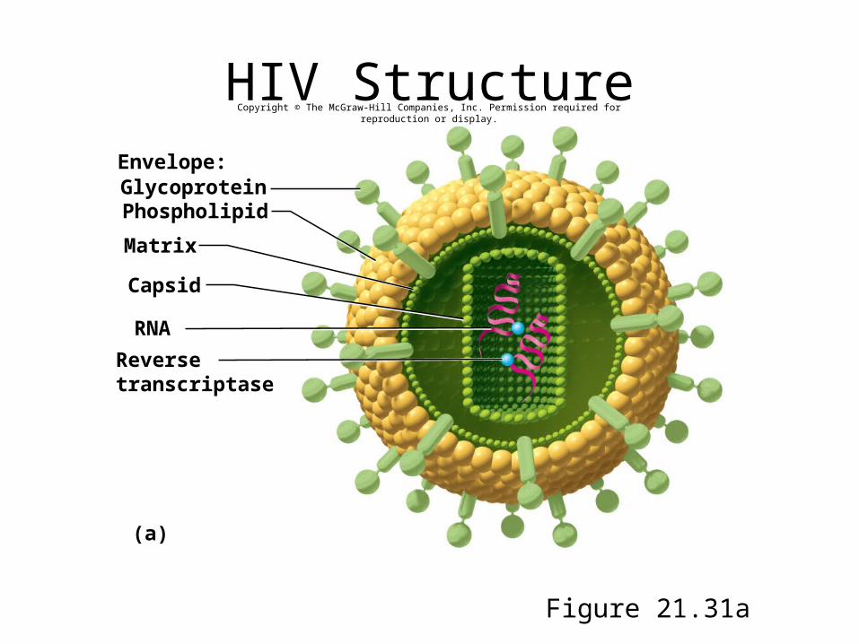
HIV Structure
Figure 21.31a
Envelope:GlycoproteinPhospholipid
Matrix
Capsid
RNA
(a)
Reversetranscriptase
Copyright © The McGraw-Hill Companies, Inc. Permission required for reproduction or display.
AIDS• by destroying TH cells, HIV strikes at the central coordinating
agent of nonspecific defense, humoral immunity, and cellular immunity
• incubation period ranges from several months to 12 years• signs and symptoms
– early symptoms: flulike symptoms of chills and fever– progresses to night sweats, fatigue, headache, extreme weight loss,
lymphadenitis– normal TH count is 600 to 1,200 cells/L of blood, but in AIDS it is less
than 200 cells/L – person susceptible to opportunistic infections (Toxoplasma,
Pneumocystis, herpes simplex virus, cytomegalovirus, or tuberculosis)– Candida (thrush): white patches on mucous membranes– Kaposi sarcoma: cancer originates in endothelial cells of blood vessels
causes purple lesions in skin
HIV Transmission• through blood, semen, vaginal secretions, breast milk,
or across the placenta
• most common means of transmission– sexual intercourse (vaginal, anal, oral)– contaminated blood products– contaminated needles
• not transmitted by casual contact
• undamaged latex condom is an effective barrier to HIV, especially with spermicide nonoxynol-9
Treatment Strategies• prevent binding to CD4 proteins of TH cells
• disrupt reverse transcriptase to inhibit assembly of new viruses or their release from host cells
• medications– none can eliminate HIV, all have serious side-effects– HIV develops drug resistance
• medicines used in combination
– AZT (azidothymidine)• first anti-HIV drug - inhibits reverse transcriptase
– protease inhibitors• inhibit enzymes HIV needs to replicate
– now more than 24 anti-HIV drugs on the market