Embed Size (px)
Citation preview
Chapter 14The Techniques of Molecular
Genetics
© John Wiley & Sons, Inc.
Chapter Outline
Basic Techniques used to Identify, Amplify, and Clone Genes
The Molecular Analysis of DNA, RNA, and Protein
© John Wiley & Sons, Inc.
Basic Techniques Used to Identify, Amplify, and Clone
GenesRecombinant DNA, gene
cloning, and DNA amplification techniques allow scientists to
isolate and characterize essentially any DNA sequence
from any organism.
© John Wiley & Sons, Inc.
Gene Cloning
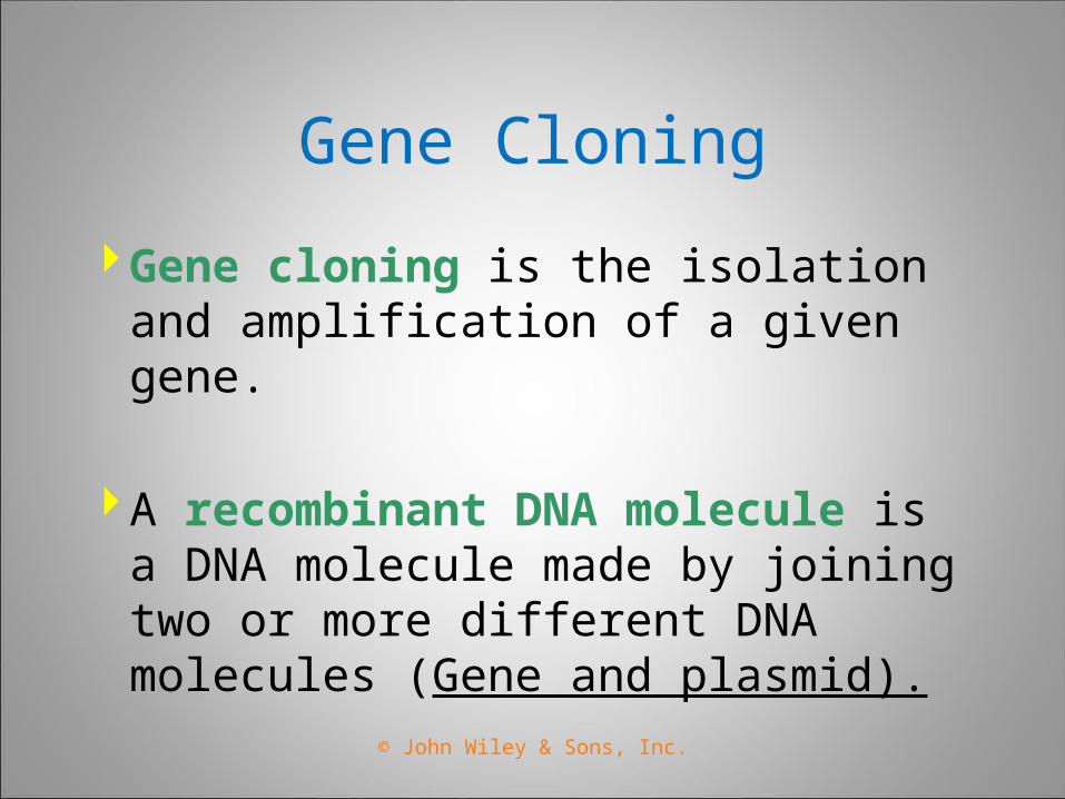
Gene cloning is the isolation and amplification of a given gene.
A recombinant DNA molecule is a DNA molecule made by joining two or more different DNA molecules (Gene and plasmid).
© John Wiley & Sons, Inc.
Amplification of a Gene In Vivo
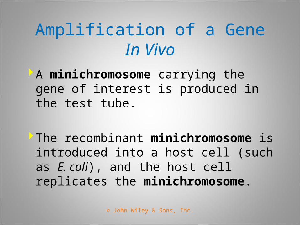
A minichromosome carrying the gene of interest is produced in the test tube.
The recombinant minichromosome is introduced into a host cell (such as E. coli), and the host cell replicates the minichromosome.
© John Wiley & Sons, Inc.
Amplification of a Gene In Vitro
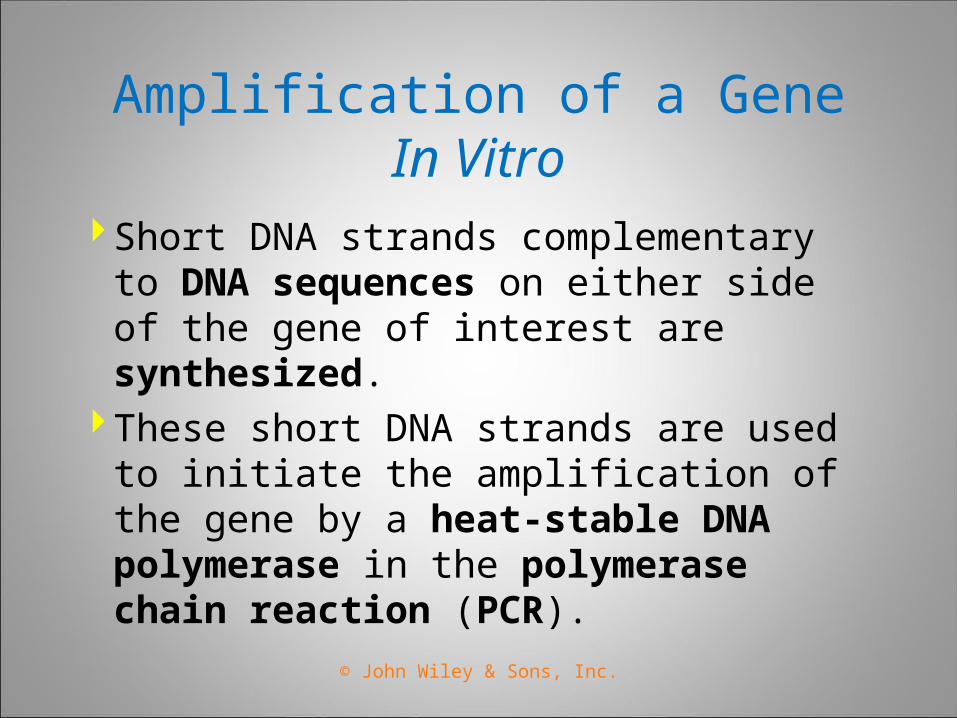
Short DNA strands complementary to DNA sequences on either side of the gene of interest are synthesized.
These short DNA strands are used to initiate the amplification of the gene by a heat-stable DNA polymerase in the polymerase chain reaction (PCR).
© John Wiley & Sons, Inc.
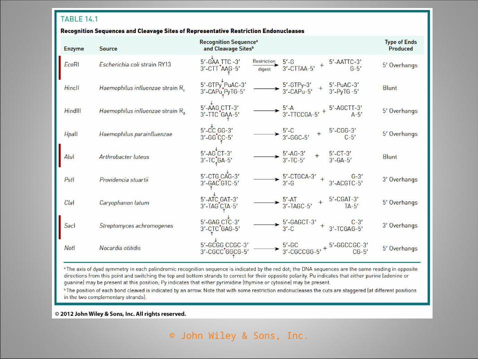
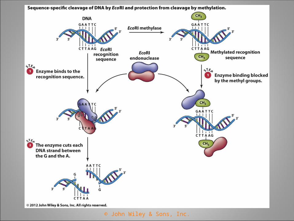
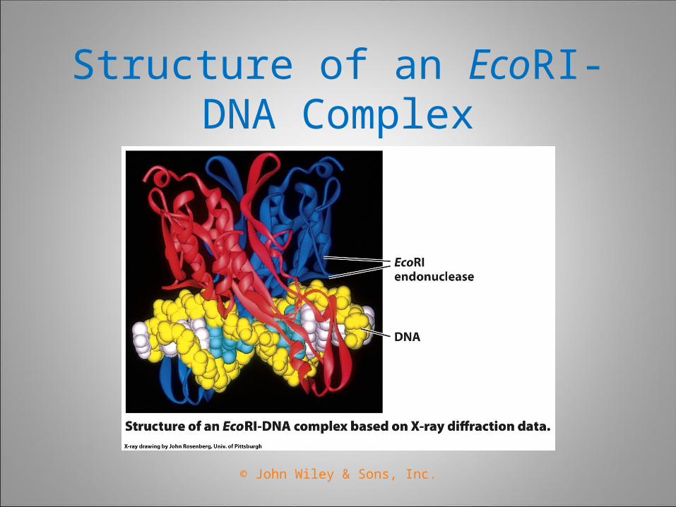
Restriction Endonucleases
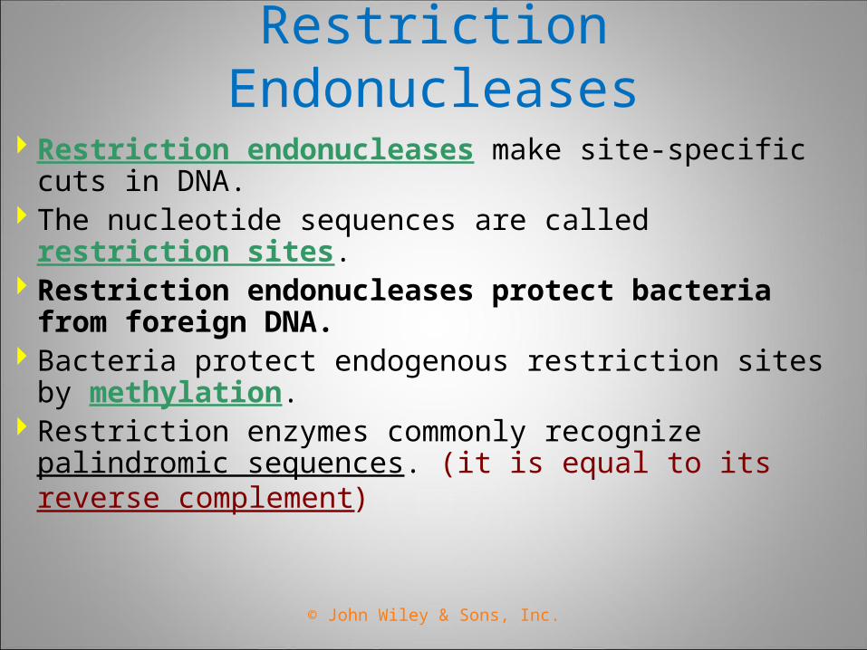
Restriction endonucleases make site-specific cuts in DNA.
The nucleotide sequences are called restriction sites.
Restriction endonucleases protect bacteria from foreign DNA.
Bacteria protect endogenous restriction sites by methylation.
Restriction enzymes commonly recognize palindromic sequences. (it is equal to its reverse complement)
© John Wiley & Sons, Inc.
© John Wiley & Sons, Inc.
© John Wiley & Sons, Inc.
Structure of an EcoRI-DNA Complex
© John Wiley & Sons, Inc.
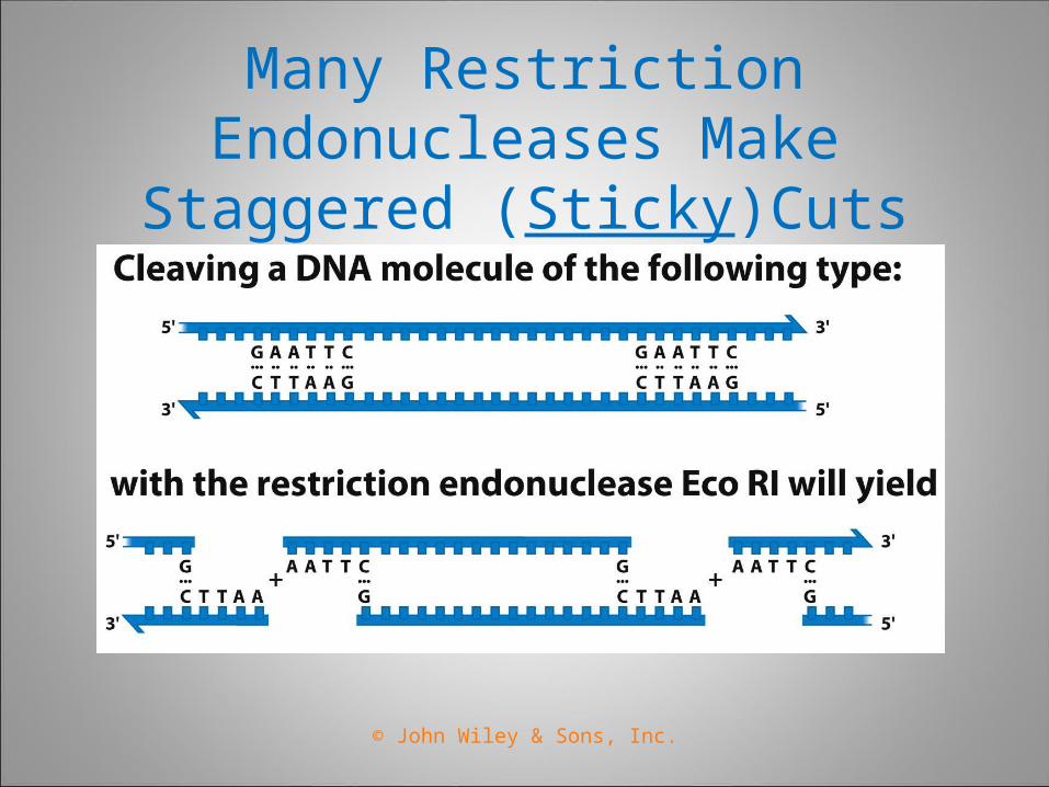
Many Restriction Endonucleases Make Staggered (Sticky)Cuts
© John Wiley & Sons, Inc.

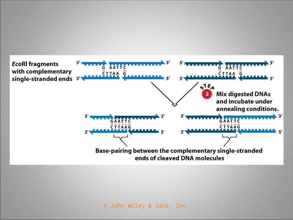
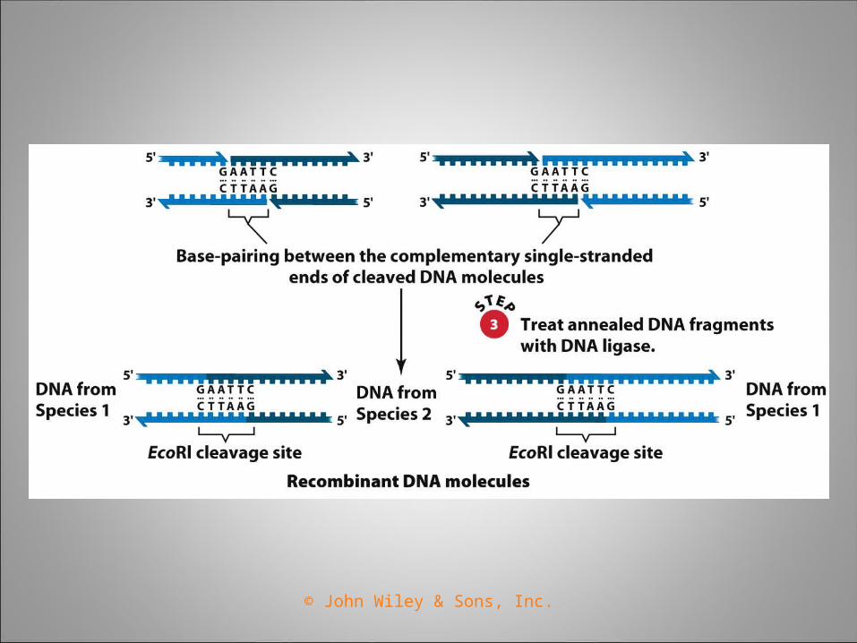
When DNA is cleaved with a restriction endonuclease that makes “sticky” cuts, all of the resulting restriction fragments have complementary single-stranded termini.
The complementary single-stranded termini can hydrogen bond with each other and be joined together by DNA ligase.
© John Wiley & Sons, Inc.
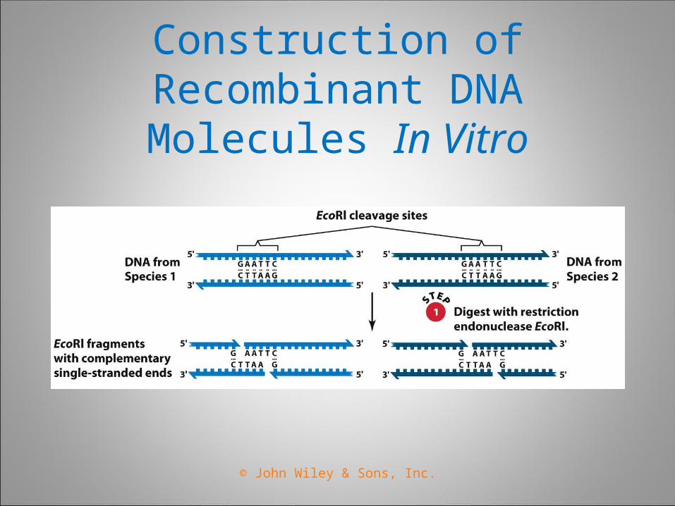
Construction of Recombinant DNA Molecules In Vitro
© John Wiley & Sons, Inc.
© John Wiley & Sons, Inc.
© John Wiley & Sons, Inc.
© John Wiley & Sons, Inc.
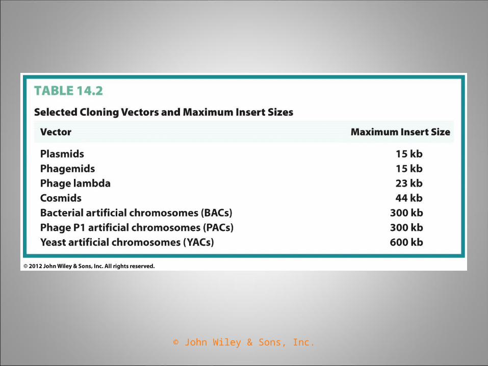
“Vector" is an agent/system that can carry a DNA fragment into a host cell.
If it is used for reproducing the DNA fragment, it is called a "cloning vector". If it is used for expressing certain gene in the DNA fragment, it is called an "expression vector".
Plasmid (Vectors)
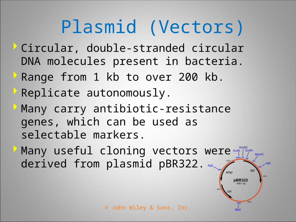
Plasmid (Vectors) Circular, double-stranded circular DNA
molecules present in bacteria. Range from 1 kb to over 200 kb. Replicate autonomously. Many carry antibiotic-resistance genes, which
can be used as selectable markers. Many useful cloning vectors were derived
from plasmid pBR322.
© John Wiley & Sons, Inc.
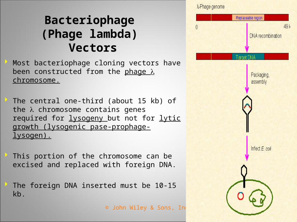
Bacteriophage (Phage lambda)
Vectors Most bacteriophage cloning vectors have
been constructed from the phage chromosome.
The central one-third (about 15 kb) of the chromosome contains genes required for lysogeny but not for lytic growth (lysogenic pase-prophage-lysogen).
This portion of the chromosome can be excised and replaced with foreign DNA.
The foreign DNA inserted must be 10-15 kb.
© John Wiley & Sons, Inc.
Phagemid Vectors
Contain components from phage chromosomes and plasmids (pBluescript II)
Replicate in E. coli as double-stranded plasmids.
Addition of a helper phage causes the phagemid to switch to the phage mode of replication, resulting in the packaging of single-stranded DNA into phage heads.
© John Wiley & Sons, Inc.
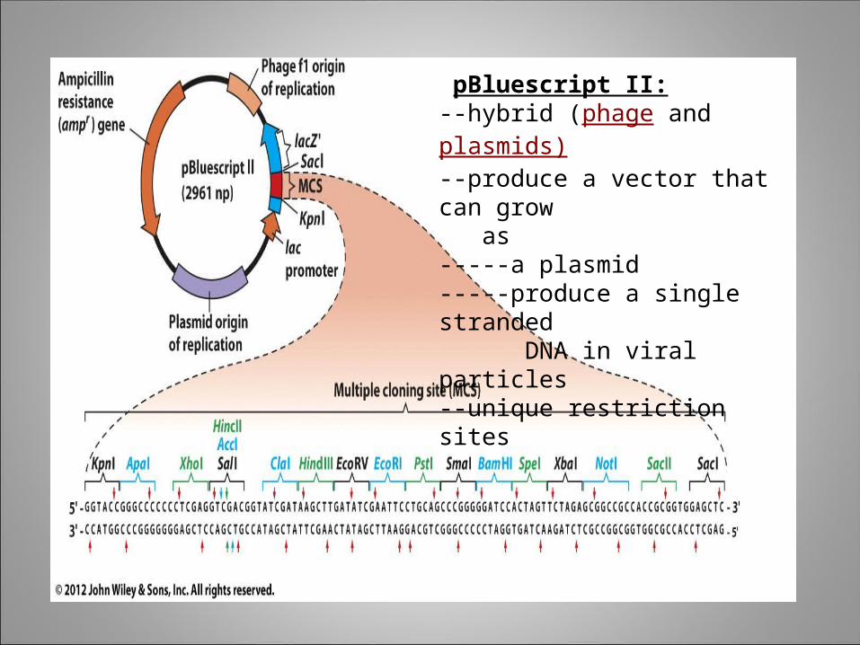
pBluescript II:--hybrid (phage and plasmids)--produce a vector that can grow as -----a plasmid-----produce a single stranded DNA in viral particles--unique restriction sites
© John Wiley & Sons, Inc.
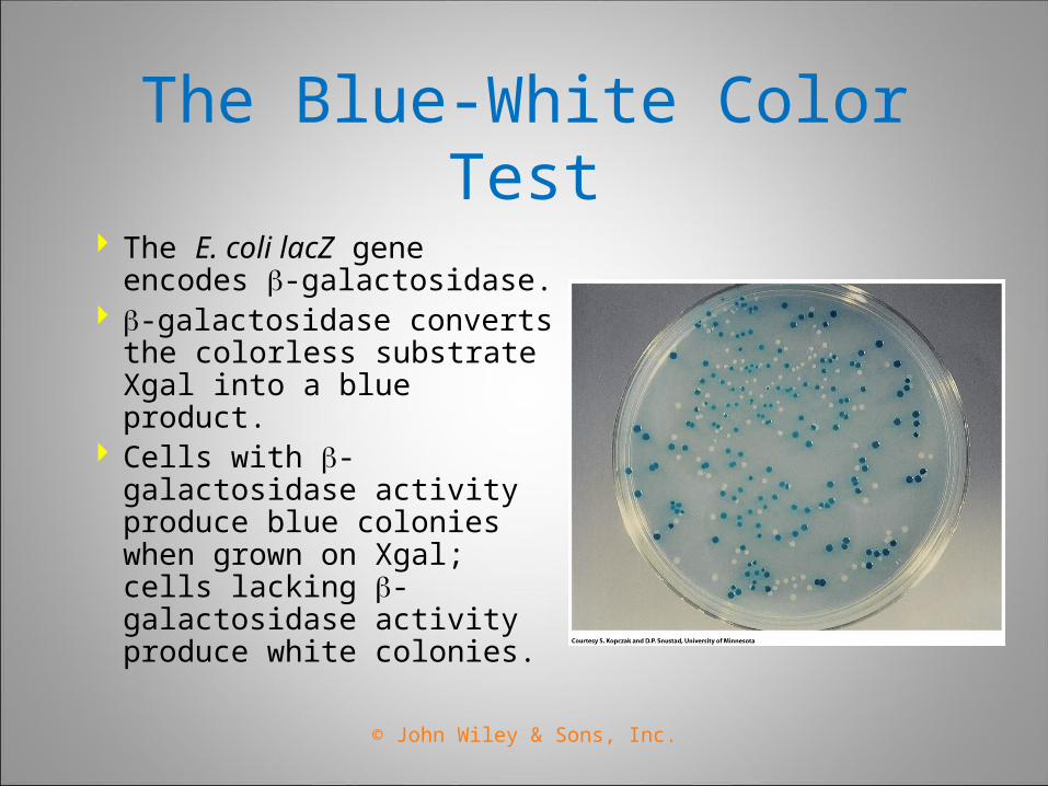
The Blue-White Color Test
The E. coli lacZ gene encodes -galactosidase.
-galactosidase converts the colorless substrate Xgal into a blue product.
Cells with -galactosidase activity produce blue colonies when grown on Xgal; cells lacking -galactosidase activity produce white colonies.
© John Wiley & Sons, Inc.
Shuttle (Eukaryotic) Vectors Because different organisms use different origins
of replication and regulatory signals, different cloning vectors must be used in different species.
Special cloning vectors can replicate in other prokaryotes and in eukaryotes.
Shuttle vectors can replicate in E. coli and in another species and it can propagate in two different host species
© John Wiley & Sons, Inc.
Importance/significance
Insulin
Antibodies
Growth Factors
Enzymes
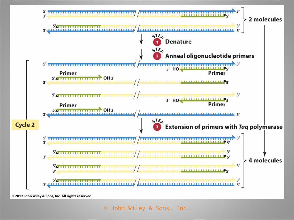
The Polymerase Chain Reaction (PCR)
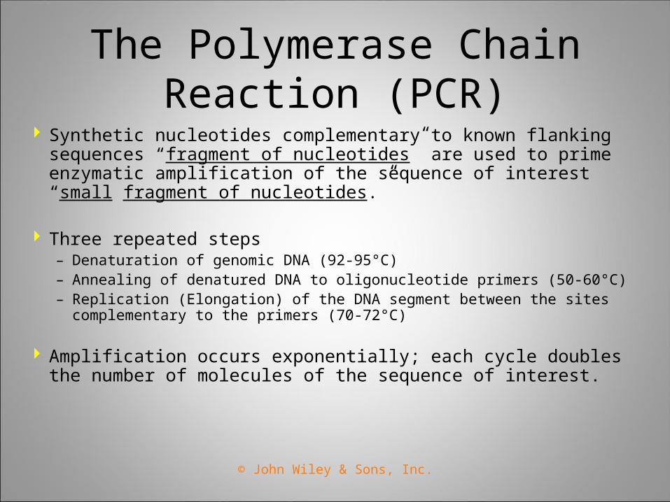
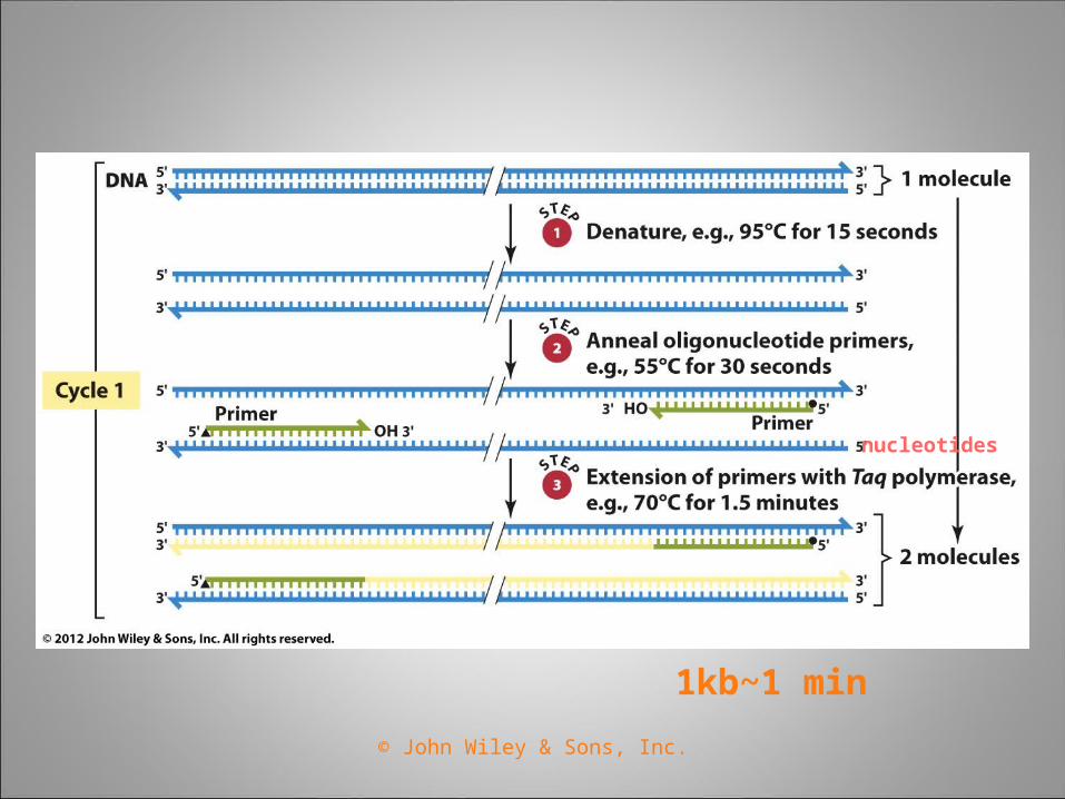
Synthetic nucleotides complementary to known flanking sequences “fragment of nucleotides” are used to prime enzymatic amplification of the sequence of interest “small fragment of nucleotides.”
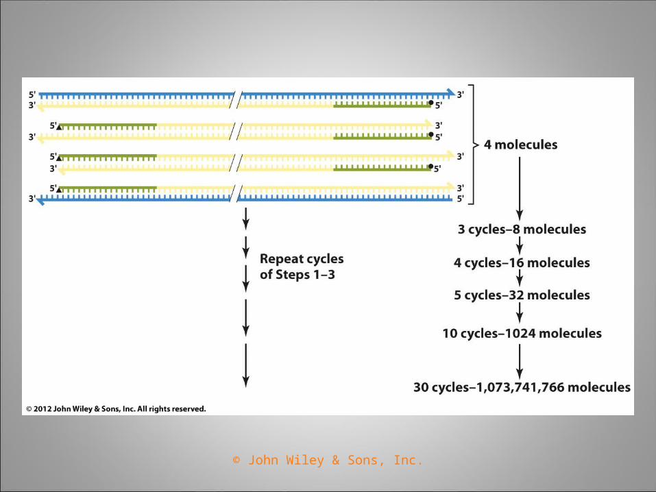
Three repeated steps– Denaturation of genomic DNA (92-95°C)– Annealing of denatured DNA to oligonucleotide primers (50-60°C)– Replication (Elongation) of the DNA segment between the sites
complementary to the primers (70-72°C)
Amplification occurs exponentially; each cycle doubles the number of molecules of the sequence of interest.
© John Wiley & Sons, Inc.
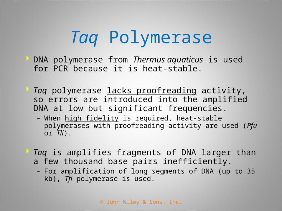
Taq Polymerase DNA polymerase from Thermus aquaticus is used for
PCR because it is heat-stable.
Taq polymerase lacks proofreading activity, so errors are introduced into the amplified DNA at low but significant frequencies.– When high fidelity is required, heat-stable polymerases with
proofreading activity are used (Pfu or Tli).
Taq is amplifies fragments of DNA larger than a few thousand base pairs inefficiently.– For amplification of long segments of DNA (up to 35 kb), Tfl
polymerase is used.
© John Wiley & Sons, Inc.
Applications of PCR
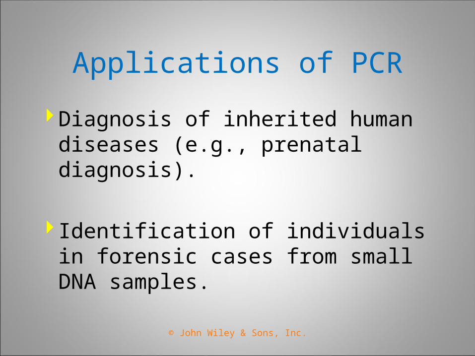
Diagnosis of inherited human diseases (e.g., prenatal diagnosis).
Identification of individuals in forensic cases from small DNA samples.
© John Wiley & Sons, Inc.
© John Wiley & Sons, Inc.
1kb~1 min
nucleotides
© John Wiley & Sons, Inc.
© John Wiley & Sons, Inc.
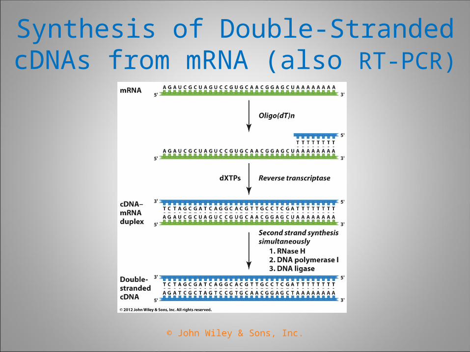
Synthesis of Double-Stranded cDNAs from mRNA (also RT-PCR)
© John Wiley & Sons, Inc.
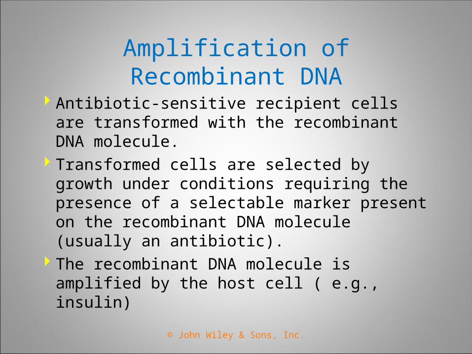
Amplification of Recombinant DNA
Antibiotic-sensitive recipient cells are transformed with the recombinant DNA molecule.
Transformed cells are selected by growth under conditions requiring the presence of a selectable marker present on the recombinant DNA molecule (usually an antibiotic).
The recombinant DNA molecule is amplified by the host cell ( e.g., insulin)
© John Wiley & Sons, Inc.
Techniques Necessary for Sequencing DNA
Restriction enzymes to prepare homogenous samples of specific segments of chromosomes.
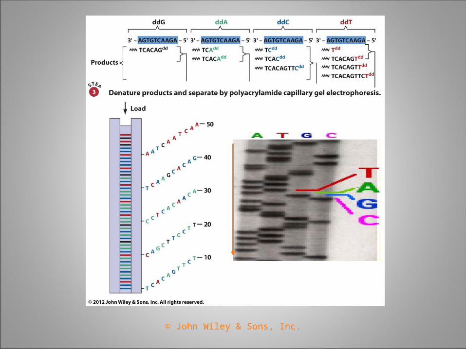
Gel electrophoresis procedures able to resolve DNA fragments differing in length by a single nucleotide.
Gene-cloning techniques allowing preparation of large quantities of a DNA molecule.
Sanger sequencing Technique is used to determine nucleotide sequences.
© John Wiley & Sons, Inc.
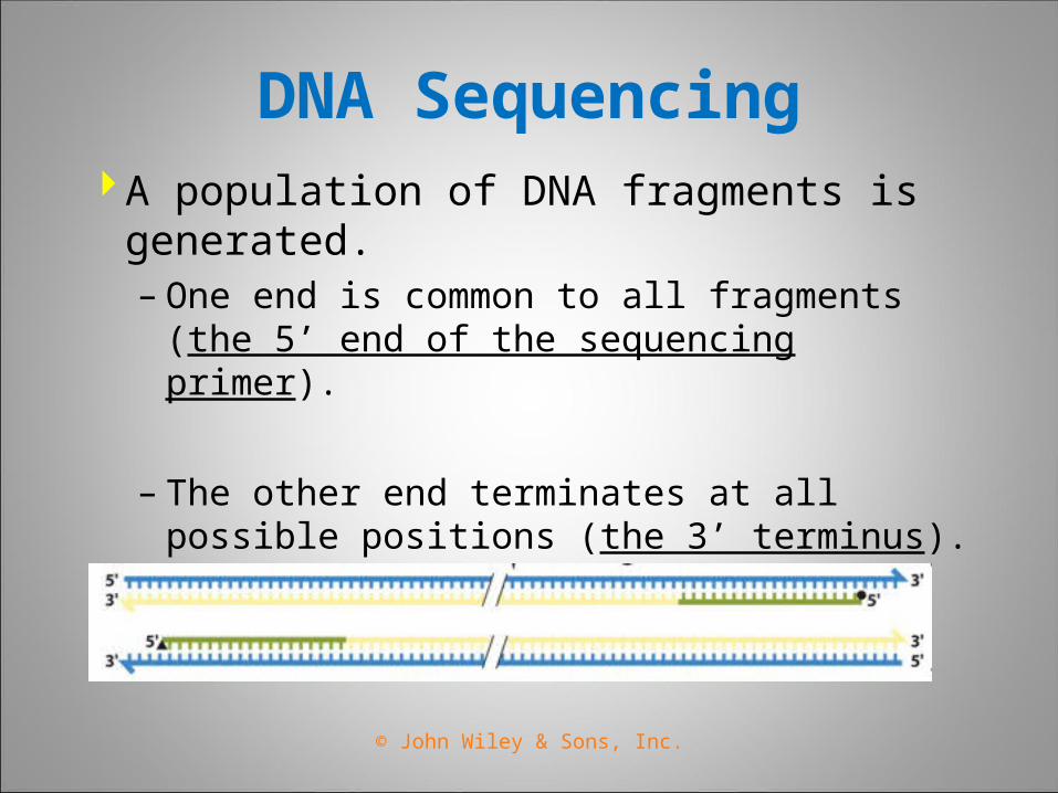
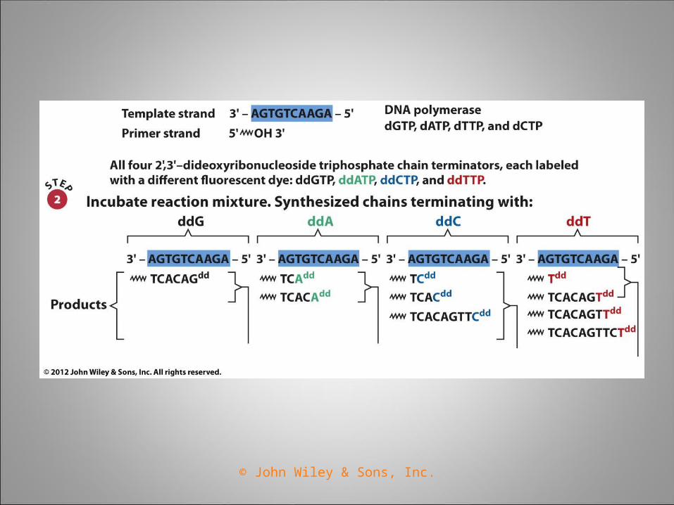
DNA SequencingA population of DNA fragments is
generated.– One end is common to all fragments (the 5’
end of the sequencing primer).
– The other end terminates at all possible positions (the 3’ terminus).
© John Wiley & Sons, Inc.
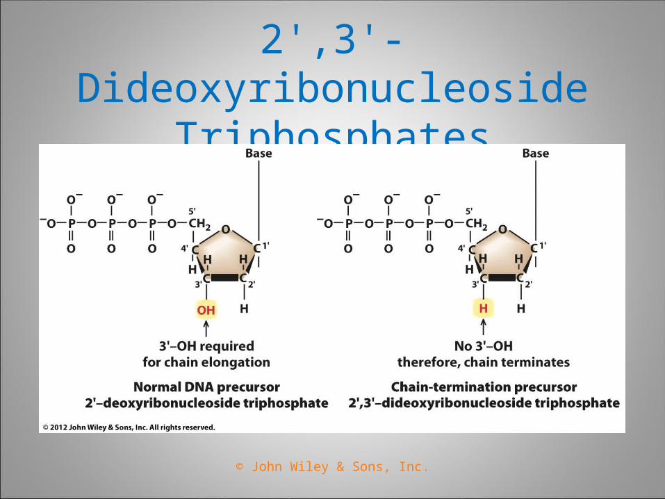
2',3'-Dideoxyribonucleoside Triphosphates
© John Wiley & Sons, Inc.
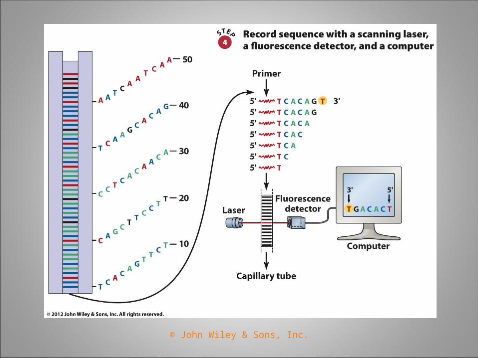
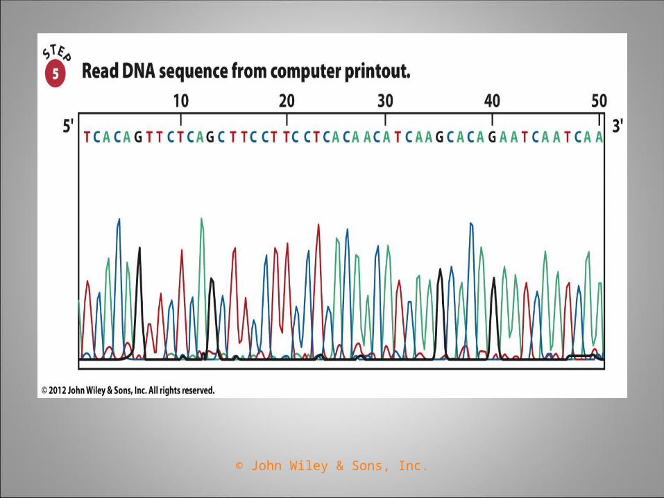
Automated DNA Sequencing Fluorescent dyes (Cy5, Cy3) are used for detection of
DNA chains instead of radioactive isotopes (32P ,35S).
Products of all four chain terminator reactions are separated through a single gel or capillary tube.
Photocells detect fluorescence of the dyes as they pass through the gel or capillary tube.
Output of the photocell is directly transferred to a computer for analysis.
© John Wiley & Sons, Inc.
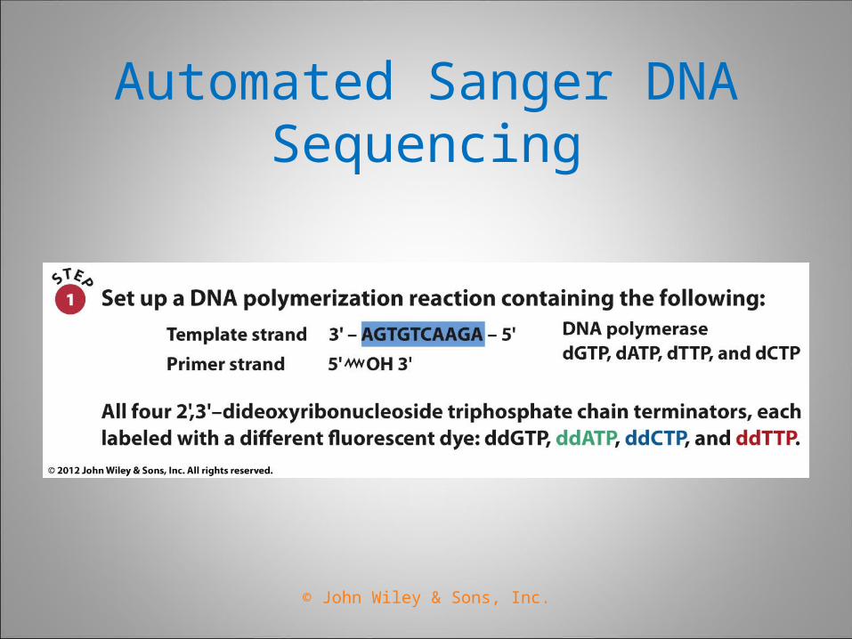
Automated Sanger DNA Sequencing
© John Wiley & Sons, Inc.
© John Wiley & Sons, Inc.
© John Wiley & Sons, Inc.
© John Wiley & Sons, Inc.
© John Wiley & Sons, Inc.