Embed Size (px)
Citation preview
CHAPTER
© 2014 by McGraw-Hill Education. This is proprietary material solely for authorized instructor use. Not authorized for sale or distribution in any manner. This document may not be copied, scanned, duplicated, forwarded, distributed, or posted on a
website, in whole or part.
49Electrocardiography
and Pulmonary Function Testing
© 2014 by McGraw-Hill Education. This is proprietary material solely for authorized instructor use. Not authorized for sale or distribution in any manner. This document may not be copied, scanned, duplicated, forwarded, distributed, or posted on a
website, in whole or part.
49-2
49.1 Discuss the medical assistant's role in electrocardiography and pulmonary function testing.
49.2 Explain the basic principles of electrocardiography and how it relates to the conduction system of the heart.
49.3 Identify the components of an electrocardiograph and what each does.
49.4 Carry out the steps necessary to obtain an ECG.
Learning Outcomes
© 2014 by McGraw-Hill Education. This is proprietary material solely for authorized instructor use. Not authorized for sale or distribution in any manner. This document may not be copied, scanned, duplicated, forwarded, distributed, or posted on a
website, in whole or part.
49-3
49.5 Summarize exercise electrocardiography and echocardiography.
49.6 Explain the procedure of Holter monitoring.
49.7 Carry out the various types of pulmonary function tests.
49.8 Describe the procedure for performing pulse oximetry testing.
Learning Outcomes
© 2014 by McGraw-Hill Education. This is proprietary material solely for authorized instructor use. Not authorized for sale or distribution in any manner. This document may not be copied, scanned, duplicated, forwarded, distributed, or posted on a
website, in whole or part.
49-4
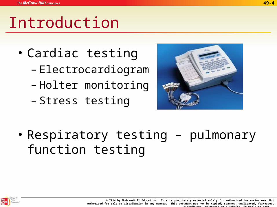
Introduction
• Cardiac testing– Electrocardiogram– Holter monitoring – Stress testing
• Respiratory testing – pulmonary function testing
© 2014 by McGraw-Hill Education. This is proprietary material solely for authorized instructor use. Not authorized for sale or distribution in any manner. This document may not be copied, scanned, duplicated, forwarded, distributed, or posted on a
website, in whole or part.
49-5
The Medical Assistant’s Role in Electrocardiography and Pulmonary Function Testing
• Medical assistant may perform – Electrocardiography
– Pulmonary function tests (PFTs)
– Diagnostic reasons
– Part of general exam
© 2014 by McGraw-Hill Education. This is proprietary material solely for authorized instructor use. Not authorized for sale or distribution in any manner. This document may not be copied, scanned, duplicated, forwarded, distributed, or posted on a
website, in whole or part.
49-6
Basic Principles of Electrocardiography
• Heartbeats produce electrical current
• Resting cardiac cell – polarized
• Depolarization – Occurs when cell loses polarity– Electrical impulse initiates contraction– Impulse detected by electrodes
© 2014 by McGraw-Hill Education. This is proprietary material solely for authorized instructor use. Not authorized for sale or distribution in any manner. This document may not be copied, scanned, duplicated, forwarded, distributed, or posted on a
website, in whole or part.
49-7
Basic Principles of Electrocardiography
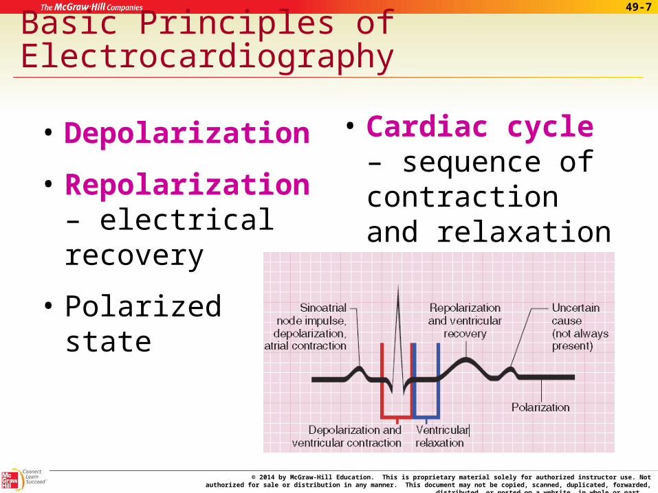
• Depolarization
• Repolarization – electrical recovery
• Polarizedstate
• Cardiac cycle – sequence of contraction and relaxation
© 2014 by McGraw-Hill Education. This is proprietary material solely for authorized instructor use. Not authorized for sale or distribution in any manner. This document may not be copied, scanned, duplicated, forwarded, distributed, or posted on a
website, in whole or part.
49-8
The Basic Pattern of the Electrocardiogram
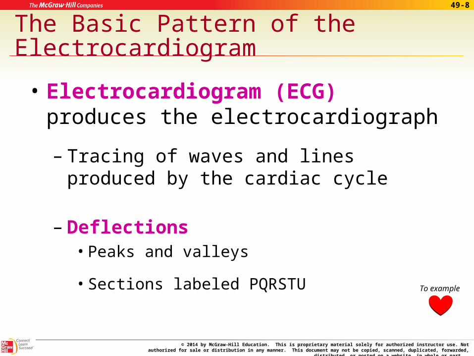
• Electrocardiogram (ECG) produces the electrocardiograph
– Tracing of waves and lines produced by the cardiac cycle
– Deflections • Peaks and valleys
• Sections labeled PQRSTU To example

© 2014 by McGraw-Hill Education. This is proprietary material solely for authorized instructor use. Not authorized for sale or distribution in any manner. This document may not be copied, scanned, duplicated, forwarded, distributed, or posted on a
website, in whole or part.
49-9
Apply Your Knowledge
1. What two procedures might the medical assistant preform?
ANSWER: Electrocardiography and pulmonary function testing.
2. What is the term for the depolarization, repolarization, and polarized state sequence?
ANSWER: The cardiac cycle.
© 2014 by McGraw-Hill Education. This is proprietary material solely for authorized instructor use. Not authorized for sale or distribution in any manner. This document may not be copied, scanned, duplicated, forwarded, distributed, or posted on a
website, in whole or part.
49-10
The Electrocardiograph
• Impulse is detected by electrodes
• Transmitted by insulated wires
• Amplified by the electrocardiograph machine
• Stylus – records the movement
• Leads – views of electrical activity
© 2014 by McGraw-Hill Education. This is proprietary material solely for authorized instructor use. Not authorized for sale or distribution in any manner. This document may not be copied, scanned, duplicated, forwarded, distributed, or posted on a
website, in whole or part.
49-11
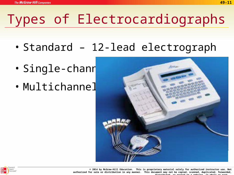
Types of Electrocardiographs
• Standard – 12-lead electrograph
• Single-channel
• Multichannel
© 2014 by McGraw-Hill Education. This is proprietary material solely for authorized instructor use. Not authorized for sale or distribution in any manner. This document may not be copied, scanned, duplicated, forwarded, distributed, or posted on a
website, in whole or part.
49-12
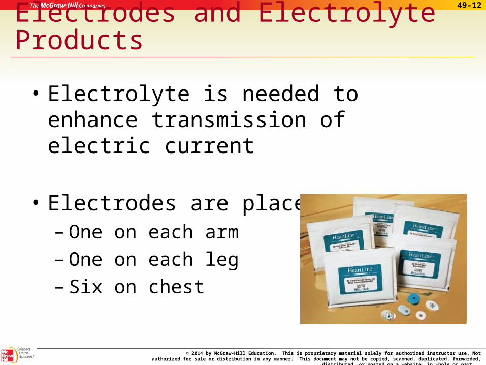
Electrodes and Electrolyte Products
• Electrolyte is needed to enhance transmission of electric current
• Electrodes are placed– One on each arm– One on each leg– Six on chest
© 2014 by McGraw-Hill Education. This is proprietary material solely for authorized instructor use. Not authorized for sale or distribution in any manner. This document may not be copied, scanned, duplicated, forwarded, distributed, or posted on a
website, in whole or part.
49-13
Leads
• Limb leads
– Three standard leads
• Bipolar
• Designation – I, II, III
– Three augmented views• Unipolar • Amplified • Designation – AVF, AVR, AVL
© 2014 by McGraw-Hill Education. This is proprietary material solely for authorized instructor use. Not authorized for sale or distribution in any manner. This document may not be copied, scanned, duplicated, forwarded, distributed, or posted on a
website, in whole or part.
49-14
Leads
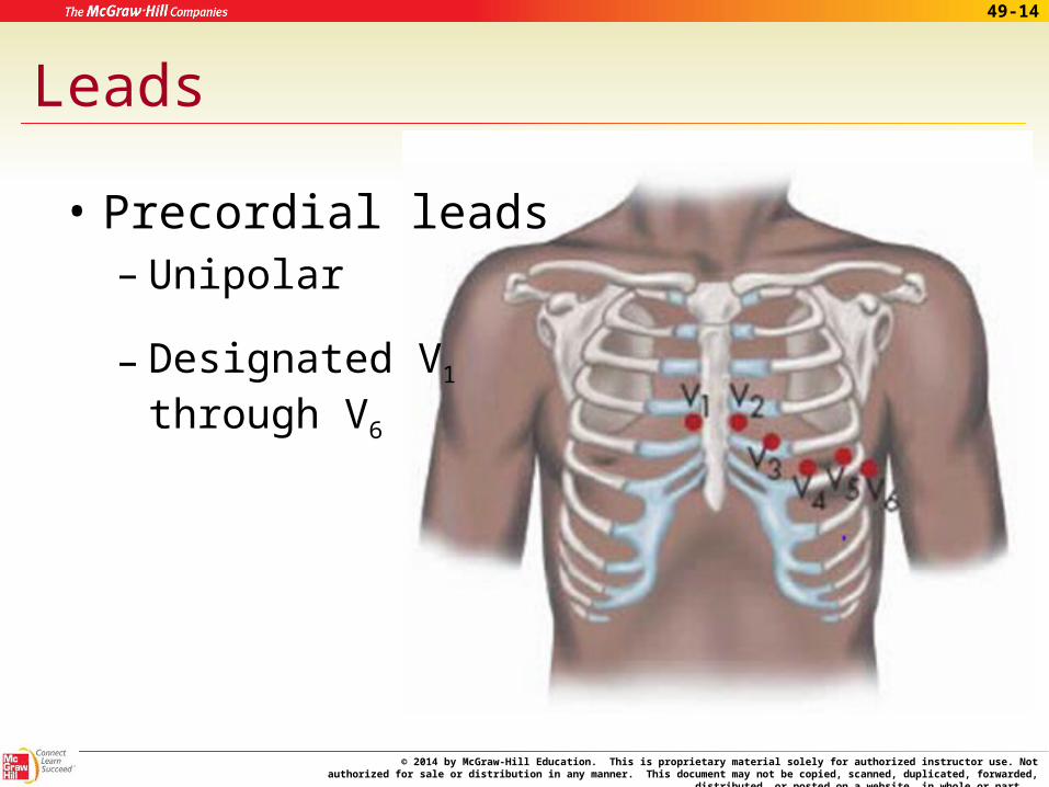
• Precordial leads– Unipolar
– Designated V1 through V6
© 2014 by McGraw-Hill Education. This is proprietary material solely for authorized instructor use. Not authorized for sale or distribution in any manner. This document may not be copied, scanned, duplicated, forwarded, distributed, or posted on a
website, in whole or part.
49-15
ECG Paper
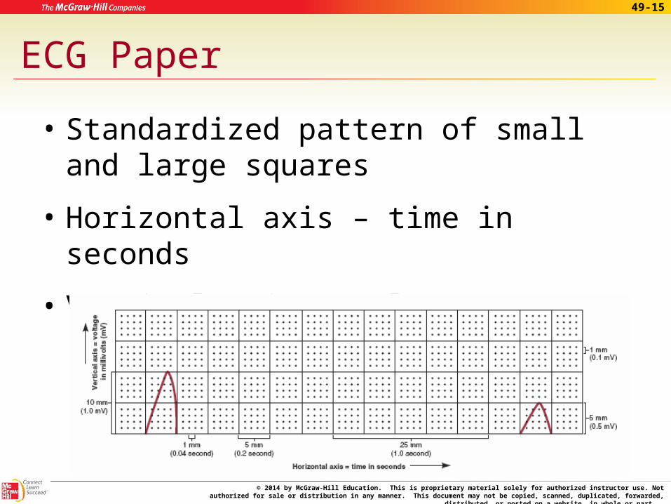
• Standardized pattern of small and large squares
• Horizontal axis – time in seconds
• Vertical axis – voltage (mV)
© 2014 by McGraw-Hill Education. This is proprietary material solely for authorized instructor use. Not authorized for sale or distribution in any manner. This document may not be copied, scanned, duplicated, forwarded, distributed, or posted on a
website, in whole or part.
49-16
The Electrocardiograph Controls
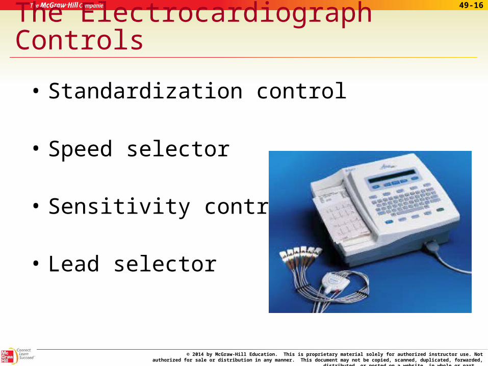
• Standardization control
• Speed selector
• Sensitivity control
• Lead selector
© 2014 by McGraw-Hill Education. This is proprietary material solely for authorized instructor use. Not authorized for sale or distribution in any manner. This document may not be copied, scanned, duplicated, forwarded, distributed, or posted on a
website, in whole or part.
49-17
The Electrocardiograph Controls
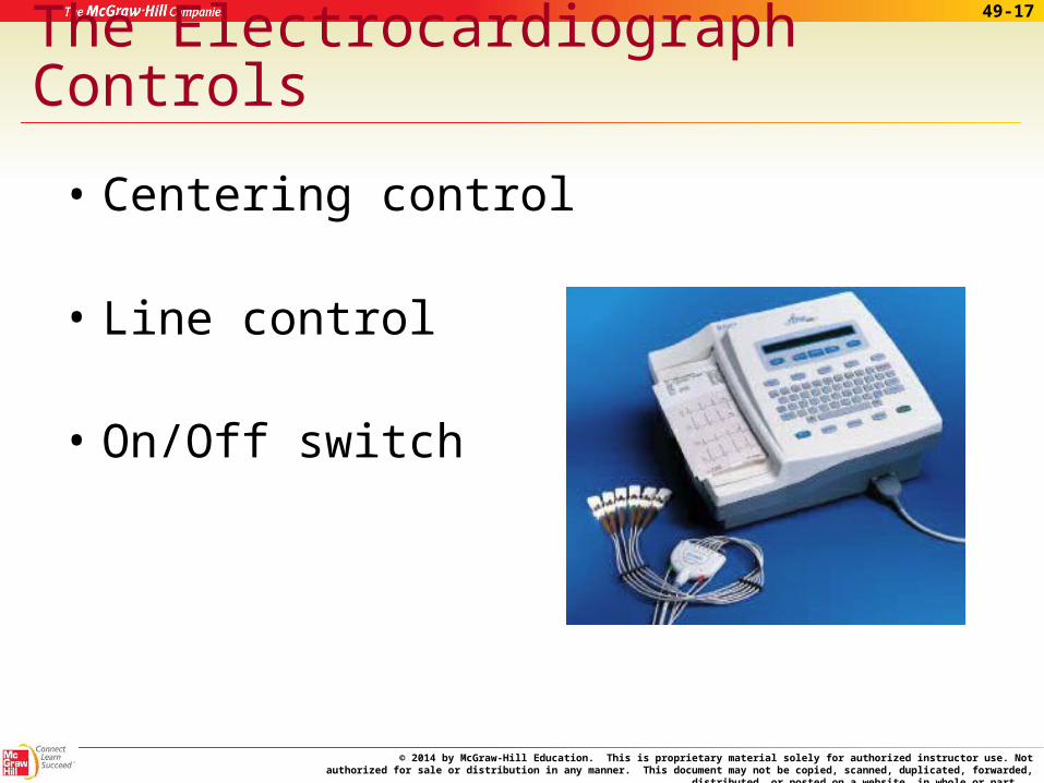
• Centering control
• Line control
• On/Off switch

© 2014 by McGraw-Hill Education. This is proprietary material solely for authorized instructor use. Not authorized for sale or distribution in any manner. This document may not be copied, scanned, duplicated, forwarded, distributed, or posted on a
website, in whole or part.
49-18
Apply Your Knowledge
Matching:
___ Adjusts position of stylus A. Vertical axis
___ Adjusts height of tracing B. Sensitivity control
___ Adjusts darkness of tracing C. Precordial leads
___ Measures strength of impulse D. Horizontal axis
___ Measures time E. Limb leads
___ AVF, AVR, AVL F. Amplification
___ V1 through V6 G. Centering control
___ Increases signal H. Line control
ANSWER:
H
G
F
E
D
C
B
A
Superbly Matched!
© 2014 by McGraw-Hill Education. This is proprietary material solely for authorized instructor use. Not authorized for sale or distribution in any manner. This document may not be copied, scanned, duplicated, forwarded, distributed, or posted on a
website, in whole or part.
49-19
Performing an ECG
• Proper technique essential
• Preparing the room and equipment– Turn off other electrical equipment
– Quiet room, comfortable temperature
– Check machine• Warm up• Adequate paper
© 2014 by McGraw-Hill Education. This is proprietary material solely for authorized instructor use. Not authorized for sale or distribution in any manner. This document may not be copied, scanned, duplicated, forwarded, distributed, or posted on a
website, in whole or part.
49-20
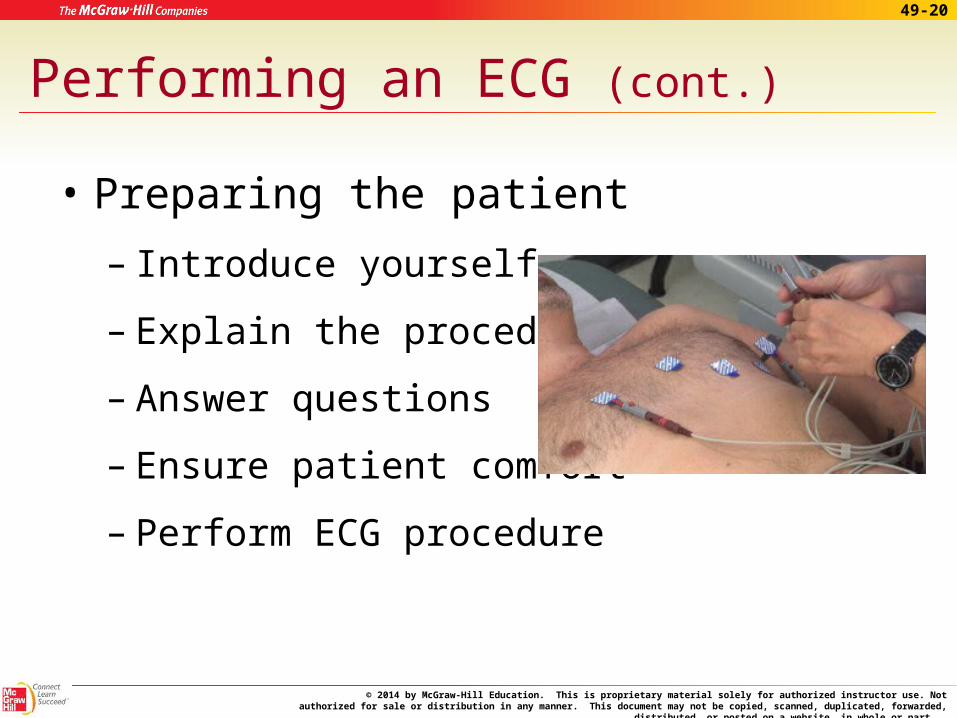
Performing an ECG (cont.)
• Preparing the patient
– Introduce yourself
– Explain the procedure
– Answer questions
– Ensure patient comfort
– Perform ECG procedure
© 2014 by McGraw-Hill Education. This is proprietary material solely for authorized instructor use. Not authorized for sale or distribution in any manner. This document may not be copied, scanned, duplicated, forwarded, distributed, or posted on a
website, in whole or part.
49-21
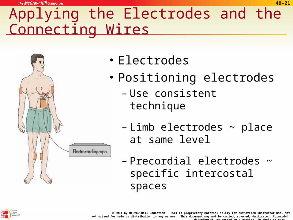
Applying the Electrodes and the Connecting Wires
• Electrodes
• Positioning electrodes – Use consistent technique
– Limb electrodes ~ place at same level
– Precordial electrodes ~ specific intercostal spaces
© 2014 by McGraw-Hill Education. This is proprietary material solely for authorized instructor use. Not authorized for sale or distribution in any manner. This document may not be copied, scanned, duplicated, forwarded, distributed, or posted on a
website, in whole or part.
49-22
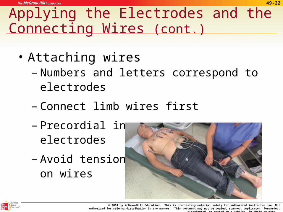
Applying the Electrodes and the Connecting Wires (cont.)
• Attaching wires– Numbers and letters correspond to electrodes
– Connect limb wires first
– Precordial in same sequence as electrodes
– Avoid tension on wires
© 2014 by McGraw-Hill Education. This is proprietary material solely for authorized instructor use. Not authorized for sale or distribution in any manner. This document may not be copied, scanned, duplicated, forwarded, distributed, or posted on a
website, in whole or part.
49-23
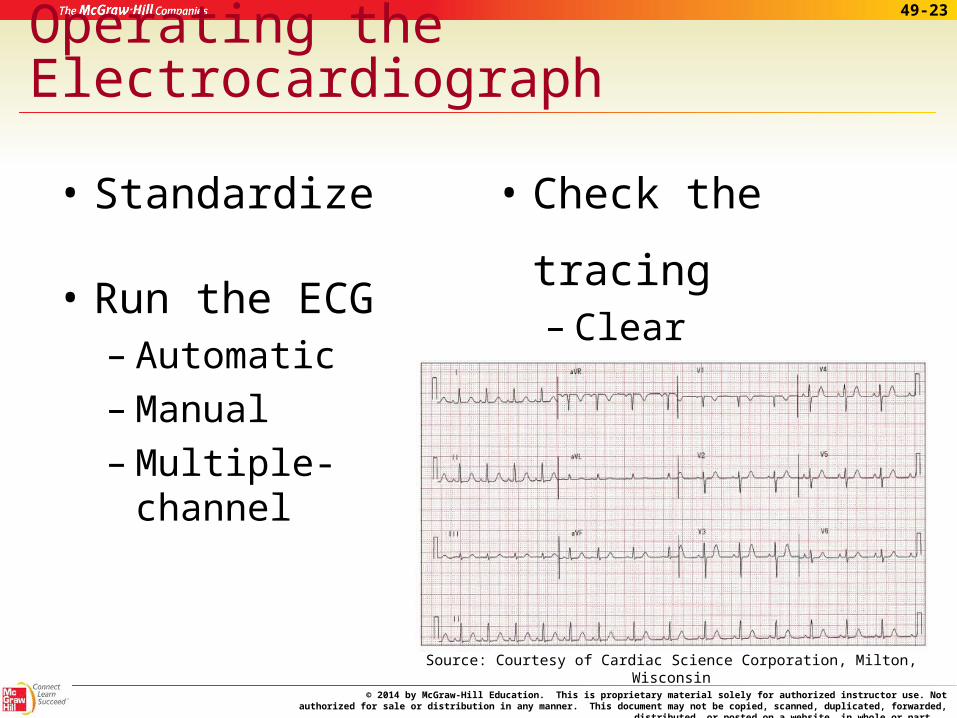
Operating the Electrocardiograph
• Standardize
• Run the ECG– Automatic– Manual– Multiple-channel
• Check the tracing– Clear– Free from artifact
Source: Courtesy of Cardiac Science Corporation, Milton, Wisconsin
© 2014 by McGraw-Hill Education. This is proprietary material solely for authorized instructor use. Not authorized for sale or distribution in any manner. This document may not be copied, scanned, duplicated, forwarded, distributed, or posted on a
website, in whole or part.
49-24
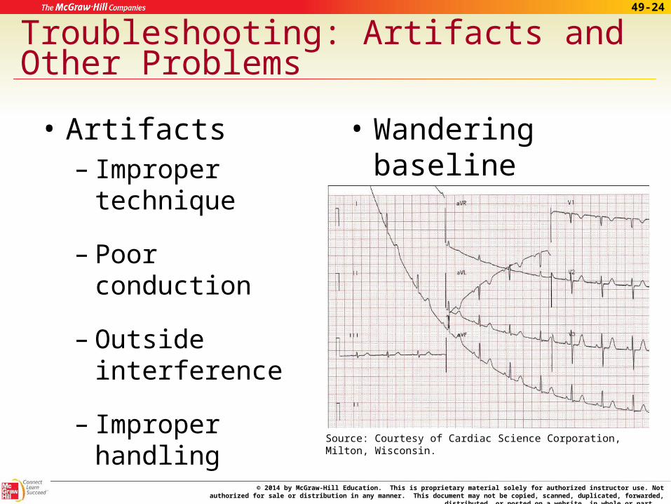
Troubleshooting: Artifacts and Other Problems
• Artifacts – Improper technique
– Poor conduction
– Outside interference
– Improper handling
• Wandering baseline
Source: Courtesy of Cardiac Science Corporation, Milton, Wisconsin.
© 2014 by McGraw-Hill Education. This is proprietary material solely for authorized instructor use. Not authorized for sale or distribution in any manner. This document may not be copied, scanned, duplicated, forwarded, distributed, or posted on a
website, in whole or part.
49-25
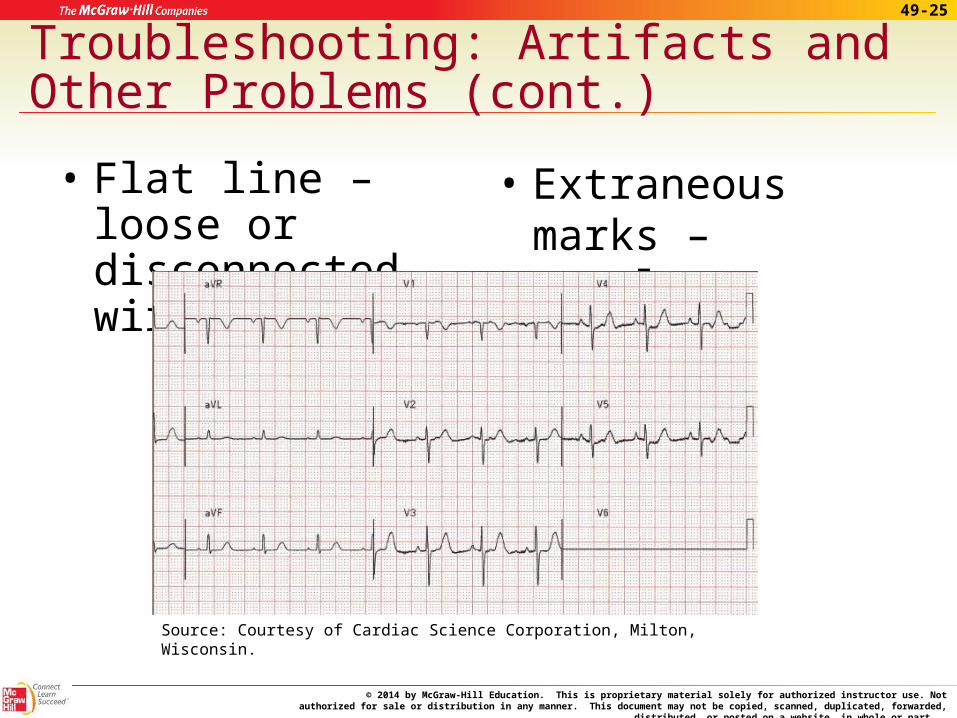
Troubleshooting: Artifacts and Other Problems (cont.)
• Flat line – loose or disconnected wire
• Extraneous marks – careless handling
Source: Courtesy of Cardiac Science Corporation, Milton, Wisconsin.
© 2014 by McGraw-Hill Education. This is proprietary material solely for authorized instructor use. Not authorized for sale or distribution in any manner. This document may not be copied, scanned, duplicated, forwarded, distributed, or posted on a
website, in whole or part.
49-26
Source: Courtesy of Cardiac Science Corporation, Milton, Wisconsin.
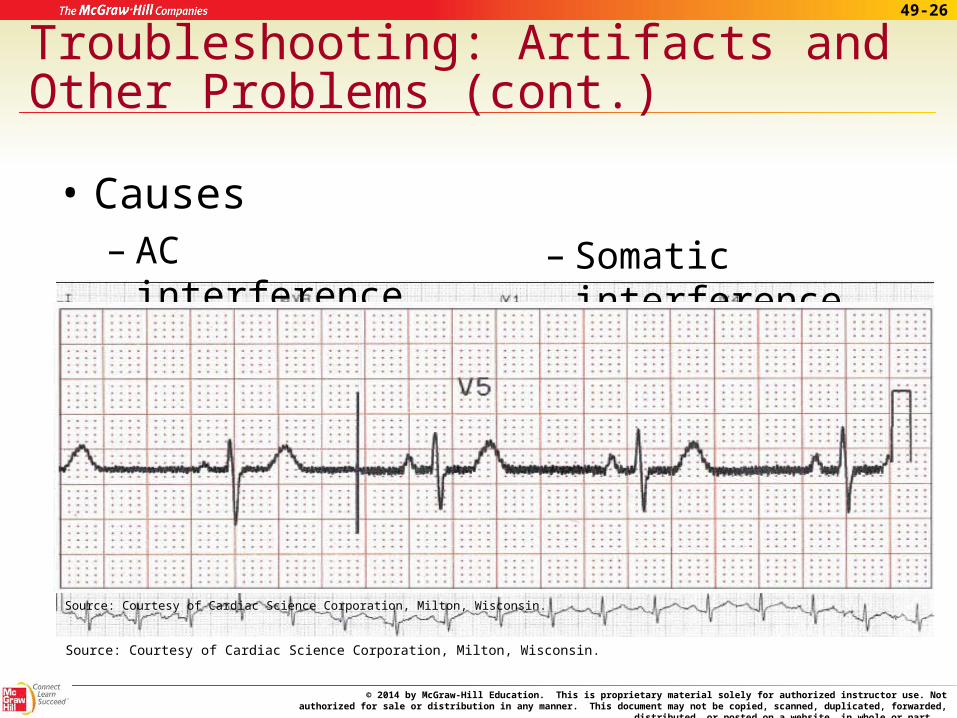
Troubleshooting: Artifacts and Other Problems (cont.)
• Causes – AC interference – Somatic interference
Source: Courtesy of Cardiac Science Corporation, Milton, Wisconsin.
© 2014 by McGraw-Hill Education. This is proprietary material solely for authorized instructor use. Not authorized for sale or distribution in any manner. This document may not be copied, scanned, duplicated, forwarded, distributed, or posted on a
website, in whole or part.
49-27
Troubleshooting: Artifacts and Other Problems (cont.)
• Identifying source – Check tracings for leads I, II, and III
– If unable to identify source, stop and notify supervisor of problem
– Leave patient connected
© 2014 by McGraw-Hill Education. This is proprietary material solely for authorized instructor use. Not authorized for sale or distribution in any manner. This document may not be copied, scanned, duplicated, forwarded, distributed, or posted on a
website, in whole or part.
49-28
Completing the Procedure
• Acceptable tracing– Label tracing properly
– Disconnect wires from electrodes
– Remove electrodes/wipe off electrolyte
– Assist patient as needed
– Prepare room appropriately
• Mount tracing if necessary
© 2014 by McGraw-Hill Education. This is proprietary material solely for authorized instructor use. Not authorized for sale or distribution in any manner. This document may not be copied, scanned, duplicated, forwarded, distributed, or posted on a
website, in whole or part.
49-29
Completing the Procedure (cont.)
• Interpreting the ECG
– Not a medical assistant responsibility
– Be able recognize a problem requiring immediate attention
© 2014 by McGraw-Hill Education. This is proprietary material solely for authorized instructor use. Not authorized for sale or distribution in any manner. This document may not be copied, scanned, duplicated, forwarded, distributed, or posted on a
website, in whole or part.
49-30
Completing the Procedure (cont.)
• Interpreting the ECG – heart rhythm– Regularity of the heartbeat
– Distances between complexes and waves is normally consistent
– Rhythm strip – lead II
To example
© 2014 by McGraw-Hill Education. This is proprietary material solely for authorized instructor use. Not authorized for sale or distribution in any manner. This document may not be copied, scanned, duplicated, forwarded, distributed, or posted on a
website, in whole or part.
49-31
Completing the Procedure (cont.)
• Heart rate – count QRS complexes in a 6-second strip and multiply by 10
• Intervals and Segments – look for variations in length and position
• Wave changes – should be similar appearance in each lead
To example
© 2014 by McGraw-Hill Education. This is proprietary material solely for authorized instructor use. Not authorized for sale or distribution in any manner. This document may not be copied, scanned, duplicated, forwarded, distributed, or posted on a
website, in whole or part.
49-33
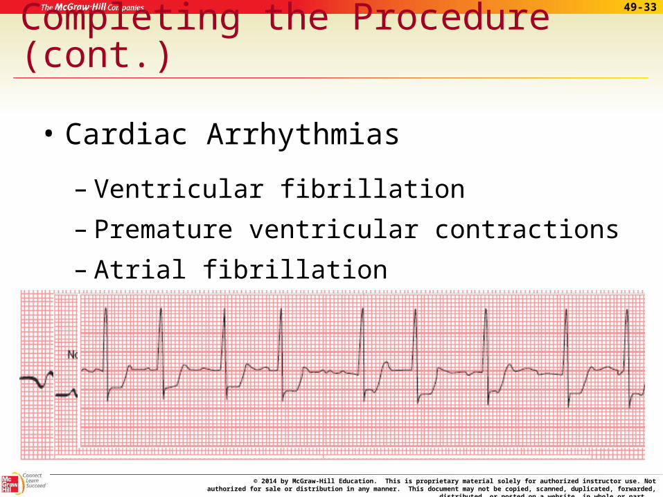
Completing the Procedure (cont.)
• Cardiac Arrhythmias
– Ventricular fibrillation
– Premature ventricular contractions
– Atrial fibrillation
© 2014 by McGraw-Hill Education. This is proprietary material solely for authorized instructor use. Not authorized for sale or distribution in any manner. This document may not be copied, scanned, duplicated, forwarded, distributed, or posted on a
website, in whole or part.
49-34
Apply Your Knowledge
1. Electrodes are placed at how many positions on the body?
ANSWER: Ten: four limb and six chest positions.
2. What should you do just prior to running the ECG to see if the machine needs adjusting? What should you do upon completion of the test?
ANSWER: Standardize the electrocardiograph prior to running the tracing. Upon completion of the ECG, you should check the tracing to be sure is it clear and free from artifact.
© 2014 by McGraw-Hill Education. This is proprietary material solely for authorized instructor use. Not authorized for sale or distribution in any manner. This document may not be copied, scanned, duplicated, forwarded, distributed, or posted on a
website, in whole or part.
49-35
Apply Your Knowledge
3. What should you do just prior to running the ECG to see if the machine needs adjusting? What should you do upon completion of the test?
ANSWER: Standardize the electrocardiograph prior to running the tracing. Upon completion of the ECG, you should check the tracing to be sure is it clear and free from artifact.
4. What are four general causes of artifacts?
ANSWER: They are improper technique, poor conduction, outside interference, and improper handling of the tracing.
© 2014 by McGraw-Hill Education. This is proprietary material solely for authorized instructor use. Not authorized for sale or distribution in any manner. This document may not be copied, scanned, duplicated, forwarded, distributed, or posted on a
website, in whole or part.
49-36
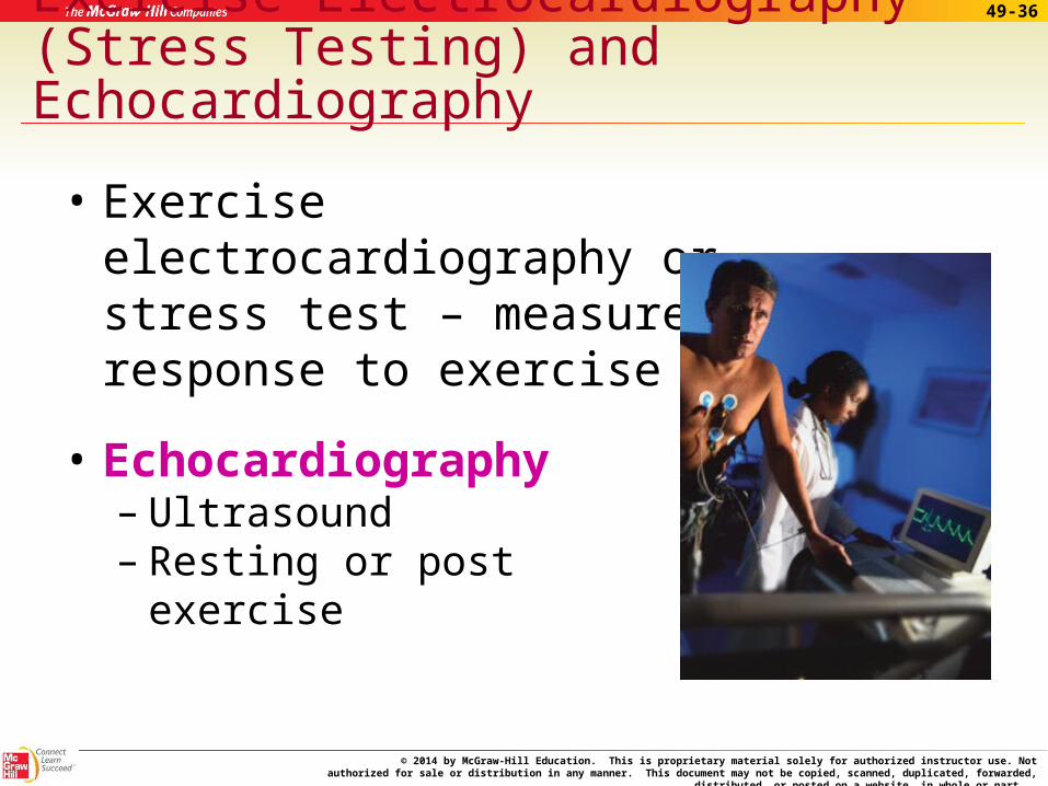
Exercise Electrocardiography (Stress Testing) and Echocardiography
• Exercise electrocardiography or stress test – measures response to exercise
• Echocardiography – Ultrasound – Resting or post exercise
© 2014 by McGraw-Hill Education. This is proprietary material solely for authorized instructor use. Not authorized for sale or distribution in any manner. This document may not be copied, scanned, duplicated, forwarded, distributed, or posted on a
website, in whole or part.
49-37
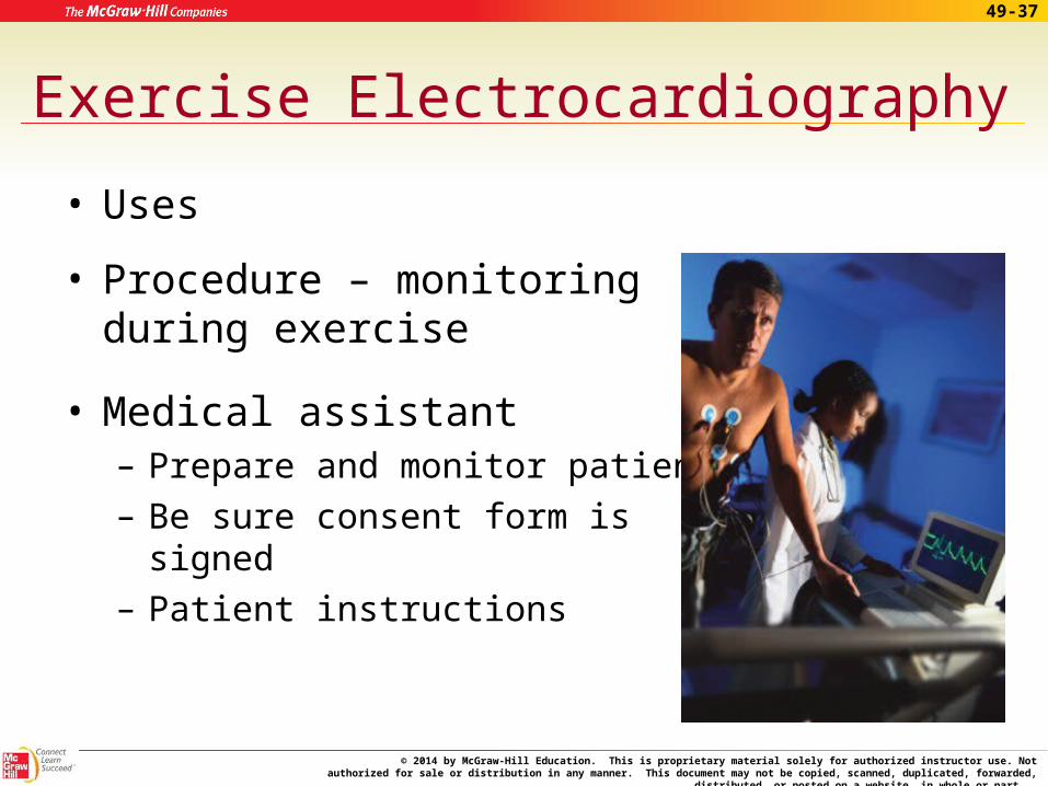
Exercise Electrocardiography
• Uses
• Procedure – monitoring during exercise
• Medical assistant– Prepare and monitor patient– Be sure consent form is signed– Patient instructions
© 2014 by McGraw-Hill Education. This is proprietary material solely for authorized instructor use. Not authorized for sale or distribution in any manner. This document may not be copied, scanned, duplicated, forwarded, distributed, or posted on a
website, in whole or part.
49-38
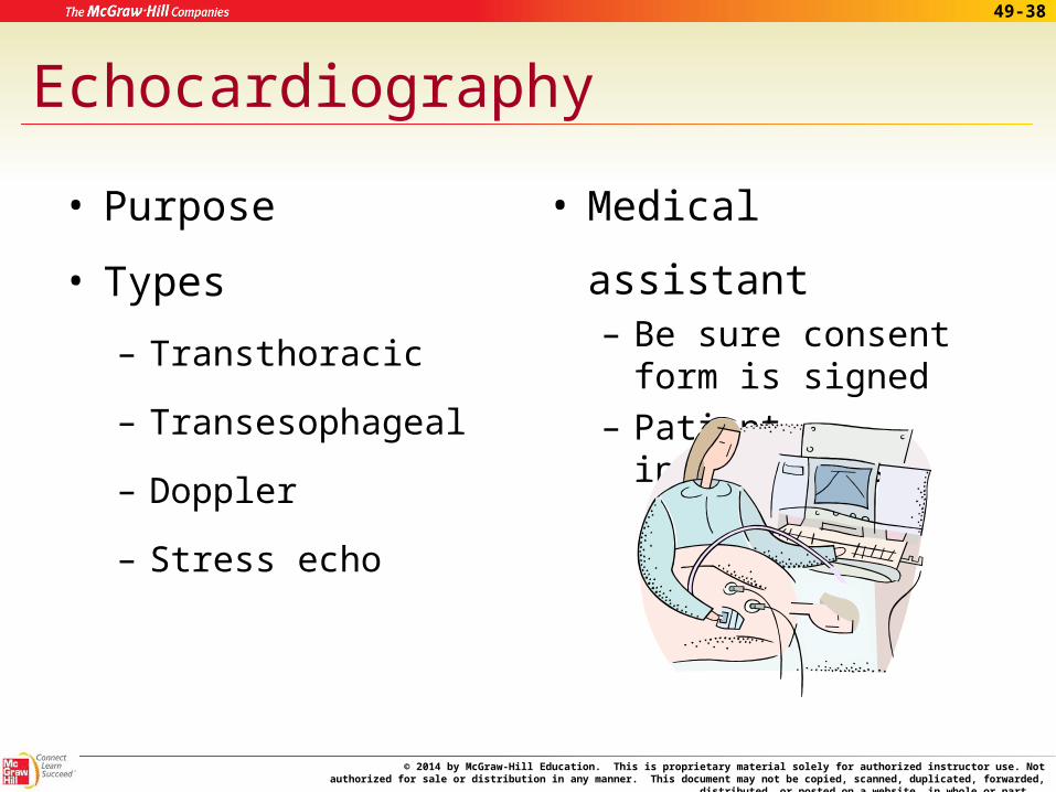
Echocardiography
• Purpose
• Types
– Transthoracic
– Transesophageal
– Doppler
– Stress echo
• Medical assistant– Be sure consent form
is signed– Patient instructions
© 2014 by McGraw-Hill Education. This is proprietary material solely for authorized instructor use. Not authorized for sale or distribution in any manner. This document may not be copied, scanned, duplicated, forwarded, distributed, or posted on a
website, in whole or part.
49-39
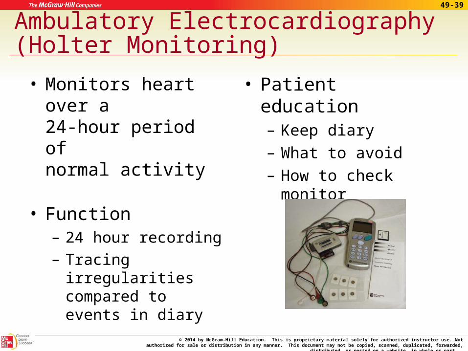
Ambulatory Electrocardiography (Holter Monitoring)
• Monitors heart over a 24-hour period of normal activity
• Function – 24 hour recording– Tracing irregularities
compared to events in diary
• Patient education– Keep diary– What to avoid– How to check monitor
© 2014 by McGraw-Hill Education. This is proprietary material solely for authorized instructor use. Not authorized for sale or distribution in any manner. This document may not be copied, scanned, duplicated, forwarded, distributed, or posted on a
website, in whole or part.
49-40
Apply Your Knowledge
What is the purpose for stress testing, echocardiogram, and Holter monitor testing?
ANSWER: Stress testing is used to measure the heart’s response to a constant or increasing workload.
An echocardiogram shows the working heart valves and chambers and how well the blood moves through the heart.
A Holter monitor is used to obtain a tracing over a period of time when a resting ECG or stress test shows no abnormalities.
All are used for diagnosing cardiac conditions or for monitoring current treatments and medications.
Correct!
© 2014 by McGraw-Hill Education. This is proprietary material solely for authorized instructor use. Not authorized for sale or distribution in any manner. This document may not be copied, scanned, duplicated, forwarded, distributed, or posted on a
website, in whole or part.
49-41
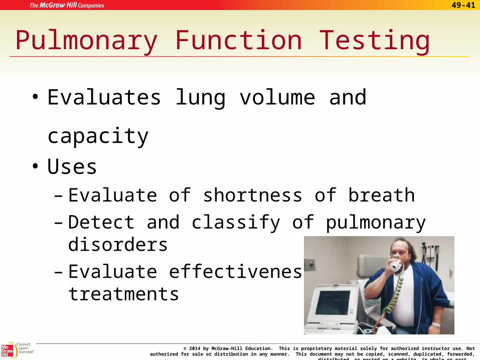
Pulmonary Function Testing
• Evaluates lung volume and capacity
• Uses– Evaluate of shortness of breath – Detect and classify of pulmonary disorders– Evaluate effectiveness of treatments
© 2014 by McGraw-Hill Education. This is proprietary material solely for authorized instructor use. Not authorized for sale or distribution in any manner. This document may not be copied, scanned, duplicated, forwarded, distributed, or posted on a
website, in whole or part.
49-42
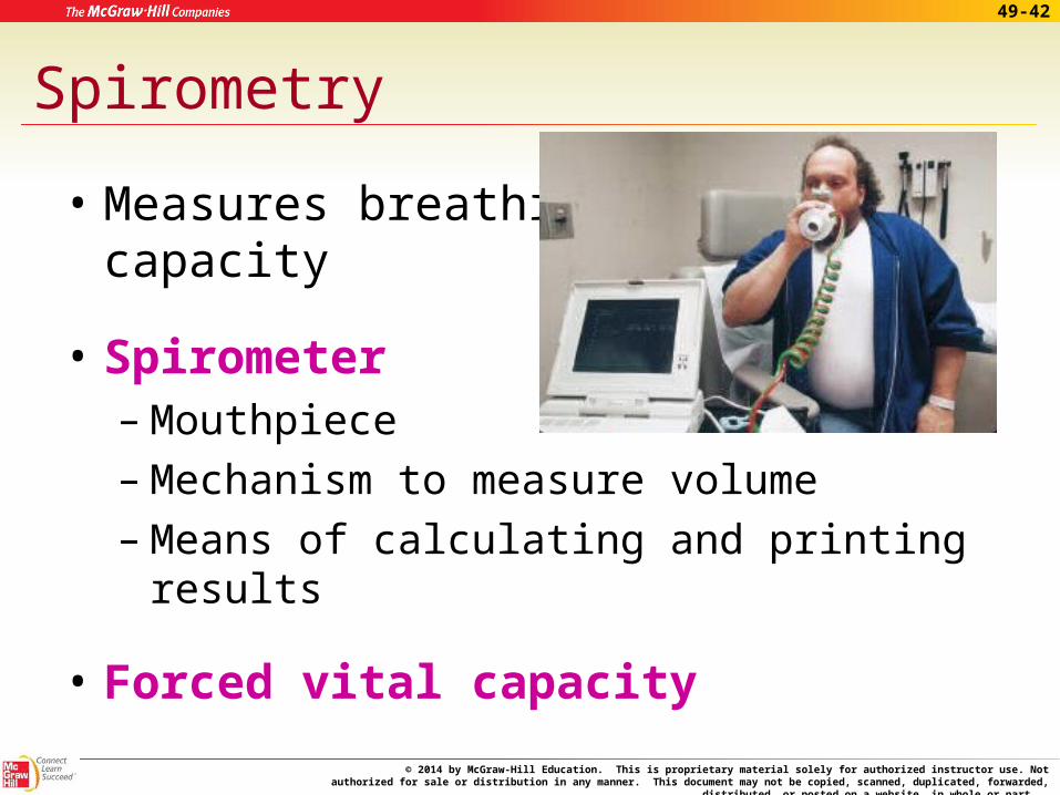
Spirometry
• Measures breathing capacity
• Spirometer – Mouthpiece – Mechanism to measure volume– Means of calculating and printing results
• Forced vital capacity
© 2014 by McGraw-Hill Education. This is proprietary material solely for authorized instructor use. Not authorized for sale or distribution in any manner. This document may not be copied, scanned, duplicated, forwarded, distributed, or posted on a
website, in whole or part.
49-43
Performing Spirometry
• Patient preparation– Inform the patient about conditions and
activities that could affect the test accuracy
– Explain procedure and its purpose
– Position the patient properly
– Explain and demonstrate correct procedure
© 2014 by McGraw-Hill Education. This is proprietary material solely for authorized instructor use. Not authorized for sale or distribution in any manner. This document may not be copied, scanned, duplicated, forwarded, distributed, or posted on a
website, in whole or part.
49-44
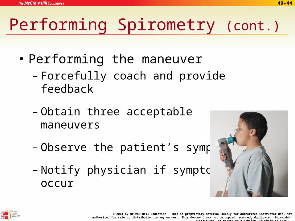
Performing Spirometry (cont.)
• Performing the maneuver– Forcefully coach and provide feedback
– Obtain three acceptable maneuvers
– Observe the patient’s symptoms
– Notify physician if symptoms occur
© 2014 by McGraw-Hill Education. This is proprietary material solely for authorized instructor use. Not authorized for sale or distribution in any manner. This document may not be copied, scanned, duplicated, forwarded, distributed, or posted on a
website, in whole or part.
49-45
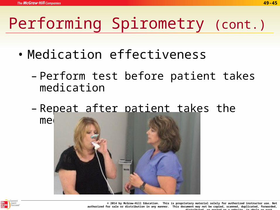
Performing Spirometry (cont.)
• Medication effectiveness
– Perform test before patient takes medication
– Repeat after patient takes the medication
© 2014 by McGraw-Hill Education. This is proprietary material solely for authorized instructor use. Not authorized for sale or distribution in any manner. This document may not be copied, scanned, duplicated, forwarded, distributed, or posted on a
website, in whole or part.
49-46
Performing Spirometry (cont.)
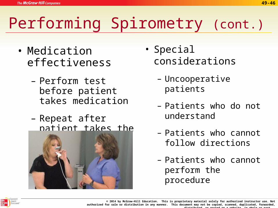
• Medication effectiveness
– Perform test before patient takes medication
– Repeat after patient takes the medication
• Special considerations
– Uncooperative patients
– Patients who do not understand
– Patients who cannot follow directions
– Patients who cannot perform the procedure
© 2014 by McGraw-Hill Education. This is proprietary material solely for authorized instructor use. Not authorized for sale or distribution in any manner. This document may not be copied, scanned, duplicated, forwarded, distributed, or posted on a
website, in whole or part.
49-47
Performing Spirometry (cont.)
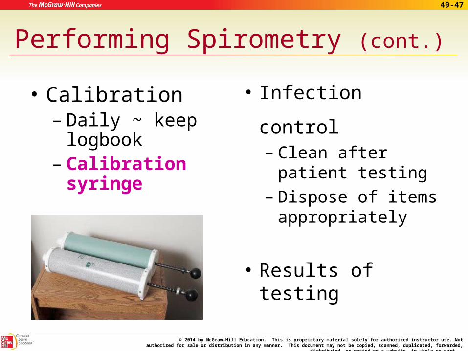
• Calibration – Daily ~ keep
logbook– Calibration
syringe
• Infection control– Clean after patient
testing– Dispose of items
appropriately
• Results of testing
© 2014 by McGraw-Hill Education. This is proprietary material solely for authorized instructor use. Not authorized for sale or distribution in any manner. This document may not be copied, scanned, duplicated, forwarded, distributed, or posted on a
website, in whole or part.
49-48
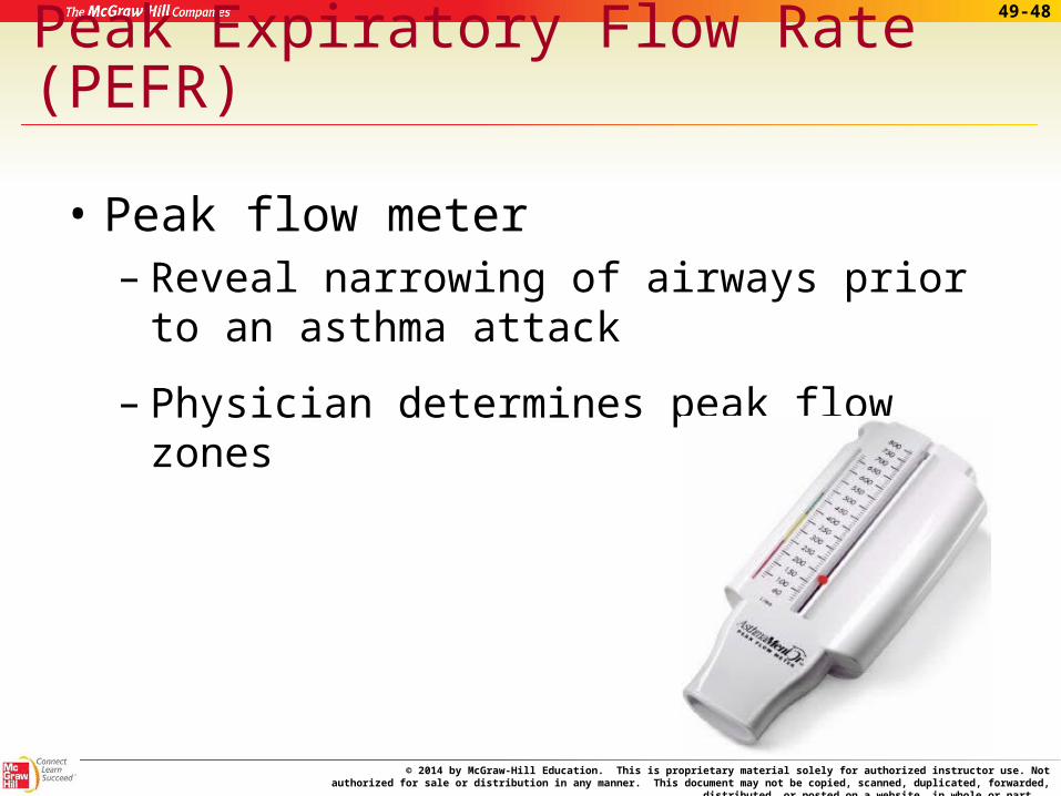
Peak Expiratory Flow Rate (PEFR)
• Peak flow meter – Reveal narrowing of airways prior to an
asthma attack
– Physician determines peak flow zones
© 2014 by McGraw-Hill Education. This is proprietary material solely for authorized instructor use. Not authorized for sale or distribution in any manner. This document may not be copied, scanned, duplicated, forwarded, distributed, or posted on a
website, in whole or part.
49-49
Peak Expiratory Flow Rate (PEFR)
© 2014 by McGraw-Hill Education. This is proprietary material solely for authorized instructor use. Not authorized for sale or distribution in any manner. This document may not be copied, scanned, duplicated, forwarded, distributed, or posted on a
website, in whole or part.
49-50
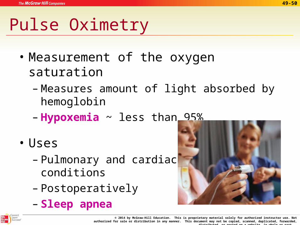
Pulse Oximetry
• Measurement of the oxygen saturation – Measures amount of light absorbed by
hemoglobin– Hypoxemia ~ less than 95%
• Uses – Pulmonary and cardiac
conditions– Postoperatively– Sleep apnea

© 2014 by McGraw-Hill Education. This is proprietary material solely for authorized instructor use. Not authorized for sale or distribution in any manner. This document may not be copied, scanned, duplicated, forwarded, distributed, or posted on a
website, in whole or part.
49-51
Apply Your Knowledge
1. What is the purpose of PFTs?
ANSWER: To evaluate lung volume and capacity.
2. What does successful spirometry testing depend on?
ANSWER: On proper patient preparation and consistent technique in preforming the procedure and analyzing the results.

© 2014 by McGraw-Hill Education. This is proprietary material solely for authorized instructor use. Not authorized for sale or distribution in any manner. This document may not be copied, scanned, duplicated, forwarded, distributed, or posted on a
website, in whole or part.
49-52
Apply Your Knowledge
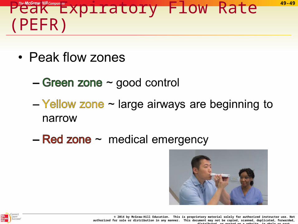
3. Joey Jackson called to ask about taking his asthma medicine. He said he has been using his peak flow meter and the readings have been in his yellow zone. What do you tell him?
ANSWER: This means that his large airways are beginning to narrow and that he should take his medication as prescribed.
4. Joey decided to come to the office and you check his oxygen saturation with the pulse oximeter. The reading was 93%. What does this mean and what should you do?
ANSWER: Joey is hypoxemic. You need to notify the physician and document findings.
Very good!
© 2014 by McGraw-Hill Education. This is proprietary material solely for authorized instructor use. Not authorized for sale or distribution in any manner. This document may not be copied, scanned, duplicated, forwarded, distributed, or posted on a
website, in whole or part.
49-53
In Summary
49.1 As a medical assistant, you will be responsible for preparing the patient for ECG and pulmonary function tests, maintaining the equipment used for these tests, and performing them.
© 2014 by McGraw-Hill Education. This is proprietary material solely for authorized instructor use. Not authorized for sale or distribution in any manner. This document may not be copied, scanned, duplicated, forwarded, distributed, or posted on a
website, in whole or part.
49-54
In Summary (cont.)
49.2 The heart’s conduction system is responsible for the electrical pathway that occurs during a heartbeat.
The pathway begins with the SA node, travels through the AV node, Bundle of HIS, Right and Left bundle branches, and ends with the Purkinje fibers.
This electrical energy pathway is measured with an electrocardiograph and a tracing of the impulses is produced. The electrical impulses are represented in wave forms or deflections. Each deflection is
labeled by letters PQRSTU and represents a part of the pattern.
© 2014 by McGraw-Hill Education. This is proprietary material solely for authorized instructor use. Not authorized for sale or distribution in any manner. This document may not be copied, scanned, duplicated, forwarded, distributed, or posted on a
website, in whole or part.
49-55
In Summary (cont.)
49.3 The electrocardiograph consists of the following components: electrodes, amplifier, stylus, leads, and ECG paper.
49.4 The steps in obtaining an accurate ECG include preparing the room and equipment, identifying the patient, properly placing the limb and chest
electrodes, attaching the lead wires, entering the patient data into the ECG machine, running the tracing, checking the tracing for artifacts, disconnecting the patient from the lead wires and removing the electrodes, and assisting the patient as required.
© 2014 by McGraw-Hill Education. This is proprietary material solely for authorized instructor use. Not authorized for sale or distribution in any manner. This document may not be copied, scanned, duplicated, forwarded, distributed, or posted on a
website, in whole or part.
49-56
In Summary (cont.)
49.5 Exercise electrocardiography is referred to as stress testing. This measures the efficiency of the heart during constant or increasing workload.
Echocardiography uses ultrasound to create a picture of the moving heart. This can be done while the patient is resting or after exercise.
49.6 A Holter monitor is used to measure the heart’s activity over a 24-hour period. This is used when the patient has intermittent chest pain or discomfort and a normal ECG and stress test.
© 2014 by McGraw-Hill Education. This is proprietary material solely for authorized instructor use. Not authorized for sale or distribution in any manner. This document may not be copied, scanned, duplicated, forwarded, distributed, or posted on a
website, in whole or part.
49-57
In Summary (cont.)
49.7 Forced vital capacity is the measurement of the greatest volume of air expelled when a patient
performs a rapid, forced expiration.
Accurate spirometry testing includes proper patient positioning, coaching the patient during the
procedure, obtaining three acceptable maneuvers, and recording the results in the patient’s chart.
A peak expiratory flow rate is obtained by having the patient sit or stand using good posture, take in as deep a breath as possible, and blow out through the peak flow meter as fast and as hard as possible three times.
© 2014 by McGraw-Hill Education. This is proprietary material solely for authorized instructor use. Not authorized for sale or distribution in any manner. This document may not be copied, scanned, duplicated, forwarded, distributed, or posted on a
website, in whole or part.
49-58
In Summary (cont.)
49.8 Pulse oximetry testing is performed by applying the pulse oximeter to the patient’s finger or toe, attaching the sensor cable to the oximeter, turning the oximeter on, setting the alarm limits for high and low oxygen saturations, and reading the patient’s oxygen
saturation levels. The oxygen saturation levels should be recorded in the patient’s chart.
© 2014 by McGraw-Hill Education. This is proprietary material solely for authorized instructor use. Not authorized for sale or distribution in any manner. This document may not be copied, scanned, duplicated, forwarded, distributed, or posted on a
website, in whole or part.
49-59
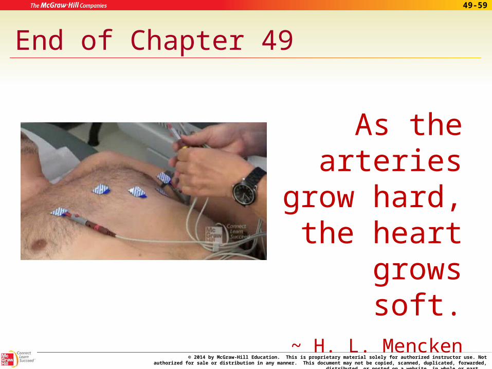
End of Chapter 49
As the arteries
grow hard, the heart
grows soft.~ H. L. Mencken
© 2014 by McGraw-Hill Education. This is proprietary material solely for authorized instructor use. Not authorized for sale or distribution in any manner. This document may not be copied, scanned, duplicated, forwarded, distributed, or posted on a
website, in whole or part.
49-6052-60
Apply Your Knowledge
1. What should you after running an ECG?
ANSWER: After making sure the tracing is acceptable, you should label it properly, disconnect wires from electrodes, remove electrodes and wipe off electrolyte, assist patient up, and prepare the room appropriately for the next patient.