Embed Size (px)
Citation preview
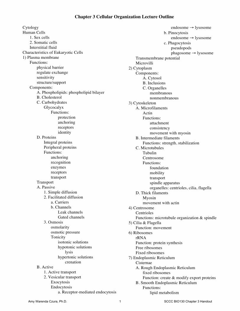
Chapter 3 Cellular Organization Lecture Outline
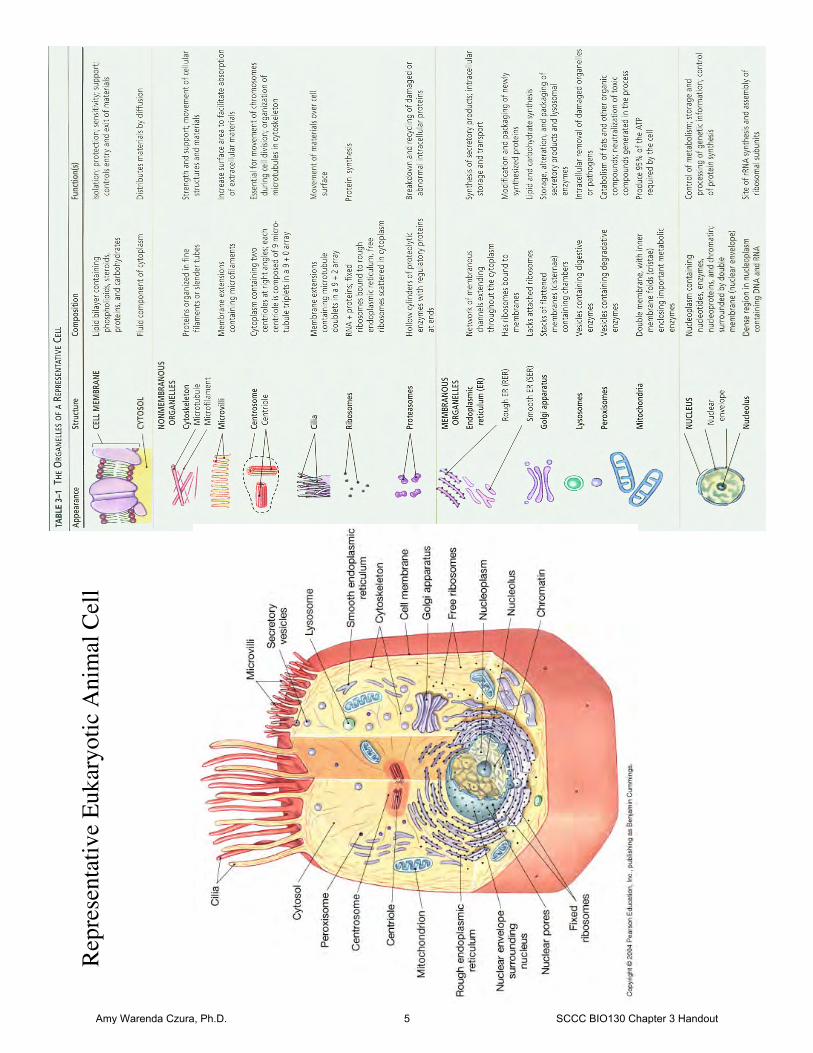
Cytology Human Cells 1. Sex cells 2. Somatic cells Interstitial fluid Characteristics of Eukaryotic Cells 1) Plasma membrane Functions: physical barrier regulate exchange sensitivity structure/support Components: A. Phospholipids: phospholipid bilayer B. Cholesterol C. Carbohydrates Glycocalyx Functions: protection anchoring receptors identity D. Proteins Integral proteins Peripheral proteins Functions: anchoring recognition enzymes receptors transport Transport A. Passive 1. Simple diffusion 2. Facilitated diffusion a. Carriers b. Channels Leak channels Gated channels 3. Osmosis osmolarity osmotic pressure Tonicity isotonic solutions hypotonic solutions lysis hypertonic solutions crenation B. Active 1. Active transport 2. Vesicular transport Exocytosis Endocytosis a. Receptor-mediated endocytosis
endosome → lysosome b. Pinocytosis endosome → lysosome c. Phagocytosis pseudopods phagosome → lysosome Transmembrane potential Microvilli 2) Cytoplasm Components: A. Cytosol B. Inclusions C. Organelles membranous nonmembranous 3) Cytoskeleton A. Microfilaments Actin Functions: attachment consistency movement with myosin B. Intermediate filaments Functions: strength, stabilization C. Microtubules Tubulin Centrosome Functions: foundation mobility transport spindle apparatus organelles: centrioles, cilia, flagella D. Thick filaments Myosin movement with actin 4) Centrosome Centrioles Functions: microtubule organization & spindle 5) Cilia & Flagella Function: movement 6) Ribosomes rRNA Function: protein synthesis Free ribosomes Fixed ribosomes 7) Endoplasmic Reticulum Cisternae A. Rough Endoplasmic Reticulum fixed ribosomes Function: create & modify export proteins B. Smooth Endoplasmic Reticulum Functions: lipid metabolism
Amy Warenda Czura, Ph.D. 1 SCCC BIO130 Chapter 3 Handout
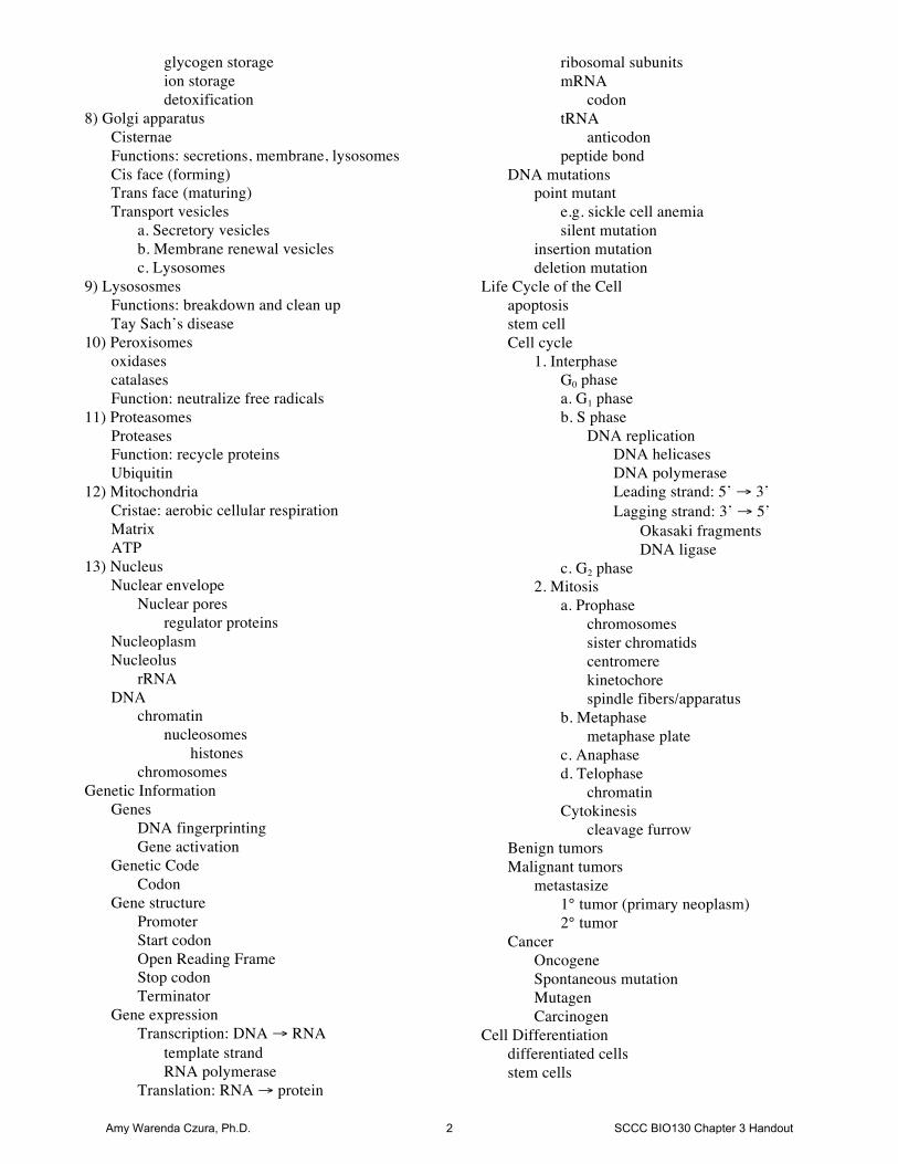
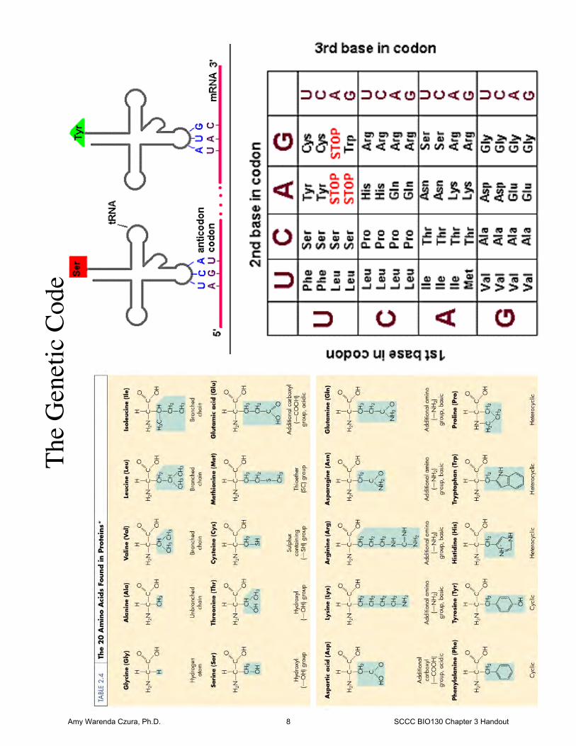
glycogen storage ion storage detoxification 8) Golgi apparatus Cisternae Functions: secretions, membrane, lysosomes Cis face (forming) Trans face (maturing) Transport vesicles a. Secretory vesicles b. Membrane renewal vesicles c. Lysosomes 9) Lysososmes Functions: breakdown and clean up Tay Sach’s disease 10) Peroxisomes oxidases catalases Function: neutralize free radicals 11) Proteasomes Proteases Function: recycle proteins Ubiquitin 12) Mitochondria Cristae: aerobic cellular respiration Matrix ATP 13) Nucleus Nuclear envelope Nuclear pores regulator proteins Nucleoplasm Nucleolus rRNA DNA chromatin nucleosomes histones chromosomes Genetic Information Genes DNA fingerprinting Gene activation Genetic Code Codon Gene structure Promoter Start codon Open Reading Frame Stop codon Terminator Gene expression Transcription: DNA → RNA template strand RNA polymerase Translation: RNA → protein
ribosomal subunits mRNA codon tRNA anticodon peptide bond DNA mutations point mutant e.g. sickle cell anemia silent mutation insertion mutation deletion mutation Life Cycle of the Cell apoptosis stem cell Cell cycle 1. Interphase G0 phase a. G1 phase b. S phase DNA replication DNA helicases DNA polymerase Leading strand: 5’ → 3’ Lagging strand: 3’ → 5’ Okasaki fragments DNA ligase c. G2 phase 2. Mitosis a. Prophase chromosomes sister chromatids centromere kinetochore spindle fibers/apparatus b. Metaphase metaphase plate c. Anaphase d. Telophase chromatin Cytokinesis cleavage furrow Benign tumors Malignant tumors metastasize 1° tumor (primary neoplasm) 2° tumor Cancer Oncogene Spontaneous mutation Mutagen Carcinogen Cell Differentiation differentiated cells stem cells
Amy Warenda Czura, Ph.D. 2 SCCC BIO130 Chapter 3 Handout
Transport across the plasma membrane
A selectively permeable barrier
Passive transport processes: require no energy from the cell, rely on diffusion principles Active transport processes: require energy for the cell to move substance against the concentration gradient Three general mechanisms:
1. Diffusion (passive) 2. Carrier-Mediated transport (active or passive) 3. Vesicular transport (active)
Passive processes: 1. Simple diffusion: net movement of molecules from an area of high concentration to an area of low
concentration across a diffusion gradient toward equilibrium
Rate depends on: distance molecule size temperature gradient size electrical forces
Transport mechanism of nonpolar and lipid soluble substances
2. Facilitated diffusion: diffusion through a transporter protein
A. Carriers: proteins that bind a particular large or polar molecule B. Channels: proteins that act as selective pores for water or ions
1. Leak channels: always open 2. Gated channels: regulated
Rate of diffusion depends on the number of carrier molecules for the specific substance
3. Osmosis: diffusion of water through a semi-permeable membrane Osmotic pressure = the pressure that must be applied to prevent further osmosis across the
membrane
Isotonic solutions: contain solute concentrations equal to that of the cell, no net movement of water into or out of the cell, no osmotic pressure.
Hypotonic solutions: contain less solutes than the cell, net movement of water into the cell. Hypertonic solutions: contain more solutes than the cell, net movement of water out of the cell.
Amy Warenda Czura, Ph.D. 3 SCCC BIO130 Chapter 3 Handout
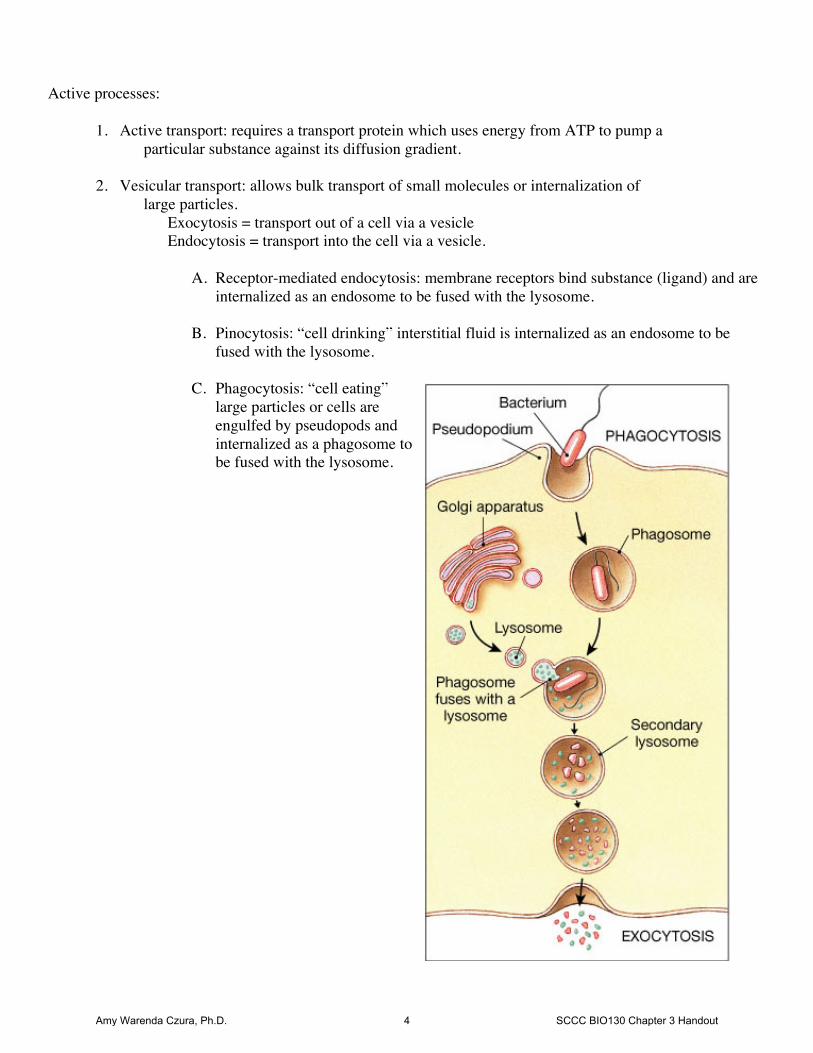
Active processes:
1. Active transport: requires a transport protein which uses energy from ATP to pump a particular substance against its diffusion gradient.
2. Vesicular transport: allows bulk transport of small molecules or internalization of large particles.
Exocytosis = transport out of a cell via a vesicle Endocytosis = transport into the cell via a vesicle.
A. Receptor-mediated endocytosis: membrane receptors bind substance (ligand) and are internalized as an endosome to be fused with the lysosome.
B. Pinocytosis: “cell drinking” interstitial fluid is internalized as an endosome to be
fused with the lysosome. C. Phagocytosis: “cell eating”
large particles or cells are engulfed by pseudopods and internalized as a phagosome to be fused with the lysosome.
Amy Warenda Czura, Ph.D. 4 SCCC BIO130 Chapter 3 Handout
Repr
esen
tativ
e Eu
kary
otic
Ani
mal
Cel
l
Amy Warenda Czura, Ph.D. 5 SCCC BIO130 Chapter 3 Handout
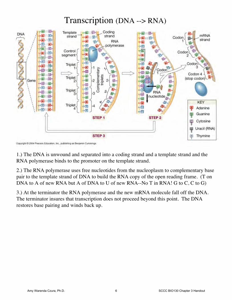
Transcription (DNA --> RNA)
1.) The DNA is unwound and separated into a coding strand and a template strand and theRNA polymerase binds to the promoter on the template strand.
2.) The RNA polymerase uses free nucleotides from the nucleoplasm to complementary basepair to the template strand of DNA to build the RNA copy of the open reading frame. (T onDNA to A of new RNA but A of DNA to U of new RNA--No T in RNA! G to C, C to G)
3.) At the terminator the RNA polymerase and the new mRNA molecule fall off the DNA.The terminator insures that transcription does not proceed beyond this point. The DNArestores base pairing and winds back up.
Amy Warenda Czura, Ph.D. 6 SCCC BIO130 Chapter 3 Handout
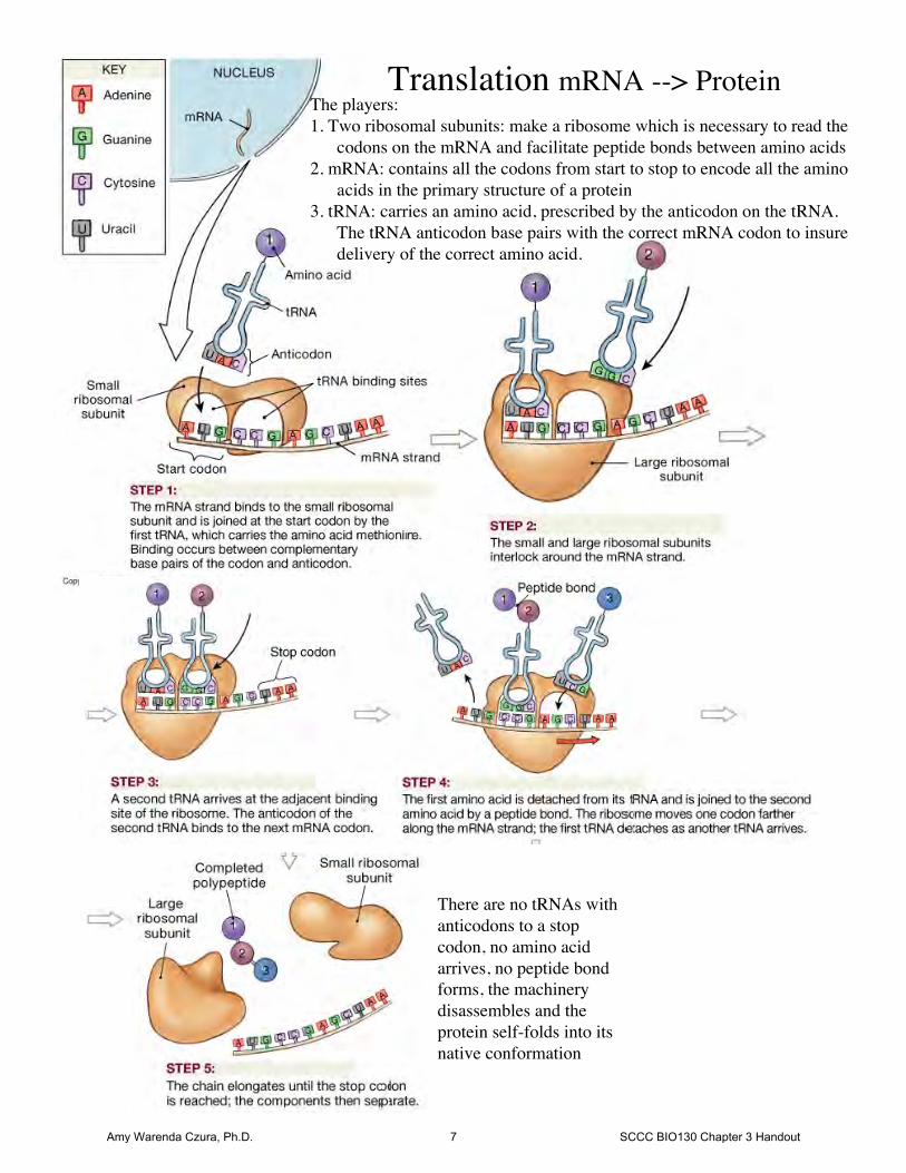
Translation mRNA --> ProteinThe players:1. Two ribosomal subunits: make a ribosome which is necessary to read the
codons on the mRNA and facilitate peptide bonds between amino acids2. mRNA: contains all the codons from start to stop to encode all the amino
acids in the primary structure of a protein3. tRNA: carries an amino acid, prescribed by the anticodon on the tRNA.
The tRNA anticodon base pairs with the correct mRNA codon to insuredelivery of the correct amino acid.
There are no tRNAs withanticodons to a stopcodon, no amino acidarrives, no peptide bondforms, the machinerydisassembles and theprotein self-folds into itsnative conformation
Amy Warenda Czura, Ph.D. 7 SCCC BIO130 Chapter 3 Handout
The
Gen
etic
Cod
e
Amy Warenda Czura, Ph.D. 8 SCCC BIO130 Chapter 3 Handout
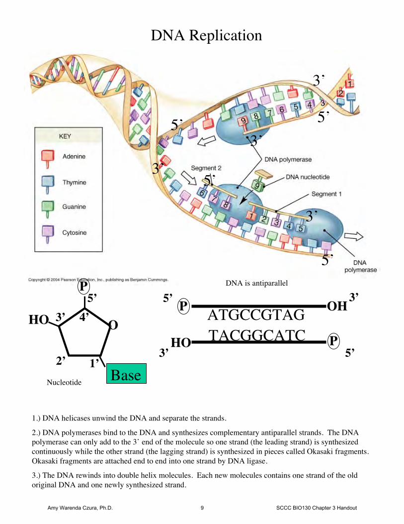
DNA Replication
5’
3’
3’5’
3’
5’
5’
3’
OOHOHO
PP
Base1’2’
3’ 4’5’
DNA is antiparallel
ATGCCGTAGTACGGCATC
PP
PPHOHO
OHOH
Nucleotide
5’
3’
3’
1.) DNA helicases unwind the DNA and separate the strands.
2.) DNA polymerases bind to the DNA and synthesizes complementary antiparallel strands. The DNApolymerase can only add to the 3’ end of the molecule so one strand (the leading strand) is synthesizedcontinuously while the other strand (the lagging strand) is synthesized in pieces called Okasaki fragments.Okasaki fragments are attached end to end into one strand by DNA ligase.
3.) The DNA rewinds into double helix molecules. Each new molecules contains one strand of the oldoriginal DNA and one newly synthesized strand.
5’
Amy Warenda Czura, Ph.D. 9 SCCC BIO130 Chapter 3 Handout
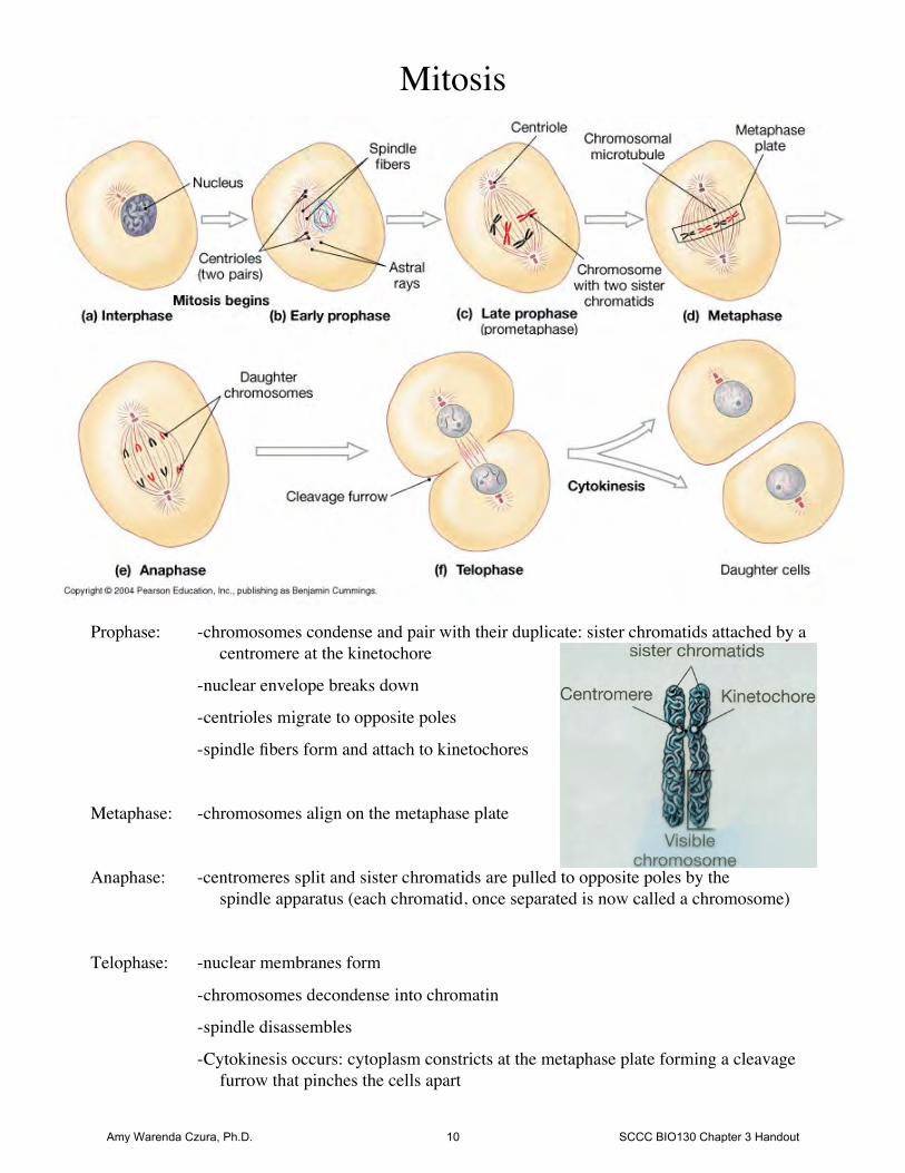
Mitosis
Prophase: -chromosomes condense and pair with their duplicate: sister chromatids attached by a centromere at the kinetochore
-nuclear envelope breaks down
-centrioles migrate to opposite poles
-spindle fibers form and attach to kinetochores
Metaphase: -chromosomes align on the metaphase plate
Anaphase: -centromeres split and sister chromatids are pulled to opposite poles by the spindle apparatus (each chromatid, once separated is now called a chromosome)
Telophase: -nuclear membranes form
-chromosomes decondense into chromatin
-spindle disassembles
-Cytokinesis occurs: cytoplasm constricts at the metaphase plate forming a cleavage furrow that pinches the cells apart
Amy Warenda Czura, Ph.D. 10 SCCC BIO130 Chapter 3 Handout