Embed Size (px)
DESCRIPTION
Chapter 47: Circulatory and Respiratory Systems. 47-1 The Circulatory System. 47-2 Blood. 47-3 The Respiratory System. 47-1 The Circulatory System. I. The Heart (THORACIC cavity, behind STERNUM). Moves blood PULMONARY and SYSTEMIC loops. - PowerPoint PPT Presentation
Citation preview

Chapter 47: Circulatory and Respiratory Systems
47-1 The Circulatory System
47-2 Blood
47-3 The Respiratory System


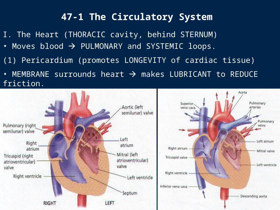
I. The Heart (THORACIC cavity, behind STERNUM)• Moves blood PULMONARY and SYSTEMIC loops.
47-1 The Circulatory System
(1) Pericardium (promotes LONGEVITY of cardiac tissue)
• MEMBRANE surrounds heart makes LUBRICANT to REDUCE friction.


(1) Some babies are born with a HOLE in the septum between the two atria. Based on what you know about BLOOD FLOW through the heart, explain why this condition would be HARMFUL to the baby.
Critical Thinking


(2) Septum (wall of tissue, PREVENTS mixing of blood)• SEPARATES heart into 2 sides (4 chambers).

(3) Atrium (Atria, RECEIVE blood returning from the lungs or body)• Smaller, RECEIVING chambers (NOTE: RA contains SA node).

(4) Ventricle (PUMP blood to the lungs or the body’s circuits)• Thicker, more muscular PUMPING chambers.

(5) Atrioventricular (AV) Valves [Tricuspid (R) and Mitral Valves (L)]• ONE-way flaps of tissue separating EACH atrium from ITS ventricle.

(6) Semilunar (SL) Valve [Pulmonary (R) and Aortic Valves (L)]• Prevent blood from flowing BACK into ventricles when ventricle relaxes.

(2) One function of the cardiovascular system is to help maintain a uniform BODY TEMPERATURE. Explain HOW the constant circulation of blood throughout the body MAY accomplish this task.
Critical Thinking

(B) Circulation in the Heart • SVC/IVC RA RV PA LUNGS PV LA LV AA/DA BODY(1) Aorta (THICKEST artery)• Receives O-blood from LV, and pumps out to SYSTEMIC loops.

(C) Control of the Heartbeat• A wave of ELECTRICAL impulses spreads so ATRIA and VENTRICLES contract in a RHYTHM.
(1) Sinoatrial Node (SA) [“Pacemaker”, located in RIGHT ATRIUM]• Specialized cardiac cells initiate their OWN electrical impulse and contract [i.e., regulating the RATE of the heartbeat].

(2) Atrioventricular Node (AV) [Located in the SEPTUM between ATRIA]• Relays SA electrical impulse to VENTRICLES, causing CONTRACTION a fraction of a second AFTER atria (1 FULL heartbeat).


(3) Systole (Heartbeat Phase 1 of 2; Blood LEAVES ventricles TO arteries)• Ventricles CONTRACT, CLOSING AV valves and OPENING SL valves which EXIT heart.

(4) Diastole (Heartbeat Phase 2 of 2; Blood ENTERS atria TO ventricles)• Ventricle RELAX, allowing back pressure of blood to CLOSE SL valves and OPEN AV valves.

(D) Blood Vessels (e.g., arteries, veins, capillaries)• Blood is contained WITHIN either heart OR blood vessels at ALL times (CLOSED LOOP)
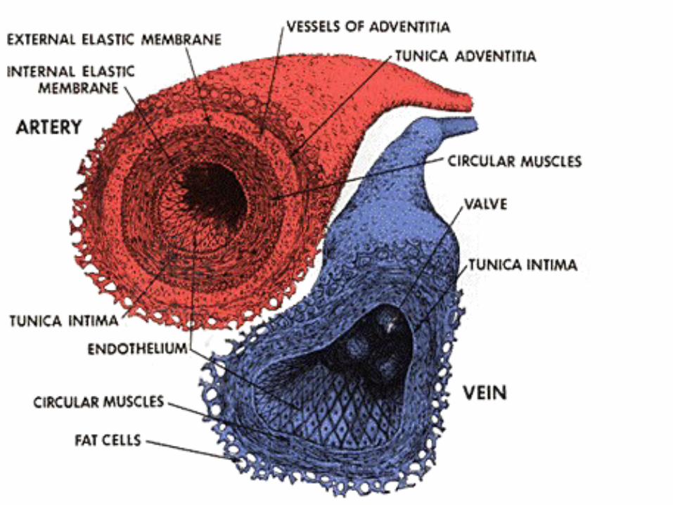

(E) Arteries (have higher BP than veins) and Blood Pressure• Each beat FORCES blood through blood vessels THROUGHOUT body.

(1) Arteries (carry blood AWAY from heart)• Large, MUSCULAR, elastic vessels begin with aorta (LARGEST artery) and split into smaller ARTERIOLES (and ultimately to capillaries).


(2) Blood Pressure (high BP risks RUPTURING an artery)• The force that BLOOD exerts against WALLS of a blood vessel.

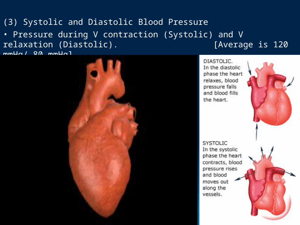
(3) Systolic and Diastolic Blood Pressure• Pressure during V contraction (Systolic) and V relaxation (Diastolic). [Average is 120 mmHg/ 80 mmHg]

(4) Hypertension (sustained High Blood Pressure)• STRAIN on WALLS of arteries INCREASES chance vessel will BURST.

(F) Capillaries and Veins• Gas EXCHANGE at CAPILLARIES, then blood returns BACK to S/I VENA CAVA via veins.

(1) Arterioles (arterioles-CAPILLARIES-venules)• Connect LARGER arteries with tiny CAPILLARIES.


(2) Capillaries (1-celled thick)• Nutrients, hormones, and gases DIFFUSE between BLOOD and CELLS.

(3) Venules and Veins (thinner, less muscular and WITH valves)• Venules unite into LARGER veins, returning BACK to HEART.


(4) Inferior Vena Cava (IVC) and Superior Vena Cava (SVC)• 2 LARGEST veins (with LOWEST BP) return d-blood from body to RA.

II. Patterns of Circulation• 2 LOOPS improve DELIVERY rate;
(1) Pulmonary Circulation• Between HEART and LUNGS.
(2) Systemic Circulation• Between HEART and BODY TISSUES.

(A) Pulmonary Circulation• D-Blood: Body SVC/IVC RA RV PA LUNGS PV O-Blood(1) Pulmonary Veins (RETURN blood from LUNGS back to LA)• ONLY veins carrying O-Blood in body.

(B) Systemic Circulation• LA LV AA/DA ALL BODY SYSTEMS.

(3) Why might it be possible that a person could turn PALE when they are frightened?
Critical Thinking


(1) Coronary Circulation (a SUBSYSTEM of systemic circulation)• Heart gets its own LOOP!

(2) Atherosclerosis (or if an artery becomes BLOCKED)• BUILDUP of fatty material on INTERIOR walls of coronary artery
(BLOCKAGE can result in a coronary thrombosis )

(3) Renal Circulation (a second SUBSYSTEM)• Kidney LOOP FILTER wastes from our blood.
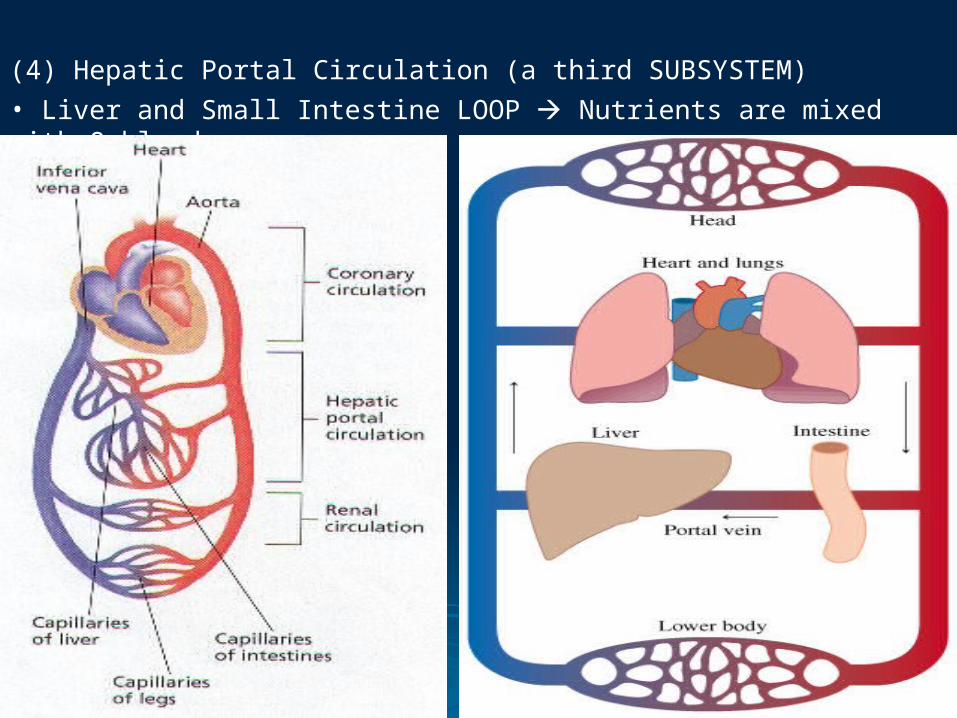
(4) Hepatic Portal Circulation (a third SUBSYSTEM)• Liver and Small Intestine LOOP Nutrients are mixed with O-blood

II. Lymphatic System (also part of the circulatory system, BUT NO pump)• A 1-WAY system of vessels (with VALVES) that DRAIN FLUIDS from tissues BACK into blood stream.


(1) Lymph (GETS CHECKED AT LYMPH NODES)• Yellowish fluid drained from tissues into lymphatic vessels.

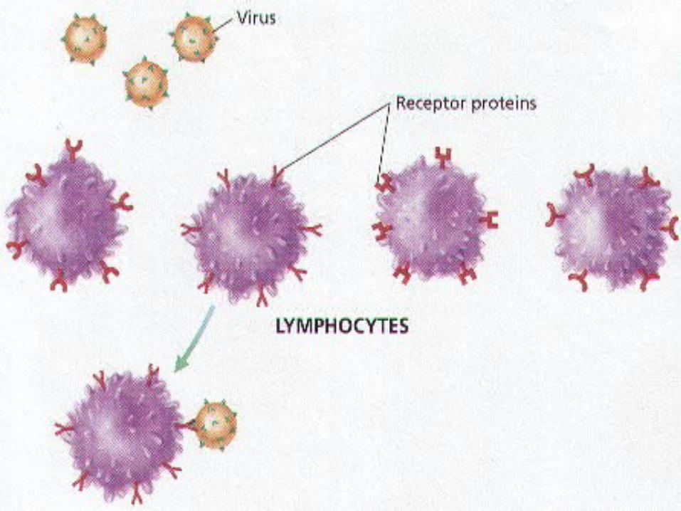
(2) Lymphocytes (stored in LYMPH NODES by the immune system)• WBCs TRAP microorganisms and tissue debris.


I. Composition of Blood (55% Liquid, 45% Solid Cells)• 4-5 L in ADULT body; ~ RBCs, WBCs, platelets, and plasma.
47-2 Blood


(A) Plasma (yellowish LIQUID of blood)• 90% water, glucose, proteins, antibodies, hormones, and wastes.

(4) Even a small increase or decrease in blood volume has an effect on BLOOD PRESSURE. When an accident victim suffers significant blood LOSS, the person is transferred with PLASMA rather than whole blood. Why might plasma be effective in meeting the IMMEDIATE threat to life?
Critical Thinking


(B) Red Blood Cells (a.k.a. Erythrocytes)• Made in RED bone marrow, SHORT span (120 days), only 1 type of cell.(1) Erythrocytes (NO nucleus, CANNOT divide)• Biconcave with HEMOGLOBIN core, ~ transports OXYGEN.


(2) Hemoglobin
• Fe-containing protein has AFFINITY for O2 across ALVEOLUS.


(C) White Blood Cells (a.k.a. Leukocytes AND Lymphocytes)• Class of IMMUNE CELLS prevent disease and pathogen infection.
(e.g., virus, bacteria, protozoan, fungus)

(1) Leukocytes (made in RED marrow, lymph nodes, and spleen)• Live up to YEARS; SEVERAL types that CAN divide (have NUCLEUS).

(2) Phagocytes (e.g., Macrophage, Dendritic Cells)• Amoeba-like cells that ENGULF foreign invaders and debris.

(D) Platelets (cell FRAGMENTS formed in RED marrow)• PLUG damaged blood vessels; ~ release “CLOTTING factors,” resulting in “FIBRIN NET.”

(1) Fibrin• Long protein NET traps RBCs and hardens to form a CLOT then a SCAB.


II. Blood Types• Determined by ANTIGEN present on SURFACE of RBCs.

(1) Blood Types (A, B, AB, O)• 4 groups based on ANTIGENS present, directs antibody production.
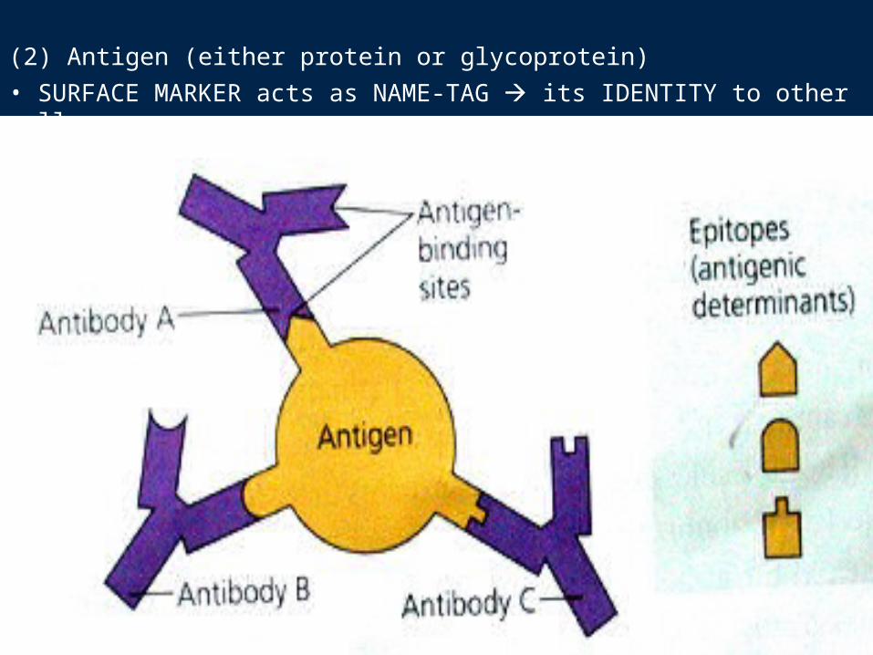
(2) Antigen (either protein or glycoprotein)• SURFACE MARKER acts as NAME-TAG its IDENTITY to other cells.

(A) A-B-O System• INCOMPATIBLE combinations result in AGGLUTINATION
(clumping of RBCs, smothered in antibodies).

(B) Rh System (85% is Rh +, 15% is Rh-)• A 2nd type of antigen found on RBCs and can cause PROBLEMS if MOTHER is Rh – and CHILD is Rh +.

(1) Rh Factor (named after Rhesus Monkey)• A MOTHER who is “–” will NOT recognize a “+” baby’s RBC and will ATTACK it if NOT prevented from encountering FOREIGN antigen.


I. The Lungs (ATMOSPHERE-BLOOD-ATMOSPHERE)
• RL (3 lobes), LL (2 lobes) inside THORACIC cavity, lined with PLEURA (secretes lubricant).
47-3 Respiratory System

(5) Polio is a disease that paralyzes muscles by affecting the nerves that make them move. Before the polio vaccine was developed, many people afflicted with polio died to a LACK of respiration. Some of the survivors had to be placed in an “iron lung” that breathed for them. From what you know about the respiratory system, explain why people stricken with polio could no longer breathe on their own.
Critical Thinking

(1) External Respiration (OUTSIDE the body)• Exchange of gases between ATMOSPHERE and BLOOD.

(2) Internal Respiration (INSIDE the body)• Exchange of gases between BLOOD and CELLS.

(A) The Passage of Air• Air is filtered, warmed, and moistened by HAIRS and MUCUS membranes.

(1) Epiglottis• Flap of cartilage COVERS trachea when swallowing, PREVENTS food from ENTERING TRACHEA.

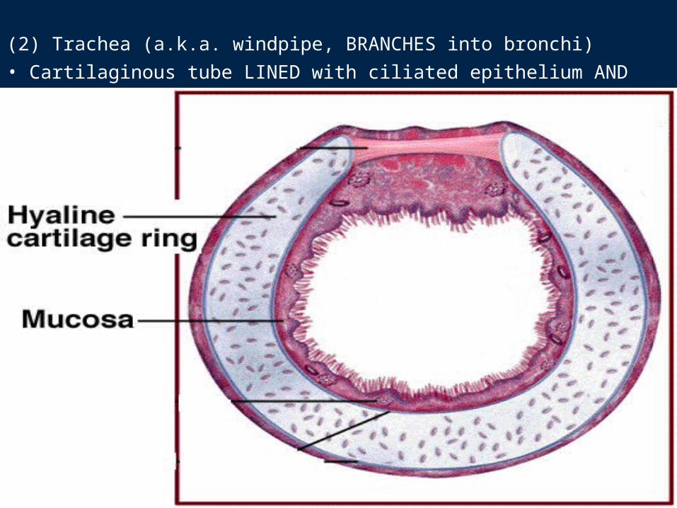
(2) Trachea (a.k.a. windpipe, BRANCHES into bronchi)• Cartilaginous tube LINED with ciliated epithelium AND mucus.


(3) Larynx (UPPER region of trachea)•2 LIGAMENTS (vocal cords) produce sound when AIR is forced BY them.

(4) Bronchi & Bronchioles (i.e., bronchi BRANCH to bronchioles) • Smooth muscle & cartilage tubes LINED with ciliated cells and mucus.


(5) Alveoli (the END of a bronchiole)• Clusters of AIR SACS where GAS EXCHANCE occurs between AIR and BLOOD; [~ 300 million inside each lung provide a surface area 40x skin]

(6) A person with anemia has TOO FEW red blood cells. The most common symptom is a LACK of energy. Why might anemia result in this symptom?
Critical Thinking


II. Gas Exchange and Transport (requires CONCENTRATION gradient)
• Gases exchanged [O2 and CO2] between ALVEOLI and BLOOD of capillaries by DIFFUSION.

(A) Gas Exchange in the Lungs (regulated by DIFFUSION)
• O2 diffuses from AIR SACS INTO BLOOD, and CO2 diffuses from BLOOD INTO AIR SACS (and OUT through lungs).

(B) Hemoglobin and Gas Exchange
• 97% of O2 with Hb, 3% dissolved in plasma 8% CO2 dissolves in plasma, 25% binds to Hb, and 67% reacts to become BICARBONATE IONS
[At lungs, REVERSES into CO2 and H2O].

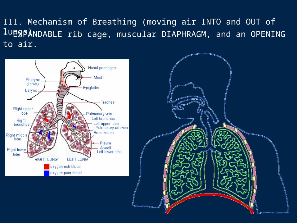
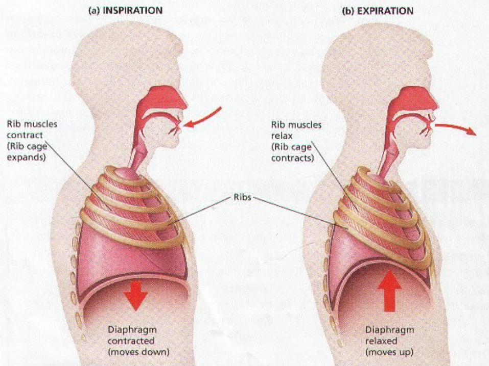
III. Mechanism of Breathing (moving air INTO and OUT of lungs)• EXPANDABLE rib cage, muscular DIAPHRAGM, and an OPENING to air.


(1) Inspiration (i.e., inhalation INCREASES volume of thoracic cage)• Diaphragm FLATTENS, pushing DOWN on abdomen, lifting ribs UP and OUT. [Result Change in air pressure, drawing air INTO lungs.]

(2) Expiration (i.e., exhalation DECREASES volume of thoracic cage)• Diaphragm and rib muscles RELAX, lungs recoil and DEFLATE. [Result Pressure difference forces air OUT of lungs.]

(A) Regulation of Breathing (breathing RATE ~ cellular METABOLISM)
• Cerebrum and brain stem SHARE regulation by monitoring [CO2] in blood (if CO2 too HIGH INCREASES breathing RATE)


Extra Slides AND Answers for Critical Thinking Questions
(1) The blood on the right side of the heart (deoxygenated blood) would mix with blood on the left side (oxygenated blood). As a result, cells would not get enough oxygen.
(2) Blood circulates very closet to every cell in the body, absorbing heat where the body is warmer than the blood and releasing heat where the body is cooler than the blood. Thus, blood distributes heat throughout the body, contributing to a uniform body temperature.
(3) Blood flow to the skin is reduced, and blood flow to muscles and glands is increased. Thus, a person may appear pale when frightened.(4) The accident victim’s loss of blood volume and the loss of body fluid can cause the victim to go into shock. Transfusing plasma replenishes the fluid, helps stabilize the patient, and can be done quickly without taking time to type. The small number of antibodies the plasma contains become harmlessly diluted in the patient’s own blood.(5) Polio can result in paralysis of the diaphragm and the muscles that control the rib cage. If these muscles cannot move, then the person cannot breathe.
(6) The fewer red blood cells there are, the less hemoglobin there is. The smaller the amount of hemoglobin, the smaller the amount of oxygen that can be transported. A smaller amount of oxygen means a decreased level of aerobic respiration and less energy (ATP).