Embed Size (px)
Citation preview
Clinical Guide for Scleral Lens SuccessMelissa Barnett, Daddi Fadel

2

Cover Image: Credited to Melissa Barnett.
Description: This manuscript is the result of a collaborative effort between the Scleral Lens Education Society (SLS) and the Accademia Italiana Lenti Sclerali (AILeS).
Acknowledgements: The authors wish to thank the Scleral Lens Education Society and Accademia Italiana Lenti Sclerali Board members, Robert Ensley, Luigi Lupelli, and Gregg Russell for reviewing this manuscript.
Publication date: 2018
Recommended Citation:
Barnett M, Fadel D. Clinical Guide for Scleral Lens Success. SLS & AILeS. 2018
Available and published by
3
Melissa Barnett OD, FAAO, FSLS, FBCLA, is the principal optometrist at the UC Davis Eye Center in Sacramento, CA. She is an internationally recognized key opinion leader, who lectures and publishes extensively on anterior segment disease and specialty contact lenses. She is a Fellow of the American Academy of Optometry and the British Contact Lens Association, a Diplomate of the American Board of Certification in Medical Optometry, and serves on the board of the American Optometric Association Cornea and Contact Lens Council, Women in Optometry and Women of Vision, Gas Permeable Lens Institute, and Ocular Surface Society of Optometry. She is past president of the Scleral Lens Education Society. Dr. Barnett is a spokesperson for the California Optometric Association and a guest lecturer for the STAPLE program. Drs. Melissa Barnett and Lynette Johns edited the book Contemporary Scleral Lenses: Theory and Application with the unique perspectives and contributions of international experts. She is an advisor to and/or has received honoraria or travel expenses from AccuLens, Alcon, Alden Optical, Allergan, Bausch + Lomb, Contamac, Johnson & Johnson Vision, Novabay, Ocusoft, Paragon Biotek, Shire, the Sjögren’s Syndrome Foundation, the Scleral Lens Society, the STAPLE Program, SynergEyes, and Visioneering Technologies.
Daddi Fadel Dip Optom, FSLS, is a lens designer and specialist in contact lenses for irregular cornea, scleral lenses and orthokeratology. She speaks five languages; Arabic, French, English, Italian and Greek. She studied optometry at Istituto Superiore di Scienze Optometriche (ISSO) in Rome (1998-2001), a four-year course achieved with honors. She started to lecture and publish in her first year of Optometry school. She has 20 years experience in optometry and specialty contact lenses. She runs an optometric practice specializing in contact lenses in Italy where she designs and fits special customized contact lenses. She lectures and publishes nationally and internationally, especially on contact lenses in irregular cornea, scleral lenses, and ortho-k. She is the Founder and President of Accademia Italiana Lenti Sclerali (AILeS), a board member of Accademia Italiana Lenti a Contatto (AILAC), and a Fellow of Scleral Lens Education Society (SLS).
4
Contents
I. Introduction . . . . . . . . . . . . . . . . . . . . . . . . . . . . . . . . . . . . . . . . . . . . . . . . . . . . . . . 6
II. Staff training . . . . . . . . . . . . . . . . . . . . . . . . . . . . . . . . . . . . . . . . . . . . . . . . . . . . . . . 6
III. Patient and family communication . . . . . . . . . . . . . . . . . . . . . . . . . . . . . . . . . . . . . 6
IV. Scleral lens indications . . . . . . . . . . . . . . . . . . . . . . . . . . . . . . . . . . . . . . . . . . . . . . 7
V. Case history . . . . . . . . . . . . . . . . . . . . . . . . . . . . . . . . . . . . . . . . . . . . . . . . . . . . . . 10
VI. Eye examination . . . . . . . . . . . . . . . . . . . . . . . . . . . . . . . . . . . . . . . . . . . . . . . . . . . 10
VII. Fitting scleral lenses . . . . . . . . . . . . . . . . . . . . . . . . . . . . . . . . . . . . . . . . . . . . . . . 101. Diameter selection. . . . . . . . . . . . . . . . . . . . . . . . . . . . . . . . . . . . . . . . . . . . . . . . . . 122. Central corneal clearance . . . . . . . . . . . . . . . . . . . . . . . . . . . . . . . . . . . . . . . . . . . . 123. Peripheral corneal clearance . . . . . . . . . . . . . . . . . . . . . . . . . . . . . . . . . . . . . . . . . . 174. Limbal clearance . . . . . . . . . . . . . . . . . . . . . . . . . . . . . . . . . . . . . . . . . . . . . . . . . . . 175. Landing zone design . . . . . . . . . . . . . . . . . . . . . . . . . . . . . . . . . . . . . . . . . . . . . . . . 176. Lens edge . . . . . . . . . . . . . . . . . . . . . . . . . . . . . . . . . . . . . . . . . . . . . . . . . . . . . . . . 197. Over-refraction. . . . . . . . . . . . . . . . . . . . . . . . . . . . . . . . . . . . . . . . . . . . . . . . . . . . . 23
VIII. Number of visits . . . . . . . . . . . . . . . . . . . . . . . . . . . . . . . . . . . . . . . . . . . . . . . . . . 24
IX. Wearing schedule. . . . . . . . . . . . . . . . . . . . . . . . . . . . . . . . . . . . . . . . . . . . . . . . . . 25
X. Patient education for correct application and removal techniques. . . . . . . . . . . 251. Scleral lens application . . . . . . . . . . . . . . . . . . . . . . . . . . . . . . . . . . . . . . . . . . . . . . 252. Scleral lens removal . . . . . . . . . . . . . . . . . . . . . . . . . . . . . . . . . . . . . . . . . . . . . . . . 26
XI. Devices for scleral lens application and removal . . . . . . . . . . . . . . . . . . . . . . . . . 26
XII. Scleral lens care . . . . . . . . . . . . . . . . . . . . . . . . . . . . . . . . . . . . . . . . . . . . . . . . . . . 27
XIII. Follow-up schedule . . . . . . . . . . . . . . . . . . . . . . . . . . . . . . . . . . . . . . . . . . . . . . . . 27
XIV. Conclusion . . . . . . . . . . . . . . . . . . . . . . . . . . . . . . . . . . . . . . . . . . . . . . . . . . . . . . . 28
Appendix A . . . . . . . . . . . . . . . . . . . . . . . . . . . . . . . . . . . . . . . . . . . . . . . . . . . . . . . 29
Appendix B . . . . . . . . . . . . . . . . . . . . . . . . . . . . . . . . . . . . . . . . . . . . . . . . . . . . . . . 30
References . . . . . . . . . . . . . . . . . . . . . . . . . . . . . . . . . . . . . . . . . . . . . . . . . . . . . . . 34
5
I. Introduction
Fitting scleral lenses has expanded in the last decade, and scleral lenses are continuously gaining popularity amongst practitioners globally. The fitting approach differs from corneal rigid gas permeable and soft contact lenses; therefore practitioners, staff, and patients should be appropriately educated. The Clinical Guide for Scleral Lens Success is a collaborative work between the Accademia Italiana Lenti Sclerali (AILeS) and the Scleral Lens Education Society (SLS) with the purpose of providing a protocol for scleral lens fitting. Each country has different laws and regulations concerning the practice of eye care. Therefore, these guidelines may be adapted accordingly. Furthermore, this guide will be constantly updated with new research in the scleral lens field. Despite the fantastic advances in lens design and materials, there are many aspects of scleral lenses that are still unknown.
II. Staff training
Scleral lenses are life changing because of the unique marriage of superior lens optics, large lens diameter, and a comfortable lens wearing experience. Patients who are interested in wearing scleral lenses have often tried other modalities without success, and have possibly searched for information from websites, social media, or other practitioners. Unfortunately, consistent information on scleral lenses is not always available, so patients may not be well informed despite their best efforts to learn about this modality.
To enhance fitting success and patient satisfaction, staff members should be trained to identify good scleral lens candidates, provide accurate information, and educate, train, and support patients throughout the fitting process [1]. Practitioners and staff members may have personal experience with scleral lenses, which makes conveying their experiences much easier. Having staff members try scleral lenses themselves may improve empathy for those patients who may be trying scleral lenses for the first time and increase conviction and confidence for this option.
The first communication with the patient occurs when they are speaking to staff at the office, either in person or on the phone. Staff should be able to provide basic information about scleral lenses and be knowledgeable on good candidate selection in order to recommend scheduling a scleral lens consultation and possible fitting with the practitioner. If needed, the staff should know how to obtain an authorization for scleral lens fitting and materials from the appropriate health authority or medical insurance provider prior to their initial appointment. Every case is unique, so it is appropriate for the practitioner to speak with the patient having had the opportunity to review all necessary information about the patient’s systemic and ocular health, and all other data that
would pertain to possible scleral lens wear prior to contact lens fitting. During the consultation, staff members should present the benefits of scleral lenses and properly educate the patient on other options, including corneal rigid gas permeable contact lenses, custom soft contact lenses, hybrid lenses, glasses, and/or surgical procedures, and further explain the fees, insurance coverage, fitting process and follow-up schedules. It is appropriate to have a signed document where the patient has acknowledged discussing this information with additional instructions regarding refunds of lens materials and fees as well as the office policy on lens changes or refits. This information should be properly vetted and discussed. Proper instruction and training on contact lens care and handling is also crucial for fitting success.
Proper staff training is the foundation for success in a scleral lens practice. Patients who have experienced vision challenges prior to scleral lens fitting are often frightened about their prognosis and are most concerned about their own future, so it is important to be professional, competent, and provide high quality customer service. It is helpful to have staff attend continuing education courses to learn and increase their awareness of scleral lenses.
III. Patient and family communication
A positive approach that effectively communicates and focuses on scleral lens benefits and their differences from other lens modalities will improve patient and family expectations and overall satisfaction. It is important to be realistic and understand what information is pertinent for patients to know. Patients are often anxious about their vision performance and ocular health or have had previous experiences with practitioners that might not have been optimal. This may color their personal view of the situation and influence their expectations. Understanding the psychological aspect of scleral lens fitting and respecting the amount of information given to a patient at one time may differ from one individual to another. Limits must be respected, and information may need to be dispersed, or even repeated over multiple visits for simpler comprehension. It may also be helpful to have a family member join the patient during instruction in order to assist the patient if they are unsure or unable to handle an aspect of care or handling on their own.
Patients should be trained on proper scleral lens care, handling, and solutions. Providing written instructions will help reinforce understanding and provide the patient with something to refer to as they adapt to this new lens modality. Written and verbal education should also be provided about potential scleral lens complications and instructions for proper course of action should these complications occur. An out-of-hours contact number is advisable and mandatory in some countries. Selecting written
6
materials in the patient’s native language will enhance the comprehension of various concepts. Brochures, posters, charts, videos and/or PowerPoint presentations may also be used. Materials and resources are not an alternative for one-on-one patient and family member education. They should supplement patient instruction and should be reviewed with the patient before their delivery. Further accurate website resources for patient education include the following:
www.sclerallens.org www.ailes.it www.gpli.info
IV. Scleral lens indications
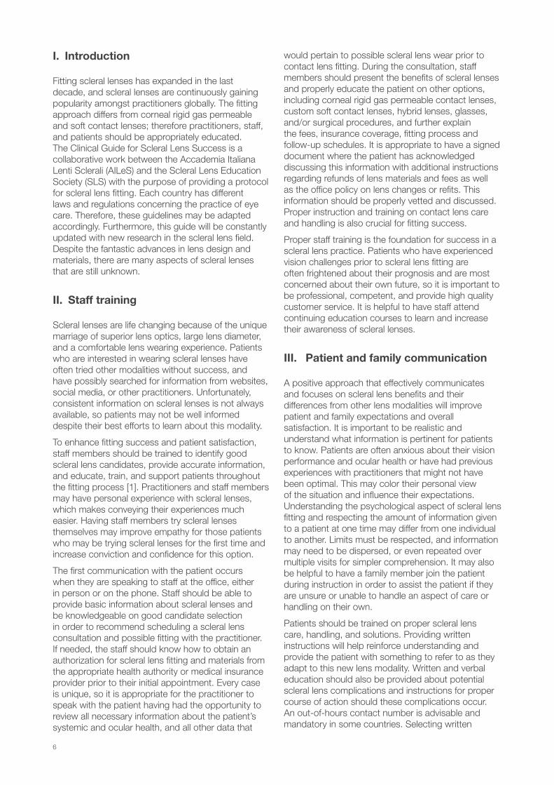
There are many clinical indications for scleral lenses. A good candidate can be identified for scleral lens fitting based on their ocular condition and health, clinical history, and their visual and comfort expectations. The most commonly encountered reasons for fitting scleral lenses include primary corneal ectasias distinguished by developed thinning of the cornea, which results in a distorted corneal surface. Keratoconus is the most common corneal ectasia and can cause monocular or bilateral asymmetric thinning and distortion of the cornea. Keratoconus typically presents with corneal protrusion which may be central or peripheral, or can be characterized as mild, moderate, and severe [2] (Figures 1 - 4). Keratoglobus is characterized by diffuse general thinning and an overall forward protrusion of the cornea circumferentially at the
limbus [3] (Figure 5). Pellucid marginal degeneration presents with a crescent-shaped band of thinning in the peripheral cornea, typically in the inferior quadrant several millimeters from the limbus. Secondary corneal ectasias have been noted after laser-assisted in situ keratomileusis (LASIK), radial keratotomy (RK), photorefractive keratectomy (PRK), and corneal keratoplasty [4,5].
Additional indications for scleral lenses include patients with corneal scars that impede best corrected vision and corneal degenerations or dystrophies such as Salzmann’s nodular or Terrien’s marginal degeneration or epithelial basement membrane dystrophy. Severe exposure keratopathy secondary to Stevens-Johnson syndrome (SJS), chronic graft vs. host disease (GvHD), or exposure keratopathy are other indications for scleral lens wear. Autoimmune conditions including Sjögren’s disease and rheumatoid arthritis benefit from scleral lenses by helping to protect the corneal surface, particularly in patients with infrequent or incomplete blinking. Additional indications include, but are not limited to; post-trauma, Graves’ disease, limbal stem cell deficiency, toxic epidermal necrolysis, neurotrophic keratopathy, superior limbic keratoconjunctivitis, ocular cicatricial pemphigoid, persistent epithelial defects, atopic keratoconjunctivitis, ptosis and superior limbic keratoconjunctivitis. Scleral lenses are extremely beneficial for neuropathic pain and can also be used after surgery of the anterior segment such as pterygium or glaucoma surgeries (Figures 6 - 22).
Figure 1. Corneas with different severities of keratoconus. A. Mild keratoconus. B. Moderate keratoconus. C. Severe keratoconus. Image credit Laura Downie.
7

Figure 2. Scleral lens on an eye with keratoconus and hydrops. Image credit Edward Boshnick.
Figure 3. Scleral lens on a different eye with keratoconus and hydrops. Image credit Edward Boshnick.
Figure 5. A profile view of a scleral lens on an eye with keratoglobus. Image credit Edward Boshnick.
Figure 8. Scleral lens on an eye that underwent three separate corneal transplant surgeries. Image credit Edward Boshnick.
Figure 11. Scleral lens on an eye with corneal scars after RK. Image credit Edward Boshnick.
Figure 4. Scleral lens on an eye with keratoconus and a conjunctival cyst. Note the superior bubbles. Image credit Edward Boshnick.
Figure 7. Scleral lens post-corneal transplantation with sutures. Image credit Edward Boshnick.
Figure 10. Irregular cornea with sutures after RK. Image credit Tom Arnold.
Figure 6. Scleral lens post-corneal transplantation. Image credit Edward Boshnick.
Figure 9. A profile view of an eye that underwent corneal transplantation. Image credit Edward Boshnick.
Figure 12. Scleral lens on an eye after LASIK with ectasia, neovascularization and hydrops. Image credit Edward Boshnick.
Figure 13. Scleral lens on an eye after LASIK with ectasia and hydrops. Image credit Edward Boshnick.
8
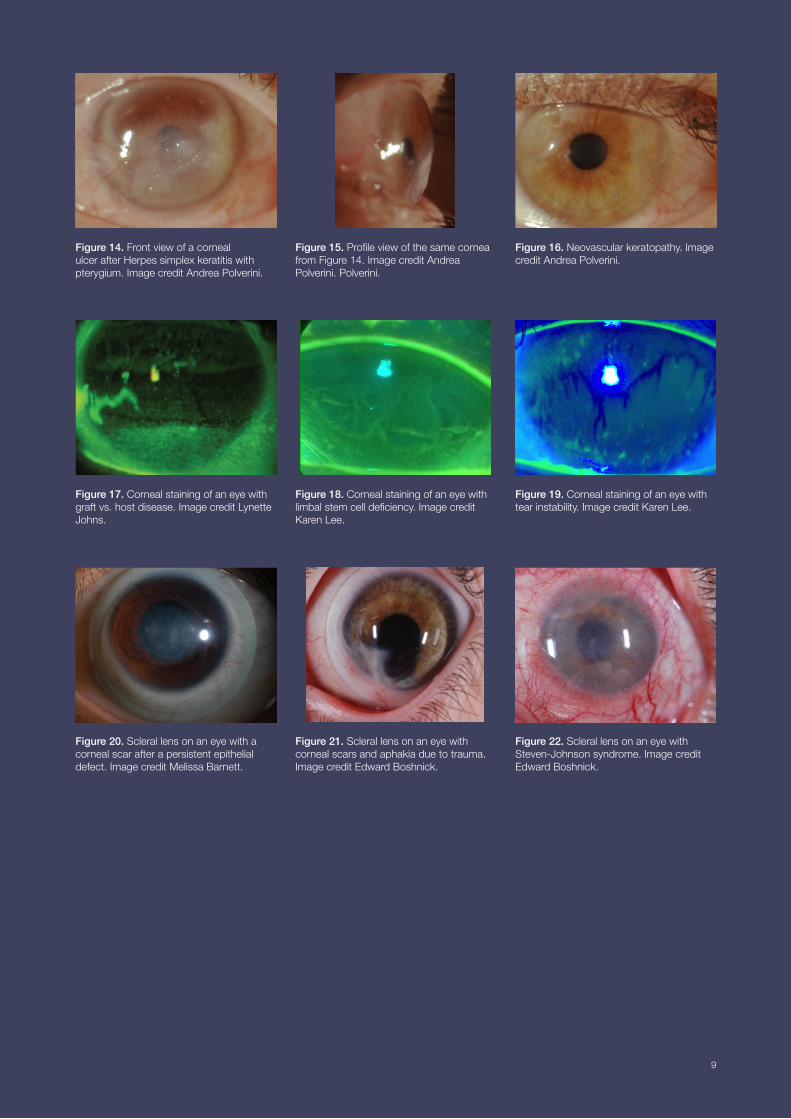
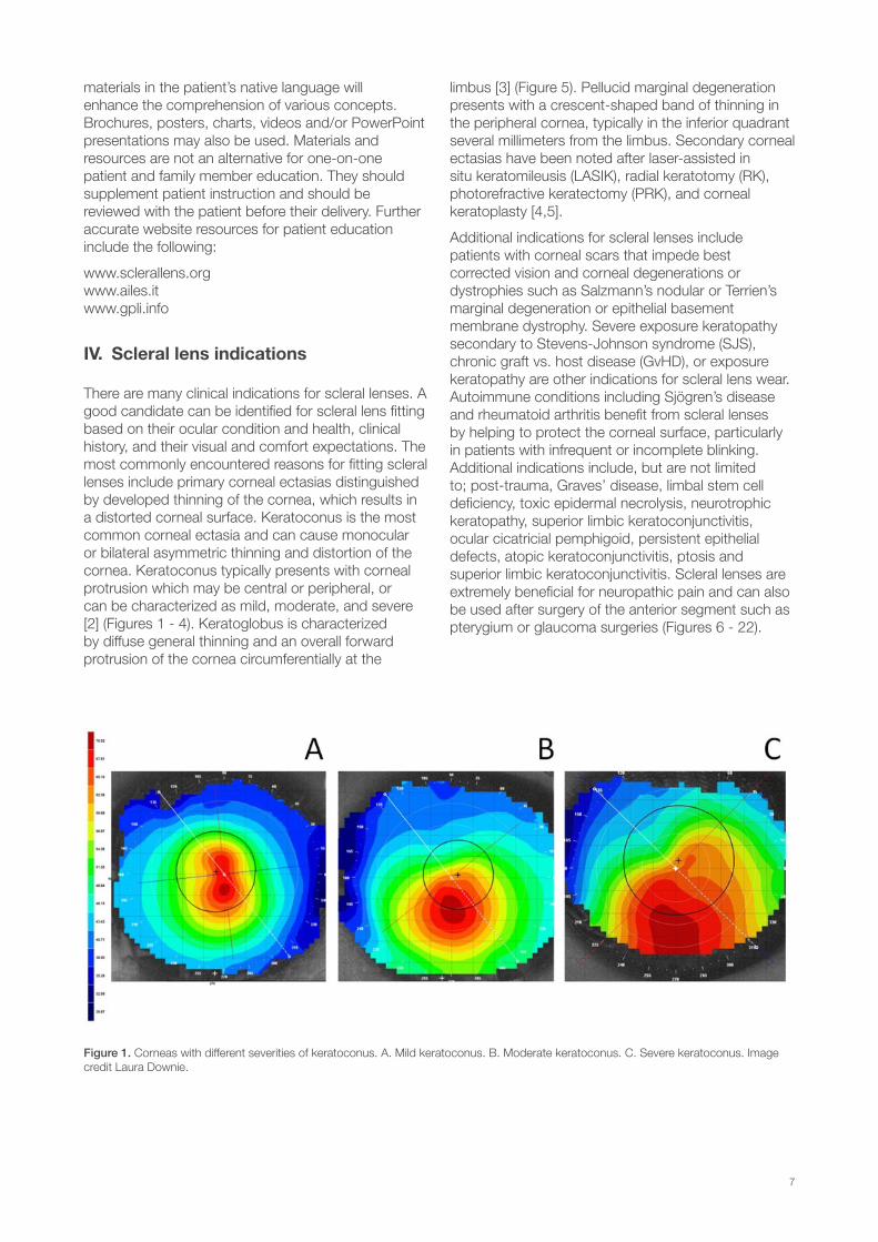
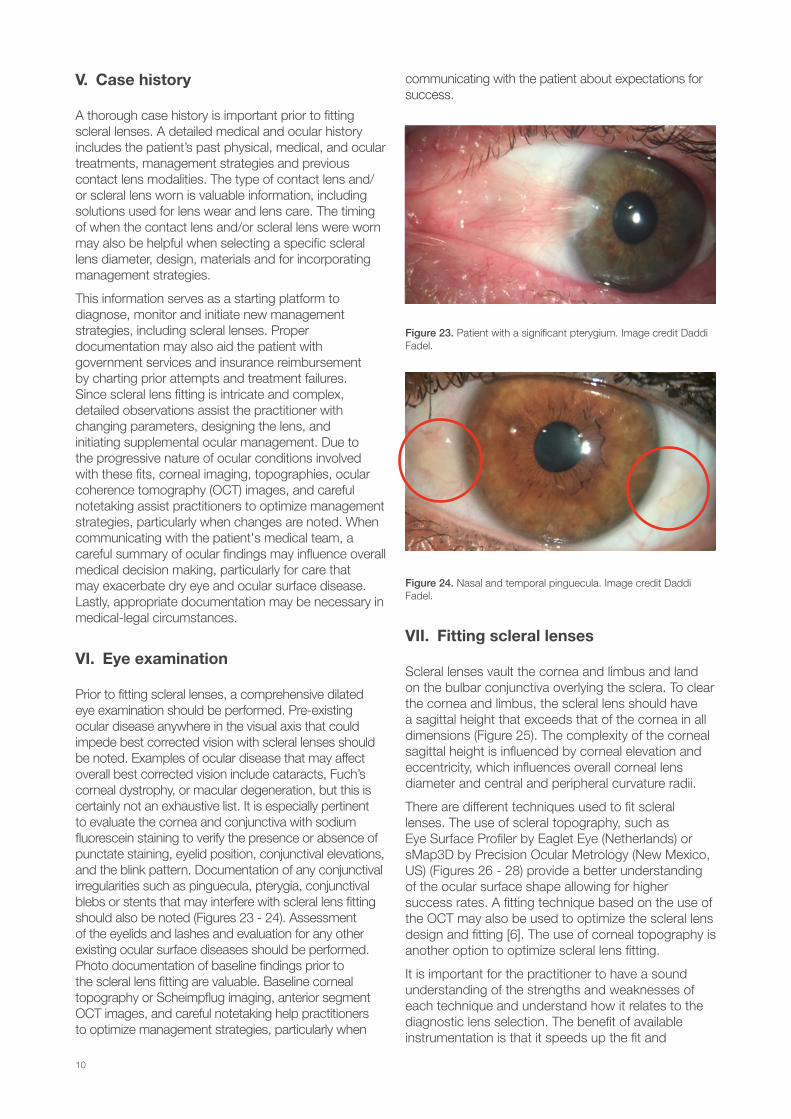
Figure 14. Front view of a corneal ulcer after Herpes simplex keratitis with pterygium. Image credit Andrea Polverini.
Figure 15. Profile view of the same cornea from Figure 14. Image credit Andrea Polverini. Polverini.
Figure 17. Corneal staining of an eye with graft vs. host disease. Image credit Lynette Johns.
Figure 20. Scleral lens on an eye with a corneal scar after a persistent epithelial defect. Image credit Melissa Barnett.
Figure 16. Neovascular keratopathy. Image credit Andrea Polverini.
Figure 19. Corneal staining of an eye with tear instability. Image credit Karen Lee.
Figure 22. Scleral lens on an eye with Steven-Johnson syndrome. Image credit Edward Boshnick.
Figure 18. Corneal staining of an eye with limbal stem cell deficiency. Image credit Karen Lee.
Figure 21. Scleral lens on an eye with corneal scars and aphakia due to trauma. Image credit Edward Boshnick.
9
V. Case history
A thorough case history is important prior to fitting scleral lenses. A detailed medical and ocular history includes the patient’s past physical, medical, and ocular treatments, management strategies and previous contact lens modalities. The type of contact lens and/or scleral lens worn is valuable information, including solutions used for lens wear and lens care. The timing of when the contact lens and/or scleral lens were worn may also be helpful when selecting a specific scleral lens diameter, design, materials and for incorporating management strategies.
This information serves as a starting platform to diagnose, monitor and initiate new management strategies, including scleral lenses. Proper documentation may also aid the patient with government services and insurance reimbursement by charting prior attempts and treatment failures. Since scleral lens fitting is intricate and complex, detailed observations assist the practitioner with changing parameters, designing the lens, and initiating supplemental ocular management. Due to the progressive nature of ocular conditions involved with these fits, corneal imaging, topographies, ocular coherence tomography (OCT) images, and careful notetaking assist practitioners to optimize management strategies, particularly when changes are noted. When communicating with the patient's medical team, a careful summary of ocular findings may influence overall medical decision making, particularly for care that may exacerbate dry eye and ocular surface disease. Lastly, appropriate documentation may be necessary in medical-legal circumstances.
VI. Eye examination
Prior to fitting scleral lenses, a comprehensive dilated eye examination should be performed. Pre-existing ocular disease anywhere in the visual axis that could impede best corrected vision with scleral lenses should be noted. Examples of ocular disease that may affect overall best corrected vision include cataracts, Fuch’s corneal dystrophy, or macular degeneration, but this is certainly not an exhaustive list. It is especially pertinent to evaluate the cornea and conjunctiva with sodium fluorescein staining to verify the presence or absence of punctate staining, eyelid position, conjunctival elevations, and the blink pattern. Documentation of any conjunctival irregularities such as pinguecula, pterygia, conjunctival blebs or stents that may interfere with scleral lens fitting should also be noted (Figures 23 - 24). Assessment of the eyelids and lashes and evaluation for any other existing ocular surface diseases should be performed. Photo documentation of baseline findings prior to the scleral lens fitting are valuable. Baseline corneal topography or Scheimpflug imaging, anterior segment OCT images, and careful notetaking help practitioners to optimize management strategies, particularly when
communicating with the patient about expectations for success.
Figure 23. Patient with a significant pterygium. Image credit Daddi Fadel.
Figure 24. Nasal and temporal pinguecula. Image credit Daddi Fadel.
VII. Fitting scleral lenses
Scleral lenses vault the cornea and limbus and land on the bulbar conjunctiva overlying the sclera. To clear the cornea and limbus, the scleral lens should have a sagittal height that exceeds that of the cornea in all dimensions (Figure 25). The complexity of the corneal sagittal height is influenced by corneal elevation and eccentricity, which influences overall corneal lens diameter and central and peripheral curvature radii.
There are different techniques used to fit scleral lenses. The use of scleral topography, such as Eye Surface Profiler by Eaglet Eye (Netherlands) or sMap3D by Precision Ocular Metrology (New Mexico, US) (Figures 26 - 28) provide a better understanding of the ocular surface shape allowing for higher success rates. A fitting technique based on the use of the OCT may also be used to optimize the scleral lens design and fitting [6]. The use of corneal topography is another option to optimize scleral lens fitting.
It is important for the practitioner to have a sound understanding of the strengths and weaknesses of each technique and understand how it relates to the diagnostic lens selection. The benefit of available instrumentation is that it speeds up the fit and
10

provides reasonable starting points for lens vault selection. However, diagnostic assessments may still be performed with diagnostic scleral lens diagnostic fitting sets if the practitioner does not have the technology to map the ocular surface.
Parameters that should be determined include lens total diameter, corneal clearance, limbal clearance, sagittal height, landing zone, and lens edge.
Figure 25. Scleral lens design on a cross-section tomogram produced by OCT showing the lens vaulting the cornea and limbus and bearing on the conjunctiva. Image credit Daddi Fadel.
Figure 26 - 27. sMap3D takes three images of the ocular surface using fluorescein for data collection. These images are then stitched together. These images are of a patient with keratoconus. Image credit Visionary Optics.
11
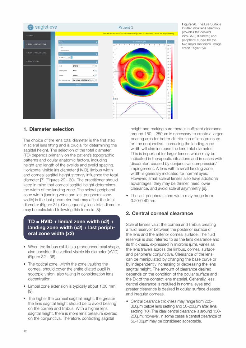
Figure 28. The Eye Surface Profiler initial lens selection provides the desired lens SAG, diameter, and peripheral curves for the two major meridians. Image credit Eaglet Eye.
1. Diameter selection
The choice of the lens total diameter is the first step in scleral lens fitting and is crucial for determining the sagittal height. The selection of the total diameter (TD) depends primarily on the patient’s topographic patterns and ocular anatomic factors, including height and length of the eyelids and eyelid spacing. Horizontal visible iris diameter (HVID), limbus width and corneal sagittal height strongly influence the total diameter [7] (Figures 29 - 30). The practitioner should keep in mind that corneal sagittal height determines the width of the landing zone. The scleral peripheral zone width (landing zone and last peripheral zone width) is the last parameter that may affect the total diameter (Figure 31). Consequently, lens total diameter may be calculated following this formula [8]:
• When the limbus exhibits a pronounced oval shape, also consider the vertical visible iris diameter (VVID) (Figure 32 - 36).
• The optical zone, within the zone vaulting the cornea, should cover the entire dilated pupil in scotopic vision, also taking in consideration lens decentration.
• Limbal zone extension is typically about 1.00 mm [9].
• The higher the corneal sagittal height, the greater the lens sagittal height should be to avoid bearing on the cornea and limbus. With a higher lens sagittal height, there is more lens pressure exerted on the conjunctiva. Therefore, controlling sagittal
height and making sure there is sufficient clearance around 150 – 250µm is necessary to create a larger bearing area for better distribution of lens pressure on the conjunctiva. Increasing the landing zone width will also increase the lens total diameter. This is important for larger lenses which may be indicated in therapeutic situations and in cases with discomfort caused by conjunctival compression/impingement. A lens with a small landing zone width is generally indicated for normal eyes. However, small scleral lenses also have additional advantages: they may be thinner, need lower clearance, and avoid scleral asymmetry [8].
• The last peripheral zone width may range from 0.20-0.40mm.
2. Central corneal clearance
Scleral lenses vault the cornea and limbus creating a fluid reservoir between the posterior surface of the lens and the anterior corneal surface. The fluid reservoir is also referred to as the lens clearance and its thickness, expressed in microns (µm), varies as the lens travels across the limbus, corneal surface and peripheral conjunctiva. Clearance of the lens can be manipulated by changing the base curve or by independently increasing or decreasing the lens sagittal height. The amount of clearance desired depends on the condition of the ocular surface and the Dk of the contact lens material. Generally, less central clearance is required in normal eyes and greater clearance is desired in ocular surface disease and irregular corneas.
• Central clearance thickness may range from 200-300µm before lens settling and 50-200µm after lens settling [10]. The ideal central clearance is around 150-250µm; however, in some cases a central clearance of 50-100µm may be considered acceptable.
TD = HVID + limbal zone width (x2) + landing zone width (x2) + last periph-eral zone width (x2)
12
• The tear permeability to oxygen has a Dk value of about 80 ([cm2/s][ml O2/ml mmHg]) [11]. The unit of oxygen permeability may be simplified with “Fatt Dk units” as has been previously suggested [12].
• Lens thickness, lens material, and lens clearance all influence oxygen delivery to the cornea [13-16]. Decreasing central clearance may also allow thinning of the post lens tear reservoir to reduce midday fogging, leading to better visual acuity.
• The amount of settling depends on individual ocular variables and may be up to 200µm [17-19]. Eighty percent of settling occurs during the first 4 hours [20].
• In eyes with keratoconus, consider adding an additional 100µm of vault to avoid central corneal apical touch in the event of possible future progression of the ectasia.
• Evaluate the central clearance at lens application, after 4 hours, at one to two weeks, after 1-6 months, and at every follow-up visit.
• The evaluation of central corneal clearance may be performed using:
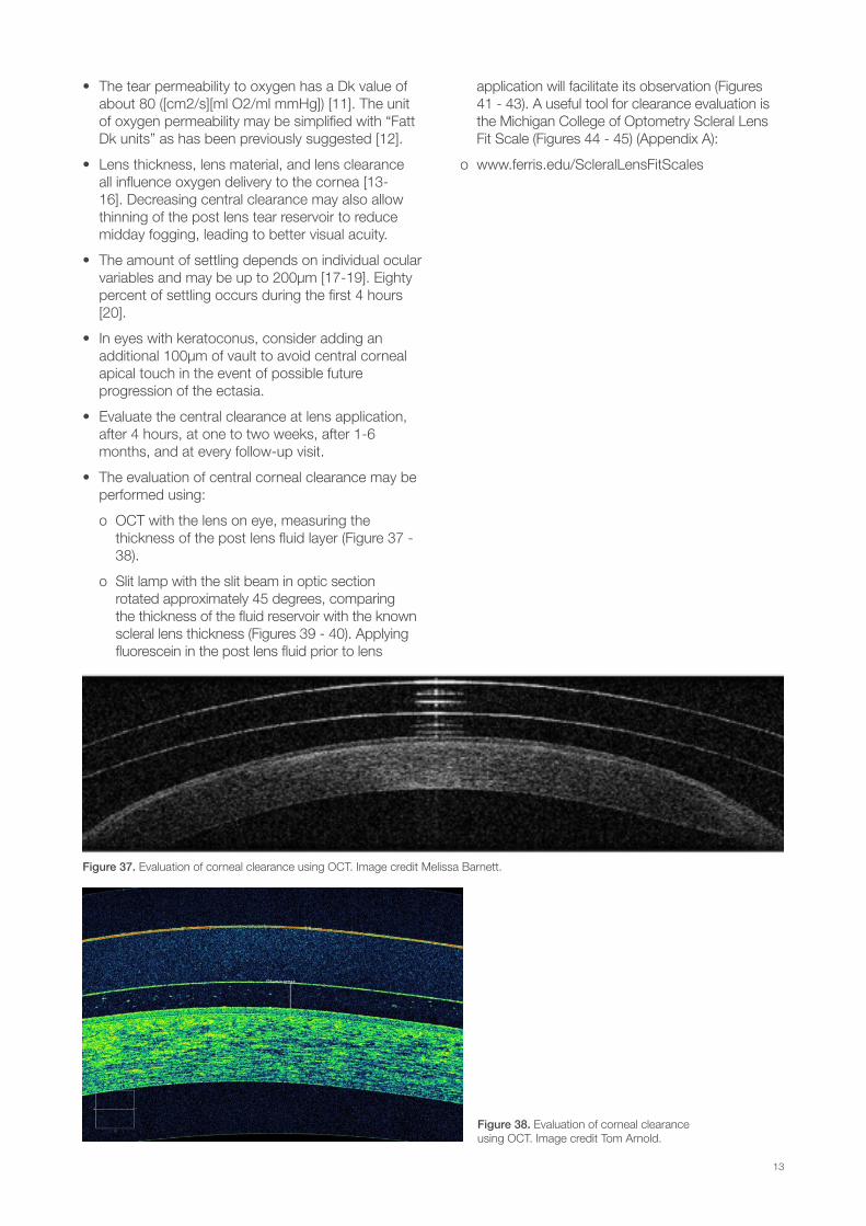
o OCT with the lens on eye, measuring the thickness of the post lens fluid layer (Figure 37 - 38).
o Slit lamp with the slit beam in optic section rotated approximately 45 degrees, comparing the thickness of the fluid reservoir with the known scleral lens thickness (Figures 39 - 40). Applying fluorescein in the post lens fluid prior to lens
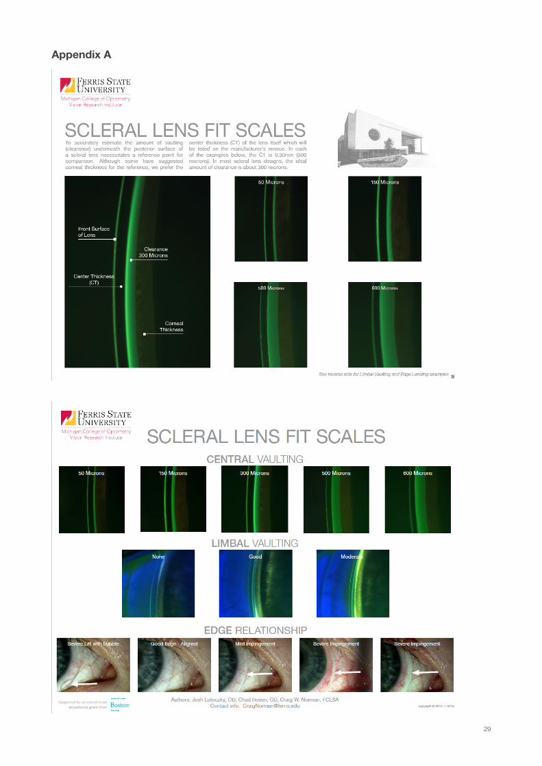
application will facilitate its observation (Figures 41 - 43). A useful tool for clearance evaluation is the Michigan College of Optometry Scleral Lens Fit Scale (Figures 44 - 45) (Appendix A):
o www.ferris.edu/ScleralLensFitScales
Figure 37. Evaluation of corneal clearance using OCT. Image credit Melissa Barnett.
Figure 38. Evaluation of corneal clearance using OCT. Image credit Tom Arnold.
13
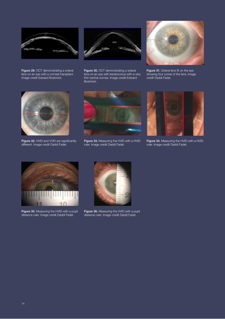
Figure 29. OCT demonstrating a scleral lens on an eye with a corneal transplant. Image credit Edward Boshnick.
Figure 30. OCT demonstrating a scleral lens on an eye with keratoconus with a very thin central cornea. Image credit Edward Boshnick.
Figure 32. HVID and VVID are significantly different. Image credit Daddi Fadel.
Figure 35. Measuring the HVID with a pupil distance ruler. Image credit Daddi Fadel.
Figure 31. Scleral lens fit on the eye showing four zones of the lens. Image credit Daddi Fadel.
Figure 33. Measuring the VVID with a HVID ruler. Image credit Daddi Fadel.
Figure 34. Measuring the HVID with a HVID ruler. Image credit Daddi Fadel.
Figure 36. Measuring the VVID with a pupil distance ruler. Image credit Daddi Fadel.
14
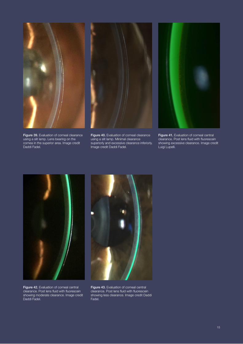
Figure 39. Evaluation of corneal clearance using a slit lamp. Lens bearing on the cornea in the superior area. Image credit Daddi Fadel.
Figure 42. Evaluation of corneal central clearance. Post lens fluid with fluorescein showing moderate clearance. Image credit Daddi Fadel.
Figure 43. Evaluation of corneal central clearance. Post lens fluid with fluorescein showing less clearance. Image credit Daddi Fadel.
Figure 40. Evaluation of corneal clearance using a slit lamp. Minimal clearance superiorly and excessive clearance inferiorly. Image credit Daddi Fadel.
Figure 41. Evaluation of corneal central clearance. Post lens fluid with fluorescein showing excessive clearance. Image credit Luigi Lupelli.
15
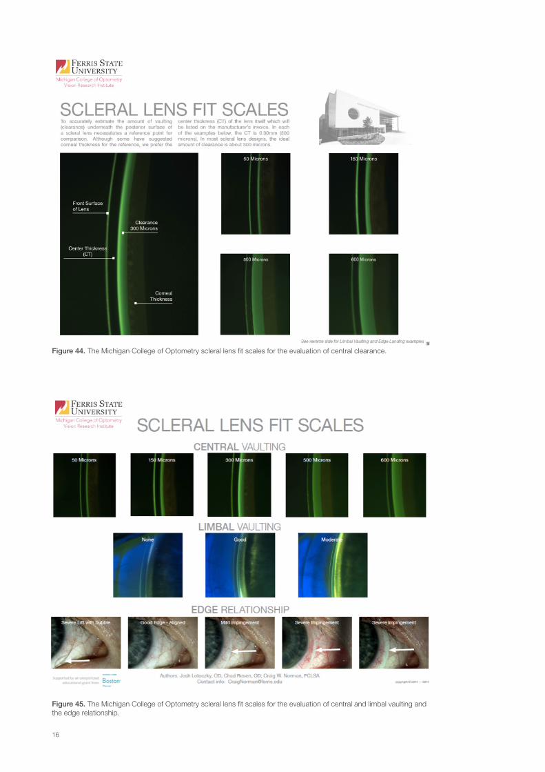
Figure 44. The Michigan College of Optometry scleral lens fit scales for the evaluation of central clearance.
Figure 45. The Michigan College of Optometry scleral lens fit scales for the evaluation of central and limbal vaulting and the edge relationship.
16
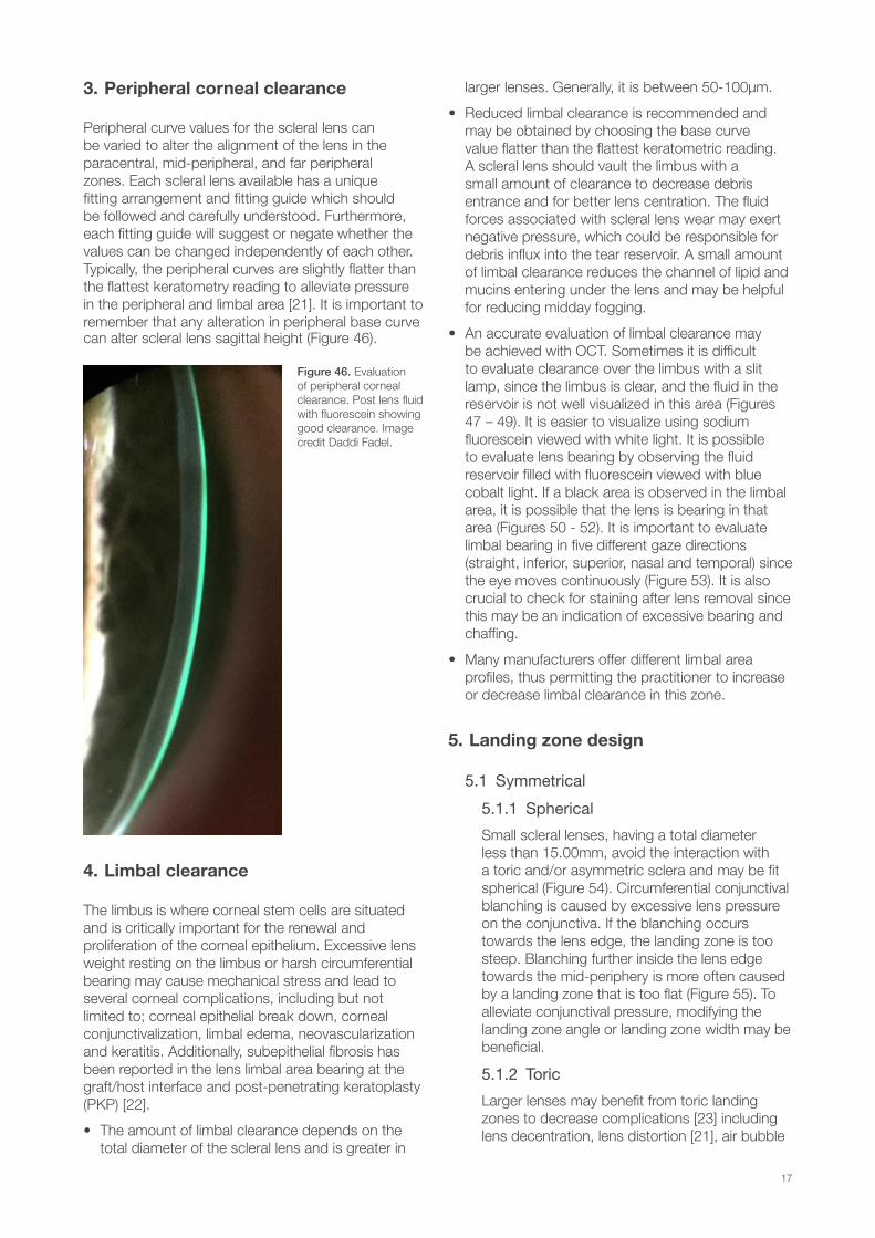
3. Peripheral corneal clearance
Peripheral curve values for the scleral lens can be varied to alter the alignment of the lens in the paracentral, mid-peripheral, and far peripheral zones. Each scleral lens available has a unique fitting arrangement and fitting guide which should be followed and carefully understood. Furthermore, each fitting guide will suggest or negate whether the values can be changed independently of each other. Typically, the peripheral curves are slightly flatter than the flattest keratometry reading to alleviate pressure in the peripheral and limbal area [21]. It is important to remember that any alteration in peripheral base curve can alter scleral lens sagittal height (Figure 46).
4. Limbal clearance
The limbus is where corneal stem cells are situated and is critically important for the renewal and proliferation of the corneal epithelium. Excessive lens weight resting on the limbus or harsh circumferential bearing may cause mechanical stress and lead to several corneal complications, including but not limited to; corneal epithelial break down, corneal conjunctivalization, limbal edema, neovascularization and keratitis. Additionally, subepithelial fibrosis has been reported in the lens limbal area bearing at the graft/host interface and post-penetrating keratoplasty (PKP) [22].
• The amount of limbal clearance depends on the total diameter of the scleral lens and is greater in
larger lenses. Generally, it is between 50-100μm.
• Reduced limbal clearance is recommended and may be obtained by choosing the base curve value flatter than the flattest keratometric reading. A scleral lens should vault the limbus with a small amount of clearance to decrease debris entrance and for better lens centration. The fluid forces associated with scleral lens wear may exert negative pressure, which could be responsible for debris influx into the tear reservoir. A small amount of limbal clearance reduces the channel of lipid and mucins entering under the lens and may be helpful for reducing midday fogging.
• An accurate evaluation of limbal clearance may be achieved with OCT. Sometimes it is difficult to evaluate clearance over the limbus with a slit lamp, since the limbus is clear, and the fluid in the reservoir is not well visualized in this area (Figures 47 – 49). It is easier to visualize using sodium fluorescein viewed with white light. It is possible to evaluate lens bearing by observing the fluid reservoir filled with fluorescein viewed with blue cobalt light. If a black area is observed in the limbal area, it is possible that the lens is bearing in that area (Figures 50 - 52). It is important to evaluate limbal bearing in five different gaze directions (straight, inferior, superior, nasal and temporal) since the eye moves continuously (Figure 53). It is also crucial to check for staining after lens removal since this may be an indication of excessive bearing and chaffing.
• Many manufacturers offer different limbal area profiles, thus permitting the practitioner to increase or decrease limbal clearance in this zone.
5. Landing zone design
5.1 Symmetrical
5.1.1 Spherical
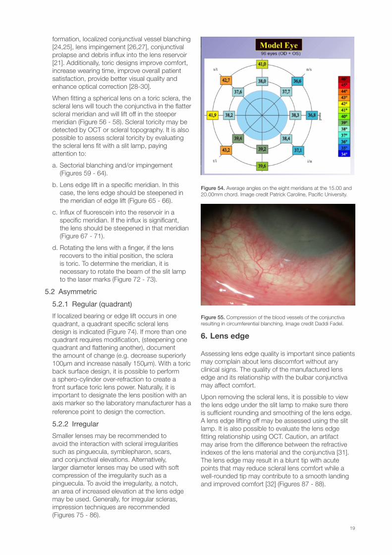
Small scleral lenses, having a total diameter less than 15.00mm, avoid the interaction with a toric and/or asymmetric sclera and may be fit spherical (Figure 54). Circumferential conjunctival blanching is caused by excessive lens pressure on the conjunctiva. If the blanching occurs towards the lens edge, the landing zone is too steep. Blanching further inside the lens edge towards the mid-periphery is more often caused by a landing zone that is too flat (Figure 55). To alleviate conjunctival pressure, modifying the landing zone angle or landing zone width may be beneficial.
5.1.2 Toric
Larger lenses may benefit from toric landing zones to decrease complications [23] including lens decentration, lens distortion [21], air bubble
Figure 46. Evaluation of peripheral corneal clearance. Post lens fluid with fluorescein showing good clearance. Image credit Daddi Fadel.
17
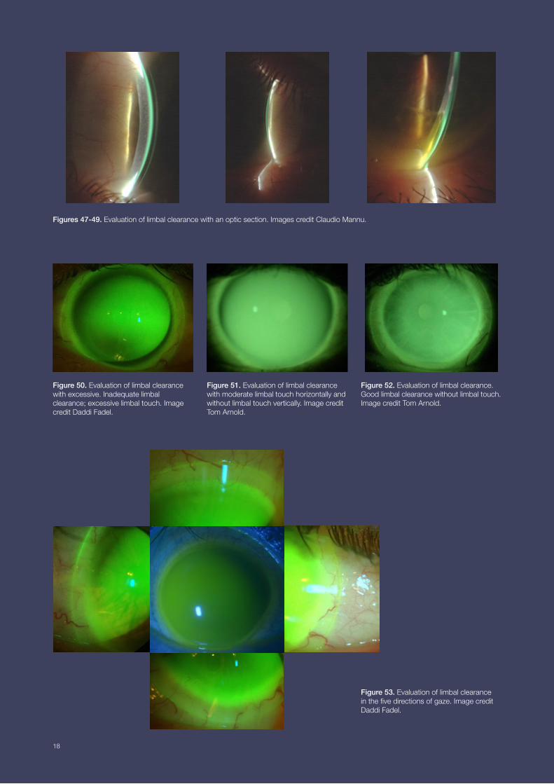
Figures 47-49. Evaluation of limbal clearance with an optic section. Images credit Claudio Mannu.
Figure 50. Evaluation of limbal clearance with excessive. Inadequate limbal clearance; excessive limbal touch. Image credit Daddi Fadel.
Figure 53. Evaluation of limbal clearance in the five directions of gaze. Image credit Daddi Fadel.
Figure 52. Evaluation of limbal clearance. Good limbal clearance without limbal touch. Image credit Tom Arnold.
Figure 51. Evaluation of limbal clearance with moderate limbal touch horizontally and without limbal touch vertically. Image credit Tom Arnold.
18
formation, localized conjunctival vessel blanching [24,25], lens impingement [26,27], conjunctival prolapse and debris influx into the lens reservoir [21]. Additionally, toric designs improve comfort, increase wearing time, improve overall patient satisfaction, provide better visual quality and enhance optical correction [28-30].
When fitting a spherical lens on a toric sclera, the scleral lens will touch the conjunctiva in the flatter scleral meridian and will lift off in the steeper meridian (Figure 56 - 58). Scleral toricity may be detected by OCT or scleral topography. It is also possible to assess scleral toricity by evaluating the scleral lens fit with a slit lamp, paying attention to:
a. Sectorial blanching and/or impingement (Figures 59 - 64).
b. Lens edge lift in a specific meridian. In this case, the lens edge should be steepened in the meridian of edge lift (Figure 65 - 66).
c. Influx of fluorescein into the reservoir in a specific meridian. If the influx is significant, the lens should be steepened in that meridian (Figure 67 - 71).
d. Rotating the lens with a finger, if the lens recovers to the initial position, the sclera is toric. To determine the meridian, it is necessary to rotate the beam of the slit lamp to the laser marks (Figure 72 - 73).
5.2 Asymmetric
5.2.1 Regular (quadrant)
If localized bearing or edge lift occurs in one quadrant, a quadrant specific scleral lens design is indicated (Figure 74). If more than one quadrant requires modification, (steepening one quadrant and flattening another), document the amount of change (e.g. decrease superiorly 100µm and increase nasally 150µm). With a toric back surface design, it is possible to perform a sphero-cylinder over-refraction to create a front surface toric lens power. Naturally, it is important to designate the lens position with an axis marker so the laboratory manufacturer has a reference point to design the correction.
5.2.2 Irregular
Smaller lenses may be recommended to avoid the interaction with scleral irregularities such as pinguecula, symblepharon, scars, and conjunctival elevations. Alternatively, larger diameter lenses may be used with soft compression of the irregularity such as a pinguecula. To avoid the irregularity, a notch, an area of increased elevation at the lens edge may be used. Generally, for irregular scleras, impression techniques are recommended (Figures 75 - 86).
6. Lens edge
Assessing lens edge quality is important since patients may complain about lens discomfort without any clinical signs. The quality of the manufactured lens edge and its relationship with the bulbar conjunctiva may affect comfort.
Upon removing the scleral lens, it is possible to view the lens edge under the slit lamp to make sure there is sufficient rounding and smoothing of the lens edge. A lens edge lifting off may be assessed using the slit lamp. It is also possible to evaluate the lens edge fitting relationship using OCT. Caution, an artifact may arise from the difference between the refractive indexes of the lens material and the conjunctiva [31]. The lens edge may result in a blunt tip with acute points that may reduce scleral lens comfort while a well-rounded tip may contribute to a smooth landing and improved comfort [32] (Figures 87 - 88).
Figure 54. Average angles on the eight meridians at the 15.00 and 20.00mm chord. Image credit Patrick Caroline, Pacific University.
Figure 55. Compression of the blood vessels of the conjunctiva resulting in circumferential blanching. Image credit Daddi Fadel.
19
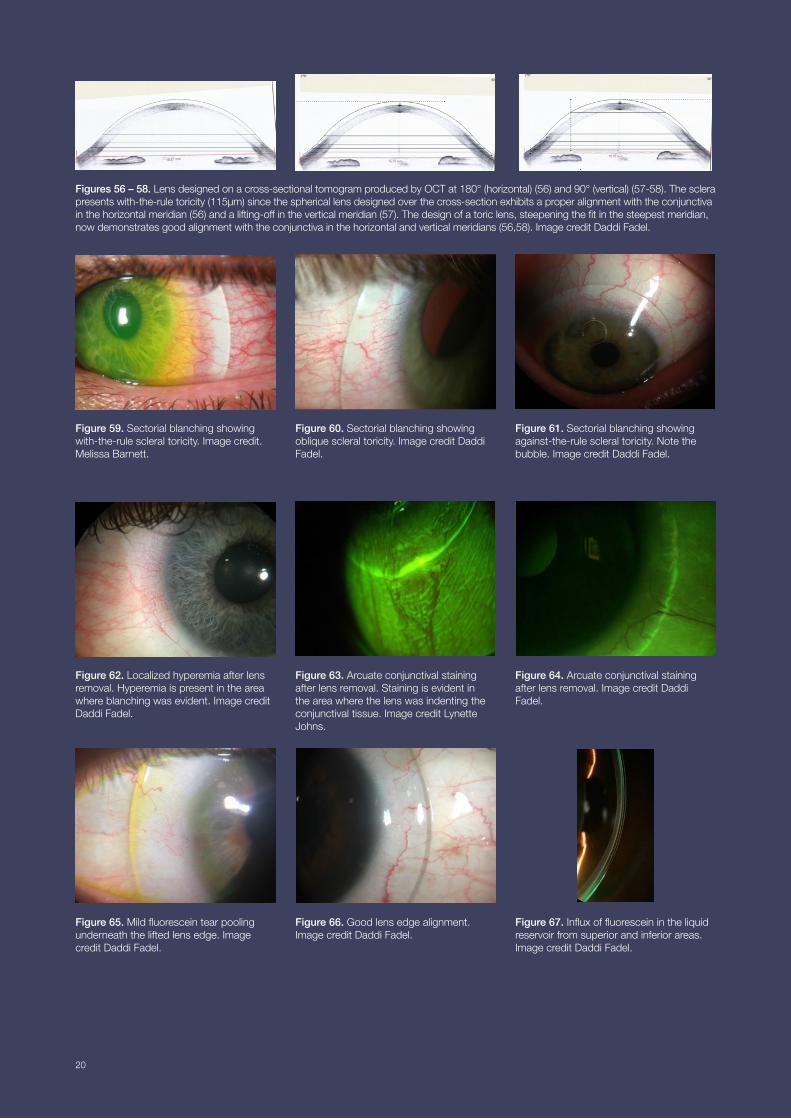
Figures 56 – 58. Lens designed on a cross-sectional tomogram produced by OCT at 180° (horizontal) (56) and 90° (vertical) (57-58). The sclera presents with-the-rule toricity (115μm) since the spherical lens designed over the cross-section exhibits a proper alignment with the conjunctiva in the horizontal meridian (56) and a lifting-off in the vertical meridian (57). The design of a toric lens, steepening the fit in the steepest meridian, now demonstrates good alignment with the conjunctiva in the horizontal and vertical meridians (56,58). Image credit Daddi Fadel.
Figure 59. Sectorial blanching showing with-the-rule scleral toricity. Image credit. Melissa Barnett.
Figure 62. Localized hyperemia after lens removal. Hyperemia is present in the area where blanching was evident. Image credit Daddi Fadel.
Figure 65. Mild fluorescein tear pooling underneath the lifted lens edge. Image credit Daddi Fadel.
Figure 61. Sectorial blanching showing against-the-rule scleral toricity. Note the bubble. Image credit Daddi Fadel.
Figure 64. Arcuate conjunctival staining after lens removal. Image credit Daddi Fadel.
Figure 67. Influx of fluorescein in the liquid reservoir from superior and inferior areas. Image credit Daddi Fadel.
Figure 60. Sectorial blanching showing oblique scleral toricity. Image credit Daddi Fadel.
Figure 63. Arcuate conjunctival staining after lens removal. Staining is evident in the area where the lens was indenting the conjunctival tissue. Image credit Lynette Johns.
Figure 66. Good lens edge alignment. Image credit Daddi Fadel.
20
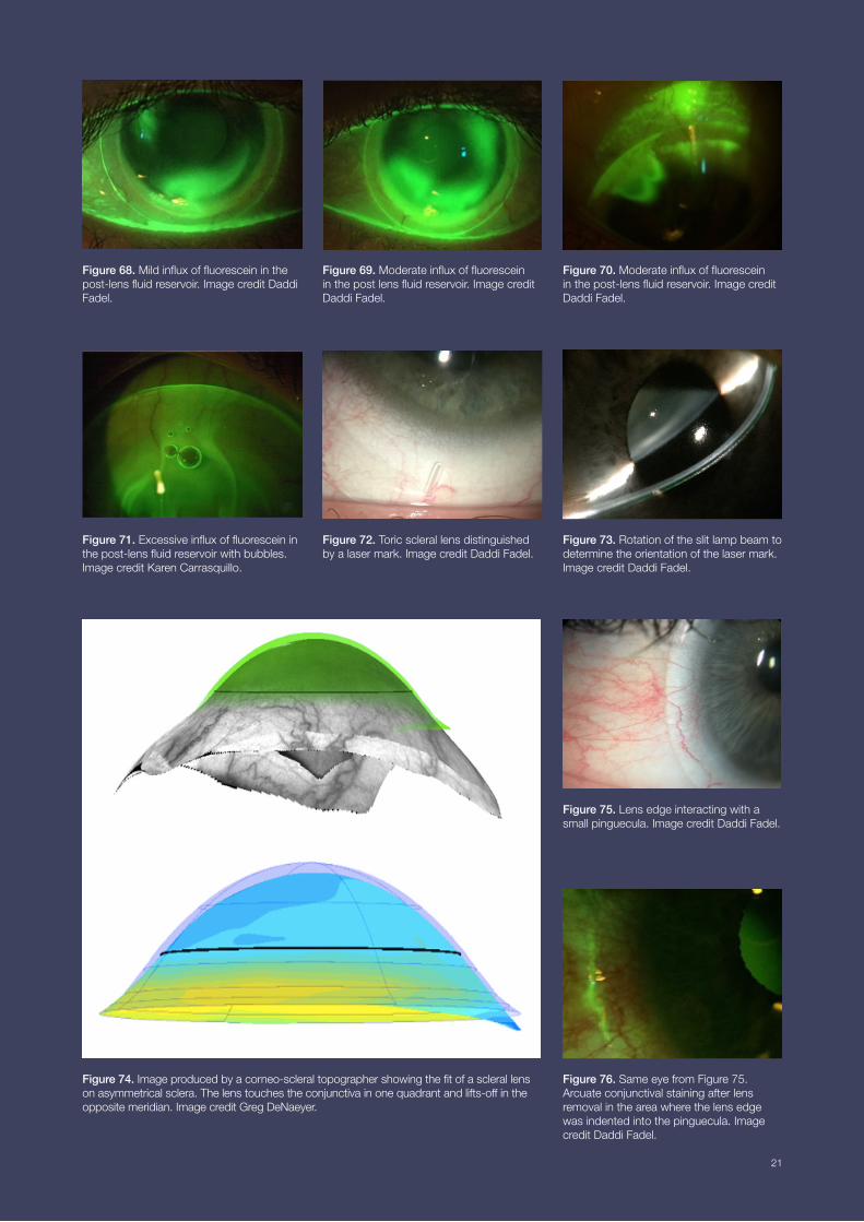
Figure 68. Mild influx of fluorescein in the post-lens fluid reservoir. Image credit Daddi Fadel.
Figure 69. Moderate influx of fluorescein in the post lens fluid reservoir. Image credit Daddi Fadel.
Figure 71. Excessive influx of fluorescein in the post-lens fluid reservoir with bubbles. Image credit Karen Carrasquillo.
Figure 74. Image produced by a corneo-scleral topographer showing the fit of a scleral lens on asymmetrical sclera. The lens touches the conjunctiva in one quadrant and lifts-off in the opposite meridian. Image credit Greg DeNaeyer.
Figure 70. Moderate influx of fluorescein in the post-lens fluid reservoir. Image credit Daddi Fadel.
Figure 73. Rotation of the slit lamp beam to determine the orientation of the laser mark. Image credit Daddi Fadel.
Figure 76. Same eye from Figure 75. Arcuate conjunctival staining after lens removal in the area where the lens edge was indented into the pinguecula. Image credit Daddi Fadel.
Figure 72. Toric scleral lens distinguished by a laser mark. Image credit Daddi Fadel.
Figure 75. Lens edge interacting with a small pinguecula. Image credit Daddi Fadel.
21
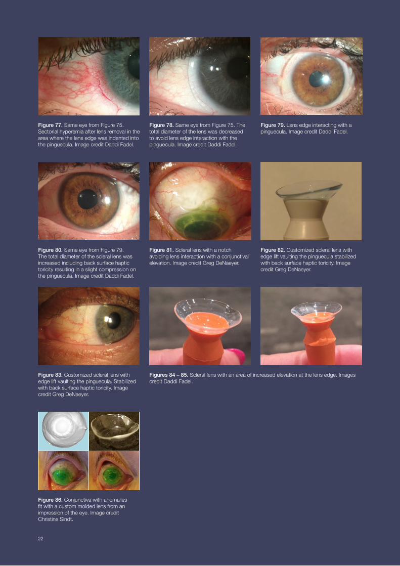
Figure 80. Same eye from Figure 79. The total diameter of the scleral lens was increased including back surface haptic toricity resulting in a slight compression on the pinguecula. Image credit Daddi Fadel.
Figure 81. Scleral lens with a notch avoiding lens interaction with a conjunctival elevation. Image credit Greg DeNaeyer.
Figure 83. Customized scleral lens with edge lift vaulting the pinguecula. Stabilized with back surface haptic toricity. Image credit Greg DeNaeyer.
Figure 86. Conjunctiva with anomalies fit with a custom molded lens from an impression of the eye. Image credit Christine Sindt.
Figure 82. Customized scleral lens with edge lift vaulting the pinguecula stabilized with back surface haptic toricity. Image credit Greg DeNaeyer.
Figures 84 – 85. Scleral lens with an area of increased elevation at the lens edge. Images credit Daddi Fadel.
Figure 77. Same eye from Figure 75. Sectorial hyperemia after lens removal in the area where the lens edge was indented into the pinguecula. Image credit Daddi Fadel.
Figure 79. Lens edge interacting with a pinguecula. Image credit Daddi Fadel.
Figure 78. Same eye from Figure 75. The total diameter of the lens was decreased to avoid lens edge interaction with the pinguecula. Image credit Daddi Fadel.
22
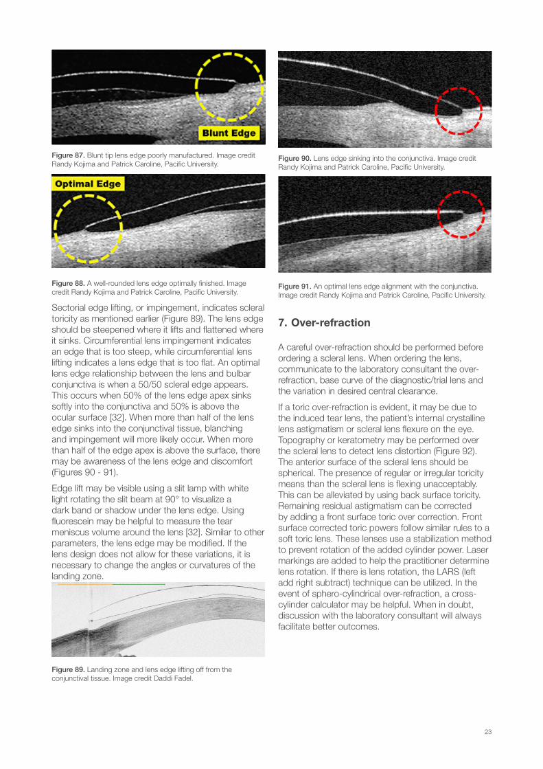
Figure 87. Blunt tip lens edge poorly manufactured. Image credit Randy Kojima and Patrick Caroline, Pacific University.
Figure 88. A well-rounded lens edge optimally finished. Image credit Randy Kojima and Patrick Caroline, Pacific University.
Sectorial edge lifting, or impingement, indicates scleral toricity as mentioned earlier (Figure 89). The lens edge should be steepened where it lifts and flattened where it sinks. Circumferential lens impingement indicates an edge that is too steep, while circumferential lens lifting indicates a lens edge that is too flat. An optimal lens edge relationship between the lens and bulbar conjunctiva is when a 50/50 scleral edge appears. This occurs when 50% of the lens edge apex sinks softly into the conjunctiva and 50% is above the ocular surface [32]. When more than half of the lens edge sinks into the conjunctival tissue, blanching and impingement will more likely occur. When more than half of the edge apex is above the surface, there may be awareness of the lens edge and discomfort (Figures 90 - 91).
Edge lift may be visible using a slit lamp with white light rotating the slit beam at 90° to visualize a dark band or shadow under the lens edge. Using fluorescein may be helpful to measure the tear meniscus volume around the lens [32]. Similar to other parameters, the lens edge may be modified. If the lens design does not allow for these variations, it is necessary to change the angles or curvatures of the landing zone.
Figure 89. Landing zone and lens edge lifting off from the conjunctival tissue. Image credit Daddi Fadel.
Figure 90. Lens edge sinking into the conjunctiva. Image credit Randy Kojima and Patrick Caroline, Pacific University.
Figure 91. An optimal lens edge alignment with the conjunctiva. Image credit Randy Kojima and Patrick Caroline, Pacific University.
7. Over-refraction
A careful over-refraction should be performed before ordering a scleral lens. When ordering the lens, communicate to the laboratory consultant the over-refraction, base curve of the diagnostic/trial lens and the variation in desired central clearance.
If a toric over-refraction is evident, it may be due to the induced tear lens, the patient’s internal crystalline lens astigmatism or scleral lens flexure on the eye. Topography or keratometry may be performed over the scleral lens to detect lens distortion (Figure 92). The anterior surface of the scleral lens should be spherical. The presence of regular or irregular toricity means than the scleral lens is flexing unacceptably. This can be alleviated by using back surface toricity. Remaining residual astigmatism can be corrected by adding a front surface toric over correction. Front surface corrected toric powers follow similar rules to a soft toric lens. These lenses use a stabilization method to prevent rotation of the added cylinder power. Laser markings are added to help the practitioner determine lens rotation. If there is lens rotation, the LARS (left add right subtract) technique can be utilized. In the event of sphero-cylindrical over-refraction, a cross-cylinder calculator may be helpful. When in doubt, discussion with the laboratory consultant will always facilitate better outcomes.
23
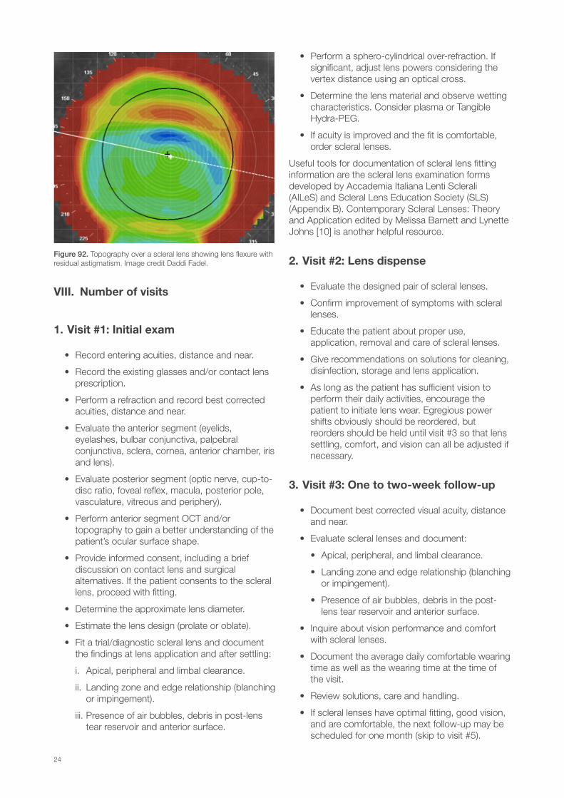
Figure 92. Topography over a scleral lens showing lens flexure with residual astigmatism. Image credit Daddi Fadel.
VIII. Number of visits
1. Visit #1: Initial exam
• Record entering acuities, distance and near.
• Record the existing glasses and/or contact lens prescription.
• Perform a refraction and record best corrected acuities, distance and near.
• Evaluate the anterior segment (eyelids, eyelashes, bulbar conjunctiva, palpebral conjunctiva, sclera, cornea, anterior chamber, iris and lens).
• Evaluate posterior segment (optic nerve, cup-to-disc ratio, foveal reflex, macula, posterior pole, vasculature, vitreous and periphery).
• Perform anterior segment OCT and/or topography to gain a better understanding of the patient’s ocular surface shape.
• Provide informed consent, including a brief discussion on contact lens and surgical alternatives. If the patient consents to the scleral lens, proceed with fitting.
• Determine the approximate lens diameter.
• Estimate the lens design (prolate or oblate).
• Fit a trial/diagnostic scleral lens and document the findings at lens application and after settling:
i. Apical, peripheral and limbal clearance.
ii. Landing zone and edge relationship (blanching or impingement).
iii. Presence of air bubbles, debris in post-lens tear reservoir and anterior surface.
• Perform a sphero-cylindrical over-refraction. If significant, adjust lens powers considering the vertex distance using an optical cross.
• Determine the lens material and observe wetting characteristics. Consider plasma or Tangible Hydra-PEG.
• If acuity is improved and the fit is comfortable, order scleral lenses.
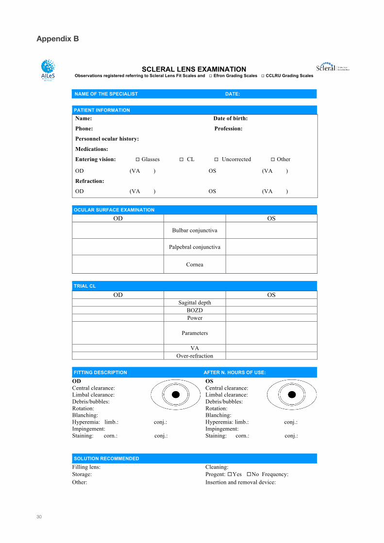
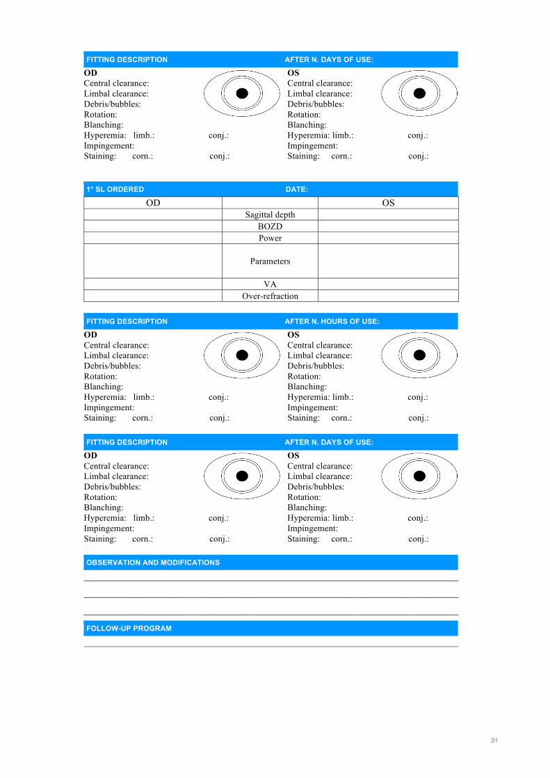
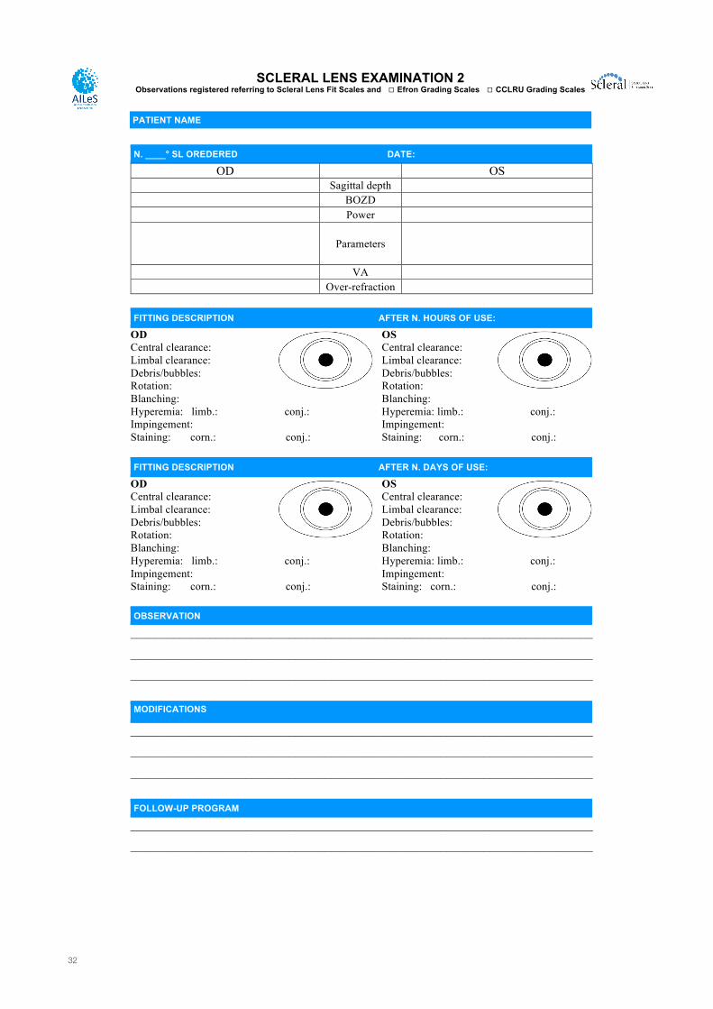
Useful tools for documentation of scleral lens fitting information are the scleral lens examination forms developed by Accademia Italiana Lenti Sclerali (AILeS) and Scleral Lens Education Society (SLS) (Appendix B). Contemporary Scleral Lenses: Theory and Application edited by Melissa Barnett and Lynette Johns [10] is another helpful resource.
2. Visit #2: Lens dispense
• Evaluate the designed pair of scleral lenses.
• Confirm improvement of symptoms with scleral lenses.
• Educate the patient about proper use, application, removal and care of scleral lenses.
• Give recommendations on solutions for cleaning, disinfection, storage and lens application.
• As long as the patient has sufficient vision to perform their daily activities, encourage the patient to initiate lens wear. Egregious power shifts obviously should be reordered, but reorders should be held until visit #3 so that lens settling, comfort, and vision can all be adjusted if necessary.
3. Visit #3: One to two-week follow-up
• Document best corrected visual acuity, distance and near.
• Evaluate scleral lenses and document:
• Apical, peripheral, and limbal clearance.
• Landing zone and edge relationship (blanching or impingement).
• Presence of air bubbles, debris in the post-lens tear reservoir and anterior surface.
• Inquire about vision performance and comfort with scleral lenses.
• Document the average daily comfortable wearing time as well as the wearing time at the time of the visit.
• Review solutions, care and handling.
• If scleral lenses have optimal fitting, good vision, and are comfortable, the next follow-up may be scheduled for one month (skip to visit #5).
24

• If scleral lenses require modification, the next appointment scheduled should be for a dispense (proceed to visit #4).
4. Visit #4: Modified lens dispense
• Evaluate scleral lenses.
• Schedule a follow-up visit at two weeks which will duplicate visit #3.
5. Visit #5: One-month follow-up
• Document best corrected visual acuity, distance and near.
• Evaluate scleral lenses.
• Perform a sphero-cylindrical over-refraction.
• Verify patient compliance.
• Additional visit frequency depends on the ocular condition and if modifications or further scleral lens evaluations are needed.
• If lenses are finalized, a follow-up evaluation may be scheduled at 4-6 months. Compromised corneas with a history of corneal transplants, a history of a persistent epithelial defect, and central scars should be seen more frequently.
IX. Wearing schedule
Much like corneal gas permeable lenses, scleral lenses require an adaptation period. Each practitioner may have a different recommended wearing schedule based on the corneal condition, Dk/t of the lens, and patient expectations. At a minimum, a specified wearing schedule is recommended. An example of an initial wearing schedule is 4-6 waking hours the first day, increasing 2 hours each subsequent day, leading to a maximum wearing time of 12 hours per day. Extended wear, or sleeping in lenses, is not recommended. If a cornea is at risk, for example a patient post penetrating-keratoplasty with a reduced endothelial cell count and a concern for corneal edema, the wearing time may be reduced. After subsequent visits without any complications, the wearing schedule may be increased.
X. Patient education for correct application and removal techniques
1. Scleral lens application
1.1. Manual method
a. Two fingers - The lens may be placed on
the index and middle finger of the dominant hand, which will serve as a support.
b. Three fingers - Three fingers may be used as a support for the lens, the thumb, index and middle fingers of the dominant hand.
1.2. Device method
The device applicator approach consists of using an inserter tool such as a large plunger, See Green® Lens Insertor, EZi Scleral Lens Applicator, Number 8 “O” Ring and orthodontic ring (see section XI – 1- Application devices).
• Wet the device with non-preserved saline before placing the lens on the device.
• Using a plunger, squeeze the side before coming in contact with the lens.
• Hold the lens from the edge and position it on the application device releasing the side pressure on the plunger.
• Overfill the scleral lens with non-preserved saline, until the solution appears convex above the lens.
• Open the eyelids, with the thumb and the index finger of the non-dominant hand.
• Leaning the chin to the chest, with the head parallel to the ground, come closer with the lens to the eye until it is applied softly without exerting pressure.
• Squeeze the plunger to release it from the lens surface and release the eyelids.
After application, check in the mirror for the presence of air bubbles. If air bubbles are detected, lenses should be removed and reapplied again (Figures 93 - 96). If air bubbles persist, use a more viscous preservative-free solution for scleral lens application.
Figure 93. Small and peripheral air bubbles resulting from lens insertion. Image credit Daddi Fadel
25
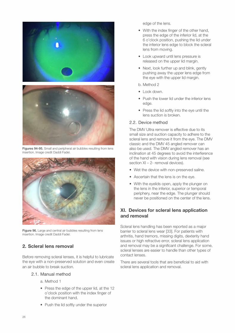
Figures 94-95. Small and peripheral air bubbles resulting from lens insertion. Image credit Daddi Fadel.
Figure 96. Large and central air bubbles resulting from lens insertion. Image credit Daddi Fadel.
2. Scleral lens removal
Before removing scleral lenses, it is helpful to lubricate the eye with a non-preserved solution and even create an air bubble to break suction.
2.1. Manual method
a. Method 1
• Press the edge of the upper lid, at the 12 o’clock position with the index finger of the dominant hand.
• Push the lid softly under the superior
edge of the lens.
• With the index finger of the other hand, press the edge of the inferior lid, at the 6 o’clock position, pushing the lid under the inferior lens edge to block the scleral lens from moving.
• Look upward until lens pressure is released on the upper lid margin.
• Next, look further up and blink, gently pushing away the upper lens edge from the eye with the upper lid margin.
b. Method 2
• Look down.
• Push the lower lid under the inferior lens edge.
• Press the lid softly into the eye until the lens suction is broken.
2.2. Device method
The DMV Ultra remover is effective due to its small size and suction capacity to adhere to the scleral lens and remove it from the eye. The DMV classic and the DMV 45 angled remover can also be used. The DMV angled remover has an inclination at 45 degrees to avoid the interference of the hand with vision during lens removal (see section XI – 2- removal devices).
• Wet the device with non-preserved saline.
• Ascertain that the lens is on the eye.
• With the eyelids open, apply the plunger on the lens in the inferior, superior or temporal periphery, near the edge. The plunger should never be positioned on the center of the lens.
XI. Devices for scleral lens application and removal
Scleral lens handling has been reported as a major barrier to scleral lens wear [33]. For patients with arthritis, hand tremors, missing digits, dexterity hand issues or high refractive error, scleral lens application and removal may be a significant challenge. For some, scleral lenses are easier to handle than other types of contact lenses.
There are several tools that are beneficial to aid with scleral lens application and removal.
26
1. Application devices:
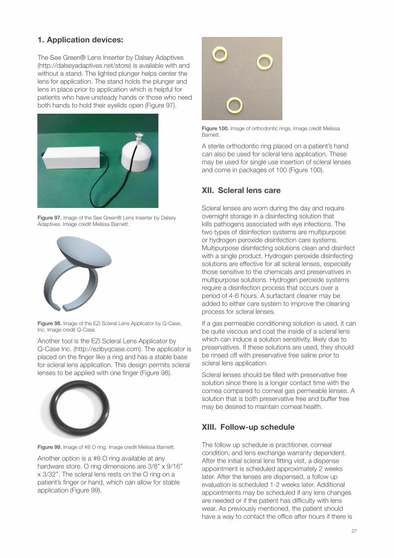
The See Green® Lens Inserter by Dalsey Adaptives (http://dalseyadaptives.net/store) is available with and without a stand. The lighted plunger helps center the lens for application. The stand holds the plunger and lens in place prior to application which is helpful for patients who have unsteady hands or those who need both hands to hold their eyelids open (Figure 97).
Figure 97. Image of the See Green® Lens Inserter by Dalsey Adaptives. Image credit Melissa Barnett.
Figure 98. Image of the EZi Scleral Lens Applicator by Q-Case, Inc. Image credit Q-Case.
Another tool is the EZi Scleral Lens Applicator by Q-Case Inc. (http://ezibyqcase.com). The applicator is placed on the finger like a ring and has a stable base for scleral lens application. This design permits scleral lenses to be applied with one finger (Figure 98).
Figure 99. Image of #8 O ring. Image credit Melissa Barnett.
Another option is a #8 O ring available at any hardware store. O ring dimensions are 3/8” x 9/16” x 3/32”. The scleral lens rests on the O ring on a patient’s finger or hand, which can allow for stable application (Figure 99).
Figure 100. Image of orthodontic rings. Image credit Melissa Barnett.
A sterile orthodontic ring placed on a patient’s hand can also be used for scleral lens application. These may be used for single use insertion of scleral lenses and come in packages of 100 (Figure 100).
XII. Scleral lens care
Scleral lenses are worn during the day and require overnight storage in a disinfecting solution that kills pathogens associated with eye infections. The two types of disinfection systems are multipurpose or hydrogen peroxide disinfection care systems. Multipurpose disinfecting solutions clean and disinfect with a single product. Hydrogen peroxide disinfecting solutions are effective for all scleral lenses, especially those sensitive to the chemicals and preservatives in multipurpose solutions. Hydrogen peroxide systems require a disinfection process that occurs over a period of 4-6 hours. A surfactant cleaner may be added to either care system to improve the cleaning process for scleral lenses.
If a gas permeable conditioning solution is used, it can be quite viscous and coat the inside of a scleral lens which can induce a solution sensitivity, likely due to preservatives. If these solutions are used, they should be rinsed off with preservative free saline prior to scleral lens application.
Scleral lenses should be filled with preservative free solution since there is a longer contact time with the cornea compared to corneal gas permeable lenses. A solution that is both preservative free and buffer free may be desired to maintain corneal health.
XIII. Follow-up schedule
The follow up schedule is practitioner, corneal condition, and lens exchange warranty dependent. After the initial scleral lens fitting visit, a dispense appointment is scheduled approximately 2 weeks later. After the lenses are dispensed, a follow up evaluation is scheduled 1-2 weeks later. Additional appointments may be scheduled if any lens changes are needed or if the patient has difficulty with lens wear. As previously mentioned, the patient should have a way to contact the office after hours if there is
27
a problem. Additional office visits can vary between one and six months, depending on the clinical judgment of the practitioner.
At each follow up visit, it is important to evaluate the scleral lens. First, use a global view outside of the slit lamp, evaluate for excessive hyperemia or obvious peripheral blanching or impingement. Next, a slit lamp examination using white light with a slit beam is used to evaluate central and limbal clearance in all lens positions. If sodium fluorescein is utilized, it is instilled prior to scleral lens removal and can seep in to evaluate the lens fit. The peripheral lens is appraised for evidence of blanching, compression, or impingement. Anterior lens surface debris, post lens reservoir debris, bubbles and conjunctival prolapse are assessed. Anterior segment OCT may also be used for evaluation.
After the scleral lens is evaluated, it is removed, and sodium fluorescein is instilled to assess corneal and conjunctival staining. Specifically, punctate epithelial keratopathy and microcystic edema of the cornea. It is important to pay special attention to the limbus. Any areas of conjunctival impression are assessed in all directions of gaze. The upper eyelid is lifted with the patient looking down to visualize the superior cornea.
If the patient’s vision and ocular health is stable, then the next appropriate office visit should be scheduled.
XIV. Conclusion
We hope this guide will simplify the scleral lens fitting process and aftercare visits. New technologies available to practitioners have allowed scleral lenses to literally change lives. While the information conveyed here should serve as an appropriate guide, nothing takes the place of good clinical judgment. For more information consult the book dedicated to scleral lenses, Contemporary Scleral Lenses: Theory and Application [10]. Colleagues from the SLS and the AILeS are available for further assistance.
28
Appendix A
29
PATIENT INFORMATION Name: Date of birth:
Phone: Profession:
Personnel ocular history: Medications:
Entering vision: ◻ Glasses ◻ CL ◻ Uncorrected ◻ Other
OD (VA ) OS (VA )
Refraction:
OD (VA ) OS (VA )
OCULAR SURFACE EXAMINATION
OD OS
Bulbar conjunctiva
Palpebral conjunctiva
Cornea
TRIAL CL
OD OS Sagittal depth BOZD Power
Parameters
VA Over-refraction
FITTING DESCRIPTION AFTER N. HOURS OF USE:
OD OS Central clearance: Central clearance: Limbal clearance: Limbal clearance: Debris/bubbles: Debris/bubbles: Rotation: Rotation: Blanching: Blanching: Hyperemia: limb.: conj.: Hyperemia: limb.: conj.: Impingement: Impingement: Staining: corn.: conj.: Staining: corn.: conj.: SOLUTION RECOMMENDED
Filling lens: Cleaning: Storage: Progent: ◻Yes ◻No Frequency: Other: Insertion and removal device:
SCLERAL LENS EXAMINATION Observations registered referring to Scleral Lens Fit Scales and ◻ Efron Grading Scales ◻ CCLRU Grading Scales
NAME OF THE SPECIALIST DATE:
Appendix B
30
FITTING DESCRIPTION AFTER N. DAYS OF USE:
OD OS Central clearance: Central clearance: Limbal clearance: Limbal clearance: Debris/bubbles: Debris/bubbles: Rotation: Rotation: Blanching: Blanching: Hyperemia: limb.: conj.: Hyperemia: limb.: conj.: Impingement: Impingement: Staining: corn.: conj.: Staining: corn.: conj.: 1° SL ORDERED DATE:
OD OS Sagittal depth BOZD Power
Parameters
VA Over-refraction
FITTING DESCRIPTION AFTER N. HOURS OF USE:
OD OS Central clearance: Central clearance: Limbal clearance: Limbal clearance: Debris/bubbles: Debris/bubbles: Rotation: Rotation: Blanching: Blanching: Hyperemia: limb.: conj.: Hyperemia: limb.: conj.: Impingement: Impingement: Staining: corn.: conj.: Staining: corn.: conj.: FITTING DESCRIPTION AFTER N. DAYS OF USE:
OD OS Central clearance: Central clearance: Limbal clearance: Limbal clearance: Debris/bubbles: Debris/bubbles: Rotation: Rotation: Blanching: Blanching: Hyperemia: limb.: conj.: Hyperemia: limb.: conj.: Impingement: Impingement: Staining: corn.: conj.: Staining: corn.: conj.: OBSERVATION AND MODIFICATIONS
____________________________________________________________________________
____________________________________________________________________________
____________________________________________________________________________
FOLLOW-UP PROGRAM
____________________________________________________________________________
____________________________________________________________________________
31
N. ____° SL OREDERED DATE:
OD OS Sagittal depth BOZD Power
Parameters
VA Over-refraction
FITTING DESCRIPTION AFTER N. HOURS OF USE:
OD OS Central clearance: Central clearance: Limbal clearance: Limbal clearance: Debris/bubbles: Debris/bubbles: Rotation: Rotation: Blanching: Blanching: Hyperemia: limb.: conj.: Hyperemia: limb.: conj.: Impingement: Impingement: Staining: corn.: conj.: Staining: corn.: conj.: FITTING DESCRIPTION AFTER N. DAYS OF USE:
OD OS Central clearance: Central clearance: Limbal clearance: Limbal clearance: Debris/bubbles: Debris/bubbles: Rotation: Rotation: Blanching: Blanching: Hyperemia: limb.: conj.: Hyperemia: limb.: conj.: Impingement: Impingement: Staining: corn.: conj.: Staining: corn.: conj.: OBSERVATION
____________________________________________________________________________
____________________________________________________________________________
____________________________________________________________________________
MODIFICATIONS
____________________________________________________________________________
____________________________________________________________________________
____________________________________________________________________________
FOLLOW-UP PROGRAM
____________________________________________________________________________
____________________________________________________________________________
SCLERAL LENS EXAMINATION 2 Observations registered referring to Scleral Lens Fit Scales and ◻ Efron Grading Scales ◻ CCLRU Grading Scales
PATIENT NAME
32
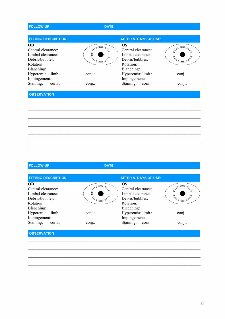
FITTING DESCRIPTION AFTER N. DAYS OF USE:
OD OS Central clearance: Central clearance: Limbal clearance: Limbal clearance: Debris/bubbles: Debris/bubbles: Rotation: Rotation: Blanching: Blanching: Hyperemia: limb.: conj.: Hyperemia: limb.: conj.: Impingement: Impingement: Staining: corn.: conj.: Staining: corn.: conj.: OBSERVATION
____________________________________________________________________________
____________________________________________________________________________
____________________________________________________________________________
____________________________________________________________________________
____________________________________________________________________________
____________________________________________________________________________
____________________________________________________________________________
FOLLOW-UP
Observations registered referring to Scleral Lens Fit Scales and ◻ Efron Grading Scales ◻ CCLRU Grading Scales
FOLLOW-UP DATE
FOLLOW-UP DATE
33

References
[1] Potter R. The importance of staff training for GP lenses. Contact Lens Spectrum. 2014; May;29;28-30,32.
[2] McMahon TT, Szczotka-Flynn L, Barr JT, Anderson RJ, Slaughter ME, Lass JH, Iyengar SK, CLEK Study Group. A new method for grading the severity of keratoconus: the Keratoconus Severity Score (KSS). Cornea. 2006;25(7), 794-800. DOI: 10.1097/01.ico.0000226359.26678.d1
[3] Wallang BS, Das S. Keratoglobus. Eye. 2013;27(9), 1004.
[4] Hersh PS, Stulting RD, Muller D, Durrie DS, Rajpal RK, U.S. Crosslinking Study Group. U.S. Multicenter Clinical Trial of Corneal Collagen Crosslinking for Treatment of Corneal Ectasia after Refractive Surgery. Ophthalmology. 2017 Oct;124(10):1475-1484. doi: 10.1016/j.ophtha.2017.05.036. Epub 2017 Jun 24.
[5] Patel SV, Malta JB, Banitt MR, Mian SI, Sugar A, Elner VM, Tester RA, Farjo QA, Soong HK. Recurrent ectasia in corneal grafts and outcomes of repeat keratoplasty for keratoconus. British Journal of Ophthalmology. 2009;93:191-197.
[6] Gemoules G. A novel method of fitting scleral lenses using high resolution optical coherence tomography. Eye & Contact Lens. 2008;3,80–3. doi: 10.1097/ICL.0b013e318166394d.
[7] Fadel D, Barnett M. Scleral Lenses: Prepare for Landing. Contact Lens Spectrum. 2017; August; 32:42-43-55.
[8] Fadel D. Modern Scleral Lenses: Mini versus Large. Cont Lens Anterior Eye. 2017;40: 200-207 http://dx.doi.org/10.1016/j.clae.2017.04.003
[9] Bergmanson JPG. Clinical Ocular Anatomy and Physiology. 24th edition, Houston, Texas Eye Research and Technology Center, 2017; p 136.
[10] Barnett M, Johns LK. Contemporary Scleral Lenses: Theory and Application. Bentham Science 2017. Volume 4 ISBN: 978-1-68108-567-8. 214-215.
[11] Benjamin WJ. Oxygen transport through contact lenses. In: Guillon M, Ruben M., editors. Contact lens practice. Chapman Hall Medical Publishers;1994:47-69.
[12] Benjamin WJ. The Dk Reference Study Group. Revised oxygen permeability (Dk)of reference materials. Investig Ophthalmol Vis Sci. 2006;47. ARVO E-Abstract 97/B385.
[13] Michaud L, van der Worp E, Brazeau D, Warde R, Giasson CJ. Predicting estimates of oxygen transmissibility for scleral lenses. Cont Lens Anterior Eye. 2012;35:266-71. doi: 10.1016/j.clae.2012.07.004.
[14] Compañ V, Aguilella-Arzo M, Edrington TB, Weissman BA. Modeling Corneal Oxygen with Scleral Gas Permeable Lens Wear. Optom Vis Sci. 2016;93(11):1339-1348. doi: 10.1097/OPX.0000000000000988.
[15] Compañ V, Oliveira C, Aguilella-Arzo M, Mollá S, Peixoto-de-Matos SC, González Méijome JM. Oxygen diffusion and edema with modern scleral rigid gas permeable contact lenses. Invest Ophthalmol Vis Sci. 2014;55:6421-9. doi:10.1167/iovs.14-14038.
[16] Jaynes JM, Edrington TB, Weissman BA. Predicting scleral GP lens entrapped tear layer oxygen tensions. Cont Lens Anterior Eye. 2015;38:44-7. doi: 10.1016/j.clae.2014.09.008.
[17] Caroline P, André M. Scleral lens settling. Contact Lens Spectrum. 2012 May;27:56.
[18] Kauffman MJ, Gilmartin CA, Bennett ES, Bassi CJ. A comparison of the short-term settling of three scleral lens designs. Optom Vis Sci. 2014;91(12):1462-1466.
[19] Mountford J. Scleral contact lens settling rates. Paper presented at the 10th Congress of the Orthokeratology Society of Oceania (OSO), Queensland, Australia, July 2012.
[20] Vincent SJ, Alonso-Caneiro D, Collins MJ. The temporal dynamics of miniscleral contact lenses: Central corneal clearance and centration. Cont Lens Anterior Eye. 2017 Jul 14. pii: S1367-0484(17)30171-6. doi: 10.1016/j.clae.2017.07.002. [Epub ahead of print].
[21] Van der Worp E. A Guide to Scleral Lens Fitting, Version 2.0 [monograph online]. Forest Grove, OR: Pacific University; 2015. Available from: http://commons.pacificu.edu/mono/10/.
[22] Severinsky B, Behrman S, Frucht-Pery J, Solomon A. Scleral contact lenses for visual rehabilitation after penetrating keratoplasty: long term outcomes, Cont Lens Anterior Eye. 2014;37:196–202. doi:http://dx.doi.org/10.1016/j. clae.2013.11.001.
[23] Barnett M, Fadel D. Scleral lenses: Benefits of toric landing zones. Contact lens Spectrum. 2017;
34

November; 32: 36-41.
[24] Visser ES, Visser R. Case report: bitorische scleralens bij keratitis sicca. Visus. 2002;2:92–95.
[25] Visser ES, Visser R, Van Lier HJJ, Otten HM. A cross sectional survey of the medical indications for and performance of scleral contact lens wear in The Netherlands. Opthalmic Res. 2004;36 (suppl 1):180.
[26] Schornack M. Toric haptics in scleral lens design: a case series. Poster presented at the Global Specialty Lens Symposium. Las Vegas, 2013 January, 27-29.
[27] Mahadevan R, Jagadeesh D, Rajan R, Arumugam AO. Unique hard scleral lens post-LASIK ectasia fitting, Optom Vis Sci. 2014;73:136–142, doi:http://dx. doi.org/10.1097/OPX.0000000000000170.
[28] Visser ES, Visser R, Van Lier HJ Advantages of toric scleral lenses. Optom Vis Sci. 2006;4,233–6. doi:http://dx.doi.org/10.1097/01.
[29] Visser ES, Visser R, Van Lier HJ, Otten HM. Modern Scleral Lenses, Part I: Clinical Features. Eye & Contact Lens. 2007;1:13–6. doi: 10.1097/01.icl.0000233217.68379.d5.
[30] Visser ES, Van der Linden BJ, Otten HM, Van der Lelij A, Visser R. Medical applications and outcomes of bitangential scleral lenses. Optom Vis Sci. 2013;90:1078–85. oi:http://dx.doi.org/10.1097/ OPX.0000000000000018.
[31] Sorbara L, Simpson TL, Maram J, Song ES, Bizheva K, Hutchings N (2015) Optical edge effects create conjunctival indentation thickness artefacts; Ophthalmic Physiol Opt. 2015 May;35(3):283-92. doi: 10.1111/opo.12196. Epub 2015 Feb 9.
[32] Kojima R, Caroline P, Walker M, Kinoshita B, André M, Lampa M. Benefits of OCT When Fitting Specialty Lenses. Contact lens spectrum. 2014; October; 29;46,48-51.
[33] Barnett, M, Lien, V, Li, JY, Durbin-Johnson, B, Mannis, MJ. Use of Scleral Lenses and Miniscleral Lenses After Penetrating Keratoplasty. Eye Contact Lens. 2015 Jul 24.
35

The Clinical Guide for Scleral Lens Success is a valuable, hands-on practical resource to use daily in clinical practice. This unified international guide covers everything from staff training, patient and family communication, step by step scleral lens fitting, scleral lens care and aftercare and follow-up schedules. The Clinical Guide for Scleral Lens Success authored by Melissa Barnett and Daddi Fadel is a collaborative work between the Accademia Italiana Lenti Sclerali (AILeS) and the Scleral Lens Education Society (SLS) with the purpose of providing a unified protocol for scleral lens fitting.
Melissa Barnett OD, FAAO, FSLS, FBCLA Daddi Fadel Dip Optom, FSLS
Download the Clinical Guide to Scleral Lens Success Ebook today at scleralsuccess.com. Available in a variety of languages.
Published by