Embed Size (px)
Citation preview
Outcomes of ab interno trabeculectomy withthe trabectome by degree of angle openingI I Bussel,1 K Kaplowitz,2 J S Schuman,1,3 N A Loewen,1 Trabectome Study Group
▸ Additional material ispublished online only. To viewplease visit the journal online(http://dx.doi.org/10.1136/bjophthalmol-2014-305577).1Department of Ophthalmology,UPMC Eye Center, Eye and EarInstitute, Ophthalmology andVisual Science Research Center,University of Pittsburgh Schoolof Medicine, Pittsburgh,Pennsylvania, USA2Department of Ophthalmology,Stony Brook University Schoolof Medicine, Stony Brook,New York, USA3Department of Bioengineering,Swanson School of Engineering,University of Pittsburgh,Pittsburgh, Pennsylvania, USA
Correspondence toDr Nils A Loewen, Departmentof Ophthalmology, UPMC EyeCenter, Eye and Ear Institute,Ophthalmology and VisualScience Research Center,University of Pittsburgh Schoolof Medicine, 203 Lothrop St,#819, Pittsburgh, PA 15235,USA;[email protected]
Received 22 May 2014Revised 17 September 2014Accepted 21 September 2014Published Online First21 October 2014
To cite: Bussel II,Kaplowitz K, Schuman JS,et al. Br J Ophthalmol2015;99:914–919.
ABSTRACTAim To analyse ab interno trabeculectomy (AIT) withthe trabectome and combined phacoemulsification withAIT (phaco-AIT) by Shaffer angle grade (SG).Methods Prospective study of AIT and phaco-AIT withnarrow angles of SG≤2 versus open angles ≥3.Outcomes included intraocular pressure (IOP),medications, complications, secondary surgery andsuccess (IOP <21 mm Hg and >20% reduction withoutfurther surgery). Exclusion criteria were missingpreoperative data and <1 year follow-up.Results Of 671 included cases, at 1 year AIT SG≤2(n=43) had an IOP reduction of 42% from 27.3±7.4 to15.7±3.0 mm Hg (p<0.01) versus AIT SG≥3 (n=271)with an IOP reduction of 37% from 26.1±7.8 to 16.4±3.9 mm Hg (p<0.01). In phaco-AIT with SG≤2 (n=48),IOP was reduced 24% from 20.7±7.0 to 15.7±3.6 mm Hg (p<0.01) versus phaco-AIT with SG≥3(n=309) with an IOP reduction of 25% from 22.6±6.4to 17.0±3.4 mm Hg (p<0.01). There was no differencebetween SG≤2 and SG≥3 in reduction of IOP ormedications, complications, secondary surgery andsuccess rates (p>0.05).Conclusions SG≤2 is not associated with worseoutcomes in AIT or phaco-AIT.
INTRODUCTIONAb interno trabeculectomy (AIT) with the trabec-tome (Neomedix Corporation, Tustin, California,USA) is a nearly 10-year old,1 minimally invasiveglaucoma surgery (MIGS) modality that lowers theintraocular pressure (IOP) by increasing aqueousoutflow using plasma-mediated ablation of trabecu-lar meshwork (TM).2 A narrow anterior chamberangle is considered a relative contraindicationbecause surgeons feel that, in addition to an oftenless direct access during surgery, peripheral anteriorsynechiae, descemetisation of the angle and fibrosismay form more readily and hasten failure.3 Thismay preclude a large numbers of glaucoma patientsfrom AIT and similar MIGS which have a highlyfavourable risk profile compared with traditionalfiltering glaucoma surgery with near absence ofvision-threatening complications: while 74% ofglaucomas present with an open angle, angle-closure glaucoma contributes up to 70% of glau-coma cases in women and to 87% of cases inAsians.4 Progressively narrow angles have beenshown to be correlated with higher IOP, presum-ably by causing higher outflow resistance.5
Conversely, cataract surgery alone has been shownto deepen the angle and lower IOP.6
Although numerous studies have described thesafety and efficacy of AIT in the treatment of glau-coma,1 7 the relationship between anterior chamber
angle grade and outcomes has not been formallyexamined. The purpose of this nonrandomisedstudy was to gain insight into whether the degreeof preoperative angle opening relates to IOP out-comes of AIT or combined phacoemulsificationwith AIT (phaco-AIT), respectively. We hypothe-sised that narrow angles would be associated withworse IOP reduction and a higher failure rate.
METHODSSubjects were enrolled from the Trabectome StudyGroup database,7 a postmarket surveillance require-ment for Neomedix, and divided into phakicpatients who had only trabectome-mediated AIT orcombined phacoemulsification with AIT(phaco-AIT). Data for this study were collected withinstitutional review board approval, in accordancewith the Declaration of Helsinki and the HealthInsurance Portability and Accountability Act.This prospective interventional cohort included
all phakic patients with a diagnosis of glaucoma(with or without visually significant cataract), whohad 12 months of follow-up. Anterior chamberangles in all patients were graded by Shaffer grade(SG),8 the most commonly used classificationsystem in which ‘0’ to ‘slit’ represents a totally orpartially closed angle with potential for angleclosure that is present or very likely, ‘1’ an anglewidth of 10° (very narrow) and closure potentialthat is probable, ‘2’ representing 20° and possiblepotential for closure, ‘3’ standing for 20° to 45°with unlikely closure and grade ‘4’ indicating awide open angle and improbable potential forangle closure. Subjects were excluded if they hadmissing preoperative IOP data or end-stage visualfield (VF) damage.The indication for AIT alone in phakic eyes con-
sisted of IOP above target with progressive glau-coma on maximally tolerated medical or lasertherapy. All surgeons were recommended to followthe standard postoperative protocol consisting of1% pilocarpine four times per day for 1 monththen three times per day for 1 month, 1% prednis-olone acetate four times per day for 1 week to betapered by one drop each week and a fourth orthird generation fluoroquinolone four times perday for 1 week. Glaucoma medications could becontinued as deemed necessary to achieve targetpressures. Deviations from this protocol wereallowed at the clinician’s discretion and notmonitored.We converted visual acuities to logarithm of the
minimum angle of resolution (logMAR). The indi-cation for phaco-AIT consisted of a visually signifi-cant cataract with at least 0.4 logMAR (20/50Snellen) visual brightness acuity testing and the
Open AccessScan to access more
free content
914 Bussel II, et al. Br J Ophthalmol 2015;99:914–919. doi:10.1136/bjophthalmol-2014-305577
Clinical science on 26 July 2018 by guest. P
rotected by copyright.http://bjo.bm
j.com/
Br J O
phthalmol: first published as 10.1136/bjophthalm
ol-2014-305577 on 21 October 2014. D
ownloaded from
need to lower IOP or the number of glaucoma medications. VFstatus was categorised as early, moderate or advanced by individ-ual glaucoma specialists based on the most recent Humphrey VFexams (Carl Zeiss Meditec AG, Jena, Germany). All patientshad a comprehensive slit lamp and ophthalmoscopy exam priorto surgery.
The major outcome measures included IOP, number of glau-coma medications, and need for secondary glaucoma surgery.Each hypotensive agent in an eye drop was counted as a glau-coma medication. Pilocarpine and oral carbonic anhydrase inhi-bitors were also counted as a glaucoma medication. We used theWilcoxon test to compare IOP and number of medicationswithin each group and between groups, and the Mann–Whitneytest and χ2 test to compare continuous and categorical variablesbetween groups, respectively. All statistical analyses were per-formed using R.9 Statistical significance was set at p<0.05.Continuous variables were expressed as mean±SD. We createdscattergrams to illustrate individual outcomes and survival plotsusing a Kaplan–Meier survival analysis. Success was defined as afinal IOP <21 mm Hg and a >20% reduction from baselinewithout further surgery. We analysed survival rates by SGs withthe log-rank test.
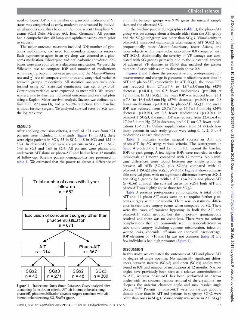
RESULTSAfter applying exclusion criteria, a total of 671 eyes from 671patients were included in this study (figure 1). In AIT, therewere eight patients in SG1, 35 in SG2, 118 in SG3 and 153 inSG4. In phaco-AIT, there were six patients in SG1, 42 in SG2,146 in SG3 and 163 in SG4. All patients were phakic andunderwent AIT alone or phaco-AIT and had at least 12 monthsof follow-up. Baseline patient demographics are presented intable 1. We estimated that the power to detect a difference of
3 mm Hg between groups was 97% given the unequal samplesizes and the observed SD.
In the baseline patient demographics (table 1), the phaco-AITgroup was on average about a decade older than the AIT groupand the SG≤2 subgroup was older than SG≥3. Visual acuity inphaco-AIT improved significantly after surgery. AIT SG≤2 hadproportionally more African–Americans, fewer Asians, andmore subjects with a cup-to-disc ratio above 0.8 compared withAIT SG≥3. Additionally, the severity of VF damage was asso-ciated with SG groups primarily due to the substantial amountof advanced VF damage in SG≥3 that matched the greaternumber of cases with a cup-to-disc ratio above 0.8.
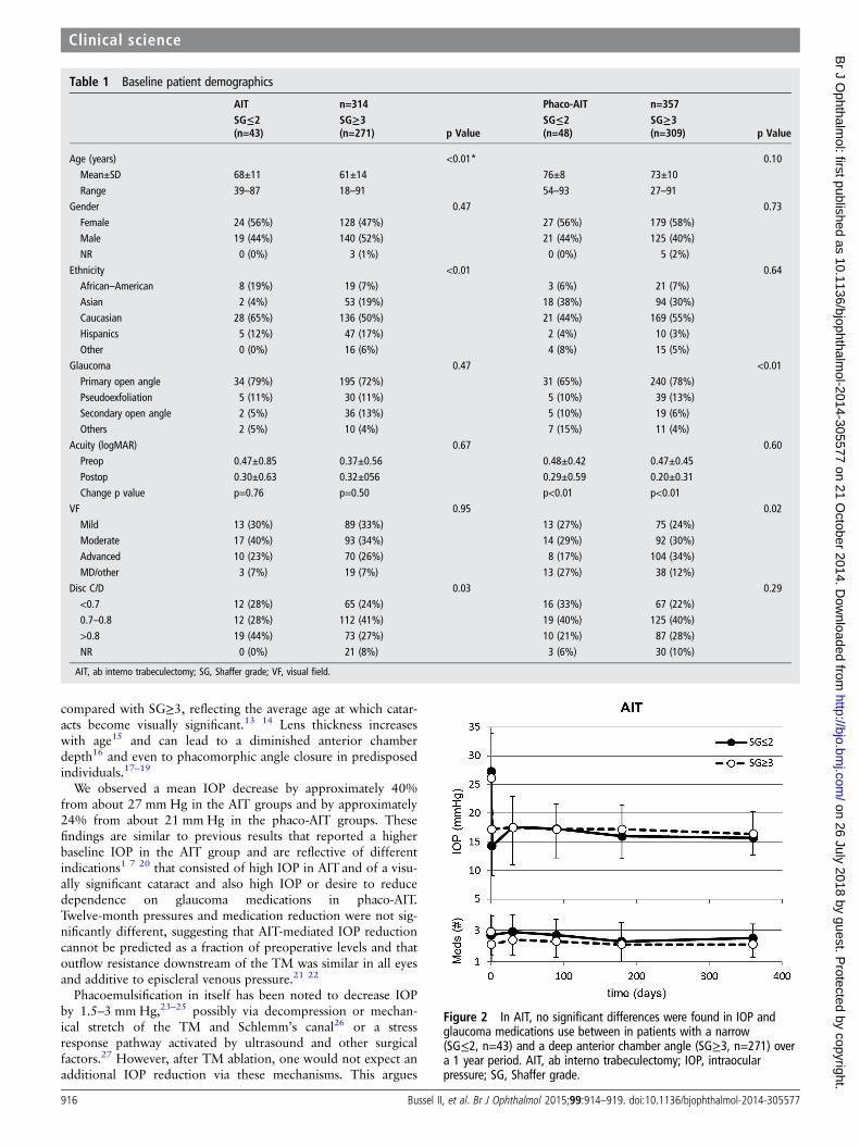
Figures 2 and 3 show the preoperative and postoperative IOPmeasurements and change in glaucoma medications over time inAIT and phaco-AIT, respectively. In AIT SG≤2, the mean IOPwas reduced from 27.3±7.4 to 15.7±3.0 mm Hg (42%decrease, p<0.01), on 0.2 fewer medications (p=1.00) at12 months. In AIT SG≥3, the mean IOP was reduced from 26.1±7.8 to 16.4±3.9 mm Hg (37% decrease, p<0.01) on 0.8fewer medications (p<0.01). In phaco-AIT SG≤2, the meanIOP was reduced from 20.7±7.0 to 15.7±3.6 mm Hg (24%decrease, p<0.01), on 0.8 fewer medications (p<0.01). Inphaco-AIT SG≥3, the mean IOP was reduced from 22.6±6.4 to17.0±3.4 mm Hg (25% decrease, p<0.01) on 0.7 fewer medi-cations (p<0.01). Online supplementary table S1 details howmany patients in each study group were using 0, 1, 2, 3 or 4medications at each time point.
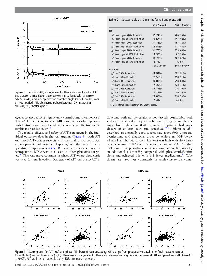
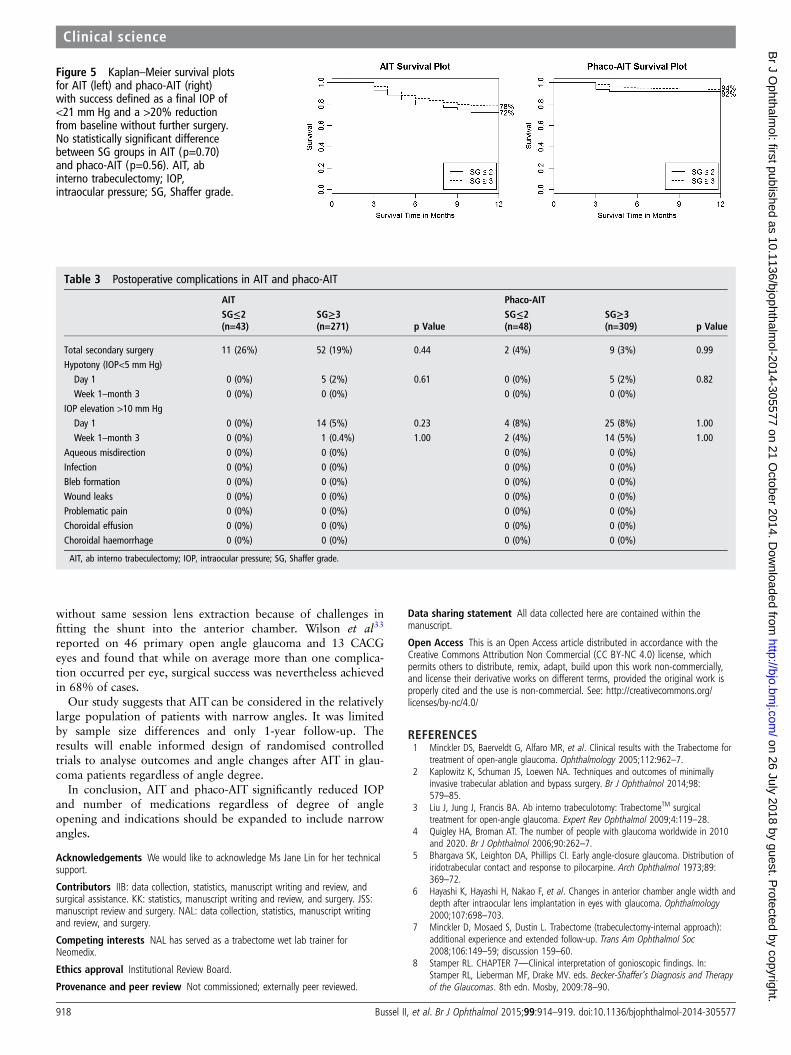
Table 2 indicates similar surgical success in AIT andphaco-AIT by SG using various criteria. The scattergrams infigure 4 plotted the 1 and 12 month IOP against the baselineIOPs of each group. A few higher IOPs were recorded in selectindividuals at 1 month compared with 12 months. No signifi-cant differences were found between any single group orbetween all AITs (SG≤2 plus SG≥3) compared with allphaco-AIT (SG≤2 plus SG≥3; p>0.05). Figure 5 shows compar-able survival plots with no significant difference between SG≤2and SG≥3 groups for neither AIT (p=0.70) nor phaco-AIT(p=0.56) although the survival curve for SG≥3 both AIT andphaco-AITwas slightly above those for SG≤2.
Table 3 presents postoperative complications. A total of 63AIT and 11 phaco-AIT cases went on to require further glau-coma surgery within 12 months. There was no statistical differ-ence in secondary surgery events when compared by SG. Therewere five cases of transient hypotony in both the AIT andphaco-AIT SG≥3 groups, but the hypotony spontaneouslyresolved and there was no vision loss. There were no seriouscomplications that are commonly seen in trabeculectomy ortube shunt surgery including aqueous misdirection, infection,wound leaks, choroidal effusions or choroidal haemorrhage.IOP elevation of >10 mm Hg was not common (table 3) andfew individuals had high pressures (figure 4).
DISCUSSIONIn this study, we evaluated the outcomes of AIT and phaco-AITby degree of angle opening. No statistically significant differ-ences between narrow (SG≤2) and open (SG≥3) angles werefound in IOP and number of medications at 12 months. Narrowangles have previously been seen as a relative contraindicationto AIT, whereas phaco-AIT has been performed in narrowangles with less concern because removal of the crystalline lensdeepens the anterior chamber angle and may resolve angleclosure.10–12 Patients in phaco-AIT were on average about adecade older than in AIT and patients in subgroup SG≤2 wereolder than ones in SG≥3. Visual acuity was worse in AIT SG≤2
Figure 1 Trabectome Study Group Database. Cases analysed afteraccounting for exclusion criteria. AIT, ab interno trabeculectomy;phaco-AIT, phacoemulsification cataract surgery combined with abinterno trabeculectomy; SG, Shaffer grade.
Bussel II, et al. Br J Ophthalmol 2015;99:914–919. doi:10.1136/bjophthalmol-2014-305577 915
Clinical science on 26 July 2018 by guest. P
rotected by copyright.http://bjo.bm
j.com/
Br J O
phthalmol: first published as 10.1136/bjophthalm
ol-2014-305577 on 21 October 2014. D
ownloaded from
compared with SG≥3, reflecting the average age at which catar-acts become visually significant.13 14 Lens thickness increaseswith age15 and can lead to a diminished anterior chamberdepth16 and even to phacomorphic angle closure in predisposedindividuals.17–19
We observed a mean IOP decrease by approximately 40%from about 27 mm Hg in the AIT groups and by approximately24% from about 21 mm Hg in the phaco-AIT groups. Thesefindings are similar to previous results that reported a higherbaseline IOP in the AIT group and are reflective of differentindications1 7 20 that consisted of high IOP in AIT and of a visu-ally significant cataract and also high IOP or desire to reducedependence on glaucoma medications in phaco-AIT.Twelve-month pressures and medication reduction were not sig-nificantly different, suggesting that AIT-mediated IOP reductioncannot be predicted as a fraction of preoperative levels and thatoutflow resistance downstream of the TM was similar in all eyesand additive to episcleral venous pressure.21 22
Phacoemulsification in itself has been noted to decrease IOPby 1.5–3 mm Hg,23–25 possibly via decompression or mechan-ical stretch of the TM and Schlemm’s canal26 or a stressresponse pathway activated by ultrasound and other surgicalfactors.27 However, after TM ablation, one would not expect anadditional IOP reduction via these mechanisms. This argues
Table 1 Baseline patient demographics
AIT n=314 Phaco-AIT n=357SG≤2(n=43)
SG≥3(n=271) p Value
SG≤2(n=48)
SG≥3(n=309) p Value
Age (years) <0.01* 0.10Mean±SD 68±11 61±14 76±8 73±10Range 39–87 18–91 54–93 27–91
Gender 0.47 0.73Female 24 (56%) 128 (47%) 27 (56%) 179 (58%)Male 19 (44%) 140 (52%) 21 (44%) 125 (40%)NR 0 (0%) 3 (1%) 0 (0%) 5 (2%)
Ethnicity <0.01 0.64African–American 8 (19%) 19 (7%) 3 (6%) 21 (7%)Asian 2 (4%) 53 (19%) 18 (38%) 94 (30%)Caucasian 28 (65%) 136 (50%) 21 (44%) 169 (55%)Hispanics 5 (12%) 47 (17%) 2 (4%) 10 (3%)Other 0 (0%) 16 (6%) 4 (8%) 15 (5%)
Glaucoma 0.47 <0.01Primary open angle 34 (79%) 195 (72%) 31 (65%) 240 (78%)Pseudoexfoliation 5 (11%) 30 (11%) 5 (10%) 39 (13%)Secondary open angle 2 (5%) 36 (13%) 5 (10%) 19 (6%)Others 2 (5%) 10 (4%) 7 (15%) 11 (4%)
Acuity (logMAR) 0.67 0.60Preop 0.47±0.85 0.37±0.56 0.48±0.42 0.47±0.45Postop 0.30±0.63 0.32±056 0.29±0.59 0.20±0.31Change p value p=0.76 p=0.50 p<0.01 p<0.01
VF 0.95 0.02Mild 13 (30%) 89 (33%) 13 (27%) 75 (24%)Moderate 17 (40%) 93 (34%) 14 (29%) 92 (30%)Advanced 10 (23%) 70 (26%) 8 (17%) 104 (34%)MD/other 3 (7%) 19 (7%) 13 (27%) 38 (12%)
Disc C/D 0.03 0.29<0.7 12 (28%) 65 (24%) 16 (33%) 67 (22%)0.7–0.8 12 (28%) 112 (41%) 19 (40%) 125 (40%)>0.8 19 (44%) 73 (27%) 10 (21%) 87 (28%)NR 0 (0%) 21 (8%) 3 (6%) 30 (10%)
AIT, ab interno trabeculectomy; SG, Shaffer grade; VF, visual field.
Figure 2 In AIT, no significant differences were found in IOP andglaucoma medications use between in patients with a narrow(SG≤2, n=43) and a deep anterior chamber angle (SG≥3, n=271) overa 1 year period. AIT, ab interno trabeculectomy; IOP, intraocularpressure; SG, Shaffer grade.
916 Bussel II, et al. Br J Ophthalmol 2015;99:914–919. doi:10.1136/bjophthalmol-2014-305577
Clinical science on 26 July 2018 by guest. P
rotected by copyright.http://bjo.bm
j.com/
Br J O
phthalmol: first published as 10.1136/bjophthalm
ol-2014-305577 on 21 October 2014. D
ownloaded from
against cataract surgery significantly contributing to outcomes inphaco-AIT in contrast to other MIGS modalities where phacoe-mulsification alone was found to be nearly as effective as thecombination under study.28
The relative efficacy and safety of AIT is apparent by the indi-vidual outcomes data in the scattergrams (figure 4): both AITand phaco-AIT contain subjects with very high preoperative IOPyet no patient had sustained hypotony or other serious post-operative complications (table 3). Few patients experienced apostoperative IOP elevation as seen in other glaucoma surger-ies.29 This was more common in phaco-AIT where viscoelasticwas used for lens injection. Our study of AIT and phaco-AIT in
glaucoma with narrow angles is not directly comparable withstudies of trabeculectomy or tube shunt surgery in chronicangle-closure glaucoma (CACG), in which patients had angleclosure of at least 180° and synechiae.30–32 Sihota et al31
described an unusually good success rate above 90% using tra-beculectomy and glaucoma drops to achieve an IOP below21 mm Hg. The rate of complications was high with flat cham-bers occurring in 40% and decreased vision in 58%. Anothertrial found that phacotrabeculectomy lowered the IOP only byan additional 1.8 mm Hg compared with phacoemulsificationalone and achieved this with 1.2 fewer medications.30 Tubeshunts are used less commonly in angle-closure glaucomas
Figure 3 In phaco-AIT, no significant differences were found in IOPand glaucoma medications use between in patients with a narrow(SG≤2, n=48) and a deep anterior chamber angle (SG≥3, n=309) overa 1 year period. AIT, ab interno trabeculectomy; IOP, intraocularpressure; SG, Shaffer grade.
Table 2 Success table at 12 months for AIT and phaco-AIT
SG≤2 (n=43) SG≥3 (n=271)
AIT≤21 mm Hg or 20% Reduction 32 (74%) 206 (76%)≤21 mm Hg and 20% Reduction 29 (67%) 157 (58%)≤18 mm Hg or 20% Reduction 31 (72%) 195 (72%)≤18 mm Hg and 20% Reduction 22 (51%) 118 (44%)≤15 mm Hg or 20% Reduction 31 (72%) 175 (65%)≤15 mm Hg and 20% Reduction 13 (30%) 67 (25%)≤12 mm Hg or 20% Reduction 30 (70%) 167 (62%)≤12 mm Hg and 20% Reduction 3 (7%) 16 (6%)
SG≤2 (n=48) SG≥3 (n=309)Phaco-AIT≤21 or 20% Reduction 44 (92%) 282 (91%)≤21 and 20% Reduction 27 (56%) 158 (51%)≤18 or 20% Reduction 39 (81%) 258 (83%)≤18 and 20% Reduction 17 (35%) 128 (41%)≤15 or 20% Reduction 35 (73%) 216 (70%)≤15 and 20% Reduction 7 (15%) 80 (26%)≤12 or 20% Reduction 29 (60%) 170 (55%)≤12 and 20% Reduction 2 (4%) 24 (8%)
AIT, ab interno trabeculectomy; SG, Shaffer grade.
Figure 4 Scattergrams for AIT (top) and phaco-AIT (bottom) demonstrating IOP change from preoperative baseline to final measurement at1 month (left) and at 12 months (right). There were no significant differences between single groups or between all AIT compared with all phaco-AIT(p>0.05). AIT, ab interno trabeculectomy; IOP, intraocular pressure.
Bussel II, et al. Br J Ophthalmol 2015;99:914–919. doi:10.1136/bjophthalmol-2014-305577 917
Clinical science on 26 July 2018 by guest. P
rotected by copyright.http://bjo.bm
j.com/
Br J O
phthalmol: first published as 10.1136/bjophthalm
ol-2014-305577 on 21 October 2014. D
ownloaded from
without same session lens extraction because of challenges infitting the shunt into the anterior chamber. Wilson et al33
reported on 46 primary open angle glaucoma and 13 CACGeyes and found that while on average more than one complica-tion occurred per eye, surgical success was nevertheless achievedin 68% of cases.
Our study suggests that AIT can be considered in the relativelylarge population of patients with narrow angles. It was limitedby sample size differences and only 1-year follow-up. Theresults will enable informed design of randomised controlledtrials to analyse outcomes and angle changes after AIT in glau-coma patients regardless of angle degree.
In conclusion, AIT and phaco-AIT significantly reduced IOPand number of medications regardless of degree of angleopening and indications should be expanded to include narrowangles.
Acknowledgements We would like to acknowledge Ms Jane Lin for her technicalsupport.
Contributors IIB: data collection, statistics, manuscript writing and review, andsurgical assistance. KK: statistics, manuscript writing and review, and surgery. JSS:manuscript review and surgery. NAL: data collection, statistics, manuscript writingand review, and surgery.
Competing interests NAL has served as a trabectome wet lab trainer forNeomedix.
Ethics approval Institutional Review Board.
Provenance and peer review Not commissioned; externally peer reviewed.
Data sharing statement All data collected here are contained within themanuscript.
Open Access This is an Open Access article distributed in accordance with theCreative Commons Attribution Non Commercial (CC BY-NC 4.0) license, whichpermits others to distribute, remix, adapt, build upon this work non-commercially,and license their derivative works on different terms, provided the original work isproperly cited and the use is non-commercial. See: http://creativecommons.org/licenses/by-nc/4.0/
REFERENCES1 Minckler DS, Baerveldt G, Alfaro MR, et al. Clinical results with the Trabectome for
treatment of open-angle glaucoma. Ophthalmology 2005;112:962–7.2 Kaplowitz K, Schuman JS, Loewen NA. Techniques and outcomes of minimally
invasive trabecular ablation and bypass surgery. Br J Ophthalmol 2014;98:579–85.
3 Liu J, Jung J, Francis BA. Ab interno trabeculotomy: TrabectomeTM surgicaltreatment for open-angle glaucoma. Expert Rev Ophthalmol 2009;4:119–28.
4 Quigley HA, Broman AT. The number of people with glaucoma worldwide in 2010and 2020. Br J Ophthalmol 2006;90:262–7.
5 Bhargava SK, Leighton DA, Phillips CI. Early angle-closure glaucoma. Distribution ofiridotrabecular contact and response to pilocarpine. Arch Ophthalmol 1973;89:369–72.
6 Hayashi K, Hayashi H, Nakao F, et al. Changes in anterior chamber angle width anddepth after intraocular lens implantation in eyes with glaucoma. Ophthalmology2000;107:698–703.
7 Minckler D, Mosaed S, Dustin L. Trabectome (trabeculectomy-internal approach):additional experience and extended follow-up. Trans Am Ophthalmol Soc2008;106:149–59; discussion 159–60.
8 Stamper RL. CHAPTER 7—Clinical interpretation of gonioscopic findings. In:Stamper RL, Lieberman MF, Drake MV. eds. Becker-Shaffer’s Diagnosis and Therapyof the Glaucomas. 8th edn. Mosby, 2009:78–90.
Figure 5 Kaplan–Meier survival plotsfor AIT (left) and phaco-AIT (right)with success defined as a final IOP of<21 mm Hg and a >20% reductionfrom baseline without further surgery.No statistically significant differencebetween SG groups in AIT (p=0.70)and phaco-AIT (p=0.56). AIT, abinterno trabeculectomy; IOP,intraocular pressure; SG, Shaffer grade.
Table 3 Postoperative complications in AIT and phaco-AIT
AIT Phaco-AITSG≤2(n=43)
SG≥3(n=271) p Value
SG≤2(n=48)
SG≥3(n=309) p Value
Total secondary surgery 11 (26%) 52 (19%) 0.44 2 (4%) 9 (3%) 0.99Hypotony (IOP<5 mm Hg)
Day 1 0 (0%) 5 (2%) 0.61 0 (0%) 5 (2%) 0.82Week 1–month 3 0 (0%) 0 (0%) 0 (0%) 0 (0%)
IOP elevation >10 mm HgDay 1 0 (0%) 14 (5%) 0.23 4 (8%) 25 (8%) 1.00Week 1–month 3 0 (0%) 1 (0.4%) 1.00 2 (4%) 14 (5%) 1.00
Aqueous misdirection 0 (0%) 0 (0%) 0 (0%) 0 (0%)Infection 0 (0%) 0 (0%) 0 (0%) 0 (0%)Bleb formation 0 (0%) 0 (0%) 0 (0%) 0 (0%)Wound leaks 0 (0%) 0 (0%) 0 (0%) 0 (0%)Problematic pain 0 (0%) 0 (0%) 0 (0%) 0 (0%)Choroidal effusion 0 (0%) 0 (0%) 0 (0%) 0 (0%)Choroidal haemorrhage 0 (0%) 0 (0%) 0 (0%) 0 (0%)
AIT, ab interno trabeculectomy; IOP, intraocular pressure; SG, Shaffer grade.
918 Bussel II, et al. Br J Ophthalmol 2015;99:914–919. doi:10.1136/bjophthalmol-2014-305577
Clinical science on 26 July 2018 by guest. P
rotected by copyright.http://bjo.bm
j.com/
Br J O
phthalmol: first published as 10.1136/bjophthalm
ol-2014-305577 on 21 October 2014. D
ownloaded from
9 R Core Team. R: A Language and Environment for Statistical Computing. Vienna,Austria. R Foundation for Statistical Computing, 2014. http://www.R-project.org/
10 Barbosa DTQ, Levison AL, Lin SC. Clear lens extraction in angle-closure glaucomapatients. Int J Ophthalmol 2013;6:406–8.
11 Shin HC, Subrayan V, Tajunisah I. Changes in anterior chamber depth andintraocular pressure after phacoemulsification in eyes with occludable angles.J Cataract Refract Surg 2010;36:1289–95.
12 Nonaka A, Kondo T, Kikuchi M, et al. Angle widening and alteration of ciliaryprocess configuration after cataract surgery for primary angle closure.Ophthalmology 2006;113:437–41.
13 Klein BE, Klein R, Lee KE. Incidence of age-related cataract: the Beaver Dam EyeStudy. Arch Ophthalmol 1998;116:219–25.
14 Leske MC, Wu SY, Nemesure B, et al. Incidence and progression of lens opacities inthe Barbados Eye Studies. Ophthalmology 2000;107:1267–73.
15 Hashemi H, Khabazkhoob M, Miraftab M, et al. The distribution of axial length,anterior chamber depth, lens thickness, and vitreous chamber depth in an adultpopulation of Shahrud, Iran. BMC Ophthalmol 2012;12:50.
16 Salmon JF. Presenting features of primary angle-closure glaucoma in patients ofmixed ethnic background. S Afr Med J 1993;83:594–7.
17 Kothari R, Tathe S, Gogri P, et al. Lens-induced glaucoma: the need to spreadawareness about early management of cataract among rural population. ISRNOphthalmol 2013;2013:581727.
18 Lee JWY, Lai JSM, Yick DWF, et al. Retrospective case series on the long-term visualand intraocular pressure outcomes of phacomorphic glaucoma. Eye2010;24:1675–80.
19 Leung CKS, Chan W-M, Ko CY, et al. Visualization of anterior chamber angledynamics using optical coherence tomography. Ophthalmology 2005;112:980–4.
20 Francis BA, Minckler D, Dustin L, et al. Combined cataract extraction andtrabeculotomy by the internal approach for coexisting cataract and open-angleglaucoma: initial results. J Cataract Refract Surg 2008;34:1096–103.
21 Nau CB, Malihi M, McLaren JW, et al. Circadian variation of aqueous humordynamics in older healthy adults. Invest Ophthalmol Vis Sci 2013;54:7623–9.
22 Fellman RL, Grover DS. Episcleral venous fluid wave: intraoperative evidence forpatency of the conventional outflow system. J Glaucoma 2014;23:347–50.
23 Mansberger SL, Gordon MO, Jampel H, et al. Reduction in intraocular pressure aftercataract extraction: the Ocular Hypertension Treatment Study. Ophthalmology2012;119:1826–31.
24 Yang HS, Lee J, Choi S. Ocular biometric parameters associated with intraocularpressure reduction after cataract surgery in normal eyes. Am J Ophthalmol2013;156:89–94.e1.
25 Shingleton BJ, Pasternack JJ, Hung JW, et al. Three and five year changes inintraocular pressures after clear corneal phacoemulsification in open angle glaucomapatients, glaucoma suspects, and normal patients. J Glaucoma 2006;15:494–8.
26 Poley BJ, Lindstrom RL, Samuelson TW, et al. Intraocular pressure reduction afterphacoemulsification with intraocular lens implantation in glaucomatous andnonglaucomatous eyes: evaluation of a causal relationship between the natural lensand open-angle glaucoma. J Cataract Refract Surg 2009;35:1946–55. http://www.sciencedirect.com/science/article/pii/S0886335009007664
27 Wang N, Chintala SK, Fini ME, et al. Ultrasound activates the TM ELAM-1/IL-1/NF-kappaB response: a potential mechanism for intraocular pressure reduction afterphacoemulsification. Invest Ophthalmol Vis Sci 2003;44:1977–81.
28 Samuelson TW, Katz LJ, Wells JM, et al. Randomized evaluation of the trabecularmicro-bypass stent with phacoemulsification in patients with glaucoma and cataract.Ophthalmology 2011;118:459–67.
29 Gedde SJ, Herndon LW, Brandt JD, et al. Surgical complications in the Tube VersusTrabeculectomy Study during the first year of follow-up. Am J Ophthalmol2007;143:23–31.
30 Tham CCY, Leung DYL, Kwong YYY, et al. Effects of phacoemulsification versuscombined phaco-trabeculectomy on drainage angle status in primary angle closureglaucoma (PACG). J Glaucoma 2010;19:119–23.
31 Sihota R, Gupta V, Agarwal HC. Long-term evaluation of trabeculectomy in primaryopen angle glaucoma and chronic primary angle closure glaucoma in an Asianpopulation. Clin Experiment Ophthalmol 2004;32:23–8.
32 Alsagoff Z, Aung T, Ang LP, et al. Long-term clinical course of primary angle-closureglaucoma in an Asian population. Ophthalmology 2000;107:2300–4.
33 Wilson MR, Mendis U, Paliwal A, et al. Long-term follow-up of primary glaucomasurgery with Ahmed glaucoma valve implant versus trabeculectomy. Am JOphthalmol 2003;136:464–70.
Bussel II, et al. Br J Ophthalmol 2015;99:914–919. doi:10.1136/bjophthalmol-2014-305577 919
Clinical science on 26 July 2018 by guest. P
rotected by copyright.http://bjo.bm
j.com/
Br J O
phthalmol: first published as 10.1136/bjophthalm
ol-2014-305577 on 21 October 2014. D
ownloaded from