Embed Size (px)
Citation preview
Complex genetic, photothermal, and photoacousticanalysis of nanoparticle-plant interactionsMariya V. Khodakovskayaa,1, Kanishka de Silvaa, Dmitry A. Nedosekinb, Enkeleda Dervishic, Alexandru S. Birisa,c,Evgeny V. Shashkovb,d, Ekaterina I. Galanzhab, and Vladimir P. Zharovb
aDepartment of Applied Science, University of Arkansas, Little Rock, AR 72204; bPhillips Classic Laser and Nanomedicine Laboratories, Winthrop P.Rockefeller Cancer Institute, University of Arkansas for Medical Sciences, Little Rock, AR 72205; cNanotechnology Center, University of Arkansas,Little Rock, AR 72204; and dProkhorov General Physics Institute, Moscow 119991, Russia
Edited by David Chandler, University of California, Berkeley, CA, and approved December 1, 2010 (received for review June 22, 2010)
Understanding the nature of interactions between engineerednanomaterials and plants is crucial in comprehending the impactof nanotechnology on the environment and agriculture with afocus on toxicity concerns, plant disease treatment, and geneticengineering. To date, little progress has been made in studyingnanoparticle-plant interactions at single nanoparticle and geneticlevels. Here, we introduce an advanced platform integrating genet-ic, Raman, photothermal, and photoacoustic methods. Using thisapproach, we discovered that multiwall carbon nanotubes inducepreviously unknown changes in gene expression in tomato leavesand roots, particularly, up-regulation of the stress-related genes,including those induced by pathogens and the water-channelLeAqp2 gene. A nano-bubble amplified photothermal/photoacous-tic imaging, spectroscopy, and burning technique demonstrated thedetection of multiwall carbon nanotubes in roots, leaves, and fruitsdown to the single nanoparticle and cell level. Thus, our integratedplatform allows the study of nanoparticles’ impact on plants withhigher sensitivity and specificity, compared to existing assays.
microarray ∣ laser spectroscopy ∣ tomato plants ∣ aquaporins ∣carbon nanomaterials
The use of nanostructures in biomedicine (1) and, recently, inagriculture (2) is one of the most intensely studied areas in
nanotechnology. Nanoscale materials have been shown to be up-taken by tumor cells (3), bacteria (4), plant cells (5), and animaltissues (6). In particular, carbon nanotubes (CNTs) with their un-ique structural and dimensional properties have been intensivelystudied for drug and gene delivery, tissue engineering, and otherbiomedical applications (7–9). It has also been shown that carbonnanotubes have the ability to penetrate plant cells (5) and inducephytotoxicity at high doses (10). We have demonstrated that sin-gle-wall CNTs at relatively low doses can penetrate even thickseed coats, stimulate germination, and activate enhanced growthof tomato plants (11). However, a thorough understanding of theeffects induced by the nano-sized engineered materials on plantphysiology at the molecular level is still lacking. In addition, themethods used for detecting such nanostructures in plant tissuesare not well established and most of them are time consumingand labor intensive. Moreover, existing nanoparticle detectiontechniques usually decompose and destroy samples to prove thepresence of nanomaterials; as a result, the same plant samplescannot be assessed for genomic/proteomic analysis. For example,the detection of magnetic nanoparticles in pumpkin plants byvibrating sample magnetometer requires drying and digestionof tissue samples with HNO3 (12). Transmission electron micro-scopy (TEM) has been used to monitor the uptake and transpor-tation of CNTs in rice (13), but it has few quantitative capabilitiesand may result in false positive interpretation because of consid-erable similarity in TEM images of CNTs and natural plant struc-tures. Consequently, the analysis has to be combined with spec-troscopic studies for the exact identification and assessment ofthe CNTs in the host plant tissue, and this requires the totaldestruction of the samples (13). Moreover, the majority of the
detection methods require additional nanoparticle labeling (14)that may unpredictably modify both the nanoparticles’ biodistri-bution properties and plant responses. It is obvious that a com-prehensive study of the nature of nanoparticles-plant cell inter-actions will require the development of ultrasensitive methodsfor in situ real-time monitoring of nanoparticle transportation inplants.
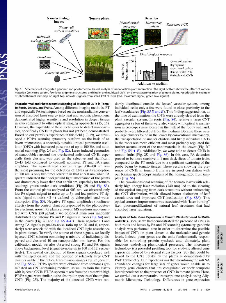
The main goal of this work is to demonstrate an integratedapproach for studying the processes that take place during theexposure of tomato plants to carbon nanotubes, e.g., changes ingene expression, and correlate these findings with the presenceof nanoparticles from roots to leaves. To perform this task, welinked the observed physiological responses of tomato plants withthe complex sets of information provided by a unique combina-tion of microarray analysis and nano-bubble amplified photother-mal and photoacoustic imaging of nanomaterials in 10-day-oldtomato seedlings grown on a medium containing various carbonnanostructural species (Fig. 1).
ResultsTo understand the role of the structural shapes of the carbonac-eous materials on plant physiology and to select those with thehighest impact, four carbon-based structures—activated carbon(AC), few-layer graphene structures, multiwall and single-wallCNTs—were added separately to the Murashige and Skoog (MS)growth medium at an identical concentration of 50 μg∕mL. Themaximum physiological responses, such as the biomass enhance-ment (both for fresh and dried plants), were observed for thesingle- and multiwall CNTs only (see Fig. 1, bottom-right andFig. S1A). The plants exposed to multiwall CNTs were then cho-sen for further detailed examination (multiwall carbon nanotubesare hereinafter referred to simply as “CNTs”). In this study,samples from the same plant seedlings exposed to CNTs were di-vided into two groups that were used in parallel for photoacoustic(PA)/photothermal (PT) detection and genetic analysis. Specifi-cally, different leaf/root tissues from the same plants were usedfor the PA/PT detection and the genetic assays. We did not usethe same identical tissues for both laser exposure and genetic ana-lysis, in order to avoid additional stress that could be induced bythe laser irradiation and that could impact the genetic results. Forthis reason, the changes in gene expression that we report couldonly be related to the presence of nanoparticles in plant tissues.
Author contributions: M.V.K., A.S.B., and V.P.Z. designed research; M.V.K., K.d.S., D.A.N.,E.D., and E.I.G. performed research; M.V.K., E.V.S., E.I.G., and V.P.Z. contributed newreagents/analytic tools; M.V.K., A.S.B., and V.P.Z. analyzed data; and M.V.K., D.A.N.,A.S.B., and V.P.Z. wrote the paper.
The authors declare no conflict of interest.
This article is a PNAS Direct Submission.
Data deposition: The data reported in this paper have been deposited in the GeneExpression Omnibus (GEO), http://www.ncbi.nlm.nih.gov/geo/ (accession no. GSE22803).1To whom correspondence should be addressed. E-mail: [email protected].
This article contains supporting information online at www.pnas.org/lookup/suppl/doi:10.1073/pnas.1008856108/-/DCSupplemental.
1028–1033 ∣ PNAS ∣ January 18, 2011 ∣ vol. 108 ∣ no. 3 www.pnas.org/cgi/doi/10.1073/pnas.1008856108
Dow
nloa
ded
by g
uest
on
June
20,
202
0
Photothermal and Photoacoustic Mapping of Multiwall CNTs in Toma-to Roots, Leaves, and Fruits.Among different imaging methods, PTand especially PA techniques based on the nonirradiative conver-sion of absorbed laser energy into heat and acoustic phenomenademonstrated higher sensitivity and resolution in deeper tissuesin vivo compared to other optical imaging approaches (15, 16).However, the capability of these techniques to detect nanoparti-cles, specifically CNTs, in plants has not yet been demonstrated.Based on our previous experience in this field (17–19), we devel-oped a PT/PA scanning cytometry platform on the basis of aninvert microscope, a spectrally tunable optical parametric oscil-lator (OPO) with increased pulse rate of up to 100 Hz, and auto-mated scanning (Fig. 2A and Fig. S2). Laser-induced generationof nanobubbles around the overheated individual CNTs, espe-cially their clusters, was used as the selective and significant(5–15 fold compared to control) nonlinear PT and PA signalamplifier. The near-infrared spectral range 800–900 nm wasthe most promising for the detection of CNTs as its absorptionat 900 nm is only two times lower than that at 600 nm, while PAspectra indicated that background light absorbance of the plantswas dramatically lower than that at 600 nm, especially for tomatoseedlings grown under dark conditions (Fig. 2B and Fig. S3).From the control plants analyzed at 903 nm, we observed onlylow PA signals (signal-to-noise up to 3), and low positive PT sig-nals (signal-to-noise ∼2–4) defined by chlorophyll and waterabsorption (Fig. S3). Negative PT signal amplitudes (nonlineareffects) from the control plant corresponded to the photodetec-tor electronic noise. For plants grown onMSmedium supplemen-ted with CNTs (50 μg∕mL), we observed numerous randomlydistributed and intense PA and PT signals in roots (Fig. S4) andin the leaves (Fig. 2C and Fig. S5 A–C). These negative PT andstrong PA signals (signal-to-noise ratio up to 20 and 8, respec-tively) were associated with the localized CNT light absorbancein plant tissues. To verify the source of these signals, we locallyinjected CNT solution containing a mixture of individually dis-persed and clustered 10 μm nanoparticles into leaves. For thiscalibration model, we also observed strong PT and PA signalsabove background level (signal-to-noise up to 100 and 12, respec-tively). The spatial distribution of these signals correlated wellwith the injection site and the position of relatively large CNTclusters visible in the optical transmission images (Fig. 2C, center,and Fig. S5G). PT/PA spectra were obtained from tomatoes ger-minated on CNT-containing medium, as well as from the leaveswith injected CNTs. PT/PA spectra taken from the areas with highPT/PA signal were similar to the absorption spectra of the originalCNTs (Fig. 2B). The majority of the detected CNTs were ran-
domly distributed outside the leaves’ vascular system, amongindividual cells; only a few were found in close proximity to theleaf vasculatures (Fig. S5D and E). This finding suggested that, atthe time of examination, the CNTs were already cleared from theplant vascular system. In roots (Fig. S4), relatively large CNTaggregates (a few of them were also visible with optical transmis-sion microscope) were located in the bulk of the root’s wall, and,probably, were filtered out from the medium. Because there wereno large clusters found in the leaves by conventional microscopy,the transportation of smaller clusters and likely individual CNTsin the roots was more efficient and most probably regulated thefurther accumulation of the nanomaterial in the leaves (Fig. 2Cand Fig. S5 A–E). Additionally, we were able to detect CNTs intomato fruits (Fig. 2D and Fig. S6). In this case, PA detectionproved to be more sensitive in 1 mm thick slices of tomato fruitscompared to the PT mode due to a significant scattering of theprobe beam by tomato tissues. These results showing the pre-sence of CNTs in tomato fruits are in good correlation withour Raman spectroscopy analysis of the homogenized fruit sam-ples (Fig. S6).
The two-dimensional scanning of the tomato leaves with rela-tively high energy laser radiation (740 nm) led to the clearingof the optical imaging from dark structures without influencingthe CNT distribution, which allowed better distinction of leafvasculatures and improved CNT localization (Fig. S5F). Thisoptical contrast improvement was associated with “laser burning”(i.e., photomodification) of natural leaf structures that hadabsorbed laser radiation.
Analysis of Total Gene Expression in Tomato Plants Exposed to Multi-wall CNTs.Because we had demonstrated the presence of CNTs inboth roots and leaves by PA and PT scanning cytometry, genomicanalysis was performed next in order to determine the possibleimpact of CNTs on plant tissues at the molecular and geneticlevels. Indeed, plant genes are the units fundamentally respon-sible for controlling protein synthesis and, ultimately, plantfunctions underlying physiological processes. The microarraytechnology is a powerful profiling tool for studying altered geneexpression induced by external stress factors (20) that could belinked to the CNT uptake by the plants as demonstrated byPA/PTcytometry. Our hypothesis was that monitoring the mRNAabundance changes for large sets of genes would enable us toidentify gene clusters that are coregulated or show temporalinterdependence to the presence of CNTs in tomato plants. Here,we carried out a comparative transcriptome analysis using Affy-metrix Microarray Technology. Differences in gene expression
Fig. 1. Schematics of integrated genomic and photothermal-based analysis of nanoparticle-plant interaction. The right bottom shows the effect of carbonmaterials (activated carbon, few-layer graphene structures, and single- andmultiwall CNTs) on biomass accumulation of tomato plants. Pseudocolor in exampleof photothermal leaf map on right top indicates signals from small CNT clusters (red- maximum signal, green low signals).
Khodakovskaya et al. PNAS ∣ January 18, 2011 ∣ vol. 108 ∣ no. 3 ∣ 1029
APP
LIED
PHYS
ICAL
SCIENCE
SAPP
LIED
BIOLO
GICAL
SCIENCE
S
Dow
nloa
ded
by g
uest
on
June
20,
202
0

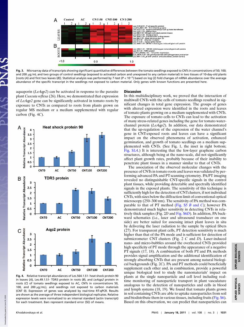
in the roots and first two leaves of 10-day-old seedlings exposedto multiwall CNTs at different concentration levels (50; 100;200 μg∕mL), activated carbon (50 μg∕mL), and the controltomato seedlings grown on regular MS medium were quantifiedby Affymetrix Tomato GeneChips. Based on these studies andafter statistical analysis (T-test, hierachical clustering), we iden-tified 91 transcripts in leaves and 49 transcripts in roots thatshowed significant differences in transcript abundance betweenthe CNT-exposed seedlings and two controls (seedlings exposed
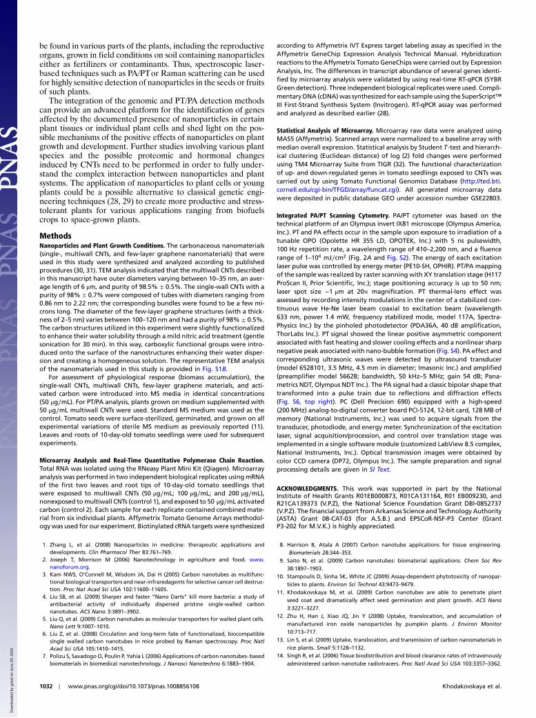
to activated carbon and seedlings unexposed to any carbonstructures) in all biological replicates that were analyzed (Fig. 3for the 16 genes with known functions; Figs. S7 and S8 presentedfull datasets). The differently expressed profiles of nanoparticle-regulated genes were categorized according to their involvementin specific biological processes as depicted in the Tomato Data-base (Table S1). In leaves, the majority of CNT-regulated geneswere involved in cellular responses (29 genes), stress responses(39 genes), transport (14 genes), signal transduction (13 genes),and metabolic and biosynthetic processes (25 genes). In roots,the largest numbers of CNT-regulated genes were involved instress responses (10 genes), cellular processes (9 genes), transport(6 genes), and catabolic, metabolic, and biosynthetic processes(22 genes). The particular functions of several genes that showedaltered expressions in response to CNTs are known. Thus, severaltomato genes (Les.17.1.S1—transcription factor; LesAffx.967.1.S1—14-3-3 family protein; Les.49.1.S1—TDR3 protein) in-volved in the regulation of transcription and hormone pathways(Les.85.1.S1—ethylene receptor; Les.3703.1.S1—IAA9 protein)were up-regulated in seedlings in response to the exposure toCNTs. Expression of mitogen-activated protein kinase (Les.699.1.S1) was also significantly up-regulated in leaves exposed toCNTs (Fig. 3). It was documented earlier that mitogen-activatedprotein kinases play a positive role in the control of plant celldivision and growth (21) and have important functions in stresssignal transduction pathways in plants (22). A number of stress-related genes (Les.564.1.S1—heat shock protein 90; Les.3048.1.S1—DB163 meloidogyne-induced giant cell protein; Les.3648.1.S1—subtilisin-like endoprotease; LesAffx.64585.1.S1—threo-nine deaminase) were differently expressed in tissues exposed toCNTs. To validate the microarray data, we generated sequence-specific primers and performed quantitative real-time reversetranscription polymerase chain reaction (RT-qPCR) using threeindependent biological replicates for four identified up-regulatedgenes in tomato tissues (Les.4264.1.A1—DB149 meloidogyne-induced giant cell protein; Les.49.1.S1—TDR3 protein; Les.564.1.S1—heat shock protein 90; Les.4373.1.S1—Dem2 protein).Data for two genes (Les.564.1.S1—heat shock protein 90 andLes.49.1.S1—TDR3 protein) are shown in Fig. 4 A and B. Thisanalysis confirmed above presented data of microarray assay andrevealed that the expression of all analyzed genes was dramati-cally higher in tissues of seedlings exposed to CNTs but not incontrol tissues or seedlings exposed to activated carbon. Theintriguing finding of the microarray analysis is that several up-regulated genes (Les.3648.1.S1—subtilisin-like endoprotease;Les.3048.1.S1—DB163 meloidogyne-induced giant cell protein;LesAffx.64585.1.S1—threonine deaminase) in response to CNTscan also be activated in response to specific biotic stress factors(pathogens or herbivores). For example, expression of threoninedeaminase was activated in tomato leaves in response to herbi-vore attack (23). In our experiment, expression of the same genewas up-regulated in tomato roots exposed to CNTs (Fig. 3). Thisobservation suggests that the penetration of nano-sized materialinto plant tissues can be sensed by plants as a stress factor similarto pathogens or herbivore attack. In this case, important stress-signaling pathways and cascades could be modified/activated inresponse to the uptake of nanoparticles. Such possible changesmight have a significant impact on most of the major physiologi-cal processes in planta. Our hypothesis was verified in a separateexperiment. The expression of the gene for the tomato water-channel protein (LeAqp2) in the control tissues and tissuesexposed to CNTs (50; 100; 200 μg∕mL) was monitored by real-time RT-qPCR (Fig. 4C). As shown before, water channels (aqua-porins) play a key role in germination and plant growth by reg-ulating the water permeability at the level of individual plant cells(24, 25). The activity of aquaporins was previously shown to beinfluenced by a number of stress factors such as heavy metals,salinity, pH, and anoxia (24). Moreover, the expression of tomato
Fig. 2. Photothermal and photoacoustic detection of multiwall CNTs intomato leaves. Schematic of integratedPA/PTscanning cytometer (A). SpectralPA and PT identification of CNTs (B); given are images of tomato leaves grownin darkness (white) and under light (green). Two-dimensional PT maps(with three-dimensional simulation) of CNTdistribution in tomato leaves com-pared to conventional optical images (C). Calibration model was constructedby injection of CNTs into leaf. PA detection of CNTs in 1 mm thick section oftomato fruit (D).
1030 ∣ www.pnas.org/cgi/doi/10.1073/pnas.1008856108 Khodakovskaya et al.
Dow
nloa
ded
by g
uest
on
June
20,
202
0
aquaporin (LeAqp2) can be activated in response to the parasiteplant Cuscuta reflexa (26). Here, we demonstrated that expressionof LeAqp2 gene can be significantly activated in tomato roots byexposure to CNTs as compared to roots from plants grown onregular MS medium or a medium supplemented with regularcarbon (Fig. 4C).
DiscussionIn this multidisciplinary work, we proved that the interaction ofmultiwall CNTs with the cells of tomato seedlings resulted in sig-nificant changes in total gene expression. The groups of geneswith altered expression were identified in the roots and leavesof tomato plants growing on a medium supplemented with CNTs.The exposure of tomato cells to CNTs can lead to the activationof many stress-related genes including the gene for tomato water-channel protein (LeAqp2). In addition, our data demonstratedthat the up-regulation of the expression of the water channel’sgene in CNT-exposed roots and leaves can have a significantimpact on the observed phenomena of activation, enhancedgermination, and growth of tomato seedlings on a medium sup-plemented with CNTs. (See Fig. 1, the inset in right bottom;Fig. S1A.) It is interesting that the few-layer graphene carbonstructures, although being at the nano-scale, did not significantlyaffect plant growth rates, probably because of their inability topenetrate plant tissues in a manner similar to that of CNTs.
The association of the observed molecular changes with thepresence of CNTs in tomato roots and leaves was validated by per-forming advanced PA and PTscanning cytometry. PA/PT imagingrevealed no distinguishable CNT-specific signals in the controlplant tissues, while providing detectable and spectrally identifiedsignals in the exposed plants. The sensitivity of this technique issufficiently high for the detection of CNTclusters, if not individualCNTs, with sizes below the diffraction limit of conventional opticalmicroscopy (250–300 nm). The sensitivity of PAmethod was com-parable to that of PT method (Fig. S5 B and C); however PAdemonstrated much higher sensitivity in detecting CNTs in rela-tively thick samples (Fig. 2D and Fig. S6D). In addition, PA back-ward schematics (i.e., laser and ultrasound transducer on oneside) are better suited for assessing intact plant leaves in situby delivering the laser radiation to the sample by optical fibers(27). For transparent plant cells, PT detection sensitivity is muchhigher than that of the PA mode and is sufficient for detection ofsubmicrometer CNT clusters (Fig. 2 C and D). Laser-inducednano- and micro-bubbles around the overheated CNTs providedhigh specificity of PT mode through the appearance of a negativePT signals (17, 18). A combination of both PT and PA methodsprovides signal amplification and the additional identification ofstrongly absorbing CNTs that are present among natural biologi-cal backgrounds (Fig. 2C). PA and PT methods could beneficiallysupplement each other and, in combination, provide a powerfulunique biological tool to study the nanomaterials’ impact onplants at the single nanoparticle and cell level including real-time monitoring of nanoparticle transport in plant vasculatureanalogous to the detection of nanoparticles and cells in bloodand lymph systems (18, 19). We found that tomato plants grownin soil containing CNTs are able to uptake nanoparticles from soiland biodistribute them in various tissues, including fruits (Fig. S6).Based on this observation, we can predict that nanoparticles can
Fig. 3. Microarray data of transcripts showing significant quantitative differences between the tomato seedlings exposed to CNTs in concentrations of 50; 100;and 200 μg∕mL and two groups of control seedlings (exposed to activated carbon and unexposed to any carbon material) in two tissues of 10-day-old plants[roots (A) and first two leaves (B)]. Statistical analysis was performed by T -test (P < 10−3) based on log (2) fold changes of mRNA abundance over the averageabundance of the specific transcript in the seedlings not exposed to carbon material. Only genes with known functions are presented here.
Fig. 4. Relative transcript abundances of Les.564.1.S1- heat shock protein 90in leaves (A), Les.49.1.S1- TDR3 protein in roots (B), and LeAqp2 protein inroots (C) of tomato seedlings exposed to AC, CNTs in concentrations 50,100, and 200 μg∕mL), and seedlings not exposed to carbon materials(CNT 0). Expression of genes was analyzed by real-time RT-qPCR. Resultsare shown as the average of three independent biological replicates. Relativeexpression levels were normalized to an internal standard (actin transcript)for each treatment. Bars represent standard error (SE) of means.
Khodakovskaya et al. PNAS ∣ January 18, 2011 ∣ vol. 108 ∣ no. 3 ∣ 1031
APP
LIED
PHYS
ICAL
SCIENCE
SAPP
LIED
BIOLO
GICAL
SCIENCE
S
Dow
nloa
ded
by g
uest
on
June
20,
202
0
be found in various parts of the plants, including the reproductiveorgans, grown in field conditions on soil containing nanoparticleseither as fertilizers or contaminants. Thus, spectroscopic laser-based techniques such as PA/PTor Raman scattering can be usedfor highly sensitive detection of nanoparticles in the seeds or fruitsof such plants.
The integration of the genomic and PT/PA detection methodscan provide an advanced platform for the identification of genesaffected by the documented presence of nanoparticles in certainplant tissues or individual plant cells and shed light on the pos-sible mechanisms of the positive effects of nanoparticles on plantgrowth and development. Further studies involving various plantspecies and the possible proteomic and hormonal changesinduced by CNTs need to be performed in order to fully under-stand the complex interaction between nanoparticles and plantsystems. The application of nanoparticles to plant cells or youngplants could be a possible alternative to classical genetic engi-neering techniques (28, 29) to create more productive and stress-tolerant plants for various applications ranging from biofuelscrops to space-grown plants.
MethodsNanoparticles and Plant Growth Conditions. The carbonaceous nanomaterials(single-, multiwall CNTs, and few-layer graphene nanomaterials) that wereused in this study were synthesized and analyzed according to publishedprocedures (30, 31). TEM analysis indicated that the multiwall CNTs describedin this manuscript have outer diameters varying between 10–35 nm, an aver-age length of 6 μm, and purity of 98.5%� 0.5%. The single-wall CNTs with apurity of 98%� 0.7% were composed of tubes with diameters ranging from0.86 nm to 2.22 nm; the corresponding bundles were found to be a few mi-crons long. The diameter of the few-layer graphene structures (with a thick-ness of 2–5 nm) varies between 100–120 nm and had a purity of 98%� 0.5%.The carbon structures utilized in this experiment were slightly functionalizedto enhance their water solubility through a mild nitric acid treatment (gentlesonication for 30 min). In this way, carboxylic functional groups were intro-duced onto the surface of the nanostructures enhancing their water disper-sion and creating a homogeneous solution. The representative TEM analysisof the nanomaterials used in this study is provided in Fig. S1B.
For assessment of physiological response (biomass accumulation), thesingle-wall CNTs, multiwall CNTs, few-layer graphene materials, and acti-vated carbon were introduced into MS media in identical concentrations(50 μg∕mL). For PT/PA analysis, plants grown on medium supplemented with50 μg∕mL multiwall CNTs were used. Standard MS medium was used as thecontrol. Tomato seeds were surface-sterilized, germinated, and grown on allexperimental variations of sterile MS medium as previously reported (11).Leaves and roots of 10-day-old tomato seedlings were used for subsequentexperiments.
Microarray Analysis and Real-Time Quantitative Polymerase Chain Reaction.Total RNA was isolated using the RNeasy Plant Mini Kit (Qiagen). Microarrayanalysis was performed in two independent biological replicates using mRNAof the first two leaves and root tips of 10-day-old tomato seedlings thatwere exposed to multiwall CNTs (50 μg∕mL; 100 μg∕mL; and 200 μg∕mL),nonexposed tomultiwall CNTs (control 1), and exposed to 50 μg∕mL activatedcarbon (control 2). Each sample for each replicate contained combined mate-rial from six individual plants. Affymetrix Tomato Genome Arrays methodol-ogywas used for our experiment. Biotinylated cRNA targets were synthesized
according to Affymetrix IVT Express target labeling assay as specified in theAffymetrix GeneChip Expression Analysis Technical Manual. Hybridizationreactions to the Affymetrix Tomato GeneChips were carried out by ExpressionAnalysis, Inc. The differences in transcript abundance of several genes identi-fied by microarray analysis were validated by using real-time RT-qPCR (SYBRGreen detection). Three independent biological replicates were used. Compli-mentaryDNA (cDNA)was synthesized for each sample using the SuperScript™III First-Strand Synthesis System (Invitrogen). RT-qPCR assay was performedand analyzed as described earlier (28).
Statistical Analysis of Microarray. Microarray raw data were analyzed usingMAS5 (Affymetrix). Scanned arrays were normalized to a baseline array withmedian overall expression. Statistical analysis by Student T -test and hierarch-ical clustering (Euclidean distance) of log (2) fold changes were performedusing TM4 Microarray Suite from TIGR (32). The functional characterizationof up- and down-regulated genes in tomato seedlings exposed to CNTs wascarried out by using Tomato Functional Genomics Database (http://ted.bti.cornell.edu/cgi-bin/TFGD/array/funcat.cgi). All generated microarray datawere deposited in public database GEO under accession number GSE22803.
Integrated PA/PT Scanning Cytometry. PA/PT cytometer was based on thetechnical platform of an Olympus invert IX81 microscope (Olympus America,Inc.). PT and PA effects occur in the sample upon exposure to irradiation of atunable OPO (Opolette HR 355 LD, OPOTEK, Inc.) with 5 ns pulsewidth,100 Hz repetition rate, a wavelength range of 410–2,200 nm, and a fluencerange of 1–104 mJ∕cm2 (Fig. 2A and Fig. S2). The energy of each excitationlaser pulse was controlled by energy meter (PE10-SH, OPHIR). PT/PA mappingof the sample was realized by raster scanning with XY translation stage (H117ProScan II, Prior Scientific, Inc.); stage positioning accuracy is up to 50 nm;laser spot size ∼1 μm at 20× magnification. PT thermal-lens effect wasassessed by recording intensity modulations in the center of a stabilized con-tinuous wave He-Ne laser beam coaxial to excitation beam (wavelength633 nm, power 1.4 mW, frequency stabilized mode, model 117A, Spectra-Physics Inc.) by the pinholed photodetector (PDA36A, 40 dB amplification,ThorLabs Inc.). PT signal showed the linear positive asymmetric componentassociated with fast heating and slower cooling effects and a nonlinear sharpnegative peak associated with nano-bubble formation (Fig. S4). PA effect andcorresponding ultrasonic waves were detected by ultrasound transducer(model 6528101, 3.5 MHz, 4.5 mm in diameter; Imasonic Inc.) and amplified(preamplifier model 5662B; bandwidth, 50 kHz–5 MHz; gain 54 dB; Pana-metrics NDT, Olympus NDT Inc.). The PA signal had a classic bipolar shape thattransformed into a pulse train due to reflections and diffraction effects(Fig. S6, top right). PC (Dell Precision 690) equipped with a high-speed(200 MHz) analog-to-digital converter board PCI-5124, 12-bit card, 128 MB ofmemory (National Instruments, Inc.) was used to acquire signals from thetransducer, photodiode, and energy meter. Synchronization of the excitationlaser, signal acquisition/procession, and control over translation stage wasimplemented in a single software module (customized LabView 8.5 complex,National Instruments, Inc.). Optical transmission images were obtained bycolor CCD camera (DP72, Olympus Inc.). The sample preparation and signalprocessing details are given in SI Text.
ACKNOWLEDGMENTS. This work was supported in part by the NationalInstitute of Health Grants R01EB000873, R01CA131164, R01 EB009230, andR21CA139373 (V.P.Z), the National Science Foundation Grant DBI-0852737(V.P.Z). The financial support fromArkansas Science and Technology Authority(ASTA) Grant 08-CAT-03 (for A.S.B.) and EPSCoR-NSF-P3 Center (GrantP3-202 for M.V.K.) is highly appreciated.
1. Zhang L, et al. (2008) Nanoparticles in medicine: therapeutic applications anddevelopments. Clin Pharmacol Ther 83:761–769.
2. Joseph T, Morrison M (2006) Nanotechnology in agriculture and food. www.nanoforum.org.
3. Kam NWS, O’Connell M, Wisdom JA, Dai H (2005) Carbon nanotubes as multifunc-tional biological transporters and near-infraredagents for selective cancer cell destruc-tion. Proc Nat Acad Sci USA 102:11600–11605.
4. Liu SB, et al. (2009) Sharper and faster “Nano Darts” kill more bacteria: a study ofantibacterial activity of individually dispersed pristine single-walled carbonnanotubes. ACS Nano 3:3891–3902.
5. Liu Q, et al. (2009) Carbon nanotubes as molecular transporters for walled plant cells.Nano Lett 9:1007–1010.
6. Liu Z, et al. (2008) Circulation and long-term fate of functionalized, biocompatiblesingle walled carbon nanotubes in mice probed by Raman spectroscopy. Proc NatlAcad Sci USA 105:1410–1415.
7. Polizu S, SavadogoO, Poulin P, Yahia L (2006) Applications of carbon nanotubes- basedbiomaterials in biomedical nanotechnology. J Nanosci Nanotechno 6:1883–1904.
8. Harrison B, Atala A (2007) Carbon nanotube applications for tissue engineering.Biomaterials 28:344–353.
9. Saito N, et al. (2009) Carbon nanotubes: biomaterial applications. Chem Soc Rev38:1897–1903.
10. Stampoulis D, Sinha SK, White JC (2009) Assay-dependent phytotoxicity of nanopar-ticles to plants. Environ Sci Technol 43:9473–9479.
11. Khodakovskaya M, et al. (2009) Carbon nanotubes are able to penetrate plantseed coat and dramatically affect seed germination and plant growth. ACS Nano3:3221–3227.
12. Zhu H, Han J, Xiao JQ, Jin Y (2008) Uptake, translocation, and accumulation ofmanufactured iron oxide nanoparticles by pumpkin plants. J Environ Monitor10:713–717.
13. Lin S, et al. (2009) Uptake, translocation, and transmission of carbon nanomaterials inrice plants. Small 5:1128–1132.
14. Singh R, et al. (2006) Tissue biodistribution and blood clearance rates of intravenouslyadministered carbon nanotube radiotracers. Proc Natl Acad Sci USA 103:3357–3362.
1032 ∣ www.pnas.org/cgi/doi/10.1073/pnas.1008856108 Khodakovskaya et al.
Dow
nloa
ded
by g
uest
on
June
20,
202
0
15. Zharov VP, Letokhov VS (1986) Laser optoacoustic spectroscopy (Springer- Verlag,Berlin Heidelberg).
16. Wang LV, ed. (2009) Photoacoustic imaging and spectroscopy (CRC, Boca Raton, FL).17. Zharov VP, Lapotko DO (2005) Photothermal imaging of nanoparticles and cells. IEEE J
Sel Topics Quant 11:733–751.18. Kim J-W, Galanzha EI, Shashkov EV, Moon H-M, Zharov VP (2009) Golden carbon
nanotubes as multimodal photoacoustic and photothermal high-contrast molecularagents. Nature Nanotechnol 4:688–694.
19. Galanzha EI, et al. (2009) In vivo magnetic enrichment and multiplex photoacousticdetection of circulating tumor cells. Nature Nanotechnol 12:855–860.
20. Rensink WA, Buell CR (2005) Microarray expression profiling resources for plantgenomics. TRENDS Plant Sci 10:603–609.
21. Krysan PJ, Jester PJ, Gottward JR, Sussman MR (2002) An Arabidopsis mitogen-activated protein kinase kinase kinase gene family encodes essential positive regula-tors of cytokinesis. Plant Cell 14:1109–1220.
22. Menke FLH, van Pelt JA, Pieterse CMJ, Klessig DF (2004) Silencing of the mitogene-activated protein kinase MPK6 compromises disease resistance in Arabidopsis. PlantCell 16:897–907.
23. Chen H, Gonzales-Vigil E, Wilkerson CG, Howe GA (2007) Stability of plant defenseproteins in the gut of insect herbivores. Plant Physiol 143:1954–1967.
24. Tyerman SD, Bohnert HJ, Maurel C, Steudle E, Smith JAC (1999) Plant aquaporins:their molecular biology, biophysics and significance for plant water relations. J ExpBot 50:1055–1071.
25. Heinen RB, Ye Q, Chaumont F (2009) Role aquaporins in leaf physiology. J Exp Bot
11:2971–2985.
26. Werner M, Uehlein N, Proksch P, Kaldenhoff R (2001) Characterization of two tomato
aquaporins and expression during incompatible interaction of tomato with the plant
parasite Cuscuta reflexa. Planta 213:550–555.
27. Galanzha EI, et al. (2009) In vivo fiber-based photoacoustic detection and photother-
mal purging of metastasis in sentinel lymph nodes targeted by nanoparticles.
J Biophotonics 2:528–539.
28. Khodakovskaya M, et al. (2010) Increasing inositol (1,4,5)-triphosphate metaboilism
affects drought tolerance, carbohydrate metabolism and phosphate-sensitive biomass
increases in tomato. Plant Biotechnol J 8:170–183.
29. Li J, et al. (2005) Arabidopsis Hþ-PPase AVP1 regulates auxin-mediated organ
development. Science 310:121–125.
30. Dervishi E, et al. (2007) Morphology of multiwall carbon nanotubes affected by the
thermal stability of the catalyst system. Chem Mater 19:179–184.
31. Dervishi E, et al. (2009) Versatile catalytic system for the large-scale and controlled
synthesis of single-wall, double-wall, multiwall, and graphene carbon nanostructures.
Chem Mater 21:5491–5498.
32. Saeed AI, et al. (2003) TM4: a free, open-source system for microarray data manage-
ment and analysis. Biotechniques 34:374–378.
Khodakovskaya et al. PNAS ∣ January 18, 2011 ∣ vol. 108 ∣ no. 3 ∣ 1033
APP
LIED
PHYS
ICAL
SCIENCE
SAPP
LIED
BIOLO
GICAL
SCIENCE
S
Dow
nloa
ded
by g
uest
on
June
20,
202
0