Embed Size (px)
Citation preview

See discussions, stats, and author profiles for this publication at: https://www.researchgate.net/publication/13890635
Corynebacterium durum sp. nov., from Human Clinical Specimens
Article in International Journal of Systematic Bacteriology · November 1997
DOI: 10.1099/00207713-47-4-1107 · Source: PubMed
CITATIONS
34READS
1,048
5 authors, including:
Some of the authors of this publication are also working on these related projects:
Coagulase-negative staphylococci virulence View project
Staphylococcus Aureus leucotoxins and NETosis View project
Remy Heller
Hopitaux Civils De Colmar
37 PUBLICATIONS 1,817 CITATIONS
SEE PROFILE
Gilles Prévost
University of Strasbourg
269 PUBLICATIONS 8,561 CITATIONS
SEE PROFILE
françois Jehl
CHRU de Strasbourg
277 PUBLICATIONS 3,710 CITATIONS
SEE PROFILE
All content following this page was uploaded by françois Jehl on 29 May 2014.
The user has requested enhancement of the downloaded file.
INTERNATIONAL JOURNAL OF SYSTEMATIC BACTERIOLOGY, OCt. 1997, p. 1107-1 11 1 0020-7713/97/$04.00+0 Copyright 0 1997, International Union of Microbiological Societies
Vol. 47, No. 4
Corynebacterium durum sp. nov., from Human Clinical SDecimens
PHILIPPE RIEGEL," REMY HELLER, GILLES PREVOST, FRANCOIS JEHL, AND HENRI MONTEIL
Institut de Bactkriologie de la Facultk de Mkdecine, Universitk Louis-Pasteur, Strasbourg, France
A new Corynebacteriurn species, Corynebacterium durum, was isolated from respiratory tract specimens of five human patients. The strains of this species exhibited similar morphologic and biochemical features that differentiated them from all recognized species. Notably, all of these strains developed irregular and strongly adherent colonies under aerobic conditions and produced acid from mannitol and galactose. The cells are long pleomorphic rods with some filaments. This species has characteristics of the genus Corynebacterium, such as 55 mol% guanine plus cytosine in the DNA and the presence of corynomycolic acids, meso-diaminopimelic acid, arabinose, and galactose in the cell wall. These isolates formed a homogeneous group in which the DNA-DNA similarity values (as determined by an S1 nuclease procedure) compared with reference strain IBS G15036T (T = type strain) ranged from 71 to 100%. The analysis of the nearly complete 16s rRNA gene sequence of IBS G15036T indicated that this new species represents a distinct taxon within the genus Corynebacterium. This new species can be identified on the basis of its colony morphology, fermentation of sugars, and enzymatic activities. Strain IBS 615036 (= CCUG 37331) is the type strain of C. durum.
The genus Corynebacterium has recently been subjected to considerable taxonomic revisions, which have resulted in the proposal of several new species and subspecies, many of them representing previous Centers for Disease Control coryneform groups defined by Hollis and Weaver (10). The recent review of Funke et al. provided a very comprehensive overview of the taxonomic changes among coryneform bacteria (7). The genus Corynebacterium is currently defined on the basis of chemot- axonomic features (3). The salient chemotaxonomic features include cell wall component type IV (arabinose, galactose, and meso-diaminopimelic acid), short-chain-length mycolic acids, and a DNA base composition ranging from 52 to 65 mol%, although there are some exceptions to this general rule, with C. amycolatum lacking mycolic acids and C. afermentans and C. auris exhibiting guanine-plus-cytosine (G+ C) contents of more than 65 mol% (7). Comparative analysis of sequences of genes coding for 16s rRNA (16s rDNA) confirmed that the mem- bers of the genus Corynebacterium constitute a single unit among the high-G+C-content gram-positive bacteria (11, 14). In contrast, species of the genus Corynebacterium exhibit sig- nificant phenotypic diversity in colony appearance (size, color, and morphology), requirements for growth factors (lipids), and biochemical activities. During the course of characterization of medically relevant coryneform rods, we isolated five strains that had the main characteristics of the genus Corynebacterium but that differed markedly from the established species be- cause these strains grew slowly and exhibited irregular colonies strongly adherent to agar. Furthermore, all these strains were found to produce acid from mannitol, which is rarely observed in coryneform bacteria. In this paper we describe a new spe- cies, Corynebacterium durum, for these strains on the basis of biochemical, morphological, and cultural data, as well as our results of DNA-DNA hybridization experiments and 16s rDNA analyses.
* Corresponding author. Mailing address: Institut de Bactkriologie de la FacultC de Medecine, 3 rue KoeberlC, F-67000 Strasbourg, France. Phone: 33.388.21.19.70. Fax: 33.388.25.11.13. E-mail: priegibsetpgnet .net.
MATERIALS AND METHODS
Strains, media, and growth conditions. The five isolates included in this study were isolated at the Institute of Bacteriology of Strasbourg, France, from respi- ratory tract specimens of different patients, mainly bronchiole washing speci- mens. As is usually done in our laboratory, these clinical materials were streaked onto 5% (vol/vol) sheep blood-Trypticase-soy agar (bioM&rieux, Marcy-I'Etoile, France) and incubated in a 10% CO, atmosphere for 24 h. In addition, the samples were inoculated on both nonselective buffered charcoal-yeast extract (BCYE) agar and vancomycin-containing BCYE agar and incubated in a 2.5% CO, atmosphere for 3 weeks in order to recover Legionella species.
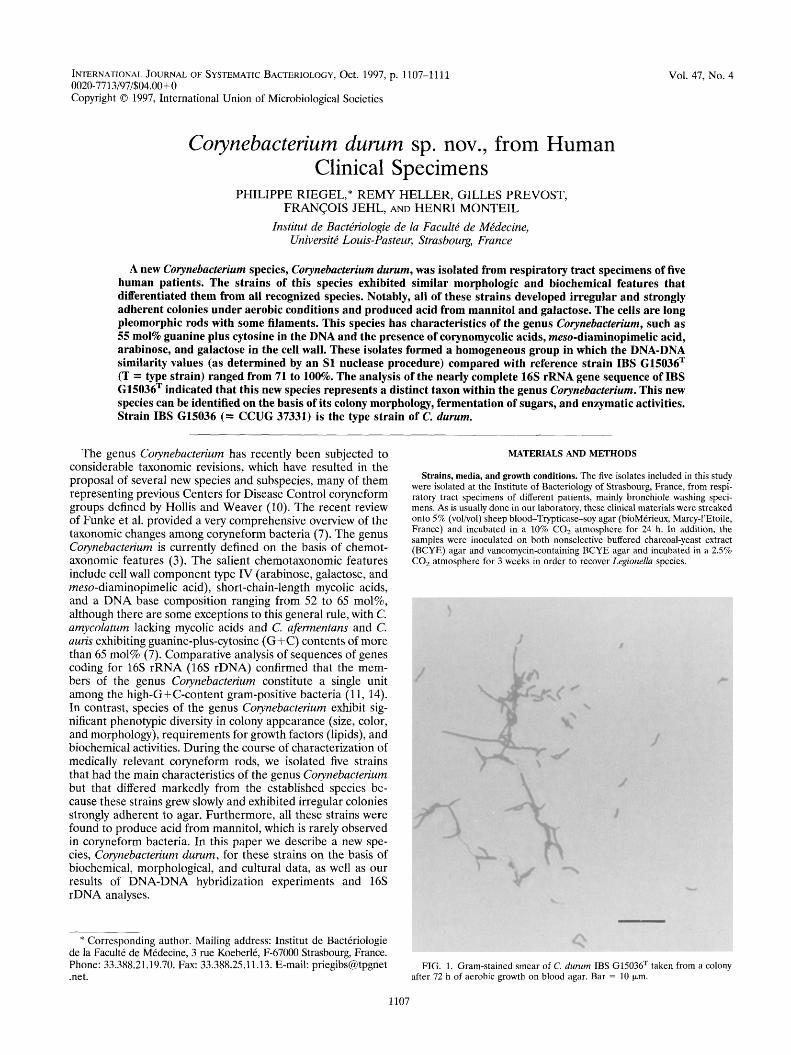
FIG. 1. Gram-stained smear of C. durum IBS G15036T taken from a colony after 72 h of aerobic growth on blood agar. Bar = 10 pm.
1107
1108 RIEGEL ET AL. INT. J. SYST. BACTERIOL.
C pseudotuberculosis CIP I02968T I X 8 19 16
C drphtherme CIP 100721T I X 82059 C vitarumen DSM 20294T I X 84680
C kutscherr CIP 103423T I X 82063
C minutisstmum NCTC 10288 T I X 82064 C strratum CIP 8 I 15T I X 81910
C accolens CCUG 28779T I X 80500 C. macgrnleyi CIP 104099T I X 80499 "C group G2" CDC G 5840 I X 80498
C propinquum CIP 103792T I X 8 19 17 L C pseudodiphfhenticum CIP 103420T I X 8 19 I7
C matruchotii CIP 8 1 82T I X 82065 3 C. durum IBS G 15036 I Z 97069 C renale CIP I0342 IT / X 8 1909
C. jeikeium CIP 103337T I X 82062 C. urealyticum ATCC 43042 T I X 8 19 13
C. bovis NCTC 3224T I X 8205 1 I C. variabilis ATCC 15753T I X 531 85
C. amycoiatum CIP 103452T I X 82057 C. xerosis DSM 207437 I X 84446
allunae CCUG 28793T I X 82053 C. glutamicum IAM I2435T I X 8206 1
C. ammoniagenes CIP 101283T I X 82056
C. afermentans CIP 103499T / X 82054 C. coyleae DSM, 441 84T I X 96497
C. pseudogenitalium" NCTC I 1860 I X 8 1872 C. auris DSM 44122T I X 82493
C, mycefpides CIP 55.51T I X 82066 C. genitaliurn" NCTC 1 1859 I X 84253
I C. cystitidis CIP 103424T I X 82058 C. glucuronolyticum DSM 44 I20T I X 86688
LC. seminaie CIP 104297T I X 84375 I Turicella otitidis DSM 882 IT J X 73976
.,._ I
8 I
6 I
4 I
2 I 0
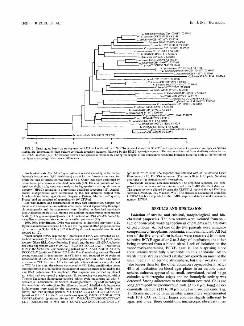
FIG. 2. Dendrogram based on an alignment of 1,423 nucleotides of the 16s rRNA genes of strain IBS G15036T and representative Colynebucteriurn species. Strains studied are designated by their culture collection accession number, followed by the EMBL accession number. The tree was inferred from similarity values by the CLUSTAL method (12). The distance between two species is obtained by adding the lengths of the connecting horizontal branches using the scale at the bottom of the figure (percentage of sequence difference).
Biochemical tests. The API-Coryne system was used according to the manu- facturer's instructions ( M I bioMvltrieux) except for the fermentation tests, for which the time of incubation was fixed at 48 h. Other tests were performed by conventional procedures as described previously (12). The end products of bac- terial metabolism of glpcose were analyzed by high-performance liquid chroma- tography (HPLC) according to a previously described procedure (13). Antimi- crobial susceptibilities were determined by the disk diffusion method with Mueller-Hinton blood agar (Sanofi Diagnostic Pasteur, Marnes-La-Coquette, France) and an inoculum of approximately lo5 CFU/ml.
Cell wall analysis and determination of DNA base composition. Samples for amino acid and sugar determinations were prepared and separated by thin-layer chromatography, and the components were identified as described previously (16). A reversed-phase HPLC method was used for the determination of mycolic acids (5) . The guanine-plus-cytosine (G+C) content of DNA was determined by a capillary electrophoresis method as described previously (13).
DNA-DNA hybridization. DNA was extracted as described previously (12). Hybridization between labeled DNA and the fragmented-DNA preparation was carried out at 60°C for 16 h in 0.42 M NaCl by the nuclease-trichloroacetic acid method (8, 12).
SmqJl-subunit rDNA sequencing. Chromosomal DNA was extracted as de- scribed previously (6). DNA amplification was performed with Tuq DNA poly- merase (Gibco BRL, Cergy-Pontoise, France), and the two 16s rDNA eubacte- rial universal primers were 5'-AGAGTTTGATCCTGGCTCAG-3' (positions 8 to 28 in the Escherichiu coZi numbering system) and 5'-AAGGAGGT(;ATCCA GCCGCA-3' (positions 1544 to 1525 in the E. coli numbering system). Thermal cycling consisted of denaturation at 93°C for 5 min, followed by 30 cycles of denaturation at 93°C for 30 s, primer annealing at 52°C for 1 min, and primer extension at 72°C for 1 min. After the last cycle, a final extension was performed at 72°C far 8 rnin and the mixture was cooled to 5°C. Thirty amplification cycles were performed in order to limit the number of sequence errors generated by the Tuq DNA polymerase. The amplified DNA fragment was purified by phenol extraction and isopropanol precipitation (1). Sequencing was performed with a Thermo Sequenase fluorescence-labelled primer cycle sequencing kit with 7- deaza-dGTP (Amersham Life Science, Buckinghamshire, England) according to the manufacturer's instructions. Six different primers 5' labelled with fluorescein isothiocyanate were used for the sequencing reactions: P8 and Pc1544 (see above) and four internal primers, 5'-GTA?TACCGCGGCTGCTGGCAC-3' (positions 535 to 514 in the E. coli numbering system), 5'-GTGCCAGCAGCCG CGGTAAKAC-3' (positions 514 to 53.9, 5'-GACTACCAGGGTATCTAAT CC-3' (positions 804 to 784), and 5'-GGAlTAGATACCCTGGGTAGTC-3'
(positions 784 to 804). The sequence was obtained with an Automated Laser Fluorescence (A.L.F.) DNA sequencer (Pharmacia Biotech, Uppsala, Sweden) according to the manufacturer's instructions.
Nucleotide sequence accession number, The amplified sequence was com- pared to other sequences of bacteria contained in the EMBL-GenBank database. The sequences were aligned by using the CLUSTAL method (9) and DNAStar software (DNAStar, Inc., Madison, Wis.). The nucleotide sequence of strain IBS G15036T has been deposited in the EMBL sequence database under accession number 297069.
RESULTS AND DISCUSSION
Isolation of strains and cultural, morphological, and bio- chemical properties. The new strains were isolated from spu- tum or bronchiole washing specimens of patients with a history of pneumonia. All but one of the five patients were immuno- compromised (neoplasms, leukemia, and renal failure). All but one of the five coryneform isolates were recovered from non- selective BCYE agar after 2 to 3 days of incubation, the other being recovered from a blood plate. Lack of isolation on the vancomycin-containing BCYE agar is not surprising since these strains were fully susceptible to this antibiotic. After- wards, these strains showed satisfactory growth on most of the usual media in an aerobic atmosphere, but their isolation may take longer than for the other common corynebacteria. After 48 h of incubation on blood agar plates in an aerobic atmo- sphere, cultures appeared as small, convoluted, raised beige colonies with irregular edges and no hemolytic activity was detected. Strong adherence to the medium occurred. Cells are long gram-positive pleomorphic rods (3 to 4 p,m long) or oc- casionally filaments (15 to 30 pm long) with swollen ends (Fig. 1). Strains incubated in an aerobic atmosphere supplemented with 10% CO, exhibited larger colonies slightly adherent to agar, and under these conditions, microscopic observation re-
VOL. 47, 1997 CORIWEBACTERIUM DURUM SP. NOV. 1109
vealed no filaments. In liquid media, the organisms produced a sediment, leading to a granular appearance of the culture after the broth was shaken. The organisms were not partially acid fast.
Biochemical reactions obtained with the MI-Coryne system or by conventional methods were remarkable for being similar among the five isolates. The strains were nitrate reductase and pyrazinamidase positive and alkaline phosphatase and p-glu- curonidase negative. Slight degradation of urea occurred for four of the five strains with the API-Coryne system, urea broth medium, or Christensen's medium. The conventional media revealed two urease-positive strains, whereas urease was pos- itive for three strains with the API-Coryne system. Some strains hydrolyzed esculin weakly (two strains were positive with tubes of esculin agar and one strain was positive with the MI-Coryne system). The strains fermented glucose, sucrose, maltose, and fructose but not lactose, trehalose, or ribose, and it should be noted that all of the strains produced acid from mannitol, which is a phenomenon rarely observed for strains belonging to the genus Corynebacterium. The MI-Coryne nu- merical code for all strains was 3000135, 3040135, or 3001135 for which there was no identification with the current database. The strains showed large inhibition zones around the following antimicrobial agents: ampicillin, amoxicillin-clavulanic acid, piperacillin, cefotaxime, gentamicin, ciprofloxacin, minocycline, erythromycin, rifampin, teicoplanin, and vancomycin.
Taxonomic analyses. The cell walls of the five isolates were found to contain cell wall component type IV (meso-diamin- opimelic acid, arabinose, and galactose) and short-chain my- colic acids (CZ6 to &). The G+C contents of two strains studied were 55.4 (strain IBS G1503fjT) and 55.0 mol%. These results support the assignment of these strains to the genus Cogmebacterium (3). DNA-DNA hybridization experiments revealed that the five isolates form a single genomic group at the species level. The levels of relatedness between labeled DNA of strain IBS G15036T (= CCUG 37331) and unlabeled DNAs of the four other isolates ranged from 71 to 100%.
PCR amplification of the 16s rDNA of strain IBS G15036T with six primers produced a 1,500-base fragment from the 3' end, and we sequenced 1,423 bases from this fragment. Com- parison of this sequence with sequences of other high-G+ C gram-positive rods available from the EMBL database dem- onstrated that strain IBS G1503tiT is related to the robust monophyletic unit grouping all available Coiynebacterium se- quences as one branch among the actinomycetes. The hylo-
resents a distinct taxon among all the Colynebacterium species. The highest levels of similarity were the levels of similarity to Coiynebacterium matruchotii, Corynebacterium macginleyi, and Corynebacterium accolens (93% , whereas the levels of similar-
of the genus Corynebacterium ranged from 90.2 to 92.8%. DNA-DNA hybridization experiments showed no significant similarity (7%) between strain IBS G15036T and the type strain of C. matruchotii. Phylogenetic analysis based on rDNA sequences was demonstrated as one of the most powerful methods for estimating relationships of Corynebacterium strains since the results of such an analysis exhibited an excel- lent correlation with physiological characteristics (11, 14). It is remarkable that the relatively close position between the new species as represented by strain IBS G15036T and C. matru- chotii confirms their cultural similarity, e.g., irregular and ad- herent slowly growing colonies. Therefore, according to recent criteria defining a species (15), the chemotaxonomic data, the 16s rDNA sequence data, and the DNA-DNA hybridization data clearly showed that the five isolates form a new genomic
genetic tree in Fig. 2 indicated that strain IBS G15036 ! rep-
ity between strain IBS G15036 4 and the other representatives
I I
+ I
+ I
C d
$ +
c +
F F!
++,
I 1
I <
c c
c c
c c
1110 RIEGEL ET AL. INT. J. SYST. BACTERIOL.
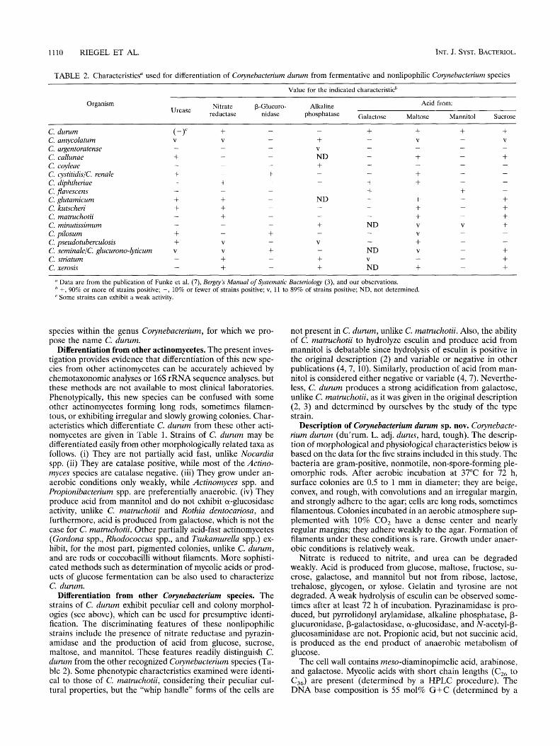
TABLE 2. Characteristics“ used for differentiation of Co ynebacterium durum from fermentative and nonlipophilic Couynebacterium species ~~~ ~
Value for the indicated characteristicb
Organism Acid from: Urease Nitrate P-Glucuro- Alkaline
reductase nidase phosphatase Galactose Maltose Mannitol Sucrose ~
C. durum C. amycolatum C. argentoratense C. callunae C. coyleae C. cystitidislc. renale C. diphtheriae C. jlavescens C. glutamicum C. kutscheri C. matruchotii C. minutissimum C. pilosum C. pseudotuberculosis C. seminalelC. glucurono-lyticum C. striatum C. xerosis
+ V -
+ + + + + +
-
-
V
V
+ V -
+ Data are from the publication of Funke et al. (7), Bergey’s Manual of Systematic Bacteriology (3), and our observations. +, 90% or more of strains positive; -, 10% or fewer of strains positive; v, 11 to 89% of strains positive; ND, not determined. Some strains can exhibit a weak activity.
species within the genus Corynebacterium, for which we pro- pose the name C. durum.
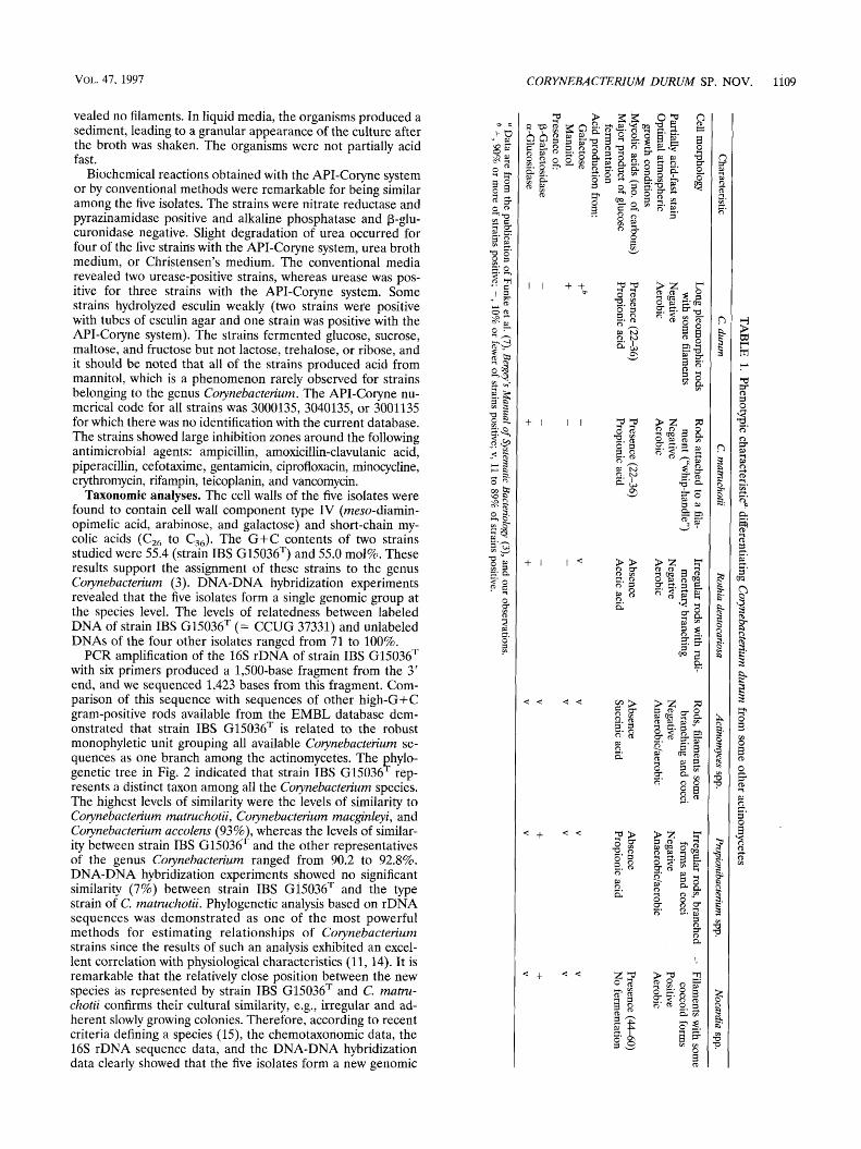
Differentiation from other actinomycetes. The present inves- tigation provides evidence that differentiation of this new spe- cies from other actinomycetes can be accurately achieved by chemotaxonomic analyses or 16s rRNA sequence analyses, but these methods are not available to most clinical laboratories. Phenotypically, this new species can be confused with some other actinomycetes forming long rods, sometimes filamen- tous, or exhibiting irregular and slowly growing colonies. Char- acteristics which differentiate C. durum from these other acti- nomycetes are given in Table 1. Strains of C. durum may be differentiated easily from other morphologically related taxa as follows. (i) They are not partially acid fast, unlike Nocardia spp. (ii) They are catalase positive, while most of the Actino- myces species are catalase negative. (iii) They grow under an- aerobic conditions only weakly, while Actinomyces spp. and Propionibacterium spp. are preferentially anaerobic. (iv) They produce acid from mannitol and do not exhibit a-glucosidase activity, unlike C. matruchotii and Rothia dentocariosa, and furthermore, acid is produced from galactose, which is not the case for C. matruchotii. Other partially acid-fast actinomycetes (Gordona spp., Rhodococcus spp., and Tsukamurella spp.) ex- hibit, for the most part, pigmented colonies, unlike C. durum, and are rods or coccobacilli without filaments. More sophisti- cated methods such as determination of mycolic acids or prod- ucts of glucose fermentation can be also used to characterize C. durum.
Differentiation from other Corynebacterium species. The strains of C. durum exhibit peculiar cell and colony morphol- ogies (see above), which can be used for presumptive identi- fication. The discriminating features of these nonlipophilic strains include the presence of nitrate reductase and pyrazin- amidase and the production of acid from glucose, sucrose, maltose, and mannitol. These features readily distinguish C. durum from the other recognized Corynebacterium species (Ta- ble 2). Some phenotypic characteristics examined were identi- cal to those of C. matruchotii, considering their peculiar cul- tural properties, but the “whip handle” forms of the cells are
not present in C. durum, unlike C. matruchotii. Also, the ability of C. matruchotii to hydrolyze esculin and produce acid from mannitol is debatable since hydrolysis of esculin is positive in the original description (2) and variable or negative in other publications (4, 7, 10). Similarly, production of acid from man- nitol is considered either negative or variable (4, 7). Neverthe- less, C. durum produces a strong acidification from galactose, unlike C. matruchotii, as it was given in the original description (2, 3) and determined by ourselves by the study of the type strain.
Description of Corynebacterium durum sp. nov. Corynebacte- rium durum (du’rum. L. adj. durus, hard, tough). The descrip- tion of morphological and physiological characteristics below is based on the data for the five strains included in this study. The bacteria are gram-positive, nonmotile, non-spore-forming ple- omorphic rods. After aerobic incubation at 37°C for 72 h, surface colonies are 0.5 to 1 mm in diameter; they are beige, convex, and rough, with convolutions and an irregular margin, and strongly adhere to the agar; cells are long rods, sometimes filamentous. Colonies incubated in an aerobic atmosphere sup- plemented with 10% CO, have a dense center and nearly regular margins; they adhere weakly to the agar. Formation of filaments under these conditions is rare. Growth under anaer- obic conditions is relatively weak.
Nitrate is reduced to nitrite, and urea can be degraded weakly. Acid is produced from glucose, maltose, fructose, su- crose, galactose, and mannitol but not from ribose, lactose, trehalose, glycogen, or xylose. Gelatin and tyrosine are not degraded. A weak hydrolysis of esculin can be observed some- times after at least 72 h of incubation. Pyrazinamidase is pro- duced, but pyrrolidonyl arylamidase, alkaline phosphatase, p- glucuronidase, P-galactosidase, a-glucosidase, and N-acetyl-P- glucosaminidase are not. Propionic acid, but not succinic acid, is produced as the end product of anaerobic metabolism of glucose.
The cell wall contains meso-diaminopimelic acid, arabinose, and galactose. Mycolic acids with short chain lengths (CZ6 to C36) are present (determined by a HPLC procedure). The DNA base composition is 55 mol% G+C (determined by a
VOL. 47, 1997 CORYNEBACTERIUM DURUM SP. NOV. 11 11
capillary electrophoresis procedure). C. dururn strains were isolated from human respiratory tract specimens. The type strain is Institut de Bactkriologie de Strasbourg (IBS) G15036 (= Culture Collection of the University of Goteborg [CCUG] 37331). It was isolated in Strasbourg, France, from the sputum of a patient. This strain has all of the above-described proper- ties for the species.
ACKNOWLEDGMENTS
We thank H. G. Truper for advice concerning the Latin name. We are grateful to B. Muller, S. Niedergang, and C. Renault for skillful technical assistance.
1.
2.
3.
4.
5.
REFERENCES
Brow, M. A. 1990. Sequencing with Tag DNA polymerase, p. 189-196. In M. A. Innis et al. (ed.), PCR protocols: a guide to methods and application. Academic Press, Inc., San Diego, Calif. Collins, M. D. 1982. Reclassification of Bactenonema matruchotii (Mendel) in the genus Corynebacterium, as Corynebacten’um matruchotii comb. nov. Zentralbl. Bakteriol. Hyg. I. Abt. Orig. C 3:364-367. Collins, M. D., and C. S. Cummins. 1986. Genus Corynebacterium Lehmann and Neumann 1896, 350AL, p. 1266-1276. In P. H. A. Sneath, N. S. Mair, M. E. Sharpe, and J. G. Holt (ed.), Bergey’s manual of systematic bacteri- ology, vol. 2. The Williams & Wilkins Co., Baltimore, Md. Coyle, M. B., and B. A. Lipsky. 1990. Coryneform bacteria in infectious diseascs: clinical and laboratory aspects. Clin. Microbiol. Rev. 3:227-246. de Briel, D., F. Couderc, P. Riegel, F. Jehl, and R. Minck. 1992. High- performance liquid chromatography of corynomycolic acids as a tool in identification of Corynebacterium species and related organisms. J. Clin. Microhiol. 3 0 1407-1417.
6.
7.
8.
9.
10.
11.
12.
13.
14.
15.
16.
de Lamhallerie, X., C. Zandotti, C. Vignoli, C. Bollet, and P. de Micco. 1992. A one-step microbial DNA extraction method using “Chelex 100” suitable for gene amplification. Res. Microbiol. 143:785-790. Funke, G., A. von Graevenitz, J. E. Clarridge 111, and K. A. Bernard. 1997. Clinical microbiology of coryneform bacteria. Clin. Microbiol. Rev. 10125- 159. Grimont, P. A. D., M. Y. Popoff, F. Grimont, C. Coynault, and M. Lemelin. 1980. Reproductibility and correlation study of three deoxyribonucleic acid hybridization procedures. Curr. Microbiol. 4325-330. Higgins, D. G., and P. M. Sharp. 1988. Clustal: a package for performing multiple sequence alignment on a microcomputer. Gene 73:237-244. Hollis, D. J., and R. E. Weaver. 1981. Gram-positive organisms: a guide to identification. Special Bacteriology Section, Centers for Disease Control, Atlanta, Ga. Pascual, C., P. A. Lawson, J. A. E. Farrow, M. Navarro Gimenez, and M. D. Collins. 1995. Phylogenetic analysis of the genus Corynebacterium based on 16s rRNA gene sequences. Int. J. Syst. Bacteriol. 45724-728. Riegel, P., D. de Briel, G. PrCvost, F. Jehl, and H. Monteil. 1994. Genomic diversity among Corynebacterium jeikeium strains and comparison with bio- chemical characteristics and antimicrobial susceptibilities. J. Clin. Microbiol.
Riegel, P., R, Ruimy, D. de Briel, G. PrCvost, F. Jehl, R. Christen, and H. Monteil. 1995. Corynebacterium seminale sp. nov., a new species associated with genital infections in male patients. J. Clin. Microbiol. 33:2244-2249. Ruimy, R., P. Riegel, P. Boiron, H. Monteil, and R. Christen. 1995. Phylog- eny of the genus Corynebacten’um deduced from analyses of small-subunit ribosomal DNA sequences. Int. J. Syst. Bacteriol. 45740-746. Stackehrandt, E., and B. M. Goebel. 1994. Taxonomic note: a place for DNA-DNA reassociation and 165 rRNA sequence analysis in the present species definition in bacteriology. Int. J. Syst. Bacteriol. 44846-849. Staneck, J. L., and G. D. Roberts. 1974. Simplified approach to identification of aerobic actinomycetes by thin-layer chromatography. Appl. Microbiol.
32~1860-1865.
28~226-231.
View publication statsView publication stats