Embed Size (px)
Citation preview
Arq Neuropsiquiatr 2011;69(4):666-669
666
Article
Critical analysis of extra peritoneal antero-lateral approach for lumbar plexusRoberto Sérgio Martins1,3, Bernardo Assumpção Monaco2,Mario Gilberto Siqueira1, Luciano Foroni1, Carlos Otto Heise1, Manoel Jacobsen Teixeira2
ABSTRACTLesions of lumbar plexus are uncommon and descriptions of surgical access are derived from vertebral spine approaches. Method: The extraperitoneal anterolateral approach to the lumbar plexus was performed in six adult fresh cadavers. The difficulties on dissection were related. Results: An exposure of all distal elements of lumbar plexus was possible, but a cranial extension of the incision was needed to reach the iliohypogastric nerve in all cases. Ligation of vessels derived from common iliac artery was necessary for genitofemoral and obturator nerves exposure in two cases. The most proximal part of the lumbar roots could be identified only after dissection and clipping of most lumbar vessels. Conclusion: The extraperitoneal anterolateral approach allows appropriate exposure of terminal nerves of lumbar plexus laterallly to psoas major muscle. Cranial extension of the cutaneous incision may be necessary for exposure of iliohypogastric nerve. Roots exposure increases the risk of vascular damage.Key words: lumbar plexus, surgical approach, peripheral nerve.
Análise crítica do acesso anterolateral retroperitoneal ao plexo lombar
RESUMOAs lesões do plexo lombar são incomuns e as descrições dos acessos cirúrgicos são derivadas de vias de acesso à coluna vertebral. Método: A via extraperitoneal anterolateral foi realizada em seis cadáveres para o acesso ao plexo lombar. Eventuais dificuldades na dissecção foram relatadas. Resultados: Tal acesso permitiu a exposição dos elementos distais do plexo lombar, mas uma extensão cranial da incisão foi necessária para a exposição do nervo iliohipogástrico. Para a exposição dos nervos genitofemoral e obturador houve a necessidade da ligadura de vasos originados da artéria ilíaca comum em 2 casos. As raízes foram identificadas somente após dissecção e ligadura dos vasos lombares. Conclusão: O acesso anterolateral extraperitoneal permite uma exposição adequada dos nervos terminais do plexo lombar lateralmente ao músculo psoas maior. Uma extensão cranial da incisão pode ser necessária para exposição do nervo iliohipogástrico. A exposição das raízes implica em maior risco de lesão vascular. Palavras-chave: plexo lombar, acesso cirúrgico, nervo periférico.
CorrespondenceRoberto S. Martins Rua Maestro Cardim 592 / cj 1101 01323-001 São Paulo SP - BrasilE-mail: [email protected]
Conflicts of interestThe authors report no conflict of interest
Received 10 February 2011Received in final form 22 March 2011Accepted 29 March 2011
1Peripheral Nerve Surgery Unit, Division of Neurosurgery, University of São Paulo Medical School, São Paulo SP, Brazil; 2Division of Neurosurgery, University of São Paulo Medical School, São Paulo SP, Brazil; 3Neurosurgical Department, Hospital do Servidor Público do Estado de São Paulo, São Paulo SP, Brazil.
The lumbar plexus is positioned inside the psoas major muscle and is formed by ventral branches of spinal lumbar nerves from L1 to L4 with a possible contribu-
tion from the subcostal nerve (T12)1. The psoas major muscle represents a funda-mental reference for localization of the principal nerves originated from the
Arq Neuropsiquiatr 2011;69(4)
667
Lumbar plexus approachMartins et al.
lumbar plexus. The iliohypogastric, ilioinguinal, lateral cutaneous nerve of the thigh and femoral nerves emerge laterally to the lateral edge of this muscle, while the gen-itofemoral and obturator nerves emerge more medially, the first on the abdominal surface of the muscle near its medial border and the second, medially to the medial edge of the psoas major muscle1. Nerves originated from lumbar plexus course to the inferior limbs anterior to the hip joint and innervate predominantly the anterior region of thigh, while nerves derived from sacral plexus reach inferior limbs posterior to this articulation to inner-vate the posterior face of thigh, most of the leg and foot.
Considering the paucity of relevant data in the litera-ture, the goal of this study is to evaluate the extra perito-neal antero-lateral approach to the lumbar plexus giving details of its limits, and relationship of its constituents, from roots to terminal nerves.
METHODThe present study was approved by Ethics Committee
of Hospital das Clinicas da Faculdade de Medicina oda Universidade de São Paulo. Six adult fresh cadavers without fixation, all male, with mean age of 61 years (range 49 to 72 years) were dissected on the Serviço de Verificação de Óbitos da Capital (SVOC). With the ca-daver in the supine position, a lateral abdominal incision was made on the left side, beginning 5cm distal to twelfth rib, and extending 10 cm obliquely, in a caudal and me-dial direction, entering the hypogastric region (Fig 1). If necessary the skin incision was extended in a cra-nial and/or caudal direction. The anterolateral abdom-inal wall muscles were sectioned in the same direction of the skin incision. Thus, the obliquus externus abdom-inis, the obliquus internus abdominis and the transversus abdominis muscles were consecutively incised allowing identification of parietal peritoneum. The transversus ad-bominis muscle was dissected from the parietal perito-neum by digital dissection, and the abdominal content, covered by this serous membrane, was displaced me-dially allowing access to the retroperitoneal space and identification of the psoas major muscle (Fig 2). Dissec-tion proceeded laterally to this muscle with incision and resection of the iliopsoas fascia and identification of the distal nerves of the lumbar plexus. For identification of femoral nerve the interval between psoas major and iliac muscles was dissected, and for isolation of genitofem-oral and obturator nerves the dissection proceeded me-dially and proximally on the psoas muscle. To reach the proximal lumbar plexus this muscle had to be partially or fully removed. At this stage of dissection, the difficul-ties on isolating neural elements were described and an-atomical variations were recorded, as well as the position of nerves in relation to the psoas muscle.
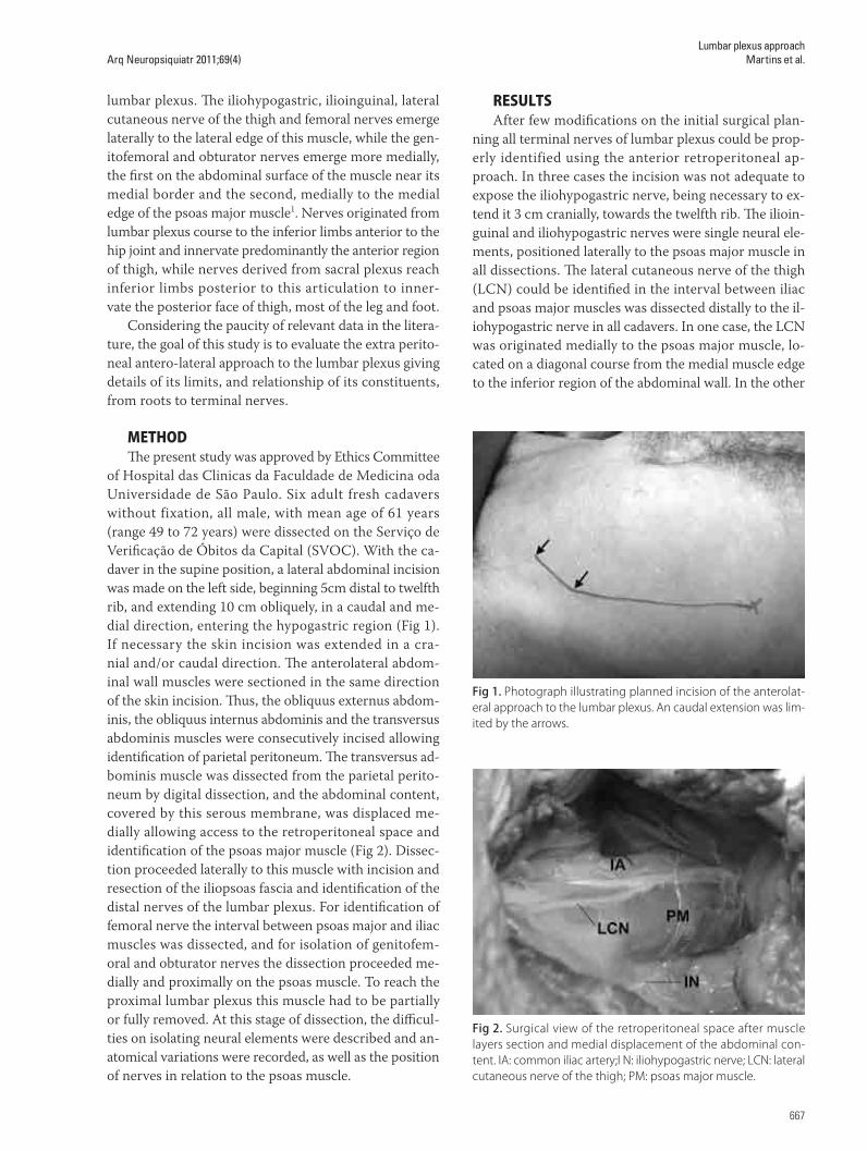
Fig 1. Photograph illustrating planned incision of the anterolat-eral approach to the lumbar plexus. An caudal extension was lim-ited by the arrows.
Fig 2. Surgical view of the retroperitoneal space after muscle layers section and medial displacement of the abdominal con-tent. IA: common iliac artery;I N: iliohypogastric nerve; LCN: lateral cutaneous nerve of the thigh; PM: psoas major muscle.
RESULTSAfter few modifications on the initial surgical plan-
ning all terminal nerves of lumbar plexus could be prop-erly identified using the anterior retroperitoneal ap-proach. In three cases the incision was not adequate to expose the iliohypogastric nerve, being necessary to ex-tend it 3 cm cranially, towards the twelfth rib. The ilioin-guinal and iliohypogastric nerves were single neural ele-ments, positioned laterally to the psoas major muscle in all dissections. The lateral cutaneous nerve of the thigh (LCN) could be identified in the interval between iliac and psoas major muscles was dissected distally to the il-iohypogastric nerve in all cadavers. In one case, the LCN was originated medially to the psoas major muscle, lo-cated on a diagonal course from the medial muscle edge to the inferior region of the abdominal wall. In the other
Arq Neuropsiquiatr 2011;69(4)
668
Lumbar plexus approachMartins et al.
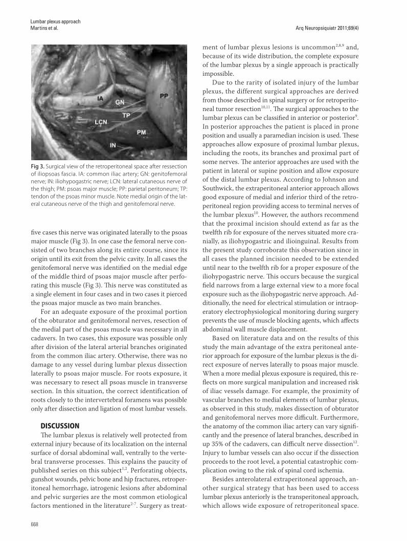
five cases this nerve was originated laterally to the psoas major muscle (Fig 3). In one case the femoral nerve con-sisted of two branches along its entire course, since its origin until its exit from the pelvic cavity. In all cases the genitofemoral nerve was identified on the medial edge of the middle third of psoas major muscle after perfo-rating this muscle (Fig 3). This nerve was constituted as a single element in four cases and in two cases it pierced the psoas major muscle as two main branches.
For an adequate exposure of the proximal portion of the obturator and genitofemoral nerves, resection of the medial part of the psoas muscle was necessary in all cadavers. In two cases, this exposure was possible only after division of the lateral arterial branches originated from the common iliac artery. Otherwise, there was no damage to any vessel during lumbar plexus dissection laterally to psoas major muscle. For roots exposure, it was necessary to resect all psoas muscle in transverse section. In this situation, the correct identification of roots closely to the intervertebral foramens was possible only after dissection and ligation of most lumbar vessels.
DISCUSSIONThe lumbar plexus is relatively well protected from
external injury because of its localization on the internal surface of dorsal abdominal wall, ventrally to the verte-bral transverse processes. This explains the paucity of published series on this subject1,2. Perforating objects, gunshot wounds, pelvic bone and hip fractures, retroper-itoneal hemorrhage, iatrogenic lesions after abdominal and pelvic surgeries are the most common etiological factors mentioned in the literature2-7. Surgery as treat-
ment of lumbar plexus lesions is uncommon2,8,9 and, because of its wide distribution, the complete exposure of the lumbar plexus by a single approach is practically impossible.
Due to the rarity of isolated injury of the lumbar plexus, the different surgical approaches are derived from those described in spinal surgery or for retroperito-neal tumor resection10,11. The surgical approaches to the lumbar plexus can be classified in anterior or posterior9. In posterior approaches the patient is placed in prone position and usually a paramedian incision is used. These approaches allow exposure of proximal lumbar plexus, including the roots, its branches and proximal part of some nerves. The anterior approaches are used with the patient in lateral or supine position and allow exposure of the distal lumbar plexus. According to Johnson and Southwick, the extraperitoneal anterior approach allows good exposure of medial and inferior third of the retro-peritoneal region providing access to terminal nerves of the lumbar plexus10. However, the authors recommend that the proximal incision should extend as far as the twelfth rib for exposure of the nerves situated more cra-nially, as iliohypogastric and ilioinguinal. Results from the present study corroborate this observation since in all cases the planned incision needed to be extended until near to the twelfth rib for a proper exposure of the iliohypogastric nerve. This occurs because the surgical field narrows from a large external view to a more focal exposure such as the iliohypogastric nerve approach. Ad-ditionally, the need for electrical stimulation or intraop-eratory electrophysiological monitoring during surgery prevents the use of muscle blocking agents, which affects abdominal wall muscle displacement.
Based on literature data and on the results of this study the main advantage of the extra peritoneal ante-rior approach for exposure of the lumbar plexus is the di-rect exposure of nerves laterally to psoas major muscle. When a more medial plexus exposure is required, this re-flects on more surgical manipulation and increased risk of iliac vessels damage. For example, the proximity of vascular branches to medial elements of lumbar plexus, as observed in this study, makes dissection of obturator and genitofemoral nerves more difficult. Furthermore, the anatomy of the common iliac artery can vary signifi-cantly and the presence of lateral branches, described in up 35% of the cadavers, can difficult nerve dissection12. Injury to lumbar vessels can also occur if the dissection proceeds to the root level, a potential catastrophic com-plication owing to the risk of spinal cord ischemia.
Besides anterolateral extraperitoneal approach, an-other surgical strategy that has been used to access lumbar plexus anteriorly is the transperitoneal approach, which allows wide exposure of retroperitoneal space.
Fig 3. Surgical view of the retroperitoneal space after ressection of iliopsoas fascia. IA: common iliac artery; GN: genitofemoral nerve; IN: iliohypogastric nerve; LCN: lateral cutaneous nerve of the thigh; PM: psoas major muscle; PP: parietal peritoneum; TP: tendon of the psoas minor muscle. Note medial origin of the lat-eral cutaneous nerve of the thigh and genitofemoral nerve.
Arq Neuropsiquiatr 2011;69(4)
669
Lumbar plexus approachMartins et al.
This approach is used mostly for the resection of large neural tumors. Its main disadvantage is the need of a laparotomy and the consequent potential morbidity. To avoid a possible complication, an ureteral catheter place-ment is recommended. This facilitates the digital identi-fication of the ureter and prevents its damage13.
In conclusion, the extra peritoneal anterolateral ap-proach allows adequate exposure of terminal nerves of the lumbar plexus laterally to psoas major muscle. Cra-nial extension of skin incision may be necessary to ex-pose the iliohypogastric nerve and roots exposure in-creases the risk of vascular injury.
REFERENCES1. Matejcík V. Anatomical variations of lumbosacral plexus. Surg Radiol Anat
2010;32:409-414. 2. Stoehr M. Traumatic and postoperative lesions of the lumbosacral plexus.
Arch Neurol 1978;35:757-760. 3. Chiou-Tan FY, Kemp K Jr, Elfenbaum M, Chan KT, Song J. Lumbosacral
plexopathy in gunshot wounds and motor vehicle accidents: comparison of electrophysiologic findings. Am J Phys Med Rehabil 2001;80:280-285.
4. Chiu WS. The syndrome of retroperitoneal hemorrhage and lumbar plexus neuropathy during anticoagulant therapy. South Med J 1976;69:595-599.
5. Durkin A, Sagi HC, Durham R, Flint L. Contemporary management of pelvic fractures. Am J Surg 2006;192:211-223.
6. Stevanato G, Vazzana L, Daramaras S, Trincia G, Saggiorno GC, Squintani G. Lumbosacral plexus lesions. Acta Neurochir 2007;100(Suppl):S15-S20.
7. Tung TH, Martin DZ, Novak CB, Lauryssen C, Mackinnon SE. Nerve recon-struction in lumbosacral plexopathy: case report and review of the litera-ture. J Neurosurg (Pediatrics 1) 2005;102:86-91.
8. Verstraete KLA, Martens F, Smeets P, et al. Traumatic lumbosacral nerve root meningoceles. The value of myelography, CT and MRI in the assessment of nerve root continuity. Neuroradiology 1989;31:425-429.
9. Lang EM, Borges J, Carlstedt T. Surgical treatment of lumbosacral plexus injuries. Neurosurg (Spine 1) 2004;1:64-71.
10. Johnson RM, Southwick WO. Surgical approaches to the lumbosacral spine. In: Rothman RH, Simeone FA (Eds). The spine. 2nd Ed. Philadelphia PA: WB Saunders, 1982;171-187.
11. Benglis Jr DM, Vanni S, Levi AD. An anatomical study of the lumbosacral plexus as related to the minimally invasive transpsoas approach to the lumbar spine. Neurosurgery (Spine) 2009;10:139-144.
12. El Mamoun BA, Demmel U. The lateral branches of the common iliac artery. Surg Radiol Anat 1988;10:161-164.
13. Bumpass DB, Keller TC, Robinson EP, et al. Implications of lmbar plexus anatomy for removal of total discs replacements through a posterior approach. Spine 2008;33:274-278.