Embed Size (px)
Citation preview
AJR:186, April 2006 1007
AJR 2006; 186:1007–1012
0361–803X/06/1864–1007
© American Roentgen Ray Society
M E D I C A L I M A G I N G
A C E N T U R Y O F
M E D I C A L I M A G I N G
A C E N T U R Y O F
Avila et al.CT of Pleural Abnormalities
C h e s t I m ag i n g • O r i g i n a l R e s e a rc h
CT of Pleural Abnormalities in Lymphangioleiomyomatosis and Comparison of Pleural Findings After Different Types of Pleurodesis
Nilo A. Avila1
Andrew J. Dwyer1
Antoinette Rabel2
Rosamma M. DeCastro2
Joel Moss2
Avila NA, Dwyer AJ, Rabel A, DeCastro RM, Moss J
Keywords: CT, lung diseases, lymphangioleiomyomatosis, pleura, pleurodesis
DOI:10.2214/AJR.04.1912
Received December 17, 2004; accepted after revision February 14, 2005.
The opinions and assertions contained herein are the private views of the authors and are not to be construed as official or as representing the views of the National Institutes of Health or any other government agency.
Presented at the 2003 annual meeting of the American Roentgen Ray Society, San Diego, CA.
1Department of Diagnostic Radiology, Warren G. Magnuson Clinical Center, Bldg. 10, Rm. 1C-660, 10 Center Dr., MSC 1182, Bethesda, MD 20892-1182. Address correspondence to N. A. Avila.
2Pulmonary-Critical Care Medicine Branch, National Heart, Lung, and Blood Institute (NHLBI), National Institutes of Health (NIH), Bethesda, MD.
OBJECTIVE. The objective of our article was to describe the spectrum and frequency ofpleural abnormalities on CT in patients with lymphangioleiomyomatosis (LAM) and the pleu-ral findings associated with different types of pleurodesis (talc, mechanical, and chemical) per-formed to treat the complications of pleural disease in these patients.
MATERIALS AND METHODS. Two hundred fifty-eight patients with LAM under-went CT of the chest. Pleural abnormalities assessed included pleural thickening, presence ofa pleural mass, areas of high attenuation, effusion, and pneumothorax. In patients who had hadpleurodesis, the CT findings were correlated with the type of procedure performed.
RESULTS. One hundred thirty-three (52%) of 258 patients had pleurodesis (unilateral,68/133; bilateral, 65/133). Pleural abnormalities were more common in patients who had pleu-rodesis (101/133, 76%) than in those who had not (47/125, 38%) and were more prevalent onthe operated side than on the unoperated side of those 68 patients who had unilateral pleurodesis.The frequencies of findings for the group without pleurodesis versus the group with pleurodesiswere pleural thickening (26% vs 65%), effusion (10% vs 13%), loculated effusion (2.4% vs11%), pneumothorax (1.6% vs 10%), areas of high attenuation (1.6% vs 23%), and mass (0.8%vs 14%), respectively. Areas of high attenuation in the pleura were present in all types of pleu-rodesis (mechanical, 8%; chemical, 13%; talc, 40%) and in two patients who had had repeatedthoracentesis or pleurectomy. Pleural masses were present in patients who had had all types ofpleurodesis (mechanical, 10%; chemical, 9%; talc, 24%) and in one patient who had had thora-centesis and thoracostomy; the masses commonly enhanced and did not change in size over time.
CONCLUSION. Pleural abnormalities are common in patients with LAM as complica-tions of the disease itself and as sequelae of pleurodesis and other pleura manipulations. Pneu-mothorax and pleural effusion result from the underlying pathophysiology of LAM, whereasareas of high attenuation and masses develop after all types of pleurodesis and other manipu-lations of the pleura (i.e., thoracentesis, thoracostomy).
ymphangioleiomyomatosis (LAM)is a rare idiopathic disease thatmost commonly affects womenand results from the accumulation
of abnormal smooth-muscle cells in the lym-phatics. Common thoracic findings are lungcysts, pneumothorax, and chylous pleural ef-fusion. Exertional dyspnea and pneumotho-rax are the major presenting complaints[1–3]. Recurrent pneumothorax and chylouspleural effusion may cause restrictive pulmo-nary dysfunction requiring pleurodesis in upto 54% of patients with LAM [1]. Because ofthe high frequency of pleurodesis in patientswith LAM, an awareness of the pleural find-ings after pleurodesis is essential for the cor-rect interpretation of pleural abnormalities inthese patients.
Because the imaging literature on this topicis limited, we describe here the spectrum andprevalence of pleural findings on CT in 258patients with LAM with and without pleurod-esis. In the patients with pleurodesis, we ana-lyze the relationship between the pleural find-ings and the type of pleurodesis performed.
Materials and MethodsThe study protocol and consent documents were
approved by the institutional review board of the Na-tional Heart, Lung, and Blood Institute. Our institu-tion is a large referral center currently studying thenatural history of LAM. Written informed consentwas obtained from all study participants. This studyincludes 258 patients referred to our institution (allwomen; age range, 23–77 years; mean, 44 years)with pulmonary LAM evaluated at our institution be-
L
Avila et al.
1008 AJR:186, April 2006
tween March 1996 and June 2003. The diagnosis ofLAM was established by lung biopsy in 186 patientsand biopsy of abdominopelvic masses in 11 patients.Sixty-one patients did not have tissue biopsy but hadclassic clinical findings (recurrent spontaneouspneumothorax, pleural effusions, or both) and pul-monary CT findings (diffusely scattered thin-walledlung cysts). We have previously reported some of thepleural findings (without correlation to type of pleu-rodesis) in 37 of these patients [4].
CT of the ChestTwo hundred forty-one patients underwent en-
hanced CT of the chest performed on a HiSpeedAdvantage, CTi, or LightSpeed scanner (GEHealthcare). The images were obtained using 5- to10-mm collimation at end inspiration after injec-tion of 120 mL of IV contrast material (iopamidol61% [Isovue 300, Bracco Diagnostics]) with thepatient supine. Seventeen patients did not receiveIV contrast material either because of a history ofallergic reactions or because of poor renal function.
Image AnalysisAn experienced board-certified radiologist,
aware of the diagnosis of LAM, reviewed the CTimages of the chest obtained in all 258 patients.Pleural abnormalities assessed included pneu-mothorax, effusion, pleural thickening (uniform in-crease in the width of pleura), calcification (areas ofhigh attenuation [i.e., > 90 H]), mass (focal, roundbulge of soft tissue attenuation [> 5 H] indentingthe lung), and loculated effusion (focal bulge of wa-ter attenuation [0–5 H] indenting the lung) [5].Rounded areas of parenchymal consolidation adja-cent to abnormal pleura were recorded as roundatelectasis.
In patients with pleural masses, we measuredwith electronic calipers the maximum diameter ofthe mass perpendicular to the pleural surface. Inthose patients with unenhanced and enhanced stud-
ies, we recorded the difference in attenuation of themass (in Hounsfield units) between the enhancedand unenhanced images. A mass was categorized asenhancing if there was an increase in attenuation of15 H or more between the enhanced and unenhancedimages. We recorded the time interval between thedate of the initial screening CT examination at ourinstitution and the date of pleurodesis and the date ofthe most recent follow-up CT examination. We alsorecorded, when present, any change in the size of thepleural mass between the initial and the most recentfollow-up CT examinations and described it as sta-ble, decreased, or increased.
In patients with areas of high attenuation in thepleura, we measured with electronic calipers thethickness (maximum diameter perpendicular to thepleural surface) and the attenuation in Hounsfieldunits. The location of the area of high attenuationwas correlated with the side of known pleurodesis,and the shape was categorized as focal plaque(punctate or coarse high-attenuation material) orcontinuous plaque (linear high-attenuation materialthat follows the outline of the pleural surface).
Medical HistoryThe patients were interviewed for admission to
the protocol, and their medical records were re-viewed. We recorded any history of pleurodesis andthe type of pleurodesis performed (talc, chemical,mechanical, or a combination of these).
Statistical AnalysisThe prevalence of each of the pleural findings in
the 133 patients who had had pleurodesis was com-pared with the prevalence in the 125 patients whohad not had pleurodesis (chi-square test). Also, tocontrol for potential confounding factors, we per-formed a matched analysis on the results from the 68patients who had had unilateral pleurodesis; specifi-cally, the prevalence of each pleural finding on theoperated side was compared with the prevalence on
the nonoperated side (McNemar test). The preva-lences of pleural findings were compared for me-chanical, chemical, and talc pleurodesis (chi-squaretest). The correlation between the degree of enhance-ment (enhanced attenuation [H]–unenhanced attenu-ation [H]) and the thickness of the pleural masseswas assessed (Pearson’s correlation coefficient [R]).
ResultsOne hundred forty-eight (57%) of the 258
patients had one or more pleural abnormali-ties on CT: pleural thickening in 117 (45%),pleural effusion in 28 (11%), loculated effu-sion in 17 (7%), pneumothorax in 15 (6%), ar-eas of high attenuation in 32 (12%), and massin 20 (8%) (Table 1 and Figs. 1–3).
One hundred thirty-three (52%) of 258 pa-tients had had pleurodesis (unilateral, 68/133;bilateral, 65/133). The prevalences of thetypes of pleurodesis performed were as fol-lows: mechanical in 40 patients (30%), talc in25 (19%), chemical in 23 (17%), a combina-tion of procedures in 38 (29%), and unknownor not recorded in seven (5%).
In both the patient-by-patient analysis andthe matched analysis, pleural abnormalitieswere more common in the presence of a previ-ous pleurodesis. The respective prevalences ofpleural abnormalities for the 133 patients whohad had pleurodesis and the 125 who had notand the levels of statistical significance of thedifferences were as follows: pleural thickening(65% vs 26%, p < 0.001), effusion (13% vs10%, p = 0.7), loculated effusion (11% vs2.4%, p = 0.13), areas of high attenuation(23% vs 1.6%, p < 0.001), and mass (14% vs0.8%, p < 0.001) (Table 1). Every type ofpleural abnormality (effusion, loculated effu-sion, pneumothorax, areas of high attenuation,and mass) was found with each type of pleu-rodesis (mechanical, chemical, talc) (Table 1).
TABLE 1: Prevalence of Pleural Abnormalities in Patients with Lymphangioleiomyomatosis With and Without Pleurodesis
Pleural Abnormality
Total No. (%)of All Patients
(n = 258)No Pleurodesis
(n = 125)Pleurodesis
(n = 133)
Type of Pleurodesis (n = 133)
Mechanical(n = 40)
Talc(n = 25)
Chemical(n = 23)
Combination(n = 38)
Unknown(n = 7)
Thickening 117 (45) 33 (26) 86 (65) 20 (50) 18 (72) 17 (74) 29 (76) 2 (29)
Mass 20 (8) 1 (0.8) 19 (14) 4 (10) 6 (24) 2 (9) 7 (18) 1 (14)
Calcification 32 (12) 2 (1.6) 30 (23) 3 (8) 10 (40) 3 (13) 14 (37) 3 (43)
Effusion 28 (11) 13 (10) 17 (13) 3 (8) 5 (20) 3 (13) 6 (16) 0
Loculated effusion 17 (7) 3 (2.4) 14 (11) 7 (18) 4 (16) 2 (9) 1 (3) 1 (14)
Pneumothorax 15 (6) 2 (1.6) 13 (10) 3 (8) 3 (12) 3 (13) 5 (13) 0
None 110 (43) 78 (62) 32 (24) 10 (25) 6 (24) 8 (35) 8 (21) 0
Total 258 125 (48) 133 (52) 40 (30) 25 (19) 23 (17) 38 (29) 7 (5)
CT of Pleural Abnormalities
AJR:186, April 2006 1009
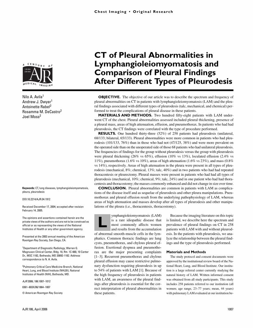
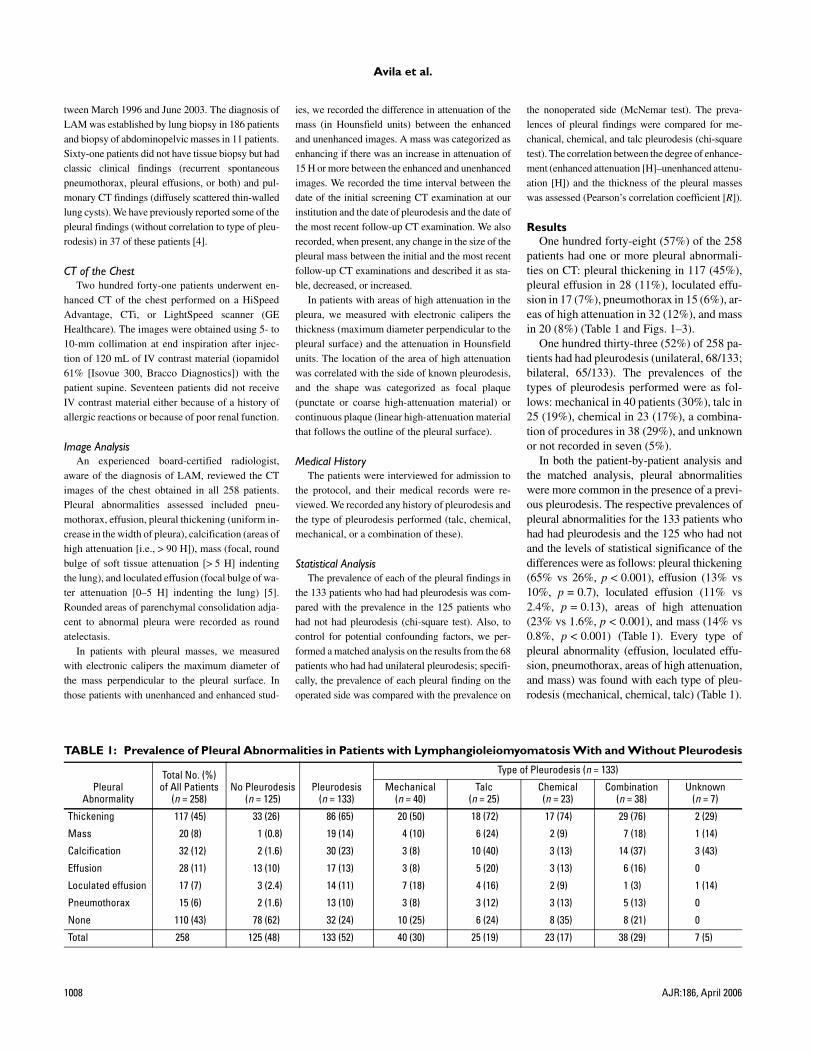
Fig. 1—39-year-old woman who had had right empyema, right thoracentesis, and right thorascopic biopsy (arrow on surgical clip) but no previous pleurodesis. Transverse contrast-enhanced CT image through lung bases shows extensive right pleural thickening (arrowheads).
Fig. 2—49-year-old woman who had had right chemical pleurodesis. Transverse contrast-enhanced CT image through lung bases shows large enhancing right pleural mass (arrows).
Fig. 3—41-year-old woman who had had bilateral talc pleurodesis. Transverse contrast-enhanced CT image through lung bases shows extensive bilateral pleural thickening and bonelike calcification (360–680 H) (arrows) in right lung base.
Similarly, in the 68 patients who had hadunilateral pleurodesis, the prevalences ofpleural abnormalities were greater on the op-erated side than on the unoperated side: pleu-ral thickening (53% vs 9%, p < 0.001), effu-sion (3% vs 1%, p = 1.0), loculated effusion(7% vs 3%, p = 0.4), pneumothorax (3% vs0%, p = 0.5), areas of high attenuation (15%vs 1%, p = 0.008), and mass (9% vs 1%,p = 0.125) (Table 2).
Twenty patients had pleural masses(Table 3). These masses ranged in size from0.8 to 3.7 cm in maximal thickness. Thepleural masses enhanced in 13 (76%) of 17patients that had had both unenhanced andenhanced studies. There was negligible cor-relation between the thickness of the massesand the degree of enhancement (R2 = 0.13).Nineteen of the 20 patients with pleuralmasses had had pleurodesis; the other pa-tient had had thoracentesis and thoracos-tomy for recurrent chylous pleural effusion(Fig. 4). The time interval between the dateof the initial screening CT at our institutionand the date of the pleurodesis in the 19 pa-tients with pleural masses after pleurodesiswas between 3 months and 12 years (mean,2.7 years). Eighteen of the 19 patients withpleural masses after pleurodesis had follow-up studies. The length of follow-up after thedate of the initial screening CT at our insti-tution was 1–6 years (mean, 3.4 years). Dur-ing follow-up, the masses remained stable in15 (83%) of 18 patients, decreased in size inthree (17%) of 18 patients, and increased insize in none (Table 3).
Thirty-two patients had areas of high atten-uation in the pleura. These areas ranged inthickness from 0.3 to 2.2 cm and in attenuationfrom 90 to 3,070 H (Fig. 5). The shape was fo-cal plaque (16/32, 50%), continuous plaque(9/32, 28%), or both (7/32, 22%). Thirty of the32 patients had had pleurodesis. Areas of highattenuation in the pleura were seen with allthree types of pleurodesis (talc, mechanical,and chemical); however, their prevalence wasgreater (p = 0.01) with talc (40%) than withchemical or mechanical pleurodesis (13% and8%, respectively). Two patients had areas ofhigh attenuation in the pleura without a historyof pleurodesis; one of these had had severalthoracentesis procedures, and the other hadhad pleurectomy complicated by empyemaand bronchopleural fistula.
Three patients had rounded lung consolida-tions adjacent to abnormal pleura with associ-ated peripheral bronchial structures consis-tent with round atelectasis (Fig. 6).
Avila et al.
1010 AJR:186, April 2006
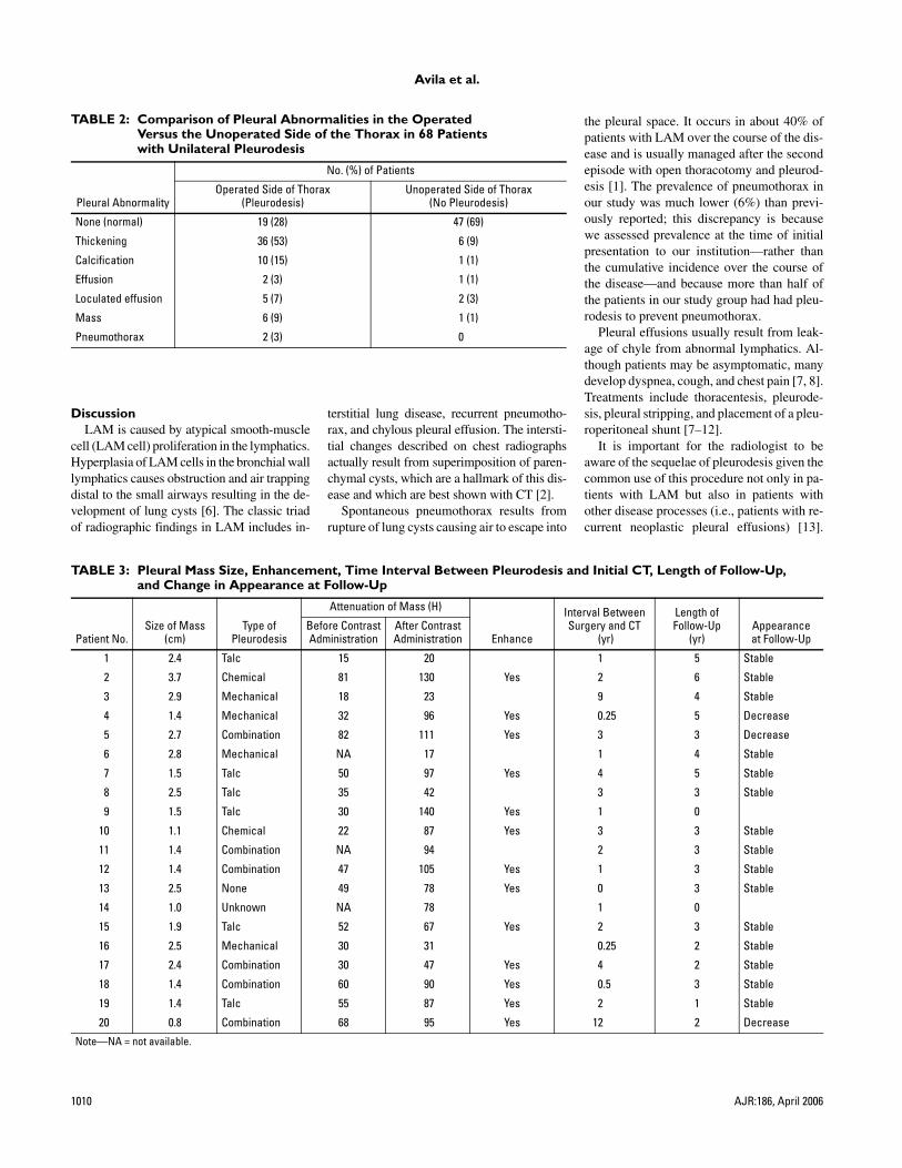
TABLE 2: Comparison of Pleural Abnormalities in the Operated Versus the Unoperated Side of the Thorax in 68 Patients with Unilateral Pleurodesis
Pleural Abnormality
No. (%) of Patients
Operated Side of Thorax(Pleurodesis)
Unoperated Side of Thorax(No Pleurodesis)
None (normal) 19 (28) 47 (69)
Thickening 36 (53) 6 (9)
Calcification 10 (15) 1 (1)
Effusion 2 (3) 1 (1)
Loculated effusion 5 (7) 2 (3)
Mass 6 (9) 1 (1)
Pneumothorax 2 (3) 0
DiscussionLAM is caused by atypical smooth-muscle
cell (LAM cell) proliferation in the lymphatics.Hyperplasia of LAM cells in the bronchial walllymphatics causes obstruction and air trappingdistal to the small airways resulting in the de-velopment of lung cysts [6]. The classic triadof radiographic findings in LAM includes in-
terstitial lung disease, recurrent pneumotho-rax, and chylous pleural effusion. The intersti-tial changes described on chest radiographsactually result from superimposition of paren-chymal cysts, which are a hallmark of this dis-ease and which are best shown with CT [2].
Spontaneous pneumothorax results fromrupture of lung cysts causing air to escape into
TABLE 3: Pleural Mass Size, Enhancement, Time Interval Between Pleurodesis and Initial CT, Length of Follow-Up, and Change in Appearance at Follow-Up
Patient No.Size of Mass
(cm)Type of
Pleurodesis
Attenuation of Mass (H)
Enhance
Interval BetweenSurgery and CT
(yr)
Length of Follow-Up
(yr)Appearanceat Follow-Up
Before ContrastAdministration
After ContrastAdministration
1 2.4 Talc 15 20 1 5 Stable
2 3.7 Chemical 81 130 Yes 2 6 Stable
3 2.9 Mechanical 18 23 9 4 Stable
4 1.4 Mechanical 32 96 Yes 0.25 5 Decrease
5 2.7 Combination 82 111 Yes 3 3 Decrease
6 2.8 Mechanical NA 17 1 4 Stable
7 1.5 Talc 50 97 Yes 4 5 Stable
8 2.5 Talc 35 42 3 3 Stable
9 1.5 Talc 30 140 Yes 1 0
10 1.1 Chemical 22 87 Yes 3 3 Stable
11 1.4 Combination NA 94 2 3 Stable
12 1.4 Combination 47 105 Yes 1 3 Stable
13 2.5 None 49 78 Yes 0 3 Stable
14 1.0 Unknown NA 78 1 0
15 1.9 Talc 52 67 Yes 2 3 Stable
16 2.5 Mechanical 30 31 0.25 2 Stable
17 2.4 Combination 30 47 Yes 4 2 Stable
18 1.4 Combination 60 90 Yes 0.5 3 Stable
19 1.4 Talc 55 87 Yes 2 1 Stable
20 0.8 Combination 68 95 Yes 12 2 Decrease
Note—NA = not available.
the pleural space. It occurs in about 40% ofpatients with LAM over the course of the dis-ease and is usually managed after the secondepisode with open thoracotomy and pleurod-esis [1]. The prevalence of pneumothorax inour study was much lower (6%) than previ-ously reported; this discrepancy is becausewe assessed prevalence at the time of initialpresentation to our institution—rather thanthe cumulative incidence over the course ofthe disease—and because more than half ofthe patients in our study group had had pleu-rodesis to prevent pneumothorax.
Pleural effusions usually result from leak-age of chyle from abnormal lymphatics. Al-though patients may be asymptomatic, manydevelop dyspnea, cough, and chest pain [7, 8].Treatments include thoracentesis, pleurode-sis, pleural stripping, and placement of a pleu-roperitoneal shunt [7–12].
It is important for the radiologist to beaware of the sequelae of pleurodesis given thecommon use of this procedure not only in pa-tients with LAM but also in patients withother disease processes (i.e., patients with re-current neoplastic pleural effusions) [13].
CT of Pleural Abnormalities
AJR:186, April 2006 1011
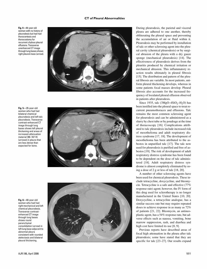
Fig. 4—44-year-old woman with no history of pleurodesis but had had thoracentesis and thoracostomy for recurrent chylous pleural effusions. Transverse unenhanced CT image through lung bases shows right pleural mass (arrow).
Fig. 5—35-year-old woman who had had bilateral chemical pleurodesis and left talc pleurodesis. Transverse contrast-enhanced CT image through lung bases shows left pleural thickening and areas of increased-attenuation material (90–167 H) (arrows) in pleura that are less dense than expected for bone.
Fig. 6—40-year-old woman who had had right mechanical and left chemical pleurodesis. Transverse contrast-enhanced CT image through lung bases shows round parenchymal consolidation (arrow) in left lung base adjacent to abnormal pleura consistent with rounded atelectasis and bilateral pleural thickening.
During pleurodesis, the parietal and visceralpleura are adhered to one another, therebyobliterating the pleural space and preventingthe accumulation of air or fluid within it.Pleurodesis may be performed by installationof talc or other sclerosing agent into the pleu-ral cavity (chemical pleurodesis) or by surgi-cal abrasion of the pleura with a dry gauzesponge (mechanical pleurodesis) [14]. Theeffectiveness of pleurodesis derives from thepleuritis produced by chemical irritation ormechanical abrasion. This inflammatory re-action results ultimately in pleural fibrosis[15]. The distribution and pattern of the pleu-ral fibrosis are variable. In most patients, uni-form pleural thickening develops, whereas insome patients focal masses develop. Pleuralfibrosis also accounts for the increased fre-quency of loculated pleural effusion observedin patients after pleurodesis.
Since 1935, talc (3MgO–4SiO2–H2O) hasbeen instilled into the pleural space to treat re-current pneumothoraces and effusions. Talcremains the most common sclerosing agentfor pleurodesis and can be administered as aslurry by chest tube or by poudrage at the timeof thoracoscopy [16]. Complications attrib-uted to talc pleurodesis include increased riskof mesothelioma and adult respiratory dis-tress syndrome [17, 18]. The development ofmesothelioma has been attributed to the as-bestos in unpurified talc [17]. The talc nowused for pleurodesis is purified and free of as-bestos [19]. The risk of development of adultrespiratory distress syndrome has been foundto be dependent on the dose of talc adminis-tered [18]. Adult respiratory distress syn-drome is almost completely eliminated by us-ing a dose of 2 g or less of talc [18, 20].
A number of other sclerosing agents havebeen used for chemical pleurodesis. These in-clude tetracycline, doxycycline, and bleomy-cin. Tetracycline is a safe and effective (77%response rate) agent; however, the IV form ofthis drug used for sclerotherapy is no longermanufactured in the United States [10, 20].Doxycycline, a tetracycline analogue, has asimilar success rate but may require repeateddoses to achieve response in as many as 72%of patients [21, 22]. Bleomycin, an antineo-plastic agent, has a 54% response rate, but ad-verse effects such as nausea, vomiting, bonemarrow suppression, rash, and diarrhea andhigh cost have limited its use [8, 9].
Previous reports have described areas offocal high attenuation in the pleura after talcpleurodesis; some have stated that they arespecific for talc [23–27]. Our results expand
Avila et al.
1012 AJR:186, April 2006
on this knowledge. In our series, focal areasof high attenuation in the pleura follow alltypes of pleurodesis; are more prevalent aftertalc pleurodesis (40%) than after other typesof pleurodesis that do not use talc (13% and8% with chemical and mechanical pleurode-sis, respectively); and are also seen in somepatients who have not had pleurodesis buthave had other pleural manipulations. Thus,these areas of high attenuation in the pleuracannot be attributed solely to deposition oftalc; rather, they may in addition be caused bydystrophic calcification of the inflamedpleura after installation of talc and other scle-rosing agents and pleural manipulation.
Pleural masses after talc pleurodesis havebeen reported in two prior case reports [28, 29].Williams et al. [28] reported an apical pleuralmass that developed after talc pleurodesis andhad CT attenuation characteristics of a cystwith a calcified capsule. Ahmed and Shrager[29] reported a calcified anterior mediastinalmass that proved to represent a giant talc gran-uloma after resection. We observed in our seriessolid pleural masses in 19 (14%) of 133 patientsdeveloping after all types of pleurodesis (chem-ical, mechanical, and talc pleurodesis). We alsoobserved a pleural mass in a patient who did nothave pleurodesis but had thoracentesis for treat-ment of recurrent pleural effusion and chesttube placement for treatment of pneumothorax.
In this series, contrast enhancement was use-ful in distinguishing solid pleural masses fromloculated effusions. A majority (76%) of thepleural masses in this series enhanced; thisfinding is important because an enhancingpleural mass may be misdiagnosed as mesothe-lioma or pleural metastasis and may prompt bi-opsy. We propose that pleural masses that resultfrom pleurodesis and other pleural manipula-tions result from the accumulation of fibrotictissue secondary to the postsurgical inflamma-tory reaction during the process of healing. Thisfibrotic tissue may enhance depending on theamount of vascular granulation tissue in it. Allpleural masses in this series remained stable ordecreased in size in the 1- to 6-year follow-upperiod. Thus, enhancing pleural masses—evenwhen associated with pleural calcification—that develop after pleurodesis in patients withLAM do not require follow-up because theircause is most likely benign. However, in pa-tients with cancer who have had pleurodesis asa palliative procedure for malignant pleural ef-fusion, the development of pleural masses mayrepresent metastasis.
Pleural abnormalities are common in pa-tients with LAM as complications of the dis-
ease itself and as sequelae of pleurodesis andother pleural manipulations. Pneumothoraxand pleural effusion result from the underly-ing pathophysiology of LAM, whereas areasof high attenuation in the pleura and massesdevelop after all types of pleurodesis andother pleural manipulations (thoracentesis,thoracostomy). Solid pleural masses com-monly enhance on CT but do not change insize over time. An awareness of this is im-portant to prevent unnecessary biopsy to ex-clude pleural malignancy such as mesothe-lioma or pleural metastasis.
References1. Chu SC, Horiba K, Usuki J, et al. Comprehensive
evaluation of 35 patients with lymphangioleio-
myomatosis. Chest 1999; 115:1041–1052
2. Muller NL, Chiles C, Kullnig P. Pulmonary lym-
phangiomyomatosis: correlation of CT with radio-
graphic and functional findings. Radiology 1990;
175:335–339
3. Aberle DR, Hansell DM, Brown K, Tashkin DP.
Lymphangiomyomatosis: CT, chest radiographic,
and functional correlations. Radiology 1990;
176:381–387
4. Avila NA, Kelly JA, Dwyer AJ, et al. Lymphan-
gioleiomyomatosis: correlation of qualitative and
quantitative thin section CT with pulmonary func-
tion tests and assessment of dependence on pleu-
rodesis. Radiology 2002; 223:189–197
5. Curry TS, Dowdey JF, Murry RC Jr. Computed to-
mography. In: Christiensen’s introduction to the
physics of diagnostic radiology, 3rd ed. Philadel-
phia, PA: Lea & Febiger, 1984:338
6. Carrington CB, Cugell DW, Gaensler EA, et al.
Lymphangiomyomatosis: physiologic–patho-
logic–radiologic correlations. Am Rev Respir
Dis 1977; 116:977–995
7. Sahn SA. Pleural effusion in lung cancer. Clin Chest
Med 1993; 14:189–200
8. Chernow B, Sahn S. Carcinomatous involvement of
the pleura. Am J Med 1977; 63:695–702
9. Andrews CO, Gora ML. Pleural effusions: patho-
physiology and management. Ann Pharmacother
1994; 28:894–903
10. Walker-Renard PB, Vaughan LM, Sahn SA. Chem-
ical pleurodesis for malignant pleural effusions.
Ann Intern Med 1994; 120:56–64
11. Windsor PG, Como JA, Windsor KS. Sclerotherapy
for malignant pleural effusions: alternatives to tet-
racycline. South Med J 1994; 87:709–714
12. Leininger RJ, Barker WL, Langston HT. A sim-
plified method for management of malignant
pleural effusion. J Thorac Cardiovasc Surg 1969;
58:758–763
13. Marom EM, Patz EF Jr, Erasmus JJ, McAdams
HP, Goodman PC, Herndon JE. Malignant pleu-
ral effusions: treatment with small-bore-catheter
thoracostomy and talc pleurodesis. Radiology
1999; 21:277–281
14. Bouros D, Froudarakis M, Siafakas NM, et al.
Pleurodesis: everything flows. Chest 2000;
118:577–579
15. Kroegel C, Antony VB. Immunobiology of pleural
inflammation: potential implications for pathogen-
esis, diagnosis, and therapy. Eur Respir J 1997;
10:2411–2418
16. Kennedy L, Sahn SA. Talc pleurodesis for the treat-
ment of pneumothorax and pleural effusion. Chest
1994; 106:1215–1222
17. Jackson JW, Bennet MH. Chest wall tumor fol-
lowing iodized talc pleurodesis. Thorax 1973;
28:788–793
18. Rinaldo JE, Owens GR, Rogers RM. Adult respi-
ratory distress syndrome following intrapleural in-
stillation of talc. J Thorac Cardiovasc Surg 1983;
85:523–526
19. Weisberg D, Ben-Zeev I. Talc pleurodesis: experi-
ence with 360 patients. J Thorac Cardiovasc Surg
1993; 106:689–695
20. Heffner JE, Unruh LC. Tetracycline pleurodesis:
adios, farewell, adieu. Chest 1992; 101:5–6
21. Seaton KG, Patz EF Jr, Goodman PC. Palliative
treatment of malignant pleural effusions: value of
small-bore catheter thoracostomy and doxycycline
sclerotherapy. AJR 1995; 164:589–591
22. Mansson T. Treatment of malignant pleural effusion
with doxycycline. Scand J Infect Dis Suppl 1988;
53:29–34
23. Murray JG, Patz EF Jr, Erasmus JJ, Gilkeson RC.
CT appearance of the pleural space after talc pleu-
rodesis. AJR 1997; 169:89–91
24. Carignan S, Samson L, Lafontaine E, Cordeau MP.
Radiological changes of talc pleurodesis in cases of
effusion [in French]. Ann Chir 1994; 48:777–784
25. Nguyen M, Varma V, Perez R, Schuster DM. CT
with histopathologic correlation of FDG uptake in
a patient with pulmonary granuloma and pleural
plaque caused by remote talc pleurodesis. AJR
2004; 182:92–94
26. Weiss N, Solomon SB. Talc pleurodesis mimics
pleural metastases: differentiation with positron
emission tomography/computed tomography. Clin
Nucl Med 2003; 28:811–814
27. Murray JG, Erasmus JJ, Bahtiarian EA, Goodman
PC. Talc pleurodesis simulating pleural metastases
on 18F-fluorodeoxyglucose positron emission to-
mography. AJR 1997; 168:359–360
28. Williams T, Gostelow B, Woods D, Spyt T. Apical
pleural mass developing following talc pleurodesis.
Respir Med 1998; 92:358–359
29. Ahmed Z, Shrager JB. Mediastinal talcoma mas-
querading as thymoma. Ann Thorac Surg 2003;
75:568–569