Embed Size (px)
Citation preview
DEVELOPMENT OF SMALL MOLECULE RGS INHIBITORS AS A MECHANISM TO MODULATE G-PROTEIN SIGNALING
by
Levi L. Blazer
A dissertation submitted in partial fulfillment of the requirements for the degree of
Doctor of Philosophy (Pharmacology)
in The University of Michigan 2010
Doctoral Committee: Professor Richard R. Neubig, Chair Professor John R. Traynor
Associate Professor John J.G. Tesmer Assistant Professor Zaneta Nikolovska-Coleska

© Levi L. Blazer 2010

ii
For My Parents

iii
Acknowledgements
I would like to thank my mentor, Dr. Rick Neubig for his guidance and
support over the last several years. I’ve always found his inexhaustible energy
and enthusiasm for research is incredibly inspiring. I would like to thank my
thesis committee members, Dr. John Traynor, Dr. John Tesmer, and Dr. Zaneta
Nikolovska-Coleska for their support, guidance, and critical analysis of my work. I
also would like to thank Dr. David Roman for his friendship and immense
assistance during the initial stages of my Ph.D. training. Heartfelt thanks also to
the rest of the Neubig lab, including our laboratory manager, Sue Wade, for
making the Neubig lab a wonderful working environment. Thanks to Dr. Roger
Sunahara and the members of his lab, including Dr. Matt Wharton, Dr. Adam
Kuszak, Giselle Ruiz, Dee Calinski, and Brian Devree for allowing me to
constantly ‘borrow’ reagents.
I also need to thank to the many collaborators with whom I have had the
pleasure of doing science. I’d like to thank Martha Larsen and the rest of the staff
at the Center for Chemical Genomics for their assistance with chemical
screening and follow-up work. I never would have implemented as successful of
a screening project without their expertise. I’d like to thank Dr. Rez Halse and the
Novartis Institute for Biomedical Research for providing screening reagents and
for their contribution to the analysis of screening results. Thanks to Dr. Qin Wang
and Dr. John Traynor for their data showing cellular activity of CCG-50014. Also
iv
thanks to Dr. John Tesmer and those in his laboratory, including David Thal and
Mark Nance for their assistance during the crystallization trials of RGS8 and for
providing a number of constructs used in this thesis.
A very heartfelt thanks to my many friends that have made Ann Arbor a
wonderful place to live and learn over the last 5 years. I especially need to thank
them for their resounding support during a difficult transition in my life. While I
could not possibly include a comprehensive list, I would like to especially
recognize, David Thal, Matt Molusky, Mike Steinbaugh, Paul & Nikki Marinec, Dr.
Daniel & Meredith Foster, Beverly Piggott, Sarah Bass, and Taylor Eves. I’ll
always cherish tailgating, the Peyronie Pirates, Team Fit, and everything else
that has made us who we are. I’d also like to thank several friends that have
supported me throughout my life. To Drew Haerer, Daniel Beckenbaugh, Kyle
Rishel: While we may not see each other often, I’ll never forget the good times
we’ve had and I look forward to the good times we’ll have in the future.
I would especially like to thank my family for their immense support and
unwavering love. None of this would have been possible without my Mother,
Penny, my Father, Jim, and my brother, Cody. I am overwhelmingly indebted to
them (and my extended family) for providing an exceptional family life that has
shaped me into the man I am today. I feel exceedingly blessed to be from a
family that has been so generous, kind, and caring to each other. Lastly, Alina:
thank you for bringing “Malsa” into the world. Your impact on my condiment
options and – more importantly – my life in general has been absolutely
wonderful and I’ll always be immensely grateful for that.
v
Table of Contents
Dedication……………………………………………………………………………….ii Acknowledgements ............................................................................................ iii
List of Figures .....................................................................................................xi
List of Tables .................................................................................................... xiv
List of Appendices ............................................................................................xv
List of Abbreviations ........................................................................................ xvi
Abstract ............................................................................................................. xxi
CHAPTER I: Introduction .................................................................................. 1
Protein-Protein Interactions: What are they & why target them? ....................... 1
Inhibiting Protein-Protein Interactions outside of the CNS ................................ 3
Rationale for targeting Protein-Protein Interactions in the CNS ........................ 6
Inhibiting protein aggregation in the CNS ......................................................... 7
Amyloid Beta Aggregation ............................................................................. 7
Alpha Synuclein Aggregation ...................................................................... 12
Modulating Signal Transduction through inhibiting protein-protein interactions
........................................................................................................................ 17
Inhibition of PDZ interactions ....................................................................... 18
Targeting elements of the G-protein signaling pathway .................................. 23
Selective Gβγ Inhibitors ............................................................................... 24
RGS Proteins .................................................................................................. 27
vi
The RGS Homology Domain ....................................................................... 33
RGS4 as a Drug Target ............................................................................... 36
Importance of selectivity in RGS inhibition .................................................. 41
Current RGS Inhibitors ................................................................................ 41
Overview of the Thesis ................................................................................... 44
References ...................................................................................................... 48
CHAPTER II: Reversible, allosteric, small-molecule inhibitors of RGS
proteins ........................................................................................................... 58
Introduction ..................................................................................................... 58
Materials & Methods ....................................................................................... 61
Reagents ..................................................................................................... 61
Compound synthesis ................................................................................... 63
Protein expression & purification ................................................................. 64
Chemical labeling of purified Gαo & RGS .................................................... 65
Time Resolved FRET .................................................................................. 67
High Throughput Screening ......................................................................... 68
TR-FRET Dose Response Experiments ...................................................... 69
Flow Cytometry Protein Interaction Assay Concentration Dependence
Experiments ................................................................................................ 70
FCPIA Reversibility Experiments ................................................................. 70
Single Turnover GTPase Measurements .................................................... 71
Thermal Stability Measurements ................................................................. 71
Results ............................................................................................................ 72

vii
Development of a High-Throughput TR-FRET RGS4-Gαo interaction screen
.................................................................................................................... 72
CCG-63802 & CCG-63808 selectively inhibit Gαo-RGS interactions .......... 74
CCG-63802 & CCG-63808 inhibit RGS4 GAP activity ................................ 75
CCG-63802 & CCG-63808 bind to RGS4 but not to Gαo ............................ 76
CCG-63802 and CCG-63808 are reversible inhibitors of the Gαo-RGS
interaction .................................................................................................... 78
Cysteine Dependence of CCG-63802 and CCG-63808 .............................. 80
Structure-Activity Studies of the CCG-63802 class of compounds .............. 85
Discussion ....................................................................................................... 98
Conclusions .................................................................................................. 104
Chapter III: Biochemical Evaluation of Class of Small Molecule RGS
inhibitors with Cellular Activity .................................................................. 108
Introduction ................................................................................................... 108
Methods ........................................................................................................ 110
Reagents ................................................................................................... 110
Protein expression and purification ........................................................... 111
Chemical labeling of purified Gαo and RGS proteins ................................. 111
FCPIA Dose Response and Reversibility experiments .............................. 111
Single Turnover GTPase Measurements .................................................. 111
Thermal Stability Measurements ............................................................... 112
Analyses of the protein adduct of RGS by ESI-LC/MS .............................. 112
Papain Activity Assay ................................................................................ 113

viii
Docking of CCG-50014 to RGS8 ............................................................... 114
Results .......................................................................................................... 114
FCPIA characterization of RGS inhibitory activity ...................................... 114
CCG-50014 inhibits the catalytic GTPase accelerating activity of RGS8 and
RGS4 ........................................................................................................ 117
CCG-50014 irreversibly inhibits RGS proteins .......................................... 117
CCG-50014 Binds to RGS Proteins but not to Gαo ................................... 120
CCG-50014 depends on cysteine residues to inhibit the AlF4-Gαo/RGS
interaction .................................................................................................. 121
CCG-50014 is a covalent sulfhydryl modifier of RGS8 .............................. 122
CCG-50014 is not a general cysteine alkylator ......................................... 128
General cysteine alkylators do not inhibit RGS proteins ............................ 129
Computational modeling of the CCG-50014-RGS8 interaction ................. 130
Limiting the reactivity of CCG-50014 diminishes potency ......................... 132
Discussion ..................................................................................................... 134
References .................................................................................................... 138
Chapter IV: Cellular and Structure-Activity Studies of the CCG-50014
Compound Class ......................................................................................... 139
Introduction ................................................................................................ 139
Materials and Methods .................................................................................. 140
Reagents and Compounds ........................................................................ 140
Protein expression, purification, and labeling ............................................ 141
FCPIA Dose Response experiments ......................................................... 141

ix
Single Turnover GTPase Measurements .................................................. 141
Solubility experiments ............................................................................... 141
WST-1 Cell Viability Studies ...................................................................... 142
Cellular Localization Studies ..................................................................... 142
Calcium Mobilization Experiments ............................................................. 143
cAMP Accumulation .................................................................................. 144
Results .......................................................................................................... 145
Structure Activity Relationship Studies of the CCG-50014 Family of
Compounds ............................................................................................... 145
CCG-50014 does not affect cell viability .................................................... 154
CCG-50014 and related analogs inhibit the RGS4/Gαo interaction in living
cells ........................................................................................................... 155
CCG-50014 potentiates signaling through the δ-opioid receptor ............... 157
CCG-203769 potentiates the M3 muscarinic receptor activity via inhibition of
RGS4 ........................................................................................................ 159
Discussion ..................................................................................................... 161
References .................................................................................................... 166
Chapter V: Conclusions ................................................................................ 167
Summary of Results ...................................................................................... 167
Future Research Directions .......................................................................... 168
Therapeutic applications of RGS modulation ................................................ 173
The Future of Small Molecule Protein-Protein Interaction Inhibitors ............. 175
References .................................................................................................... 177
x
Appendix I: CCG-50014 Analogs Inhibit The RGS4-Gαo Protein-Protein
Interaction in Living Cells ............................................................................... 179
Appendix II: Tag Localization on RGS4 Does Not Effect Translocation or
Sensitivity to CCG-203769 ............................................................................. 180
xi
List of Figures Figure 1.1 Predicted “hot spots” on the Regulator of G protein signaling 4 (RGS4)/Gαi1 protein-protein interaction interface. ............................................... 5 Figure 1.2 Examples of amyloid beta aggregation inhibitors. ............................ 10 Figure 1.3 Examples of small molecule α-synuclein aggregation inhibitors ....... 16 Figure 1.4 Crystal structure of the first PDZ domain from MAGI bound to the PDZ ligand of HPV18 E6 .................................................................................... 20 Figure 1.5 Canonical G-protein signaling mechanism ....................................... 24 Figure 1.6 Structure of M119, a pathway selective inhibitor of Gβγ signaling .... 27 Figure 1.7 The RGS homology (RH) domain fold of RGS4 ............................... 34 Figure 1.8 Conserved lysine residues in RGS4 that have been implicated in calmodulin and acidic phospholipid binding ........................................................ 36 Figure 1.9 RGS-inhibitors increase the tissue specificity of an agonist .............. 37 Figure 1.10 Crystal structure of RGS4 in complex with Gαi1 .............................. 39 Figure 2.1 Characterization of the RGS4 TR-FRET high-throughput Assay ...... 62 Figure 2.2 Reaction scheme for the synthesis of CCG-63802 ........................... 64 Figure 2.3 RGS specificity of CCG-63802 and CCG-63808 determined by multiplex FCPIA analysis .................................................................................... 75 Figure 2.4 Single Turnover GAP analysis of small molecule RGS inhibitors with RGS4 .................................................................................................................. 76 Figure 2.5 Gαo is thermally stabilized in presence of nucleotide ........................ 77 Figure 2.6 CCG-63802 specifically binds to RGS4 and not to Gαo .................... 78 Figure 2.7 CCG-63802 and CCG-63808 are reversible inhibitors ...................... 79
xii
Figure 2.8 CCG-63802 and CCG-63808 are much less potent on a mutant form of RGS4 that lacks cysteine residues in the RH domain .................................... 81 Figure 2.9 CCG-63802 is less sensitive to glutathione than other RGS4 inhibitors ........................................................................................................................... 83 Figure 2.10 CCG-63802 and CCG-63808 inhibit the GAP activity of a cysteine-null RGS4 mutant ............................................................................................... 85 Figure 2.11 CCG-203687 and CCG-203680 (100 µM) are incapable of inhibiting the ability of RGS4 to accelerate the rate of GTP hydrolysis by Gαo .................. 87 Figure 2.12 CCG-203695 and CCG-203696 dose-dependently inhibit the RGS4-Gαo interaction ................................................................................................... 94 Figure 3.1 The chemical structure of CCG-50014 ........................................... 115 Figure 3.2 CCG-50014 inhibits RGS4 and RGS8 binding and function ........... 116 Figure 3.3 CCG-50014 does not affect the intrinsic rate of GTP hydrolysis by Gαo ................................................................................................................... 117 Figure 3.4 CCG-50014 is an irreversible inhibitor of RGS4 and RGS8 and its effects are partially reversed the thiol reductant DTT ....................................... 119 Figure 3.5 CCG-50014 thermally destabilizes RGS8 in a dose-dependent manner, but has no effect on the thermal stability of Gαo ................................. 120 Figure 3.6 CCG-50014 requires at least one cysteine residue on RGS8 for full activity .............................................................................................................. 122 Figure 3.7 CCG-50014 forms a covalent adduct on RGS8 .............................. 124 Figure 3.8 CCG-50014-induced protein aggregation is dependent on the presence of 160C ............................................................................................. 125 Figure 3.9 Irreversible inhibition of RGS8 is predominantly mediated by Cys 160 ......................................................................................................................... 127 Figure 3.10 Development of irreversible inhibition after exposure to CCG-50014 differs between the individual cysteine mutants ............................................... 128 Figure 3.11 CCG-50014 does not inhibit the general cysteine protease, papain ......................................................................................................................... 129
xiii
Figure 3.12 CCG-50014 is a much more potent RGS inhibitor than two general cysteine alkylators N-ethyl maleimide (NEM) and iodoacetamide (IA) ............. 130 Figure 3.13 Hypothesized binding site of CCG-50014 on RGS8 ..................... 131 Figure 4.1 The chemical structure of CCG-50014 ........................................... 145 Figure 4.2 CCG-50014 induces a calcium transient in HEK293 cells .............. 153 Figure 4.3 CCG-50014 does not affect HEK-293 viability ................................ 155 Figure 4.4 RGS4 is recruited to the plasma membrane by coexpression of Gαo and this recruitment is inhibited by CCG-50014 ............................................... 157 Figure 4.5 CCG-50014 potentiates the activity of the δ-opioid receptor ligand SNC-80 in SH-SY5Y cells ................................................................................ 158 Figure 4.6 CCG-203769 partially reverses the RGS4-mediated suppression of carbachol responsiveness in HEK293 cells expressing the M3 muscarinic receptor ............................................................................................................ 160
xiv
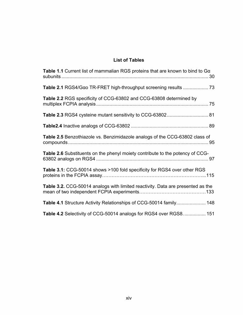
List of Tables Table 1.1 Current list of mammalian RGS proteins that are known to bind to Gα subunits .............................................................................................................. 30 Table 2.1 RGS4/Gαo TR-FRET high-throughput screening results ................... 73 Table 2.2 RGS specificity of CCG-63802 and CCG-63808 determined by multiplex FCPIA analysis .................................................................................... 75 Table 2.3 RGS4 cysteine mutant sensitivity to CCG-63802 ............................... 81 Table2.4 Inactive analogs of CCG-63802 .......................................................... 89 Table 2.5 Benzothiazole vs. Benzimidazole analogs of the CCG-63802 class of compounds ......................................................................................................... 95 Table 2.6 Substituents on the phenyl moiety contribute to the potency of CCG-63802 analogs on RGS4 .................................................................................... 97 Table 3.1: CCG-50014 shows >100 fold specificity for RGS4 over other RGS proteins in the FCPIA assay………………………………………………………...115 Table 3.2. CCG-50014 analogs with limited reactivity. Data are presented as the mean of two independent FCPIA experiments……………………………………133 Table 4.1 Structure Activity Relationships of CCG-50014 family ...................... 148 Table 4.2 Selectivity of CCG-50014 analogs for RGS4 over RGS8. ................ 151
xv
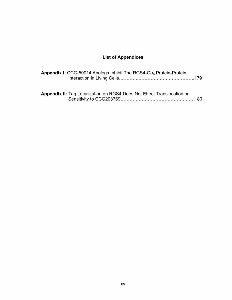
List of Appendices
Appendix I: CCG-50014 Analogs Inhibit The RGS4-Gαo Protein-Protein Interaction in Living Cells………….……………………………….179
Appendix II: Tag Localization on RGS4 Does Not Effect Translocation or Sensitivity to CCG203769………………………………………….180

xvi
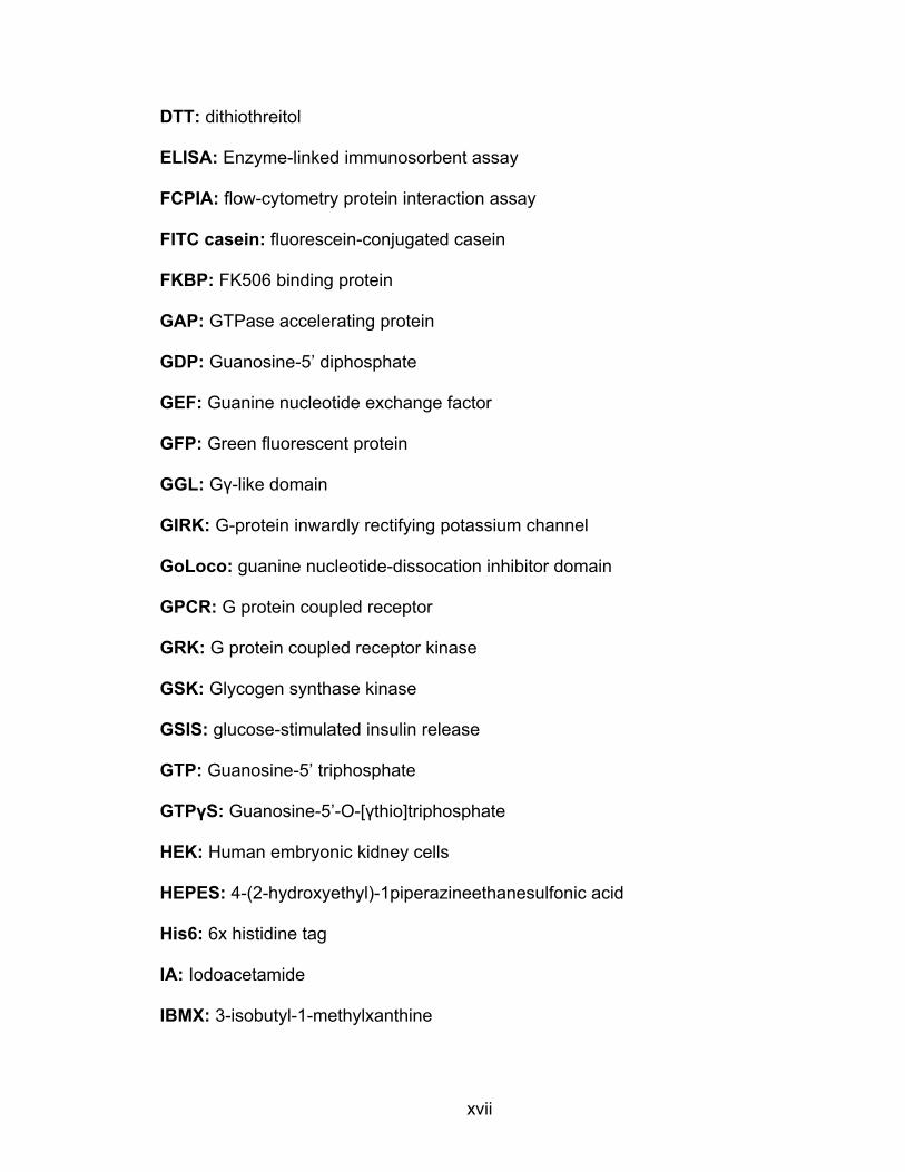
List of Abbreviations 1-8ANS: 1-anilinonapthalene-8-sulfonic acid
AchE: Acetylcholinesterase
AD: Alzheimer’s Disease
AF488: AlexaFluor 488
AF532: AlexaFluor-532
AlCl3: aluminum chloride
ANOVA: Analysis of variance
APP: Amyloid precursor protein
BSA: Bovine serum albumin
cAMP: 3’-5’-cyclic adenosine monophosphate
CCG: Center for Chemical Genomics, University of Michigan
CNS: Central nervous system
δ receptor: delta opioid receptor
DAX: Domain present in Dishevelled and Axin
DEP: Dishevelled/EGL10/Plextrin homology domain
DH: Dbl homologous domain
DMEM: Dulbecco’s Modified Eagle Medium
DMSO: Dimethyl sulphoxide
Dox: doxycycline
DRC: Dose-response curve
xvii
DTT: dithiothreitol
ELISA: Enzyme-linked immunosorbent assay
FCPIA: flow-cytometry protein interaction assay
FITC casein: fluorescein-conjugated casein
FKBP: FK506 binding protein
GAP: GTPase accelerating protein
GDP: Guanosine-5’ diphosphate
GEF: Guanine nucleotide exchange factor
GFP: Green fluorescent protein
GGL: Gγ-like domain
GIRK: G-protein inwardly rectifying potassium channel
GoLoco: guanine nucleotide-dissocation inhibitor domain
GPCR: G protein coupled receptor
GRK: G protein coupled receptor kinase
GSK: Glycogen synthase kinase
GSIS: glucose-stimulated insulin release
GTP: Guanosine-5’ triphosphate
GTPγS: Guanosine-5’-O-[γthio]triphosphate
HEK: Human embryonic kidney cells
HEPES: 4-(2-hydroxyethyl)-1piperazineethanesulfonic acid
His6: 6x histidine tag
IA: Iodoacetamide
IBMX: 3-isobutyl-1-methylxanthine
xviii
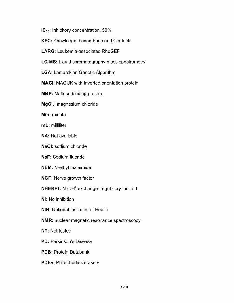
IC50: Inhibitory concentration, 50%
KFC: Knowledge–based Fade and Contacts
LARG: Leukemia-associated RhoGEF
LC-MS: Liquid chromatography mass spectrometry
LGA: Lamarckian Genetic Algorithm
MAGI: MAGUK with Inverted orientation protein
MBP: Maltose binding protein
MgCl2: magnesium chloride
Min: minute
mL: milliliter
NA: Not available
NaCl: sodium chloride
NaF: Sodium fluoride
NEM: N-ethyl maleimide
NGF: Nerve growth factor
NHERF1: Na+/H+ exchanger regulatory factor 1
NI: No inhibition
NIH: National Institutes of Health
NMR: nuclear magnetic resonance spectroscopy
NT: Not tested
PD: Parkinson’s Disease
PDB: Protein Databank
PDEγ: Phosphodiesterase γ

xix
PDZ: PSD95/Dlg/Z0-1 domain
PH domain: Plextrin homology domain
PI3K: Phosphoinositide 3-kinase
PIP3: (3,4,5)P3-phosphotidyl inositol
PLCβ: Phospholipase C, β isoform
PPI: Protein-protein interaction
PPII: Protein-protein interaction inhibitors
PTB: phosphotyrosine binding domain
PTEN: Phosphatase and tensin homolog
RBD: Ras binding domain
RGS: Regulator of G Protein Signaling
RGS4c: a mutant RGS4 with no cysteines in the RH domain
RH domain: RGS homology domain
RhoGEF: Rho guanine nucleotide exchange factor
SAR: Structure-activity relationship
SD: Standard deviation
Sec: Second
SEM: standard error of the mean
siRNA: short interfering ribonucleic acid
SMPPII: small molecule protein-protein interaction inhibitors
S/T Kinase: serine/threonine kinase domain
TCEP: tris(2-carboxyethyl) phosphine
TFA: tetrafluoroacetic acid
xx
Tm: melting temperature
TR-FRET: time-resolved fluorescence resonance energy transfer
µ receptor: mu opioid receptor
µL: microliter
WST-1: 4-[3-(4-iodophenyl)-2-(4-nitrophenyl)-2H-5-tetrazolio]-1,3-benzene
disulfonate

xxi
Abstract
DEVELOPMENT OF SMALL MOLECULE RGS INHIBITORS AS A MECHANISM TO MODULATE G-PROTEIN SIGNALING
by
Levi L. Blazer
Chair: Richard R. Neubig
Regulator of G-protein Signaling (RGS) proteins are important regulatory
molecules in the transduction of G-Protein Coupled Receptor (GPCR)
signaling. They function by directly binding to G alpha subunits and
accelerating GTP hydrolysis, thus potently inhibiting GPCR signaling. We and
others have proposed that small molecule inhibitors of RGS proteins may
provide a novel mechanism for therapeutic intervention in diseases stemming
from deficiencies in GPCR signaling. This thesis details the identification and
characterization of two novel classes of small molecule RGS inhibitors with
unique properties. These compounds were identified from a series of high
throughput screens performed by myself and others in our laboratory. The
CCG-63802 class of molecules includes the first examples of reversible
inhibitors of RGS4. These compounds can inhibit the in vitro binding and
activity of several RGS proteins with IC50 values in the 3-100 micromolar
xxii
range. They function by binding to RGS4 near a site thought to be important
for allosteric regulation by endogenous acidic phospholipids. The second
class of molecules, typified by CCG-50014, includes the most potent RGS4
inhibitors identified to date. This compound irreversibly inhibits RGS4 with
nanomolar potency (IC50 30±6 nM) by covalently interacting with at least one
cysteine on the RGS. In spite of the thiol dependence of these compounds,
several members of this class can inhibit RGS binding and activity on G
protein alpha subunits in living cells. Future work with these compounds is
focused upon testing their activity in a variety of isolated organ and whole-
animal studies. It is hoped that these compounds will provide a foundation for
the development of new, more active RGS inhibitors with potential clinical
and/or research utility.
1
CHAPTER I: Introduction
The crux of this thesis is based upon the development of two classes of
small molecules that inhibit a therapeutically interesting and academically
intriguing family of protein-protein interactions (PPI). Generally speaking, PPIs
are a particularly challenging class of drug targets that have the potential for
great therapeutic benefit in a number of different diseases. While this thesis
focuses primarily upon one specific PPI, the work contained herein provides
technical insight and warnings of potential pitfalls that can be expected when
performing high-throughput screening for small molecule inhibitors of any PPI.
Because of the immense potential of small molecule PPI modulators as
therapeutic agents and as research tools, it is hoped that this work will accelerate
the development of this important class of compounds. It should be noted that
parts of this chapter were compiled into a review article published in
Neuropsychopharmacology [1].
Protein-Protein Interactions: What are they & why target them?
Protein-protein interactions are essential components of virtually all
cellular processes. The binding of two or more proteins in a cell can have a wide
array of effects, including modulating or initiating signal transduction, regulating
patterns of gene transcription, providing cytoskeletal stability, and promoting

2
cellular replication or death. Because the cellular network of PPIs is vast and
essential, in theory it should contain many potential sites at which a drug may be
targeted. In the past several years, there has been much effort focused towards
identifying specific inhibitors of PPIs. Currently, there are a number of clinically
relevant therapies that target PPI interfaces. Most currently used PPI inhibitors
(PPIIs) in the clinic are based upon humanized monoclonal antibodies. While this
class of therapeutics possesses some very desirable drug properties (e.g. high
specificity, low toxicity) it also has several drawbacks that make the approach
less applicable to the widespread development of PPIIs (e.g. lack of cell/blood-
brain barrier permeability, poor oral bioavailability, high cost of manufacture).
While all organ systems contain PPIs that are potential drug targets, the
central nervous system (CNS) is, in particular, ripe for targeting of protein-protein
interactions. This is due, in part, to the fact that the highly organized nature of
CNS signal transduction relies heavily on localization and compartmentalization
of signaling functions. Blocking the protein-protein interactions underlying this
compartmentalization (e.g. PSD95/Dlg/Z0-1 domain, (PDZ)) domain targets)
could provide more subtle tissue-specific therapeutic actions than would blocking
the signal pathway itself. Furthermore, highly specific neural transcriptional
patterns of regulatory molecules (e.g. Regulator of G Protein Signaling (RGS)
proteins, see below) provide great opportunities for cell-type selective modulation
of signaling. This burgeoning field is only starting to be developed and entails a
large number of unexplored potential drug targets of which some of the best-
developed examples will be discussed.

3
Inhibiting Protein-Protein Interactions outside of the CNS:
Directly targeting PPIs with small molecules has only recently become a
feasible approach to drug development. Over the last two decades, significant
progress has been made in the development of small, drug-like molecules that
are capable of inhibiting the interaction between two proteins. However, this
progress has not come easily - PPI interfaces have proven to be particularly
difficult drug targets and had been deemed intractable in many instances [2, 3].
The difficulties encountered in targeting a PPI are substantial and it takes a great
deal of work to develop useful lead compounds. The most obvious obstacle is the
sheer size and geometry of the standard protein interaction interface. These
regions are often relatively featureless expanses of protein surface that cover
750-1500 Ǻ2[4] and are devoid of traditional ‘pockets’ into which a small molecule
can dock in an energetically favorable manner. While developing a cell-
permeant, bioavailable small molecule that is capable of occluding such a large
interaction surface was considered exceedingly difficult by many, recent
advances in the field have shown that this conclusion was premature. Numerous
families of small molecule protein-protein inhibitors have been developed for a
number of targets, the majority of which are directed towards potential application
for cancer therapy. For example, much progress has been made in the
development of inhibitors of the p53/MDM2 interaction, the Bak/Bcl2 interaction,
or the Myc/Max interaction [4, 5]. While the development of these inhibitors is of
great academic and clinical interest, they are beyond the scope of this thesis and
4
as such will not be discussed further. Several good reviews have been published
on small molecule PPIIs that function as cancer therapeutics [4-8], so I will focus
here on CNS-related targets.
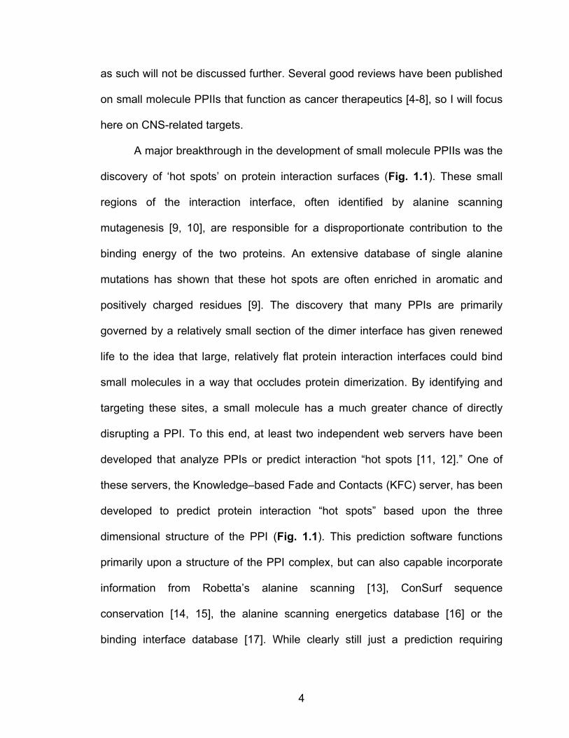
A major breakthrough in the development of small molecule PPIIs was the
discovery of ‘hot spots’ on protein interaction surfaces (Fig. 1.1). These small
regions of the interaction interface, often identified by alanine scanning
mutagenesis [9, 10], are responsible for a disproportionate contribution to the
binding energy of the two proteins. An extensive database of single alanine
mutations has shown that these hot spots are often enriched in aromatic and
positively charged residues [9]. The discovery that many PPIs are primarily
governed by a relatively small section of the dimer interface has given renewed
life to the idea that large, relatively flat protein interaction interfaces could bind
small molecules in a way that occludes protein dimerization. By identifying and
targeting these sites, a small molecule has a much greater chance of directly
disrupting a PPI. To this end, at least two independent web servers have been
developed that analyze PPIs or predict interaction “hot spots [11, 12].” One of
these servers, the Knowledge–based Fade and Contacts (KFC) server, has been
developed to predict protein interaction “hot spots” based upon the three
dimensional structure of the PPI (Fig. 1.1). This prediction software functions
primarily upon a structure of the PPI complex, but can also capable incorporate
information from Robetta’s alanine scanning [13], ConSurf sequence
conservation [14, 15], the alanine scanning energetics database [16] or the
binding interface database [17]. While clearly still just a prediction requiring

5
experimental confirmation, algorithms such as these may provide a rapid
mechanism to determine if a particular PPI contains a well-defined “hot spot” that
may be amenable to small molecule targeting.
Figure 1.1 Predicted “hot spots” on the Regulator of G protein signaling 4 (RGS4)/Gαi1 protein-protein interaction interface. The highlighted residues on both surfaces (spacefill) are predicted by the KFC Server to be energetically important for the protein-protein interaction [11]. Structure from PDB ID 1AGR [18].
6
Rationale for targeting Protein-Protein Interactions in the CNS:
The importance of PPIs in proper cellular function is particularly striking in
the nervous system. In the CNS, a host of PPIs is required for virtually all cellular
processes, including neurite outgrowth, synapse formation and modulation,
neurotransmission, signal transduction, and the induction of apoptosis [19-22].
Indeed, the highly specialized structures and discrete localization of signaling
molecules in the synapse are dependent on a large network of PPIs. Targeting
specific PPIs in the CNS may provide novel mechanisms to modulate neural
function downstream of receptor activation or to disrupt localization signals that
contribute to the efficiency or specificity of signaling. Furthermore, by targeting
these processes, it may be possible to more subtly and specifically tune neural
functioning than can be achieved by administering a receptor agonist/antagonist.
Most receptor-targeted drugs do not have the ability to selectively act upon
receptors in a particular region of the body. For example, µ-opioid receptor (µ)
agonists (e.g. morphine, fentanyl) commonly cause constipation due to their
effects on µ receptors in the intestine. The benefit of targeting localization signals
or downstream members of a signaling pathway is that, in many instances, those
factors are expressed in a much more tissue specific manner than are the
receptors themselves. By using this approach, it may be possible to provide a
measure of tissue specificity in the intrinsic mechanism of a drug. This benefit
could be particularly important in the development of centrally acting drugs, as
many broadly acting drugs in the CNS tend to have serious side effects limiting
their use [23]. Theoretically, this selectivity could be achieved at various points in
7
the signaling cascade, as there are often several steps in a signal transduction
pathway that are dependent on PPIs. Another mechanism that targeting PPIs
affords is the potential ability to localize two important signaling molecules with a
bifunctional molecule that facilitates the interaction [24]. Such a molecule is
comprised of two protein binding moieties joined by a short linker region and
functions to localize the two potential binding partners by non-covalently tethering
them together. While these bifunctional molecules are more of a PPI facilitator (or
agonist) than an inhibitor, they may also provide a mechanism to specifically
modulate neural signaling. Overall, targeting a downstream signaling modulator
is likely to provide an increase in tissue specificity of the therapeutic effect and
may also provide a mechanism to subtly modulate neural firing downstream of
natural neurotransmission.
Inhibiting protein aggregation in the CNS:
Amyloid Beta Aggregation:
Alzheimer’s disease (AD), Parkinson’s disease (PD), and other
‘plaqueopathies’ are becoming increasingly prevalent in our society and there is
growing interest in the mechanism, prevention, and treatment of these protein
aggregation diseases. Therapies for these diseases, typified by accumulation of
aggregated protein plaques, have largely dealt solely with the symptoms of the
disease (i.e. dyskinesias, decline of cognitive abilities). While these treatments
can offer some benefit, they offer no real chance of disease reversal nor can they
halt its progression. There has been great interest, however, in understanding
8
the biochemistry and pathophysiology of the plaque development and in
discovering methods to inhibit or reverse plaque formation. Emphasis recently
has shifted to finding compounds that inhibit the development of the small
oligomeric species that both lead to the macroscopic plaques and are believed to
be the pathogenic factor in these diseases [25]. Several of these methods rely
upon directly inhibiting the aggregation of the protein, while a subset are focused
upon modulating the expression levels of the plaque-forming protein or the
chaperones that assist it into its native conformation. I will focus on the former.
Identifying compounds that selectively disrupt protein aggregates or that
prevent plaque formation by inhibiting protein aggregation could be a viable
approach to the treatment of protein aggregation diseases. As such, there has
been a push for the discovery and development of compounds that selectively
inhibit protein aggregation. Compounds have been identified that inhibit the
aggregation of a variety of proteins including, huntingtin [26, 27], amyloid beta
[25, 28-31], and tau [32]. Particular attention has been paid to the proteins that
form the basis of plaque formation in AD, namely amyloid beta and tau. It has
long been known that a variety of dyes bind to and can destabilize or inhibit
plaque formation (for an extensive list, see [33]). Histopathological evaluation of
brains from AD patients has shown at least two distinct types of plaques form
during this disease. In the brain of an AD patient, aggregates of amyloid beta
form in the extracellular matrix and neurofilbrillary tangles of aggregated tau
protein form intracellularly. Both of these aggregates are correlated with AD, but
it has yet to be conclusively shown that these plaques cause the observed

9
neurodegeneration and are not merely coincident with it or even a result of it. In
fact, significant plaque development has been observed in a population of
cognitively normal 70-year olds [34]. A current hypothesis states that it is not the
mature plaques that are the triggering factor for neurodegeration, but rather the
protofibrils – small oligermeric complexes of the protein – that are the basis of (or
are at least correlated with) disease progression [35]. Due to the lack of in vivo
imaging methods for visualizing protofibril formation, this hypothesis has yet to be
tested in living human patients. This suggests that by inhibiting the development
of protofibrils it might be possible to slow the disease progression. Indeed,
several drugs that inhibit amyloid beta fibril formation via distinct mechanisms are
currently in or have been tested in clinical trials [25, 28, 36]. One of these drugs,
Alzhemed (Fig. 1.2A, tramiprosate) is a PPII that functions by sequestering
monomeric amyloid beta protein [25, 28, 36]. This drug passed through phase II
clinical trials, but failed in phase III clinical trials [37]. While tramiprosate
ultimately failed in the clinical trials, it provides a proof of concept that small
molecule inhibitors of amyloid beta protofibril formation are capable of reaching
late stage development and that analogs with better
pharmacokinetic/pharmacodynamic properties may still provide a viable
approach to AD treatment.
10
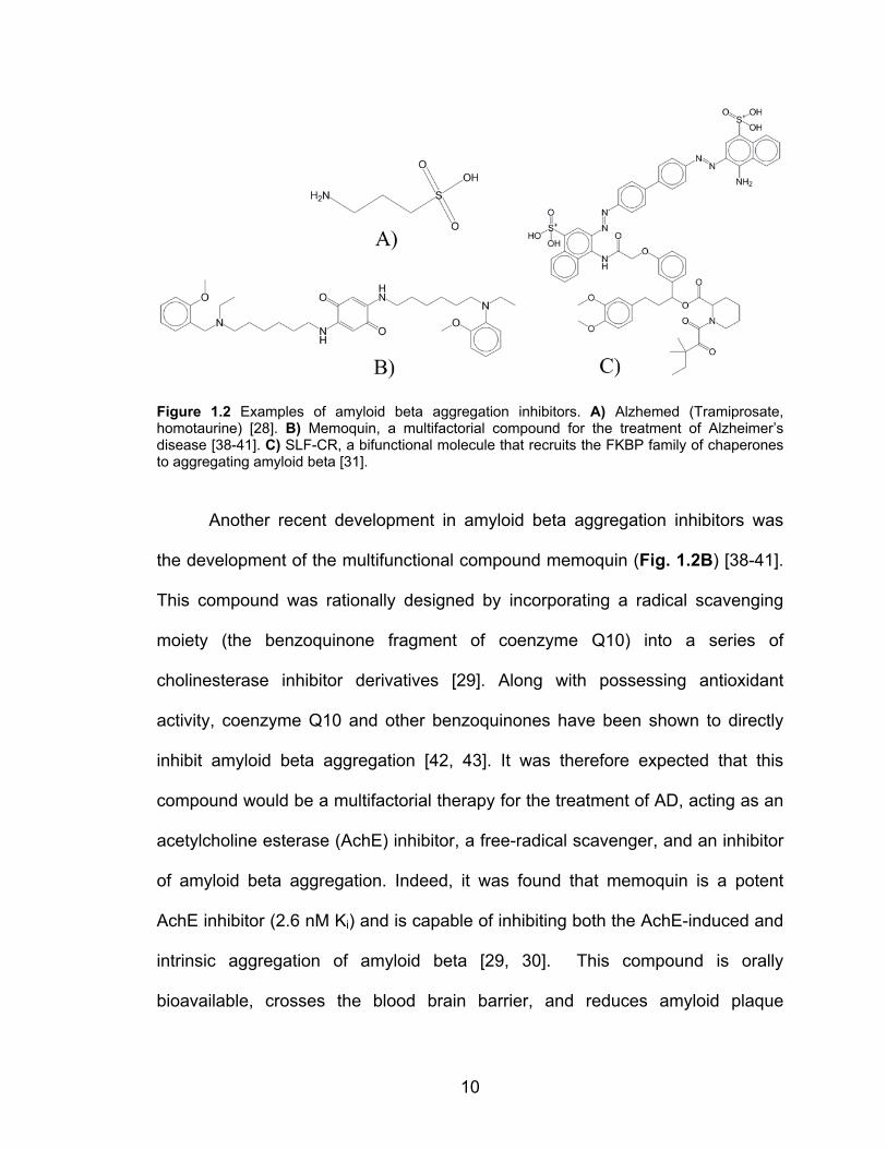
Figure 1.2 Examples of amyloid beta aggregation inhibitors. A) Alzhemed (Tramiprosate, homotaurine) [28]. B) Memoquin, a multifactorial compound for the treatment of Alzheimer’s disease [38-41]. C) SLF-CR, a bifunctional molecule that recruits the FKBP family of chaperones to aggregating amyloid beta [31].
Another recent development in amyloid beta aggregation inhibitors was
the development of the multifunctional compound memoquin (Fig. 1.2B) [38-41].
This compound was rationally designed by incorporating a radical scavenging
moiety (the benzoquinone fragment of coenzyme Q10) into a series of
cholinesterase inhibitor derivatives [29]. Along with possessing antioxidant
activity, coenzyme Q10 and other benzoquinones have been shown to directly
inhibit amyloid beta aggregation [42, 43]. It was therefore expected that this
compound would be a multifactorial therapy for the treatment of AD, acting as an
acetylcholine esterase (AchE) inhibitor, a free-radical scavenger, and an inhibitor
of amyloid beta aggregation. Indeed, it was found that memoquin is a potent
AchE inhibitor (2.6 nM Ki) and is capable of inhibiting both the AchE-induced and
intrinsic aggregation of amyloid beta [29, 30]. This compound is orally
bioavailable, crosses the blood brain barrier, and reduces amyloid plaque
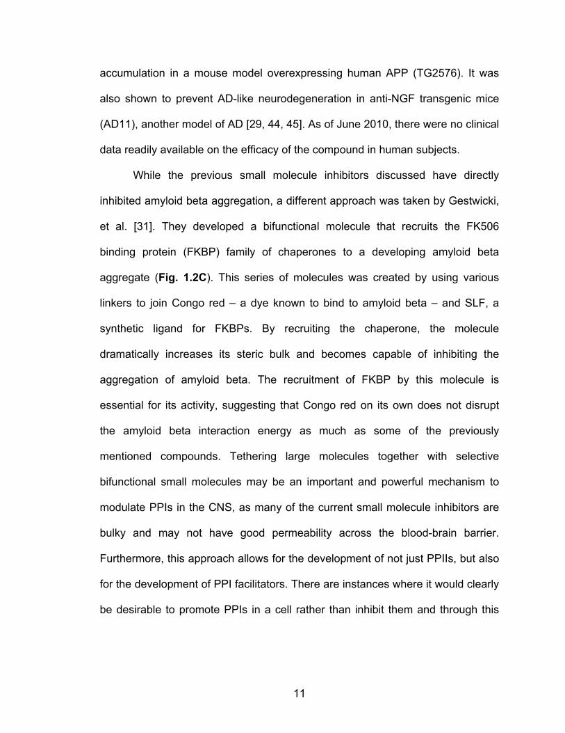
11
accumulation in a mouse model overexpressing human APP (TG2576). It was
also shown to prevent AD-like neurodegeneration in anti-NGF transgenic mice
(AD11), another model of AD [29, 44, 45]. As of June 2010, there were no clinical
data readily available on the efficacy of the compound in human subjects.
While the previous small molecule inhibitors discussed have directly
inhibited amyloid beta aggregation, a different approach was taken by Gestwicki,
et al. [31]. They developed a bifunctional molecule that recruits the FK506
binding protein (FKBP) family of chaperones to a developing amyloid beta
aggregate (Fig. 1.2C). This series of molecules was created by using various
linkers to join Congo red – a dye known to bind to amyloid beta – and SLF, a
synthetic ligand for FKBPs. By recruiting the chaperone, the molecule
dramatically increases its steric bulk and becomes capable of inhibiting the
aggregation of amyloid beta. The recruitment of FKBP by this molecule is
essential for its activity, suggesting that Congo red on its own does not disrupt
the amyloid beta interaction energy as much as some of the previously
mentioned compounds. Tethering large molecules together with selective
bifunctional small molecules may be an important and powerful mechanism to
modulate PPIs in the CNS, as many of the current small molecule inhibitors are
bulky and may not have good permeability across the blood-brain barrier.
Furthermore, this approach allows for the development of not just PPIIs, but also
for the development of PPI facilitators. There are instances where it would clearly
be desirable to promote PPIs in a cell rather than inhibit them and through this
12
general schema it may be possible to selectively colocalize different molecules in
a single cell by varying one component of the bifunctional molecule.
While the pathophysiological mechanism behind the development of AD
has yet to be fully understood, it seems reasonable to hypothesize that amyloid
beta protofibril formation plays a significant role in the progression of the disease.
Several small molecules have been developed that inhibit the oligomerization of
amyloid beta either in vitro or in vivo. While inhibiting amyloid beta aggregation
may provide therapeutic benefit on its own, the development of multifactorial
agents such as memoquin has the potential to be much more efficacious in terms
of treating the underlying disease.
Alpha Synuclein Aggregation:
Parkinson’s disease is the second most common neurodegenerative
disorder in most Western countries [46, 47]. This disease is characterized by the
loss of dopaminergic neurons in several brain regions, including the substantia
nigra pars compacta and other regions important for higher order functioning
[48]. Histopathological evaluation of the postmortem brains of Parkinson’s
patients has revealed the presence of large intraneuronal aggregates termed
“Lewy bodies.” These aggregates are primarily composed of a 140 amino acid
protein, α-synuclein, although they are generally not as homogenous as amyloid
beta plaques [48]. It has been shown that overexpression of alpha-synuclein in
several model organisms causes the development of Parkinsonian-like
symptoms [49-51]. Further study of α-synuclein has shown that the protein
13
contains a highly amyloidogenic domain that, when misfolded, oligomerizes and
forms a series of self-associating β-pleated sheets that spontaneously form Lewy
bodies [52, 53]. Like amyloid beta oligomers in AD, it is believed that it is the α-
synuclein oligomers and not the fully formed Lewy bodies that are the
pathological factor in PD. The current hypothesis states that α-synuclein
oligomers are capable of forming membrane pores that disrupt organelle
function, leading to cell dysfunction and death. [48]
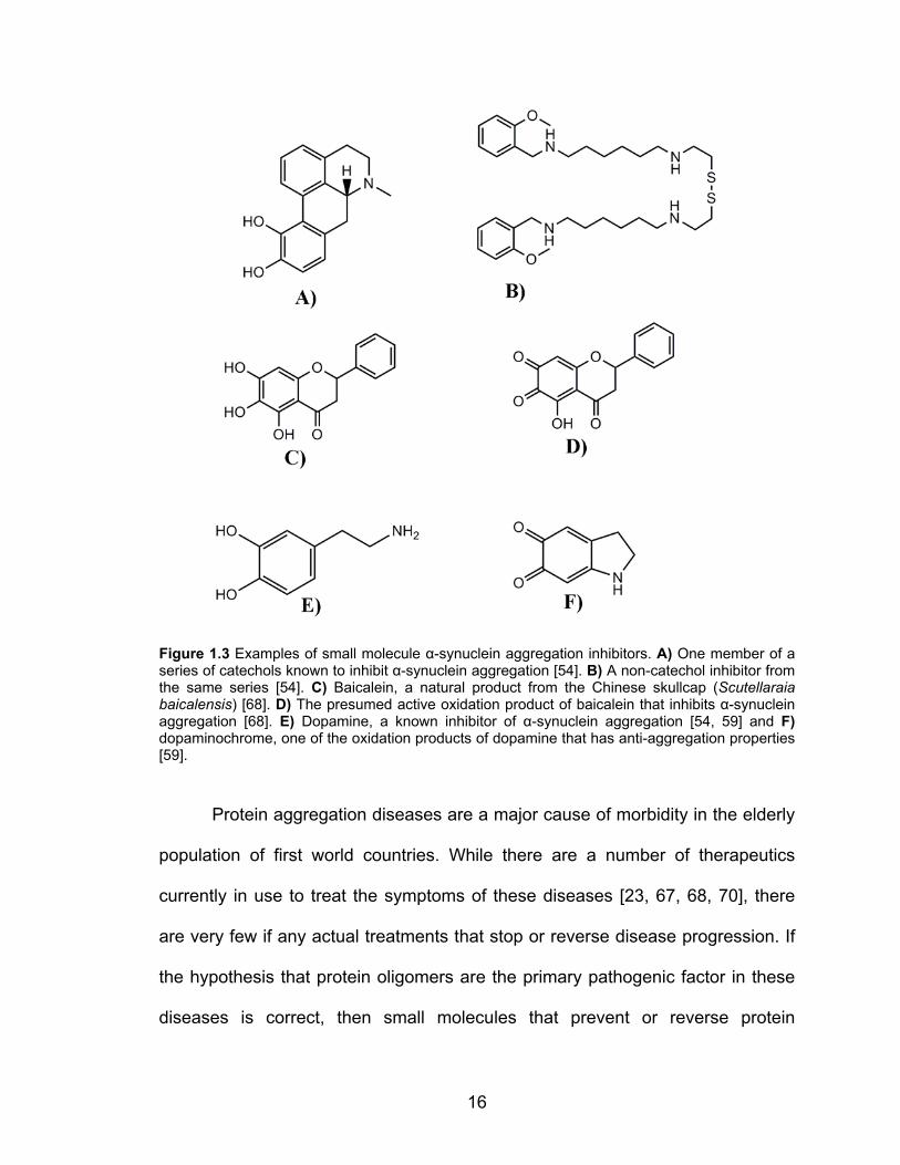
Several inhibitors of α-synuclein aggregation have been identified [54-58]
(Fig. 1.3). An intriguing finding is that catecholamines can inhibit α-synuclein
aggregation [54, 55]. This has also been shown in a mouse model of α-synuclein
aggregation, where Lewy bodies were dissolved in brain slices by the addition of
l-dopa [59]. The oxidation state of the catecholamines are important for this
activity, whereby the several oxidation products of dopamine are more potent at
inhibiting α-synuclein aggregation than is the parent neurotransmitter [59] (Fig.
1.3). The link, if any, between dopaminergic neuron loss and the ability of
catecholamines to inhibit α-synuclein aggregation has yet to be fully understood,
but remains an intriguing concept in the pathophysiology of Parkinson’s disease.
A series of peptide inhibitors of α-synuclein aggregation were identified by
developing a library of overlapping heptapeptides that span the α-synuclein
sequence. The active peptides were centered around residues 69-72 of α-
synuclein, suggesting that this region of the molecule was important for self-
association [60, 61]. It appears that short peptide fragments of α-synuclein also
occur naturally, as the serine protease neurosin degrades α-synuclein into

14
fragments that can inhibit α-synuclein polymerization [62]. Current work is
focused upon developing peptidomimetics and identifying small molecule
inhibitors of α-synuclein using both high-throughput screening (HTS) and rational
design from the information obtained in the peptide library study [63].
Other small molecule inhibitors of α-synuclein have been identified.
Rifampicin and several of its derivatives can inhibit both α-synuclein [64]and
amyloid beta [65, 66] aggregation in a concentration-dependent manner with
reasonable potency (< 10 µM IC50). A conclusive mechanism of rifampicin action
has not been fully elucidated, but it has been suggested that it could act by
binding directly to the developing plaque [65] and/or by acting as a free radical
scavenger [66]. Panacea Pharmaceuticals had also developed a pair of α-
synuclein inhibitors, PAN-408 and PAN-527, that had progressed to preclinical
trials. However, there have been no recent reports of compounds with these
names [67, 68].
Polyphenolic compounds, like flavonoids or Congo red, have been
proposed to be α-synuclein aggregation inhibitors [69]. Many of these
compounds are derived from natural sources and have low micromolar IC50
values for protein aggregation inhibition. Baicalein (Fig. 1.3), a flavonoid isolated
from the Chinese Skullcap plant (Scutellaraia baicalensis), can directly bind to a
single site on α-synuclein with submicromolar affinity and inhibit oligomerization
[70]. It is likely that a quinone oxidation product of this compound is responsible
for the observed inhibitory activity (Fig. 1.3) [70]. Interestingly, this compound
can inhibit α-synuclein aggregate nucleation but does not affect fibril elongation

15
or dissolve aggregates, suggesting that the molecule may act by stabilizing the
monomeric α-synuclein [70]. This mechanism could be beneficial, as plaque
disruption could generate free protofibrils and lead to increased cellular damage.
Circular dichroism studies confirmed that binding of baicalein stabilized the semi-
folded state of α-synuclein [70]. Unfortunately, baicalein also stabilized an
oligomeric species of α-synuclein as well as the monomer [70]. It is not known
whether the oligomeric species stabilized by baicalein has neurodegenerative
properties, however this finding does not bode well for this family of polyphenoic
compounds as inhibitors of α-synuclein function. Unfortunately, it is possible that
these molecules could stabilize the formation of the protofibrils that, as the
current hypothesis states, are the pathogenic factor in protein aggregation
diseases.
16
Figure 1.3 Examples of small molecule α-synuclein aggregation inhibitors. A) One member of a series of catechols known to inhibit α-synuclein aggregation [54]. B) A non-catechol inhibitor from the same series [54]. C) Baicalein, a natural product from the Chinese skullcap (Scutellaraia baicalensis) [68]. D) The presumed active oxidation product of baicalein that inhibits α-synuclein aggregation [68]. E) Dopamine, a known inhibitor of α-synuclein aggregation [54, 59] and F) dopaminochrome, one of the oxidation products of dopamine that has anti-aggregation properties [59].
Protein aggregation diseases are a major cause of morbidity in the elderly
population of first world countries. While there are a number of therapeutics
currently in use to treat the symptoms of these diseases [23, 67, 68, 70], there
are very few if any actual treatments that stop or reverse disease progression. If
the hypothesis that protein oligomers are the primary pathogenic factor in these
diseases is correct, then small molecules that prevent or reverse protein
17
oligomerization may provide a mechanism to target the actual cause of the
disease. There has been substantial work put forth to develop inhibitors of
protein oligomerization and significant progress has been made. There is,
however, much more work that needs to be done in this field before a clinically
useful agent will be available for general use.
Modulating Signal Transduction through inhibiting protein-protein
interactions:
Signal transduction cascades are required for nearly all biological
functions. The importance of these systems is further illustrated by the fact that a
large proportion of all clinically used therapeutics modulate signaling [23]. The
most common method to modulate information processing through a signal
transduction pathway is to alter activity of the most upstream molecule in the
system: the receptor. These receptors come in many forms including G-protein
coupled receptors, intracellular steroid/glucocorticoid receptors, and tyrosine
kinase linked receptors. Currently ~30-50% of all clinically used drugs target
GPCRs and a substantial portion of the remaining drugs target other receptor
systems [23]. While many of these drugs are effective therapeutics, targeting
regulation systems or molecules further downstream in the signaling pathway
may provide advantages not readily available when solely modulating receptor
activity.
Targeting downstream signaling molecules in a signal transduction
pathway requires overcoming several significant hurdles in drug development,

18
including cell permeability of the compound, achieving pathway specificity, and
avoiding unwanted or unexpected side effects. There are currently several
examples of clinically used drugs or drug candidates that target downstream
signaling molecules in a pathway. The majority of these are kinase inhibitors,
exemplified by Gleevec, that inhibit an enzymatic step in a signal transduction
cascade [71]. As compared to a standard PPIs, enzymes are much more
amenable to small molecule targeting due to their well-defined active site binding
pocket. Furthermore, kinases represent a critical step in the signal transduction
pathway that can be selectively inhibited. With all of these qualities, it is easy to
understand why a kinase inhibitor could be a useful therapeutic.
Many signal transduction steps do not rely upon an enzymatic process but
rather use PPIs to relay information, often in the context of a signalosome that is
tightly regulated by scaffolding components in the cell (e.g. lipid rafts, scaffold
proteins). Targeting these steps requires the development of small molecules
that inhibit the PPIs required for signal transduction. One of the more obvious
drug targets in this case would be the scaffolding proteins that pull these
signalosomes together.
Inhibition of PDZ interactions:
PDZ domains are important scaffolding components in many signaling
systems, with an extensive role in the development and maintenance of both pre-
and post-synaptic structures [72, 73]. Development of reversible small molecule
inhibitors that target PDZ domains would provide useful tools to probe the many
19
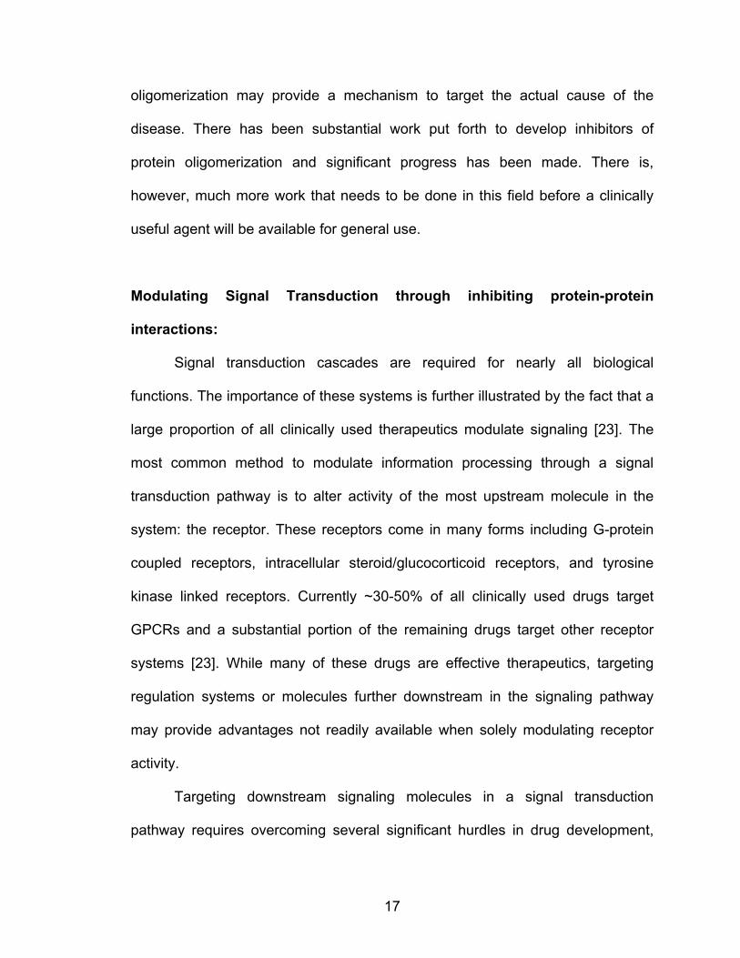
functions of these important scaffolds [74]. Of all canonical PPIs, PDZ domains
are possibly the most similar to a traditional ligand-receptor interaction, as the
interaction interface is comprised of a groove on the PDZ domain binding to the
last few (3-5) amino acid residues in its partner (Fig. 1.4A) [73]. The small
interaction interface requires that the few amino acids compromising the PDZ
ligand contribute a great deal to the energetics of binding. Having such a small
PPI interface might suggest that these interactions would be amenable to small
molecule disruption. To this end, there have been a few PDZ inhibitors described
based either upon rational design or from random high-throughput screening
(Fig. 1.4B) [75-82]. Rational design of PDZ inhibitors would appear to be
relatively straightforward, as the PDZ ligand is comprised of so few residues and
the binding pockets of many PDZ domains have been characterized structurally
by NMR or crystallographically. Indeed, several peptidomimetic scaffolds have
been developed that inhibit PDZ interactions (Fig. 1.4B) [76, 77, 80, 81]
.
20
Figure 1.4 Crystal structure of the first PDZ domain from MAGI bound to the PDZ ligand of HPV18 E6. A) MAGI shown in surface with HPV18 E6 shown as sticks and balls. Note how in this protein-protein interaction, only a few (generally 3-5) residues play a predominant role in the binding energetics. Structure from PDB ID 2I04. [83] B) Examples of small molecule PDZ inhibitors. 1. General scaffold for a wide array of PDZ domains [77-79]. Analogs of this structure have been shown to inhibit the second PDZ domain of NHERF1 [78]. 2. Beta-hairpin peptidomimetic developed to inhibit the α1-syntrophin PDZ domain [80]. 3. Peptidomimetic developed to inhibit the NHERF1 PDZ domains [81].

21
Cell permeant small molecule inhibitors of PDZ domains will provide a
mechanism with which to probe the complex functions of these scaffolding
proteins. For example, the Na+/H+ exchanger regulatory factor 1 (NHERF1)
contains two PDZ domains and has been shown to have altered expression in
many cancers [84-88]. The role of NHERF1 in cancer is complicated and
appears to be dependent upon cellular context. Outside the realm of oncology,
NHERF1 has been shown to be a multifunctional scaffolding protein that is
capable of regulating the trafficking and localization of many membrane
associated proteins [89]. Clearly, a tool which would allow for the acute and
reversible inhibition of NHERF1 PDZ function could provide a powerful
mechanism with which to determine the physiological role of this protein in
different cellular contexts.
Currently, the best defined PDZ inhibitors are directed against the
dishevelled and NHERF1 PDZ domains. These compounds were originally
designed as a treatment for beta-catenin dependent tumor growth or to study the
controversial role of NHERF1 in cancer progression, respectively. While these
compounds have limited utility as centrally acting agents, they provide a clear
example of how a PDZ inhibitor could be developed for one of the many PDZ
domains that are important in neural functioning (see for reviews [19, 72, 73, 90-
94]).
The first cell permeable PDZ inhibitor was developed by Fujii, et al [77].
This irreversible inhibitor was rationally designed to bind to the second PDZ
22
domain of MAGI [77]. The compound dose-dependently (IC50 ~10-30 µM)
inhibited the binding of a peptide corresponding to the PDZ ligand of the lipid
phosphatase PTEN to the membrane-associated MAGI protein in a fluorescence
polarization assay. It also increased the activity of PKB (or Akt) in cells,
consistent with increased phosphatidyl inositol 3,4,5 trisphosphate levels due to
reduced PTEN recruitment to the membrane [77]. Eventually, this indole scaffold
was developed into a reversible, albeit weak (IC50 ~1 mM), inhibitor of the second
PDZ domain of MAGI [76]. A similar indole scaffold was used to develop an
inhibitor of the disheveled PDZ domain, an important scaffold in the Wnt/β-
catenin pathway [79]. This compound, named FJ9, blocked the interaction
between the PDZ ligand at the C-terminus of the 7TM receptor Frizzled 7 with the
disheveled PDZ domain both in vitro and in cells (In vitro IC50 30-60 µM). It also
suppressed the growth of tumor cells in a β-catenin dependent manner [79].
Another inhibitor of the disheveled PDZ domain has been described. This
relatively weak (~200 µM IC50) inhibitor was identified in a virtual screen against
the disheveled PDZ domain and it inhibited Wnt signaling in a zebrafish embryo
model of Wnt signaling [82]. Inhibition of PDZ domains has the potential to
provide very useful pharmacologic tools for the study of protein trafficking,
synaptic function, and other scaffolding-dependent processes. While current
compounds still have only modest affinities, a selective inhibitor of some
particular PDZ domains may also provide useful therapeutic agents, although this
hypothesis needs to be tested.
23
Targeting elements of the G-protein signaling pathway:
Another class of PPIs for which drug targeting is attractive is the multitude
of PPIs formed by heterotrimeric G protein subunits. These trimeric proteins are
formed of α and βγ subunits which, when activated (and thus dissociated into
free α and βγ subunits), interact via PPIs with a large number of downstream
effectors, including adenylate cyclase, phosphoinosotide-3 kinase,
phospholipase Cβ, voltage gated Ca2+ channels, G protein coupled inwardly-
rectifying potassium channels, and others. G-proteins, especially Gα subunits,
also bind to regulatory proteins that can alter the temporal and spatial signaling
pattern. Developing specific inhibitors of various G-protein/effector or G-
protein/regulator interactions could provide a mechanism to selectively modulate
GPCR signaling pathways. It is not difficult to imagine several scenarios whereby
modulating GPCR signaling could provide significant therapeutic benefit, either
by potentiating positive actions of a drug or by inhibiting undesirable side effects.
The progress in this field will be discussed throughout the rest of this thesis,
including the contributions that I have made during the course of my Ph.D work.

24
Figure 1.5 Canonical G-protein signaling mechanism. See text for explanation. Note that the hydrolysis of GTP by Gα is accelerated by members of the RGS family. Reproduced with permission from Nature Publishing Group from [1].
Selective Gβγ Inhibitors:
Due to the prevalence of clinically important drugs that target GPCRs,
there has been great interest in the therapeutic modulation of signaling
downstream of these receptors. Canonical signaling through GPCRs (Fig. 1.5)
progresses through the activation of a receptor by ligand binding, which
stimulates the exchange of GDP for GTP on the Gα subunit of a heterotrimeric G
protein. The GTP-bound Gα subunit and Gβγ subunit of the G protein then
dissociate or at least undergo a conformational change to expose interaction
surfaces to act upon downstream effectors in the signaling pathway. Since the
first signaling molecule downstream of a GPCR is the G protein heterotrimer, it
has become an interesting target for small molecule inhibition. While there have
been no published reports of a small molecule inhibitor of Gα/effector PPIs, there
have been a family of compounds identified by Smrcka and colleagues that bind
25
to Gβγ and selectively inhibit its interaction with downstream effectors [95-97].
The strategy used to identify these inhibitors provides a clear example of a
protocol being used to identify small molecule PPIIs. The first step that the
investigators took was to screen a random-peptide phage display library to
identify binding sites on Gβγ [98]. A series of peptides was identified and one
inhibited the Gβγ regulation of PI3K and PLCβ. It did not however, inhibit
regulation of type I adenylate cyclase or N-type Ca2+ channels, suggesting that
effector selectivity may be possible with small molecule modulators of Gβγ
activity. By analyzing the crystal structure of Gβ1γ2 bound to this selective
peptide inhibitor, it was possible to define the binding pocket for the peptide.
Using this site as a binding pocket in virtual screening, the investigators identified
85 small molecules (top 1% in the screen) that were predicted to bind to the Gβγ
‘hotspot [95]. Analysis of these compounds using an ELISA assay based upon
displacement of the peptide ligand identified 9 compounds with reasonable IC50
values (100 nM – 60 µM). One of these compounds, M119 (Fig. 1.6A), inhibited
the Gβγ stimulation of PLCβ and PI3Kγ activity in vitro and it also inhibited the
Gβγ-dependent calcium release from activation of the Gi-linked N-formyl peptide
receptor in differentiated HL-60 cells. The compound had no inhibitory activity
upon the calcium mobilization initiated by carbachol in HEK cells stably
expressing the Gq-linked M3 muscarinic receptor, showing that M119 is selective
for Gβγ-dependent calcium mobilization. M119 also showed in vivo activity when
tested in a morphine antinociception assay in mice. PLCβ3-/- mice have been
shown to be ten times more sensitive to the antinociceptive effects of morphine
26
and an intracerebroventricular injection of M119 recapitulated this augmentation
of morphine activity in wild type animals [95, 99]. Since opioid receptors have
many Gβγ-dependent functions, the fact that M119 potentiates morphine-induced
antinociception instead of inhibiting it provides evidence that this compound is
not globally inhibiting Gβγ activity. Another structurally distinct compound
identified by this approach, M201, also showed an interesting selectivity profile in
its ability to inhibit Gβγ-effector interactions. This compound showed no ability to
inhibit PLCβ2 activity, potentiated PLCβ3 and PI3K activity, and inhibited GRK2
binding. The discovery of effector-selective modulators of Gβγ signaling M119 &
M201, has thus provided a clear example of how targeting downstream signaling
molecules can be a viable approach to modulating the pharmacological
properties of a common drug (e.g. morphine).

27
Figure 1.6 Structure of M119, a pathway selective inhibitor of Gβγ signaling A) Chemical structure of M119 [95]. B) Crystal structure of Gβγ bound to peptide SIGK. This peptide, identified by phage display, binds to the identified “hot spot” on Gβγ [100]. Structure from PDB ID 1XHM.
RGS Proteins:
Another approach to modulating GPCR signaling is to alter the activity of
key proteins that regulate signal transduction. The G protein pathways are
regulated by a number of proteins including scaffolding proteins such as the PDZ
domains discussed previously, RGS proteins, G protein coupled receptor kinases
(GRKs), and arrestins. These molecules are critical for the proper temporal and
spatial regulation of GPCR signaling. By selectively modulating the actions of
28
these molecules it might be possible to more finely tune GPCR signaling for
therapeutic purposes.
A particularly interesting approach to modulating GPCR signaling is to
target RGS proteins. These molecules function as GTPase accelerating proteins
(GAP), by binding directly to Gα subunits and accelerating the intrinsic hydrolysis
rate of GTP (Fig. 1.1, 1.5) [18, 101, 102]. The discovery of these proteins
provided a solution to the paradox of how rapid regulation of GPCR signaling
could occur given the slow intrinsic rate of GTP hydrolysis by purified Gα
subunits. They explain the subsecond regulation of G protein signals observed in
excitable cells [18, 101, 103, 104] and RGS proteins can strongly inhibit cellular
responses [105-107]. There are over twenty identified RGS proteins that interact
with limited selectivity for most Gα subtypes (Table 1.1, [101, 103, 108-110]).
The only exception to this is Gαs, for which no RGS interaction has been
confirmed. There are increasing reports of RGS selectivity for signaling by
specific GPCRs, suggesting that targeting an RGS may provide a mechanism to
selectively augment signaling through a particular GPCR [111, 112]. It is also
increasingly appreciated that RGS proteins are heterogeneously expressed
throughout the body, including in individual neuron types in specific brain regions
[113-115]. The distinct expression patterns, presumed GPCR selectivity, and the
dependence on an active signaling pathway for function all suggest that small
molecules that modulate RGS activity could potentially be useful therapeutics.
Indeed, mice expressing a mutated (G184S) form of Gαi2 or Gαo that render
these G proteins insensitive to RGS effects exhibit markedly enhanced potency

29
of agonists and substantial physiological phenotypes [116-119]. Specifically,
mice with the Gαi2 G184S mutation show reduced fat mass and resistance to
high fat diet, possibly due to CNS actions [119]. They also show behaviors
consistent with enhanced 5-HT1A signaling and a spontaneously antidepressant-
like state as well as 10-fold increased potency of 5HT-based antidepressant
drugs [120]. Mice with the RGS-insensitive mutant Gαo show increased
antiepileptiform activity in hippocampal slices by α2a agonists [121]. Strikingly, the
effects are quite specific where the 5-HT1A potentiation is only seen for
antidepressant-like and not for hypothermia effects [120]. This exquisite
specificity suggests that RGS proteins may play a targeted role in the regulation
of different physiological effects that are elicited through the same receptor.
Since there are a number of thorough reviews of RGS structure and function
(e.g. [1, 101-103, 108, 115, 122-129]), I will focus primarily upon RGS4 and its
potential as a drug target.

30
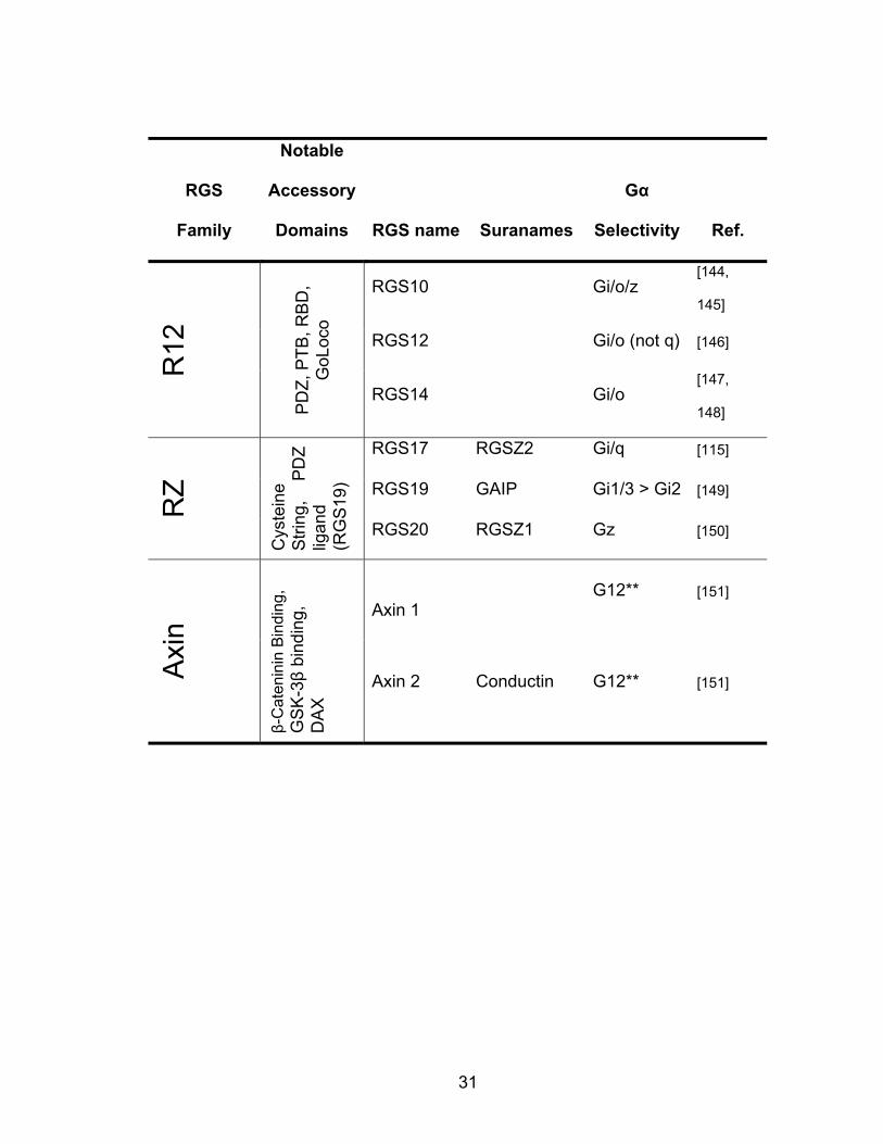
RGS
Family
Notable
Accessory
Domains RGS name Suranames
Gα
Selectivity Ref. R
4
N-te
rmin
al a
mph
ipat
hic
helix
RGS1 BL34 Gi/o/q [130]
RGS2 GOS8 Gq [131,
132]
RGS3 Gi/q/11 [133]
RGS4 Gi/q [134]
RGS5 Gi/o/q [135]
RGS8 Gi/o/q [136]
RGS13 Gi/q [137]
RGS16 Gi/o/q/13* [135,
138]
RGS18 Gi/q [139]
RGS21 Gi/q [140]
R7
DE
P, G
GL
RGS6 Go [141]
RGS7 Go/i [142]
RGS9 RGS-r Go/Gt [142,
143]
RGS11 Go/i [142]
Table 1.1 Current list of mammalian RGS proteins that are known to bind to Gα subunits. DEP: Dishevelled/EGL10/Plextrin homology domain; GGl: Gγ-like domain; PDZ: PSD95/Dlg/Z0-1/2 domain; PTB: phosphotyrosine binding domain; RBD: Ras binding domain; GoLoco: guanine nucleotide-dissocation inhibitor domain; DH: Dbl homologous domain; PH: Plextrin homology domain; DAX: Domain present in Dishevelled and Axin; S/T kinase: serine/threonine kinase domain; RhoGEF: Rho guanine nucleotide exchange factor; GRK: G-protein coupled receptor kinase. *RGS16 inhibits signaling through G13, but not via GAP activity. **No GAP activity, data remain to be independently confirmed. *** Weak or no GAP activity, physiological importance in question.
31
RGS
Family
Notable
Accessory
Domains RGS name Suranames
Gα
Selectivity Ref.
R12
PD
Z, P
TB, R
BD
, G
oLoc
o
RGS10 Gi/o/z [144,
145]
RGS12 Gi/o (not q) [146]
RGS14 Gi/o [147,
148]
RZ
Cys
tein
e S
tring
, P
DZ
ligan
d (R
GS
19)
RGS17 RGSZ2 Gi/q [115]
RGS19 GAIP Gi1/3 > Gi2 [149]
RGS20 RGSZ1 Gz [150]
Axi
n
β-C
aten
inin
Bin
ding
, G
SK
-3β
bind
ing,
D
AX
Axin 1 G12** [151]
Axin 2 Conductin G12** [151]

32
RGS
Family
Notable
Accessory
Domains RGS name Suranames
Gα
Selectivity Ref. R
hoG
EFs
DH
/PH
LARG G12/13/q** [152,
153]
P115
RhoGEF G12/13 [154]
PDZ
RhoGEF G12/13 [155]
GR
K
S/T
Kin
ase,
PH
(PH
on
GR
K2/
3 on
ly)
GRK1 Rhodopsin
Kinase
None
Known [124]
GRK2 βARK1 Gq*** [156,
157]
GRK3 βARK2 Gq*** [156]
GRK4 IT-11 kinase G13, Gs*** [158]
GRK5 None
Known
[124,
156]
GRK6 None
known
[124,
156]
GRK7 None
Known [124]

33
The RGS Homology Domain: The RGS Homology (RH) domain is comprised of nine helices that form a
bi-lobed structure (Fig 1.7). This fold, originally identified for RGS4 in complex
with Gαi1, has been observed in at least 22 other proteins and bears little to no
homology with the folds of small G-protein GAPs. The α4-α7 helices
predominantly provide the structural components and stability required for
interaction with Gα subunits, but the loop regions between helices α3-α4, α5-α6,
and α7-α8 form the primary interaction interface with the G protein. These loops
bind to the three ‘switch’ regions of Gα. The GAP activity of RGS is believed to
stem from the stabilization of these ‘switch regions into the transition state
conformation. As such, the RGS protein actually does not provide any residues
that are crucial for catalytic activity, as is seen with some GAPs for small
GTPases. [124]

34
Figure 1.7 The RGS homology (RH) domain fold of RGS4. That the loop regions between α3-α4, α5-α6, and α7-α8 provide the surface required for Gα binding. Structure from PDB ID 1AGR [18].
A particularly promising feature of the RH domain of RGS4 as a target for
small molecule inhibitors is that it contains an endogenous small molecule
allosteric regulatory site. Kurachi and colleagues showed that (3,4,5)P3-
phosphotidyl inositol (PIP3) and lysophosphatidic acid – but not other
phosphoinositides - directly bind to an allosteric site on the RGS and inhibit its
activity [159]. This binding inhibits RGS4 GAP activity in vitro and also inhibits the
effects of RGS4 upon the muscarinic control of GIRK currents in a reconstituted
Xenopus oocyte system [159].
35
While there is no direct structural information (e.g x-ray crystal structure)
of the phospholipid interactions with the RGS, there is significant evidence that
the binding of PIP3 occurs at a site that is independent of the G-protein binding
interaction interface. There are four conserved lysine residues in the α4/α5
helices (K99, K100, K112, K113) of RGS4 that have been shown to be important
for PIP3 binding (Fig. 1.8). Charge-swapping mutation of these lysines (e.g.
K99E/K100E) renders RGS4 unable to bind activated Gαi1 as well as inhibiting
RGS modulation of the M2-activation of G-protein inwardly rectifying potassium
(GIRK) channels [159].
Two of these lysine residues (K99/K100) lie in a site analogous to the site
of binding of adenomatous polyposis coli protein to the RH domain of Axin,
suggesting that this site might be a more generalized accessory site for
protein/small molecule binding to RH domains. On RGS4, this site has also been
shown to bind calmodulin in a Ca2+-dependent manner [159, 160]. Mutation of
K99/K100 to glutamate in this system inhibited the interaction with calmodulin,
suggesting that the binding sites of acidic phospholipids and calmodulin overlap
[161]. Further strengthening this notion, binding of Ca2+-calmodulin reverses the
PIP3–induced RGS4 inhibition [160]. Since RGS4 has been shown to be efficient
at inhibiting calcium release induced by Gq-coupled receptors ([162], Chapter IV)
this calcium-dependent activation of RGS4 is likely to be a physiological
feedback mechanism for dampening overactive Gq signaling [160, 163].
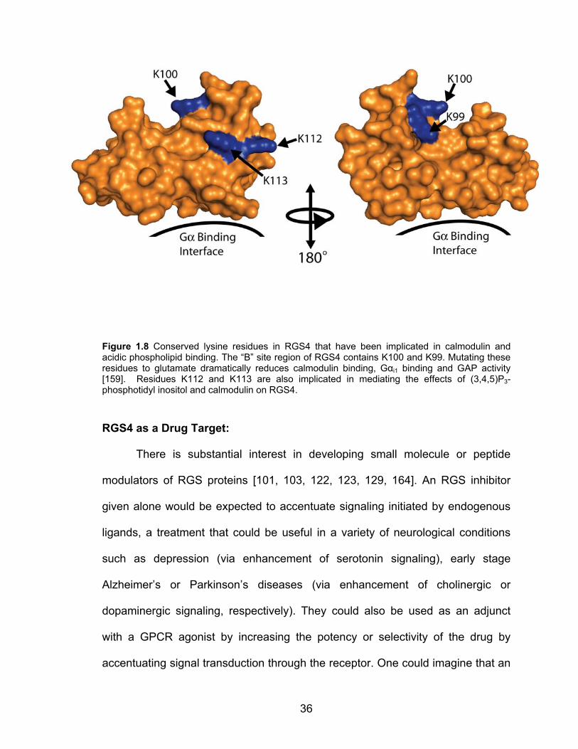
36
Figure 1.8 Conserved lysine residues in RGS4 that have been implicated in calmodulin and acidic phospholipid binding. The “B” site region of RGS4 contains K100 and K99. Mutating these residues to glutamate dramatically reduces calmodulin binding, Gαi1 binding and GAP activity [159]. Residues K112 and K113 are also implicated in mediating the effects of (3,4,5)P3-phosphotidyl inositol and calmodulin on RGS4.
RGS4 as a Drug Target:
There is substantial interest in developing small molecule or peptide
modulators of RGS proteins [101, 103, 122, 123, 129, 164]. An RGS inhibitor
given alone would be expected to accentuate signaling initiated by endogenous
ligands, a treatment that could be useful in a variety of neurological conditions
such as depression (via enhancement of serotonin signaling), early stage
Alzheimer’s or Parkinson’s diseases (via enhancement of cholinergic or
dopaminergic signaling, respectively). They could also be used as an adjunct
with a GPCR agonist by increasing the potency or selectivity of the drug by
accentuating signal transduction through the receptor. One could imagine that an

37
RGS9 inhibitor that accentuated dopaminergic signaling selectively in the
striatum where RGS9 is expressed could be a useful adjunctive therapy with L-
dopa or synthetic dopamine agonists in Parkinson’s disease (Fig. 1.9).
Furthermore, an RGS inhibitor that selectively accentuated opioid signaling in
neurons in the pain pathway may provide a mechanism to selectively increase
the analgesic properties of opioids but might leave alone the undesirable actions
of these drugs (i.e. constipation, abuse liability) which might be regulated by
different RGS proteins. Thus, RGS inhibitors could serve as GPCR agonist
potentiators but would also enhance agonist specificity in a cell-type or pathway-
specific manner.
Figure 1.9 RGS-inhibitors increase the tissue specificity of an agonist. Left Panel: Graphical representation of the RGS4 & RGS9 protein expression patterns in the brain [151]. The Red/Green color denotes regions of high expression, specifically cortex and thalamic regions for RGS4, and basal ganglia for RGS9. Center Panel: Upon addition of a low or high concentration of agonist, a response would be seen across all brain regions that express the receptor. Left Panel: In the presence of an RGS inhibitor, there would be tissue-specific enhancement of agonist effect in the tissues where the RGS is expressed. Reproduced with permission from Nature Publishing Group from [1].
38
RGS4 is a prototypical RGS protein that is widely expressed throughout
the CNS with limited expression in peripheral tissues. It controls a variety of
signaling systems, and has been implicated as a risk factor for schizophrenia
[165-188]. RGS4, like other RGS proteins, interacts strongly with several
members of the Gαi/o and Gαq families and shows limited selectivity between
these proteins in in vitro binding and functional studies [18, 189-191]. The
interaction interface between RGS4 and Gαi1 was revealed by the crystal
structure by Tesmer et al [18]. This relatively flat interface includes the three
switch regions in Gαi1 (residues 179-185 in switch 1, 204-213 in switch 2, and
235-237 in switch 3) and several of the loops in the RH domain of RGS4 [18].
The interaction interface covers approximately 1100Å2 and utilizes primarily van
der Waals and hydrogen bond interactions (Fig. 1.10). The binding affinity of the
Gα/RGS complex is dependent upon the conformational state of the Gα. There is
very little to no interaction when Gα is in the GDP-bound state, some moderate
affinity when bound with GTP, and a strong interaction (Kd ~1-5nM) when the Gα
is bound to GDP-aluminum fluoride, a state believed to serve as a mimic for the
transition state of GTP hydrolysis [189, 192].
The physiological effects of RGS4 have been studied in a number of
systems. Two lines of RGS4 knockout mice have been developed [193, 194].
The original whole body knockout of RGS4 had a subtle phenotype [193], which
was attributed in part to compensation by other RGS proteins. Recently, whole-
body or brain-region specific inducible RGS4 knockout strains of mice have been

39
developed [194]. The whole-body knockout displayed reduced sensitivity to
fentanyl and methadone but wild-type sensitivity to morphine, presumably
through complex neurological interactions that are as of yet misunderstood.
Interestingly, these animals displayed worsened withdrawal effects to morphine
as compared to their littermate wild-type controls, suggesting that RGS4 plays a
role at distinct sites in the reward and withdrawal pathways.
Figure 1.10 Crystal structure of RGS4 in complex with Gαi1 A) RGS4 (Blue ribbon) binding to Gαi1 (red surface). This stereotypical protein-protein interaction buries approximately 1100Å 2 and is relatively featureless in terms of readily identifiable small molecule binding sites. B) Structure of RGS4 that has been rotated to display the regions of the surface (red) that lie within 5Å of Gαi1. Note the large diffuse contact interface. Structures from PDB ID 1AGR [18].
The brain-region specific RGS4 knockout animals are particularly useful
for probing the physiological role of RGS4 in pain processing and opiate reward
mechanisms [194]. When RGS4 was depleted from the nucleus accumbens, the
animals showed decreased sensitivity to fentanyl antinociception. Deletion of

40
RGS4 from the accumbens also had no effect upon stereotypical withdrawal
behavior. This is in clear contradiction with the whole body knockout animals,
suggesting a complex role of RGS4 in vivo. Because of the redundancy of RGS
proteins, future studies utilizing tissue- or cell-type specific deletions of RGS
proteins are likely to provide much more compelling information regarding the
true physiological roles of RGS proteins.
RGS proteins also play a role in glucose homeostasis. Mice expressing an
RGS-insensitive (G148S) mutation in Gαi2 display improved insulin sensitivity as
compared to wild-type controls [119]. Another recent study showed that RGS4
plays a role in regulating the muscarinic control of glucose-stimulated insulin
release (GSIS) from the pancreatic β cells [162]. Activation of the M3 muscarinic
receptor potentiates insulin release in response to high glucose both in vitro and
in vivo [195, 196]. Using siRNA knockdown of RGS4 in the MIN6 insulinoma cell
line, Wess and colleagues were able to cause a left-shift the dose-response
curve for oxotremorine-M potentiated GSIS [162]. Islets isolated from RGS4
knockout animals also displayed increased sensitivity to oxotremorine-M
potentiation of GSIS. Furthermore, those authors showed that the RGS4
knockout animals had a potentiated insulin release response to the muscarinic
agonist bethanecol as compared to wildtype littermate controls. Taken together,
these data strongly suggest a role of RGS4 in the regulation of parasympathetic
control of pancreatic β cell function. In this setting, an RGS4 inhibitor could
function to potentiate insulin release.
41
Importance of selectivity in RGS inhibition:
From a research standpoint, an exquisitely selective RGS inhibitor would
be of great value in elucidating the physiological role of RGS proteins in vivo.
However, from a clinical standpoint an RGS inhibitor that targeted a small select
group of RGS proteins might be more valuable. Most diseases (e.g. cardiac
failure, depression) coincide with changes in a complex set of signaling pathways
that are regulated by several different RGS proteins. While still hypothetical, it is
possible to imagine that an RGS inhibitor with a specific activity profile against
several different RGS proteins might have very different physiological effects
than an inhibitor specific for one RGS protein. Furthermore, one could imagine
that the activity profile that would provide the most prominent effect for one
disease may not be the best for another disease. Therefore, it is possible that an
RGS inhibitor with a specific set of activity against several different RGS proteins
might be very valuable in the treatment of certain diseases. However, which RGS
proteins to target and in what combination for a given disease are questions that
remain to be fully answered.
Current RGS Inhibitors:
Two groups have published independent series of peptide inhibitors of
RGS4 function. One series from our lab was rationally designed to mimic the
switch I region of Gαi and expanded by screening of a constrained peptide library
[190, 197]. The other peptide inhibitor series was developed by a random yeast-
two hybrid screening campaign [198]. The lead peptide from this latter campaign
42
bears no resemblance to the sequence of any known RGS4 interacting protein
and its mechanism of action is unclear. Both of these series produced lead
peptides with modest (mid-low micromolar) activity in both binding and functional
assays, suggesting that small molecule inhibition of RGS function may be more
tractable than previously thought [190, 198]. The first small molecule inhibitor of
RGS4 was published in 2007 [189]. This compound, CCG-4986 (4-chloro-N-[N-
(4-nitrophenyl)methoxysulfanyl]benzene-1-sulfonamide), was identified through a
flow-cytometry protein interaction assay (FCPIA)-based high throughput screen
on a diverse compound library. This compound has a 4 micromolar IC50 value for
the inhibition of RGS4 binding to Gαo and shows significant selectivity for RGS4
over RGS8, its closest relative based upon sequence homology. The activity of
the compound was confirmed by single turnover GTP hydrolysis assay, in which
CCG-4986 blocked the GTPase accelerating protein (GAP) activity of the RGS.
The compound bound directly to RGS4 as determined by changes in intrinsic
fluorescence of the RGS upon compound addition. Further study of the
mechanism of CCG-4986 action showed that it did not function in a cellular
environment. Subsequent mechanistic studies determined that the compound
irreversibly forms a covalent adduct with the RGS in both orthosteric (i.e. at the
site of Gα binding) [199],[200] and allosteric interaction sites [200]. While this lack
of cellular activity limits the utility of the compound as a pharmacological tool, the
development of CCG-4986 was nonetheless exciting, as it clearly shows that
RGS proteins are susceptible to small molecule inhibition and also to allosteric
modulation which may provide greater specificity among RGS proteins. Current
43
efforts toward developing small molecule RGS inhibitors using high-throughput
screening and rational design approaches are ongoing and are the main focus of
this thesis.
While RGS inhibitors have the potential to accentuate signaling through
GPCRs, it is also possible to imagine a scenario where augmenting RGS activity
with “RGS agonists” could be therapeutically beneficial. As mentioned above,
RGS4 has been shown to be reciprocally regulated by acidic phospholipids and
calmodulin [159-161]. A small molecule that inhibits the interaction of RGS4 with
acidic phospholipids or calmodulin could provide a mechanism to ‘activate’
RGS4. By activating RGS4, it may be possible to attenuate signaling from
aberrant or overactive GPCR neurotransmitter receptors. Phosphodiesterase γ
(PDEγ) can also positively modulate RGS activity. PDEγ has been shown to
selectively potentiate the GAP activity of RGS9 but not RGS4, 16, or 19. It does
this in part by forming a ternary complex with Gα and the RGS9/Gβγ complex
[201-204]. It may be possible to develop compounds that mimic PDEγ or alter its
ability to bind to the Gα-RGS complex providing a novel mechanism to enhance
RGS activity.
Whether the goal is to produce RGS inhibitors or RGS activators, there
are clear challenges as most actions do require modulation of PPIs. However,
small molecules that regulate RGS function would provide a novel approach to
the treatment of diseases stemming from or benefited by changes in GPCR
signaling.

44
Overview of the Thesis:
My work focused upon identifying small molecule inhibitors of the RGS-Gα
interaction. The compounds generated from this work should provide
pharmacological probes to better understand the physiological role of RGS
proteins both in vitro and in vivo. Furthermore, these compounds may serve as
leads for the development of therapeutics for a number of different disease states
(e.g. depression [120], diabetes [119, 162] ).
Chapter II describes results from a high-throughput screen that I
developed using a time-resolved fluorescence resonance energy transfer (TR-
FRET) assay to monitor the interaction between RGS4 and Gαo. From this
screen of approximately 40,000 small molecules, the first series of reversible
inhibitors of an RGS protein were identified. The prototype compound from this
family, CCG-63802, selectively inhibits RGS4 in both the TR-FRET (IC50 1 µM)
and FCPIA (IC50 10 µM) assays. Furthermore, CCG-63802 inhibits the GAP
activity RGS4 upon Gαo. Using biophysical studies, I have shown that the
compound binds to the RGS domain and mutagenesis studies have narrowed
down the potential binding sites on this protein to two interfaces that are known
to play a role in the regulation of the RGS-Gα interaction. Structure-Activity
studies in this family of compounds have determined a number of important
factors regarding the mechanism of action of these compounds, including: 1) a
reversible Michael addition of the compound via the vinyl cyanide moiety is
crucial for activity, 2) all three heterocycles are important for full activity, and 3)
optimization of the phenyl ring substituents can provide ½ Log improvement in
45
potency. The future development of this class of compounds is focused upon
increasing potency, improving the cellular activity of these compounds, and
increasing solubility.
In Chapter III, I characterize the most potent in vitro family of RGS4
inhibitors identified to date. The prototype compound from this family, CCG-
50014 was originally discovered in a high throughput screen performed by a
postdoctoral fellow in our laboratory [205]. I undertook the detailed
characterization of the biochemical mechanism of action, structure-activity
relationships, and cellular activity of this compound. CCG-50014 potently inhibits
the RGS4-Gαo interaction in vitro (IC50 30 nM). Like the compounds in Chapter II,
this family of inhibitors blocks the RGS-Gα PPI in a variety of biochemical assays
including single turnover GAP and FCPIA. The binding mechanism of CCG-
50014 to RGS8 is studied in detail with thermal stability measurements,
mutagenesis, and mass spectrometry. This mechanism of action is further
examined through the synthesis and analysis of a number of analogs of CCG-
50014. These analogs also allowed for the generation of structure-activity
knowledge leading to the identification of more potent, more soluble compounds
that possess less non-specific effects on other non-RGS functions.
In Chapter IV, I detail the ability of CCG-50014 and related analogs to
function in a cellular environment. While it was known from the functional studies
that CCG-50014 loses activity under reducing conditions that mimic the
intracellular environment, the remarkable potency of this family of compounds
prompted us to study the cellular action of these compounds. RGS4 is a cytosolic
46
protein that translocates to the plasma membrane in the presence of activated
Gα subunits [206]. Using this information, I expressed a GFP-tagged form of
RGS4 in HEK293T cells and induced membrane translocation by coexpression
of Gαo. The massive overexpression of the Gα pulled the RGS to the membrane
in the absence of receptor stimulation. This interaction was blocked by several of
the compounds tested, with observable activity down to 1 micromolar. While
these data proved that we could inhibit the RGS4-Gα PPI in cells, it did not
discern whether or not this effect altered a GPCR signaling pathway. To
determine this, we tested CCG-50014 for its ability to block the RGS4 effect on
the δ-opioid receptor (δ) using endogenous protein in SH-SY5Y neuroblastoma
cells. It was previously shown that in these cells, endogenous RGS4 inhibits the
δ signaling, but not µ. In this assay, CCG-50014 enhanced the response through
δ but not µ, consistent with inhibition of RGS4. To further study the cellular
activity of these compounds, I tested several analogs for their ability to
accentuate signaling through the M3 muscarinic receptor after suppression of the
receptor response by co-expression of RGS4. Surprisingly, many of these
compounds produced a significant calcium transient on their own, suggesting an
off-target effect. Of the analogs that did not have this effect, several partially
reversed the RGS4-mediated suppression of M3 signaling. The ability of these
compounds to function in a cellular system has prompted us to pursue future
work with this class of compounds in isolated organ systems and in whole
animals.

47
The work from this thesis significantly advanced the field by producing the
first reversible small molecule RGS inhibitor, by characterizing the most potent
RGS inhibitor identified to date, and by providing a fundamental framework for
the development of future inhibitors with improved cellular and animal activity. It
is possible that a promising lead compound for preclinical and clinical trials may
be developed with further development of these two classes of RGS inhibitors.
48
References:
1. Blazer, L.L. and R.R. Neubig, Small molecule protein-protein interaction inhibitors as CNS therapeutic agents: current progress and future hurdles. Neuropsychopharmacology, 2009. 34(1): p. 126-41.
2. Hajduk, P.J. and D.J. Burns, Integration of NMR and high-throughput screening. Comb Chem High Throughput Screen, 2002. 5(8): p. 613-21.
3. Whitty, A. and G. Kumaravel, Between a rock and a hard place? Nat Chem Biol, 2006. 2(3): p. 112-8.
4. Arkin, M.R. and J.A. Wells, Small-molecule inhibitors of protein-protein interactions: progressing towards the dream. Nat Rev Drug Discov, 2004. 3(4): p. 301-17.
5. Pagliaro, L., et al., Emerging classes of protein-protein interaction inhibitors and new tools for their development. Curr Opin Chem Biol, 2004. 8(4): p. 442-9.
6. Fotouhi, N. and B. Graves, Small molecule inhibitors of p53/MDM2 interaction. Curr Top Med Chem, 2005. 5(2): p. 159-65.
7. Laurie, N.A., C. Schin-Shih, and M.A. Dyer, Targeting MDM2 and MDMX in retinoblastoma. Curr Cancer Drug Targets, 2007. 7(7): p. 689-95.
8. Sarek, G. and P.M. Ojala, p53 reactivation kills KSHV lymphomas efficiently in vitro and in vivo: new hope for treating aggressive viral lymphomas. Cell Cycle, 2007. 6(18): p. 2205-9.
9. Bogan, A.A. and K.S. Thorn, Anatomy of hot spots in protein interfaces. J Mol Biol, 1998. 280(1): p. 1-9.
10. Wells, J.A., Systematic mutational analyses of protein-protein interfaces. Methods Enzymol, 1991. 202: p. 390-411.
11. Darnell, S.J., L. LeGault, and J.C. Mitchell, KFC Server: interactive forecasting of protein interaction hot spots. Nucleic Acids Res, 2008. 36(Web Server issue): p. W265-9.
12. Shulman-Peleg, A., et al., MultiBind and MAPPIS: webservers for multiple alignment of protein 3D-binding sites and their interactions. Nucleic Acids Res, 2008. 36(Web Server issue): p. W260-4.
13. Kortemme, T., D.E. Kim, and D. Baker, Computational alanine scanning of protein-protein interfaces. Sci STKE, 2004. 2004(219): p. pl2.
14. Glaser, F., et al., ConSurf: identification of functional regions in proteins by surface-mapping of phylogenetic information. Bioinformatics, 2003. 19(1): p. 163-4.
15. Landau, M., et al., ConSurf 2005: the projection of evolutionary conservation scores of residues on protein structures. Nucleic Acids Res, 2005. 33(Web Server issue): p. W299-302.
16. Thorn, K.S. and A.A. Bogan, ASEdb: a database of alanine mutations and their effects on the free energy of binding in protein interactions. Bioinformatics, 2001. 17(3): p. 284-5.
17. Fischer, T.B., et al., The binding interface database (BID): a compilation of amino acid hot spots in protein interfaces. Bioinformatics, 2003. 19(11): p. 1453-4.
18. Tesmer, J.J., et al., Structure of RGS4 bound to AlF4--activated G(i alpha1): stabilization of the transition state for GTP hydrolysis. Cell, 1997. 89(2): p. 251-61.
19. Collins, M.O. and S.G. Grant, Supramolecular signalling complexes in the nervous system. Subcell Biochem, 2007. 43: p. 185-207.
20. Morrison, R.S., et al., Neuronal survival and cell death signaling pathways. Adv Exp Med Biol, 2002. 513: p. 41-86.
21. Planas, A.M., R. Gorina, and A. Chamorro, Signalling pathways mediating inflammatory responses in brain ischaemia. Biochem Soc Trans, 2006. 34(Pt 6): p. 1267-70.
22. Redies, C., Cadherins and the formation of neural circuitry in the vertebrate CNS. Cell Tissue Res, 1997. 290(2): p. 405-13.
23. Brunton, L.L., J.S. Lazo, and K.L. Parker, eds. Goodman & Gilman's: The Pharmacological Basis of Therapeutics. 11 ed. 2006, McGraw-Hill Companies, Inc.: New York.
24. Gestwicki, J.E. and P.S. Marinec, Chemical control over protein-protein interactions: beyond inhibitors. Comb Chem High Throughput Screen, 2007. 10(8): p. 667-75.
49
25. Aisen, P.S., The development of anti-amyloid therapy for Alzheimer's disease : from secretase modulators to polymerisation inhibitors. CNS Drugs, 2005. 19(12): p. 989-96.
26. Heiser, V., et al., Identification of benzothiazoles as potential polyglutamine aggregation inhibitors of Huntington's disease by using an automated filter retardation assay. Proc Natl Acad Sci U S A, 2002. 99 Suppl 4: p. 16400-6.
27. Heiser, V., et al., Inhibition of huntingtin fibrillogenesis by specific antibodies and small molecules: implications for Huntington's disease therapy. Proc Natl Acad Sci U S A, 2000. 97(12): p. 6739-44.
28. Aisen, P.S., et al., Alzhemed: a potential treatment for Alzheimer's disease. Curr Alzheimer Res, 2007. 4(4): p. 473-8.
29. Cavalli, A., et al., A small molecule targeting the multifactorial nature of Alzheimer's disease. Angew Chem Int Ed Engl, 2007. 46(20): p. 3689-92.
30. Cavalli, A., et al., Multi-target-directed ligands to combat neurodegenerative diseases. J Med Chem, 2008. 51(3): p. 347-72.
31. Gestwicki, J.E., G.R. Crabtree, and I.A. Graef, Harnessing chaperones to generate small-molecule inhibitors of amyloid beta aggregation. Science, 2004. 306(5697): p. 865-9.
32. Necula, M., C.N. Chirita, and J. Kuret, Cyanine dye N744 inhibits tau fibrillization by blocking filament extension: implications for the treatment of tauopathic neurodegenerative diseases. Biochemistry, 2005. 44(30): p. 10227-37.
33. Necula, M., et al., Small molecule inhibitors of aggregation indicate that amyloid beta oligomerization and fibrillization pathways are independent and distinct. J Biol Chem, 2007. 282(14): p. 10311-24.
34. Dickson, D.W. and J. Rogers, Neuroimmunology of Alzheimer's disease: a conference report. Neurobiol Aging, 1992. 13(6): p. 793-8.
35. Lansbury, P.T. and H.A. Lashuel, A century-old debate on protein aggregation and neurodegeneration enters the clinic. Nature, 2006. 443(7113): p. 774-9.
36. Wright, T.M., Tramiprosate. Drugs Today (Barc), 2006. 42(5): p. 291-8. 37. Hébert, L. Neurochem Announces results from Tramiprosate (ALZHEMED(TM)) North
American Phase III Clinical Trial. 2007 August 26, 2007 [cited 2008 June 10]; Available from: http://72.232.136.18/~neurochem/getpage.php?url=http%3A%2F%2Fmicro.newswire.ca%2Frelease.cgi%3Frkey%3D1508262369%26view%3D96347-2%26Start%3D0&date=August+26%2C+2007&title=Neurochem+Announces+results+from+Tramiprosate+%28ALZHEMED%28TM%29%29+North+American+Phase+III+Clinical+Trial.
38. Bolognesi, M.L., et al., Propidium-based polyamine ligands as potent inhibitors of acetylcholinesterase and acetylcholinesterase-induced amyloid-beta aggregation. J Med Chem, 2005. 48(1): p. 24-7.
39. Melchiorre, C., et al., Acetylcholinesterase noncovalent inhibitors based on a polyamine backbone for potential use against Alzheimer's disease. J Med Chem, 1998. 41(22): p. 4186-9.
40. Rosini, M., et al., Rational approach to discover multipotent anti-Alzheimer drugs. J Med Chem, 2005. 48(2): p. 360-3.
41. Piazzi, L., et al., 3-(4-[[Benzyl(methyl)amino]methyl]phenyl)-6,7-dimethoxy-2H-2-chromenone (AP2238) inhibits both acetylcholinesterase and acetylcholinesterase-induced beta-amyloid aggregation: a dual function lead for Alzheimer's disease therapy. J Med Chem, 2003. 46(12): p. 2279-82.
42. Bragin, V., et al., Integrated treatment approach improves cognitive function in demented and clinically depressed patients. Am J Alzheimers Dis Other Demen, 2005. 20(1): p. 21-6.
43. Ono, K., et al., Preformed beta-amyloid fibrils are destabilized by coenzyme Q10 in vitro. Biochem Biophys Res Commun, 2005. 330(1): p. 111-6.
44. Capsoni, S. and A. Cattaneo, On the molecular basis linking Nerve Growth Factor (NGF) to Alzheimer's disease. Cell Mol Neurobiol, 2006. 26(4-6): p. 619-33.
45. Capsoni, S., et al., Alzheimer-like neurodegeneration in aged antinerve growth factor transgenic mice. Proc Natl Acad Sci U S A, 2000. 97(12): p. 6826-31.
50
46. Elbaz, A. and C. Tranchant, Epidemiologic studies of environmental exposures in Parkinson's disease. J Neurol Sci, 2007. 262(1-2): p. 37-44.
47. Weintraub, D., C.L. Comella, and S. Horn, Parkinson's disease--Part 1: Pathophysiology, symptoms, burden, diagnosis, and assessment. Am J Manag Care, 2008. 14(2 Suppl): p. S40-8.
48. Lee, V.M. and J.Q. Trojanowski, Mechanisms of Parkinson's disease linked to pathological alpha-synuclein: new targets for drug discovery. Neuron, 2006. 52(1): p. 33-8.
49. Bilen, J. and N.M. Bonini, Drosophilia as a model for human neurodegenerative diseases. Annu Rev Genet, 2005. 39: p. 153-171.
50. Feany, M.B., New genetic insights into Parkinson's disease. N Engl J Med, 2004. 351(19): p. 1937-40.
51. Giasson, B.I., V.M.-Y. Lee, and J.Q. Trojanowski, Animal models of neurodegenerative dementing disorders other than Alzheimer's Disease. Clin. Neurosci. Res., 2004. 3: p. 427-436.
52. Duda, J.E., V.M. Lee, and J.Q. Trojanowski, Neuropathology of synuclein aggregates. J Neurosci Res, 2000. 61(2): p. 121-7.
53. Norris, E.H., B.I. Giasson, and V.M. Lee, Alpha-synuclein: normal function and role in neurodegenerative diseases. Curr Top Dev Biol, 2004. 60: p. 17-54.
54. Conway, K.A., et al., Kinetic stabilization of the alpha-synuclein protofibril by a dopamine-alpha-synuclein adduct. Science, 2001. 294(5545): p. 1346-9.
55. Norris, E.H., et al., Reversible inhibition of alpha-synuclein fibrillization by dopaminochrome-mediated conformational alterations. J Biol Chem, 2005. 280(22): p. 21212-9.
56. Rochet, J.C., et al., Interactions among alpha-synuclein, dopamine, and biomembranes: some clues for understanding neurodegeneration in Parkinson's disease. J Mol Neurosci, 2004. 23(1-2): p. 23-34.
57. Savitt, J.M., V.L. Dawson, and T.M. Dawson, Diagnosis and treatment of Parkinson disease: molecules to medicine. J Clin Invest, 2006. 116(7): p. 1744-54.
58. Skovronsky, D.M., V.M. Lee, and J.Q. Trojanowski, Neurodegenerative diseases: new concepts of pathogenesis and their therapeutic implications. Annu Rev Pathol, 2006. 1: p. 151-70.
59. Li, J., et al., Dopamine and L-dopa disaggregate amyloid fibrils: implications for Parkinson's and Alzheimer's disease. Faseb J, 2004. 18(9): p. 962-4.
60. El-Agnaf, O.M., et al., A strategy for designing inhibitors of alpha-synuclein aggregation and toxicity as a novel treatment for Parkinson's disease and related disorders. Faseb J, 2004. 18(11): p. 1315-7.
61. Bodles, A.M., et al., Inhibition of fibril formation and toxicity of a fragment of alpha-synuclein by an N-methylated peptide analogue. Neurosci Lett, 2004. 359(1-2): p. 89-93.
62. Iwata, A., et al., Alpha-synuclein degradation by serine protease neurosin: implication for pathogenesis of synucleinopathies. Hum Mol Genet, 2003. 12(20): p. 2625-35.
63. Amer, D.A., G.B. Irvine, and O.M. El-Agnaf, Inhibitors of alpha-synuclein oligomerization and toxicity: a future therapeutic strategy for Parkinson's disease and related disorders. Exp Brain Res, 2006. 173(2): p. 223-33.
64. Li, J., et al., Rifampicin inhibits alpha-synuclein fibrillation and disaggregates fibrils. Chem Biol, 2004. 11(11): p. 1513-21.
65. Tomiyama, T., et al., Rifampicin prevents the aggregation and neurotoxicity of amyloid beta protein in vitro. Biochem Biophys Res Commun, 1994. 204(1): p. 76-83.
66. Tomiyama, T., et al., Inhibition of amyloid beta protein aggregation and neurotoxicity by rifampicin. Its possible function as a hydroxyl radical scavenger. J Biol Chem, 1996. 271(12): p. 6839-44.
67. Johnston, T.H. and J.M. Brotchie, Drugs in development for Parkinson's disease. Curr Opin Investig Drugs, 2004. 5(7): p. 720-6.
68. Johnston, T.H. and J.M. Brotchie, Drugs in development for Parkinson's disease: an update. Curr Opin Investig Drugs, 2006. 7(1): p. 25-32.

51
69. Porat, Y., A. Abramowitz, and E. Gazit, Inhibition of amyloid fibril formation by polyphenols: structural similarity and aromatic interactions as a common inhibition mechanism. Chem Biol Drug Des, 2006. 67(1): p. 27-37.
70. Zhu, M., et al., The flavonoid baicalein inhibits fibrillation of alpha-synuclein and disaggregates existing fibrils. J Biol Chem, 2004. 279(26): p. 26846-57.
71. Traxler, P., et al., Tyrosine kinase inhibitors: from rational design to clinical trials. Med Res Rev, 2001. 21(6): p. 499-512.
72. Garner, C.C., J. Nash, and R.L. Huganir, PDZ domains in synapse assembly and signalling. Trends Cell Biol, 2000. 10(7): p. 274-80.
73. Jelen, F., et al., PDZ domains - common players in the cell signaling. Acta Biochim Pol, 2003. 50(4): p. 985-1017.
74. Lamprecht, G. and U. Seidler, The emerging role of PDZ adapter proteins for regulation of intestinal ion transport. Am J Physiol Gastrointest Liver Physiol, 2006. 291(5): p. G766-77.
75. Chen, X., et al., High-throughput screen for small molecule inhibitors of Mint1-PDZ domains. Assay Drug Dev Technol, 2007. 5(6): p. 769-83.
76. Fujii, N., et al., Rational design of a nonpeptide general chemical scaffold for reversible inhibition of PDZ domain interactions. Bioorg Med Chem Lett, 2007. 17(2): p. 549-52.
77. Fujii, N., et al., A selective irreversible inhibitor targeting a PDZ protein interaction domain. J Am Chem Soc, 2003. 125(40): p. 12074-5.
78. Fujii, N., et al., Design of a selective chemical probe for class I PDZ domains. Bioorg Med Chem Lett, 2007. 17(2): p. 546-8.
79. Fujii, N., et al., An antagonist of dishevelled protein-protein interaction suppresses beta-catenin-dependent tumor cell growth. Cancer Res, 2007. 67(2): p. 573-9.
80. Hammond, M.C., et al., Beta strand peptidomimetics as potent PDZ domain ligands. Chem Biol, 2006. 13(12): p. 1247-51.
81. Mayasundari, A., et al., Rational design of the first small-molecule antagonists of NHERF1/EBP50 PDZ domains. Bioorg Med Chem Lett, 2008. 18(3): p. 942-5.
82. Shan, J., et al., Identification of a specific inhibitor of the dishevelled PDZ domain. Biochemistry, 2005. 44(47): p. 15495-503.
83. Zhang, Y., et al., Structures of a human papillomavirus (HPV) E6 polypeptide bound to MAGUK proteins: mechanisms of targeting tumor suppressors by a high-risk HPV oncoprotein. J Virol, 2007. 81(7): p. 3618-26.
84. Cardone, R.A., et al., The NHERF1 PDZ2 domain regulates PKA-RhoA-p38-mediated NHE1 activation and invasion in breast tumor cells. Mol Biol Cell, 2007. 18(5): p. 1768-80.
85. Kreimann, E.L., et al., Cortical stabilization of beta-catenin contributes to NHERF1/EBP50 tumor suppressor function. Oncogene, 2007. 26(36): p. 5290-9.
86. Pan, Y., L. Wang, and J.L. Dai, Suppression of breast cancer cell growth by Na+/H+ exchanger regulatory factor 1 (NHERF1). Breast Cancer Res, 2006. 8(6): p. R63.
87. Shibata, T., et al., EBP50, a beta-catenin-associating protein, enhances Wnt signaling and is over-expressed in hepatocellular carcinoma. Hepatology, 2003. 38(1): p. 178-86.
88. Voltz, J.W., E.J. Weinman, and S. Shenolikar, Expanding the role of NHERF, a PDZ-domain containing protein adapter, to growth regulation. Oncogene, 2001. 20(44): p. 6309-14.
89. Shenolikar, S. and E.J. Weinman, NHERF: targeting and trafficking membrane proteins. Am J Physiol Renal Physiol, 2001. 280(3): p. F389-95.
90. Dev, K.K., et al., Part I: parkin-associated proteins and Parkinson's disease. Neuropharmacology, 2003. 45(1): p. 1-13.
91. Kennedy, M.B., Signal transduction molecules at the glutamatergic postsynaptic membrane. Brain Res Brain Res Rev, 1998. 26(2-3): p. 243-57.
92. Nagano, T., H. Jourdi, and H. Nawa, Emerging roles of Dlg-like PDZ proteins in the organization of the NMDA-type glutamatergic synapse. J Biochem, 1998. 124(5): p. 869-75.
93. O'Brien, R.J., L.F. Lau, and R.L. Huganir, Molecular mechanisms of glutamate receptor clustering at excitatory synapses. Curr Opin Neurobiol, 1998. 8(3): p. 364-9.
52
94. Sarrouilhe, D., et al., Spinophilin: from partners to functions. Biochimie, 2006. 88(9): p. 1099-113.
95. Bonacci, T.M., et al., Differential targeting of Gbetagamma-subunit signaling with small molecules. Science, 2006. 312(5772): p. 443-6.
96. Smrcka, A.V., D.M. Lehmann, and A.L. Dessal, G Protein betagamma Subunits as Targets for Small Molecule Therapeutic Development. Comb Chem High Throughput Screen, 2008. 11(5): p. 382-95.
97. Lehmann, D.M., A.M. Seneviratne, and A.V. Smrcka, Small molecule disruption of G protein beta gamma subunit signaling inhibits neutrophil chemotaxis and inflammation. Mol Pharmacol, 2008. 73(2): p. 410-8.
98. Scott, J.K., et al., Evidence that a protein-protein interaction 'hot spot' on heterotrimeric G protein betagamma subunits is used for recognition of a subclass of effectors. Embo J, 2001. 20(4): p. 767-76.
99. Xie, W., et al., Genetic alteration of phospholipase C beta3 expression modulates behavioral and cellular responses to mu opioids. Proc Natl Acad Sci U S A, 1999. 96(18): p. 10385-90.
100. Davis, T.L., et al., Structural and molecular characterization of a preferred protein interaction surface on G protein beta gamma subunits. Biochemistry, 2005. 44(31): p. 10593-604.
101. Zhong, H. and R.R. Neubig, Regulator of G protein signaling proteins: novel multifunctional drug targets. J Pharmacol Exp Ther, 2001. 297(3): p. 837-45.
102. Hollinger, S. and J.R. Hepler, Cellular regulation of RGS proteins: modulators and integrators of G protein signaling. Pharmacol Rev, 2002. 54(3): p. 527-59.
103. Neubig, R.R. and D.P. Siderovski, Regulators of G-protein signalling as new central nervous system drug targets. Nat Rev Drug Discov, 2002. 1(3): p. 187-97.
104. Garzon, J., et al., RGSZ1 and GAIP regulate mu- but not delta-opioid receptors in mouse CNS: role in tachyphylaxis and acute tolerance. Neuropsychopharmacology, 2004. 29(6): p. 1091-104.
105. Clark, M.J., et al., Endogenous RGS protein action modulates mu-opioid signaling through Galphao. Effects on adenylyl cyclase, extracellular signal-regulated kinases, and intracellular calcium pathways. J Biol Chem, 2003. 278(11): p. 9418-25.
106. Bodenstein, J., R.K. Sunahara, and R.R. Neubig, N-terminal residues control proteasomal degradation of RGS2, RGS4, and RGS5 in human embryonic kidney 293 cells. Mol Pharmacol, 2007. 71(4): p. 1040-50.
107. Fu, Y., et al., Endogenous RGS proteins and Galpha subtypes differentially control muscarinic and adenosine-mediated chronotropic effects. Circ Res, 2006. 98(5): p. 659-66.
108. Siderovski, D.P. and F.S. Willard, The GAPs, GEFs, and GDIs of heterotrimeric G-protein alpha subunits. Int J Biol Sci, 2005. 1(2): p. 51-66.
109. Tosetti, P., Evaluating chick Galpha-interacting protein selectivity. Methods Enzymol, 2004. 390: p. 3-17.
110. Woulfe, D.S. and J.M. Stadel, Structural basis for the selectivity of the RGS protein, GAIP, for Galphai family members. Identification of a single amino acid determinant for selective interaction of Galphai subunits with GAIP. J Biol Chem, 1999. 274(25): p. 17718-24.
111. Bernstein, L.S., et al., RGS2 binds directly and selectively to the M1 muscarinic acetylcholine receptor third intracellular loop to modulate Gq/11alpha signaling. J Biol Chem, 2004. 279(20): p. 21248-56.
112. Hague, C., et al., Selective inhibition of alpha1A-adrenergic receptor signaling by RGS2 association with the receptor third intracellular loop. J Biol Chem, 2005. 280(29): p. 27289-95.
113. Doupnik, C.A., T. Xu, and J.M. Shinaman, Profile of RGS expression in single rat atrial myocytes. Biochim Biophys Acta, 2001. 1522(2): p. 97-107.
114. Grafstein-Dunn, E., et al., Regional distribution of regulators of G-protein signaling (RGS) 1, 2, 13, 14, 16, and GAIP messenger ribonucleic acids by in situ hybridization in rat brain. Brain Res Mol Brain Res, 2001. 88(1-2): p. 113-23.

53
115. Nunn, C., et al., RGS17/RGSZ2 and the RZ/A family of regulators of G-protein signaling. Semin Cell Dev Biol, 2006. 17(3): p. 390-9.
116. Lan, K.L., et al., A point mutation in Galphao and Galphai1 blocks interaction with regulator of G protein signaling proteins. J Biol Chem, 1998. 273(21): p. 12794-7.
117. Fu, Y., et al., RGS-insensitive G-protein mutations to study the role of endogenous RGS proteins. Methods Enzymol, 2004. 389: p. 229-43.
118. Huang, X., et al., Pleiotropic phenotype of a genomic knock-in of an RGS-insensitive G184S Gnai2 allele. Mol Cell Biol, 2006. 26(18): p. 6870-9.
119. Huang, X., et al., Resistance to diet-induced obesity and improved insulin sensitivity in mice with a regulator of G protein signaling-insensitive G184S Gnai2 allele. Diabetes, 2008. 57(1): p. 77-85.
120. Talbot, J.N., et al., RGS inhibition at G{alpha}i2 selectively potentiates 5-HT1A-mediated antidepressant effects. Proc Natl Acad Sci U S A.
121. Goldenstein, B.L., et al., Regulator of G protein signaling protein suppression of Galphao protein-mediated alpha2A adrenergic receptor inhibition of mouse hippocampal CA3 epileptiform activity. Mol Pharmacol, 2009. 75(5): p. 1222-30.
122. Neubig, R.R., Regulators of G protein signaling (RGS proteins): novel central nervous system drug targets. J Pept Res, 2002. 60(6): p. 312-6.
123. Traynor, J.R. and R.R. Neubig, Regulators of G protein signaling & drugs of abuse. Mol Interv, 2005. 5(1): p. 30-41.
124. Tesmer, J.J., Chapter 4 Structure and Function of Regulator of G Protein Signaling Homology Domains. Prog Mol Biol Transl Sci, 2009. 86C: p. 75-113.
125. Anderson, G.R., E. Posokhova, and K.A. Martemyanov, The R7 RGS protein family: multi-subunit regulators of neuronal G protein signaling. Cell Biochem Biophys, 2009. 54(1-3): p. 33-46.
126. Bansal, G., K.M. Druey, and Z. Xie, R4 RGS proteins: regulation of G-protein signaling and beyond. Pharmacol Ther, 2007. 116(3): p. 473-95.
127. Gu, S., et al., RGS proteins: identifying new GAPs in the understanding of blood pressure regulation and cardiovascular function. Clin Sci (Lond), 2009. 116(5): p. 391-9.
128. Hurst, J.H. and S.B. Hooks, Regulator of G-protein signaling (RGS) proteins in cancer biology. Biochem Pharmacol, 2009. 78(10): p. 1289-97.
129. Riddle, E.L., et al., Multi-tasking RGS proteins in the heart: the next therapeutic target? Circ Res, 2005. 96(4): p. 401-11.
130. Hong, J.X., et al., Isolation and characterization of a novel B cell activation gene. J Immunol, 1993. 150(9): p. 3895-904.
131. Heximer, S.P., et al., G protein selectivity is a determinant of RGS2 function. J Biol Chem, 1999. 274(48): p. 34253-9.
132. Heximer, S.P., et al., RGS2/G0S8 is a selective inhibitor of Gqalpha function. Proc Natl Acad Sci U S A, 1997. 94(26): p. 14389-93.
133. Scheschonka, A., et al., RGS3 is a GTPase-activating protein for g(ialpha) and g(qalpha) and a potent inhibitor of signaling by GTPase-deficient forms of g(qalpha) and g(11alpha). Mol Pharmacol, 2000. 58(4): p. 719-28.
134. Soundararajan, M., et al., Structural diversity in the RGS domain and its interaction with heterotrimeric G protein alpha-subunits. Proc Natl Acad Sci U S A, 2008. 105(17): p. 6457-62.
135. Chen, C., et al., Characterization of a novel mammalian RGS protein that binds to Galpha proteins and inhibits pheromone signaling in yeast. J Biol Chem, 1997. 272(13): p. 8679-85.
136. Saitoh, O. and K. Yoshihiro, Biochemical and electrophysiological analyses of RGS8 function. Methods Enzymol, 2004. 390: p. 129-48.
137. Johnson, E.N. and K.M. Druey, Functional characterization of the G protein regulator RGS13. J Biol Chem, 2002. 277(19): p. 16768-74.
138. Johnson, E.N., et al., RGS16 inhibits signalling through the G alpha 13-Rho axis. Nat Cell Biol, 2003. 5(12): p. 1095-103.
139. Nagata, Y., et al., A novel regulator of G-protein signaling bearing GAP activity for Galphai and Galphaq in megakaryocytes. Blood, 2001. 97(10): p. 3051-60.
54
140. von Buchholtz, L., et al., RGS21 is a novel regulator of G protein signalling selectively expressed in subpopulations of taste bud cells. Eur J Neurosci, 2004. 19(6): p. 1535-44.
141. Lan, K.L., et al., Rapid kinetics of regulator of G-protein signaling (RGS)-mediated Galphai and Galphao deactivation. Galpha specificity of RGS4 AND RGS7. J Biol Chem, 2000. 275(43): p. 33497-503.
142. Hooks, S.B., et al., RGS6, RGS7, RGS9, and RGS11 stimulate GTPase activity of Gi family G-proteins with differential selectivity and maximal activity. J Biol Chem, 2003. 278(12): p. 10087-93.
143. He, W., C.W. Cowan, and T.G. Wensel, RGS9, a GTPase accelerator for phototransduction. Neuron, 1998. 20(1): p. 95-102.
144. Hunt, T.W., et al., RGS10 is a selective activator of G alpha i GTPase activity. Nature, 1996. 383(6596): p. 175-7.
145. Popov, S., et al., The regulators of G protein signaling (RGS) domains of RGS4, RGS10, and GAIP retain GTPase activating protein activity in vitro. Proc Natl Acad Sci U S A, 1997. 94(14): p. 7216-20.
146. Snow, B.E., et al., GTPase activating specificity of RGS12 and binding specificity of an alternatively spliced PDZ (PSD-95/Dlg/ZO-1) domain. J Biol Chem, 1998. 273(28): p. 17749-55.
147. Traver, S., et al., RGS14 is a novel Rap effector that preferentially regulates the GTPase activity of galphao. Biochem J, 2000. 350 Pt 1: p. 19-29.
148. Mittal, V. and M.E. Linder, Biochemical characterization of RGS14: RGS14 activity towards G-protein alpha subunits is independent of its binding to Rap2A. Biochem J, 2006. 394(Pt 1): p. 309-15.
149. De Vries, L., et al., GAIP, a protein that specifically interacts with the trimeric G protein G alpha i3, is a member of a protein family with a highly conserved core domain. Proc Natl Acad Sci U S A, 1995. 92(25): p. 11916-20.
150. Wang, J., et al., RGSZ1, a Gz-selective RGS protein in brain. Structure, membrane association, regulation by Galphaz phosphorylation, and relationship to a Gz gtpase-activating protein subfamily. J Biol Chem, 1998. 273(40): p. 26014-25.
151. Stemmle, L.N., T.A. Fields, and P.J. Casey, The regulator of G protein signaling domain of axin selectively interacts with Galpha12 but not Galpha13. Mol Pharmacol, 2006. 70(4): p. 1461-8.
152. Booden, M.A., D.P. Siderovski, and C.J. Der, Leukemia-associated Rho guanine nucleotide exchange factor promotes G alpha q-coupled activation of RhoA. Mol Cell Biol, 2002. 22(12): p. 4053-61.
153. Vogt, S., et al., Receptor-dependent RhoA activation in G12/G13-deficient cells: genetic evidence for an involvement of Gq/G11. J Biol Chem, 2003. 278(31): p. 28743-9.
154. Kozasa, T., et al., p115 RhoGEF, a GTPase activating protein for Galpha12 and Galpha13. Science, 1998. 280(5372): p. 2109-11.
155. Fukuhara, S., et al., A novel PDZ domain containing guanine nucleotide exchange factor links heterotrimeric G proteins to Rho. J Biol Chem, 1999. 274(9): p. 5868-79.
156. Carman, C.V., et al., Selective regulation of Galpha(q/11) by an RGS domain in the G protein-coupled receptor kinase, GRK2. J Biol Chem, 1999. 274(48): p. 34483-92.
157. Day, P.W., et al., Characterization of the GRK2 binding site of Galphaq. J Biol Chem, 2004. 279(51): p. 53643-52.
158. Keever, L.B., J.E. Jones, and B.T. Andresen, G protein-coupled receptor kinase 4gamma interacts with inactive Galpha(s) and Galpha13. Biochem Biophys Res Commun, 2008. 367(3): p. 649-55.
159. Ishii, M., et al., Phosphatidylinositol 3,4,5-trisphosphate and Ca2+/calmodulin competitively bind to the regulators of G-protein-signalling (RGS) domain of RGS4 and reciprocally regulate its action. Biochem J, 2005. 385(Pt 1): p. 65-73.
160. Popov, S.G., et al., Ca2+/Calmodulin reverses phosphatidylinositol 3,4, 5-trisphosphate-dependent inhibition of regulators of G protein-signaling GTPase-activating protein activity. J Biol Chem, 2000. 275(25): p. 18962-8.
55
161. Ishii, M., M. Ikushima, and Y. Kurachi, In vivo interaction between RGS4 and calmodulin visualized with FRET techniques: possible involvement of lipid raft. Biochem Biophys Res Commun, 2005. 338(2): p. 839-46.
162. Ruiz de Azua, I., et al., RGS4 is a negative regulator of insulin release from pancreatic beta-cells in vitro and in vivo. Proc Natl Acad Sci U S A. 107(17): p. 7999-8004.
163. Ishii, M., et al., Ca(2+) elevation evoked by membrane depolarization regulates G protein cycle via RGS proteins in the heart. Circ Res, 2001. 89(11): p. 1045-50.
164. Burchett, S.A., Psychostimulants, madness, memory... and RGS proteins? Neuromolecular Med, 2005. 7(1-2): p. 101-27.
165. Bakker, S.C., et al., The PIP5K2A and RGS4 genes are differentially associated with deficit and non-deficit schizophrenia. Genes Brain Behav, 2007. 6(2): p. 113-9.
166. Bowden, N.A., R.J. Scott, and P.A. Tooney, Altered expression of regulator of G-protein signalling 4 (RGS4) mRNA in the superior temporal gyrus in schizophrenia. Schizophr Res, 2007. 89(1-3): p. 165-8.
167. Campbell, D.B., et al., Ethnic stratification of the association of RGS4 variants with antipsychotic treatment response in schizophrenia. Biol Psychiatry, 2008. 63(1): p. 32-41.
168. Chowdari, K.V., et al., Linkage disequilibrium patterns and functional analysis of RGS4 polymorphisms in relation to schizophrenia. Schizophr Bull, 2008. 34(1): p. 118-26.
169. Chowdari, K.V., et al., Association and linkage analyses of RGS4 polymorphisms in schizophrenia. Hum Mol Genet, 2002. 11(12): p. 1373-80.
170. Gu, Z., Q. Jiang, and Z. Yan, RGS4 modulates serotonin signaling in prefrontal cortex and links to serotonin dysfunction in a rat model of schizophrenia. Mol Pharmacol, 2007. 71(4): p. 1030-9.
171. Guo, S., et al., RGS4 polymorphisms and risk of schizophrenia: an association study in Han Chinese plus meta-analysis. Neurosci Lett, 2006. 406(1-2): p. 122-7.
172. Ishiguro, H., et al., RGS4 is not a susceptibility gene for schizophrenia in Japanese: association study in a large case-control population. Schizophr Res, 2007. 89(1-3): p. 161-4.
173. Lane, H.Y., et al., RGS4 polymorphisms predict clinical manifestations and responses to risperidone treatment in patients with schizophrenia. J Clin Psychopharmacol, 2008. 28(1): p. 64-8.
174. Lang, U.E., et al., Molecular mechanisms of schizophrenia. Cell Physiol Biochem, 2007. 20(6): p. 687-702.
175. Levitt, P., et al., Making the case for a candidate vulnerability gene in schizophrenia: Convergent evidence for regulator of G-protein signaling 4 (RGS4). Biol Psychiatry, 2006. 60(6): p. 534-7.
176. Li, D. and L. He, Association study of the G-protein signaling 4 (RGS4) and proline dehydrogenase (PRODH) genes with schizophrenia: a meta-analysis. Eur J Hum Genet, 2006. 14(10): p. 1130-5.
177. Mirnics, K., et al., Disease-specific changes in regulator of G-protein signaling 4 (RGS4) expression in schizophrenia. Mol Psychiatry, 2001. 6(3): p. 293-301.
178. Morris, D.W., et al., Confirming RGS4 as a susceptibility gene for schizophrenia. Am J Med Genet B Neuropsychiatr Genet, 2004. 125B(1): p. 50-3.
179. Nicodemus, K.K., et al., Evidence for statistical epistasis between catechol-O-methyltransferase (COMT) and polymorphisms in RGS4, G72 (DAOA), GRM3, and DISC1: influence on risk of schizophrenia. Hum Genet, 2007. 120(6): p. 889-906.
180. Nicodemus, K.K., et al., Serious obstetric complications interact with hypoxia-regulated/vascular-expression genes to influence schizophrenia risk. Mol Psychiatry, 2008.
181. O'Tuathaigh, C.M., et al., Susceptibility genes for schizophrenia: characterisation of mutant mouse models at the level of phenotypic behaviour. Neurosci Biobehav Rev, 2007. 31(1): p. 60-78.
182. Sanders, A.R., et al., No significant association of 14 candidate genes with schizophrenia in a large European ancestry sample: implications for psychiatric genetics. Am J Psychiatry, 2008. 165(4): p. 497-506.
56
183. So, H.C., et al., An association study of RGS4 polymorphisms with clinical phenotypes of schizophrenia in a Chinese population. Am J Med Genet B Neuropsychiatr Genet, 2008. 147B(1): p. 77-85.
184. Sutrala, S.R., et al., Gene copy number variation in schizophrenia. Am J Med Genet B Neuropsychiatr Genet, 2007. 147B(5): p. 606-611.
185. Talkowski, M.E., et al., Evaluation of a susceptibility gene for schizophrenia: genotype based meta-analysis of RGS4 polymorphisms from thirteen independent samples. Biol Psychiatry, 2006. 60(2): p. 152-62.
186. Vilella, E., et al., Association of schizophrenia with DTNBP1 but not with DAO, DAOA, NRG1 and RGS4 nor their genetic interaction. J Psychiatr Res, 2008. 42(4): p. 278-88.
187. Williams, N.M., et al., Support for RGS4 as a susceptibility gene for schizophrenia. Biol Psychiatry, 2004. 55(2): p. 192-5.
188. Wood, L.S., E.H. Pickering, and B.M. Dechairo, Significant support for DAO as a schizophrenia susceptibility locus: examination of five genes putatively associated with schizophrenia. Biol Psychiatry, 2007. 61(10): p. 1195-9.
189. Roman, D.L., et al., Identification of small-molecule inhibitors of RGS4 using a high-throughput flow cytometry protein interaction assay. Mol Pharmacol, 2007. 71(1): p. 169-75.
190. Roof, R.A., et al., Mechanism of action and structural requirements of constrained peptide inhibitors of RGS proteins. Chem Biol Drug Des, 2006. 67(4): p. 266-74.
191. Zhong, H., et al., A spatial focusing model for G protein signals. Regulator of G protein signaling (RGS) protien-mediated kinetic scaffolding. J Biol Chem, 2003. 278(9): p. 7278-84.
192. Berman, D.M., T. Kozasa, and A.G. Gilman, The GTPase-activating protein RGS4 stabilizes the transition state for nucleotide hydrolysis. J Biol Chem, 1996. 271(44): p. 27209-12.
193. Grillet, N., et al., Generation and characterization of Rgs4 mutant mice. Mol Cell Biol, 2005. 25(10): p. 4221-8.
194. Han, M.H., et al., Brain region specific actions of regulator of G protein signaling 4 oppose morphine reward and dependence but promote analgesia. Biol Psychiatry. 67(8): p. 761-9.
195. Gautam, D., et al., A critical role for beta cell M3 muscarinic acetylcholine receptors in regulating insulin release and blood glucose homeostasis in vivo. Cell Metab, 2006. 3(6): p. 449-61.
196. Gautam, D., et al., Metabolic roles of the M3 muscarinic acetylcholine receptor studied with M3 receptor mutant mice: a review. J Recept Signal Transduct Res, 2008. 28(1-2): p. 93-108.
197. Jin, Y., et al., Structure-based design, synthesis, and activity of peptide inhibitors of RGS4 GAP activity. Methods Enzymol, 2004. 389: p. 266-77.
198. Wang, Y., et al., Identification of Peptides That Inhibit Regulator of G Protein Signaling 4 Function. Pharmacology, 2008. 82(2): p. 97-104.
199. Kimple, A.J., et al., The RGS protein inhibitor CCG-4986 is a covalent modifier of the RGS4 Galpha-interaction face. Biochim Biophys Acta, 2007. 1774(9): p. 1213-20.
200. Roman, D., et al., Allosteric Inhibition of the RGS-G{alpha} Protein-Protein Interaction by CCG-4986. Mol Pharmacol.
201. Natochin, M., A.E. Granovsky, and N.O. Artemyev, Regulation of transducin GTPase activity by human retinal RGS. J Biol Chem, 1997. 272(28): p. 17444-9.
202. Nekrasova, E.R., et al., Activation of transducin guanosine triphosphatase by two proteins of the RGS family. Biochemistry, 1997. 36(25): p. 7638-43.
203. Slep, K.C., et al., Structural determinants for regulation of phosphodiesterase by a G protein at 2.0 A. Nature, 2001. 409(6823): p. 1071-7.
204. Wieland, T., C.K. Chen, and M.I. Simon, The retinal specific protein RGS-r competes with the gamma subunit of cGMP phosphodiesterase for the alpha subunit of transducin and facilitates signal termination. J Biol Chem, 1997. 272(14): p. 8853-6.

57
205. Roman, D.L., S. Ota, and R.R. Neubig, Polyplexed Flow Cytometry Protein Interaction Assay: A Novel High-Throughput Screening Paradigm for RGS Protein Inhibitors. J Biomol Screen, 2009.
206. Roy, A.A., K.E. Lemberg, and P. Chidiac, Recruitment of RGS2 and RGS4 to the plasma membrane by G proteins and receptors reflects functional interactions. Mol Pharmacol, 2003. 64(3): p. 587-93.
58
CHAPTER II: Reversible, allosteric, small-molecule
inhibitors of RGS proteins Introduction:
. There has been significant interest in developing small molecule
inhibitors of specific protein-protein interactions (SMPPIIs) for use as research
probes and potential therapeutic agents [1-5]. The development of SMPPIIs has
been difficult. One challenge has been the lack of clearly identifiable small
molecule binding sites on the relatively featureless protein-protein interaction
interface. A promising approach is the use of allosteric pockets on the protein
target to bypass this problem and – as outlined in Chapter I - there has been
solid progress in SMPPII development [1-4, 6-8]. The compounds described in
this chapter appear to be acting in an allosteric manner to inhibit the RGS/Gαo
PPI..
There is substantial interest in the therapeutic potential of small molecule
modulators of RGS proteins [4, 9-12]. In brief, RGS inhibitors may potentiate
signaling though GPCRs in a tissue-specific manner due to the localized
expression patterns of many RGS proteins. This effect could be used to reduce
side effects of clinically used GPCR agonists that stem from non-target tissue
receptor activation (e.g. µ-opioid receptor dependent constipation during post-
operative analgesia [13]). Due to the wealth of information on the structure and
59
function of the prototypical RGS, RGS4, we chose this protein as our primary
target for validating the druggability of RGS proteins.
There have been several reported peptide inhibitors of RGS4 and related
family members [14-16] and one disclosed small molecule inhibitor [17]. Due to
the physical properties of the peptides, none of them function in a cellular
environment unless they are introduced intracellularly (e.g. by dialysis via a patch
pipette [15]). The small molecule compound CCG-4986 irreversibly inhibits RGS4
by reacting with one or more cysteine residues [18, 19]) and is inactive in a
cellular setting Consequently, we undertook this study to identify novel RGS
inhibitors that retain activity under reducing conditions and ones that have a
reversible mechanism of action.
This chapter describes the identification and characterization of the first
class of reversible small molecule inhibitors of an RGS protein, typified by CCG-
63802 and CCG-63808. They were identified in a biochemical high-throughput
screen that I developed based upon time-resolved fluorescence resonance
energy transfer between fluorescently labeled RGS4 and Gαo. The RGS-
inhibitory activity of the compounds identified in this screen was confirmed using
three different biochemical assays (TR-FRET, FCPIA, Single Turnover GTPase).
FCPIA reversibility studies revealed that these compounds inhibit the binding
RGS4 to Gαo in a reversible manner.
RGS and Gα thermal stability measurements showed that the compounds
bind solely to the RGS. To identify the compound binding site on RGS4, I
performed a series of mutagenesis studies. An initial finding was that compound

60
activity required the presence of at least one cysteine residue on the RGS. More
detailed analysis showed that these compounds require the presence of at least
two cysteines on the RGS for full activity. These two cysteine residues (Cys 148
and Cys 95) are located in an allosteric site on the RGS, suggesting that these
compounds may be binding to the RGS at a site far from the Gα interaction
interface.
A series of analogs of CCG-63802 were tested using FCPIA to determine
the structure-activity relationships of this novel compound class. CCG-63802
contains three heterocycles with a vinyl cyanide linker. It was found that for full
activity, all three heterocycles were necessary. The effects of modifying each of
the heterocycles are described in detail. The vinyl cyanide group, a known
Michael acceptor, is also required for activity of the compounds. This fact, in
conjunction with the compounds dependence on cysteine residues for function,
suggests that CCG-63802 acts by reversibly reacting with one or more cysteine
residues on the RGS. These compounds represent an important step towards
the development of tools for the study of RGS functions in physiological and
pathophysiological situations.
The work in this chapter was performed by me with the following
exceptions: 1) Dr. Stephen Husbands and Dr. Benjamin Greedy synthesized all
of the compounds; 2) The thermal stability experiments were performed by Alfred
Chung under my direct guidance. I also independently replicated these
experiments using a different thermal stability system and obtained similar
results; 3) High throughput screening was performed in conjunction with Martha

61
Larsen; 4) Analysis of the screening data was performed in conjunction with Rez
Halze, Paul Kirchhoff, and Dave Roman; 5) Thermal stability experiments on
RGS4c were performed under my guidance by Andrew Storaska. Parts of this
chapter have been compiled into a publication in Molecular Pharmacology [20].
Materials & Methods:
Reagents:
Chemicals were purchased from Sigma-Aldrich (St. Louis, MO) or Fisher
Scientific (Hampton, NH) and were reagent grade or better. AlexaFluor™-488
succinimidyl ester and LanthaScreen™ Thiol reactive Tb chelate were obtained
from Invitrogen (Carlsbad, CA). γ[32P]GTP (10 mCi/mL) and [35S]GTPγS (12.5
mCi/mL) were obtained from Perkin Elmer Life and Analytical Sciences, (Boston,
MA) and were isotopically diluted with unlabeled nucleotide before use. Amylose
resin was purchased from New England Biolabs (Ipswich, MA). Ni-NTA resin was
purchased from Qiagen (Valencia, CA). Avidin-coated microspheres were
purchased from Luminex (Austin, TX). The screening library was comprised of a
commercially available subset of compounds from Chem Div (San Diego, CA)
provided through a collaboration between the University of Michigan Center for
Chemical Genomics and the Novartis Institute for Biomedical Research. CCG-
63802 (((2E)-2-(1,3-benzothiazol-2-yl)-3-[9-methyl-2-(3-methylphenoxy)-4-oxo-
4H-pyrido[1,2-a]pyrimidin-3-yl]prop-2-enenitrile)) and CCG-63808 (((2E)-2-(1,3-
benzothiazol-2-yl)-3-[9-methyl-2-(4-fluorolphenoxy)-4-oxo-4H-pyrido[1,2-
a]pyrimidin-3-yl]prop-2-enenitrile)) (see structures in Fig. 2.1) were purchased
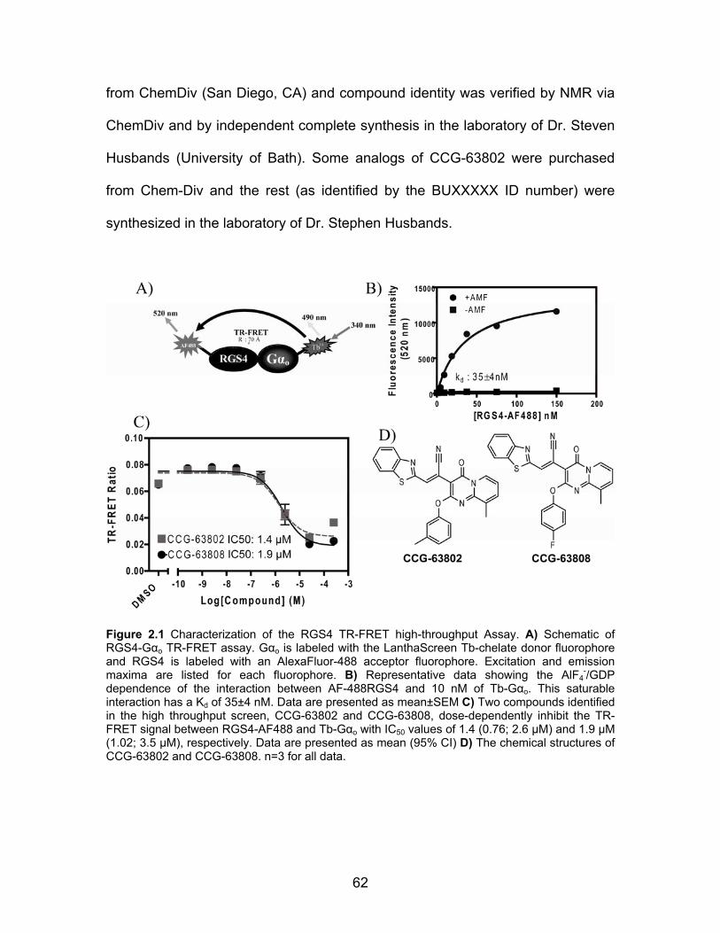
62
from ChemDiv (San Diego, CA) and compound identity was verified by NMR via
ChemDiv and by independent complete synthesis in the laboratory of Dr. Steven
Husbands (University of Bath). Some analogs of CCG-63802 were purchased
from Chem-Div and the rest (as identified by the BUXXXXX ID number) were
synthesized in the laboratory of Dr. Stephen Husbands.
Figure 2.1 Characterization of the RGS4 TR-FRET high-throughput Assay. A) Schematic of RGS4-Gαo TR-FRET assay. Gαo is labeled with the LanthaScreen Tb-chelate donor fluorophore and RGS4 is labeled with an AlexaFluor-488 acceptor fluorophore. Excitation and emission maxima are listed for each fluorophore. B) Representative data showing the AlF4
-/GDP dependence of the interaction between AF-488RGS4 and 10 nM of Tb-Gαo. This saturable interaction has a Kd of 35±4 nM. Data are presented as mean±SEM C) Two compounds identified in the high throughput screen, CCG-63802 and CCG-63808, dose-dependently inhibit the TR-FRET signal between RGS4-AF488 and Tb-Gαo with IC50 values of 1.4 (0.76; 2.6 µM) and 1.9 µM (1.02; 3.5 µM), respectively. Data are presented as mean (95% CI) D) The chemical structures of CCG-63802 and CCG-63808. n=3 for all data.
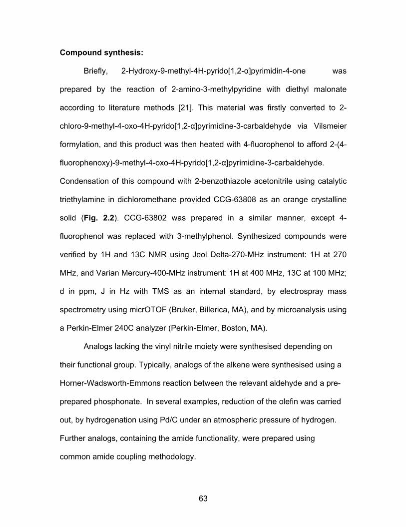
63
Compound synthesis:
Briefly, 2-Hydroxy-9-methyl-4H-pyrido[1,2-α]pyrimidin-4-one was
prepared by the reaction of 2-amino-3-methylpyridine with diethyl malonate
according to literature methods [21]. This material was firstly converted to 2-
chloro-9-methyl-4-oxo-4H-pyrido[1,2-α]pyrimidine-3-carbaldehyde via Vilsmeier
formylation, and this product was then heated with 4-fluorophenol to afford 2-(4-
fluorophenoxy)-9-methyl-4-oxo-4H-pyrido[1,2-α]pyrimidine-3-carbaldehyde.
Condensation of this compound with 2-benzothiazole acetonitrile using catalytic
triethylamine in dichloromethane provided CCG-63808 as an orange crystalline
solid (Fig. 2.2). CCG-63802 was prepared in a similar manner, except 4-
fluorophenol was replaced with 3-methylphenol. Synthesized compounds were
verified by 1H and 13C NMR using Jeol Delta-270-MHz instrument: 1H at 270
MHz, and Varian Mercury-400-MHz instrument: 1H at 400 MHz, 13C at 100 MHz;
d in ppm, J in Hz with TMS as an internal standard, by electrospray mass
spectrometry using micrOTOF (Bruker, Billerica, MA), and by microanalysis using
a Perkin-Elmer 240C analyzer (Perkin-Elmer, Boston, MA).
Analogs lacking the vinyl nitrile moiety were synthesised depending on
their functional group. Typically, analogs of the alkene were synthesised using a
Horner-Wadsworth-Emmons reaction between the relevant aldehyde and a pre-
prepared phosphonate. In several examples, reduction of the olefin was carried
out, by hydrogenation using Pd/C under an atmospheric pressure of hydrogen.
Further analogs, containing the amide functionality, were prepared using
common amide coupling methodology.
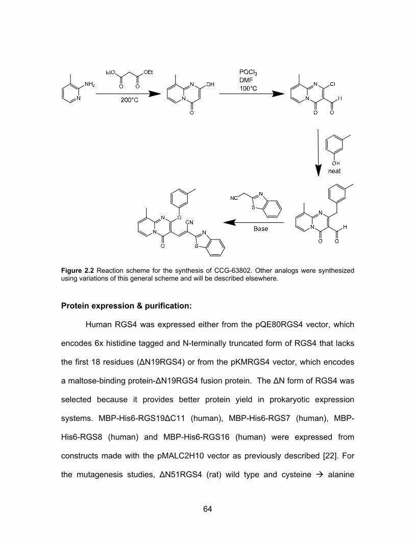
64
Figure 2.2 Reaction scheme for the synthesis of CCG-63802. Other analogs were synthesized using variations of this general scheme and will be described elsewhere.
Protein expression & purification:
Human RGS4 was expressed either from the pQE80RGS4 vector, which
encodes 6x histidine tagged and N-terminally truncated form of RGS4 that lacks
the first 18 residues (ΔN19RGS4) or from the pKMRGS4 vector, which encodes
a maltose-binding protein-ΔN19RGS4 fusion protein. The ΔN form of RGS4 was
selected because it provides better protein yield in prokaryotic expression
systems. MBP-His6-RGS19ΔC11 (human), MBP-His6-RGS7 (human), MBP-
His6-RGS8 (human) and MBP-His6-RGS16 (human) were expressed from
constructs made with the pMALC2H10 vector as previously described [22]. For
the mutagenesis studies, ΔN51RGS4 (rat) wild type and cysteine alanine
65
mutants were expressed from the pMALC2H10 vector. Mutagenesis was
performed as described [19] using the QuickChange Multi Site Directed
Mutagenesis kit (Agilent, La Jolla CA) where one or more of the cysteine
residues in the RGS domain of RGS4 were mutated to alanine.
All proteins were expressed in and harvested from BL21-DE3 E. coli via
standard transformation, growth, and lysis protocols [16, 17, 23-25]. Histidine-
tagged RGS4 was purified over a Nickel NTA affinity column (Qiagen, Valenica,
CA) followed by cation exchange chromatography and size exclusion
chromatography. MBP-tagged RGS proteins were purified with an amylose
affinity column followed by size exclusion chromatography. Hexahistidine-tagged
rat Gαo was expressed and purified as previously described [25]. G protein
activity was determined by [35S]GTPγS binding [26]. In all cases, proteins were
purified to >90% homogeneity before use.
Chemical labeling of purified Gαo & RGS:
AlexaFluor-488 labeling of RGS4: ΔN19RGS4 was labeled with
AlexaFluor-488 succinimidyl ester (Invitrogen, Carlsbad CA) at a 5:1
(label:protein) stoichiometry in a total volume of 2.0 mL of 50 mM HEPES pH 8.2
at 4˚C, 100 mM NaCl, 1 mM DTT. The reaction was performed while rotating
samples in the dark for 1.5 hr at 4˚C. The reaction was quenched by the addition
of 1 mM glycine for 10 minutes at 4˚C. Labeled RGS4 was resolved from the
reaction mixture by size exclusion chromatography using a 20 mL Sephadex G-

66
25 desalting column (GE Biosciences, Piscataway NJ). Degree of labeling was
determined spectroscopically to be approximately 1:1.
Tb chelate labeling of Gαo: Gαo was labeled with the LanthaScreen™ Tb
thiol reactive reagent (Invitrogen, Carlsbad CA) at a 5:1 (label:protein)
stoichiometry in a total volume of 1.0 mL of 50 mM HEPES pH 7.25 at 4˚C, 100
mM NaCl, supplemented with 10 μM GDP and 0.8 mM TCEP. The reaction was
allowed to proceed at 4˚C for 1.5 hr while rotating in the dark. The reaction was
quenched by the addition 1 mM DTT for 20 minutes at 4˚C. Labeled protein was
purified from the reaction mixture by size exclusion chromatography using a
Sephadex G-25 desalting column (GE Biosciences, Piscataway NJ). Degree of
labeling was determined spectroscopically to be approximately 1:1. The activity
and effective concentration of the labeled G protein was determined by
[35S]GTPγS binding as described [26].
Biotinylation of RGS proteins: RGS protein was mixed at a 3:1
(label:protein) molar ratio with biotinamidohexanoic acid N-hydroxysuccinimide
ester (Sigma, St. Louis, MO) in a buffer of 50 mM HEPES pH 8.5 at 4°C, 100 mM
NaCl, & 1 mM DTT. The reaction was allowed to proceed at 4°C while rotating for
2 hours and then was quenched by the addition of a large molar excess of
glycine for 10 minutes. Labeled protein was purified from the reaction mixture by
size exclusion chromatography using a Sephadex G-25 desalting column (GE
Biosciences, Piscataway NJ).
AlexaFluor-532 labeling of Gαo: Gαo labeling was performed as previously
described [17]. Labeled protein was purified from the reaction mixture by size

67
exclusion chromatography using a Sephadex G-25 desalting column (GE
Biosciences, Piscataway NJ).
Time Resolved FRET:
TR-FRET experiments were performed on a PHERAstar multipurpose
microplate reader (BMG Labtech, Offenberg, Germany) using the LanthaScreen
filter set. These experiments were based on the method of Leifert et al [27]. For
the saturation experiments, Tb-Gαo was diluted to 20 nM in 50 mM HEPES pH
8.0, 100 mM NaCl, 0.1% lubrol, 30 µM GDP, 5 mM NaF, 5 mM MgCl2, 5 μM AlCl3
and allowed to activate for 10 minutes on ice before use. RGS4-AF488 was
serially diluted in 50 mM HEPES pH 8.0 at room temperature, 100 mM NaCl,
0.1% lubrol (TR-FRET buffer). Ten microliters of the RGS4 dilution was added to
a black non-stick low-volume 384 well plate (Corning 3676) with a minimum of
duplicate measurements. Ten microliters of the Tb-Gαo was added (10 nM final)
and the mixture was allowed to incubate at room temperature for 15 minutes in
the dark. The non-specific TR-FRET signal was determined by excluding the
AlCl3, MgCl2 and NaF from a set of samples. The fluorescence emission at both
490 and 520 nm was measured from 50 flashes of 340 nm excitation light per
well. The data were collected in 10 μs bins and the delayed emission signal was
integrated from 100 to 500 μs after each flash. TR-FRET data was analyzed as
the ratio of emission at 520 nm/490 nm.

68
High Throughput Screening:
High throughput screening was performed with the University of Michigan
Center for Chemical Genomics. The ca. 40,000 compound screening collection
was provided by the Novartis Institute for Biomedical Research (East Hanover
NJ) and was comprised of compounds selected from the ChemDiv screening
library. Five microliters of 50 mM HEPES pH 8.0 at room temperature, 100 mM
NaCl, 0.1% lubrol, 1 mM DTT (TR-FRET buffer) was dispensed with a
Multidrop™ (Thermo Fisher Scientific, Waltham MA) into every well of a black,
non-stick, low-volume 384-well plate. Two hundred nanoliters of each compound
(2 mM stock, 20 µM final assay concentration) or DMSO control was added to
the plate with a pin tool using a Beckman BioMek FX liquid handler (Beckman
Coulter, Fullerton, CA). To this compound dilution, 5 microliters of 200 nM
AlexFluor 488 labeled RGS4 was added and incubated for 15 minutes at room
temperature in the dark. Then, 10 microliters of 20 nM Tb labeled Gαo was added
to the mixture. For this assay, the positive inhibition control (i.e. no RGS4/Gαo
binding) was Tb-labeled Gαo in the inactive GDP-bound state and the negative
control (i.e. full RGS4/Gαo binding) utilized Gαo in the GDP/AlF4 bound state.
This mixture was incubated at room temperature in the dark for 15 minutes
before analysis with the PHERAstar plate reader. Data were compiled and
analyzed using the M-Screen database, an in-house chemoinformatics suite
developed by the Center for Chemical Genomics at the University of Michigan.
Compounds that inhibited the TR-FRET signal >2SD from the negative control

69
were considered “actives” and were chosen for dose response follow-up
experiments.
TR-FRET Dose Response Experiments:
Actives from the primary screen were evaluated for concentration-
dependent activity in the TR-FRET assay. Compound dilutions were performed in
DMSO and 200 nL of diluted compound was spotted into the wells of a non-stick,
low volume black 384-well plate that contained 5 µL of TR-FRET buffer. To the
well, 5 µL of 200 nM AlexFluor 488 labeled RGS4 was added and incubated at
room temperature in the dark for 15 minutes. Then, 10 µL of 20 nM Tb labeled
Gαo GDP/AlF4 was added to the mixture and incubated at room temperature in
the dark for 30 minutes before analysis on the PHERAstar plate reader.
Compound dilutions covered a final concentration range from 200 µM to 1.6 µM.
Positive and negative controls were performed as in the primary screening
assay. Compounds whose dose response curves were not fully defined by these
concentrations were repeated using a more appropriate dilution scheme.
Nonlinear least-squares regression fitting of the data was performed using the
data analysis component of the MScreen database.

70
Flow Cytometry Protein Interaction Assay Concentration Dependence
Experiments:
Compounds that confirmed in the follow up TR-FRET dose response
assay were tested as described [17] in the Flow Cytometry Protein Interaction
Assay (FCPIA). This was done in part to provide a complementary set of
biochemical data to filter out any compounds that might produce spectroscopic
artifacts in the TR-FRET assay. Briefly, biotinylated RGS proteins (5nM, final
assay concentration) were immobilized on Luminex LumAvidin beads and
incubated with diluted compound in 50 mM HEPES pH 8.0 at room temperature,
100 mM NaCl, 0.1% lubrol, 1 mM DTT, supplemented with 1% BSA. To each
well of a 96-well PCR plate (Axygen, Union City, CA) was added AlexaFluor 532
labeled Gαo to a final concentration of 30nM. This mixture was incubated for 30
minutes at room temperature in the dark and then it was analyzed on a Luminex
200 flow cytometer for the bead associated fluorescence (median value).
Nonlinear regression analysis of inhibition curves was performed with Prism 5.0
(Graphpad Software, San Diego CA).
FCPIA Reversibility Experiments:
RGS-coated beads were prepared as above and were treated with 50 µM
compound or vehicle (DMSO) for 15 minutes at room temperature. The RGS-
containing beads were then washed by resuspension in 1mL of phosphate
buffered saline, pH 7.4 supplemented with 1% BSA, vortexing briefly, then
pelleting the beads by centrifugation. This procedure was repeated a total of
71
three times before 1,000 beads were added to each quadruplicate well of a 96-
well PCR plate that contained AlexaFluor-532-labeled Gαo at a final
concentration of 20 nM in the presence or absence of 50µM test compound. The
mixture was incubated for 30 minutes at room temperature and then analyzed on
a Luminex 200 flow cytometer for bead-associated fluorescence. Data analysis
was performed with Prism 5.0 (Graphpad Software, San Diego CA).
Single Turnover GTPase Measurements:
Compounds were tested for the ability to inhibit the RGS4-stimulated
increase in GTP hydrolysis by Gαo as described previously [15, 17].
Thermal Stability Measurements:
Untagged ΔN19RGS4 or His6-Gαo was added to the well of a 96-well ABI
Prism optical reaction plate (Applied Biosystems, Foster City CA) to a final
concentration of 5 or 2.5 µM, respectively in 50-60 µL of 50 mM HEPES pH 8.0
with 150 mM NaCl. Test compounds were added to the protein at the desired
concentration and allowed to interact for 15 minutes at room temperature. To
each well, Sypro Orange dye (Invitrogen, Carlsbad, CA) was added to a 5X final
concentration (as described by the supplier) and the plate was sealed with an
optically clear adhesive film. Sypro Orange fluorescence was measured
continuously in an ABI HT7900 real-time PCR system during a stepwise gradient
from ambient temperature to 90°C in 1°C steps lasting 30 seconds each. Data
are analyzed by fitting the obtained curves to a Boltzmann model:

72
aTTm
e
LULI )(
1
)(−
+
−=
Where I = Fluorescence Intensity (AU), L = the lower limit of the curve (°C), U =
the upper limit of the curve (°C), T = temperature (°C), and a = is a slope factor.
Values obtained after the fluorescence maximum occurred were excluded from
the analysis.
Results:
Development of a High-Throughput TR-FRET RGS4-Gαo interaction screen:
I developed a biochemical TR-FRET assay using purified human RGS4
labeled with the AlexaFluor-488 acceptor fluorophore and purified Gαo labeled
with the LanthaScreen Tb probe donor fluorophore (Fig. 2.1A). Using this
system, I observed a saturable, aluminum fluoride-dependent interaction
between RGS4 and Gαo that has an affinity consistent with other reports of this
PPI in the literature (Fig. 2.1B) [17]. In collaboration with the Center for Chemical
Genomics at the University of Michigan, this assay was scaled to 384-well format
and used to screen ~44,000 small molecules for inhibition of RGS4/Gαo binding
in the presence of a thiol reducing agent (Table 2.1). Compounds from this
screen were re-tested in the primary screening assay to confirm the initial result
and to assess the concentration dependence of the inhibition using the original
TR-FRET assay. Of the 162 compounds that met the 2 standard deviation
selection criteria for inhibition, 48 were either unavailable or predicted to be
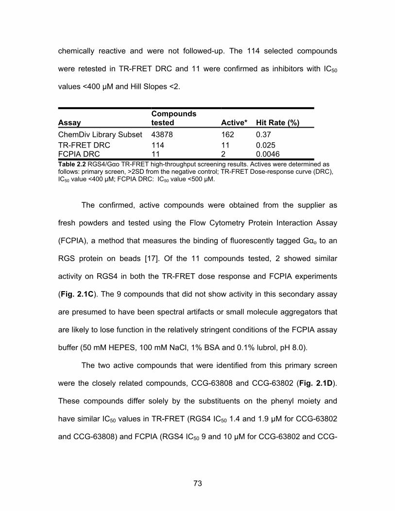
73
chemically reactive and were not followed-up. The 114 selected compounds
were retested in TR-FRET DRC and 11 were confirmed as inhibitors with IC50
values <400 µM and Hill Slopes <2.
Assay Compounds tested Active* Hit Rate (%)
ChemDiv Library Subset 43878 162 0.37 TR-FRET DRC 114 11 0.025 FCPIA DRC 11 2 0.0046 Table 2.2 RGS4/Gαo TR-FRET high-throughput screening results. Actives were determined as follows: primary screen, >2SD from the negative control; TR-FRET Dose-response curve (DRC), IC50 value <400 µM; FCPIA DRC: IC50 value <500 µM.
The confirmed, active compounds were obtained from the supplier as
fresh powders and tested using the Flow Cytometry Protein Interaction Assay
(FCPIA), a method that measures the binding of fluorescently tagged Gαo to an
RGS protein on beads [17]. Of the 11 compounds tested, 2 showed similar
activity on RGS4 in both the TR-FRET dose response and FCPIA experiments
(Fig. 2.1C). The 9 compounds that did not show activity in this secondary assay
are presumed to have been spectral artifacts or small molecule aggregators that
are likely to lose function in the relatively stringent conditions of the FCPIA assay
buffer (50 mM HEPES, 100 mM NaCl, 1% BSA and 0.1% lubrol, pH 8.0).
The two active compounds that were identified from this primary screen
were the closely related compounds, CCG-63808 and CCG-63802 (Fig. 2.1D).
These compounds differ solely by the substituents on the phenyl moiety and
have similar IC50 values in TR-FRET (RGS4 IC50 1.4 and 1.9 µM for CCG-63802
and CCG-63808) and FCPIA (RGS4 IC50 9 and 10 µM for CCG-63802 and CCG-

74
63808). The compounds also contain a vinyl cyanide moiety that may function as
a Michael acceptor.
CCG-63802 & CCG-63808 selectively inhibit Gαo-RGS interactions:
Using TR-FRET to assess the RGS4-Gαo interaction, CCG-63802 and
CCG-63808 had IC50 values of 1.4 and 1.9 µM, respectively (Fig. 2.1C). To
determine the selectivity of these compounds for different RGS proteins, they
were tested in an FCPIA competition experiment against a panel of 5 different
RGS proteins (Fig. 2.3, Table 2.2). The compounds are 6- to 7-fold less potent in
blocking Gαo/RGS4 interactions when tested using FCPIA (IC50 9 or 10 µM for
CCG-62802 or CCG-63808) than with the TR-FRET method. This is probably
due to the high level of BSA (1%) in the FCPIA buffer sequestering compound
and decreasing its apparent concentration in the assay. These compounds did
not inhibit Gα binding to RGS7, which is distantly related to RGS4, and they are
two- to ten-fold more potent at RGS4 than on the other closely related R4 family
members, RGS8 and RGS16 (Table 2.2). They are also active (IC50 20-50 µM)
on the one RZ family member tested, RGS19.

75
Figure 2.3 RGS specificity of CCG-63802 and CCG-63808 determined by multiplex FCPIA analysis (n≥3). RGS coated beads were treated with the indicated concentration of A) CCG-63802 or B) CCG-63808 for 15 minutes at room temperature, after which GDP/AlF4 bound Gαo-AF532 was added and allowed to incubate with the RGS/compound mixture for 30 minutes prior to analysis. See table 2.2 for IC50 values. All data was calculated using nonlinear least squares regression with the bottom of the curves constrained to 0% binding. Data are presented as mean ± SEM from 3 separate experiments.
CCG-63802 CCG-63808 RGS Protein IC50 (µM) Hill Slope IC50 (µM) Hill Slope RGS4 9 -0.9 10 -1.4 RGS4c >400 -0.4 >400 -0.8 RGS8 112 -0.6 74 -1.1 RGS16 42 -1.4 21 -2.1 RGS19 20 -0.6 46 -0.8 RGS7 NI NI NI NI
Table 2.3 RGS specificity of CCG-63802 and CCG-63808 determined by multiplex FCPIA analysis (n≥3). All data was calculated from at least three independent experiments using nonlinear least squares regression with the bottom of the curves constrained to 0% binding. NI: No inhibition observed at highest concentration tested (100 µM). CCG-63802 & CCG-63808 inhibit RGS4 GAP activity:
For RGS inhibitors to be functionally relevant, they need to inhibit the
catalytic activity of the RGS in addition to blocking Gα/RGS binding. The two
compounds inhibit the GAP activity of RGS4 as shown by measurements using
the [32P]GTP single turnover GAP assay (Fig. 2.4). Under these conditions, GTP
hydrolysis by Gαo is accelerated ~10-fold by the addition of wtRGS4 and this
effect can be inhibited by the previously described [17] RGS4 inhibitor CCG-
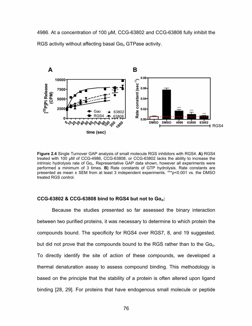
76
4986. At a concentration of 100 µM, CCG-63802 and CCG-63808 fully inhibit the
RGS activity without affecting basal Gαo GTPase activity.
0 10 20 30 40 50 60 70 80 90100
0
2500
5000
7500
10000
GaoRGS4 63808
63802
300
1800
time (sec)
[32P]
Pi R
elea
se (C
PM)
DMSO DMSO 4986 63808 638020.00
0.02
0.04
0.06
0.08
RGS4
******
***
Rate
con
stan
t (se
c-1)
A B
Figure 2.4 Single Turnover GAP analysis of small molecule RGS inhibitors with RGS4. A) RGS4 treated with 100 µM of CCG-4986, CCG-63808, or CCG-63802 lacks the ability to increase the intrinsic hydrolysis rate of Gαo. Representative GAP data shown, however all experiments were performed a minimum of 3 times. B) Rate constants of GTP hydrolysis. Rate constants are presented as mean ± SEM from at least 3 independent experiments. ***p<0.001 vs. the DMSO treated RGS control.
CCG-63802 & CCG-63808 bind to RGS4 but not to Gαo:
Because the studies presented so far assessed the binary interaction
between two purified proteins, it was necessary to determine to which protein the
compounds bound. The specificity for RGS4 over RGS7, 8, and 19 suggested,
but did not prove that the compounds bound to the RGS rather than to the Gαo.
To directly identify the site of action of these compounds, we developed a
thermal denaturation assay to assess compound binding. This methodology is
based on the principle that the stability of a protein is often altered upon ligand
binding [28, 29]. For proteins that have endogenous small molecule or peptide

77
ligands (e.g. enzymes or receptors), binding of the ligand often increases the
thermal stability. Upon binding GDP, Gαo experiences a >5°C increase in melting
temperature (Tm) when compared to nucleotide-free protein (Fig 2.5). This
increases to a >20°C increase in Tm for Gαo binding the exceptionally high affinity
nucleotide GTPγS. Using this assay, we observed a concentration-dependent
10°C reduction (See Discussion) in the melting temperature of RGS4 in the
presence of CCG-63802 (Fig. 2.6A). The concentration dependence of this
effect corresponds with the IC50 values obtained in the FCPIA assay. Even at a
maximal concentration of CCG-63802 (100 µM), there was no change in the
melting temperature of Gαo (Fig. 2.6B).
20
40
60
80
***
***
GTPγSno nt GDP
Tm (°
C)
Figure 2.5 Gαo is thermally stabilized in presence of nucleotide. Purified Gαo was stripped of nucleotide by gel filtration in a buffer containing EDTA. Lack of nucleotide was confirmed by spectroscopic analysis. The melting temperature (Tm) of Gαo (2.5 µM) was determined by the thermal stability assay as described (see Methods) in the presence or absence of 50 µM GDP or GTPγS. Nucleotide free (no nt) Gαo has a Tm of 38.9±0.7°C. The protein is stabilized in the presence of 50 µM GDP by 6°C (Tm: 45.0±0.3C°) and is stabilized by 23°C in the presence of 50 µM GTPγS (Tm 62.7±0.5°C). Data are presented as mean ± SEM from 3 separate experiments.

78
-6.0 -5.5 -5.0 -4.5 -4.0 -3.530
32
34
36
38
40
42
44
DMSOCCG-63802
Log [CCG-63802] (M)
Tm (C
o )
Vehicle CCG-6380225
30
35
40
45
50 ns
Tm ( °
C)
A B
Figure 2.6 CCG-63802 specifically binds to RGS4 and not to Gαo. A) Purified RGS4 shows a dose dependent change in melting temperature in the presence of CCG-63802 (EC50 26 µM). B) A saturating concentration of CCG-63802 (100 µM) does not affect the melting temperature of Gαo. Data are presented as mean±SEM of 3 separate experiments.
CCG-63802 and CCG-63808 are reversible inhibitors of the Gαo-RGS
interaction:
The effects of CCG-4986, our previously described RGS4 inhibitor [17],
could not be reversed by dilution and washing away the compound, showing that
it acts irreversibly to inhibit the function of RGS4 [18, 19], and Fig. 2.7). Also, its
activity was blocked in the presence of reducing agents. These effects are likely
due to the formation of a covalent adduct of the compound with a cysteine
residue in the RGS [18, 19]. Since our new compounds were identified through
screens in the presence of DTT, we tested the reversibility of their inhibition.
RGS-coated microspheres were treated with 50 µM compound or vehicle
(DMSO), extensively washed (see Methods for details), and then assayed for
Gαo binding (Fig 2.7). In contrast to the effects of CCG-4986, full binding was
restored to compound-treated RGS-beads after washing (Fig 2.7), showing that

79
CCG-63802 and CCG-63808 are reversible on the 10-minute time scale required
for the washing procedure. Consequently, these new compounds represent the
first examples of reversible small molecule inhibitors of an RGS protein.
RGS4
0
25
50
75
100
CCG-63808CCG-63802
Washed- - -+ + +CCG-4986
% In
hibi
tion
RGS19
0
25
50
75
100
CCG-63808CCG-63802
Washed- - -+ + +CCG-4986
% In
hibi
tion
A
B
Figure 2.7 CCG-63802 and CCG-63808 are reversible inhibitors of A) RGS4 and B) RGS19. CCG-4986 is an irreversible inhibitor of RGS4 and RGS19. In all cases, RGS coated FCPIA beads were treated with 50 µM of compound (or vehicle, DMSO) and then extensively washed. The beads were then split into two groups and tested for the ability to interact with Gαo-AF532 in the presence or absence of 50 µM Compound. Data shown are the mean±SEM of three separate experiments.

80
Cysteine Dependence of CCG-63802 and CCG-63808:
To further explore the mechanism of these compounds and the role of
cysteines in their action, they were tested on a mutant of RGS4 where all
cysteines in the RGS domain were mutated to alanine (RGS4c). In FCPIA
measures of Gα binding to RGS4c, CCG-63802 and CCG-63808 show only
modest activity, indicating a role for RGS cysteines in the actions of these
compounds (Fig 2.8, Table 2.3). Consequently, we tested CCG-63808 and
CCG-63802 with a panel of RGS4 RGS domain cysteine mutants using FCPIA
(Table 2.3). The G protein binding affinity of these RGS mutants has been
previously described [30] and the Kd values ranged from 3-12 nM, not drastically
different from that of wild type RGS4. No single cysteine could fully account for
the effects of these compounds, but it appears that three cysteines, Cys 148, Cys
132, and Cys 95 are important for full sensitivity to CCG-63808 and CCG-63802.
Cysteine 95 and Cys 148 are located close to each other on RGS4, however
they are at a site distinct from the Gα interaction interface. It appears that Cys 95
plays a more significant role than Cys 148, possibly suggesting that the
compound docks onto the RGS at a site that is either closer to this cysteine or
that requires this residue for proper formation of the compound binding pocket.

81
RGS4 Mutant
IC50 (µM)
pIC50 Log(M)
Hill Slope
Inhibition at 100µM (%)
n
WT 9 5.02 ± 0.07 -0.86 ± 0.11 87 9 C148A 43 4.37 ± 0.07 -0.95 ± 0.16 63 3 C132A 41 4.39 ± 0.07 -0.97 ± 0.18 66 3 C95A/C132A 32 4.50 ± 0.13 -0.78 ± 0.20 70 3 C148A/C132A 92 4.04 ± 0.07 -0.75 ± 0.11 57 3 C148A/C132A/C95A ~3000 2.55 ± 0.64 -0.33 ± 0.12 16 3 RGS4c ~8000 2.10 ± 1.50 -0.36 ± 0.30 13 6 A148C ~390 3.41 ± 0.17 -0.62 ± 0.14 30 3 A132C 174 3.76 ± 0.19 -0.80 ± 0.29 31 3 A95C 170 3.77 ± 0.23 -1.20 ± 0.82 30 3 A148C/A132C 33 4.47 ± 0.05 -1.48 ± 0.23 92 3 A148C/A95C 17 4.77±0.12 -1.06±0.28 100 3 A95C/A148C/A132C 16 4.79 ± 0.12 -0.63 ± 0.12 64 3
Table 2.4 RGS4 cysteine mutant sensitivity to CCG-63802. IC50 values calculated from FCPIA concentration-response experiments. Data are presented as mean ±SEM.
Figure 2.8 CCG-63802 and CCG-63808 are much less potent on a mutant form of RGS4 that lacks cysteine residues in the RH domain. FCPIA dose-response curves for A) CCCG-63802 and B) CCG-63808. Data are presented as mean ± SEM from 3 separate FCPIA experiments.
Since thiol-reactive compounds may have difficulty functioning in the
reducing environment of a cell, it is important to assess the activity of any such
leads under conditions mimicking the intracellular environment. Therefore, CCG-
63802, CCG-63808, and CCG-4986 were tested for activity using FCPIA in the
presence of 2 mM reduced glutathione (Fig. 2.9). This concentration of

82
glutathione was selected because it is similar to intracellular concentrations.
CCG-63802 and CCG-63808 lose approximately 0.5-1 Log of potency (IC50 6 µM
40 µM for CCG-63802; 4 µM 21 µM for CCG-63808) in the presence of 2
mM glutathione, but still retain the ability to fully inhibit the interaction between
RGS4 and Gαo. In contrast, CCG-4986 loses over 2-Logs in potency (IC50
from1.4 µM 215 µM) in the presence of 2 mM glutathione and it is not capable
of fully inhibiting the RGS-Gαo interaction up to concentrations nearing its
aqueous solubility (Fig. 2.9).
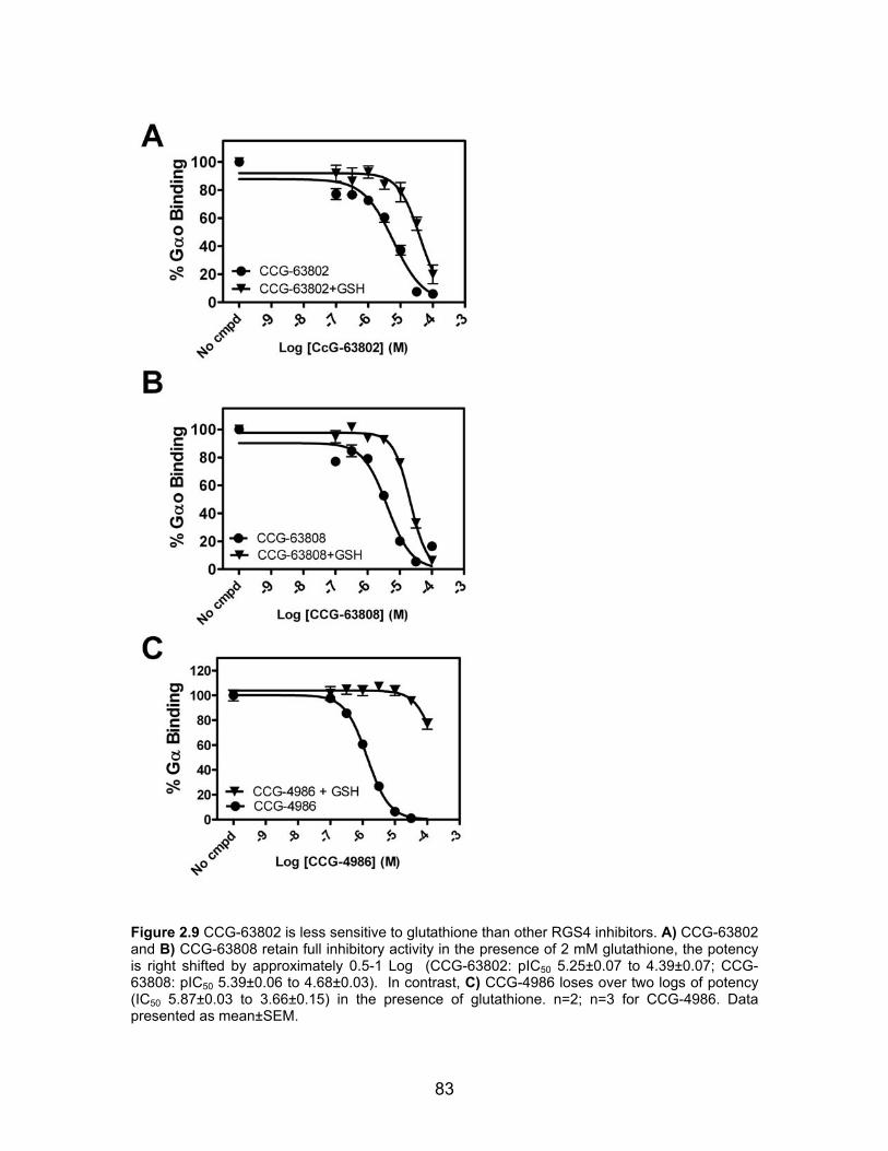
83
Figure 2.9 CCG-63802 is less sensitive to glutathione than other RGS4 inhibitors. A) CCG-63802 and B) CCG-63808 retain full inhibitory activity in the presence of 2 mM glutathione, the potency is right shifted by approximately 0.5-1 Log (CCG-63802: pIC50 5.25±0.07 to 4.39±0.07; CCG-63808: pIC50 5.39±0.06 to 4.68±0.03). In contrast, C) CCG-4986 loses over two logs of potency (IC50 5.87±0.03 to 3.66±0.15) in the presence of glutathione. n=2; n=3 for CCG-4986. Data presented as mean±SEM.
84
I originally found that CCG-63802 and CCG-63808 inhibit the single
turnover GAP activity of RGS4c (Fig. 2.10) mutant despite their much lower
potency to inhibit Gαo/RGS4c binding in FCPIA (Fig 2.8). These data suggested
that this class of compounds can inhibit the functional activity of the cysteine-null
RGS4 mutant, while having much less effect on the high-affinity binding to GDP-
AMF bound Gαo (see Discussion). This inhibitory effect does not appear to be
due to compound aggregation, as is not reversed in the presence of 0.01% Triton
X-100 (data not shown), which generally blocks the activity of promiscuous small
molecule aggregators [31]. Unfortunately, the compounds did not have an effect
on RGS4c in steady state GTPase assays and thermal stability experiments
(data not shown, experiments performed by Andrew Storaska). While it is
possible that the single turnover data are correct, the lack of consistency with
other biochemical assays raises questions regarding the validity of the single
turnover data. As such, it is believed that while these data are solid, their
physiological significance is likely to be limited. Although disappointing, this result
is in line with the observed cysteine dependence in FCPIA.
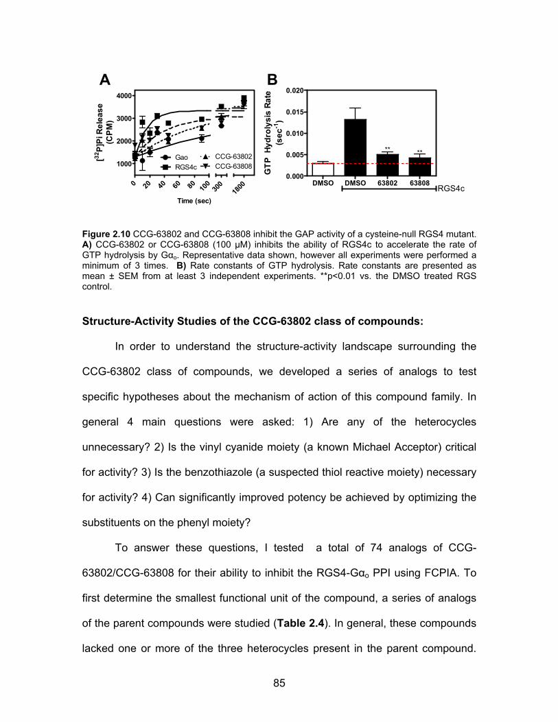
85
DMSO DMSO 63802 638080.000
0.005
0.010
0.015
0.020
RGS4c
** **
GTP
Hyd
roly
sis
Rate
(sec
-1)
0 20 40 60 80 100
1000
2000
3000
4000
GaoRGS4c
CCG-63802CCG-63808
300
1800
Time (sec)
[32P]
Pi R
elea
se(C
PM)
A B
Figure 2.10 CCG-63802 and CCG-63808 inhibit the GAP activity of a cysteine-null RGS4 mutant. A) CCG-63802 or CCG-63808 (100 µM) inhibits the ability of RGS4c to accelerate the rate of GTP hydrolysis by Gαo. Representative data shown, however all experiments were performed a minimum of 3 times. B) Rate constants of GTP hydrolysis. Rate constants are presented as mean ± SEM from at least 3 independent experiments. **p<0.01 vs. the DMSO treated RGS control. Structure-Activity Studies of the CCG-63802 class of compounds:
In order to understand the structure-activity landscape surrounding the
CCG-63802 class of compounds, we developed a series of analogs to test
specific hypotheses about the mechanism of action of this compound family. In
general 4 main questions were asked: 1) Are any of the heterocycles
unnecessary? 2) Is the vinyl cyanide moiety (a known Michael Acceptor) critical
for activity? 3) Is the benzothiazole (a suspected thiol reactive moiety) necessary
for activity? 4) Can significantly improved potency be achieved by optimizing the
substituents on the phenyl moiety?
To answer these questions, I tested a total of 74 analogs of CCG-
63802/CCG-63808 for their ability to inhibit the RGS4-Gαo PPI using FCPIA. To
first determine the smallest functional unit of the compound, a series of analogs
of the parent compounds were studied (Table 2.4). In general, these compounds
lacked one or more of the three heterocycles present in the parent compound.

86
Compounds CCG-203673-5 contain primarily the pyridopyrimidone ring structure
and did not inhibit RGS4- or RGS8-Gαo interaction at any concentration below
100µM. Compounds CCG-203676-80 contain the pyridopyrimidone ring with the
fluorophenoxy moiety present in CCG-63808. These likewise did not show
inhibitory activity against the RGS4/RGS8-Gαo PPI in FCPIA or GAP (Fig. 2.11).
The compounds CCG-203686 and CCG-203687 contain the
pyridopyrimidone/fluorphenoxy rings and the vinyl cyanide moiety (CCG-203686)
or the cyano group lacking the vinyl (CCG-203687). Neither of these compounds
have activity, suggesting that the benzothiazole is important in the mechanism of
action of this compound.
The presence of the vinyl cyanide is important for the Michael-style
reactivity of this group. To determine the importance of this reactivity in the
mechanism of action of CCG-63802, I tested three analogs (CCG-203683-5) that
do not contain a vinyl cyanide group (Table 2.4). In place of this group, these
compounds contain an alkyl chain (CCG-203685), a alkene (CCG-203684) or an
alkyl chain with a cyano group (CCG-203683) It was found that none of these
compounds have activity towards RGS4, suggesting that the vinyl cyanide moiety
is a major driving force between the compound-RGS interaction.
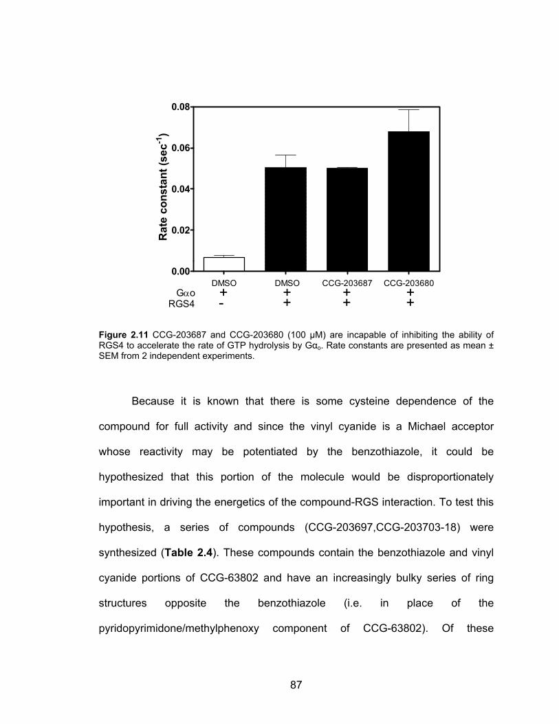
87
0.00
0.02
0.04
0.06
0.08
GαoRGS4
DMSO DMSO CCG-203687 CCG-203680+ + + +- + + +
Rat
e co
nsta
nt (s
ec-1
)
Figure 2.11 CCG-203687 and CCG-203680 (100 µM) are incapable of inhibiting the ability of RGS4 to accelerate the rate of GTP hydrolysis by Gαo. Rate constants are presented as mean ± SEM from 2 independent experiments.
Because it is known that there is some cysteine dependence of the
compound for full activity and since the vinyl cyanide is a Michael acceptor
whose reactivity may be potentiated by the benzothiazole, it could be
hypothesized that this portion of the molecule would be disproportionately
important in driving the energetics of the compound-RGS interaction. To test this
hypothesis, a series of compounds (CCG-203697,CCG-203703-18) were
synthesized (Table 2.4). These compounds contain the benzothiazole and vinyl
cyanide portions of CCG-63802 and have an increasingly bulky series of ring
structures opposite the benzothiazole (i.e. in place of the
pyridopyrimidone/methylphenoxy component of CCG-63802). Of these
88
compounds, only CCG-203714 showed any activity, although this activity was
weak (>200 µM IC50) and inconsistent. Thus, these data suggest that the vinyl
cyanide/benzothiazole groups on this class of compound are necessary, but not
sufficient for RGS inhibitory activity. It will be shown below, however, that with the
appropriate ring structure, the benzothiazole can be replaced. When taken in
conjunction with the pyridopyrimidone analog data, these data suggest that all
three rings and the vinyl cyanide are required for full activity of this class of
compounds. In certain instances (e.g. CCG-203687 and CCG-203680, Fig. 2.11)
compounds from these groups of compounds were tested at a high concentration
(100 µM) in a single turnover RGS4 GAP assay to confirm the observations that
were made using FCPIA. In all cases, there was direct correlation between lack
of activity in FCPIA and lack of activity in the single turnover GAP assay.
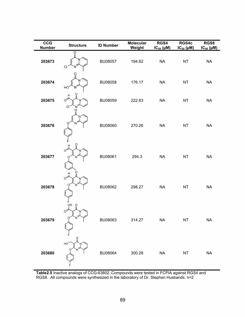
89
CCG Number Structure ID Number Molecular
Weight RGS4
IC50 (µM) RGS4c
IC50 (µM) RGS8
IC50 (µM)
203673
O
N
NCl
BU08057 194.62 NA NT NA
203674
BU08058 176.17 NA NT NA
203675
BU08059 222.63 NA NT NA
203676
BU08060 270.26 NA NT NA
203677
O
N
NO
HO
BU08061 294.3 NA NT NA
203678
O
N
NO
F
HO
BU08062 298.27 NA NT NA
203679
O
N
NO
F
OH
O
BU08063 314.27 NA NT NA
203680
O
N
NO
F
HO
BU08064 300.28 NA NT NA
Table2.5 Inactive analogs of CCG-63802. Compounds were tested in FCPIA against RGS4 and RGS8. All compounds were synthesized in the laboratory of Dr. Stephen Husbands. n=2
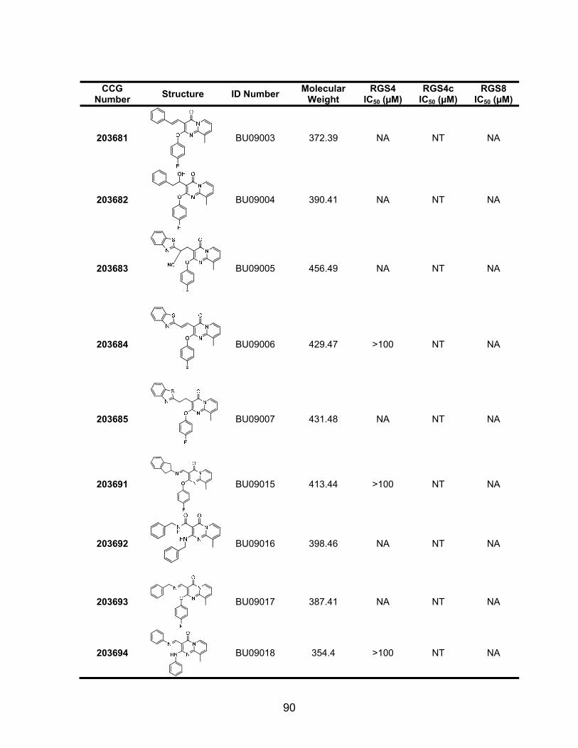
90
CCG
Number Structure ID Number Molecular Weight
RGS4 IC50 (µM)
RGS4c IC50 (µM)
RGS8 IC50 (µM)
203681
BU09003 372.39 NA NT NA
203682
BU09004 390.41 NA NT NA
203683
BU09005 456.49 NA NT NA
203684
BU09006 429.47 >100 NT NA
203685
BU09007 431.48 NA NT NA
203691
BU09015 413.44 >100 NT NA
203692
BU09016 398.46 NA NT NA
203693
BU09017 387.41 NA NT NA
203694
BU09018 354.4 >100 NT NA
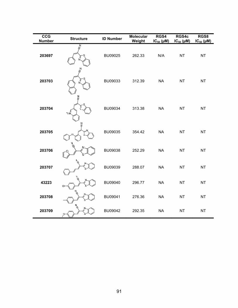
91
CCG
Number Structure ID Number Molecular Weight
RGS4 IC50 (µM)
RGS4c IC50 (µM)
RGS8 IC50 (µM)
203697
N
NS
BU09025 262.33 N/A NT NT
203703
N
NS
BU09033 312.39 NA NT NT
203704
BU09034 313.38 NA NT NT
203705
BU09035 354.42 NA NT NT
203706
BU09038 252.29 NA NT NT
203707
BU09039 288.07 NA NT NT
43223 BU09040 296.77 NA NT NT
203708 N
N
S
BU09041 276.36 NA NT NT
203709
BU09042 292.35 NA NT NT
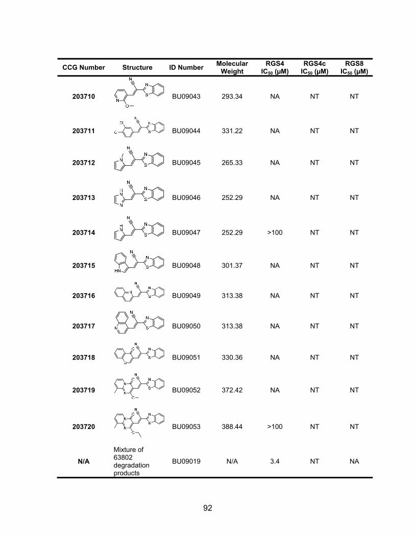
92
CCG Number Structure ID Number Molecular Weight
RGS4 IC50 (µM)
RGS4c IC50 (µM)
RGS8 IC50 (µM)
203710
BU09043 293.34 NA NT NT
203711
BU09044 331.22 NA NT NT
203712
BU09045 265.33 NA NT NT
203713
BU09046 252.29 NA NT NT
203714
BU09047 252.29 >100 NT NT
203715
BU09048 301.37 NA NT NT
203716
BU09049 313.38 NA NT NT
203717
BU09050 313.38 NA NT NT
203718
BU09051 330.36 NA NT NT
203719
BU09052 372.42 NA NT NT
203720
BU09053 388.44 >100 NT NT
N/A Mixture of 63802 degradation products
BU09019 N/A 3.4 NT NA
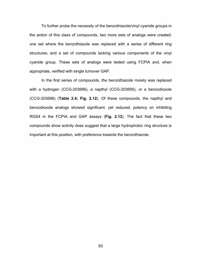
93
To further probe the necessity of the benzothiazole/vinyl cyanide groups in
the action of this class of compounds, two more sets of analogs were created:
one set where the benzothiazole was replaced with a series of different ring
structures; and a set of compounds lacking various components of the vinyl
cyanide group. These sets of analogs were tested using FCPIA and, when
appropriate, verified with single turnover GAP.
In the first series of compounds, the benzothiazole moiety was replaced
with a hydrogen (CCG-203686), a napthyl (CCG-203695), or a benzodioxole
(CCG-203696) (Table 2.4; Fig. 2.12). Of these compounds, the napthyl and
benzodioxole analogs showed significant, yet reduced, potency on inhibiting
RGS4 in the FCPIA and GAP assays (Fig. 2.12). The fact that these two
compounds show activity does suggest that a large hydrophobic ring structure is
important at this position, with preference towards the benzothiazole.
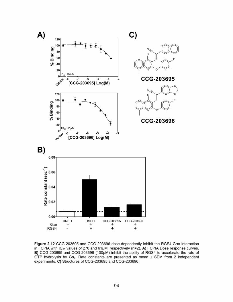
94
0.00
0.02
0.04
0.06
0.08
GαoRGS4
DMSO DMSO CCG-203695 CCG-203696+ + + +- + + +
Rat
e co
nsta
nt (s
ec-1
)
C)
0
20
40
60
80
100
120
-8 -7 -6 -5 -4 -3
Vehicl
e
IC50: 270μM
[CCG-203695] Log(M)
% B
indi
ng
0
20
40
60
80
100
120
-8 -7 -6 -5 -4 -3
IC50: 61μM
Vehicl
e[CCG-203696] Log(M)
% B
indi
ng
B)
A)
N
O
N
O
F
N
N
O
NO
O
O
F
N
CCG-203696
CCG-203695
Figure 2.12 CCG-203695 and CCG-203696 dose-dependently inhibit the RGS4-Gαo interaction in FCPIA with IC50 values of 270 and 61µM, respectively (n=2). A) FCPIA Dose response curves. B) CCG-203695 and CCG-203696 (100µM) inhibit the ability of RGS4 to accelerate the rate of GTP hydrolysis by Gαo. Rate constants are presented as mean ± SEM from 2 independent experiments. C) Structures of CCG-203695 and CCG-203696.

95
The second series of these compounds were designed to test the
importance of the sulfur atom in the benzothiazole group. These compounds
were of particular interest because of the potential importance of cysteine
reactivity in the mechanism of action of the compound. In this series, several
benzothiazole analogs with different substituents on the phenyl moiety (the
importance of which is discussed below) were compared to the benzimidazole
analogs thereof. Five such parings of these analogs were tested using FCPIA
(Table 2.5). In the majority of cases, the benzimidazole compounds were not
able to inhibit the RGS4-Gαo interaction, due at least in part to solubility
limitations. In the one instance where there was appreciable activity (CCG-
63798), compound solubility was less of an issue and therefore it is believed that
it is possible that a benzimidazole is an acceptable group at this position, as long
as solubility can be maintained.
CCG ID R1 R2: S
(Benzothiazole) IC50 (µM)
R2: N (Benzimidazole)
IC50 (µM) 63804, 63806 pEt 4 >100
203669, 203670 H 7 >100 63826, 63828 mBis-Me 8 >100 63742, 63776 ptBu 10 >100 203673, 63798 pOMe 28 31
Table 2.6 Benzothiazole vs. Benzimidazole analogs of the CCG-63802 class of compounds. IC50 values are presented as the mean from at least 2 FCPIA experiments measuring the inhibition of Gαo binding to RGS4.
This class of compounds requires at least two of the three heterocycles for
full activity – the benzothiazole (or some other bulky substituent) and the
pyridopyrimidone. To better understand the chemical space around the phenyl
96
moiety we developed a series of compounds with varying substituents on or in
place of the phenyl group (Table 2.6). It was found that replacing this group with
an alkyl chain (e.g. CCG203719-20) results in compounds with weak, if any,
activity. If however, the phenyl ring is maintained but the substituents thereon are
varied, limited variation (3 to 30 µM IC50) in the structure-activity landscape can
be observed. While the increase in potency achieved is small, it can be
concluded that the optimal moiety on this phenyl ring is a small, hydrophobic
substituent in the para position. Furthermore, this site can accommodate larger
hydrophobic substituents (up to t-butyl); however, it is at the cost of reduced
potency and compound solubility.

97
CCG ID R1 R2 R3 RGS4 IC50 (µM) RGS8 IC50 (µM) 63804 S pEt CH3 4 53 63794 S pBr CH3 4 50 63770 S m,oMe H 5 57 203671 S pMe CH3 5 67 63766 S mMe, pCl H 7 >100 203701 S pBu CH3 7 >100 203669 S H CH3 7 57 63826 S mBis-Me CH3 8 41 63764 S pF H 8 65 63772 S o,pMe H 8 80 63762 S pBr H 9 >100 63768 S mBis-Me H 9 >100 63742 S ptBu H 10 >100 203666 S pEt H 11 >100 203665 S H H 14 >100 203700 S pIsopropyl CH3 23 NT 203675 S pIsopropyl H 23 >100 203672 S pCl H 24 81 203677 S ppropyl H 25 >100 203702 S ppropyl CH3 25 NT 203699 S p,mCl CH3 27 NT 203698 S pOMe CH3 27 NT 203674 S p,mCl H 27 >100 203673 S pOMe H 28 >100 63798 N pOMe H 31 >100 203668 N mMe, pCl H >100 >100 203667 N oCl H >100 >100 63806 N pEt CH3 >100 >100 63776 N ptBu CH3 >100 >100 96665 N mMe CH3 >100 >100 63828 N mBis-Me CH3 >100 >100 63830 N oOMe CH3 >100 >100 203670 N H CH3 >100 >100
Table 2.7 Substituents on the phenyl moiety contribute to the potency of CCG-63802 analogs on RGS4. All compounds were tested using FCPIA. Data are presented as the mean of at least two independent experiments.
98
Discussion:
RGS proteins play a strong modulatory role in GPCR signaling leading to
substantial interest in small molecule inhibitors targeting this class of proteins [9-
12, 32]. The localized expression of RGS proteins [33] suggested that RGS
inhibitors could provide enhanced tissue specificity for GPCR agonist actions [9,
10, 32]. Furthermore, up-regulation of RGS proteins in various disease states, for
example RGS4 in neuropathic pain models [34], also provides an important
rationale for targeting RGS proteins. In this study, we report the second family of
RGS SMPPIIs. Unlike the our previously reported RGS inhibitor, CCG-4986 [17]
which is irreversible and loses function in the presence of reducing agents ([18,
19]), the new compounds identified here, act reversibly and retain substantial
function in the presence of glutathione, a predominant intracellular reductant.
These compounds, with their reversibility and activity in glutathione, therefore
represent a significant step forward in the development RGS SMPPIIs.
Unfortunately, these compounds do not appear to possess significant cellular
activity, likely due permeability issues stemming from the large polar surface area
of the most potent members of this family.
Similar to CCG-4986, CCG-63802 and CCG-63808 are relatively selective
for RGS4 over other R4 family members, including the closely related RGS8 and
RGS16. They have no detectable activity for the more distantly related RGS7.
They also have dependence on cysteine residues because they very weakly
inhibit the cysteine-null (C A) mutant of RGS4 (RGS4c) in the FCPIA assay.
However, both compounds at 100 µM fully inhibit the GAP activity of RGS4c.

99
There are a few potential explanations for this discrepancy. First, the
compounds, which are of modest affinity (10 µM) in the FCPIA studies, may have
a very short RGS-bound lifetime and therefore have difficulty competing with the
constitutive binding of AlF4-/GDP-bound Gαo to the RGS. In the GTPase assay
they may be more efficient at inhibiting the transient interaction between GTP-
bound Gαo and RGS4 during the catalytic cycle. Also, since the compounds
appear to act via an allosteric site (see below), the induced conformational
change in RGS4 may have a more dramatic impact on binding to or GAP activity
at the Gα-GTP than for the GDP-AlF4- conformation of the Gα subunits. While
this explanation is tempting, subsequent studies using a steady state GTPase
assay and a thermal stability assay with RGS4c failed to reproduce the effects
observed in the single turnover assay, lessening the strength of the single
turnover data (Data not shown, experiments performed by Andrew Storaska).
The limited and inconsistent activity of CCG-63802 and CCG-63808 on RGS4c
corresponds with the cysteine reactivity that is believed to play a role in the
mechanism of action of this family of compounds.
The partial cysteine-dependence of the actions of these compounds
suggests a tethering model in which a reactive group binds to an RGS cysteine
residue. This is supported by the cysteine mutagenesis studies and also by the
presence of the potential Michael acceptor functionality (vinyl cyanide) in both of
the compounds. Tethered ligands can provide enhanced potency for small
molecules acting on difficult targets [35, 36]. Our ability to detect these
compounds in the original screen may have derived from potency enhancement
100
from a slow off-rate due to tethering. The reaction, however, is clearly reversible
on the 10-minute time scale and attempts to demonstrate covalent binding by
mass spectroscopy have been unsuccessful. While uncommon, there are other
well-described examples of reversible Michael acceptor reactions with thiols [37,
38]. While most drug molecules are designed to avoid such reactive groups,
there are a number of examples of clinically used drugs (e.g. omeprazole) or
drug candidates (CI-1033) that are thiol reactive [39, 40]. The clear margin of
safety and commercial success of these drugs suggests that irreversible
inhibitors may not be as intractable as therapeutic agents as previously thought.
Furthermore, tethered ligands have been used to develop SAR in the context of
the higher affinity starting structure that is then transferred to analogs without the
reactive group [41].
I have shown that wild type RGS4 is inhibited at 10 μM by CCG-63802
(Table 2.2, Table 2.3). Mutation of all four cysteines in the RH domain to alanine
completely desensitizes the RGS to this family of compounds. By using this
discrepancy, I was able to determine the cysteines that are important for the
modulation of RGS function by CCG-63802 and related analogs. The compounds
described here require two cysteines for full potency of RGS4 inhibition: Cys 95
and Cys 148. These residues are positioned in the “B site” of RGS proteins [10]
which is proposed to participate in the allosteric modulation of RGS4 by acidic
phospholipids and calmodulin [42, 43]. The presence of either Cys 95 or Cys 148
alone provided only modest sensitivity to CCG-63802, however, a mutant that

101
contains both Cys 95 and Cys 148 displays nearly wild-type levels of sensitivity
to this family of compounds (Table 2.3).
Cysteine 132 is located on the outer edge of the Gα interaction interface. It
is tempting to hypothesize that labeling this cysteine with CCG-63802 would
provide significant steric occlusion of the RGS-Gαo PPI. However, that contained
only Cys 132 displayed modest (IC50: 174 µM, Table 2.3) sensitivity to CCG-
63802. Furthermore, addition of the Cys 132 to the double-add back mutant that
contains Cys 95 and Cys 148 was unable to further potentiate the effects of
CCG-63802 (Table 2.3). These data strongly suggest that, while Cys 132 may
play a minor role in the action of these compounds, the primary driving force
behind CCG-63802 inhibition is through the interactions with Cys 95 and Cys
148.
The binding of CCG-63802 induces a destabilizing effect on RGS4 in the
thermal stability studies. This reduced stability of the RGS4 may be related to
conformational perturbation induced upon compound binding to the cysteines in
the allosteric site. In most instances, proteins with endogenous small molecule
ligands (e.g. Gα proteins) are stabilized by the presence of their ligand [44, 45].
This notion was recently borne out by the crystallization of several GPCRs [46-
50]. In all cases (the notable exception being opsin), crystals were only obtained
in the presence of a small molecule antagonist. This strongly suggests that these
ligands are important for the structural stability of this class receptor in solution.
Furthermore, our data (Fig. 2.5) and others [51, 52] also confirm that binding of
natural or artificial ligands to sites that have evolved the capacity for small

102
molecule binding causes a stabilization of the protein. This stabilizing effect may
be due to the decrease in free energy derived from the binding event and also
the conformational restriction required for high-affinity ligand-protein interaction.
On the surface, it would appear that this paradigm is contradicted by the
compounds CCG-63802 and CCG-63808 which potently destabilize RGS4 even
though they appear to bind close to the site on RGS4 that binds native acidic
phospholipids (See Introduction for more details). It is possible that these
compounds bind to a site near, yet independent of, the acidic lipid site on the
RGS and binding to this non-natural site might not be expected to produce the
same stabilization effect as binding of small molecules to sites that have evolved
the capacity for such small molecule-protein interactions. Also, insertion of the
compounds into the 4-helix bundle, stabilized by the reversible Michael addition
to a cysteine thiol, could unfold the RGS4 structure leading to destabilization.
When a chemical entity is discovered to have a particularly interesting
biological activity, it is wise to explore the chemical space surrounding the
molecule in search of higher potency compounds or for compounds with better
physicochemical properties (e.g. improved solubility). To characterize the
structure activity landscape surrounding the CCG-63802 class of compounds, we
synthesized (or purchased, if available) a series of analogs to test specific
hypotheses about the mechanism of action of CCG-63802. These compounds
were tested using FCPIA and those compounds of particular interest were further
studied in follow up experiments using a single turnover GAP assay. In total, 74
analogs were tested for their RGS inhibitory activity. Analysis of the IC50 vales of

103
the compounds using FCPIA (Tables 2.4-2.6) reveals the following: 1) the
compound can accommodate a series of substituents on the phenyl ring while
maintaining an IC50 value within ½ Log of the parent compound (4-30 µM); 2) the
benzothiazole is generally required for full activity, yet measurable activity is
retained when the benzothiazole is replaced with a benzodioxole (IC50 61 µM),
napthyl (IC50 270 µM), or in one instance, a benzimidazole (IC50 31 µM); 3) the
minimal structural unit of the compound class includes all three heterocycles and
the vinyl cyanide. The GAP data available correlate well with the FCPIA data,
suggesting that the PPI-inhibitory effects we observe are correlated to functional
inhibition of RGS activity – at least in vitro.
Overall, the SAR landscape surrounding the CCG-63802 class of
molecules is particularly steep when altering any of the moieties that are believed
to relate to its reactivity. Outside of that, the SAR is rather shallow. The primary
observation from the analogs that retain full reactivity (e.g. that contain the vinyl
cyanide and benzothiazole moieties) is that a small hydrophobic substituent on
the phenyl moiety, preferably in the para position provides the greatest increase
in potency. The dependence upon reactivity suggests that a large part of this
compound’s mechanism of action is through a reversible adduct formation with a
cysteine residue in RGS4, most likely at the 95/148 positions in the molecule.
While this is discouraging from a compound development standpoint, this class
of compounds does show that RGS proteins are capable of being reversibly
inhibited – an important proof of concept. It is likely that future work focusing up
on this family of compounds, especially the elucidation of the structural contacts
104
of CCG-63802 with RGS4, should provide valuable insight into the development
of future generations of small molecule RGS inhibitors.
Conclusions:
In this study we have identified the first examples of reversible SMPPIIs
that disrupt RGS protein function. CCG-63808 and CCG-63802 are selective
inhibitors of the RGS-Gα interaction and R4 family GAP activity. Their
mechanism appears to, at least in part, involve an allosteric action at the “B” site
on the RGS [10], which has been implicated in the physiological allosteric
modulation of RGS proteins by acidic phospholipids and calmodulin [43, 53].
While these compounds have yet to show any cellular activity, it is possible that
future generations of the CCG-63802 family of compounds that have better
physicochemical properties (e.g. improved solubility and lower polar surface
area), and improved potency will provide more tractable lead compounds for
cellular and whole-animal studies.
105
References: 1. Arkin, M.R. and J.A. Wells, Small-molecule inhibitors of protein-protein interactions:
progressing towards the dream. Nat Rev Drug Discov, 2004. 3(4): p. 301-17. 2. Berg, T., Modulation of protein-protein interactions with small organic molecules. Angew
Chem Int Ed Engl, 2003. 42(22): p. 2462-81. 3. Berg, T., Small-molecule inhibitors of protein-protein interactions. Curr Opin Drug Discov
Devel, 2008. 11(5): p. 666-74. 4. Blazer, L.L. and R.R. Neubig, Small Molecule Protein-Protein Interaction Inhibitors as
CNS Therapeutic Agents: Current Progress and Future Hurdles. Neuropsychopharmacology, 2008.
5. Gadek, T.R. and J.B. Nicholas, Small molecule antagonists of proteins. Biochem Pharmacol, 2003. 65(1): p. 1-8.
6. Arkin, M.R. and A. Whitty, The road less traveled: modulating signal transduction enzymes by inhibiting their protein-protein interactions. Curr Opin Chem Biol, 2009. 13(3): p. 284-90.
7. Busschots, K., et al., In search of small molecules blocking interactions between HIV proteins and intracellular cofactors. Mol Biosyst, 2009. 5(1): p. 21-31.
8. Niu, G. and X. Chen, From protein-protein interaction to therapy response: molecular imaging of heat shock proteins. Eur J Radiol, 2009. 70(2): p. 294-304.
9. Neubig, R.R. and D.P. Siderovski, Regulators of G-protein signalling as new central nervous system drug targets. Nat Rev Drug Discov, 2002. 1(3): p. 187-97.
10. Zhong, H. and R.R. Neubig, Regulator of G protein signaling proteins: novel multifunctional drug targets. J Pharmacol Exp Ther, 2001. 297(3): p. 837-45.
11. Traynor, J.R., et al., RGS9-2: probing an intracellular modulator of behavior as a drug target. Trends Pharmacol Sci, 2009. 30(3): p. 105-11.
12. Riddle, E.L., et al., Multi-tasking RGS proteins in the heart: the next therapeutic target? Circ Res, 2005. 96(4): p. 401-11.
13. Bueno, L. and J. Fioramonti, Action of opiates on gastrointestinal function. Baillieres Clin Gastroenterol, 1988. 2(1): p. 123-39.
14. Wang, Y., et al., Identification of Peptides That Inhibit Regulator of G Protein Signaling 4 Function. Pharmacology, 2008. 82(2): p. 97-104.
15. Roof, R.A., et al., Mechanism of action and structural requirements of constrained peptide inhibitors of RGS proteins. Chem Biol Drug Des, 2006. 67(4): p. 266-74.
16. Roof, R.A., et al., Novel peptide ligands of RGS4 from a focused one-bead, one-compound library. Chem Biol Drug Des, 2008. 72(2): p. 111-9.
17. Roman, D.L., et al., Identification of small-molecule inhibitors of RGS4 using a high-throughput flow cytometry protein interaction assay. Mol Pharmacol, 2007. 71(1): p. 169-75.
18. Kimple, A.J., et al., The RGS protein inhibitor CCG-4986 is a covalent modifier of the RGS4 Galpha-interaction face. Biochim Biophys Acta, 2007. 1774(9): p. 1213-20.
19. Roman, D., et al., Allosteric Inhibition of the RGS-G{alpha} Protein-Protein Interaction by CCG-4986. Mol Pharmacol.
20. Blazer, L.L., et al., Reversible, allosteric, small-molecule inhibitors of RGS proteins. Mol Pharmacol.
21. Ingalls EA, P.F., The Preparation, Structure, and Reactions of Some "Malonyl-α-aminopyridines". J Heterocyclic Chem, 1967. 4: p. 523-26.
22. Roman, D.L., S. Ota, and R.R. Neubig, Polyplexed Flow Cytometry Protein Interaction Assay: A Novel High-Throughput Screening Paradigm for RGS Protein Inhibitors. J Biomol Screen, 2009.
23. Lan, K.L., et al., A point mutation in Galphao and Galphai1 blocks interaction with regulator of G protein signaling proteins. J Biol Chem, 1998. 273(21): p. 12794-7.
24. Lan, K.L., et al., Rapid kinetics of regulator of G-protein signaling (RGS)-mediated Galphai and Galphao deactivation. Galpha specificity of RGS4 AND RGS7. J Biol Chem, 2000. 275(43): p. 33497-503.

106
25. Lee, E., M.E. Linder, and A.G. Gilman, Expression of G-protein alpha subunits in Escherichia coli. Methods Enzymol, 1994. 237: p. 146-64.
26. Sternweis, P.C. and J.D. Robishaw, Isolation of two proteins with high affinity for guanine nucleotides from membranes of bovine brain. J Biol Chem, 1984. 259(22): p. 13806-13.
27. Leifert, W.R., et al., Measurement of heterotrimeric G-protein and regulators of G-protein signaling interactions by time-resolved fluorescence resonance energy transfer. Anal Biochem, 2006. 355(2): p. 201-12.
28. Lo, M.C., et al., Evaluation of fluorescence-based thermal shift assays for hit identification in drug discovery. Anal Biochem, 2004. 332(1): p. 153-9.
29. Senisterra, G.A., et al., Application of high-throughput isothermal denaturation to assess protein stability and screen for ligands. J Biomol Screen, 2008. 13(5): p. 337-42.
30. Roof, R.A., et al., A covalent peptide inhibitor of RGS4 identified in a focused one-bead, one compound library screen. BMC Pharmacol, 2009. 9(1): p. 9.
31. Feng, B.Y., et al., A high-throughput screen for aggregation-based inhibition in a large compound library. J Med Chem, 2007. 50(10): p. 2385-90.
32. Blazer, L.L. and R.R. Neubig, Small molecule protein-protein interaction inhibitors as CNS therapeutic agents: current progress and future hurdles. Neuropsychopharmacology, 2009. 34(1): p. 126-41.
33. Kurrasch, D.M., et al., Quantitative real-time polymerase chain reaction measurement of regulators of G-protein signaling mRNA levels in mouse tissues. Methods Enzymol, 2004. 389: p. 3-15.
34. Garnier, M., et al., Up-regulation of regulator of G protein signaling 4 expression in a model of neuropathic pain and insensitivity to morphine. J Pharmacol Exp Ther, 2003. 304(3): p. 1299-306.
35. Erlanson, D.A., et al., Site-directed ligand discovery. Proc Natl Acad Sci U S A, 2000. 97(17): p. 9367-72.
36. Arkin, M.R., et al., Binding of small molecules to an adaptive protein-protein interface. Proc Natl Acad Sci U S A, 2003. 100(4): p. 1603-8.
37. Ettari, R., et al., Development of peptidomimetics with a vinyl sulfone warhead as irreversible falcipain-2 inhibitors. J Med Chem, 2008. 51(4): p. 988-96.
38. Jin, F., et al., Structural requirements of 2',4',6'-tris(methoxymethoxy) chalcone derivatives for anti-inflammatory activity: the importance of a 2'-hydroxy moiety. Arch Pharm Res, 2007. 30(11): p. 1359-67.
39. Ocana, A. and E. Amir, Irreversible pan-ErbB tyrosine kinase inhibitors and breast cancer: current status and future directions. Cancer Treat Rev, 2009. 35(8): p. 685-91.
40. Sachs, G., et al., Gastric acid secretion: activation and inhibition. Yale J Biol Med, 1994. 67(3-4): p. 81-95.
41. Erlanson, D.A., et al., In situ assembly of enzyme inhibitors using extended tethering. Nat Biotechnol, 2003. 21(3): p. 308-14.
42. Popov, S.G., et al., Ca2+/Calmodulin reverses phosphatidylinositol 3,4, 5-trisphosphate-dependent inhibition of regulators of G protein-signaling GTPase-activating protein activity. J Biol Chem, 2000. 275(25): p. 18962-8.
43. Ishii, M., et al., Phosphatidylinositol 3,4,5-trisphosphate and Ca2+/calmodulin competitively bind to the regulators of G-protein-signalling (RGS) domain of RGS4 and reciprocally regulate its action. Biochem J, 2005. 385(Pt 1): p. 65-73.
44. Grasberger, B.L., et al., Discovery and cocrystal structure of benzodiazepinedione HDM2 antagonists that activate p53 in cells. J Med Chem, 2005. 48(4): p. 909-12.
45. Wan, K.F., et al., Differential scanning fluorimetry as secondary screening platform for small molecule inhibitors of Bcl-XL. Cell Cycle, 2009. 8(23): p. 3943-52.
46. Cherezov, V., et al., High-resolution crystal structure of an engineered human beta2-adrenergic G protein-coupled receptor. Science, 2007. 318(5854): p. 1258-65.
47. Jaakola, V.P., et al., The 2.6 angstrom crystal structure of a human A2A adenosine receptor bound to an antagonist. Science, 2008. 322(5905): p. 1211-7.
48. Rasmussen, S.G., et al., Crystal structure of the human beta2 adrenergic G-protein-coupled receptor. Nature, 2007. 450(7168): p. 383-7.
107
49. Warne, T., et al., Structure of a beta1-adrenergic G-protein-coupled receptor. Nature, 2008. 454(7203): p. 486-91.
50. Scheerer, P., et al., Crystal structure of opsin in its G-protein-interacting conformation. Nature, 2008. 455(7212): p. 497-502.
51. Abad, M.C., et al., Structural determination of estrogen-related receptor gamma in the presence of phenol derivative compounds. J Steroid Biochem Mol Biol, 2008. 108(1-2): p. 44-54.
52. Matulis, D., et al., Thermodynamic stability of carbonic anhydrase: measurements of binding affinity and stoichiometry using ThermoFluor. Biochemistry, 2005. 44(13): p. 5258-66.
53. Ishii, M., M. Ikushima, and Y. Kurachi, In vivo interaction between RGS4 and calmodulin visualized with FRET techniques: possible involvement of lipid raft. Biochem Biophys Res Commun, 2005. 338(2): p. 839-46.

108
Chapter III: Biochemical Evaluation of Class of
Small Molecule RGS inhibitors with Cellular Activity Introduction:
In the prior chapter I characterized the mechanism of action of the first
family of reversible RGS inhibitors. While the CCG-63802 class of compounds
serves as a useful proof-of-concept, the structure-activity relationship studies
thus far have not yielded compounds with significant cellular activity. In this
chapter I characterize the biochemical mechanism of action of the first example
of an RGS inhibitor that can potentiate G protein signaling in a number of living-
cell systems. Like the previously identified RGS inhibitor, CCG-4986, this
compound is an irreversible modifier of cysteine residues.
The development of cysteine-reactive small molecule inhibitors into useful
research probes and therapeutic agents is particularly challenging. A major
difficulty is obtaining sufficient target specificity. Overly reactive compounds are
often capable of non-specifically reacting with most solvent accessible thiols,
leading to deleterious off-target effects in a physiological setting. Strikingly,
covalent interactions of certain compounds (e.g. sulfonamide metabolites) with
plasma proteins can lead to potentially life-threatening immune responses [1].
The reducing intracellular environment of the cell is also biased against cysteine-
reactive compounds. The major intracellular reductant is glutathione - a cysteine-
containing tripeptide present in the cell at a concentration of ~2 mM. Thiol-

109
reactive compounds also are likely to have poor pharmacokinetic profiles due to
the number of metabolic enzymes that act upon cysteine residues. These issues
constitute the major challenges to the development of the compound class
described in this chapter.
There are, however, a few successful therapeutics that function by
covalently binding to cysteine. For example, the acid-reflux drug omeprazole
operates in the stomach by covalently modifying a proton exchanger [2]. In this
case, the compound does not reach the systemic circulation to any great extent,
so side effects are minimized. There is also a class of cysteine-reactive
irreversible tyrosine kinase inhibitors, typified by CI-1033, that are currently in
clinical trials [3]. Cysteine reactive compounds thus have a place in modern
pharmacology. To be truly useful however, these compounds require significantly
more development than their non-reactive counterparts.
The compound discussed in this chapter, CCG-50014, was discovered in
a high throughput biochemical screen designed to identify inhibitors of 5 different
RGS proteins. This screen was performed by a postdoctoral fellow in our
laboratory and has been previously described in the literature [4]. CCG-50014
was identified as the most potent inhibitor from this screen with an IC50 value
<300 nM. In this chapter I characterize the biochemical mechanism of action of
this compound. The information obtained from these studies allowed for the
development of novel analogs of CCG-50014 (see Chapter IV) that have cellular
activity.
110
All work in this chapter was performed by myself, with the following
exceptions: 1) The computational modeling of CCG-50014 docking to RGS8 was
performed by Haoming Zhang in the laboratory of Dr. Paul Hollenberg; 2) LC-MS
analysis of CCG-50014 or vehicle-treated RGS proteins was performed by
Haoming Zhang in the laboratory of Dr. Paul Hollenberg and myself; 3) CCG-
50014 and analogs were synthesized by Dr. Benjamin Greedy and/or Dr. Emma
Casey in the laboratory of Dr. Stephen Husbands.
Methods:
Reagents:
Chemicals were purchased from Sigma-Aldrich (St. Louis, MO) or Fisher
Scientific (Hampton, NH) and were reagent grade or better. γ[32P]GTP (10
mCi/mL) and [35S]GTPγS (12.5 mCi/mL) was obtained from Perkin Elmer Life
and Analytical Sciences, (Boston, MA) and was isotopically diluted before use.
Amylose resin was purchased from New England Biolabs (Ipswich, MA). Ni-NTA
resin was purchased from Qiagen (Valencia, CA). Avidin-coated microspheres
were purchased from Luminex (Austin, TX). CCG-50014 (4-[(4-
fluorophenyl)methyl]-2-(4-methylphenyl)-1,2,4-thiadiazolidine-3,5-dione) and
analogs were purchased from Fisher Scientific (Waltham, MA) from the
Maybridge compound collection or were synthesized in the laboratory of Dr.
Stephen Husbands.
111
Protein expression and purification:
With the exception of RGS8 mutants, all RGS and G proteins were
prepared as previously described [5]. For the RGS8 cysteine serine mutants,
site directed mutagenesis was performed using the following primers for 107C
(C160S) (Sense: 5’-GCAGGAGCCATCCCTGACTAGCTTTGACCAAG-3’;
Antisense: 5’-CGTCCTCGGTAGGGACTGATCGAAACTGGTTC-3’), and 160C
(C107S) (Sense: 5’-TGGAATTCTGGTTGGCCAGTGAGGAGTTCAAGAAG-3’;
Antisense: 5’-ACCTTAAGACCAACCGGTCACTCCTCAAGTTCTTC-3’).
Mutagenesis was performed using the QuickChange Multi-site Directed
Mutagenesis kit (Agilent, La Jolla CA). G protein activity was determined by
[35S]GTPγS binding [6]. In all cases, proteins were purified to >90% homogeneity
before use.
Chemical labeling of purified Gαo and RGS proteins:
RGS proteins were biotinylated and Gαo was labeled with AlexaFluor-532
as previously described [7].
FCPIA Dose Response and Reversibility experiments:
FCPIA was performed as previously described using chemically
biotinylated RGS proteins and AlexaFluor-532 labeled Gαo. ([4, 8])
Single Turnover GTPase Measurements:
Compounds were tested for the ability to inhibit the RGS4 and RGS8-
stimulated increase in GTP hydrolysis by Gαo as described previously [9, 10].
112
Thermal Stability Measurements:
The thermal denaturation of RGS8 and Gαo was measured using a
ThermoFluor Instrument (Johnson & Johnson, Langhorne, PA). Protein (5 µM
RGS8 or Gαo) was incubated with CCG-50014 or vehicle control for 15 minutes
at room temperature in 50 mM HEPES pH 8.2, 500 mM NaCl, 5% glycerol in a
volume of 15 µL in a black 384-well PCR microtiter plate (ThermoFisher Cat #
TF-0384/K). To this mixture was added 1-anilinonapthalene-8-sulfonic acid to a
final concentration of 200 µM. The samples were overlaid with 5 µL of silicone oil
and subjected to a temperature ramp using the following parameters: ramp
temperature range: 30-90°C; temperature increment: 1°C; image collection
temperature: 25°C; temperature holds: 30 seconds for ramp temperature, 15
seconds for image collection temperature. The samples were cooled to 25˚C
between temperature increments for image capture to maximize signal:noise.
Melting temperatures (Tm) were calculated from the data using the sigmoidal
fitting procedure in the ThermoFluor++ software package (version 1.3.7).
Analyses of the protein adduct of RGS by ESI-LC/MS:
The molecular mass of the RGS protein was analyzed by ESI-LC/MS
using a LCQ ion-trap mass spectrometer (ThermoScientific, Waltham, MA).
RGS8 wild-type or mutant proteins were diluted to 2 µM in 50 mM potassium
phosphate buffer, pH 7.4 and CCG-50014 or an equivalent volume of DMSO was
added to the sample. Following treatment with CCG-50014, an aliquot (~50 μL)

113
of the protein solution was applied to a reverse-phase Zorbax 300-SB C3 column
(2×150 mm, 5 µm) (Agilent Technologies, CA). The RGS protein was subjected
to high performance liquid chromatography with a binary solvent system
consisting of 0.1% TFA in water (Solvent A) and 0.1% TFA in acetonitrile
(Solvent B) using the following gradient: 30% B for 5 min., linearly increased to
90% B in 20 min., and held at 90% B for 30 min. The flow rate was 0.25 mL/min.
The mass spectrometer was tuned with horse heart cytochrome c and the
instrumental settings for the mass spectrometer were: spray voltage, 3.5 kV;
capillary temperature, 220oC; sheath gas flow, 80 (arbitrary units); auxiliary gas
flow, 20 (arbitrary units). The molecular masses of the unmodified and inhibitor-
modified RGS proteins were determined by deconvolution of the apoprotein
charge envelopes using the Bio-works software (Thermo Scientific, Waltham,
MA).
Papain Activity Assay:
Papain (Sigma-Aldrich, St. Louis, MO) activity was monitored by the
increase in fluorescence caused by the liberation of fluorescein from auto-
quenched fluorescein-conjugated casein (AnaSpec, San Jose, CA). Papain
(0.625 U) was diluted into 20 mM sodium acetate pH 6.5, 2 mM EDTA. The
enzyme was treated with iodoacetamide, N-ethyl maleimide, CCG-50014, or
vehicle control for 30 minutes at room temperature. To this, FITC-casein was
added to a final concentration of 250 nM. The reaction was allowed to proceed at
room temperature in the dark. At time various points along the reaction, the
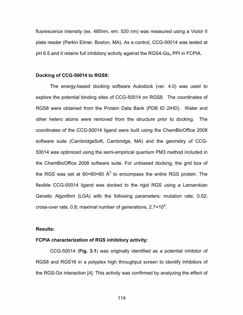
114
fluorescence intensity (ex. 485nm, em. 520 nm) was measured using a Victor II
plate reader (Perkin Elmer, Boston, MA). As a control, CCG-50014 was tested at
pH 6.5 and it retains full inhibitory activity against the RGS4-Gαo PPI in FCPIA.
Docking of CCG-50014 to RGS8:
The energy-based docking software Autodock (ver. 4.0) was used to
explore the potential binding sites of CCG-50014 on RGS8. The coordinates of
RGS8 were obtained from the Protein Data Bank (PDB ID 2IHD). Water and
other hetero atoms were removed from the structure prior to docking. The
coordinates of the CCG-50014 ligand were built using the ChemBioOffice 2008
software suite (CambridgeSoft, Cambridge, MA) and the geometry of CCG-
50014 was optimized using the semi-empirical quantum PM3 method included in
the ChemBioOffice 2008 software suite. For unbiased docking, the grid box of
the RGS was set at 60×60×60 Å3 to encompass the entire RGS protein. The
flexible CCG-50014 ligand was docked to the rigid RGS using a Lamarckian
Genetic Algorithm (LGA) with the following parameters: mutation rate, 0.02;
cross-over rate, 0.8; maximal number of generations, 2.7×10⁴.
Results:
FCPIA characterization of RGS inhibitory activity:
CCG-50014 (Fig. 3.1) was originally identified as a potential inhibitor of
RGS8 and RGS16 in a polyplex high throughput screen to identify inhibitors of
the RGS-Gα interaction [4]. This activity was confirmed by analyzing the effect of
115
CCG-50014 on several different RGS proteins with freshly reordered compound
using multiplexed FCPIA. CCG-50014 fully inhibited several different RGS
proteins including RGS4, 8, 16, and 19, but did not have activity on RGS7 or a
mutated form of RGS4 that lacks cysteine residues (Fig. 3.2A, Table 3.1). The
30 nM IC50 value observed for the inhibition of RGS4 makes CCG-50014 the
most potent small molecule RGS inhibitor discovered to date.
NN
FOS
O Figure 3.1 The chemical structure of CCG-50014 (4-[(4-fluorophenyl)methyl]-2-(4-methylphenyl)-1,2,4-thiadiazolidine-3,5-dione). RGS IC50 (µM) ± SEM Hill Slope RGS4 wild Type 0.030 ± 0.006 -1.53 RGS4 Cys-null Mutant N/A N/A RGS8 11 ± 2 -0.57 RGS16 3.5 ± 2.4 -1.33 RGS19 0.12 ± 0.02 -0.61 RGS7 N/A N/A Table 3.1: CCG-50014 shows >100 fold specificity for RGS4 over other RGS proteins in the FCPIA assay. Data are presented as: mean IC50 values ± SEM from at least three independent experiments (for RGS4 and RGS8, n >28). N/A: No inhibition below the aqueous solubility limit of the compound.

116
0
25
50
75
100
-10 -9 -8 -7 -6 -5 -4 -3
RGS4RGS8
DMSOLog [CCG-50014] (M)
% B
indi
ngA)
DMSO DMSO 10uM 1uM0.00
0.05
0.10
0.15
*** ***
RGS4
GTP
Hyd
roly
sis
Rate
(s-1
)
DMSO DMSO100uM 10uM 1uM 100nM0.00
0.02
0.04
0.06
0.08
0.10
* *
RGS8
GTP
Hyd
roly
sis
Rate
(s-1
)
0 100 200 300 4000
250500750
100012501500
GaoRGS4
10um 500141um 50014
Time (s)
[32P]
Pi R
elea
se (C
PM)
0 100 200 300 4000
500
1000
1500
2000
2500
GaoRGS8
100uM 5001410uM 50014
Time (s)
[32P]
Pi R
elea
se (C
PM)
B) C)
D) E)
Figure 3.2 CCG-50014 inhibits RGS4 and RGS8 binding and function. A) CCG-50014 dose-dependently inhibits the binding between aluminum fluoride-activated Gαo and RGS4 or RGS8. Data shown are an average of three independent experiments. This experiment has been independently repeated 28 times, producing average IC50 values of 30 nM against RGS4 and 1.1 µM against RGS8. B,C) CCG-50014 also inhibits the GAP activity of RGS4 and D,E) RGS8. Using a single-turnover GAP assay, CCG-50014 dose-dependently inhibits the GAP activity of both RGS4 and RGS8. * P <0.05, *** P <0.0001. All experiments were independently repeated a minimum of three times.
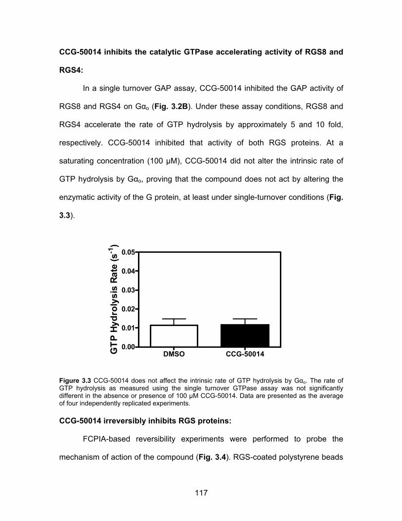
117
CCG-50014 inhibits the catalytic GTPase accelerating activity of RGS8 and
RGS4:
In a single turnover GAP assay, CCG-50014 inhibited the GAP activity of
RGS8 and RGS4 on Gαo (Fig. 3.2B). Under these assay conditions, RGS8 and
RGS4 accelerate the rate of GTP hydrolysis by approximately 5 and 10 fold,
respectively. CCG-50014 inhibited that activity of both RGS proteins. At a
saturating concentration (100 µM), CCG-50014 did not alter the intrinsic rate of
GTP hydrolysis by Gαo, proving that the compound does not act by altering the
enzymatic activity of the G protein, at least under single-turnover conditions (Fig.
3.3).
DMSO CCG-500140.00
0.01
0.02
0.03
0.04
0.05
GTP
Hyd
roly
sis
Rate
(s-1
)
Figure 3.3 CCG-50014 does not affect the intrinsic rate of GTP hydrolysis by Gαo. The rate of GTP hydrolysis as measured using the single turnover GTPase assay was not significantly different in the absence or presence of 100 µM CCG-50014. Data are presented as the average of four independently replicated experiments. CCG-50014 irreversibly inhibits RGS proteins:
FCPIA-based reversibility experiments were performed to probe the
mechanism of action of the compound (Fig. 3.4). RGS-coated polystyrene beads
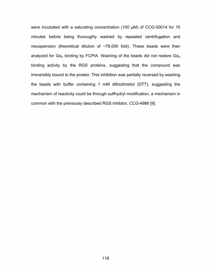
118
were incubated with a saturating concentration (100 µM) of CCG-50014 for 15
minutes before being thoroughly washed by repeated centrifugation and
resuspension (theoretical dilution of ~78,000 fold). These beads were then
analyzed for Gαo binding by FCPIA. Washing of the beads did not restore Gαo
binding activity by the RGS proteins, suggesting that the compound was
irreversibly bound to the protein. This inhibition was partially reversed by washing
the beads with buffer containing 1 mM dithiothreitol (DTT), suggesting the
mechanism of reactivity could be through sulfhydryl modification, a mechanism in
common with the previously described RGS inhibitor, CCG-4986 [9].
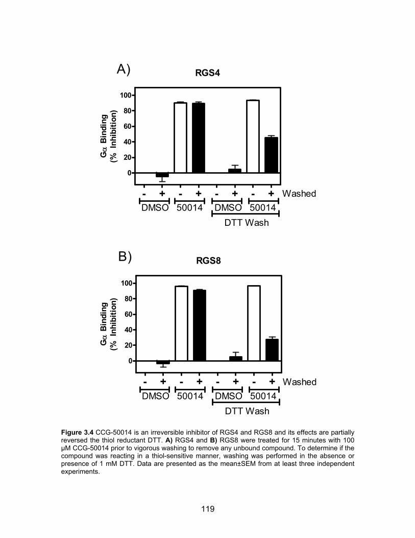
119
RGS4
- + - + - + - +
0
20
40
60
80
100
DMSO 50014 DMSO 50014DTT Wash
Washed
A)
Gα
Bin
ding
(% I
nhib
ition
)
RGS8
- + - + - + - +
0
20
40
60
80
100
DMSO 50014 DMSO 50014DTT Wash
Washed
B)
Gα
Bin
ding
(% I
nhib
ition
)
Figure 3.4 CCG-50014 is an irreversible inhibitor of RGS4 and RGS8 and its effects are partially reversed the thiol reductant DTT. A) RGS4 and B) RGS8 were treated for 15 minutes with 100 µM CCG-50014 prior to vigorous washing to remove any unbound compound. To determine if the compound was reacting in a thiol-sensitive manner, washing was performed in the absence or presence of 1 mM DTT. Data are presented as the mean±SEM from at least three independent experiments.
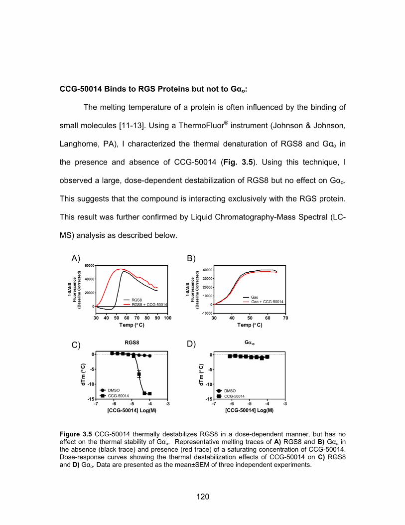
120
CCG-50014 Binds to RGS Proteins but not to Gαo:
The melting temperature of a protein is often influenced by the binding of
small molecules [11-13]. Using a ThermoFluor® instrument (Johnson & Johnson,
Langhorne, PA), I characterized the thermal denaturation of RGS8 and Gαo in
the presence and absence of CCG-50014 (Fig. 3.5). Using this technique, I
observed a large, dose-dependent destabilization of RGS8 but no effect on Gαo.
This suggests that the compound is interacting exclusively with the RGS protein.
This result was further confirmed by Liquid Chromatography-Mass Spectral (LC-
MS) analysis as described below.
RGS8
-7 -6 -5 -4 -3-15
-10
-5
0
DMSOCCG-50014
[CCG-50014] Log(M)
dTm
( °C
)
30 40 50 60 70 80 90 100
0
20000
40000
60000
RGS8RGS8 + CCG-50014
Temp (°C)
1-8A
NS
Fluo
resc
ence
(Bas
elin
e C
orre
cted
)
30 40 50 60 70-10000
0
10000
20000
30000
40000
GaoGao + CCG-50014
Temp (°C)
1-8A
NS
Fluo
resc
ence
(Bas
elin
e C
orre
cted
)
Gαo
-7 -6 -5 -4 -3-15
-10
-5
0
DMSOCCG-50014
[CCG-50014] Log(M)
dTm
( °C
)
A) B)
C) D)
Figure 3.5 CCG-50014 thermally destabilizes RGS8 in a dose-dependent manner, but has no effect on the thermal stability of Gαo. Representative melting traces of A) RGS8 and B) Gαo in the absence (black trace) and presence (red trace) of a saturating concentration of CCG-50014. Dose-response curves showing the thermal destabilization effects of CCG-50014 on C) RGS8 and D) Gαo. Data are presented as the mean±SEM of three independent experiments.

121
CCG-50014 depends on cysteine residues to inhibit the AlF4-Gαo/RGS
interaction:
To identify the potential cysteine targets of CCG-50014, we studied the
compound’s effects on RGS8. This protein only contains two cysteines in the
RGS homology domain, making it a simpler model system to study than other
RGS proteins. Each cysteine from the RGS8 RGS homology (RH) domain was
individually mutated to serine and the activity of the compound was analyzed via
FCPIA (Fig. 3.6). These mutants have been named according to the cysteine
residue that they maintain (e.g. 107C contains Cys 107 and a serine at position
160). Neither cysteine was fully necessary for function of the compound, but
mutating both cysteines reduced the potency of CCG-50014 by >100 fold. An
interesting trend was noticed whereby the Hill coefficients for the inhibition of
each individual mutant was significantly shallower than that of the wild-type
protein, possibly suggesting some form of cooperativity between the two cysteine
residues. However, this interpretation is dependent upon the assumptions of the
Hill equation, including that the binding has reached equilibrium. In the case of an
irreversible inhibitor, clearly this is not the case. Therefore, these data could also
be explained by differences in the rate of compound reaction with these two
mutants. The insensitivity of the RGS8 cysteine null mutant corresponds well with
the insensitivity of the RGS4 cysteine null mutant (Table 3.1), suggesting a
similar mechanism of action across the two proteins.
122
0
25
50
75
100
-9 -8 -7 -6 -5 -4
WT107C160CRGS8c
DMSO
[CCG-50014] Log(M)
% B
indi
ng
Figure 3.6 CCG-50014 requires at least one cysteine residue on RGS8 for full activity. Mutating both cysteines to serine (RGS8c) produced a protein that was completely insensitive to the effect of CCG-50014. The presence of either Cys 107 (107C) or Cys 160 (160C) provided sensitivity to CCG-50014. The inhibition parameters (IC50 (µM), Hill Coefficient) for CCG-50014 on these proteins were as follows: wildtype RGS8 (wt): 6.1 µM, -0.79; 107C: 46.5 µM, -0.54; 160C: 0.71 µM, -0.36; Cysteine-null RGS8 (RGS8c): >100 µM. Data are presented as the mean±SEM of three independent experiments. CCG-50014 is a covalent sulfhydryl modifier of RGS8:
The data thus far suggest that CCG-50014 covalently modifies RGS
proteins. To test this hypothesis, we performed high performance liquid
chromatography–mass spectral analysis on RGS8 samples treated with CCG-
50014 (Fig. 3.7). After compound treatment, there was a peak shift in RGS8
corresponding to a full mass adduct of CCG-50014. At high concentrations of
CCG-50014 (100 µM) with wild-type RGS8, a second minor peak corresponding
to two full adducts was also observed, suggesting that CCG-50014 at this
concentration can react with both cysteine residues in the protein. To confirm that
this action was via cysteine reactivity, the mutant RGS8 where the two cysteines
in the RH domain were mutated to serine was also analyzed and no adduct was

123
observed (Fig 3.7B). RGS8 individual cysteine mutants (107C, 160C) were also
tested for covalent adduct formation in MS. A single adduct was observed on
107C, while no adduct was observed on 160C under the conditions tested. The
lack of an observable adduct on 160C was of particular interest since this mutant
was also irreversibly modified by CCG-50014. Indeed, it was inhibited more
potently than the 107C mutant. To further probe this observation, WT RGS8 and
the two cysteine mutant proteins were treated with a saturating concentration
(100 µM) of CCG-50014 before removal of the compound via gel filtration
chromatography (Fig. 3.8). CCG-50014-treated wild-type RGS8 showed a minor
mobility shift compared to vehicle-treated WT RGS8 and showed a 14-fold
decrease in its ability to compete for Gαo binding to RGS8 beads. The CCG-
50014- and vehicle-treated 107C mutant protein migrated through the column in
an identical manner and no discernable difference in Gαo binding was observed.
The 160C mutant, however, formed aggregates upon treatment with a saturating
concentration of CCG-50014 and no monomeric, soluble protein was recovered
from the experiment. This suggests that labeling at Cys 160 causes a dramatic
decrease in protein stability. It is likely that this aggregation accounts for the lack
of an observable adduct in the mass spectral experiments, as these aggregates
would not migrate on the HPLC as a standard protein peak for MS analysis.
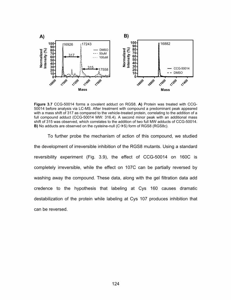
124
1680
017
000
1720
017
400
1760
00
102030405060708090
100DMSO
100uM50uM
16926 17243
17558
317
315
Mass
Norm
aliz
edIn
tens
ity (%
)
1660
016
800
1700
017
200
1740
00
102030405060708090
100
CCG-50014DMSO
Mass
Norm
aliz
edIn
tens
ity (%
)
16882
A) B)
Figure 3.7 CCG-50014 forms a covalent adduct on RGS8. A) Protein was treated with CCG-50014 before analysis via LC-MS. After treatment with compound a predominant peak appeared with a mass shift of 317 as compared to the vehicle-treated protein, correlating to the addition of a full compound adduct (CCG-50014 MW: 316.4). A second minor peak with an additional mass shift of 315 was observed, which correlates to the addition of two full MW adducts of CCG-50014. B) No adducts are observed on the cysteine-null (C S) form of RGS8 (RGS8c).
To further probe the mechanism of action of this compound, we studied
the development of irreversible inhibition of the RGS8 mutants. Using a standard
reversibility experiment (Fig. 3.9), the effect of CCG-50014 on 160C is
completely irreversible, while the effect on 107C can be partially reversed by
washing away the compound. These data, along with the gel filtration data add
credence to the hypothesis that labeling at Cys 160 causes dramatic
destabilization of the protein while labeling at Cys 107 produces inhibition that
can be reversed.
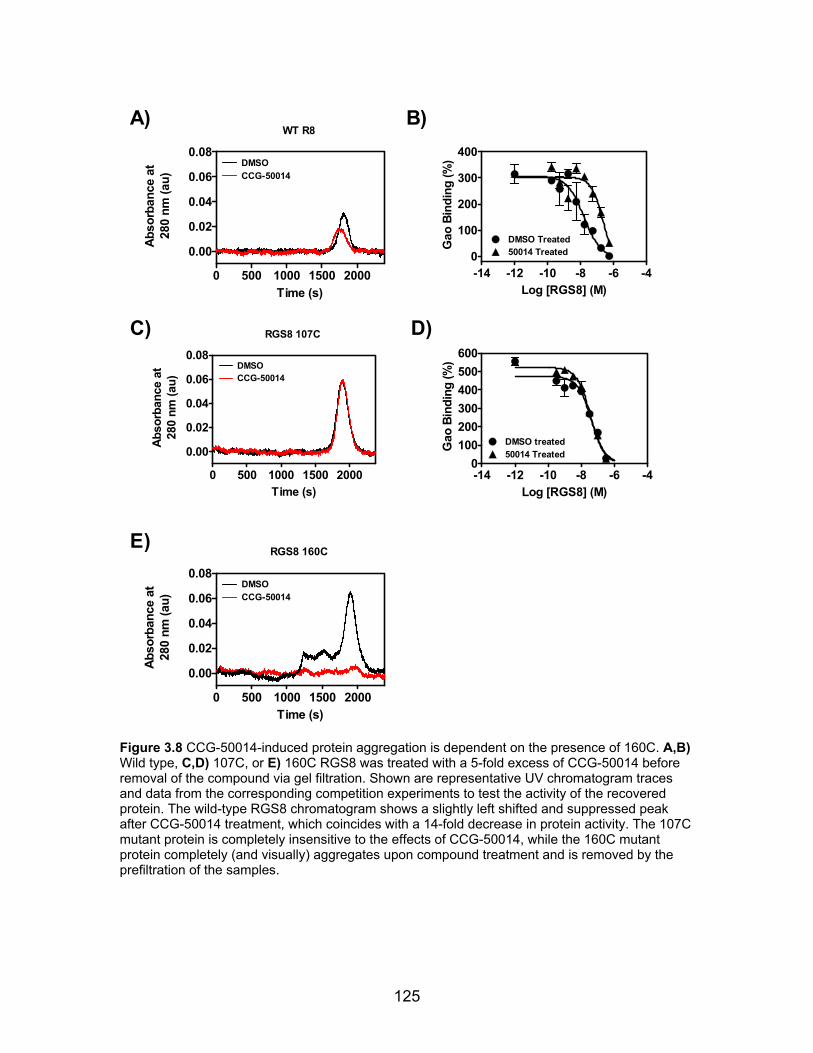
125
WT R8
0 500 1000 1500 2000
0.00
0.02
0.04
0.06
0.08DMSOCCG-50014
Time (s)
Abso
rban
ce a
t28
0 nm
(au)
-14 -12 -10 -8 -6 -40
100
200
300
400
DMSO Treated50014 Treated
Log [RGS8] (M)
Gao
Bin
ding
(%)
RGS8 107C
0 500 1000 1500 2000
0.00
0.02
0.04
0.06
0.08DMSOCCG-50014
Time (s)
Abso
rban
ce a
t28
0 nm
(au)
-14 -12 -10 -8 -6 -40
100200300400500600
DMSO treated50014 Treated
Log [RGS8] (M)
Gao
Bin
ding
(%)
RGS8 160C
0 500 1000 1500 2000
0.00
0.02
0.04
0.06
0.08DMSOCCG-50014
Time (s)
Abso
rban
ce a
t28
0 nm
(au)
A) B)
C) D)
E)
Figure 3.8 CCG-50014-induced protein aggregation is dependent on the presence of 160C. A,B) Wild type, C,D) 107C, or E) 160C RGS8 was treated with a 5-fold excess of CCG-50014 before removal of the compound via gel filtration. Shown are representative UV chromatogram traces and data from the corresponding competition experiments to test the activity of the recovered protein. The wild-type RGS8 chromatogram shows a slightly left shifted and suppressed peak after CCG-50014 treatment, which coincides with a 14-fold decrease in protein activity. The 107C mutant protein is completely insensitive to the effects of CCG-50014, while the 160C mutant protein completely (and visually) aggregates upon compound treatment and is removed by the prefiltration of the samples.
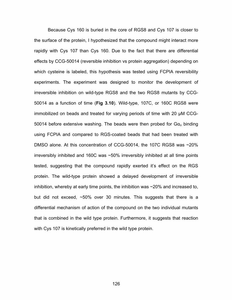
126
Because Cys 160 is buried in the core of RGS8 and Cys 107 is closer to
the surface of the protein, I hypothesized that the compound might interact more
rapidly with Cys 107 than Cys 160. Due to the fact that there are differential
effects by CCG-50014 (reversible inhibition vs protein aggregation) depending on
which cysteine is labeled, this hypothesis was tested using FCPIA reversibility
experiments. The experiment was designed to monitor the development of
irreversible inhibition on wild-type RGS8 and the two RGS8 mutants by CCG-
50014 as a function of time (Fig 3.10). Wild-type, 107C, or 160C RGS8 were
immobilized on beads and treated for varying periods of time with 20 µM CCG-
50014 before extensive washing. The beads were then probed for Gαo binding
using FCPIA and compared to RGS-coated beads that had been treated with
DMSO alone. At this concentration of CCG-50014, the 107C RGS8 was ~20%
irreversibly inhibited and 160C was ~50% irreversibly inhibited at all time points
tested, suggesting that the compound rapidly exerted it’s effect on the RGS
protein. The wild-type protein showed a delayed development of irreversible
inhibition, whereby at early time points, the inhibition was ~20% and increased to,
but did not exceed, ~50% over 30 minutes. This suggests that there is a
differential mechanism of action of the compound on the two individual mutants
that is combined in the wild type protein. Furthermore, it suggests that reaction
with Cys 107 is kinetically preferred in the wild type protein.
127
RGS8 160C
0
20
40
60
80
DMSO CCG-50014Washed: -+ - +
% In
hibi
tion
of Gα
Bin
ding
RGS8 107C
0
20
40
60
80
DMSO CCG-50014Washed: - + - +
***
% In
hibi
tion
of Gα
Bin
ding
Figure 3.9 Irreversible inhibition of RGS8 is predominantly mediated by Cys 160. Mutant proteins were exposed to 20 µM CCG-50014 and reversibility experiments were performed as in Fig. 3.4. Data are presented as the mean±SEM from three independent experiments. ***P<0.0001 using an unpaired t test.
A potential confounding factor in this analysis is that there is a significant
difference in basal melting temperatures between 107C RGS8 (Tm 53.2±0.2)
and 160C RGS8 (Tm 42.5±0.1), whereby 160C is dramatically less stable
overall. It is possible that labeling of the 160C mutant may have a more
exacerbated sensitivity to CCG-50014 than that of the 107C mutant, solely due to

128
the intrinsic instability of this protein. One could therefore imagine that labeling of
the 160C mutant with smaller, less disruptive, analogs CCG-50014 may not
always force protein unfolding in the wild-type protein.
0 5 10 15 20 25 30 350
20
40
60
80
100 WT160C107C
CCG-50014 Exposure (min)
% In
hibi
tion
reta
ined
Figure 3.10 Development of irreversible inhibition after exposure to CCG-50014 differs between the individual cysteine mutants and provides a means to understand the compound’s mechanism of action. Wild-type, 160C or 107C RGS8 was treated with 20 µM CCG-50014 for the desired amount of time before compound removal by extensive washing. The developed amount of irreversible inhibition was quantified by comparing the G-protein binding of CCG-50014 treated beads to DMSO treated beads. Data are presented as the mean±SEM from three independent experiments. CCG-50014 is not a general cysteine alkylator:
Cysteine reactive compounds might be expected to have more off-target
effects than non-reactive compounds. To determine if this compound could bind
to and inhibit any reactive cysteine, we tested the ability of CCG-50014 and a
known general cysteine alkylator (iodoacetamide) to inhibit a standard cysteine
protease (Fig. 3.11). Iodoacetamide inhibited the proteolytic activity of papain in
a dose-dependent manner. However, even at high concentrations (100 µM),
CCG-50014 had no effect on papain. This suggests that there is at least a basal
level of selectivity of this class of compounds for cysteines in the RGS over other
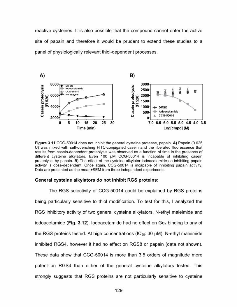
129
reactive cysteines. It is also possible that the compound cannot enter the active
site of papain and therefore it would be prudent to extend these studies to a
panel of physiologically relevant thiol-dependent processes.
0 5 10 15 20 25 302000
4000
6000
8000 DMSOIodoacetamideCCG-50014No enzyme
Time (min)
Cas
ein
prot
eoly
sis
(FI 5
20)
-7.0 -6.5 -6.0 -5.5 -5.0 -4.5 -4.0 -3.50
50010001500200025003000
DMSOIodoacetamideCCG-50014
Log[cmpd] (M)C
asei
n pr
oteo
lysi
s(F
I 520
)
A) B)
Figure 3.11 CCG-50014 does not inhibit the general cysteine protease, papain. A) Papain (0.625 U) was mixed with self-quenching FITC-conjugated casein and the liberated fluorescence that results from casein-dependent proteolysis was observed as a function of time in the presence of different cysteine alkylators. Even 100 µM CCG-50014 is incapable of inhibiting casein proteolysis by papain. B) The effect of the cysteine alkylator iodoacetamide on inhibiting papain activity is dose-dependent. Once again, CCG-50014 is incapable of inhibiting papain activity. Data are presented as the mean±SEM from three independent experiments. General cysteine alkylators do not inhibit RGS proteins:
The RGS selectivity of CCG-50014 could be explained by RGS proteins
being particularly sensitive to thiol modification. To test for this, I analyzed the
RGS inhibitory activity of two general cysteine alkylators, N-ethyl maleimide and
iodoacetamide (Fig. 3.12). Iodoacetamide had no effect on Gαo binding to any of
the RGS proteins tested. At high concentrations (IC50: 30 µM), N-ethyl maleimide
inhibited RGS4, however it had no effect on RGS8 or papain (data not shown).
These data show that CCG-50014 is more than 3.5 orders of magnitude more
potent on RGS4 than either of the general cysteine alkylators tested. This
strongly suggests that RGS proteins are not particularly sensitive to cysteine

130
modification and the effect observed by CCG-50014 is more than just random
thiol alkylation.
0
25
50
75
100
125
-7 -6 -5 -4
RGS4RGS8
DMSO[NEM] Log(M)
Gα
o Bi
ndin
g (%
)
025
50
75
100
125
-7 -6 -5 -4
RGS8RGS4
DMSO [IA] Log(M)
Gao
Bin
ding
(%)
A) B)
Figure 3.12 CCG-50014 is a much more potent RGS inhibitor than two general cysteine alkylators N-ethyl maleimide (NEM) and iodoacetamide (IA). Dose response curves for A) NEM and B) IA. The only protein that displayed any sensitivity to the alkylators tested was RGS4, which was inhibited by NEM with an IC50 value >3.5 Log higher than that of CCG-50014. Data are presented as the mean±SEM from three independent experiments. Computational modeling of the CCG-50014-RGS8 interaction:
To identify potential binding sites for CCG-50014 on RGS8, I in
collaboration with Haoming Zhang performed an unbiased molecular docking
simulation. CCG-50014 docked preferentially to a site on the RGS that is located
near the region of the surface of RGS8 that corresponds to the “B”-site of RGS4
(Fig. 3.13). The compound docked at this site with a calculated free energy of
-6.4 kCal/mol, which translates to an estimated Ki of 18 µM. This affinity is
approximately in line with experimentally derived Ki value of 0.3µM (Table 1).
This binding site places the compound a considerable distance from the two
cysteine residues known to play a role in the compound’s inhibitory activity (Fig.
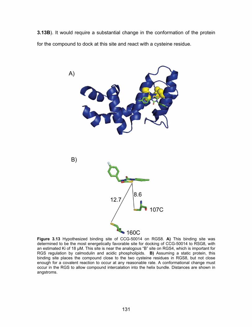
131
3.13B). It would require a substantial change in the conformation of the protein
for the compound to dock at this site and react with a cysteine residue.
Figure 3.13 Hypothesized binding site of CCG-50014 on RGS8. A) This binding site was determined to be the most energetically favorable site for docking of CCG-50014 to RSG8, with an estimated Ki of 18 µM. This site is near the analogous “B” site on RGS4, which is important for RGS regulation by calmodulin and acidic phospholipids. B) Assuming a static protein, this binding site places the compound close to the two cysteine residues in RGS8, but not close enough for a covalent reaction to occur at any reasonable rate. A conformational change must occur in the RGS to allow compound intercalation into the helix bundle. Distances are shown in angstroms.
132
Limiting the reactivity of CCG-50014 diminishes potency:
To probe the chemical space around CCG-50014, Dr. Stephen Husbands,
Dr. Benjamin Greedy and Dr. Emma Casey synthesized a series of 76 analogs of
the lead compound and we analyzed them in an attempt to identify compounds
with optimized physicochemical and pharmacological properties (see Chapter
IV). While the detailed activity of these compounds is described in the next
chapter, there are a few interesting findings that contribute to the understanding
of the mechanism of action of this compound class. The first and most prominent
trend is that the center heterocycle (the thiadiazolidine dione) is absolutely
required for function. This is not particularly surprising, because it is likely to be
the site of cysteine thiol reactivity. I hypothesized that the extremely potent IC50
value on RGS4 meant that the compound interacted with the protein in a way
that was governed by more than simple covalent reactivity. To determine if there
was significant non-covalent affinity of this compound, non-reactive, or less-
reactive analogs of CCG-50014 were synthesized and tested for activity (Table
3.2). These compounds showed limited, if any, activity in the FCPIA assay,
suggesting that the main mechanism of action of CCG-50014 is through covalent
reactivity with one or more cysteine residues on the RGS. However, since this
compound is dramatically more potent than two other general cysteine alkylators,
it is likely that there is a non-covalent docking mechanism at play that drives the
affinity of CCG-50014 and analogs for the RGS.

133
Compound ID Structure Putative Reactivity
RGS4 IC50 (µM)
RGS8 IC50 (µM)
CCG-50014 N
SN
O
O
F
Reactive 0.030 1.1
CCG-203778 N N
O
O
Non-reactive >100 >100
CCG-203779 N
O
O
Less reactive 93 >100
CCG-203780 N
BrO
O
Less Reactive 32 >100
Table 3.2. CCG-50014 analogs with limited reactivity. Data are presented as the mean of two independent FCPIA experiments.
134
Discussion:
Molecules disrupting the RGS/Gα interaction are likely to have significant
physiological effects by increasing the magnitude and/or duration of Gα signaling
responses. Inhibition of RGS protein activity genetically produces dramatic
physiological phenotypes (see Chapter I), suggesting that a small molecule RGS
inhibitor might provide similar actions in vivo.
CCG-50014 is the most potent small molecule RGS inhibitor identified to
date. It inhibits the in vitro interaction between RGS4 and Gαo with a low
nanomolar IC50 value. It is nearly 3 orders of magnitude selective for RGS4 over
two closely related RGS proteins, RGS8 and RGS16 (Fig. 3.2, Table 3.1). I also
show that CCG-50014 is a covalent modifier of cysteine residues (Fig. 3.4, 3.6,
3.7), raising concerns about the therapeutic potential of this class of compounds.
Even so, studying this compound has provided significant insight into the
mechanism of allosteric RGS inhibition and as shown in Chapter IV, it is active in
a series of cellular assays, suggesting that we may be close to the physiological
modulation of RGS activity by small molecules.
CCG-50014 does not inhibit a mutant RGS4 where all cysteine residues in
the RH domain were mutated to alanine (Table 3.1 and [4]). Furthermore, the
compound was inactive on RGS7, an RGS protein that naturally has no cysteine
residues in its RH domain (Table 3.1). These two pieces of information
suggested that the mechanism of action of CCG-50014 requires at least one
cysteine residue – a hallmark of a sulfhydryl-reactive irreversible inhibitor. This
135
hypothesis was confirmed by the FCPIA reversibility experiments (Fig. 3.4) and
subsequent mass spectral analysis of CCG-50014 treated RGS8 (Fig. 3.7).
Based on the chemical structure of the compound and the full molecular weight
adduct observed in the LC-MS experiments, it is likely that the mechanism of
reaction of CCG-50014 with a cysteine residue on an RGS protein is by
nucleophilic attack of the cysteine thiol onto the sulfur atom of the central
heterocycle causing a ring opening event. Consistent with the DTT-induced
reversibility of CCG-50014 inhibition (Fig. 3.4), this newly formed disulfide is
likely to be sensitive to reductants.
Interestingly, CCG-50014 interacts with cysteine residues in RGS8 that
are not near the Gα interaction interface (Fig 3.13), suggesting an allosteric
mechanism of action. Unbiased computational modeling predicts that CCG-
50014 could non-covalently bind to a site on RGS8 that is near to the acidic
phospholipid binding site on RGS4. Binding in this site would place the reactive
group of CCG-50014 within 8-13 Å of the two cysteines in the RGS8 RH domain.
While at this distance it is unlikely that a covalent bond could be formed, I
propose that the compound may initially bind to this pocket and a subsequent
conformational change in the protein provides access to the cysteine thiol. This
conformational change is likely to be the fundamental mechanism by which the
allosteric modulation of G protein binding activity is conferred.
The differential sensitivities of the cysteine mutants to CCG-50014 are
also explained by this binding modality. The decreased activity and increased
reversibility of CCG-50014 on 107C RGS8 (Fig 3.6, 3.8, 3.9) is in accord with the

136
fact that Cys 107 is more solvent accessible and is closer to the hypothesized
binding site of the compound. Compound reacting with Cys 160 causes drastic
protein unfolding (Fig. 3.9), which also fits with this model.
The data accumulated in this chapter allow for the development of a
reasonable mechanism of action for RGS inhibition by CCG-50014. I propose
that the compound originally binds to a surface equivalent to the “B” site on
RGS4 in a manner that does not produce an inhibitory effect. This interaction
may provide enough interaction energy to keep the compound in close proximity
to the protein long enough for the RGS to enter a cysteine-exposed
conformation. Assuming that the compound binding site is as modeled (Fig
3.13), the first cysteine to become exposed to the compound is likely to be Cys
107. Upon reacting with this cysteine, CCG-50014 can trap the RGS in a
conformation that is incapable of binding to Gα. Reversal of this reaction is
possible, leading to reactivation of the RGS. If the compound interacts with the
more deeply buried cysteine, Cys 160, it causes a dramatic disruption of the
hydrophobic core of the protein, leading to protein denaturation. This is shown
primarily by the gel filtration data (Fig. 3.8). My data also suggest that Cys 107 is
labeled more rapidly than Cys 160 in the wild type protein (Fig. 3.10). These data
are consistent with the hypothesis that the compound initially interacts with Cys
107 to form a weak, DTT- or time/dilution-reversible inhibition of RGS activity.
Then, either the Cys 107-bound compound transfers to Cys 160 or a second
CCG-50014 molecule binds to Cys 160 to produce the completely irreversible
reaction observed the gel filtration experiments (Fig. 3.8). The mechanism
137
behind the irreversible inhibition after labeling of Cys 160 is likely due to a
massive destabilization of the hydrophobic core of the RH domain that would
occur by the intercalation of CCG-50014.
Selectivity is a significant issue when studying reactive compounds. I
undertook a series of experiments to determine if CCG-50014 is a just a general,
non-specific cysteine alkylator, or if it has some intrinsic selectivity for RGS
proteins. While a comprehensive analysis of CCG-50014 effects upon all
cysteine-dependent processes in a cell is clearly intractable, I showed that CCG-
50014 does not inhibit the activity of the cysteine protease papain at
concentrations over 3000 times higher than that required for RGS inhibition (Fig.
3.11). In contrast, the cysteine alkylator iodoacetamide dose-dependently
inhibited the activity of this protease but had no effect on RGS4. Furthermore, the
cellular activity observed (see Chapter IV), also suggests that these compounds
do not dramatically affect a large number of cellular processes.
In this chapter I have characterized the mechanism of action of the most
potent RGS inhibitor identified to date. This compound irreversibly inhibits RGS4
with nanomolar potency and the mechanism of this inhibition is predominantly
through reacting with cysteine residues at an allosteric site on the RGS. As will
be shown in the next chapter, this compound and related analogs are also the
first examples of RGS inhibitors that are active in living cells.
138
References:
1. Brunton, L.L., J.S. Lazo, and K.L. Parker, eds. Goodman & Gilman's: The Pharmacological Basis of Therapeutics. 11 ed. 2006, McGraw-Hill Companies, Inc.: New York.
2. Sachs, G., et al., Gastric acid secretion: activation and inhibition. Yale J Biol Med, 1994. 67(3-4): p. 81-95.
3. Ocana, A. and E. Amir, Irreversible pan-ErbB tyrosine kinase inhibitors and breast cancer: current status and future directions. Cancer Treat Rev, 2009. 35(8): p. 685-91.
4. Roman, D.L., S. Ota, and R.R. Neubig, Polyplexed Flow Cytometry Protein Interaction Assay: A Novel High-Throughput Screening Paradigm for RGS Protein Inhibitors. J Biomol Screen, 2009.
5. Roman, D., et al., Allosteric Inhibition of the RGS-G{alpha} Protein-Protein Interaction by CCG-4986. Mol Pharmacol.
6. Sternweis, P.C. and J.D. Robishaw, Isolation of two proteins with high affinity for guanine nucleotides from membranes of bovine brain. J Biol Chem, 1984. 259(22): p. 13806-13.
7. Blazer, L.L., et al., Use of flow cytometric methods to quantify protein-protein interactions. Curr Protoc Cytom. Chapter 13: p. Unit 13 11 1-15.
8. Blazer, L.L., et al., Reversible, allosteric, small-molecule inhibitors of RGS proteins. Mol Pharmacol.
9. Roman, D.L., et al., Identification of small-molecule inhibitors of RGS4 using a high-throughput flow cytometry protein interaction assay. Mol Pharmacol, 2007. 71(1): p. 169-75.
10. Roof, R.A., et al., Mechanism of action and structural requirements of constrained peptide inhibitors of RGS proteins. Chem Biol Drug Des, 2006. 67(4): p. 266-74.
11. Lo, M.C., et al., Evaluation of fluorescence-based thermal shift assays for hit identification in drug discovery. Anal Biochem, 2004. 332(1): p. 153-9.
12. Niesen, F.H., H. Berglund, and M. Vedadi, The use of differential scanning fluorimetry to detect ligand interactions that promote protein stability. Nat Protoc, 2007. 2(9): p. 2212-21.
13. Pantoliano, M.W., et al., High-density miniaturized thermal shift assays as a general strategy for drug discovery. J Biomol Screen, 2001. 6(6): p. 429-40.
139
Chapter IV: Cellular and Structure-Activity Studies
of the CCG-50014 Compound Class Introduction:
The work presented thus far focuses solely upon the biochemical
mechanism of action of either novel or previously identified small molecule
inhibitors of the RGS-Gαo PPI. While this knowledge is academically useful,
pharmacologically important chemical entities need to have activity in a
physiological (preferably clinical) setting. Prior to the work described in this
chapter, there were no examples of RGS inhibitors that functioned in a living,
whole-cell system. I present here work performed by myself and collaborators
that shows for the first time a class of irreversible small molecule inhibitors that
produce substantial RGS-inhibitory effects in several cell-based systems.
There are a number of issues involved with the use of covalent,
irreversible inhibitors for research or clinical applications. A major problem is
target specificity. If the compound is too reactive, it will bind many different
proteins and will likely cause a barrage of undesired side effects However, there
are a number of clinically useful irreversible inhibitors (e.g. lactams,
cyclophosphamide), some of which function by reacting with thiols (e.g.
omeprazole) [1]. A final issue that pertains primarily with the clinical utility of
irreversible inhibitors is that their effects are often difficult to quickly reverse.
Medicine, like all human endeavors, is prone to mistake. Having a mechanism to
140
compensate for or to correct dosing errors can play a critical role in patient
outcome. The challenging nature of reversing the physiological effects of
irreversible inhibitors is likely to diminish their clinical utility.
Irreversible inhibition can however be a benefit in the research setting.
With this, I present the following data showing the effects of CCG-50014 and
selected analogs in a series of cellular assays. It is shown here that CCG-50014
and related analogs are able to inhibit the RGS4/Gαo PPI in living cells and to
potentiate signaling through the δ opioid receptor and the M3 muscarinic
receptor. The success of this work required a substantial amount of structure-
activity data, both at the biochemical and cellular levels, to minimize off target
effects and to improve aqueous solubility. These compounds and the information
presented in this chapter should accelerate the development of small molecule
modulators of RGS function by providing a fundamental groundwork from which
to design future analogs and to judge future novel scaffolds.
The work in this chapter was performed by me with the following
exceptions: 1) All compound synthesis was performed by Dr. Benjamin Greedy
and/or Dr. Emma Casey in the laboratory of Dr. Stephen Husbands; 2) cAMP
experiments with SH-SY5Y cells were performed by Dr. Qin Wang in the
laboratory of Dr. John Traynor.
Materials and Methods:
Reagents and Compounds:
Reagents were purchased from Sigma-Aldrich (St. Louis, MO) or Fisher
Scientific (Hampton, NH) and were reagent grade or better. Avidin-coated
141
microspheres were purchased from Luminex (Austin, TX). CCG-50014 (4-[(4-
fluorophenyl)methyl]-2-(4-methylphenyl)-1,2,4-thiadiazolidine-3,5-dione) and
analogs were purchased from Thermo-Fisher (Waltham, MA) from the Maybridge
compound collection or were provided from complete synthesis in the laboratory
of Dr. Stephen Husbands.
Protein expression, purification, and labeling:
All RGS and G proteins were prepared as previously described [2]. G
protein activity was determined by [35S]GTPγS binding [3]. In all cases, proteins
were purified to >90% homogeneity before use. RGS proteins were biotinylated
and Gαo was labeled with AlexaFluor-532 as previously described [4].
FCPIA Dose Response experiments:
FCPIA was performed as previously described using chemically
biotinylated RGS proteins and AlexaFluor-532 labeled Gαo. ([5, 6])
Single Turnover GTPase Measurements:
Compounds were tested for the ability to inhibit the RGS4 and RGS8-
stimulated increase in GTP hydrolysis by Gαo as previously described [7, 8].
Solubility experiments:
Compounds were diluted to 100 mM in DMSO and the further diluted to
500 µM in H2O, vortexed and centrifuged for 10 minutes at 13,000 x g at ambient

142
temperature. Solubility was quantified by visual inspection of pellet formation on
a scale from 0 – 5, with 0 being no pellet and 5 being a pellet of the same size as
that of CCG-50014. Since the key metric was improved solubility, in the rare
instance that a compound was deemed to be more insoluble than CCG-50014, it
was given a value of 5.
WST-1 Cell Viability Studies:
HEK-293 cells were plated to a density of 30,000/well in 96-well plates
and grown for 48 hours in Dulbecco’s Modified Eagle Medium (DMEM)
containing 10% fetal bovine serum and Penicillin (100 units/ml)-Streptomycin
(100 µg/ml) under 5% CO2 at 37oC. Compound treatment was performed
overnight in DMEM containing 0.1% bovine serum albumin under 5% CO2 at
37oC. After compound incubation ten microliters of the WST-1 reagent
(Hoffmann-La Roche, Switzerland) was added to every well and the cells were
incubated for 1 hour under 5% CO2 at 37oC. Absorbance was measured at 450
nm using a Victor II plate reader (Perkin-Elmer, Picastaway, NJ).
Cellular Localization Studies:
HEK-293T cells grown to 80-90% confluency in 6-well dishes in DMEM
supplemented with 10% fetal bovine serum and Penicillin (100 units/ml)-
Streptomycin (100 µg/ml) under 5% CO2 at 37 oC. RGS and Gαo expression was
induced by transient co-transfection with either 250 ng of full-length human
RGS4 with an N-terminal GFP tag (RGS4pEGFP-C1) or a C-terminal RGS4-GFP
143
(RGS4pDEST47) and 250 ng of pcDNA3.1 or pcDNA with wildtype human Gαo.
Cells were split onto poly-D-lysine coated glass coverslips and cultured for 24-48
hours after transfection before live cell imaging. Images were acquired on an
Olympus Fluoview 500 confocal microscope with a 60 x 1.40 numerical aperture
(N.A) oil objective. Images were obtained by taking a series of stacks every 0.5
µm through the cell and combining the images into a composite stack. The light
source for the fluorescent studies was a 488 nm laser with a 505-525 nm
bandpass filter. Images were quantified using NIH ImageJ software version
1.43r.
Calcium Mobilization Experiments:
A stable cell line was developed based upon the HEK-293 Flp-In TREx
cell line (Invitrogen, Carlsbad, CA) that stably express the muscarinic M3
receptor and have human RGS4 expression under doxycycline control. Cells
were maintained in DMEM supplemented with 10% fetal bovine serum and
Penicillin (100 units/ml)-Streptomycin (100 µg/ml) under 5% CO2 at 37°C. For
experiments, cells were split into 96-well black, clear bottom, poly-D-lysine
coated microtiter plates (Nunc, Cat. # 152037) at a density of 20,000 cells/well in
DMEM containing 10% fetal bovine serum and Penicillin (100 units/ml)-
Streptomycin (100 µg/ml). RGS4 expression was induced by supplementing the
medium with 1 µg/mL doxycycline for 24-48 hours before experimentation. Cells
were loaded with Fluo-4 No Wash dye in buffer for 30 minutes at 37°C.
Compounds and/or carbachol were added to the wells and the fluorescence
144
intensity was measured using a FlexStation (Molecular Devices, Sunnyvale, CA)
plate reader. Data analysis was performed by calculating the area under the
curve or maximal fluorescence intensity from a 120 second kinetic measurement.
cAMP Accumulation:
SH-SY5Y cells were grown in DMEM containing 10% fetal bovine serum
and Penicillin (100 units/ml)-Streptomycin (100 µg/ml) under 5% CO2 at 37oC.
Cells were plated into 24-well plates to reach ~ 90% confluency on the day of
assay and washed once with fresh serum-free medium, then the medium was
replaced with 1 mM IBMX (3-isobutyl-1-methylxanthine) in serum-free medium
for 15 min at 37°C, and changed to the medium containing 1 mM IBMX, 30 µM
forskolin, and 100 nM of either morphine or SNC80 with or without compound
CCG-50014 for 5 min at 37 °C. Reactions were stopped by replacing the medium
with ice-cold 3% perchloric acid and samples were kept at 4 °C for at least 30
min. An aliquot (0.4 ml) from each sample was removed, neutralized with 0.08 ml
of 2.5 M KHCO3, vortexed, and centrifuged at 15,000 x g for 1 min to pellet the
precipitates. Accumulated cAMP was measured by radioimmunoassay in a 10-15
µl aliquot of the supernatant from each sample following the manufacturer’s
instructions (cAMP radioimmunoassay kit from GE Healthcare, Piscataway, NJ).
Data are from four separate experiments, each carried out in duplicates and
calculated as percent inhibition. The basal cAMP accumulation with forskolin
alone with or without compound CCG-50014 did not differ.

145
Results:
Structure Activity Relationship Studies of the CCG-50014 Family of
Compounds:
Biochemical Optimization: To explore the chemical space around the thiadiazolidine scaffold, we
analyzed a series of 76 analogs of CCG-50014 using a variety of cellular and
biochemical experiments (Table 4.1). As a primary screening methodology, a
RGS4/RGS8 duplex FCPIA assay was performed to obtain biochemical IC50
values for the inhibition of Gαo binding to both proteins. Secondary screening
was performed to assess compound solubility and the ability to induce a calcium
transient in HEK cells, an off-target effect that was originally observed with the
lead compound CCG-50014. The primary goals of the SAR studies were to
optimize compound solubility and potency and to minimize off target effects. A
secondary goal of this study was to determine if it was possible to remove or
minimize the reactivity of this scaffold and still retain RGS inhibitory activity.
Figure 4.14 The chemical structure of CCG-50014, (4-[(4-fluorophenyl)methyl]-2-(4-methylphenyl)-1,2,4-thiadiazolidine-3,5-dione)

146
The majority of the analogs of CCG-50014 that were tested contain the
thiadiazolidinedione core structure with varied substituents at the 2 and 4
positions. Because CCG-50014 is only modestly soluble in aqueous solutions,
we wanted to identify active compounds with improved solubility for use in
cellular and whole-animal studies. Compound solubility was assessed on a 0-5
scale with 5 being as insoluble as the lead compound, CCG-50014 (solubility
~100 µM in aqueous) and 0 being completely soluble at 500 µM in double-
distilled H2O. As expected, it was found that the key determinant of compound
solubility was size, whereby large hydrophobic substituents lead to more
insoluble compounds. While this was not particularly surprising, I did identify a
number of compounds that were much more soluble than CCG-50014 that
retained similar potency for RGS4 inhibition (e.g. CCG-203759, CCG-203769).
To attempt to identify a pharmacophore for the CCG-50014 class of
compounds, I ranked all of the compounds in order of potency on RGS4 (Table
4.1). Unfortunately, no clear trends emerge. I then probed this data for
compounds that possessed increased selectivity for RGS4 over RGS8 (Table
4.2). This analysis shows that small alkyl groups at the R1/R2 positions provide
the greatest RGS4/RGS8 selectivity. However, there are several compounds with
phenyl-containing sidechains that show >1000 fold selectivity (e.g. CCG-
203742). These compounds all contain a -CH3-Ph-p-Me (or in one instance a -
CH3-Ph-p-OMe, CCG-203705) at the R2 position, a moiety that appears to confer
selectivity regardless of group at the R1 position. This suggests that this region of
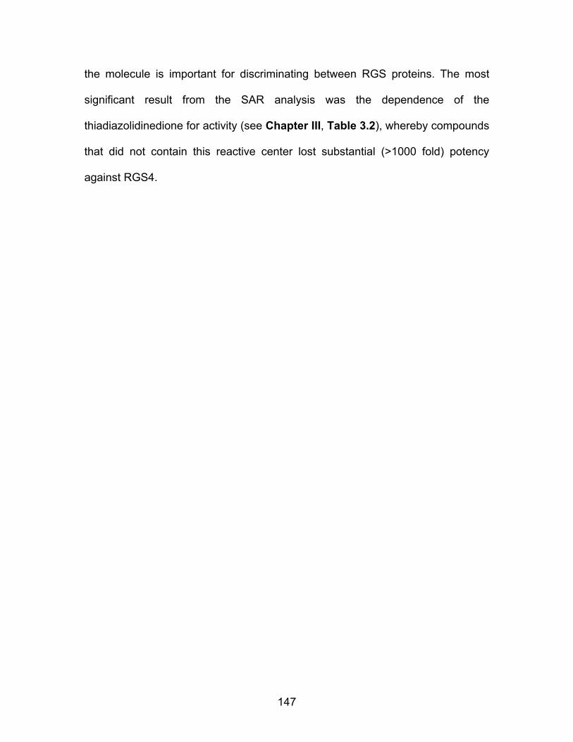
147
the molecule is important for discriminating between RGS proteins. The most
significant result from the SAR analysis was the dependence of the
thiadiazolidinedione for activity (see Chapter III, Table 3.2), whereby compounds
that did not contain this reactive center lost substantial (>1000 fold) potency
against RGS4.

48
R1 N
O
S
N
R2O
CCG # R1 R2 Solubility
Scale (1-5)
Stock Form
Calcium transient (% 50014)
RGS4 IC50 (µM)
RGS4 Hill
Slope RGS8
IC50 (µM) RGS8
Hill Slope
Fold Selectivity
(R8/R4) 203731 CH3-Ph-p-Cl Ph-p-Me 5 Solid -2.6 0.005 -0.70 11.8 -1.50 2360 203734 CH3-Ph CH3-Ph-p-Me 5 Solid 591.3 0.007 -0.84 20.4 -0.66 2914 203732 CH3-Ph-p-Me Ph 3 Solid 12.2 0.009 -1.50 8.3 -0.87 922 203736 CH3-Ph-p-Me Ph-p-Me 4 Solid 4.9 0.009 -1.35 11.6 -1.45 1289 203735 CH3-Ph-p-F Ph-p-OMe 4 Solid 6.2 0.011 -1.33 11.4 -1.27 1036 203741 CH3-Ph-p,m-Cl Ph-p-OMe 3 Solid 202.4 0.013 -0.86 5.9 -0.64 454 203742 CH3-Ph-p-F CH3-Ph-p-Me 5 Solid 108.5 0.013 -1.19 39.8 -0.22 3062 203724 CH3-Ph Ph-p-Me 5 Solid 176.8 0.014 -3.63 7.5 -0.94 536 203727 CH3-Ph-p-F Ph-p-Cl 5 Solid 158.5 0.014 -1.66 7.6 -1.06 543 203761 iBu Ph-p-Me 4 Solid 98.9 0.014 -1.47 7.7 -0.92 550 203769 Bu Et 0 Oil 2.7 0.014 -0.71 83.5 -0.56 5964 203781 CH3-Ph-m-Me Ph 3 Solid 242.8 0.015 -1.65 8.4 -1.17 560 203726 CH3-Ph-p-F Ph 2 Solid 20.9 0.016 -1.42 6.2 -0.82 388 203765 CH3-Ph-p-F Bu 3 Solid 183.8 0.016 -0.81 31.6 -0.59 1975 203733 CH3-Ph-p-Me Ph-p-Cl 5 Solid 20.0 0.017 -1.59 12.8 -1.17 753 203777 CH3-Ph-m-Cl Ph-p-Me 1 Solid 179.0 0.017 -0.96 17.5 -1.00 1029 203759 Me Ph-p-Me 0 Solid 238.0 0.019 -1.56 8.4 -1.06 442 203767 Bu Ph-p-Me 1 Solid 123.0 0.020 -1.46 9.5 -0.96 475 203757 Bu Me 0 Oil 138.3 0.023 -1.01 28.4 -1.10 1235 203722 CH3-Ph Ph 1 Solid 3.8 0.024 -1.89 5.6 -0.80 233 Table 4.8 Structure Activity Relationships of CCG-50014 family. All FCPIA IC50 and calcium transient values presented are an average from two independent experiments. The calcium transient data are presented as the effect observed by 10 µM compound expressesed as a percentange of the effect observed by 10 µM CCG-50014. All compounds were synthesized in the laboratory of Dr. Stephen Husbands
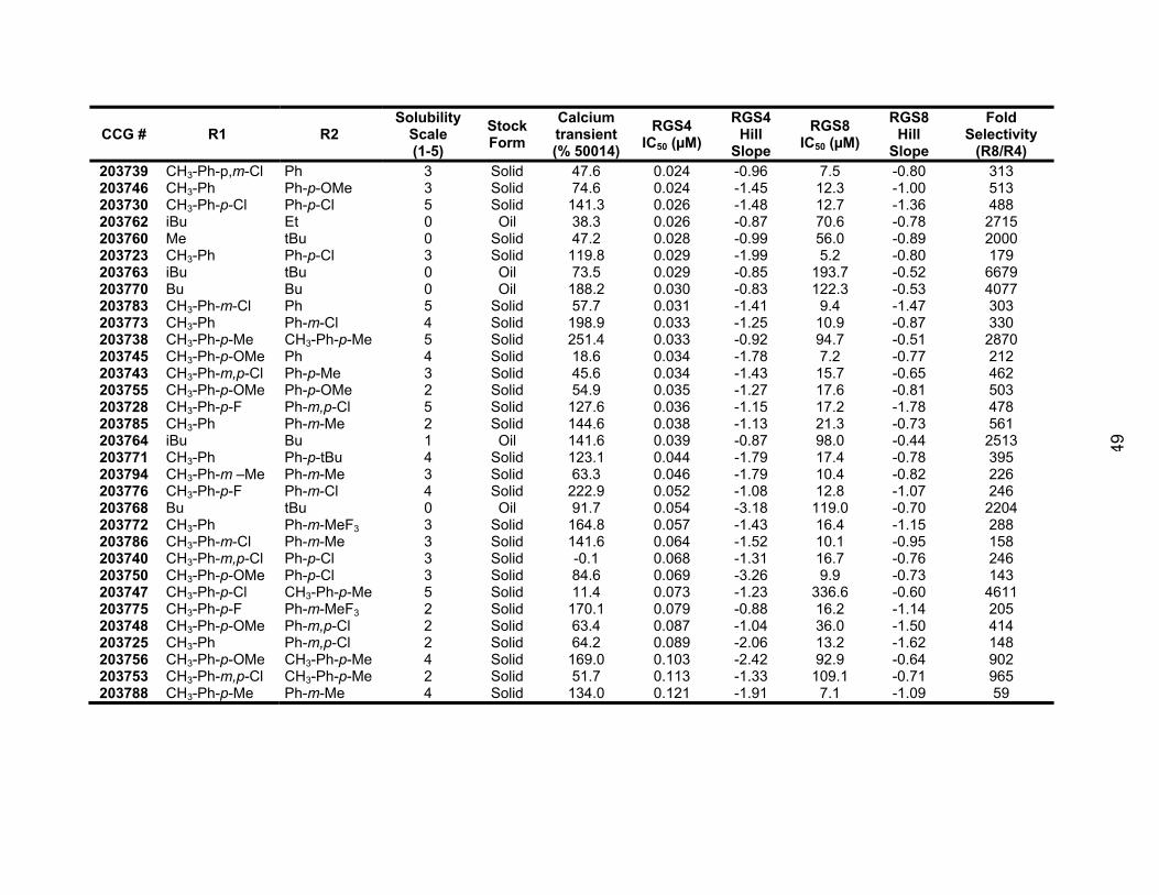
49
CCG # R1 R2 Solubility
Scale (1-5)
Stock Form
Calcium transient (% 50014)
RGS4 IC50 (µM)
RGS4 Hill
Slope RGS8
IC50 (µM) RGS8
Hill Slope
Fold Selectivity
(R8/R4) 203739 CH3-Ph-p,m-Cl Ph 3 Solid 47.6 0.024 -0.96 7.5 -0.80 313 203746 CH3-Ph Ph-p-OMe 3 Solid 74.6 0.024 -1.45 12.3 -1.00 513 203730 CH3-Ph-p-Cl Ph-p-Cl 5 Solid 141.3 0.026 -1.48 12.7 -1.36 488 203762 iBu Et 0 Oil 38.3 0.026 -0.87 70.6 -0.78 2715 203760 Me tBu 0 Solid 47.2 0.028 -0.99 56.0 -0.89 2000 203723 CH3-Ph Ph-p-Cl 3 Solid 119.8 0.029 -1.99 5.2 -0.80 179 203763 iBu tBu 0 Oil 73.5 0.029 -0.85 193.7 -0.52 6679 203770 Bu Bu 0 Oil 188.2 0.030 -0.83 122.3 -0.53 4077 203783 CH3-Ph-m-Cl Ph 5 Solid 57.7 0.031 -1.41 9.4 -1.47 303 203773 CH3-Ph Ph-m-Cl 4 Solid 198.9 0.033 -1.25 10.9 -0.87 330 203738 CH3-Ph-p-Me CH3-Ph-p-Me 5 Solid 251.4 0.033 -0.92 94.7 -0.51 2870 203745 CH3-Ph-p-OMe Ph 4 Solid 18.6 0.034 -1.78 7.2 -0.77 212 203743 CH3-Ph-m,p-Cl Ph-p-Me 3 Solid 45.6 0.034 -1.43 15.7 -0.65 462 203755 CH3-Ph-p-OMe Ph-p-OMe 2 Solid 54.9 0.035 -1.27 17.6 -0.81 503 203728 CH3-Ph-p-F Ph-m,p-Cl 5 Solid 127.6 0.036 -1.15 17.2 -1.78 478 203785 CH3-Ph Ph-m-Me 2 Solid 144.6 0.038 -1.13 21.3 -0.73 561 203764 iBu Bu 1 Oil 141.6 0.039 -0.87 98.0 -0.44 2513 203771 CH3-Ph Ph-p-tBu 4 Solid 123.1 0.044 -1.79 17.4 -0.78 395 203794 CH3-Ph-m –Me Ph-m-Me 3 Solid 63.3 0.046 -1.79 10.4 -0.82 226 203776 CH3-Ph-p-F Ph-m-Cl 4 Solid 222.9 0.052 -1.08 12.8 -1.07 246 203768 Bu tBu 0 Oil 91.7 0.054 -3.18 119.0 -0.70 2204 203772 CH3-Ph Ph-m-MeF3 3 Solid 164.8 0.057 -1.43 16.4 -1.15 288 203786 CH3-Ph-m-Cl Ph-m-Me 3 Solid 141.6 0.064 -1.52 10.1 -0.95 158 203740 CH3-Ph-m,p-Cl Ph-p-Cl 3 Solid -0.1 0.068 -1.31 16.7 -0.76 246 203750 CH3-Ph-p-OMe Ph-p-Cl 3 Solid 84.6 0.069 -3.26 9.9 -0.73 143 203747 CH3-Ph-p-Cl CH3-Ph-p-Me 5 Solid 11.4 0.073 -1.23 336.6 -0.60 4611 203775 CH3-Ph-p-F Ph-m-MeF3 2 Solid 170.1 0.079 -0.88 16.2 -1.14 205 203748 CH3-Ph-p-OMe Ph-m,p-Cl 2 Solid 63.4 0.087 -1.04 36.0 -1.50 414 203725 CH3-Ph Ph-m,p-Cl 2 Solid 64.2 0.089 -2.06 13.2 -1.62 148 203756 CH3-Ph-p-OMe CH3-Ph-p-Me 4 Solid 169.0 0.103 -2.42 92.9 -0.64 902 203753 CH3-Ph-m,p-Cl CH3-Ph-p-Me 2 Solid 51.7 0.113 -1.33 109.1 -0.71 965 203788 CH3-Ph-p-Me Ph-m-Me 4 Solid 134.0 0.121 -1.91 7.1 -1.09 59

50
CCG # R1 R2 Solubility
Scale (1-5)
Stock Form
Calcium transient (% 50014)
RGS4 IC50 (µM)
RGS4 Hill
Slope RGS8
IC50 (µM) RGS8
Hill Slope
Fold Selectivity
(R8/R4) 203797 203749 203752
CH3-Ph-p-F CH3-Ph-m,p-Cl CH3-Ph- p-Cl
Ph-p-N(CH3)2 Ph-m,p-Cl Ph-p-OMe
5 5 4
Oil Solid Solid
111.2 49.6
104.1
0.146 0.155 0.166
-1.65 -0.87 -1.73
10.3 125.0 40.2
-1.28 -0.85 -0.91
71 806 242
203790 CH3-Ph-p-tBu Ph-p-Me 5 Solid 18.9 0.178 -1.36 >100 N/A N/A 203784 Ph-p-tBu Ph-m-Cl 2 Solid 31.8 0.224 -2.03 32.2 -0.58 144 203791 CH3-Ph-p-tBu Ph 5 Solid 14.4 0.245 -1.72 >100 N/A N/A 203796 CH3-Ph-p-tBu Ph-p-tBu 5 Solid 14.8 0.351 -1.70 >100 N/A N/A 203787 CH3-Ph-o-Me Ph-m-Cl 4 Solid 78.2 0.381 -2.01 31.0 -1.10 81 203792 CH3-Ph-m-Cl Ph-m-Cl 4 Solid 67.8 0.419 -1.55 21.1 -1.66 50 203793 CH3-Ph-m-Me Ph-m-MeF3 5 Solid 65.2 N/A N/A 15.9 -1.03 N/A
151
CCG Number R1 R2 Fold Selectivity
(RGS8 IC50 /RGS4 IC50 )
203763 iBu tBu 6679 203769 Bu Et 5964 203770 Bu Bu 4077 203742 CH3-Ph-p-F CH3-Ph-p-Me 3062 203734 CH3-Ph CH3-Ph-p-Me 2914 203738 CH3-Ph-p-Me CH3-Ph-p-Me 2870 203762 iBu Et 2715 203764 iBu Bu 2513 203731 CH3-Ph-p-Cl Ph-p-Me 2360 203760 Me tBu 2000 203765 CH3-Ph-p-F Bu 1975 203736 CH3-Ph-p-Me Ph-p-Me 1289 203757 Bu Me 1235 203735 CH3-Ph-p-F Ph-p-OMe 1036 203777 CH3-Ph-m-Cl Ph-p-Me 1029 203732 CH3-Ph-p-Me Ph 922 203733 CH3-Ph-p-Me Ph-p-Cl 753 203785 CH3-Ph Ph-m-Me 561 203781 CH3-Ph-m-Me Ph 560 203761 iBu Ph-p-Me 550 203727 CH3-Ph-p-F Ph-p-Cl 543 203724 CH3-Ph Ph-p-Me 536 203746 CH3-Ph Ph-p-OMe 513 203755 CH3-Ph-p-OMe Ph-p-OMe 503 203730 CH3-Ph-p-Cl Ph-p-Cl 488 203728 CH3-Ph-p-F Ph-m,p-Cl 478 203767 Bu Ph-p-Me 475 203743 CH3-Ph-m,p-Cl Ph-p-Me 462 203741 CH3-Ph-p,m-Cl Ph-p-OMe 454 203759 Me Ph-p-Me 442 203771 CH3-Ph Ph-p-tBu 395 203726 CH3-Ph-p-F Ph 388 203773 CH3-Ph Ph-m-Cl 330 203739 CH3-Ph-p,m-Cl Ph 313 203783 CH3-Ph-m-Cl Ph 303 203722 CH3-Ph Ph 233 203794 CH3-Ph-m –Me Ph-m-Me 226 203745 CH3-Ph-p-OMe Ph 212 203723 CH3-Ph Ph-p-Cl 179
Table 4.9 Selectivity of CCG-50014 analogs for RGS4 over RGS8. For clarity, only compounds with IC50 values on RGS4 <50 nM are included in this table. Selectivity was determined by comparing the IC50 values from duplex FCPIA assays. n≥2.
152
Identification of compounds with minimized off-target effects: I initially attempted to test CCG-50014 in a Ca2+ mobilization assay using
a cell line stably expressing the M3 muscarinic receptor and expressing RGS4
under control of a doxycycline promoter. A 30-minute pretreatment of these cells
with CCG-50014 completely inhibited the Ca2+ response to carbachol. To probe
the mechanism of this effect, I reduced the compound preoccupation time so that
I could monitor intracellular calcium levels directly after compound addition (Fig.
4.2A). I found that CCG-50014 induced a marked calcium transient in these cells
that after prolonged treatment would be expected to deplete calcium stores.
Since proper calcium handling is crucial for cellular functioning, I tested all of the
available CCG-50014 analogs for their ability to induce calcium mobilization at a
concentration of 10 µM. As shown in Table 4.1 and Fig 4.2B, there were a
number of analogs that did not produce a significant calcium effect in this cell
system, several of which still potently inhibit RGS4.

153
0 25 50 75 100 125 150 175-50
-25
0
25
50
75
100
125
150
10 μM CCG-50014 (or vehicle)Carbachol
A)
Time (sec)
Fluo
-4 C
a++
(% o
f bas
elin
e)
N
O
S
NO
NS
O
N
O
N
OS
N
O
CCG-203760
CCG-203762
CCG-203769
B)
Figure 4.15 A) CCG-50014 induces a calcium transient in HEK293 cells. Fluo4-loaded cells were exposed to 10 µM CCG-50014 (red traces) or DMSO control (black traces) before an injection of 1 nM carbachol. CCG-50014 induced a calcium mobilization event on its own, suggesting that this compound has an as-of-yet unidentified off-target effect. Representative data shown from three wells for each condition. B) Chemical structures of the 3 compounds that did not show calcium mobilization effects and were more potent and soluble than CCG-50014.
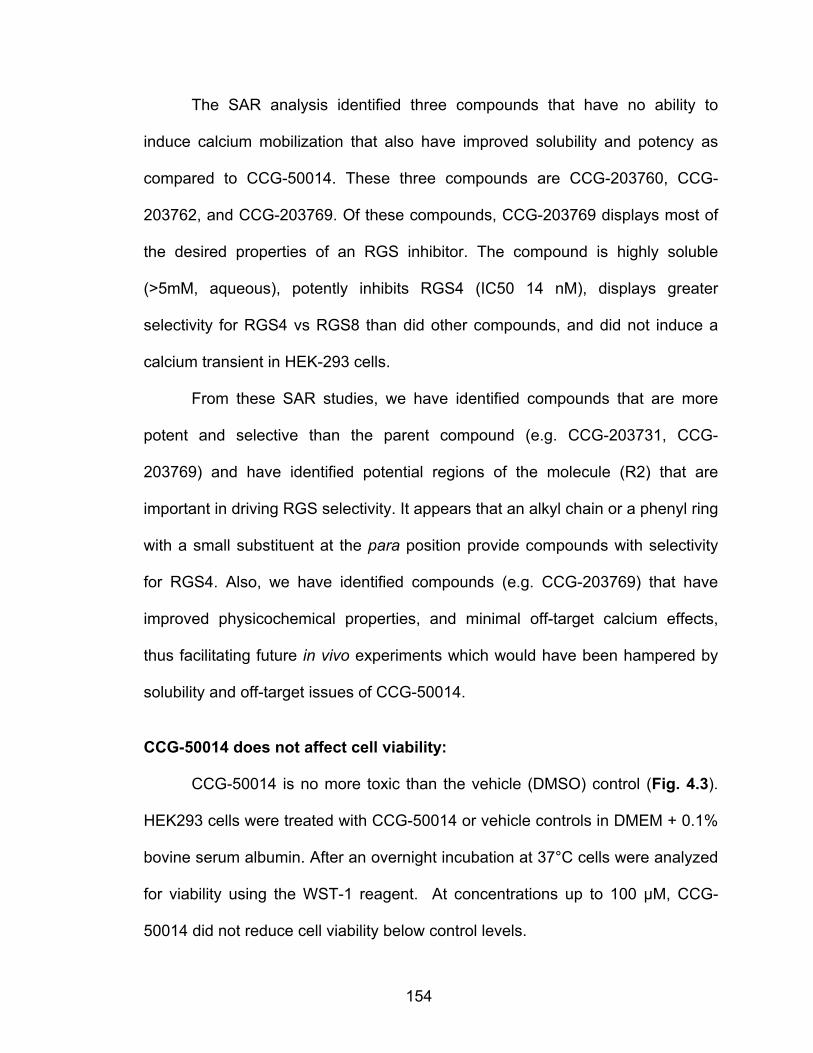
154
The SAR analysis identified three compounds that have no ability to
induce calcium mobilization that also have improved solubility and potency as
compared to CCG-50014. These three compounds are CCG-203760, CCG-
203762, and CCG-203769. Of these compounds, CCG-203769 displays most of
the desired properties of an RGS inhibitor. The compound is highly soluble
(>5mM, aqueous), potently inhibits RGS4 (IC50 14 nM), displays greater
selectivity for RGS4 vs RGS8 than did other compounds, and did not induce a
calcium transient in HEK-293 cells.
From these SAR studies, we have identified compounds that are more
potent and selective than the parent compound (e.g. CCG-203731, CCG-
203769) and have identified potential regions of the molecule (R2) that are
important in driving RGS selectivity. It appears that an alkyl chain or a phenyl ring
with a small substituent at the para position provide compounds with selectivity
for RGS4. Also, we have identified compounds (e.g. CCG-203769) that have
improved physicochemical properties, and minimal off-target calcium effects,
thus facilitating future in vivo experiments which would have been hampered by
solubility and off-target issues of CCG-50014.
CCG-50014 does not affect cell viability:
CCG-50014 is no more toxic than the vehicle (DMSO) control (Fig. 4.3).
HEK293 cells were treated with CCG-50014 or vehicle controls in DMEM + 0.1%
bovine serum albumin. After an overnight incubation at 37°C cells were analyzed
for viability using the WST-1 reagent. At concentrations up to 100 µM, CCG-
50014 did not reduce cell viability below control levels.
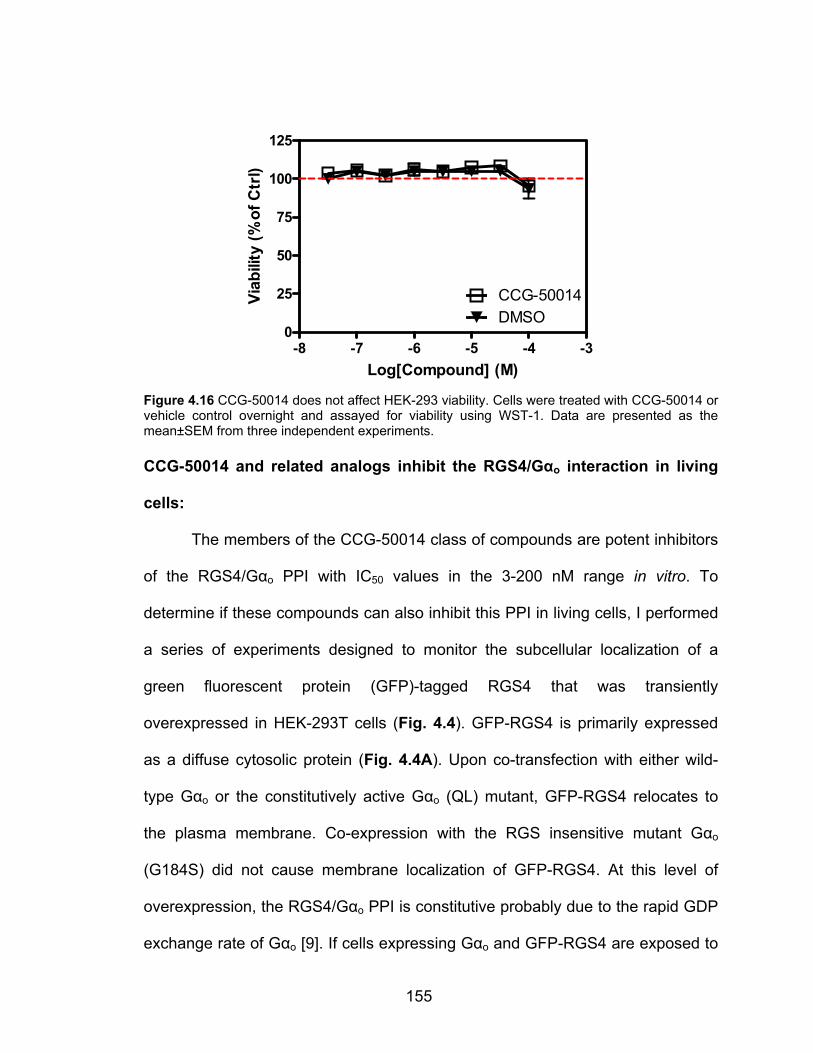
155
-8 -7 -6 -5 -4 -30
25
50
75
100
125
CCG-50014DMSO
Log[Compound] (M)
Viab
ility
(%of
Ctr
l)
Figure 4.16 CCG-50014 does not affect HEK-293 viability. Cells were treated with CCG-50014 or vehicle control overnight and assayed for viability using WST-1. Data are presented as the mean±SEM from three independent experiments. CCG-50014 and related analogs inhibit the RGS4/Gαo interaction in living
cells:
The members of the CCG-50014 class of compounds are potent inhibitors
of the RGS4/Gαo PPI with IC50 values in the 3-200 nM range in vitro. To
determine if these compounds can also inhibit this PPI in living cells, I performed
a series of experiments designed to monitor the subcellular localization of a
green fluorescent protein (GFP)-tagged RGS4 that was transiently
overexpressed in HEK-293T cells (Fig. 4.4). GFP-RGS4 is primarily expressed
as a diffuse cytosolic protein (Fig. 4.4A). Upon co-transfection with either wild-
type Gαo or the constitutively active Gαo (QL) mutant, GFP-RGS4 relocates to
the plasma membrane. Co-expression with the RGS insensitive mutant Gαo
(G184S) did not cause membrane localization of GFP-RGS4. At this level of
overexpression, the RGS4/Gαo PPI is constitutive probably due to the rapid GDP
exchange rate of Gαo [9]. If cells expressing Gαo and GFP-RGS4 are exposed to
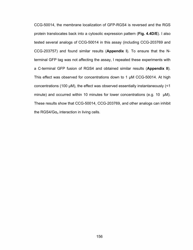
156
CCG-50014, the membrane localization of GFP-RGS4 is reversed and the RGS
protein translocates back into a cytosolic expression pattern (Fig. 4.4D/E). I also
tested several analogs of CCG-50014 in this assay (including CCG-203769 and
CCG-203757) and found similar results (Appendix I). To ensure that the N-
terminal GFP tag was not affecting the assay, I repeated these experiments with
a C-terminal GFP fusion of RGS4 and obtained similar results (Appendix II).
This effect was observed for concentrations down to 1 µM CCG-50014. At high
concentrations (100 µM), the effect was observed essentially instantaneously (<1
minute) and occurred within 10 minutes for lower concentrations (e.g. 10 µM).
These results show that CCG-50014, CCG-203769, and other analogs can inhibit
the RGS4/Gαo interaction in living cells.
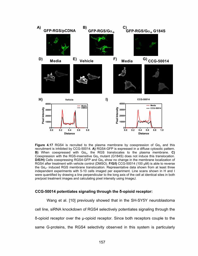
157
Vehicle
0.0 0.2 0.4 0.6 0.80
1000
2000
3000
4000DMSOMedia
Distance
Pixe
l Int
ensi
ty
CCG-50014
0.0 0.2 0.4 0.6 0.8 1.00
1000
2000
3000
4000MediaCCG-50014
Distance
Pixe
l Int
ensi
ty
Media Vehicle Media CCG-50014D)
H)
E)
I)
GFP-RGS/pCDNA GFP-RGS/Gαo GFP-RGS/Gαo G184SA) B) C)
F) G)
Figure 4.17 RGS4 is recruited to the plasma membrane by coexpression of Gαo and this recruitment is inhibited by CCG-50014. A) RGS4-GFP is expressed in a diffuse cytosolic pattern. B) When coexpressed with Gαo, the RGS translocates to the plasma membrane. C) Coexpression with the RGS-insensitive Gαo mutant (G184S) does not induce this translocation. D/E/H) Cells coexpressing RGS4-GFP and Gαo show no change in the membrane localization of RGS4 after treatment with vehicle control (DMSO). F/G/I) CCG-50014 (100 µM) is able to reverse the Gαo- induced RGS membrane translocation. Representative data shown from at least three independent experiments with 5-10 cells imaged per experiment. Line scans shown in H and I were quantified by drawing a line perpendicular to the long axis of the cell at identical sites in both pre/post treatment images and calculating pixel intensity using ImageJ. CCG-50014 potentiates signaling through the δ-opioid receptor:
Wang et al. [10] previously showed that in the SH-SY5Y neuroblastoma
cell line, siRNA knockdown of RGS4 selectively potentiates signaling through the
δ-opioid receptor over the µ-opioid receptor. Since both receptors couple to the
same G-proteins, the RGS4 selectivity observed in this system is particularly

158
useful in dissecting the compound’s action. A low concentration (100 nM) of
either the µ-opioid receptor agonist morphine or the δ-opioid receptor agonist
SNC-80 produced a small (10-20%) inhibition of forskolin-stimulated adenylate
cyclase activity. CCG-50014 (100 µM) significantly potentiated the effect of the δ-
opioid agonist (44% inhibition vs 22% inhibition without the compound, p<0.001),
as would be expected of an RGS4 inhibitor in this system (Fig. 4.5), with only a
modest effect upon the µ-receptor signaling. The observed receptor selectivity of
compound action is consistent with CCG-50014 acting by inhibiting RGS4.
The non-significant increase in µ-opioid signaling in response to
compound should be addressed. It is possible that this effect is due to inhibition
of other RGS proteins in the cell or because there is some low-level control of µ-
opioid receptor signaling by RGS4. Regardless, there is significant potentiation of
the δ-opioid signaling as would be expected of an RGS inhibitor.
- + - +0
20
40
60
morphine
11%
18% 22%
44%
CCG-50014
**
SNC-80
% In
hibi
tion
ofFo
rsko
lin-s
timul
ated
AC a
ctiv
ity
Figure 4.18 CCG-50014 potentiates the activity of the δ-opioid receptor ligand SNC-80 in SH-SY5Y cells. Inhibition of forskolin-activated adenylate cyclase activity by either the µ-opioid receptor or the δ-opioid receptor was measured in the absence and presence of 100 µM CCG-50014. Data are presented as the mean±SEM from three independent experiments. ** P<0.001
159
CCG-203769 potentiates the M3 muscarinic receptor activity via inhibition
of RGS4:
To further probe the ability of the CCG-50014 class of compounds to
inhibit RGS actions in a cellular setting, I performed a series of experiments
using the engineered cell system expressing the M3 muscarinic receptor with
RGS4 under doxycycline control. Upon addition of carbachol to these cells, a
Gαq-dependent intracellular calcium mobilization is initiated by the activation of
phospholipase C. After induction of RGS4 expression, calcium signaling is
suppressed by ~80%. A compound that inhibited RGS4 should relieve this
suppression.
As noted above, when these experiments were performed with CCG-
50014 it was found that the compound itself induced a sizable calcium transient.
This off-target effect clearly makes it difficult to interpret data from these
experiments. To circumvent this issue, I identified CCG-50014 analogs that were
unable to induce a calcium flux in HEK-293 cells. One of these compounds,
CCG-203769, is more potent at inhibiting RGS4 than CCG-50014 and is more
soluble and was therefore chosen for study in this system.
Incubation with CCG-203769 for 15 minutes prior to carbachol stimulation
resulted in a partial reversal of the RGS4-mediated inhibition (Fig. 4.6) without
affecting M3-mediated calcium signal in the absence of RGS4 expression. This
result further confirms this class of compounds is acting as an RGS4 inhibitor in
living cells. Furthermore, the response occurs at micromolar concentrations,
which is more in concordance with what is observed in vitro. This assay also

160
measures Gq signaling (as opposed to Gi/o in the opioid studies), proving that the
effect of this compound is not G-protein dependent.
The enhanced Ca++ signaling induced by CCG-203769 occurs at
concentrations as low as 1 µM. While this compound was chosen for study
because of its inability to stimulate calcium mobilization by itself, it is possible
that a weak effect on calcium handling is starting to play a significant role over
the time course of the experiment. It is also possible that at high concentrations
of compound other, as of yet unknown off-target effects are occurring that disrupt
calcium handling. Regardless, it does appear that CCG-203769 can inhibit RGS4
in living cells at concentrations that do not cause any measurable off-target
effects.
0 25 50 75 1000
20
40
60
80
DMSODMSO +DoxCCG-203769
CCG-203769 +Dox
Carb. Time (sec)
Ca++
(Flu
o-4
fluor
esce
nce)
0
20
40
60
80
100
***
DMSO 1 μM 3 μM 5 μMDox - + - + - + - +M
ax F
luo-
4 Fl
uore
scen
ce (A
U)
A) B)
Figure 4.19 CCG-203769 partially reverses the RGS4-mediated suppression of carbachol responsiveness in HEK293 cells expressing the M3 muscarinic receptor. A) Representative traces of cells responding to 3 nM carbachol. Doxycycline (+Dox) treated cells express RGS4 and show a significant suppression of Ca2+ mobilization. Pretreatment of these cells for 15 minutes with 1 µM CCG-302769 (CCG-203769 +Dox) partially rescues the Ca2+ response. Data are presented as the mean±SEM of 12 wells per condition from a single experiment. B) Concentration dependence of the effect observed in A. Data are presented as the mean±SEM from three independent experiments. * P< 0.05; ** P < 0.01.

161
Discussion:
The development of RGS inhibitors has till now been dominated by
compounds that lacked efficacy in a cellular environment. The reasons for this
lack of cellular activity stem from: permeability issues (e.g. peptides and possibly
the CCG-638x family), weak in vitro activity (e.g. CCG-638x family and CCG-
4986), and inactivation in the presence of reductants (e.g. CCG-4986 and the
CCG-50014 family). In this chapter I have presented the first evidence for a
family of RGS inhibitors that functions in a cellular environment.
Early biochemical studies of this compound class suggested that it would
likely lack cellular activity. As noted in Chapter III, this family of compounds is
inactivated by reductants, including physiologically relevant concentrations of the
intracellular reductant glutathione. Furthermore, the cysteine-dependent reactivity
of the compounds raised concerns about target specificity and potential off-target
effects. It was with these issues in mind that we attempted cellular studies with
CCG-50014 and some selected analogs.
I developed a method to directly measure the Gαo/RGS4 PPI in living
cells. This approach allowed me to determine if my compounds can inhibit this
PPI in living cells like they do in vitro (Fig. 4.4A). In this system, the RGS is
expressed as a diffuse cytosolic protein until co-expression with Gαo, which
drives membrane association of RGS4. This co-localization is not induced by the
co-expression of a mutant Gαo (G184S) that is insensitive to RGS GAP activity.
The lack of interaction between the G184S Gαo mutant and RGS4 confirms the
findings in cell and whole animal knock-in models [11-17]. CCG-50014 and CCG-

162
203769 are both able to inhibit the membrane localization of RGS4 in this assay,
strongly suggesting that these compounds can inhibit the Gαo/RGS4 interaction
in living cells. These data lend considerable credence to the notion that the
functional effects observed in the subsequent studies stem from an inhibition of
one (or more) RGS proteins.
To further confirm that the compounds could function in a cellular setting,
we performed a series of cellular studies to test the ability of these compounds to
block the negative regulation of GPCR signaling by RGS proteins. Dr. Qin Wang,
working in the laboratory of Dr. John Traynor, was able to show that CCG-50014
could potentiate the signaling through the δ-opioid receptor selectively over the
µ-opioid receptor in SH-SY5Y cells. The signaling pathways used in this study
are all endogenously expressed in SH-SY5Y cells and this result correlates well
with their previous work showing RGS4 selectively inhibits the δ-opioid receptor
[10]. These data are important because they show for the first time a small
molecule RGS inhibitor having a functional effect on an endogenous signaling
pathway.
While it is important to show that the compound can function on
endogenous signaling pathways, I wanted to also probe the actions of this
compound in a more controlled manner. I tested several compounds using cells
stably expressing the muscarinic M3 receptor with RGS4 expression under
doxycycline control. By measuring the Gq-dependent calcium mobilization
induced by the M3 receptor, I was able to show that CCG-230769 partially
inhibits the effects of RGS4. CCG-50014, however, induced a calcium transient
163
on its own (Fig 4.2A). These studies were important for two reasons: 1) it
showed that the compound inhibits RGS4 under more controlled conditions than
the SH-SY5Y experiments; and 2) these experiments revealed a previously
unknown off-target effect elicited by some members of the CCG-50014 family.
I undertook a series of experiments to probe the chemical space around
the CCG-50014 scaffold. By analyzing ~80 analogs of CCG-50014 for a variety
of parameters including potency, RGS selectivity, and solubility, I was able to
identify a number of structure-activity relationships around this scaffold. Potency
against RGS proteins can be improved by shortening the side chains (R1/R2 in
Table 4.1) to small alkyl chains. This also corresponded to an increase in RGS4
selectivity, although some analogs with phenyl rings at the R1/R2 position that
have a small substituent in the para position on the R2 phenyl ring (e.g. CCG-
203702) also display prominent selectivity for RGS4 over RGS8. Modification of
the thiadiazolidine ring to a less reactive center drastically reduces activity.
This SAR analysis also provided a means to identify compounds that did
not produce the Ca2+ mobilization off-target effect noted for CCG-50014. I
identified three compounds (Fig. 4.2B) that potently inhibit RGS4 yet lack the
ability to induce a Ca2+ mobilization. Study of the SAR landscape surrounding
CCG-50014 has yielded several compounds with improved properties and in vitro
activity. These compounds, including CCG-203769, are currently being used in
isolated organ and whole animal studies to determine their physiological effects
on RGS activity and GPCR signaling.

164
There are at least two advantages that the CCG-50014 class of molecules
has over previous generations of RGS inhibitors. The first – and presumably
most important – is potency. CCG-50014 is nearly 3 orders of magnitude more
potent at inhibiting RGS4 than any of the other small molecule RGS inhibitors
described to date. Therefore, even if a substantial fraction of the compound is
rapidly metabolized or reacts with glutathione (or other non-target thiols), an
active concentration of compound is likely to be present. Another set of
advantages this compound has over prior generations of RGS inhibitors
(especially CCG-63802) is improved cellular permeability and aqueous solubility.
While solubility is variant across this family of compounds, we have identified
several that have aqueous solubility >5 mM yet still retain cellular activity. These
factors allow us generate pharmacologically relevant concentrations of
compound in our assay systems without interfering artifacts such as compound
precipitation.
The CCG-50014 class of compounds contains the most potent RGS
inhibitors identified to date and the first examples of small molecules that can
inhibit the cellular activity of an RGS protein. While this is a significant step
forward in the development of small molecule RGS inhibitors, the true value of
this compound has yet to be fully elucidated. One major concern that has not
been addressed is the pharmacokinetic parameters of these compounds.
Furthermore, we have yet to fully determine the physiological effects of systemic
administration of CCG-50014 or related analogs. Preliminary data has shown no
gross physiological/toxicological effects to low doses of CCG-203769, however,
165
the appropriate full-scale studies have yet to be performed. Also, the RGS
specificity of CCG-203769 has yet to be determined in a cellular setting.
Regardless, this family of compounds provides a foothold for development of
new RGS inhibitors.

166
References: 1. Brunton, L.L., J.S. Lazo, and K.L. Parker, eds. Goodman & Gilman's: The
Pharmacological Basis of Therapeutics. 11 ed. 2006, McGraw-Hill Companies, Inc.: New York.
2. Roman, D., et al., Allosteric Inhibition of the RGS-G{alpha} Protein-Protein Interaction by CCG-4986. Mol Pharmacol.
3. Sternweis, P.C. and J.D. Robishaw, Isolation of two proteins with high affinity for guanine nucleotides from membranes of bovine brain. J Biol Chem, 1984. 259(22): p. 13806-13.
4. Blazer, L.L., et al., Use of flow cytometric methods to quantify protein-protein interactions. Curr Protoc Cytom. Chapter 13: p. Unit 13 11 1-15.
5. Blazer, L.L., et al., Reversible, allosteric, small-molecule inhibitors of RGS proteins. Mol Pharmacol.
6. Roman, D.L., S. Ota, and R.R. Neubig, Polyplexed Flow Cytometry Protein Interaction Assay: A Novel High-Throughput Screening Paradigm for RGS Protein Inhibitors. J Biomol Screen, 2009.
7. Roman, D.L., et al., Identification of small-molecule inhibitors of RGS4 using a high-throughput flow cytometry protein interaction assay. Mol Pharmacol, 2007. 71(1): p. 169-75.
8. Roof, R.A., et al., Mechanism of action and structural requirements of constrained peptide inhibitors of RGS proteins. Chem Biol Drug Des, 2006. 67(4): p. 266-74.
9. Remmers, A.E., et al., Interdomain interactions regulate GDP release from heterotrimeric G proteins. Biochemistry, 1999. 38(42): p. 13795-800.
10. Wang, Q., L.Y. Liu-Chen, and J.R. Traynor, Differential modulation of mu- and delta-opioid receptor agonists by endogenous RGS4 protein in SH-SY5Y cells. J Biol Chem, 2009. 284(27): p. 18357-67.
11. Fu, Y., et al., RGS-insensitive G-protein mutations to study the role of endogenous RGS proteins. Methods Enzymol, 2004. 389: p. 229-43.
12. Huang, X., et al., Pleiotropic phenotype of a genomic knock-in of an RGS-insensitive G184S Gnai2 allele. Mol Cell Biol, 2006. 26(18): p. 6870-9.
13. Lan, K.L., et al., A point mutation in Galphao and Galphai1 blocks interaction with regulator of G protein signaling proteins. J Biol Chem, 1998. 273(21): p. 12794-7.
14. Goldenstein, B.L., et al., Regulator of G protein signaling protein suppression of Galphao protein-mediated alpha2A adrenergic receptor inhibition of mouse hippocampal CA3 epileptiform activity. Mol Pharmacol, 2009. 75(5): p. 1222-30.
15. Goldenstein, B.L., et al., RGS Protein Suppression of G{alpha}o Protein-Mediated {alpha}2A Adrenergic Receptor Inhibition of Mouse Hippocampal CA3 Epileptiform Activity. Mol Pharmacol, 2009.
16. Huang, X., et al., Resistance to diet-induced obesity and improved insulin sensitivity in mice with a regulator of G protein signaling-insensitive G184S Gnai2 allele. Diabetes, 2008. 57(1): p. 77-85.
17. Talbot, J.N., et al., RGS inhibition at G(alpha)i2 selectively potentiates 5-HT1A-mediated antidepressant effects. Proc Natl Acad Sci U S A. 107(24): p. 11086-91.

167
Chapter V: Conclusions
Summary of Results:
The work presented in this thesis has significantly advanced our
understanding of how to target RGS proteins with small molecules. The major
advances that are described in this thesis are 1) the first examples of reversible
small molecule RGS inhibitors; 2) characterization of the most potent RGS
inhibitors identified to date; and 3) the first example of small molecule RGS
inhibitors with cellular activity. The information in this thesis provides both novel
methodology and direction for the development of future RGS inhibitors.
In Chapter I, I describe the first set of compounds that can reversibly
inhibit RGS4. The prototypical compound from this class, CCG-63802, inhibits
RGS4 with an in vitro IC50 value of 10 micromolar in the FCPIA assay. This family
of compounds provided the first proof-of-concept that RGS proteins can be
inhibited by small molecules in a reversible fashion. While CCG-63802 may not
be the optimal lead for cellular/animal studies, it is nonetheless an important step
forward in the development of small molecules targeting RGS proteins.
Chapter II detailed the molecular mechanism of the most potent small
molecule RGS inhibitor, CCG-50014. This compound irreversibly inhibits RGS4
with a 30 nM IC50 value and has significant selectivity for RGS4 over RGS
proteins including RGS7, RGS8, RGS16 and RGS19. While irreversible, this

168
compound provides several advantages for studying the molecular mechanism of
RGS allosteric inhibition.
In Chapter IV I further develop the CCG-50014 class of compounds to
show, for the first time, small molecules that can inhibit both the RGS/Gα PPI and
RGS activity in a living cell. To our knowledge, this is the first time that a small
molecule RGS inhibitor has been shown to directly inhibit the RGS/Gα interaction
in a cellular setting. Second generation members from this family, including
CCG-203769, are also described. These compounds retain the RGS inhibitory of
CCG-50014, but have improved aqueous solubility and were selected for the
inability to invoke a calcium transient. The data presented in Chapter IV clearly
show that this class of small molecules can inhibit the RGS/Gα PPI in a manner
that has a measurable effect upon GPCR signaling. This is a dramatic step
forward in the development of small molecule RGS inhibitors and it is hoped that,
with continued development, descendents from this class of compounds will
become useful research tools and potentially even clinically relevant
therapeutics.
Future Research Directions:
The work presented in this thesis opens many potential avenues for
further research and development of small molecule RGS inhibitors. The novel
methodology described throughout this thesis (TR-FRET high throughput
screening, RGS/Gα thermal stability assays, RGS membrane translocation, etc.)
provides a framework against which novel RGS inhibitor scaffolds can be tested.
Furthermore, the compound classes that have been expounded upon in this
169
thesis provide the opportunity to both learn more about small molecule binding
sites on RGS and also two novel scaffolds for the development of future
generations of RGS inhibitors.
The CCG-63802 class of molecules may be able to provide structural
information regarding the non-covalent binding of small molecules to RGS. It is
possible that many of the small fragments of CCG-63802 that lack RGS inhibitory
activity might weakly bind to the protein in a manner that does not produce a
functional effect. By performing nuclear magnetic resonance experiments with
isotopically labeled RGS4, we should be able to map the binding sites of these
fragments on the RGS. Similar studies have been performed before using a
variety of different proteins [19-24]. Indeed, experiments along these lines are
already in progress by a graduate student in our lab, Andrew Storaska.
Based upon mutagenesis data, it is expected that the mechanism of action
of CCG-63802 will primarily be through a conformational change induced by
compound binding to the allosteric “B” site on RGS4. The B site is the location on
RGS4 that is important for the binding of calmodulin and acidic phospholipids.
Previous work by Ishii and colleagues has shown that these two molecules can
reciprocally regulate RGS GAP activity, whereby phospholipid binding inhibit the
RGS function and this effect can be displaced by calmodulin binding [25, 26]. It is
possible that CCG-638002 mimics the effects of acidic phospholipids on RGS4
GAP activity. While technically challenging, it would be very interesting to test
this hypothesis by using a series of protection or displacement studies to
determine if calmodulin or acidic phospholipids can affect CCG-63802 binding.
170
Similar experiments could test any RGS inhibitors that are expected to bind via
that “B” site (e.g. CCG-50014).
One of the main motivations for the development of small molecule
modulators of RGS activity is so that we can better understand the molecular
mechanisms behind RGS activity and regulation. While the “B” site hypothesis
has been studied from a variety of functional angles, to date there are no data
that specifically address how this allosteric binding results in altered GAP activity.
This is likely due in part to the challenging nature of performing biophysical
studies on proteins in the presence of phospholipids. It is possible that a small
molecule that binds to the “B” site to inhibit RGS function could provide insights
into the conformational changes that occur during this allosteric regulation.
While initial studies have not been particularly promising, it might also be
useful to test selected analogs of CCG-63802 in cellular assays. For these
compounds to have significant cellular activity, it is likely that a new analog with
the following properties will have to be generated. First, the compound needs to
have a greater potency at RGS4. Most compounds identified in this class
possess micromolar IC50 values in vitro, which is likely not potent enough for in
vivo studies. Ideally, the candidate compound would be >2 Log more potent than
the CCG-63802. It would also be beneficial if the compound possessed a smaller
polar surface area than CCG-63802 to improve membrane permeability. If we
can identify a soluble, stable compound that meets these two conditions, it is
likely that it would have cellular activity.
171
The CCG-50014 class of compounds has already progressed to the point
of containing members with documented cellular activity. For this class of
compounds, it is most important to focus upon transitioning from
biochemical/cellular assays to isolated organ/whole animal experimentation.
Several of these studies are currently underway. Through collaboration, we have
started testing CCG50014 and CCG-203769 in the langendorff isolated heart
model to determine if the compounds can potentiate carbachol-stimulated
bradycardia. It is known that RGS4 is expressed in sinoatrial nodal cells, where
parasympathetic M2 muscarinic receptor activation slows the heart. We are also
approaching this same system in a whole-rat model. Sprague-Dawley rats are
implanted with indwelling venous catheters and telemetry probes that monitor
heart rate, blood pressure and body temperature. After recovery, these animals
are infused with a dose of CCG-203769 and then given a small dose of
carbachol to induce a transient bradycardia. It is expected that if the compounds
inhibit RGS in the SA node, the carbachol effect will be potentiated. Currently, we
have not observed any significant physiological effects with CCG-203769, but the
studies are not complete and due to solubilization issues, we have not been able
to inject the animals with as large of doses as we would like. Currently, Dr.
Stephen Husbands is producing analogs of CCG-50014 that will likely be much
more soluble in aqueous environments.
Also through collaboration, we are testing the ability of these compounds
to potentiate the activity of the M3 muscarinic receptor in pancreatic islet β cells.
In β cells, M3 activation potentiates glucose-stimulated insulin release [27]. Wess
172
and colleagues showed that RGS4 potently regulates the M3 activity in β cells
[28]. Using isolated mouse islets, we have been attempting to determine if CCG-
50014 or CCG-203769 can accentuate the M3 activity on glucose-stimulated
insulin release. Preliminary data from these experiments have been promising
and we are in the process of developing protocols to test the glucose tolerance of
animals that were treated with CCG-50014 analogs.
While important, the animal studies need to be accompanied by a full
pharmacokinetic profile of the compound before any conclusions regarding its
usefulness as a potential therapeutic may be drawn. We, in collaboration, are
currently in the process of developing a method to detect the CCG-50014
analogs in whole blood and isolated tissues. With this methodology in hand, we
will be able to perform single dose pharmacokinetic profiles of important CCG-
50014 analogs. These data will be critical for the animal studies to ensure that
we are reaching and maintaining appropriate blood and tissue concentrations.
The CCG-63802 and CCG-50014 classes of molecules have provided
significant advances to the field of RGS inhibitors. Both of these families also
have the potential to provide further knowledge into the location and geometry of
small molecule binding sites on RGS4. Furthermore, they may function as
pharmacological tools to study the effects of RGS proteins in vivo. It is
anticipated that these molecules will provide many new discoveries in the fields
of RGS biology and small molecule protein-protein interaction inhibitors.
173
Therapeutic applications of RGS modulation:
Modulating RGS activity could have a variety of potential therapeutic
applications. While clearly much work needs to be done to generate a drug
candidate targeting an RGS protein, the potential benefit of such a drug in
several disease states could be significant.
Recent work has shown that RGS4 negatively modulates the
parasympathetic stimulation of insulin release [28]. The cholinergic stimulation of
β cells does not directly cause a release of insulin, but it does potentiate the
responsiveness of these cells to high blood glucose. In this setting, a selective
RGS4 inhibitor would be expected to potentiate the autonomic stimulation of
insulin release in a glucose-dependent manner. This could be a very beneficial
mechanism for treatment of early-stage type II diabetes and could require much
less monitoring than conventional insulin replacement therapies.
Another potential application for an RGS inhibitor is in the treatment of
depression. Traynor and colleagues have shown that the pan-inhibition of RGS
activity on Gαi2 produces a striking antidepressant-like effect [18]. While we still
do not know which RGS protein(s) to target for antidepression therapy, these
results suggest that subtle modulation of endogenous neurotransmitter signaling
might provide an effective treatment for depression and potentially other
neurological disorders. I find this approach to be particularly elegant, especially
when compared to the commonly used drugs to treat neurological disorders –
many of which affect many different signaling systems in ways that we still do not
fully understand [29-31].
174
Another potential target is RGS9-2, the brain specific isoform of RGS9.
This molecule, is strongly and specifically expressed in certain parts of the brain,
including the striatum and nucleus accumbens [32]. Knockout of RGS9 increases
supra-spinal and spinal cord mediated analgesia and delays the development of
tolerance to morphine [33]. Small molecules specifically targeting RGS9 may
provide a mechanism to potentiate morphine analgesia under conditions that
may slow the development of tolerance.
Unfortunately, the application of RGS9 inhibitors might be expected to
have a variety of undesired side effects. An isoform of RGS9, RGS9-1, is the
major RGS controlling phototransduction [34]. Unless the compound is
pharmacokinetically excluded from the eyes or is exquisitely specific for RGS9-2
over RGS9-1, vision problems (likely trailing images) may occur. Furthermore,
RGS9-2 knockout mice are sensitized to the dyskinesias associated with
activation of D2 dopamine receptor after prolonged pharmacological dopamine
receptor antagonism [35]. These potential issues will have to be addressed
before inhibitors of RGS9 will be adopted as viable drug candidates.
While the work in this thesis has focused upon developing inhibitors of
RGS function, there are several situations in which an activator of RGS activity
could provide significant therapeutic benefit. Gold and colleagues have shown
that overexpression of RGS9-2 in the striatum of non-human primates can
suppress the dyskinesias associated with chronic L-dopa treatment [36]. These
data suggest that potentiating RGS9-2 expression in the striatum or
175
pharmacological activation of RGS9-2 in patients with Parkinson’s disease may
provide a level of relief from the side effects of L-dopa treatment.
Modulating RGS proteins with small molecules could provide significant
therapeutic benefit. RGS modulation could provide a mechanism to increase the
efficacy of endogenous signaling in the body, something that current GPCR-
targeted approaches often cannot attain. Furthermore, the unique expression
patterns of RGS proteins may provide a level of tissue specificity not achievable
with GPCR agonists/antagonists. It is hoped that future generations of RGS
inhibitors will provide therapeutic benefit to mankind.
The Future of Small Molecule Protein-Protein Interaction Inhibitors:
It is clear that substantial challenges lie ahead in the development of PPI
modulators. Continued progress is being made in the cancer arena where the
requirements of oral absorption and/or blood-brain-barrier penetration are less
critical. Indeed, the affinity of PPIIs in the cancer field has increased (to IC50
values <10 nM in vitro and 10s-100s of nM in cells) and the molecular weight of
these PPIIs has decreased over the last 10 years [37, 38]. That work and
continued academic efforts to develop tool compounds blocking PPIs should
advance this field substantially. The need to maintain small size and appropriate
physicochemical properties of compounds may require novel approaches. Rather
than targeting the immediate PPI site, the identifying and targeting of allosteric
sites on the target protein may permit the use of more suitable chemical
structures. In the cancer drug design arena, fragment-based design of PPI
176
inhibitors has proven useful so applications to other targets should be pursued.
Also, identifying compounds that could make use of cellular uptake mechanisms
could permit compounds to achieve higher intracellular concentrations and result
in additional cell-type specificity of action.
There are a large number of potential PPI targets that could provide subtle
modulation of cellular processes if a successful drug could be developed. As with
the history of protein kinase inhibitors, overcoming initial reluctance to embrace
the concept will likely require a success story. At present, it is hard to predict a
major breakthrough in this field but continued refinement of existing approaches
and further development of existing targets is likely to reach a threshold of
success in the not too distant future. In the meantime, substantial genetic and
biological studies will continue to define novel PPI targets. With the rapid
advances in target identification, an increased pace of chemical discovery related
to PPIs will be critical to exploit the potential of this novel field.
177
References:
1. Berg, T., Small-molecule inhibitors of protein-protein interactions. Curr Opin Drug Discov Devel, 2008. 11(5): p. 666-74.
2. Brunton, L.L., J.S. Lazo, and K.L. Parker, eds. Goodman & Gilman's: The Pharmacological Basis of Therapeutics. 11 ed. 2006, McGraw-Hill Companies, Inc.: New York.
3. Roman, D., et al., Allosteric Inhibition of the RGS-G{alpha} Protein-Protein Interaction by CCG-4986. Mol Pharmacol.
4. Sternweis, P.C. and J.D. Robishaw, Isolation of two proteins with high affinity for guanine nucleotides from membranes of bovine brain. J Biol Chem, 1984. 259(22): p. 13806-13.
5. Blazer, L.L., et al., Use of flow cytometric methods to quantify protein-protein interactions. Curr Protoc Cytom. Chapter 13: p. Unit 13 11 1-15.
6. Blazer, L.L., et al., Reversible, allosteric, small-molecule inhibitors of RGS proteins. Mol Pharmacol.
7. Roman, D.L., S. Ota, and R.R. Neubig, Polyplexed Flow Cytometry Protein Interaction Assay: A Novel High-Throughput Screening Paradigm for RGS Protein Inhibitors. J Biomol Screen, 2009.
8. Roman, D.L., et al., Identification of small-molecule inhibitors of RGS4 using a high-throughput flow cytometry protein interaction assay. Mol Pharmacol, 2007. 71(1): p. 169-75.
9. Roof, R.A., et al., Mechanism of action and structural requirements of constrained peptide inhibitors of RGS proteins. Chem Biol Drug Des, 2006. 67(4): p. 266-74.
10. Remmers, A.E., et al., Interdomain interactions regulate GDP release from heterotrimeric G proteins. Biochemistry, 1999. 38(42): p. 13795-800.
11. Wang, Q., L.Y. Liu-Chen, and J.R. Traynor, Differential modulation of mu- and delta-opioid receptor agonists by endogenous RGS4 protein in SH-SY5Y cells. J Biol Chem, 2009. 284(27): p. 18357-67.
12. Fu, Y., et al., RGS-insensitive G-protein mutations to study the role of endogenous RGS proteins. Methods Enzymol, 2004. 389: p. 229-43.
13. Huang, X., et al., Pleiotropic phenotype of a genomic knock-in of an RGS-insensitive G184S Gnai2 allele. Mol Cell Biol, 2006. 26(18): p. 6870-9.
14. Lan, K.L., et al., A point mutation in Galphao and Galphai1 blocks interaction with regulator of G protein signaling proteins. J Biol Chem, 1998. 273(21): p. 12794-7.
15. Goldenstein, B.L., et al., Regulator of G protein signaling protein suppression of Galphao protein-mediated alpha2A adrenergic receptor inhibition of mouse hippocampal CA3 epileptiform activity. Mol Pharmacol, 2009. 75(5): p. 1222-30.
16. Goldenstein, B.L., et al., RGS Protein Suppression of G{alpha}o Protein-Mediated {alpha}2A Adrenergic Receptor Inhibition of Mouse Hippocampal CA3 Epileptiform Activity. Mol Pharmacol, 2009.
17. Huang, X., et al., Resistance to diet-induced obesity and improved insulin sensitivity in mice with a regulator of G protein signaling-insensitive G184S Gnai2 allele. Diabetes, 2008. 57(1): p. 77-85.
18. Talbot, J.N., et al., RGS inhibition at G(alpha)i2 selectively potentiates 5-HT1A-mediated antidepressant effects. Proc Natl Acad Sci U S A. 107(24): p. 11086-91.
19. Carr, R.A., et al., Fragment-based lead discovery: leads by design. Drug Discov Today, 2005. 10(14): p. 987-92.
20. Dalvit, C., NMR methods in fragment screening: theory and a comparison with other biophysical techniques. Drug Discov Today, 2009. 14(21-22): p. 1051-7.
21. Erlanson, D.A., Fragment-based lead discovery: a chemical update. Curr Opin Biotechnol, 2006. 17(6): p. 643-52.
22. Jhoti, H., et al., Fragment-based screening using X-ray crystallography and NMR spectroscopy. Curr Opin Chem Biol, 2007. 11(5): p. 485-93.
23. Schade, M. and H. Oschkinat, NMR fragment screening: tackling protein-protein interaction targets. Curr Opin Drug Discov Devel, 2005. 8(3): p. 365-73.

178
24. Zartler, E.R. and M.J. Shapiro, Fragonomics: fragment-based drug discovery. Curr Opin Chem Biol, 2005. 9(4): p. 366-70.
25. Ishii, M., et al., Phosphatidylinositol 3,4,5-trisphosphate and Ca2+/calmodulin competitively bind to the regulators of G-protein-signalling (RGS) domain of RGS4 and reciprocally regulate its action. Biochem J, 2005. 385(Pt 1): p. 65-73.
26. Ishii, M., M. Ikushima, and Y. Kurachi, In vivo interaction between RGS4 and calmodulin visualized with FRET techniques: possible involvement of lipid raft. Biochem Biophys Res Commun, 2005. 338(2): p. 839-46.
27. Gilon, P. and J.C. Henquin, Mechanisms and physiological significance of the cholinergic control of pancreatic beta-cell function. Endocr Rev, 2001. 22(5): p. 565-604.
28. Ruiz de Azua, I., et al., RGS4 is a negative regulator of insulin release from pancreatic beta-cells in vitro and in vivo. Proc Natl Acad Sci U S A. 107(17): p. 7999-8004.
29. Bianchi, M.T., Promiscuous modulation of ion channels by anti-psychotic and anti-dementia medications. Med Hypotheses. 74(2): p. 297-300.
30. Bianchi, M.T., J. Pathmanathan, and S.S. Cash, From ion channels to complex networks: magic bullet versus magic shotgun approaches to anticonvulsant pharmacotherapy. Med Hypotheses, 2009. 72(3): p. 297-305.
31. Musk, P., Magic shotgun methods for developing drugs for CNS disorders. Discov Med, 2004. 4(23): p. 299-302.
32. Gold, S.J., et al., Regulators of G-protein signaling (RGS) proteins: region-specific expression of nine subtypes in rat brain. J Neurosci, 1997. 17(20): p. 8024-37.
33. Zachariou, V., et al., Essential role for RGS9 in opiate action. Proc Natl Acad Sci U S A, 2003. 100(23): p. 13656-61.
34. He, W., C.W. Cowan, and T.G. Wensel, RGS9, a GTPase accelerator for phototransduction. Neuron, 1998. 20(1): p. 95-102.
35. Kovoor, A., et al., D2 dopamine receptors colocalize regulator of G-protein signaling 9-2 (RGS9-2) via the RGS9 DEP domain, and RGS9 knock-out mice develop dyskinesias associated with dopamine pathways. J Neurosci, 2005. 25(8): p. 2157-65.
36. Gold, S.J., et al., RGS9-2 negatively modulates L-3,4-dihydroxyphenylalanine-induced dyskinesia in experimental Parkinson's disease. J Neurosci, 2007. 27(52): p. 14338-48.
37. Berg, T., Modulation of protein-protein interactions with small organic molecules. Angew Chem Int Ed Engl, 2003. 42(22): p. 2462-81.
38. Arkin, M.R. and J.A. Wells, Small-molecule inhibitors of protein-protein interactions: progressing towards the dream. Nat Rev Drug Discov, 2004. 3(4): p. 301-17.
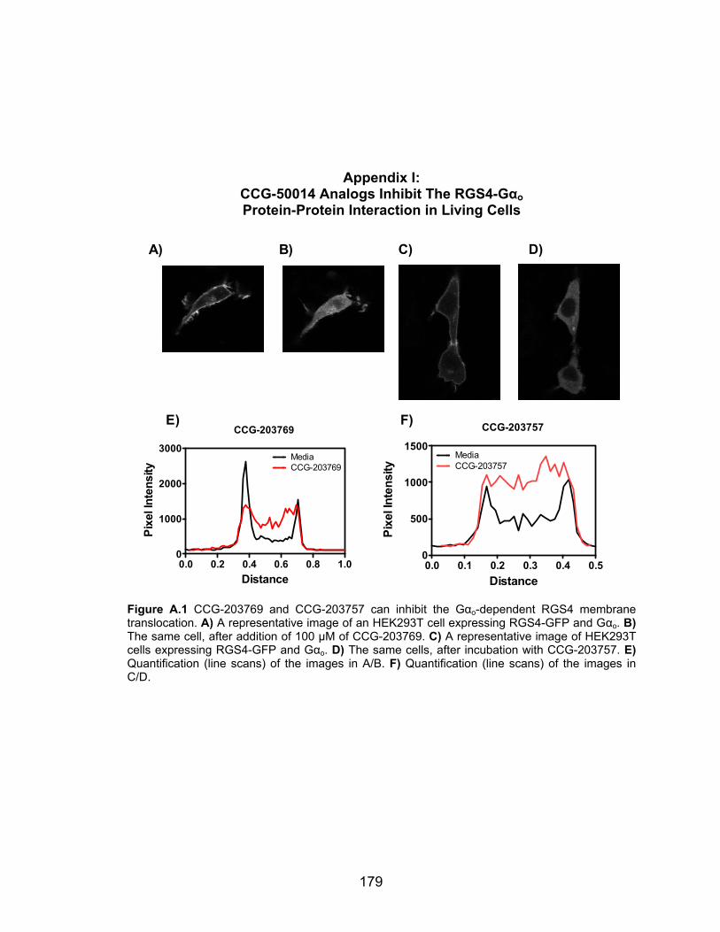
179
Appendix I: CCG-50014 Analogs Inhibit The RGS4-Gαo Protein-Protein Interaction in Living Cells
CCG-203769
0.0 0.2 0.4 0.6 0.8 1.00
1000
2000
3000MediaCCG-203769
Distance
Pixe
l Int
ensi
ty
CCG-203757
0.0 0.1 0.2 0.3 0.4 0.50
500
1000
1500MediaCCG-203757
Distance
Pixe
l Int
ensi
ty
A) B) C) D)
E) F)
Figure A.1 CCG-203769 and CCG-203757 can inhibit the Gαo-dependent RGS4 membrane translocation. A) A representative image of an HEK293T cell expressing RGS4-GFP and Gαo. B) The same cell, after addition of 100 µM of CCG-203769. C) A representative image of HEK293T cells expressing RGS4-GFP and Gαo. D) The same cells, after incubation with CCG-203757. E) Quantification (line scans) of the images in A/B. F) Quantification (line scans) of the images in C/D.
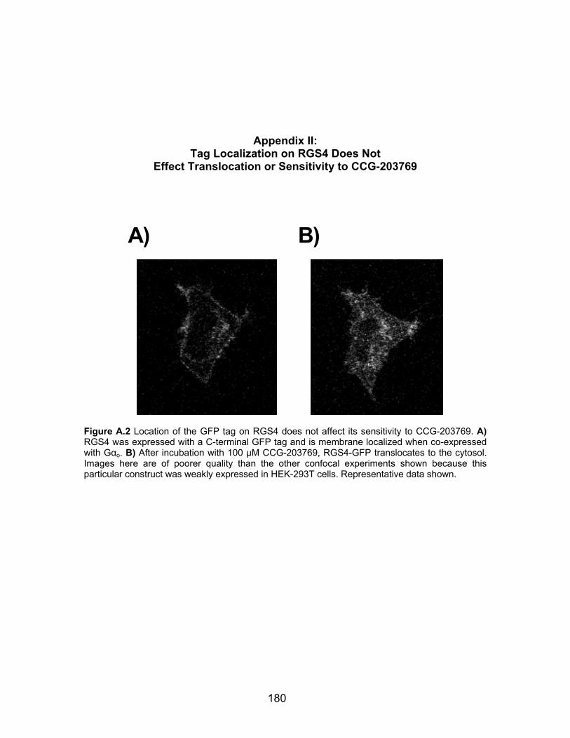
180
Appendix II: Tag Localization on RGS4 Does Not
Effect Translocation or Sensitivity to CCG-203769
A) B)
Figure A.2 Location of the GFP tag on RGS4 does not affect its sensitivity to CCG-203769. A) RGS4 was expressed with a C-terminal GFP tag and is membrane localized when co-expressed with Gαo. B) After incubation with 100 µM CCG-203769, RGS4-GFP translocates to the cytosol. Images here are of poorer quality than the other confocal experiments shown because this particular construct was weakly expressed in HEK-293T cells. Representative data shown.