Embed Size (px)
Citation preview
1
Department of Internal Medicine and Clinical Nutrition, Institution of Medicine, Sahlgrenska Academy,
University of Gothenburg, Gothenburg, Sweden
Diagnosis & dietary intervention in patients with diabetic
gastroparesis
Eva A Olausson
University of Gothenburg
Gothenburg 2013
2
Abstract Diagnosis & dietary intervention in patients with diabetic gastroparesis Eva A Olausson Department of Internal Medicine and Clinical Nutrition, Sahlgrenska University Hospital, University of Gothenburg, Gothenburg, Sweden. Background: Gastroparesis is a diabetic complication, which is often under-recognised. Existing treatment options are limited and diagnostic methods not easily accessible. Aims of the thesis: To compare two alternative diagnostic methods for gastroparesis in patients with insulin-treated diabetes (DM) and to compare gastrointestinal (GI) symptoms and metabolic control after intake of diet with a large (LP) and small particle size (SP) in patients with insulin-treated DM with gastroparesis. Methods: In Paper I, gastric emptying of radiopaque markers (ROM) from the stomach using fluoroscopy was compared to gastric scintigraphy (current gold standard for diagnosing gastroparesis)) and the link to GI symptom severity was determined. In Paper II, the plasma glucose response after a test meal with a LP was compared to gastric scintigraphy in diabetic subjects with and without gastroparesis and the association with GI symptom severity was evaluated. In Papers III and IV, the effects of meals with a LP or SP were compared in randomised controlled trials (RCT). In Paper III, the effect of SP and LP diets on gastric emptying measured using scintigraphy and on the postprandial glucose response were studied in subjects with DM type 1 and gastroparesis and in healthy controls. In Paper IV, subjects with insulin-treated DM and gastroparesis were treated with a SP or LP diet for 20 weeks and the effects on GI symptoms and metabolic control were compared between the groups. Results: Paper I: 115 patients with insulin-treated DM were included and 83 subjects had gastroparesis determined using scintigraphy. A moderately strong correlation was demonstrated between scintigraphic (% retained radioactivity at 120 min, T120) and ROM emptying (markers retained at 6 h) (r = 0.47; p <0.0001).The sensitivity and specificity of the ROM test was 34% and 97% respectively. Only scintigraphic gastric emptying correlated significantly with GI symptom severity with the strongest associations for fullness/early satiety (r = 0.34; p <0.001) and nausea/vomiting (r = 0.30; p <0.001). Paper II: We included 83 patients with insulin-treated DM – 53 with gastroparesis and 30 with normal gastric emptying determined by gastric scintigraphy. The patients with gastroparesis had a blunted postprandial glucose response and demonstrated a lower maximum postprandial plasma glucose increase (p <0.05) and a lower incremental area under the plasma glucose curve (p <0.05). GI symptom severity had the best discriminative value to positively identify gastroparesis (sensitivity 87%, specificity 80%). By adding the plasma glucose response to GI symptom severity to identify patients with gastroparesis, the specificity increased (100%), but the sensitivity decreased (37%). Paper III: We studied seven patients with DM type 1 and gastroparesis and seven healthy controls. The lag phase in the stomach and the T120 was significantly shorter and the postprandial blood glucose dip in diabetic subjects was less and of shorter duration after a SP meal compared to a LP meal. Gastric emptying did not differ significantly between groups after a SP meal. Paper IV: We randomised 56 subjects with insulin-treated DM and gastroparesis to eating diet with a SP ('intervention diet'), compared to the recommended diet for DM ('control diet') for 20 weeks. A significantly greater reduction in the severity of the key gastroparetic symptoms – nausea/vomiting, postprandial fullness and bloating - were seen in patients on the intervention diet compared to the control diet. Conclusions: The alternative diagnostic methods for gastroparesis tested in this thesis – gastric emptying of ROM, plasma glucose response after a standardized test meal and GI symptoms assessment – can add information in the clinical setting, but cannot replace the current gold standard, gastric scintigraphy. A meal with a small particle size increases the gastric emptying rate and reduces the postprandial blood glucose dip in DM Type 1 subjects with gastroparesis. Dietary treatment with a SP significantly improves the key symptoms of gastroparesis in patients with DM. Key words: diabetic gastroparesis, gastrointestinal symptoms, scintigraphy, radiopaque markers, postprandial glucose, hypoglycaemia, gastric emptying, quality of life. ISBN 978-91-628-8743-8

3
Till Björn, Cecilia och Per
Att våga är att förlora fotfästet en liten stund.
Att inte våga är att förlora sig själv.
(Sören Kirkegaard)

4
Diagnosis & dietary intervention in patients with diabetic gastroparesis © Eva A Olausson [email protected] ISBN: 978-91-628-8743-8 Published by:
Department of Internal Medicine and Clinical Nutrition
Institution of Medicine
Sahlgrenska Academy
University of Gothenburg
Gothenburg, Sweden
Printed by:
Ineko AB, Gothenburg
5
List of papers 1. Olausson EA, Brock C, Drewes AM, Grundin H, Isaksson M, Stotzer
P, Abrahamsson H, Attvall S, Simrén M. Measurement of gastric
emptying by radiopaque markers in patients with diabetes: correlation
with scintigraphy and upper gastrointestinal symptoms.
Neurogastroenterol Motil. 2013 Mar; 25(3):e224-32.
2. Olausson EA, Grundin H, Isaksson M, Brock C, Drewes AM, Attvall S,
Simrén, M. Plasma glucose response after a test meal, and
gastrointestinal symptom severity in patients with diabetic
gastroparesis. Submitted.
3. Olausson EA, Alpsten M, Larsson A, Mattsson H, Andersson H,
Attvall S. Small particle size of a solid meal increases gastric
emptying and late postprandial glycaemic response in diabetic
subjects with gastroparesis. Diabetes Res Clin Pract. 2008
May;80:231-7.
4. Olausson EA, Störsrud S, Attvall S, Simrén M. A small particle size
diet reduces upper gastrointestinal symptoms in patients with diabetic
gastroparesis: A randomized controlled trial. Submitted.
6
Contents
Page
Abstract 2
List of papers 5
Abbreviations 8
Introduction 9
Historical background 10
Background 11
Development of diabetes care 11
Gastrointestinal autonomy neuropathy-delayed gastric
emptying 12
Gastrointestinal symptoms 13
Metabolic control 14
Nutritional status 14
Physical and psychological wellbeing 16
Care-intensive patients 16
Normal gastric emptying 17
Pathophysiology of diabetic gastroparesis 18
Diagnosis 18
Incidence 19
Prevalence 20
Diagnostic methods 21
Scintigraphy 22
Breath test 22
Ultrasonography 22
SmartPill 22
The paracetamol absorption test 23
Radiopaque markers 23
Magnetic resonance imaging 23
Limitations with existing diagnostic methods 23
Treatment options for patients with diabetic gastroparesis 24
Dietary/nutritional treatment 24
Pharmacologic therapy 25
Surgery, including gastric electric stimulation 27
General and specific aims of this thesis 29
Methodological considerations 31
Subjects 31
7
Radiation Safety Committee and Ethical Review Board 33
Study design 33
Methods 36
Statistical analyses 43
Results and comments 45
Paper I 45
Paper II 49
Paper III 53
Paper IV 57
General discussion 67
General conclusions 73
Summary in Swedish 75
Future perspective 77
Acknowledgements 79
References 81
Appendix 1: Intervention diet 97
Appendix 2: Control diet 101
Appendix 3: Questionnaire: PAGI-SYM 102
Appendix 4: Questionnaire: HADS 104
Appendix 5: Questionnaire: SF-36 107
8
Abbreviations
BMI Body Mass Index
DCCT Diabetes Control and Complications Trial
DM Diabetes Mellitus
GCSI Gastroparesis Cardinal Symptom Index
GCSI-DD Gastroparesis Cardinal Symptom Index Daily Diary
GERD Gastroesophageal Reflux Disease
GFR Glomular Filtration Rate
GI Gastrointestinal
HADS Hospital Anxiety and Depression Scale
HbA1c Glycosylated haemoglobin
IAUC Incremental area under the curve
LADA Latent Autoimmune Diabetes in the Adult
LP Large Particle size
MODY Maturity Onset Diabetes in Young diabetes
MMC Migrating Motor Complex
PAGI-SYM Patient Assessment of upper Gastrointestinal
Symptom severity index
PN Parenteral Nutrition
ROM Radiopaque Markers
SF36 Short Form 36
PCS Physical component score
MCS Mental component score
SP Small Particle size
99mTc Technetium-99m
T50 At the time, 50% of the isotope has passed
through the stomach
T120=R120 The remaining isotope percentage in the stomach 120
minutes after ingested meal

9
Introduction Over the past 25 years, my work has been focused on giving dietary advice
to patients with diabetes mellitus (DM) at a specialized clinic at the
Sahlgrenska University Hospital. The idea behind the research in this thesis
stems from an incident some twenty years ago when I was privileged to
participate in a one-day training programme for physicians on the subject of
diabetic gastroparesis (i.e. delayed gastric emptying due to DM). One
conclusion from the day was that the physicians had tried to help patients
through dietary measures but always with the same disappointing results:
“Dietary intervention for diabetic gastroparesis does not work”. This made
me think: "Why does it not work – it must work." I had to try. I then recalled
that, Inga Thorsdottir at the Department of Clinical Nutrition, a couple of
years earlier had defended a thesis entitled: "Gastric emptying and
postprandial glucose in healthy subjects and patients with diabetes mellitus
type 2." In her thesis it was obvious that a diet with a small particle size
(SP), such as mashed potatoes, passed through the stomach much more
quickly than a diet with a large particle size (LP), such as pasta and rice,
which ought to be valuable for patients with delayed gastric emptying.
Based on this idea, I informed my patients with DM and gastroparesis- that
the stomach needs to knead the diet into very small particles for it to pass
through the pylorus as part of postprandial motility. I asked them to tell me
what kind of food they thought suited them and told them about my dietary
idea and asked them to try the SP diet in order to improve their symptoms.
I specifically remember one patient with DM and severe neuropathy. The
patient had been in hospital for two months because of nausea, bloating,
diarrhoea and faecal incontinence. Now, after a few weeks at home, she
was back in the hospital. She was instructed to eat a SP diet and she did
not need to be hospitalised again for gastrointestinal (GI) symptoms.
Another patient, who had a loss of appetite, nausea, abdominal fullness and
bloating, as well as suicidal thoughts told me that it is acceptable to have
poorer eyesight and to suffer from renal insufficiency but he could not
handle being constantly nauseous. After eating diet with a SP, his appetite
returned and he had no GI symptoms anymore. He looked forward to eating
and no longer had suicidal thoughts.
10
I thereafter used these ideas about a SP diet to consecutive patients in my
outpatient clinic and was heartened when I assessed the outcome in my
first 65 patients who had clearly improved GI symptoms, fewer and milder
episodes of postprandial hypoglycaemia and improved quality of life. I then
realized that I had to perform proper studies to evaluate this diet in a
scientifically sound way and the idea of starting a PhD project was born.
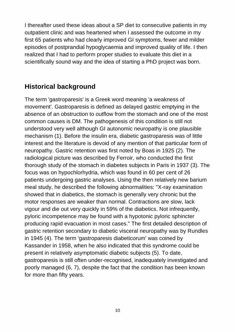
Historical background
The term 'gastroparesis' is a Greek word meaning 'a weakness of
movement'. Gastroparesis is defined as delayed gastric emptying in the
absence of an obstruction to outflow from the stomach and one of the most
common causes is DM. The pathogenesis of this condition is still not
understood very well although GI autonomic neuropathy is one plausible
mechanism (1). Before the insulin era, diabetic gastroparesis was of little
interest and the literature is devoid of any mention of that particular form of
neuropathy. Gastric retention was first noted by Boas in 1925 (2). The
radiological picture was described by Ferroir, who conducted the first
thorough study of the stomach in diabetes subjects in Paris in 1937 (3). The
focus was on hypochlorhydria, which was found in 60 per cent of 26
patients undergoing gastric analyses. Using the then relatively new barium
meal study, he described the following abnormalities: "X-ray examination
showed that in diabetics, the stomach is generally very chronic but the
motor responses are weaker than normal. Contractions are slow, lack
vigour and die out very quickly in 59% of the diabetics. Not infrequently,
pyloric incompetence may be found with a hypotonic pyloric sphincter
producing rapid evacuation in most cases." The first detailed description of
gastric retention secondary to diabetic visceral neuropathy was by Rundles
in 1945 (4). The term 'gastroparesis diabeticorum' was coined by
Kassander in 1958, when he also indicated that this syndrome could be
present in relatively asymptomatic diabetic subjects (5). To date,
gastroparesis is still often under-recognised, inadequately investigated and
poorly managed (6, 7), despite the fact that the condition has been known
for more than fifty years.
11
Background
The incidence of DM is increasing worldwide. Population growth, ageing
populations and urbanisation, with an associated lifestyle change, are likely
to result in a 54% increase in worldwide numbers of people with DM
between 2010 and 2030.
It is estimated that worldwide DM prevalence will be 6.4% and 7.7% for in
2010 and 2030 respectively (8). The total number among the adult
population (aged 20-79 years) was about 285 million in 2010 and will be
439 million in 2030.
There is a difference between developed and developing countries.
Between 2010 and 2030, there will be a 69% increase in the number of
adults with DM in developing countries and a 20% increase in developed
countries (8). The highest increase in prevalence will be in Africa with 98%
in 2030 due to a growing population and increasing incidence.
After several years of DM, the patient could develop, micro- and macro-
angiopathy complications. Retinopathy, nephropathy and neuropathy are
micro-angiopathy complications and cardiovascular disease is a macro-
angiopathy complication (9-13). These complications mean that the
individual is suffering and is a financial burden on society as their care is
expensive. The burden of DM complications is a growing problem.
Development of diabetes care
Knowledge of DM has increased and national and local diabetic care
programmes have developed significantly over the past 20-30 years.
Today, it is well known that good metabolic control, treatment of
hypertension and hyperlipidaemia and absence of smoking are important in
preventing complications (9, 10, 14-16).
Severe complications can be prevented by, for example, using fundus
photography, where fundus changes can be detected at an early stage.
Laser treatment can prevent blindness and foot care can prevent different
foot problems and amputations.
12
The development of a method to examine metabolic control over the past 8-
12 weeks, glycosylated haemoglobin (HbA1c), is a valuable tool for both
patients and health care professionals (17, 18). Technical equipment that
allows the patient to self-measure plasma glucose increases the options for
achieving better metabolic control. Sophisticated technical equipment for
continuous measuring of plasma glucose and insulin pumps with different
technical solutions, e.g. different infusion rates at different times during the
day, and delay of meal insulin doses, make it is easier to achieve good
metabolic control. Other possible technical advantages with an insulin pump
are that it can calculate the amount of insulin that is present in the body and
how much insulin that is needed for a meal. All these developments
increase the possibility of achieving good metabolic control, but despite this
still many patients do not achieve good metabolic control and therefore
develop complications.
Gastrointestinal autonomy neuropathy – delayed gastric
emptying
Diabetic gastroparesis, i.e. delayed gastric emptying in the absence of an
obstruction to outflow from the stomach, caused by gastrointestinal
autonomic neuropathy (19) is another complication in DM (20, 21) and
occurs in both DM type 1 (5, 22, 23) and DM type 2 (24, 25). The
pathogenesis of this disabling condition is still not understood very well (21,
26). A common view is that it is only patients with long disease duration that
may have diabetic gastroparesis and that gastroparesis occurs only after at
least 10 years of DM (25, 27, 28). However, in individual cases,
gastroparesis may occur after just a few years of diabetes (29). Usually, the
patient not only has the micro-complications and Gl autonomic neuropathy,
but also other diabetic complications. Many patients with DM type 1 may
have triopathy, i.e. neuropathy, retinopathy and nephropathy (28). Kockar et
al. found a strong correlation between nephropathy, retinopathy and cardiac
autonomic denervation and gastroparesis in subjects with DM type 1 and 2
(30). Hyett et al. demonstrated a correlation between gastroparesis and
cardiovascular disease, hypertension and retinopathy (31). For the patient,
each of these complications is a burden and together they lead to a
considerable reduction in quality of life and a conspicuous increase in
mortality (32). Gastroparesis is sometimes associated with GI symptoms
13
and/or poor metabolic control (25), dehydration, electrolytes disturbances
and/or nutritional deficiencies (33).
Gastrointestinal symptoms
Some patients with delayed gastric emptying have GI symptoms (31, 34-
37), while others are often asymptomatic, even with markedly delayed
gastric emptying (5, 31, 38, 39). Conversely, Stanghellini et al. confirmed a
high prevalence of delayed gastric emptying and gastroparesis in patients
with upper GI symptoms (40). On the other hand, up to 50% of patients with
markedly delayed gastric emptying had no GI symptoms whatsoever (39,
41). In addition, in a population-based study, DM was associated with
increased prevalence of upper and lower GI symptoms. This was linked to
poor glycaemic control (42) but not to duration of DM or type of treatment
(43). GI symptoms that occur in patients with gastroparesis are upper GI
symptoms such as loss of appetite, meal-related cough, early satiety,
nausea, vomiting, abdominal fullness, bloating and regurgitation, but also
lower GI symptoms such as constipation, diarrhoea with gas, particularly
nocturnal, and faecal incontinence, which cannot be attributed to the
delayed gastric emptying per se, but to a generalized visceral neuropathy
(44). However, these GI symptoms are not specific for diabetic autonomic
neuropathy, but also occur in other conditions, such as irritable bowel
syndrome, functional dyspepsia and a multitude of organic GI diseases.
GI symptoms that correlate with gastroparesis are early satiety (25, 36),
upper abdominal bloating/fullness (25, 34, 37), nausea/vomiting (25) and
abdominal pain (45).However, it should be noted that the severity of GI
symptoms does not always correlate with the rate of gastric emptying (34,
38).
Clinical manifestations are often underestimated and autonomic neuropathy
should therefore be suspected in all diabetic patients with unexplained GI
symptoms (7). Lack of GI symptoms does not exclude severe gastric
emptying abnormalities (39). The lack of correlation between impaired
gastric motility in the fasting state and dyspeptic symptoms shows that, on
the basis of the clinical symptom analysis, the prevalence of such motor
disorders could be underestimated (46). The early recognition of GI motility
14
alterations in autonomy neuropathy may be important for the better long-
term management of patients with DM (46, 47).
Metabolic control
Both symptomatic and asymptomatic diabetic gastroparesis seem to be
associated with poor glycaemic control by causing a mismatch between the
onset of action of the exogenous insulin or oral hypoglycaemic drug, gastric
emptying and the absorption of nutrients from the small intestine (48).
Decreased postprandial glycaemia and late increased glycaemia, resulted
in higher glucose fluctuations in subjects with abnormal gastric emptying
than in those with normal gastric emptying (49). Clinical incidence of
severe, emergency hypoglycaemic events occur sometimes in diabetic
gastroparesis (50). Hypoglycaemic activation of counter-regulation
(Somogyi phenomenon) in all subjects with insulin-treated DM is best
defined as hyperglycaemia following hypoglycaemia and is caused by
insulin resistance induced by hypoglycaemic activation of counter-
regulation (51). This counter-regulation sometimes occurs for several hours,
resulting in higher HbA1c (22, 39).
In addition, acute changes in blood glucose concentration affect gastric
motor function; gastric emptying is slowed down during hyperglycaemia (52,
53) and is accelerated during hypoglycaemia (54). Hyperglycaemia should
be taken into account in the treatment of the patient, as gastric emptying is
slower than necessary. At a glucose level of 8 mmol/L, gastric emptying is
already slower than at 4 mmol/L (53). It is therefore logical that DM patients
have more severe postprandial fullness during hyperglycaemia than during
euglycaemia (55).
Nutritional status
In patients with gastroparesis, the presence of GI symptoms is associated
with eating problems, potentially resulting in food aversion. Patients with
gastroparesis are thus at risk of weight loss and malnutrition.
15
In 2011, Parkman et al. published the first large study aimed at determining
nutritional deficiencies in diabetic and idiopathic gastroparesis. In total, 194
patients (64%) reported caloric-deficient diets, defined as <60% of
estimated daily total energy requirements. More severe symptoms (bloating
and constipation) were characteristic of patients who reported an energy-
deficient diet, and 82% of the subjects had protein intake <0.6 g/kg body
weight. Some patients have protracted nausea, vomiting and diarrhoea,
making it difficult to maintain hydration and electrolyte balance.
Deficiencies in the intake of vitamins and minerals from diet ranged from
30% to 86% of patients (56). However, nutritional consultation took place
infrequently (56).
The current dietary therapy for patients with diabetic gastroparesis is based
on a few studies that changed one parameter at a time in normal volunteers
and measured the effects on gastric emptying. Although the results from
some of these interventions have reached statistical significance, they have
not been shown to be clinically significant (57). Theoretically, a host of
factors can slow gastric emptying but no clinical trials have been conducted
in patients with gastroparesis to determine the success of any dietary
interventions (57).
No randomised dietary study has been performed in patients with
gastroparesis (58). For patients who fail to stabilise their weight loss or who
cannot gain weight with oral feeding, enteral nutrition support may be
indicated. Enteral nutrition not only allows the patient to stay hydrated and
nourished but also provides ready access for consistent medication delivery
(57). Occasionally, a jejunostomy may be helpful to maintain nutrition (59).
Parenteral nutrition (PN) is associated with more infectious complications
and is significantly more expensive (59). However, many patients with
gastroparesis are drug refractory and invariably do not do well with enteral
or parenteral access. Historically, these patients have been without
effective therapeutic options. The development of gastric electrical
stimulation has allowed patients with severe drug-refractory gastroparesis
to be treated successfully and it may also improve survival rates (60). To
summarize, nutritional but also psychological support is important for these
patients, but unfortunately often neglected (61).
16
Physical and psychological well-being
Several studies have addressed the prevalence of GI symptoms in DM and
their importance for physical and psychological well-being. GI symptoms in
unselected DM subjects impact negatively on health-related quality of life in
DM and this is similar in community and outpatient DM subjects. Increased
levels of state anxiety, depression and neuroticism are associated with
upper and lower GI symptoms in DM (62).
Parkman et al. arrived at the conclusion that quality of life in gastroparesis
subjects, as assessed by SF 36, was below that of the US population.
Gastroparetic subjects with DM type 1 had the lowest mental health
summary score and in diabetes type 2 the lowest physical health summary
scores were seen (63). Also the fact that GI symptoms in subjects with
gastroparesis decrease quality of life has been confirmed in several studies
(64, 65). Not surprisingly, nausea, vomiting (66) and bloating symptoms
correlate with poorer quality of life in patients with DM and gastroparesis
(67).
Also depressive symptoms have been demonstrated in diabetic
gastroparesis, and by using the Beck Depression Index, 25% of the patients
in the diabetic gastroparesis type 1 group were found to have severe
depression and 17% of the patients in the diabetic gastroparesis type 2
group. Moreover, with the State-Trait Anxiety Inventory questionnaire, state
and trait anxiety scores were found to be severe in a large proportion of
these patients – around 45% and 30% in types 1 and 2 respectively (63).
To summarize, gastroparesis can be extremely troublesome, causing poor
metabolic control and GI symptoms and result in poor quality of life (33, 68).
Care-intensive patients
Around 20% of hospitalised DM type 2 patients had delayed gastric
emptying (36). A study in the USA found that gastroparesis-related
hospitalisations from 1995-2004 had risen. Admission with gastroparesis as
the primary diagnosis increased by 158% and 138% in those with the
condition as a secondary diagnosis (69). The potential explanations for this
17
rise include the wider recognition of the disease, higher prevalence of DM
and longer survival of DM patients (70).
Jung et al. (28) provided conclusive evidence that gastroparesis was
associated with higher mortality but also with morbidity (31), increased
hospitalisation and emergency department and doctor visits by DM types 1
and 2 patients with classic symptoms of gastroparesis. Patients with a
documented delay in gastric emptying were more likely to have
cardiovascular disease, hypertension and retinopathy, suggesting that the
underlying complication might be related to micro-angiopathies or macro-
angiopathies, which are known complications of poor metabolic control (28).
In a recent study, the healthcare consumption and the GI symptom severity
in a small group of patients with gastroparesis and GI symptoms was
determined, and it was confirmed that these patients form a high-risk group
in terms of cost, quality of life, morbidity and mortality (71).
Normal gastric emptying
The stomach is composed of several regions with different yet
complementary actions. The fundus is a storage area for food and the
antrum and pylorus ensure grinding and move the stomach contents from
the fundus to the antrum. The stomach is an innervated muscle and the
motor function is co-ordinated with that of the pylorus to ensure
fragmentation of the food bolus and ensure digestion regularity (50).
Peristaltic activity is dependent on the pacemaker in the stomach, located
on the greater curvature (Interstitial cells of Cajal). Peristaltic activity
propagates down to the antrum and pylorus at a frequency controlled by a
natural rhythm related to the gastric slow waves, and occurs at a maximum
frequency of about three cycles per minute (50). The slow waves arrive
continuously while the peristaltic contractions occur only after food intake.
Solid nutrients usually empty in two phases over 3-4 hours. An initial lag
phase (i.e. the retention phase) is followed by a propellant phase of
relatively constant emptying. During the first phase, diet is churned while
antral contractions propel particles towards the closed pylorus. Diet is
emptied once it has been broken down into particles approximately 2 mm in
diameter (72). Liquid diet is usually emptied faster, especially with large
18
volumes. If there is an increased number of calories in the liquid, emptying
is relatively constant over time (72).
In the fasting state, gastric motility undergoes a cyclical pattern, termed the
'migrating motor complex' (MMC). This consists of phase I (motor
quiescence, ~40 min), phase II (irregular contractions, ~50 min) and phase
III (regular contractions of three per minute for ~5–10 min). Large,
indigestible solid particles are usually emptied from the stomach into the
small intestine during phase III (73).
Pathophysiology of diabetic gastroparesis
Gastroduodenal motor abnormalities in DM patients with delayed gastric
emptying include parasympathetic nerve damage. However, changes in the
secretion of hormones, such as motilin and ghrelin, are also involved in the
pathophysiological process. These abnormalities lead to excessive
relaxation in the corpus, less frequent antral contractions, antroduodenal
incoordination (74, 75) and pyloric spasm (75). It should be noted that the
latter rarely occurs in isolation and is typically associated with antral
hypomotility (75). Abnormalities in small bowel motility might result in
delayed gastric emptying of solids. Gastric motor dysfunction might be
associated with small bowel dysmotility caused by a common mechanism
(74). In addition, disturbances of proximal gastric compliance, either
increased (76) or decreased (77), have been reported and could also
contribute to symptoms.
In a fasting condition, the chronic influence of diabetic neuropathy on the
stomach is decreased amplitude of the pressure waves in the antrum during
MMC phase III, significantly fewer contractions and in half of the diabetic
gastroparesis subjects a total loss of MMC phase III in the stomach (78).
Diagnosis
Gastroparesis is most often idiopathic, diabetic or postsurgical in nature,
with other causes representing a minority of cases (79). Diabetic
gastroparesis is found in about one-third of all gastroparesis patients (80).
19
Kassander coined the term 'gastroparesis diabeticorum' in asymptomatic
patients with DM type 1 (5). However, other definitions were formulated
later. Gastroparesis, literally defined as 'weakness' of the stomach, is
characterised by an abnormal delay in gastric emptying in the absence of
mechanical obstruction (81). There is a lack of a standard definition for
gastroparesis, although it is frequently defined as a rate of emptying that is
at least two standard deviations above the normal mean, usually
determined using a scintigraphic technique (82). It has been suggested that
a distinction should be made between gastroparesis and delayed gastric
emptying. It has been suggested that gastroparesis, or gastropathy, be
defined based on the presence of upper GI symptoms alone when other
causes, including gastric outlet obstruction, have been excluded (83, 84).
However, there is a lack of consensus as to its appropriateness. While
many patients with gastroparesis have upper GI symptoms, there is a
relatively poor relationship between symptoms and the rate of gastric
emptying (82, 85). Gastroparesis can also have other important clinical
manifestations, such as impaired glycaemic control in insulin-treated DM
patients (85). It should be recognised that in many cases the delay in
gastric emptying of solids or liquids is relatively modest and it can be
argued that a distinction should be made between gastroparesis and
delayed gastric emptying, i.e. that a diagnosis of gastroparesis should be
restricted to those patients in whom emptying is grossly delayed (83).
Incidence
Few studies have addressed the incidence of gastroparesis. The
cumulative proportions of patients who developed gastroparesis over a 10-
year period were 5.2% for type 1 DM, 1.0% for type 2 DM, and 0.2% for
non-diabetic controls (70). The age-adjusted incidence of definite
gastroparesis per 100,000 person years for the period 1996-2006 was 2.4
(95% confidence interval, CI, 1.2-3.8) for men and 9.8 (95% CI, 7.5–12.1)
for women (28). When comparing DM type 1 and type 2, patients with DM
type 2 are 13 years older at onset of symptoms associated with
gastroparesis than patients with DM type 1 (63).
20
Prevalence
The prevalence of gastroparesis in the community is difficult to estimate
due to the relatively poor correlation of symptoms with gastric emptying rate
and the need to apply a diagnostic test in a community setting. It is unclear
whether the majority of patients with gastroparesis seek healthcare or how
often they are referred to gastroenterologists. The true prevalence of
gastroparesis is therefore unknown. In the DM population, the prevalence of
gastroparesis is also difficult to estimate due to the apparently higher
prevalence of the disorder in university centres than in smaller hospitals, in
a primary care setting and in the community. A widely available diagnostic
test that could be applied in a standard fashion is currently lacking in the
primary care setting (86, 87).
Gastric scintigraphy is considered the gold standard worldwide. This
method is expensive and requires specialised staff. One problem is that
scintigraphy is not always performed according to the same standards. In
gastric scintigraphy, measurement takes place either during two- or four
hours and different test meals are used, hence consensus
recommendations are not always followed (86, 88). In most centres
different reference values are used for men and women (86), as women
have slower gastric emptying than men. However, there are also
differences between pre- and post-menopausal women depending on
hormones (89). Moreover, in diabetic gastroparesis it is important to assess
gastric emptying in a euglycaemic condition as hyperglycaemia delays
gastric emptying (52, 53, 90). The study design is also important. Are the
samples selected or unselected and which inclusion criteria are applied?
Most population-based DM studies in subsets of patients at risk of
developing gastroparesis have focused on symptoms rather than gastric
scintigraphy findings (81). In such investigations, 10–18% of individuals with
DM report symptoms consistent with upper GI dysmotility, such as nausea
and vomiting (43, 91, 92). Jung et al. made an attempt to study the
prevalence of all kinds of gastroparesis in the general population. After
studying the complete medical records in Olmsted County, Minnesota from
1996 to 2006, there were 3,604 potential cases of gastroparesis. After
exclusion criteria were applied, 432 subjects remained. The inclusion
criteria were: Definite, probable and possible gastroparesis and gastric
emptying investigated using scintigraphy. The age-adjusted (compared to
21
the white population in the US in 2000) prevalence of definite gastroparesis
per 100,000 persons was 24.2 (95% confidence interval [CI], 15.7– 32.6) for
both genders, 9.6 (95% CI, 1.8 –17.4) for men and 37.8 (95% CI, 23.3–
52.4) for women (28).
In another study, the prevalence of gastroparesis was 36% in symptomatic
subjects with DM type 1 and 2 (93). Other studies have also confirmed that
the prevalence in females is higher than in males (36, 70).
A study in the DM population assessed gastric emptying of a solid meal
with scintigraphy under euglycaemic conditions (23), confirming the
prevalence of gastroparesis in unselected patients with DM type 1 at 34%.
In another study, the prevalence was 28% in DM type 1 (35). In two studies
investigating selected groups of patients with long-standing type 1 DM, the
prevalence was 40-44% (22, 48).
The prevalence of gastroparesis in subjects with DM type 2 is in the range
30-58% (94-96). However, these studies are limited by the small sample
sizes, the presence of hyperglycaemia during testing and the variety of test
meals and diagnostic methods used. In the study performed by Annese et
al., 34% of patients had peripheral and autonomic neuropathy and 36% of
the patients showed abnormal esophageal and gastric motor function (96).
Diagnostic methods
There are various means of assessing gastric emptying (97). The current
gold standard is gastric scintigraphy, but other techniques have been
evaluated and/or are under evaluation. Some of these will be reviewed
below.
Scintigraphy. Using scintigraphy, the emptying time and intragastric
distribution of solid and/or liquid meal components can be evaluated.
Scintigraphy, which is non-invasive and reproducible, remains the most
sensitive and accurate method and is considered to be the 'gold standard'
technique. It measures the intragastric distribution and emptying of a solid
or liquid meal labelled with Technetium- 99m,99mTc. The isotope binds to
meal protein and a gamma camera registers the 99mTc passage through the
stomach. Gastric emptying is monitored for 3-4 hours after meal
22
consumption. The gastric emptying is evaluated as follows: Lag phase = the
time from finished meal before the gastric emptying begins, T50 = at that
time, 50% of the isotope has passed through the stomach and R120 or T120 =
the remaining isotope percentage in the stomach 120 minutes after the
ingested meal. This method is expensive and requires specialised staff
(86), and is not easy accessible for many clinicians. Moreover, radiation
exposure is another drawback with this technique. Alternative methods are
therefore needed.
Breath tests. These tests assess gastric emptying using meals labelled
with stable isotopes, such as 13C-acetate or 13C-octanoate and, in contrast
to scintigraphy, does not involve exposure to ionising radiation. It has good
reproducibility and the results have been reported to correlate well with
scintigraphy, with a sensitivity and specificity of 86-89% and 80%
respectively for the presence of delayed gastric emptying, including a DM
population (98, 99). Furthermore, they are easy to administer and are
inexpensive (100, 101). After ingestion, the labelled meal passes through
the stomach to the small intestine, where the 13C-acetate or 13C-octanoate
is absorbed, metabolised into 13CO2 in the liver and exhaled via the breath. 13CO2 in breath samples is analysed by means of mass spectrometry (45,
101). Although this technique has advantages over scintigraphy,
information relating to the validity of breath tests in patients with markedly
delayed gastric emptying is limited.
Ultrasonography. Two-dimensional ultrasonography is a convenient, valid,
non-invasive method of measuring the emptying of liquids or semi-solids as
well as antral contractions and transpyloric flow (102, 103). The more
recently applied three-dimensional ultrasonography can provide
comprehensive imaging of the stomach, including information about
intragastric meal distribution, and has been validated against scintigraphy to
measure gastric emptying in both healthy subjects and in patients with
diabetic gastroparesis (104). Although ultrasonography is readily available
and does not involve exposure to radiation, obesity, abdominal gas and the
need for an experienced operator limit its widespread use.
SmartPill®. Swallowed capsule telemetry (SmartPill®) uses an ingestible
capsule that measures intraluminal pH to determine the gastric emptying
rate. Moreover, the capsule can also record pressure during the passage
through the GI tract as a measure of contractions, as well as temperature.
23
This method has been reported to correlate well with scintigraphy with good
sensitivity (82%) and specificity (83%) to diagnose delayed gastric
emptying(61) but has only recently been approved for use in Europe.
Emptying of the capsule presumably occurs after that of digestible meal
components, and this technique has been criticised for measuring re-
appearance of gastric fasting motility (MMC, phase III) rather than emptying
of a meal.
The paracetamol (acetaminophen) absorption test. Another principle
used to measure gastric emptying is the absorption of paracetamol
assessed with serial blood samples after intake of paracetamol. The
paracetamol (acetaminophen) absorption test is a simple bedside test, but
its usefulness is limited to evaluation of the emptying of liquids and is
generally not recommended as a diagnostic tool as its accuracy is
moderate at best (105).
Radiopaque markers. Emptying of radiopaque markers (ROM)
(indigestible solids) from the stomach using fluoroscopy is an easy and
inexpensive method and is available at all hospitals. There is a significant
correlation between gastric emptying of ROM with gastric scintigraphy in
healthy subjects and in a small group of subjects with insulin-dependent DM
(106). However, the correlation between gastric emptying of ROM and
gastric scintigraphic emptying in larger groups of patients with gastroparesis
has not been studied.
Magnetic resonance imaging. Magnetic resonance imaging has also been
used to measure gastric emptying and wall motions of the stomach.
Abdominal scans are generally obtained in the supine position every 15 min
before and after a predominantly liquid meal. This method is still not widely
spread and is under development. An advantage with this method is that it
does not expose the subject to radiation, but it is limited by the use of
specialized equipment, by being expensive and by using the supine
position.
Limitations with existing diagnostic methods
Several of the existing diagnostic methods have limitations, such as
radiation exposure, expenses etc., which has been described above. One
24
of the major limitations is availability, and there is a limited number of
methods for investigating gastroparesis at small hospitals, which may be
one reason why this condition has been investigated inadequately. In order
to improve the diagnostic management of gastroparesis access to different
diagnostic methods should be available in all hospitals. Fluoroscopy is
available at all hospitals, and therefore we aimed to study the correlation
between gastric scintigraphy and the ROM method in patients with
gastroparesis, to evaluate if this widely available method could be used
more in the diagnostic approach in patients with suspected delayed gastric
emptying. Moreover, as patients with DM and delayed gastric emptying
have an altered glucose response after meal intake (39, 107, 108) and
some of them also report upper GI symptoms (25, 34, 36), we also
hypothesized that measuring blood glucose after meal intake and to assess
the symptom profile could be helpful in finding DM patients with suspected
gastroparesis. Our research question was therefore if GI symptoms in
recent weeks and/or glucose response after a test meal could be a
screening method to strengthen the suspicion of gastroparesis?
Treatment options for patients with diabetic gastroparesis
The treatment for patients with diabetic gastroparesis is aimed at reducing
GI symptoms, optimising glucose control and correcting fluid, electrolyte
and nutritional deficiencies (109). The principles in the management of
patients with diabetic gastroparesis is based on dietary/nutritional
adjustments, optimisation of the glycaemic control, pharmacologic therapy
with prokinetics or antiemetics, avoidance of opioids, and in very select
cases specialized therapies such as surgery, botox injections or gastric
electrical stimulation (110).
Dietary/nutritional treatment. To date, the recommendation for
management of gastroparesis using dietary/nutritional measures is as
follows:
1. The first line of management for gastroparesis patients should include
restoration of fluids and electrolytes, nutritional support and, in DM patients,
optimisation of glycaemic control. (Strong recommendation, moderate level
of evidence.)
25
2. Oral intake is preferable for nutrition and hydration. Patients should
receive counselling from a dietician regarding the consumption of frequent,
small-volume nutrient meals that are low in fat and soluble fibre. If they are
unable to tolerate solid food, then the use of homogenised or liquid nutrient
meals is recommended. (Conditional recommendation, low level of
evidence.)
3. Oral intake is the preferred route for nutrition and hydration. If oral intake
is insufficient, then enteral alimentation by jejunostomy tube feeding should
be pursued (after testing nasoenteric tube feeding). Indications for enteral
nutrition include unintentional loss of 10% or more of the usual body weight
during a period of 3-6 months and/or repeated hospitalisation for refractory
symptoms. (Strong recommendation, moderate level of evidence.)
4. For enteral alimentation, postpyloric feeding is preferable to gastric
feeding, as gastric delivery can be associated with erratic nutritional
support. (Conditional recommendation, low level of evidence.)
5. Enteral feeding is preferable to parenteral nutrition. (Conditional
recommendation, low level of evidence.) (109).
However, so far there is no randomised, controlled diet trial for
gastroparesis. Dietary advice comprises suggestions that stem from the
results of studies performed in subjects with normal gastric emptying. The
major features in the dietary recommendations are to decrease fat (111-
113) and fibre (114), as these slow gastric emptying. However, most
studies added soluble fibre to liquid meals, which may have more impact of
gastric emptying than soluble fibre added to a solid meal. In healthy
subjects, a fat liquid test meal increased the lag period and decreased the
slope of the emptying curve. During the lag period, there was initial filling of
the distal stomach, followed by redistribution of the distal stomach contents
back into the proximal stomach. At the onset of total gastric emptying, the
proximal stomach also emptied (111). This redistribution must be taken
account in patients with delayed gastric emptying.
In order to prevent formation of bezoars in the stomach of patients with
delayed gastric emptying, advise to avoid foods that may lead to
phytobezoar formation, such as oranges, persimmons, coconuts, berries,
green beans, figs, apples, sauerkraut and Brussels sprouts, should be
given (115). Moreover, small, frequent meals are suggested as large meals
slow down gastric emptying in healthy subjects (116), and the lag phase
26
was prolonged in both solid and liquid large meals compared with smaller
meals (117).
Pharmacologic therapy. The aim of the therapy is to improve the
efficiency of the gastric pump and to relieve GI symptoms. Two classes are
available to treat affected patients – prokinetic and antiemetic drugs. Of
these, only prokinetics have been specifically studied in gastroparesis,
whereas antiemetics, such as phenotiazines, antihistamine agents and
serotonin 5HT3 receptor antagonists are used empirically based on their
effect on nausea and vomiting. Only prokinetics will be described in some
detail here. Prokinetics are the most commonly used medications for
gastroparesis. These stimulate peristalsis and could specifically improve
gastric pump function by influencing antral contractility and rhythm as well
as antroduodenal co-ordination. Established prokinetic agents, including
metoclopramide, domperidone and erythromycin, form the mainstay of the
treatment. Metoclopramide has both prokinetic and antiemetic properties.
The long-term efficacy of metoclopramide, a dopamine D2 receptor
antagonist, has not been established clearly and the prokinetic effects may
potentially diminish over time (118). Metoclopramide is available in several
formulations. It can be given orally, intravenously and subcutaneously. The
use of metoclopramide is marked by a high prevalence of adverse effects
on the central nervous system. Mild central neurological effects, including
restlessness, agitation, dizziness and drowsiness, occur in up to 40% of
patients, while dystonic reactions occur in approximately 1% (118). During
more prolonged use, tardive dyskinesia may develop, which is potentially
irreversible and may occur in 1-10% of patients who have been taking the
drug for more than three months (119). These side effects greatly
diminishes the clinical usefulness of metoclopramide.
Domperidone, another dopamine antagonist, appears to be effective in the
management of symptomatic gastroparesis, including that associated with
DM (120), with fewer side effects than metoclopramide (121). It is relatively
impermeable to the blood-brain barrier and therefore causes fewer adverse
central neurological effects (122). Acute administration of domperidone has
been reported to accelerate gastric emptying of solids and liquids in both
healthy subjects and in DM patients with autonomic neuropathy. The
greatest effect is observed in patients with a marked delay in gastric
27
emptying (123). Domperidone has also been shown to improve quality of
life in patients with DM through its alleviation of GI symptoms (120).
Erythromycin is a macrolide antibiotic that also has the ability to act as a
motilin agonist through its interaction with motilin receptors. Erythromycin
accelerates gastric emptying by increasing the frequency and amplitude of
antral and duodenal contractions in the proximal stomach (85). The effect of
erythromycin on GI symptoms is controversial, although there is evidence to
suggest that it improves bloating in patients with functional dyspepsia and
gastroparesis (82). However, long-term use of erythromycin for
gastroparesis is less well substantiated and tachyphylaxis is thought to
develop due to down-regulation of motilin receptors with chronic use.
Furthermore, there is the potential risk of bacterial resistance (124). The
major adverse effects of erythromycin are well recognised and include
abdominal pain and cramping, nausea, diarrhoea, vomiting and skin rash.
Erythromycin may also cause adverse cardiac effects (124).
Surgery and gastric electric stimulation
Some subjects with gastroparesis remain unsatisfactory despite
dietary/nutritional adjustments, optimisation of the glycaemic control, and
the use of prokinetic and/or antiemetic drugs. In these cases, gastric
electrical stimulation is an option. Also surgical procedures, such as venting
gastrostomy, gastrectomy or pyloroplasty, may be used in very select cases
(110). Gastric electrical stimulation delivers high frequency, low energy
electrical stimulation to the stomach and through an unknown mechanism
many patients with diabetic gastroparesis demonstrate a substantial
reduction of the key gastroparetic symptoms, especially nausea and
vomiting (21). In the first multi-centre trial, thirty-eight highly symptomatic
patients with drug-refractory gastroparesis with differing aetiology, including
diabetic gastroparesis, received gastric electrical stimulation. In 97% of
these patients more than 80% reduction in vomiting and nausea (including
diabetic gastroparesis) was reported. (125). Several studies have followed
and confirmed these initial positive results, especially in diabetic
gastroparesis (21). However, access to a gastric pacing is limited to a few
hospitals and few patients are in a position to benefit from this method. The
cost is also rather high, limiting a widespread use of this treatment option.

28
29
General and specific aims of this thesis
Based on gaps in the existing literature and building on previous clinical and
scientific experience with these patients our aims with the studies included
in this thesis were:
1. To compare two alternative diagnostic methods for gastroparesis
using gastric scintigraphy, the current golden standard, in patients
with insulin-treated DM.
2. To compare GI symptoms and metabolic control after intake of diet
with a LP and SP in patients with insulin-treated DM with
gastroparesis.
Paper I
To evaluate the correlation between emptying of ROM from the stomach
using fluoroscopy and gastric scintigraphy in a large group of patients with
insulin-treated DM in order to define a potential role for ROM emptying in
the management of patients with DM and clinical suspicion of delayed
gastric emptying. We also wanted to assess the link between different GI
symptoms, ROM emptying and scintigraphic gastric emptying of solids.
Paper II
To characterise thoroughly the postprandial glucose response after a test
meal with LP in patients with DM and clinical suspicion of gastroparesis and
to assess whether this, together with a questionnaire-based assessment of
the severity of GI symptoms during the preceding two weeks, could help to
predict delayed gastric emptying determined with gastric scintigraphy.
30
Paper III
To evaluate the effect of meal particle size on gastric emptying and
postprandial blood glucose variations after a meal. To achieve this, we
measured the gastric emptying time and postprandial blood glucose
response in patients with DM Type 1 and gastroparesis and in healthy
controls by comparing meal with a LP with the same meal with a SP.
Paper IV
To compare the effects of a dietary intervention that recommends diet with
SP with the standard dietary recommendations given to patients with DM, in
insulin-treated DM subjects with gastroparesis. Our primary aim was to
improve GI symptoms associated with gastroparesis and the secondary
aims were to assess the effects on body weight, nutritional intake,
metabolic control, mental health and quality of life.
31
Methodological considerations
Subjects
The thesis summarises four studies incorporating insulin-treated DM
subjects with and without gastroparesis and healthy control subjects;
Papers I and II deal with validation of diagnostic and screening methods for
gastroparesis. Papers III and IV deal with randomised trials studying the
relationship between LP and SP in the diet and gastric emptying,
postprandial plasma glucose and GI symptoms in subjects with insulin
treated DM and gastroparesis.
The subjects included in Papers I, II and IV in this thesis were insulin
treated DM patients recruited from DM outpatient clinics in eleven hospitals
and four primary care teams in Region Västra Götaland of Sweden. The
teams were informed about the study through personal visits to the clinics
by EO and through e-mails. During the visits, the teams were informed
about signs and symptoms associated with gastroparesis in order to find
patients who had still not received a diagnosis. The recruitment process
began in August 2007 and was completed in November 2011.
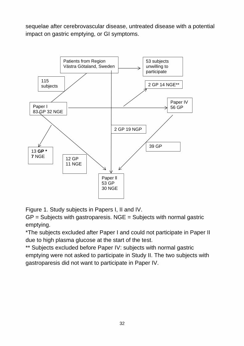
Paper I. 115 subjects with insulin treated DM and with a wide range of GI
symptom severe were included.
Paper II. Insulin-treated DM patients with and without suspected
gastroparesis were included.
Paper IV. Insulin treated DM patients with suspected gastroparesis were
included. Only 14 DM subjects had normal gastric emptying and 2 subjects
with gastroparesis were unwilling to participate in the study. Figure 1.
Paper III. These patients with gastroparesis were recuited from the
Diabetes outpatient clinics, Sahlgrenska University Hospital and Kungälv
Hospital in September 1996 and March 1998. The gender, age and body
mass index matched healthy controls were recruited via an newspaper
advertisement.
Exclusion criteria for all the studies were as follows: previous
gastrointestinal surgery except appendectomy, severe psychiatric disease,
32
sequelae after cerebrovascular disease, untreated disease with a potential
impact on gastric emptying, or GI symptoms.
Figure 1. Study subjects in Papers I, II and IV.
GP = Subjects with gastroparesis. NGE = Subjects with normal gastric
emptying.
*The subjects excluded after Paper I and could not participate in Paper II
due to high plasma glucose at the start of the test.
** Subjects excluded before Paper IV: subjects with normal gastric
emptying were not asked to participate in Study II. The two subjects with
gastroparesis did not want to participate in Paper IV.
Patients from Region Västra Götaland, Sweden
115 subjects
Paper I 83 GP 32 NGE
Paper ll 53 GP 30 NGE
12 GP 11 NGE
Paper IV 56 GP
39 GP
2 GP 19 NGP
13 GP * 7 NGE
53 subjects unwilling to participate
2 GP 14 NGE**
33
Approval by the Radiation Safety Committee and Ethical
Review Board
The studies were approved by the Radiation Safety Committee at
Sahlgrenska University Hospital, Sahlgrenska Academy, University of
Gothenburg, and the Regional Ethical Review Board in Gothenburg,
Sweden. Each participant received verbal and written information about the
study and gave their written, informed consent before any study related
procedure was performed.
Study design
Papers I and II. Before the test, the patients had completed the 'Patient
Assessment of Gastrointestinal Disorders Symptom Severity Index (PAGI-
SYM)' and Gastroparesis Cardinal Symptom Index (GCSI). The subjects
arrived at the hospital at 8 am in a fasting condition (10 hours). Alcoholic
beverages were not permitted 24 hours before the test and drugs with an
effect on GI motility were not permitted for 48 hours before the test.
Smoking was not permitted during the test. The plasma glucose had to be ≤
10.0 mmol/L at the beginning of the meal and the subjects had taken their
ordinary dose of insulin for breakfast, adjusted to the carbohydrate content
of the test meal (units insulin/g carbohydrates). Plasma glucose samples
were taken before the meal and throughout the whole study period.
Paper I. On the same day, we measured solid gastric emptying using
scintigraphy of a 99mTc labelled standard meal for three hours and followed
emptying by fluoroscopy of ROM (non-digestible solids; 20 spherical
radiopaque markers) added to the meal 4-6 h after meal intake. The
fluoroscopy measurement of ROM was followed each hour for three hours
or until all the ROM were emptied. The number of ROM in the stomach was
counted each hour after meal intake. We focused on the time period 4–6 h
after meal intake to test for delayed gastric emptying of ROM since
indigestible solids or ROM are emptied with a time delay of 1.5–2 h
relative to digestible solids (106).
Paper II. Before the study, gastric emptying was confirmed using a
scintigraphic method. The ingestion time of the test meal of food in LP was
34
25 minutes. Plasma glucose was followed for three hours from the
beginning of the meal.
Paper III. The subjects arrived at the hospital at 11.15 am after ingesting a
standard breakfast at 07.30 am and a snack at 9 am. The study started at
noon. Insulin-dependent DM patients with suspected gastroparesis took
their ordinary dose of regular human insulin for lunch (units insulin/g
carbohydrates) 30 minutes before the meal. Plasma glucose before the
meal was ≤ 10.0 mmol/L. In random order, the subjects ingested solid
meals with a LP and SP. Ingestion time was up to 25 minutes. Gastric
emptying was followed by scintigraphy for three hours from the beginning of
the meal. Plasma glucose and insulin were followed throughout the study
period.
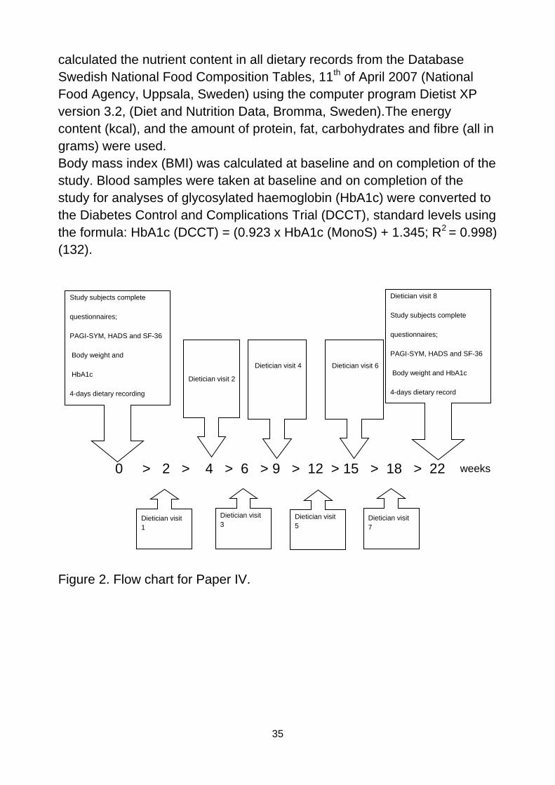
Paper IV. Insulin-treated DM subjects with confirmed gastroparesis were
randomised to receive dietary advice, which differed regarding the particle
size of the food (for more detailed information, see appendices). Otherwise
the nutritional composition in the two dietary treatment groups was the
same and in line with the recommendation for diabetic patients (126), with
the exception of the recommendation that the fat content was reduced to
25-30% of total energy and the fibre content to 15 gram/1000 kcal
according to Paper III, and in order to remain in line with the current
recommendations for patients with gastroparesis (109). The study period
was 20 weeks.
The subjects were also randomised to one of two dieticians, who provided
the dietary advice, and they received dietary counselling on seven
occasions. Both groups were advised to have the same meal schedule:
breakfast, snack, lunch, snack, dinner and evening snack.
GI symptoms, quality of life, anxiety and depression were assessed at
baseline and on completion of the study. The subjects completed the
questionnaires: PAGI-SYM (127, 128), GCSI (129), Short-Form Health
Survey (SF36) (130) and Hospital Anxiety and Depression Scale
(HADS)(131). Nutritional intake was investigated at baseline and on
completion of the study by keeping a four-day dietary record at home. One
dietician provided the subjects with instructions regarding the
questionnaires and dietary recording. Any ambiguities were resolved when
the questionnaires and dietary records were returned. The same dietician
35
calculated the nutrient content in all dietary records from the Database
Swedish National Food Composition Tables, 11th of April 2007 (National
Food Agency, Uppsala, Sweden) using the computer program Dietist XP
version 3.2, (Diet and Nutrition Data, Bromma, Sweden).The energy
content (kcal), and the amount of protein, fat, carbohydrates and fibre (all in
grams) were used.
Body mass index (BMI) was calculated at baseline and on completion of the
study. Blood samples were taken at baseline and on completion of the
study for analyses of glycosylated haemoglobin (HbA1c) were converted to
the Diabetes Control and Complications Trial (DCCT), standard levels using
the formula: HbA1c (DCCT) = (0.923 x HbA1c (MonoS) + 1.345; R2 = 0.998)
(132).
Figure 2. Flow chart for Paper IV.
0 > 2 > 4 > 6 > 9 > 12 > 15 > 18 > 22
Dietician visit
1
Dietician visit 2
Dietician visit
3
Dietician visit 4
Dietician visit
5
Dietician visit 6
Dietician visit
7
weeks
Dietician visit 8
Study subjects complete
questionnaires;
PAGI-SYM, HADS and SF-36
Body weight and HbA1c
4-days dietary record
Study subjects complete
questionnaires;
PAGI-SYM, HADS and SF-36
Body weight and
HbA1c
4-days dietary recording
36
Methods
Measurement of gastric emptying using a scintigraphic technique
(Paper I).
Assessment of gastric emptying using gastric scintigraphy was registered
for three hours in a fasting condition. A gamma camera registered one
mean value every 5 minutes for 180 minutes. The gamma camera
registered gamma radiation from 99mTc in anterior and posterior views of the
stomach, mean value for three minutes followed by a pause for two
minutes. The test meal was according to Swedish standard (133) and
reference values were used (134). The nutrient content of the test meal,
based on eggs, was 310 kcal, 19 g protein (25% of the total energy), 18 g
fat (52 % of the total energy), and 17 g carbohydrates (23 % of the total
energy) and 150 g water.
Comments. Because gastric emptying using scintigraphy is the worldwide
'gold standard', this method was also our choice. The energy percentage of
fat was high in this meal, contrary to what is proposed to be used as the
initial screening test for gastric emptying in the consensus of the American
Neurogastroenterology and Motility Society and the Society of Nuclear
Medicine, U.S. (86). However, it is suggested that in some patients a low-
fat, egg white meal may not prove to be an adequate functional challenge,
especially for patients who report symptom exacerbations after eating lipid-
rich diets. These subjects probably need a high-fat meal (86).
In our papers, separate reference values are presented for men and women
before and after the menopause. Because gastric emptying in females is
slower than in men and women before menopause have a lower gastric
emptying rate, it is important to have different reference values for women
(134). For the most part, certain time points have been used to evaluate
gastric emptying: the time until gastric emptying begins after a finished meal
(lag phase), the time point after a finished meal for emptying 50% of the
isotope (T50) and/or percentage isotope remaining in the stomach at time
point 120 minutes from the finished meal (R120 or T120). For diagnosis, T50
and/or R120 or T120 are most widely used. In this study, the measurement T50
could not be used because not all subjects had emptied 50% of the
radioactivity from the ventricle during the study period (3 h). R120 has
therefore been used to establish a diagnosis of gastric emptying. We
37
measured gastric emptying for three hours and not four hours as advocated
in the literature (86). Abell et al. suggest measurements 1, 2, 3 and 4 hours
after finishing the meal. The choice of three hours measurement only in our
study was because the subjects were in a sitting position during the whole
test, not just at four time points as recommended (86). It is very difficult to
be in a sitting position for three hours and we considered four hours
impossible.
Gastric emptying by radiopaque markers (Paper I).
To assess gastric emptying of ROM we used fluoroscopy and counted the
number of ROM remaining in the stomach 3,4, 5 and 6 h after meal intake,
which is a standard method used at our hospital (106).
Comments. Measurement of gastric emptying with ROM using fluoroscopy
has been compared to gastric scintigraphy in healthy individuals and in a
small group of patients with DM. This validation revealed a significant
correlation between the methods (106). One of several reasons that
gastroparesis is under-recognised and inadequately investigated is poor
access to scintigraphy. However, as fluoroscopy is available at all hospitals,
emptying of ROM could serve as a screening method for delayed gastric
emptying. This method has also been suggested as being a better means
of detecting gastroparesis compared to scintigraphy (135). However, results
with a lower correlation also exist (136). No comparison has been made
between gastric emptying with ROM and scintigraphy in a large group of
gastroparetic subjects. We therefore wanted to validate the method in
gastroparetic patients.
Gastric emptying by scintigraphic technique (Paper III).
Gastric emptying was performed in a sitting position with the gamma
camera positioned behind the subjects throughout the whole study period
(0–180 minutes). The gamma camera registered the mean value every
second minute throughout the 180 minutes. Gastric emptying was
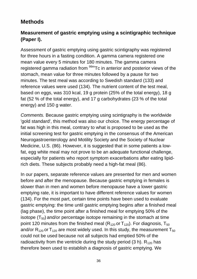
measured using two test meals. They consisted of 100 g of meat, 40 g of
pasta, 150 g of carrot and 5 g of canola oil. The nutrient content was 375
kcal (1.57 MJ), 26 g protein, 13 g fat (31% of the total energy), 38 g
carbohydrates and 4.8 g of fibre (3 g/MJ), calculated using the National
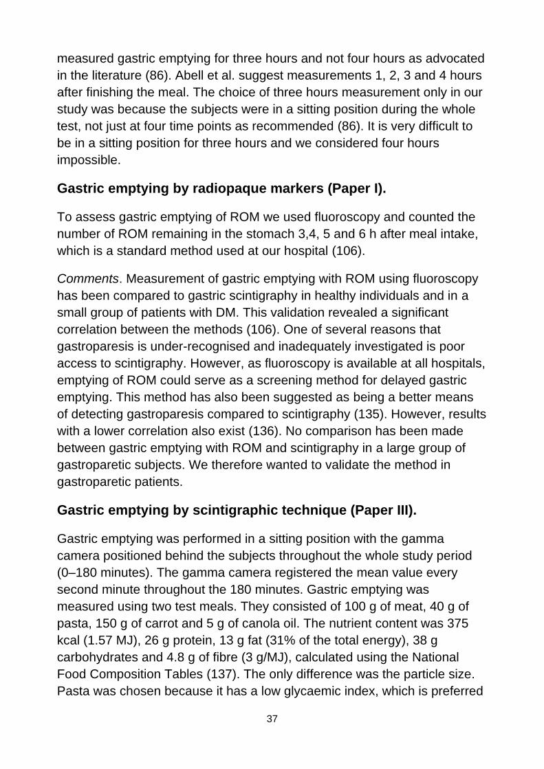
Food Composition Tables (137). The only difference was the particle size.
Pasta was chosen because it has a low glycaemic index, which is preferred
38
in a DM diet (138, 139). Five Mega Becquerel (MBq) of the tracer 99mTc
were added to the uncooked pasta. Supplementary pictures 1-3.
Comments. Because the relationship between the different particle sizes
and gastric emptying was being studied, it was not possible to use any
standard test meal for the scintigraphic method (133), and reference values
were therefore not available. Gender, age and BMI-matched healthy
subjects were therefore included as references for normal gastric emptying
rate.
Supplementary picture 1. Ingredients: carrot, meat and pasta
39
Supplementary picture 2.Test meal with large particle size
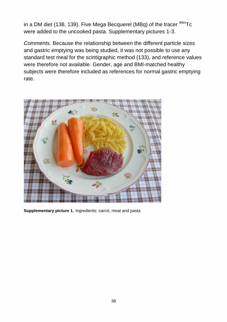
Supplementary picture 3.Test meal with small particle size
Plasma glucose response after a solid test meal (Papers I, II and
III).
In Paper 1, plasma glucose was taken at the beginning of the meal (00), at
the end of the meal (0), and after 15, 30, 45, 60, 90, 120, 150, 180, 210,
40
240, 270, 300, 330 and 360 minutes, or until all ROM had emptied from the
stomach. In Paper II, blood samples were taken before the meal (0) and
after 15, 30, 45, 60, 90, 120, 150, 180 minutes. In Paper III, the first blood
sample was taken at the beginning of the LP and SP meal and then every
15 minutes throughout the whole study period of 180 minutes. Plasma
glucose samples were analysed immediately in an automated plasma
glucose analyser using a glucose oxidase method (Merck, Darmstadt,
Germany) using HemoCue NAD-NADH (HemoCue AB, Ängelholm,
Sweden).
Comments. If the postprandial glucose correlated with gastric emptying, this
measurement should be a very cheap and widely available screening
method for gastroparesis. Every diabetic team could take plasma glucose
samples and thus have a cheap tool at their disposal.
During the testing in Paper II, the subjects were in a sitting position and
were not allowed to walk around as it can affect both gastric emptying (140)
and plasma glucose response. They only walked to the toilet close to the
test room. In Paper III, the subjects were in a sitting position the whole time.
Metabolic control (Papers I, II, III and IV)
HbA1c was converted to the Diabetes Control and Complications Trial,
standard levels using the formula: HbA1c (DCCT) = (0.923 x HbA1c
(MonoS) + 1.345; R2 = 0.998) (132).
Comment. To assess the metabolic control HbA1c was analysed in all
studies as it reflects the metabolic control during 8-12 weeks (17, 141). The
glycosylated haemoglobin (HbA1c) is presented according to the Diabetes
Control and Complications Trial standard.
Measurement of insulin concentration (Paper III)
The insulin concentration was analysed every 15 minutes for 0 -180
minutes. The serum insulin samples were centrifuged and stored at -20ºC
until analysis. The insulin levels were determined using a radio
immunochemical RIA assay technique (Diagnostic Products Corporation,
Los Angeles).
41
Comment. The insulin concentration has an impact on the postprandial
glucose curve. To sort out the impact of gastric emptying on postprandial
glucose, the impact of insulin concentration and gastric emptying needs to
be differentiated.
Gastrointestinal symptoms (Papers I, II and IV)
The severity of GI symptoms were assessed using PAGI-SYM (Appendix
3) and GCSI. PAGI-SYM is developed to evaluate symptom severity during
the preceding two weeks. The 20-item PAGI-SYM includes six subscales:
heartburn/regurgitation, fullness/early satiety, nausea/vomiting, bloating,
upper abdominal pain and lower abdominal pain, analysed on a 6-point
Likert scale ranging from 0 (no symptoms) to 5 (very severe symptoms).
PAGI-SYM has good reliability, responsiveness and evidence supporting
construct validity in subjects with GERD, dyspepsia or gastroparesis (127).
The Gastroparesis Cardinal Symptom Index-Daily Diary (GCSI-DD), which
is a part of PAGI-SYM, consists of nine items with three subscales –
fullness/early satiety, nausea/vomiting and bloating – has demonstrated
excellent test-retest reliability and good validity and responsiveness to
treatments for gastroparesis (142).
Comments. GI symptoms occur in some patients with gastroparesis,
although there are gastroparetic patients without GI symptoms. To optimize
treatment and management, it is important to detect how often patients are
asymptomatic, and to find out which symptoms most often correlate with
gastroparesis. PAGI-SYM is suitable for this as it has been validated and
used in studies with gastroparetic patients (66, 143).
Anxiety and depression (Paper IV).
The severity of anxiety and depression were determined using the self-
reporting questionnaire Hospital Anxiety and Depression Scale (HADS)
(Appendix 4) (131, 144, 145). HADS contains 14 items, seven items on
anxiety and seven items on depression. Patients score the extent to which
they agree with each statement on a four-point scale, ranging from 0 to 3.
Cut-off points for severity in each domain are the scores: 0–7 = normal, 8–
10 = mild, 11–14 = moderate and 15–21 = severe. A score of 8 or above is
considered abnormal. The HADS questionnaire has been reported to have
good validity in DM subjects (145). Factor structure, discriminant validity
and internal consistency were studied in a review paper and the authors
42
found that the sensitivity and specificity for detecting anxiety and
depression were 0.80 (144).
Comment. HADS has previously been used in DM patients with
gastroparesis and found to be useful in this patient group (67).
Quality of life (Paper IV)
The 36-item Short-Form Health Survey (SF-36) was used to evaluate
quality of life(Appendix 5) (146, 147). SF-36 consists of eight different
domains and these can be summarised in a physical component summary
(PCS): physical functioning, role physical, bodily pain and general health;
and a mental component summary (MCS): vitality, social functioning, role-
emotional and mental health. The highest score possible is 100, where no
limitations or disabilities are reported, and the lowest score possible is 0,
where the greatest degree of limitation or disability is reported.
Comments. SF-36 is a general questionnaire and is not disease specific. It
has been used in many studies in different diseases and in health surveys
in the general population. This makes it possible to compare the quality of
life in different groups of patients.
Dietary intake (Paper IV)
The subjects kept four-day dietary diaries at home. Three weekdays and
one weekend day were chosen. The amounts of all food and beverages
were recorded in weight and household measures. The nutrient content
was calculated in all dietary records using the Database Swedish National
Food Composition Tables, April 11, 2007 (148) and the computer program
Dietist XP version 3.2, (Diet and Nutrition Data, Bromma, Sweden). For this
paper, the caloric content (kcal), and the amount of protein, fat,
carbohydrates and fibre (all in grams) were calculated.
Comments. Methodological error was minimised by the same dietician
providing instructions on how the dietary diary should be kept. This dietician
interviewed each subject and any ambiguities were resolved when the
dietary records were returned. The same dietician thereafter calculated the
nutrient content in all dietary records to reduce variability in the
assessment.
43
Nutrition status (Paper IV)
Body weight was recorded with the person in their underwear and without
shoes to the nearest 0.1 kg. Calibrated, electronic scales were used (Serial
no 11087, system 31, Advanced Weighing Co Ltd, New Haven, East
Sussex, BN9 0DU, UK). Height was measured in a standing position to the
nearest 0.5 cm. Body mass index (BMI) was calculated as weight (kg)
divided by height squared (m2) (kg/m2).
Comment. In the literature, it is suggested that gastroparetic DM subjects
have nutritional deficiencies (56). Therefore we considered BMI to reflect
energy intake.
Statistical analysis
Statistical evaluations were performed using the statistical software
package IBM SPSS/ PC statistics 19 (Chicago, IL, US).
Results are generally presented as median and range, unless otherwise
noted. Correlations were calculated using Spearman's correlation
coefficients. Nominal data were compared by using the Pearson’s Chi-
squared test. Comparisons between groups were made with the Mann-
Whitney U-test.
In Papers I and II, the sensitivity, specificity and positive and negative
predictive value were estimated in order to diagnose gastroparesis with
gastric scintigraphy being the gold standard. In Paper I, these were
analysed using ROM emptying at different time points. In Paper II, a
receiver operating characteristics (ROC) curve was used to determine
sensitivity and specificity of upper GI symptom severity (GCSI) and
postprandial glucose parameters to make a diagnosis of gastroparesis,
again with scintigraphy being the gold standard. An area under the ROC
curve (AUROC) of >0.7 is considered to be fair, >0.8 is considered to be
good and >0.9 is considered to reflect excellent discriminating ability. The
best cut-off values for discriminating between patients with and without
gastroparesis were determined.
In Paper II a mixed between-within subjects analysis of variance (ANOVA)
was conducted to assess the effect of delayed gastric emptying, i.e.
gastroparesis, on postprandial glucose response across the three-hour
44
period after intake of the test meal. Postprandial plasma glucose response
is also presented as the maximum increase in plasma glucose after meal
intake (peak glucose response) and the time to reach the peak glucose
value after meal intake, and these parameters were compared between
patients with and without gastroparesis.
In both Papers II and III the incremental area under the curve (IAUC) of the
postprandial glucose values was calculated using the trapezium rule (149).
In Paper IV, the demographic characteristics of the patients and the
outcome variables at baseline and after the treatment period in the two
groups are shown as mean ± SD and median (range). For comparisons
within the groups, the Wilcoxon Signed Rank test was used and
comparisons between the groups for outcome variables were made using
Analysis of Covariance (ANCOVA), adjusting for baseline values, and the
between-group differences in changes in the outcome variables are shown
as a mean and 95% CI. In all papers statistical significance was accepted at
p < 0.05.
45
Results
Paper I
We included 115 subjects with insulin-treated DM (56 men, 59 women,
median age 53, range 21-69 years) and all patients completed the
investigation. Of these, 72 were referred to our unit for evaluation of
suspected gastroparesis and 43 were recruited from a trial aimed at
assessing GI symptoms in patients with insulin-treated diabetes. On clinical
grounds, the principal investigator (EO) suspected that 99 of the patients
suffered from delayed gastric emptying. The subjects were grouped into two
main DM groups. In the type 1 DM group, we included 78 subjects with type
1 DM, three patients with Latent Autoimmune Diabetes in the Adult (LADA)
and one subject with secondary diabetes (caused by pancreatitis). Included
in the type 2 DM group were 31 subjects with type 2 DM, one with Maturity
Onset Diabetes in Young (MODY) and one with secondary diabetes caused
by corticosteroid medication. The characteristics of the subjects are shown
in Table 1.
The plasma glucose level was median 8.3, range 4.0–10.0 mmol/L at the
beginning of the test meal. During the test meal, the lowest plasma glucose
value was 3.0 mmol/L and the highest value was 17.2 mmol/L. The plasma
glucose level during the first three hours of the test was 8.5, 4.7–13.1
(median, range) mmol/L and 8.3, 4.7–13.1 mmol/L during the entire test (six
hours). Ten subjects needed glucose supplementation during the study
because of a plasma glucose level below 4 mmol/L.
The correlation between gastric emptying using a scintigraphic method at 1,
2 and 3 hours and ROM by fluoroscopy at 3, 4, 5 and 6 hours was 0.41 to
0.54, (p <0.0001). (Figure 3 and Table 2).
Eighty-three (72%) patients had a delayed gastric emptying rate using the
scintigraphic technique (delayed emptying at 2 hours), whilst 29 (25%)
patients had delayed emptying of ROM at 6 hours. Of the 29 patients with
delayed gastric emptying of ROM, 28 also had delayed scintigraphic
emptying, whereas 55 of the patients with a normal ROM test (total 86
patients) had delayed gastric emptying with scintigraphy, indicating poor
sensitivity (34%) and negative predictive value (36%) of ROM emptying and
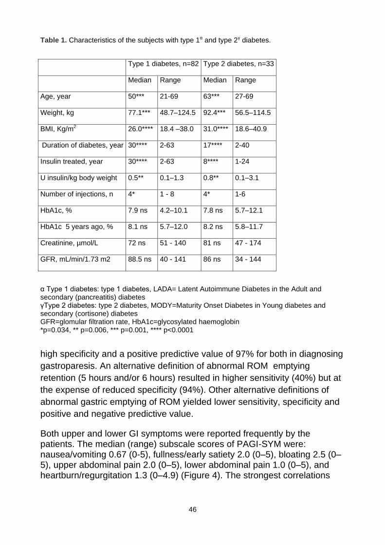
46
Table 1. Characteristics of the subjects with type 1α and type 2γ diabetes.
α Type 1 diabetes: type 1 diabetes, LADA= Latent Autoimmune Diabetes in the Adult and secondary (pancreatitis) diabetes γType 2 diabetes: type 2 diabetes, MODY=Maturity Onset Diabetes in Young diabetes and secondary (cortisone) diabetes GFR=glomular filtration rate, HbA1c=glycosylated haemoglobin *p=0.034, ** p=0.006, *** p=0.001, **** p<0.0001
high specificity and a positive predictive value of 97% for both in diagnosing
gastroparesis. An alternative definition of abnormal ROM emptying
retention (5 hours and/or 6 hours) resulted in higher sensitivity (40%) but at
the expense of reduced specificity (94%). Other alternative definitions of
abnormal gastric emptying of ROM yielded lower sensitivity, specificity and
positive and negative predictive value.
Both upper and lower GI symptoms were reported frequently by the patients. The median (range) subscale scores of PAGI-SYM were: nausea/vomiting 0.67 (0-5), fullness/early satiety 2.0 (0–5), bloating 2.5 (0–5), upper abdominal pain 2.0 (0–5), lower abdominal pain 1.0 (0–5), and heartburn/regurgitation 1.3 (0–4.9) (Figure 4). The strongest correlations
Type 1 diabetes, n=82 Type 2 diabetes, n=33
Median Range Median Range
Age, year 50*** 21-69 63*** 27-69
Weight, kg 77.1*** 48.7–124.5 92.4*** 56.5–114.5
BMI, Kg/m2 26.0**** 18.4 –38.0 31.0**** 18.6–40.9
Duration of diabetes, year 30**** 2-63 17**** 2-40
Insulin treated, year 30**** 2-63 8**** 1-24
U insulin/kg body weight 0.5** 0.1–1.3 0.8** 0.1–3.1
Number of injections, n 4* 1 - 8 4* 1-6
HbA1c, % 7.9 ns 4.2–10.1 7.8 ns 5.7–12.1
HbA1c 5 years ago, % 8.1 ns 5.7–12.0 8.2 ns 5.8–11.7
Creatinine, µmol/L 72 ns 51 - 140 81 ns 47 - 174
GFR, mL/min/1.73 m2 88.5 ns 40 - 141 86 ns 34 - 144
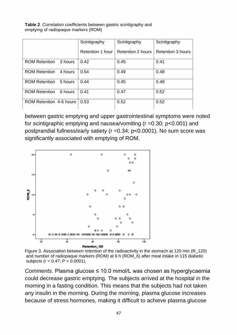
47
Table 2. Correlation coefficients between gastric scintigraphy and emptying of radiopaque markers (ROM)
between gastric emptying and upper gastrointestinal symptoms were noted
for scintigraphic emptying and nausea/vomiting (r =0.30; p<0.001) and
postprandial fullness/early satiety (r =0.34; p<0.0001). No sum score was
significantly associated with emptying of ROM.
Figure 3. Association between retention of the radioactivity in the stomach at 120 min (R_120) and number of radiopaque markers (ROM) at 6 h (ROM_6) after meal intake in 115 diabetic subjects (r = 0.47; P < 0.0001).
Comments. Plasma glucose ≤ 10.0 mmol/L was chosen as hyperglycaemia
could decrease gastric emptying. The subjects arrived at the hospital in the
morning in a fasting condition. This means that the subjects had not taken
any insulin in the morning. During the morning, plasma glucose increases
because of stress hormones, making it difficult to achieve plasma glucose
Scintigraphy
Retention 1 hour
Scintigraphy
Retention 2 hours
Scintigraphy
Retention 3 hours
ROM Retention 3 hours 0.42 0.45 0.41
ROM Retention 4 hours 0.54 0.49 0.48
ROM Retention 5 hours 0.44 0.45 0.48
ROM Retention 6 hours 0.41 0.47 0.52
ROM Retention 4-6 hours 0.53 0.52 0.52

48
of 4-6 mmol/L (150). Moreover, several of the subjects had travelled to the
hospital for 2-3 hours during the morning before the test. When the subjects
finished registration using a gamma camera, they were transported
immediately in a wheelchair to the fluoroscopy equipment. This was done
as walking has been shown to increase gastric emptying (140). The time
between final registration by scintigraphy and the first fluoroscopy
measurement was no more than five minutes. This means that we tried our
best to remove potential confounders, but despite this the ROM test were
only found to be moderately helpful in the clinical setting. However, a
positive ROM test seems to be reliable, whereas a negative test is less
reliable.
Figure 4. Severity of GI symptoms in subjects with insulin-treated diabetes with and without gastroparesis. Distribution of PAGI-SYM summary scores (0–5); white represents the highest score (very severe symptoms), and with increasing darkness of the gray colour the severity of the symptoms decreases (darkest gray colour = no symptoms).
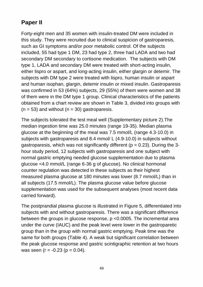
49
Paper II
Forty-eight men and 35 women with insulin-treated DM were included in
this study. They were recruited due to clinical suspicion of gastroparesis,
such as GI symptoms and/or poor metabolic control. Of the subjects
included, 55 had type 1 DM, 23 had type 2, three had LADA and two had
secondary DM secondary to cortisone medication. The subjects with DM
type 1, LADA and secondary DM were treated with short-acting insulin,
either lispro or aspart, and long-acting insulin, either glargin or detemir. The
subjects with DM type 2 were treated with lispro, human insulin or aspart
and human isophan, glargin, detemir insulin or mixed insulin. Gastroparesis
was confirmed in 53 (64%) subjects, 29 (55%) of them were women and 38
of them were in the DM type 1 group. Clinical characteristics of the patients
obtained from a chart review are shown in Table 3, divided into groups with
(n = 53) and without (n = 30) gastroparesis.
The subjects tolerated the test meal well (Supplementary picture 2).The
median ingestion time was 25.0 minutes (range 19-35). Median plasma
glucose at the beginning of the meal was 7.5 mmol/L (range 4.3-10.0) in
subjects with gastroparesis and 8.4 mmol/ L (4.9-10.0) in subjects without
gastroparesis, which was not significantly different (p = 0.23). During the 3-
hour study period, 12 subjects with gastroparesis and one subject with
normal gastric emptying needed glucose supplementation due to plasma
glucose <4.0 mmol/L (range 6-36 g of glucose). No clinical hormonal
counter regulation was detected in these subjects as their highest
measured plasma glucose at 180 minutes was lower (8.7 mmol/L) than in
all subjects (17.5 mmol/L). The plasma glucose value before glucose
supplementation was used for the subsequent analyses (most recent data
carried forward).
The postprandial plasma glucose is illustrated in Figure 5, differentiated into
subjects with and without gastroparesis. There was a significant difference
between the groups in glucose response, p <0.0005. The incremental area
under the curve (IAUC) and the peak level were lower in the gastroparetic
group than in the group with normal gastric emptying. Peak time was the
same for both groups (Table 4). A weak but significant correlation between
the peak glucose response and gastric scintigraphic retention at two hours
was seen (r = -0.23 (p = 0.04).
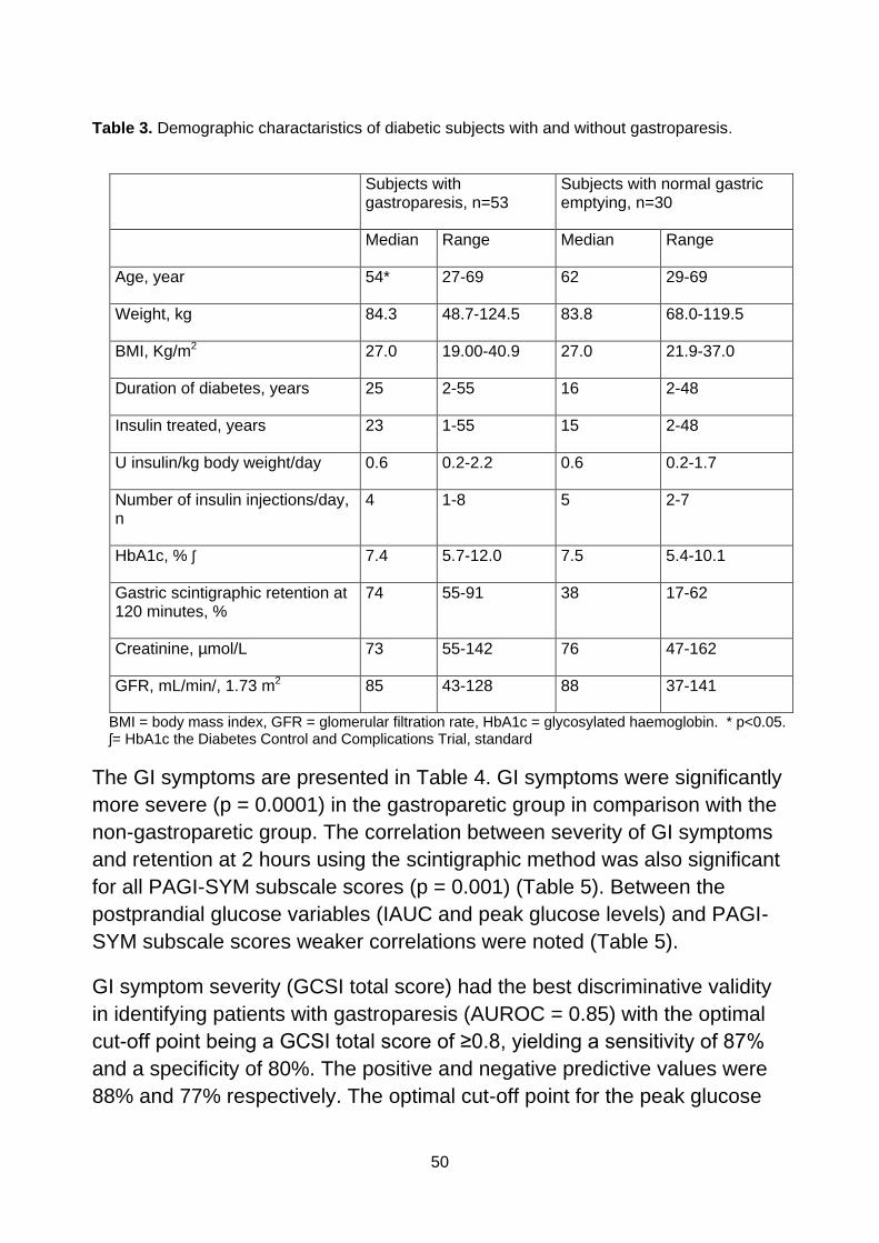
50
Table 3. Demographic charactaristics of diabetic subjects with and without gastroparesis.
Subjects with gastroparesis, n=53
Subjects with normal gastric emptying, n=30
Median Range Median Range
Age, year 54* 27-69 62 29-69
Weight, kg 84.3 48.7-124.5 83.8 68.0-119.5
BMI, Kg/m2 27.0 19.00-40.9 27.0 21.9-37.0
Duration of diabetes, years 25 2-55 16 2-48
Insulin treated, years 23 1-55 15 2-48
U insulin/kg body weight/day 0.6 0.2-2.2 0.6 0.2-1.7
Number of insulin injections/day, n
4 1-8 5 2-7
HbA1c, % ʃ 7.4 5.7-12.0 7.5 5.4-10.1
Gastric scintigraphic retention at 120 minutes, %
74 55-91 38 17-62
Creatinine, µmol/L 73 55-142 76 47-162
GFR, mL/min/, 1.73 m2 85 43-128 88 37-141
BMI = body mass index, GFR = glomerular filtration rate, HbA1c = glycosylated haemoglobin. * p<0.05. ʃ= HbA1c the Diabetes Control and Complications Trial, standard
The GI symptoms are presented in Table 4. GI symptoms were significantly
more severe (p = 0.0001) in the gastroparetic group in comparison with the
non-gastroparetic group. The correlation between severity of GI symptoms
and retention at 2 hours using the scintigraphic method was also significant
for all PAGI-SYM subscale scores (p = 0.001) (Table 5). Between the
postprandial glucose variables (IAUC and peak glucose levels) and PAGI-
SYM subscale scores weaker correlations were noted (Table 5).
GI symptom severity (GCSI total score) had the best discriminative validity
in identifying patients with gastroparesis (AUROC = 0.85) with the optimal
cut-off point being a GCSI total score of ≥0.8, yielding a sensitivity of 87%
and a specificity of 80%. The positive and negative predictive values were
88% and 77% respectively. The optimal cut-off point for the peak glucose
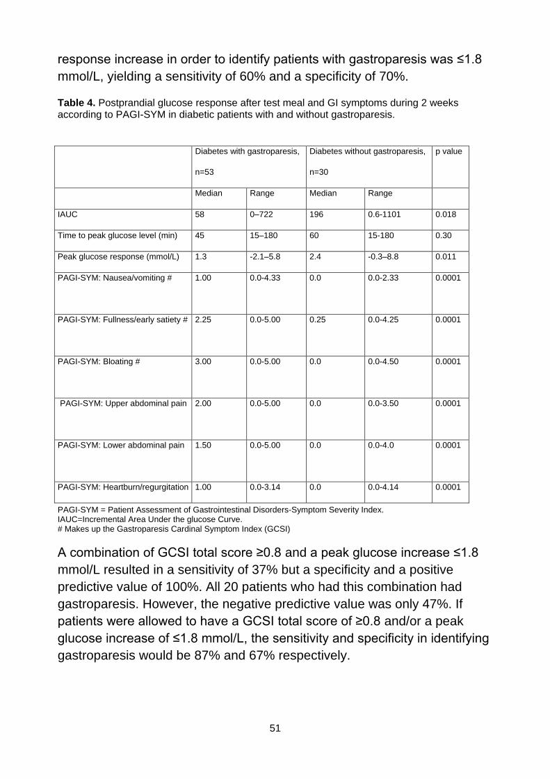
51
response increase in order to identify patients with gastroparesis was ≤1.8
mmol/L, yielding a sensitivity of 60% and a specificity of 70%.
Table 4. Postprandial glucose response after test meal and GI symptoms during 2 weeks according to PAGI-SYM in diabetic patients with and without gastroparesis.
PAGI-SYM = Patient Assessment of Gastrointestinal Disorders-Symptom Severity Index. IAUC=Incremental Area Under the glucose Curve. # Makes up the Gastroparesis Cardinal Symptom Index (GCSI)
A combination of GCSI total score ≥0.8 and a peak glucose increase ≤1.8
mmol/L resulted in a sensitivity of 37% but a specificity and a positive
predictive value of 100%. All 20 patients who had this combination had
gastroparesis. However, the negative predictive value was only 47%. If
patients were allowed to have a GCSI total score of ≥0.8 and/or a peak
glucose increase of ≤1.8 mmol/L, the sensitivity and specificity in identifying
gastroparesis would be 87% and 67% respectively.
Diabetes with gastroparesis,
n=53
Diabetes without gastroparesis,
n=30
p value
Median Range Median Range
IAUC 58 0–722 196 0.6-1101 0.018
Time to peak glucose level (min) 45 15–180 60 15-180 0.30
Peak glucose response (mmol/L) 1.3 -2.1–5.8 2.4 -0.3–8.8 0.011
PAGI-SYM: Nausea/vomiting #
1.00
0.0-4.33 0.0
0.0-2.33
0.0001
PAGI-SYM: Fullness/early satiety # 2.25
0.0-5.00
0.25
0.0-4.25
0.0001
PAGI-SYM: Bloating # 3.00
0.0-5.00
0.0
0.0-4.50
0.0001
PAGI-SYM: Upper abdominal pain 2.00
0.0-5.00
0.0
0.0-3.50
0.0001
PAGI-SYM: Lower abdominal pain 1.50
0.0-5.00
0.0
0.0-4.0
0.0001
PAGI-SYM: Heartburn/regurgitation 1.00 0.0-3.14 0.0 0.0-4.14 0.0001
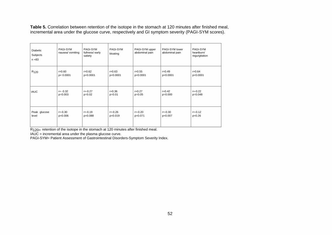
52
Table 5. Correlation between retention of the isotope in the stomach at 120 minutes after finished meal,
incremental area under the glucose curve, respectively and GI symptom severity (PAGI-SYM scores).
Diabetic
Subjects
n =83
PAGI-SYM nausea/ vomiting
PAGI-SYM fullness/ early satiety
PAGI-SYM
bloating
PAGI-SYM upper abdominal pain
PAGI-SYM lower abdominal pain
PAGI-SYM heartburn/ regurgitation
R120
r=0.60
p< 0.0001
r=0.62
p<0.0001
r=0.63
p<0.0001
r=0.55
p<0.0001
r=0.49
p<0.0001
r=0.64
p<0.0001
IAUC
r= -
0.32
p=0.003
r=-0.27 p=0.02
r=0.36 p=0.01
r=0.27 p=0.05
r=0.42 p=0.000
r=-0.22 p=0.048
Peak glucose
level
r=-0.30
p=0.006
r=-0.19
p=0.088
r=-0.26
p=0.019
r=-0.20
p=0.071
r=-0.30
p=0.007
r=-0.12
p=0.26
R120= retention of the isotope in the stomach at 120 minutes after finished meal.
IAUC = incremental area under the plasma glucose curve. PAGI-SYM= Patient Assessment of Gastrointestinal Disorders-Symptom Severity Index.
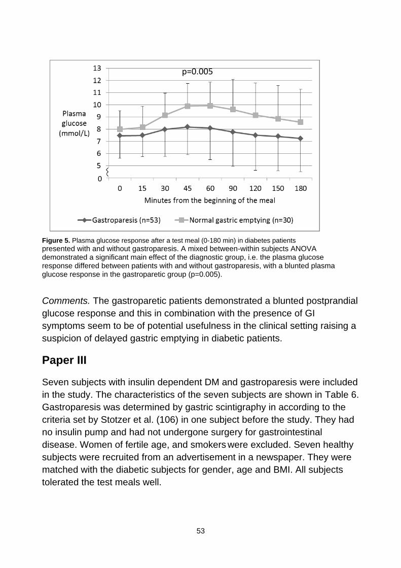
53
Figure 5. Plasma glucose response after a test meal (0-180 min) in diabetes patients
presented with and without gastroparesis. A mixed between-within subjects ANOVA demonstrated a significant main effect of the diagnostic group, i.e. the plasma glucose response differed between patients with and without gastroparesis, with a blunted plasma glucose response in the gastroparetic group (p=0.005).
Comments. The gastroparetic patients demonstrated a blunted postprandial
glucose response and this in combination with the presence of GI
symptoms seem to be of potential usefulness in the clinical setting raising a
suspicion of delayed gastric emptying in diabetic patients.
Paper III
Seven subjects with insulin dependent DM and gastroparesis were included
in the study. The characteristics of the seven subjects are shown in Table 6.
Gastroparesis was determined by gastric scintigraphy in according to the
criteria set by Stotzer et al. (106) in one subject before the study. They had
no insulin pump and had not undergone surgery for gastrointestinal
disease. Women of fertile age, and smokers were excluded. Seven healthy
subjects were recruited from an advertisement in a newspaper. They were
matched with the diabetic subjects for gender, age and BMI. All subjects
tolerated the test meals well.
p=0.005
0
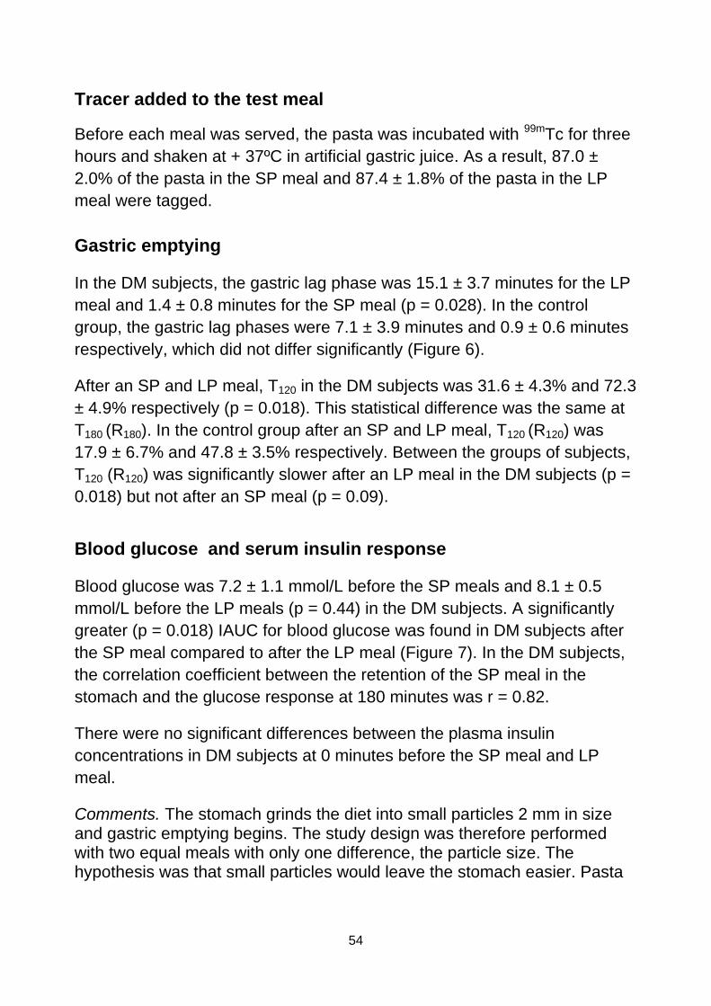
54
Tracer added to the test meal
Before each meal was served, the pasta was incubated with 99mTc for three
hours and shaken at + 37ºC in artificial gastric juice. As a result, 87.0 ±
2.0% of the pasta in the SP meal and 87.4 ± 1.8% of the pasta in the LP
meal were tagged.
Gastric emptying
In the DM subjects, the gastric lag phase was 15.1 ± 3.7 minutes for the LP
meal and 1.4 ± 0.8 minutes for the SP meal (p = 0.028). In the control
group, the gastric lag phases were 7.1 ± 3.9 minutes and 0.9 ± 0.6 minutes
respectively, which did not differ significantly (Figure 6).
After an SP and LP meal, T120 in the DM subjects was 31.6 ± 4.3% and 72.3
± 4.9% respectively (p = 0.018). This statistical difference was the same at
T180 (R180). In the control group after an SP and LP meal, T120 (R120) was
17.9 ± 6.7% and 47.8 ± 3.5% respectively. Between the groups of subjects,
T120 (R120) was significantly slower after an LP meal in the DM subjects (p =
0.018) but not after an SP meal (p = 0.09).
Blood glucose and serum insulin response
Blood glucose was 7.2 ± 1.1 mmol/L before the SP meals and 8.1 ± 0.5
mmol/L before the LP meals (p = 0.44) in the DM subjects. A significantly
greater (p = 0.018) IAUC for blood glucose was found in DM subjects after
the SP meal compared to after the LP meal (Figure 7). In the DM subjects,
the correlation coefficient between the retention of the SP meal in the
stomach and the glucose response at 180 minutes was r = 0.82.
There were no significant differences between the plasma insulin
concentrations in DM subjects at 0 minutes before the SP meal and LP
meal.
Comments. The stomach grinds the diet into small particles 2 mm in size and gastric emptying begins. The study design was therefore performed with two equal meals with only one difference, the particle size. The hypothesis was that small particles would leave the stomach easier. Pasta
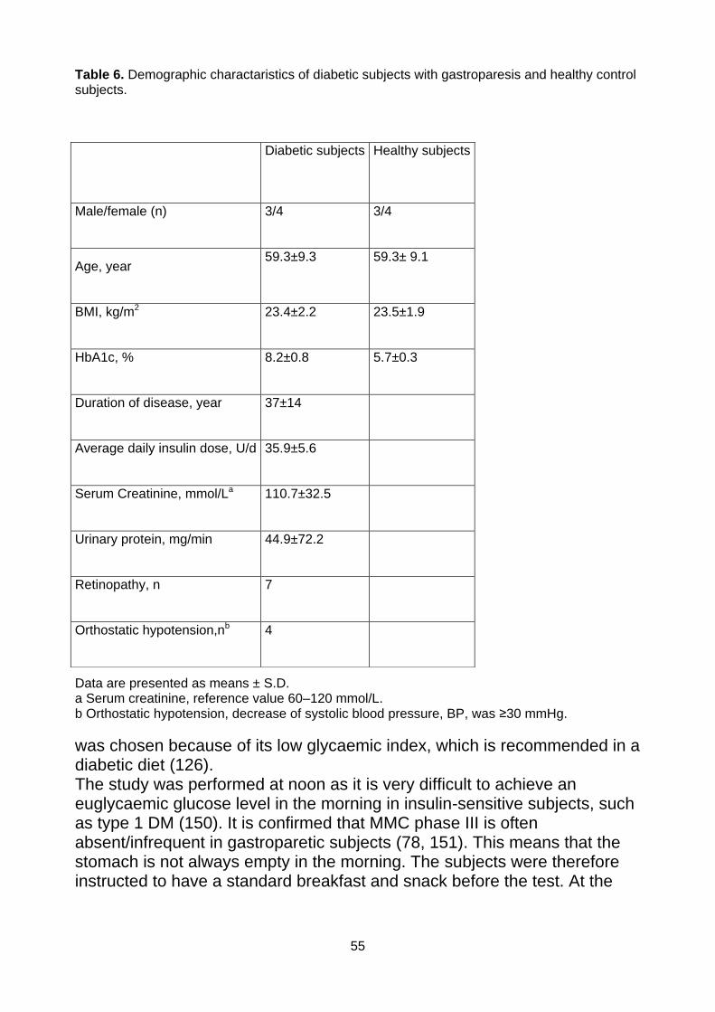
55
Table 6. Demographic charactaristics of diabetic subjects with gastroparesis and healthy control subjects.
Data are presented as means ± S.D. a Serum creatinine, reference value 60–120 mmol/L. b Orthostatic hypotension, decrease of systolic blood pressure, BP, was ≥30 mmHg.
was chosen because of its low glycaemic index, which is recommended in a diabetic diet (126). The study was performed at noon as it is very difficult to achieve an euglycaemic glucose level in the morning in insulin-sensitive subjects, such as type 1 DM (150). It is confirmed that MMC phase III is often absent/infrequent in gastroparetic subjects (78, 151). This means that the stomach is not always empty in the morning. The subjects were therefore instructed to have a standard breakfast and snack before the test. At the
Diabetic subjects
Healthy subjects
Male/female (n) 3/4 3/4
Age, year 59.3±9.3 59.3± 9.1
BMI, kg/m2 23.4±2.2 23.5±1.9
HbA1c, % 8.2±0.8 5.7±0.3
Duration of disease, year 37±14
Average daily insulin dose, U/d 35.9±5.6
Serum Creatinine, mmol/La 110.7±32.5
Urinary protein, mg/min 44.9±72.2
Retinopathy, n 7
Orthostatic hypotension,nb 4
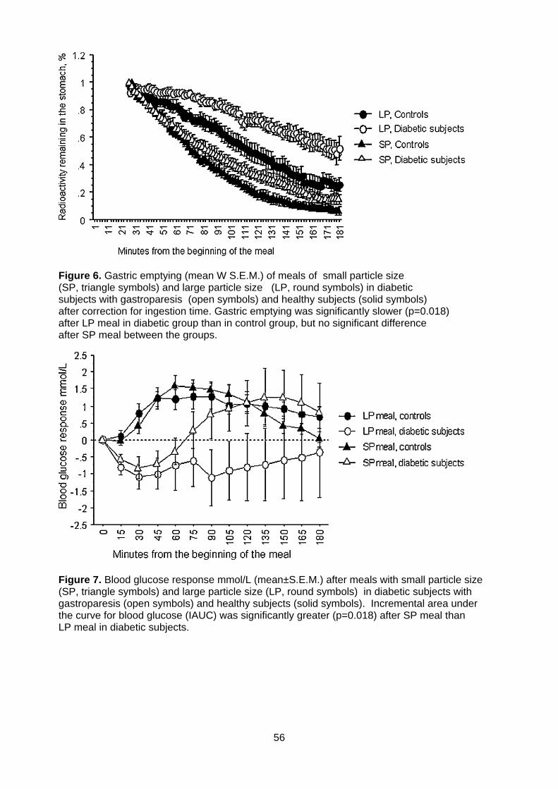
56
Figure 6. Gastric emptying (mean W S.E.M.) of meals of small particle size (SP, triangle symbols) and large particle size (LP, round symbols) in diabetic subjects with gastroparesis (open symbols) and healthy subjects (solid symbols) after correction for ingestion time. Gastric emptying was significantly slower (p=0.018) after LP meal in diabetic group than in control group, but no significant difference after SP meal between the groups.
Figure 7. Blood glucose response mmol/L (mean±S.E.M.) after meals with small particle size (SP, triangle symbols) and large particle size (LP, round symbols) in diabetic subjects with gastroparesis (open symbols) and healthy subjects (solid symbols). Incremental area under the curve for blood glucose (IAUC) was significantly greater (p=0.018) after SP meal than LP meal in diabetic subjects.
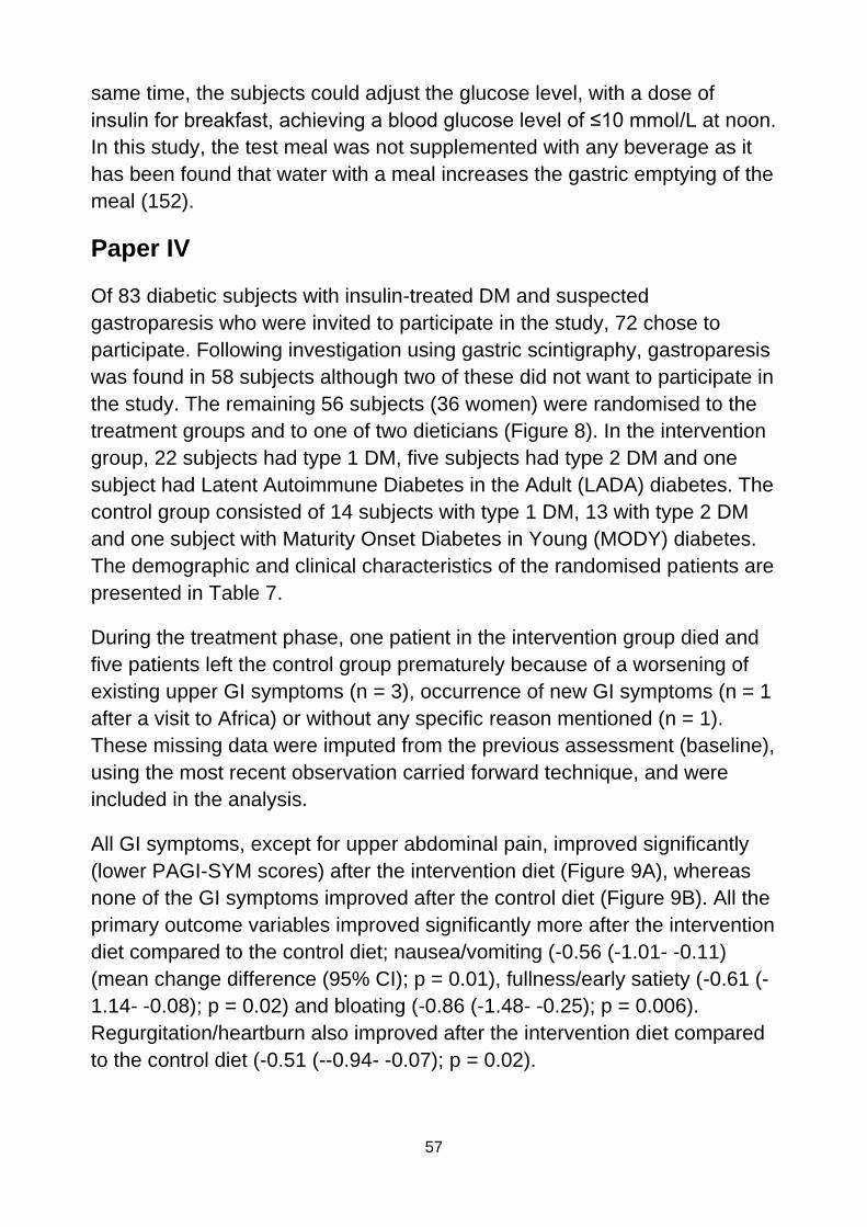
57
same time, the subjects could adjust the glucose level, with a dose of
insulin for breakfast, achieving a blood glucose level of ≤10 mmol/L at noon.
In this study, the test meal was not supplemented with any beverage as it
has been found that water with a meal increases the gastric emptying of the
meal (152).
Paper IV
Of 83 diabetic subjects with insulin-treated DM and suspected
gastroparesis who were invited to participate in the study, 72 chose to
participate. Following investigation using gastric scintigraphy, gastroparesis
was found in 58 subjects although two of these did not want to participate in
the study. The remaining 56 subjects (36 women) were randomised to the
treatment groups and to one of two dieticians (Figure 8). In the intervention
group, 22 subjects had type 1 DM, five subjects had type 2 DM and one
subject had Latent Autoimmune Diabetes in the Adult (LADA) diabetes. The
control group consisted of 14 subjects with type 1 DM, 13 with type 2 DM
and one subject with Maturity Onset Diabetes in Young (MODY) diabetes.
The demographic and clinical characteristics of the randomised patients are
presented in Table 7.
During the treatment phase, one patient in the intervention group died and
five patients left the control group prematurely because of a worsening of
existing upper GI symptoms (n = 3), occurrence of new GI symptoms (n = 1
after a visit to Africa) or without any specific reason mentioned (n = 1).
These missing data were imputed from the previous assessment (baseline),
using the most recent observation carried forward technique, and were
included in the analysis.
All GI symptoms, except for upper abdominal pain, improved significantly
(lower PAGI-SYM scores) after the intervention diet (Figure 9A), whereas
none of the GI symptoms improved after the control diet (Figure 9B). All the
primary outcome variables improved significantly more after the intervention
diet compared to the control diet; nausea/vomiting (-0.56 (-1.01- -0.11)
(mean change difference (95% CI); p = 0.01), fullness/early satiety (-0.61 (-
1.14- -0.08); p = 0.02) and bloating (-0.86 (-1.48- -0.25); p = 0.006).
Regurgitation/heartburn also improved after the intervention diet compared
to the control diet (-0.51 (--0.94- -0.07); p = 0.02).
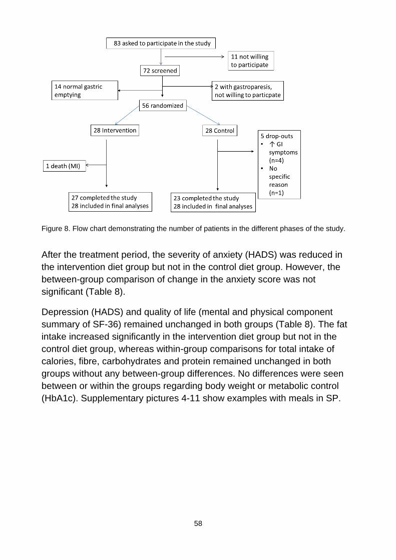
58
Figure 8. Flow chart demonstrating the number of patients in the different phases of the study.
After the treatment period, the severity of anxiety (HADS) was reduced in
the intervention diet group but not in the control diet group. However, the
between-group comparison of change in the anxiety score was not
significant (Table 8).
Depression (HADS) and quality of life (mental and physical component
summary of SF-36) remained unchanged in both groups (Table 8). The fat
intake increased significantly in the intervention diet group but not in the
control diet group, whereas within-group comparisons for total intake of
calories, fibre, carbohydrates and protein remained unchanged in both
groups without any between-group differences. No differences were seen
between or within the groups regarding body weight or metabolic control
(HbA1c). Supplementary pictures 4-11 show examples with meals in SP.
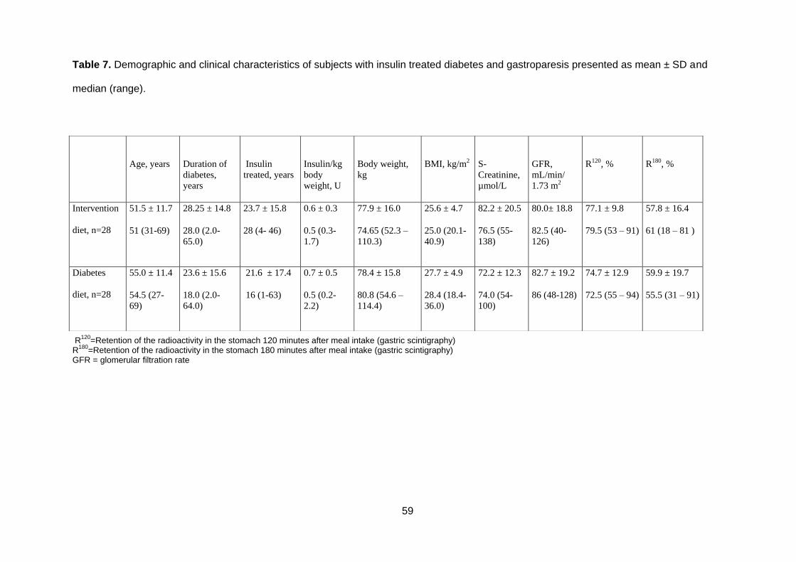
59
Table 7. Demographic and clinical characteristics of subjects with insulin treated diabetes and gastroparesis presented as mean ± SD and
median (range).
R
120=Retention of the radioactivity in the stomach 120 minutes after meal intake (gastric scintigraphy)
R180
=Retention of the radioactivity in the stomach 180 minutes after meal intake (gastric scintigraphy) GFR = glomerular filtration rate
Age, years
Duration of
diabetes,
years
I
Insulin
treated, years
Insulin/kg
body
weight, U
Body weight,
kg
BMI, kg/m2
S-
Creatinine,
µmol/L
GFR,
mL/min/
1.73 m2
R120, %
R180, %
Intervention
diet, n=28
51.5 ± 11.7
51 (31-69)
28.25 ± 14.8
28.0 (2.0-
65.0)
23.7 ± 15.8
28 (4- 46)
0.6 ± 0.3
0.5 (0.3-
1.7)
77.9 ± 16.0
74.65 (52.3 –
110.3)
25.6 ± 4.7
25.0 (20.1-
40.9)
82.2 ± 20.5
76.5 (55-
138)
80.0± 18.8
82.5 (40-
126)
77.1 ± 9.8
79.5 (53 – 91)
57.8 ± 16.4
6 61 (18 – 81 )
Diabetes
diet, n=28
55.0 ± 11.4
54.5 (27-
69)
23.6 ± 15.6
18.0 (2.0-
64.0)
21.6 ± 17.4
16 (1-63)
0.7 ± 0.5
0.5 (0.2-
2.2)
78.4 ± 15.8
80.8 (54.6 –
114.4)
27.7 ± 4.9
28.4 (18.4-
36.0)
72.2 ± 12.3
74.0 (54-
100)
82.7 ± 19.2
86 (48-128)
74.7 ± 12.9
72.5 (55 – 94)
59.9 ± 19.7
5 55.5 (31 – 91)
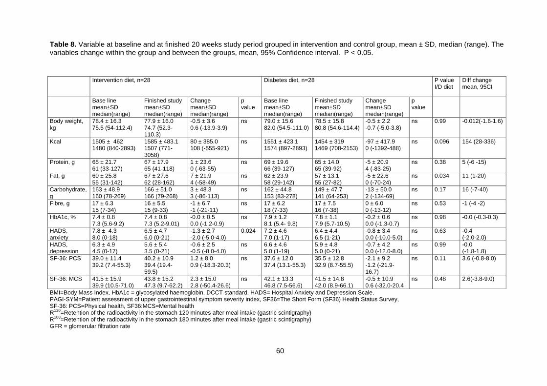
60
Table 8. Variable at baseline and at finished 20 weeks study period grouped in intervention and control group, mean ± SD, median (range). The variables change within the group and between the groups, mean, 95% Confidence interval. P < 0.05.
BMI=Body Mass Index, HbA1c = glycosylated haemoglobin, DCCT standard, HADS= Hospital Anxiety and Depression Scale, PAGI-SYM=Patient assessment of upper gastrointestinal symptom severity index, SF36=The Short Form (SF36) Health Status Survey, SF-36: PCS=Physical health, SF36:MCS=Mental health R
120=Retention of the radioactivity in the stomach 120 minutes after meal intake (gastric scintigraphy)
R180
=Retention of the radioactivity in the stomach 180 minutes after meal intake (gastric scintigraphy) GFR = glomerular filtration rate
Intervention diet, n=28 Diabetes diet, n=28 P value I/D diet
Diff change mean, 95CI
Base line mean±SD median(range)
Finished study mean±SD median(range)
Change mean±SD median(range)
p value
Base line mean±SD median(range)
Finished study mean±SD median(range)
Change mean±SD median(range)
p value
Body weight, kg
78.4 ± 16.3 75.5 (54-112.4)
77.9 ± 16.0 74.7 (52.3-110.3)
-0.5 ± 3.6 0.6 (-13.9-3.9)
ns 79.0 ± 15.6 82.0 (54.5-111.0)
78.5 ± 15.8 80.8 (54.6-114.4)
-0.5 ± 2.2 -0.7 (-5.0-3.8)
ns 0.99 -0.012(-1.6-1.6)
Kcal 1505 ± 462 1480 (840-2893)
1585 ± 483.1 1507 (771-3058)
80 ± 385.0 108 (-555-921)
ns 1551 ± 423.1 1574 (897-2893)
1454 ± 319 1469 (708-2153)
-97 ± 417.9 0 (-1392-488)
ns 0.096 154 (28-336)
Protein, g 65 ± 21.7 61 (33-127)
67 ± 17.9 65 (41-118)
1 ± 23.6 0 (-63-55)
ns 69 ± 19.6 66 (39-127)
65 ± 14.0 65 (39-92)
-5 ± 20.9 4 (-83-25)
ns 0.38 5 (-6 -15)
Fat, g 60 ± 25.8 55 (31-142)
67 ± 27.6 62 (28-162)
7 ± 21.9 4 (-58-49)
ns 62 ± 23.9 58 (29-142)
57 ± 13.1 55 (27-82)
-5 ± 22.6 0 (-70-24)
ns 0.034 11 (1-20)
Carbohydrate, g
163 ± 48.9 160 (78-269)
166 ± 51.0 166 (79-268)
3 ± 48.3 3 (-86-113)
ns 162 ± 44.8 153 (83-278)
149 ± 47.7 141 (64-253)
-13 ± 50.0 2 (-134-69)
ns 0.17 16 (-7-40)
Fibre, g 17 ± 6.3 15 (7-34)
16 ± 5.5 15 (9-33)
-1 ± 6.7 -1 (-21-11)
ns 17 ± 6.2 18 (7-33)
17 ± 7.5 16 (7-38)
0 ± 6.0 0 (-13-12)
ns 0.53 -1 (-4 -2)
HbA1c, % 7.4 ± 0.8 7.3 (5.6-9.2)
7.4 ± 0.8 7.3 (5.2-9.01)
-0.0 ± 0.5 0.0 (-1.2-0.9)
ns 7.9 ± 1.2 8.1 (5.4- 9.8)
7.8 ± 1.1 7.9 (5.7-10.5)
-0.2 ± 0.6 0.0 (-1.3-0.7)
ns 0.98 -0.0 (-0.3-0.3)
HADS, anxiety
7.8 ± 4.3 8.0 (0-18)
6.5 ± 4.7 6.0 (0-21)
-1.3 ± 2.7 -2.0 (-5.0-4.0)
0.024 7.2 ± 4.6 7.0 (1-17)
6.4 ± 4.4 6.5 (1-21)
-0.8 ± 3.4 0.0 (-10.0-5.0)
ns 0.63 -0.4 (-2.0-2.0)
HADS, depression
6.3 ± 4.9 4.5 (0-17)
5.6 ± 5.4 3.5 (0-21)
-0.6 ± 2.5 -0.5 (-8.0-4.0)
ns 6.6 ± 4.6 5.0 (1-19)
5.9 ± 4.8 5.0 (0-21)
-0.7 ± 4.2 0.0 (-12.0-8.0)
ns 0.99 -0.0 (-1.8-1.8)
SF-36: PCS 39.0 ± 11.4 39.2 (7.4-55.3)
40.2 ± 10.9 39.4 (19.4-59.5)
1.2 ± 8.0 0.9 (-18.3-20.3)
ns 37.6 ± 12.0 37.4 (13.1-55.3)
35.5 ± 12.8 32.9 (8.7-55.5)
-2.1 ± 9.2 -1.2 (-21.9-16.7)
ns 0.11 3.6 (-0.8-8.0)
SF-36: MCS 41.5 ± 15.9 39.9 (10.5-71.0)
43.8 ± 15.2 47.3 (9.7-62.2)
2.3 ± 15.0 2.8 (-50.4-26.6)
ns 42.1 ± 13.3 46.8 (7.5-56.6)
41.5 ± 14.8 42.0 (8.9-66.1)
-0.5 ± 10.9 0.6 (-32.0-20.4
ns 0.48 2.6(-3.8-9.0)

61
Figure 9A. GI symptom severity, as measured with PAGI-SYM, at baseline and after the dietary treatment period (20 weeks) in the group who received advice to eat the intervention diet (small particle size). PAGI-SYM domains: N/V = nausea/vomiting; Fullness = fullness/early satiety; U / L abd pain = Upper / Lower Abdominal Pain; H/R = Heartburn/regurgitation. * p<0.05, ** p<0.01, *** p<0.001 vs. baseline.
Figure 9B. Figure 2. GI symptom severity, as measured with PAGI-SYM, at baseline and after the dietary treatment period (20 weeks) in the group who were instructed to follow the control diet (B). PAGI-SYM domains: N/V = nausea/vomiting; Fullness = fullness/early satiety; U / L abd pain = Upper / Lower Abdominal Pain; H/R = Heartburn/regurgitation. * p<0.05, ** p<0.01, *** p<0.001 vs. baseline.
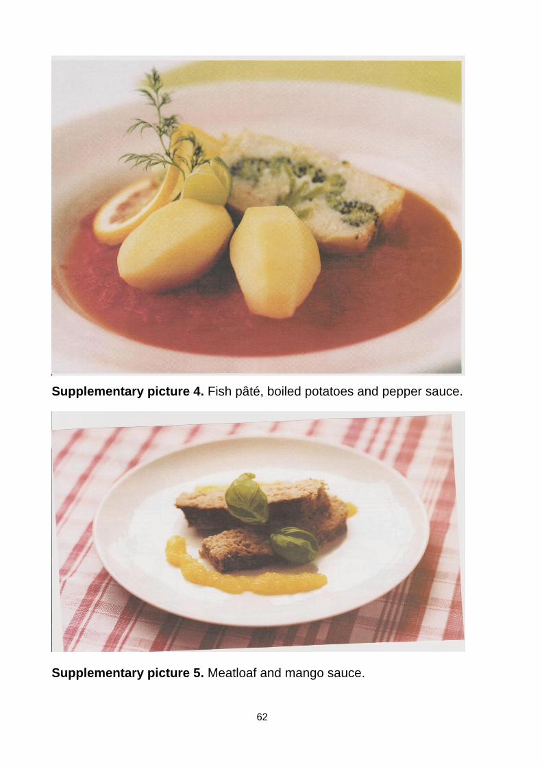
62
Supplementary picture 4. Fish pâté, boiled potatoes and pepper sauce.
Supplementary picture 5. Meatloaf and mango sauce.
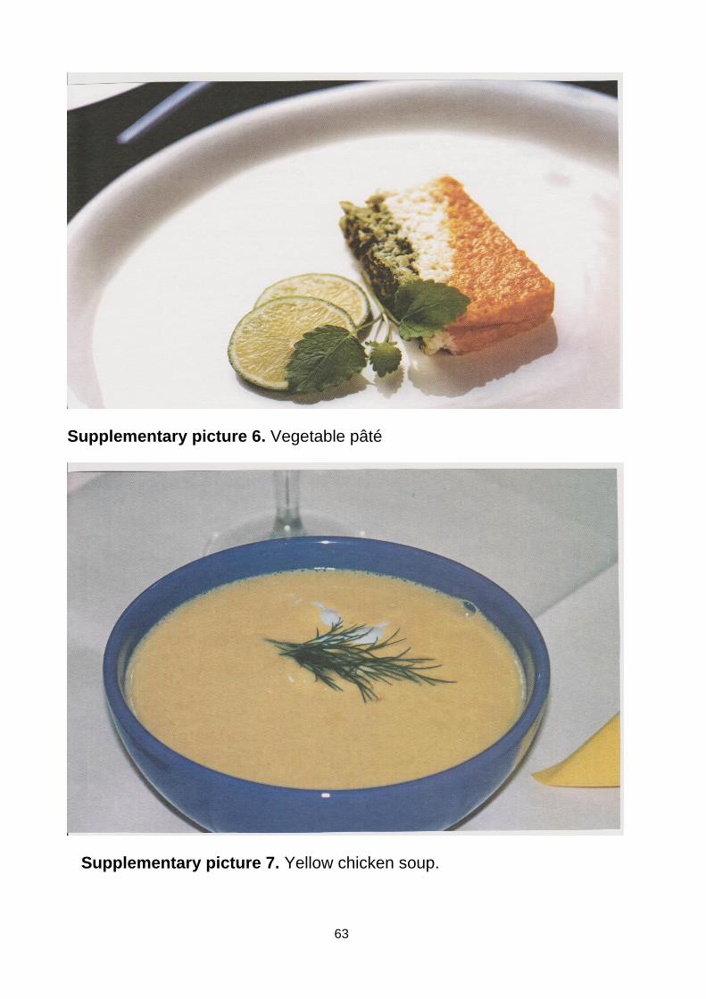
63
Supplementary picture 6. Vegetable pâté
Supplementary picture 7. Yellow chicken soup.
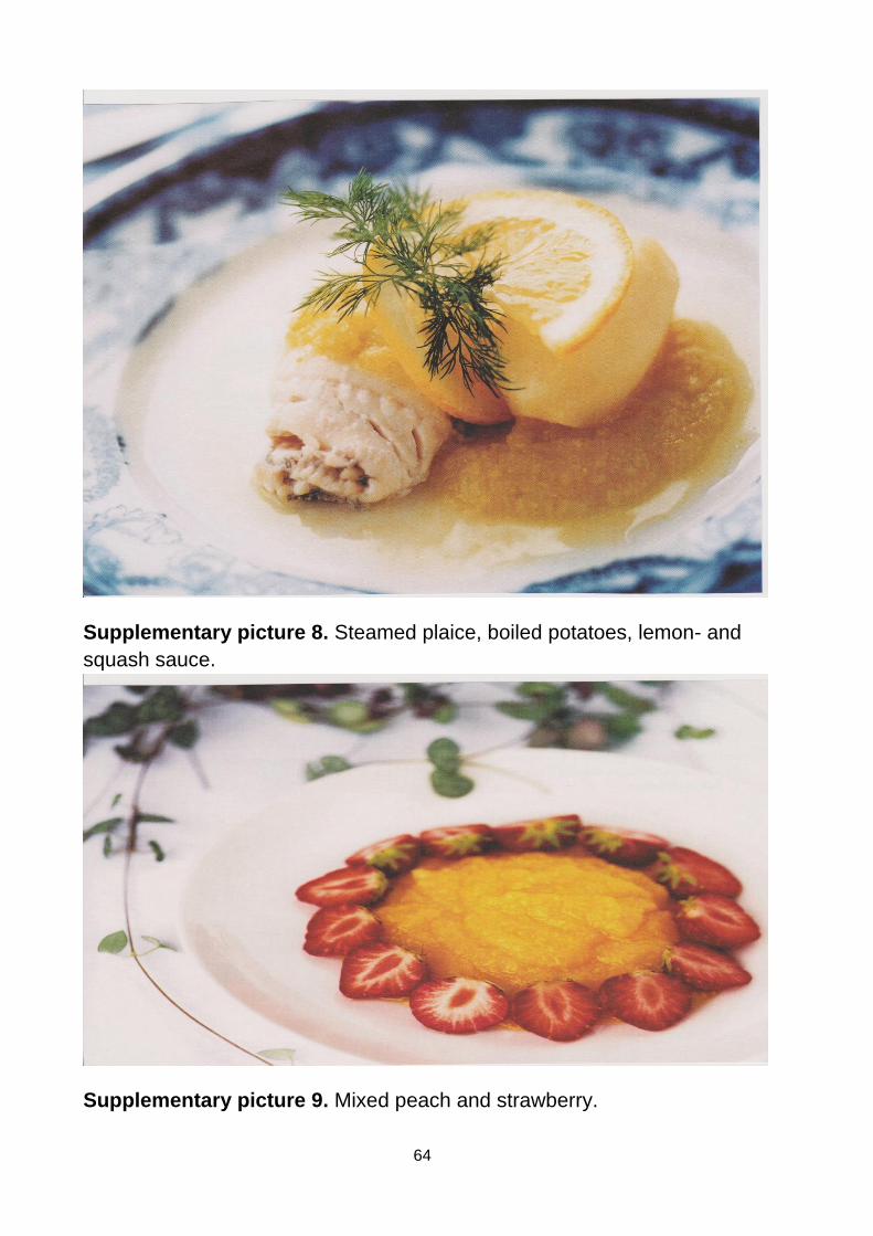
64
Supplementary picture 8. Steamed plaice, boiled potatoes, lemon- and
squash sauce.
Supplementary picture 9. Mixed peach and strawberry.
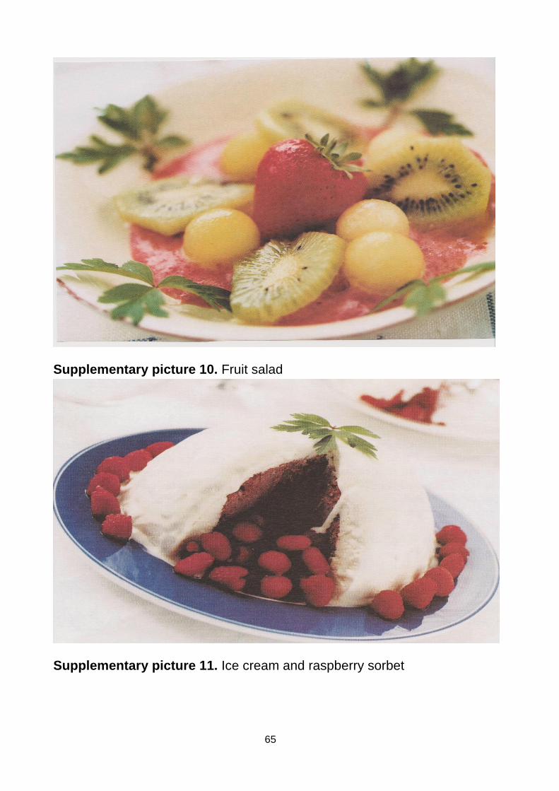
65
Supplementary picture 10. Fruit salad
Supplementary picture 11. Ice cream and raspberry sorbet

66
67
General discussion
In the present thesis, I have demonstrated that gastric emptying of ROM
and plasma glucose response after a standardized test meal can add
information in the clinical management of DM patients, but cannot replace
the current gold standard to diagnose gastroparesis, gastric scintigraphy.
Moreover, a meal with SP increases the gastric emptying rate and
reduces the postprandial blood glucose dip in Type 1 DM subjects with
gastroparesis. In line with this, dietary treatment with SP significantly
improves the key symptoms of gastroparesis in patients with DM.
In Paper I, we noted a moderate association between gastric emptying of
digestible solids, measured using gastric scintigraphy, and gastric
emptying of indigestible solids, measured using ROM. The difference in
results between the methods could be due in part to the fact that the
methods do not measure exactly the same variables. However, based on
our results it seems as if gastric emptying with ROM, using fluoroscopy
could be used as a screening method for gastroparesis. If the
measurement shows delayed emptying at 6 h, this is probably true. If the
gastric emptying is normal but there is still a clinical suspicion question of
gastroparesis, further investigation is required. The specificity and positive
predictive value of the method were 97%, if ROM emptying at 6 h was
used, but both the sensitivity and negative predictive value were clearly
lower. In another study, emptying of ROM seemed to be very potent in
predicting gastroparesis (135). The discrepancy between our study and
Feldman et al. may be partly be due to different patient populations and
potentially also different size of the markers.
The correlation between GI symptoms and the gastric emptying rate was
strongest for nausea/vomiting and postprandial fullness/early satiety when
using a scintigraphic method, but there was no significant correlation with
the fluoroscopy method in Paper I.
Many factors are involved in gastric emptying, such as blood glucose level
and a number of dietary factors. It is now well known that hyperglycaemia
decreases gastric emptying. It is therefore important to have as normal
plasma glucose as possible before and during the investigation. Chang et
al. suggest a plasma glucose level of 4-10 mmol/L at the beginning of the
test (85), which we used in our studies. It seems to be of great importance
that departments measuring gastric scintigraphic emptying should have
68
equipment to measure plasma glucose, and as far as possible normalize
the glucose levels before commencing and during the test. A blood
glucose level of 8 mmol/L decreases gastric emptying compared to 4
mmol/L (53) although there is an increase in the gastric emptying at 2
mmol/L (54). Hyperglycaemia is also known to inhibit the antral
component of MMC phase III (90). Moreover, Jones et al. found that
hyperglycaemia masks the medical effect of prokinetics (153). Based on
these results we followed glucose levels carefully in our studies, and when
needed normalized the levels in order to avoid the confounding effect of
glucose levels on gastric emptying as far as possible.
In Paper II the plasma glucose response was blunted in subjects with
gastroparesis compared with subjects without gastroparesis. However, the
GI symptom severity during the preceding two weeks was an even
stronger predictor for gastroparesis than the plasma glucose response.
In the literature, different correlations are presented between GI symptoms
and gastric emptying and/or a diagnosis of gastroparesis (34, 36, 38, 55,
68, 154). In Papers I and II, the correlation between these parameters
was different. This could probably be caused by the inclusion criteria,
since all subjects in Paper I had significant upper GI symptoms, but in
Paper II also subjects with milder GI symptoms were included. Moreover,
the proportion of subjects with normal gastric emptying was higher in
Paper II than in Paper I, which could have an impact on the association
between gastric emptying rate and GI symptom severity. Plasma glucose
level before the test investigation the diagnosis was the same in both
studies. Therefore the effect on gastric emptying and the diagnosis by the
plasma glucose level was most likely the same in both studies. Moreover,
Byzter et al. found in DM subjects in general that they had more severe GI
symptoms when there was poor metabolic control (43). In Paper I and II
HbA1c was not significantly different between the groups , ruling out
differences in metabolic control as an explanation behind the discrepancy.
In Paper III, the gastric emptying after SP meal was not significantly
different in DM type 1 subjects with gastroparesis than in healthy subjects.
However, after LP meal the gastric emptying rate was slower in subjects
with gastroparesis than in healthy subjects. The IAUC of the plasma
glucose response after SP meal in gastroparetic subjects was also
significantly larger than after LP meal. These findings strengthened my
69
hypothesis that a meal with SP is beneficial for DM patients with
gastroparesis, and this diet was then further tested in Paper IV.
Up to now there has been no randomised trial of dietary interventions in
gastroparesis and consequently there are no evidence-based
recommendations (58). It has been advocated that the diet in diabetic
gastroparesis should be low in fat and fibre (109). The test meals used in
Papers II and III have a fat content of 31 energy per cent and 3 gram/MJ.
The recommendation was 25-30 energy per cent fat and 27-30 gram fibre
at the time Paper III was planned (155). Despite the fact that gastric
emptying was more rapid after a test meal with SP in both gastroparetic
subjects and in healthy controls, there was no significant difference in
terms of gastric emptying rate between the groups after a meal with SP.
However, gastric emptying after the LP meal was significantly slower in
the patient group than in the healthy group. Both test meals were identical
– only the particle size differed. The impact of different diet particle sizes
ceased earlier, resulting in a significantly different gastric emptying rate
(156). This means that fat and fibre content play a lesser role than particle
size in gastric emptying; knowledge that we used in our controlled dietary
intervention in Paper IV.
Fat has a different impact on gastric emptying depending on whether the
fat is in a liquid or a solid meal and when the fat was ingested. When fat
was ingested in a liquid meal 30 minutes before a carbohydrate-containing
solid meal, the lag phase was prolonged – the gastric emptying slope did
not change in healthy subjects or in subjects with DM type 2. If fat was
added to a solid meal and ingested immediately, the lag phase was short
and the slope was reduced (112, 113). Often it has been suggested that a
liquid meal should leave the stomach easier. However, Houghton et al.
found that in young, healthy subjects a fat liquid meal caused the contents
to move into the distal stomach, followed by redistribution of the contents
in the distal stomach and then back into the proximal stomach in healthy
subjects. When gastric emptying started, the contents moved down into
the distal stomach again (111). In addition, the lag phase was prolonged
and the slope decreased after a fat liquid meal. Based on these
observations, other factors than merely the fat content per se should be
considered when giving dietary advice to patients with delayed gastric
emptying.
70
It can be noted that soluble fibre in a liquid and a solid meal may also not
affect the gastric emptying rate in the same way. Bianci et al. studied
gastric emptying after solid meals with 5 g of added soluble fibre. There
were no significant changes in gastric emptying between the meals with
and without added fibre in healthy subjects (157). Leiper et al. found that
osmolality had a greater impact on gastric emptying than the viscosity of
the meal (158). Insoluble fibre sometimes contributes to bezoars (115). It
is therefore logical to decrease the fibre content in the diet for DM patients
with gastroparesis. If the fibre is SP it may not form bezoars as diet with
SP leaves the stomach more easily. On the other hand, there are many
gastroparetic patients who suffer from constipation and a diet low in fibre
could worsen the constipation (25).
Other factors that have an impact on gastric emptying are the
temperature, drinks with the meal and the volume of the meal. A meal with
a temperature of <4ºC and >50ºC slows down gastric emptying (159).
When a DM subject needs glucose due to hypoglycaemia, the usual
advice is to take water with the glucose tablets. It is important that the
drink is not too cold. The drink must then be warmed by the stomach
before moving from the stomach into the duodenum. This will delay
glucose absorption into the blood. The test meals in Papers II and III were
served without a drink due to the fact that Thorsdottir et al. confirmed
increased gastric emptying when 300 ml water was added to a meal
compared to without (152). It has also been confirmed that a large meal
volume delays gastric emptying in healthy subjects (160). Moreover, the
meal size may be a problem specifically for patients with DM and
neuropathy, as these patients have impaired relaxation of the proximal
stomach after an ingested meal and this correlates with bloating (77).
Therefore, taking the meal size into consideration when giving dietary
advice to DM patients seems to be of importance.
When assessing gastric emptying by means of scintigraphy, the lag phase
and T50 and T120 (R120) are commonly used measures. When assessing
insulin-treated gastroparetic patients, the duration of the insulin profile
must be taken into account. Otherwise, reading the slope will be incorrect
and the lag phase needs to be taken into account. The dietary intake also
needs to match gastric emptying and plasma glucose with the specific
profile of the insulin duration.

71
The aim behind dietary treatment of gastroparesis is to optimise metabolic
control, decrease GI symptoms, improve nutritional intake and quality of
life. In our study in Paper IV, a diet with SP improved GI symptoms, but no
clear effects on metabolic control or quality of life were noted.
In Paper IV HbA1c were not changed in any group. This could probably
depend on 20 weeks being a too short study period, as HbA1c is a mirror
of metabolic control for up to 12 weeks (141). On the other hand, the
change is a process that takes place over time – and the patient needs
time to learn the new diet. Studies with longer duration are needed in
order to evaluate if diet with SP can affect the metabolic control in the long
run.
All upper GI symptoms except upper abdominal pain were improved in the
intervention group, but not in the control group. There was a small
increase in fat intake, but this did not result in gain in body weight,
probably because of the short study period. It is interesting to note that in
the intervention group, the GI symptoms improved, but not in the control
group, even though they had not increased their fat intake. No other
nutritional intake changes were noted in any of the groups. It may again
depend on the short study period. GI symptoms in DM subjects generally
is associated with psychological distress (161) and decreased quality of
life (62). The GI symptoms, such as nausea, vomiting, bloating, abdominal
pain, fullness, early satiety, diarrhoea and faecal incontinence, are
disabling for the patient. The patient cannot eat all types of food and do
not take part in any social life. The patients never know when
hypoglycaemia could occur, thus further contributing to anxiety and
depression. Impaired quality of life is also associated with GI symptoms
(66). In our study in Paper IV, a decrease in severity of anxiety was only
seen in the intervention group from baseline to the end of study. This
result is in line with the results in earlier studies (162). However, the
severity of depression did not was no change, neither within nor between
the groups. Probably a longer study period than 20 weeks is needed,
since the subjects need time to learn to change to SP diet and time to
affect the depression. The quality of life assessed by SF-36 (mental and
physical component) was not changed in any group. This may also
depend on the short study period, as the effect on quality of life probably
comes after the full effect of the dietary intervention. However, it also
depends on other diabetic complications. The subjects in Paper IV had
72
several other severe diabetic complications, which has an impact on
quality of life, as well as on psychological symptoms. To summarize,
further studies with larger samples and longer study periods needs to
evaluate the full dietary effect of SP diet on metabolic control, nutritional
intake, body weight, psychological well-being and quality of life.
The main cornerstone in our dietary treatment of insulin-treated DM
gastroparesis was the particle size. In Paper III, it was shown that a diet
with SP produces a more rapid gastric emptying rate. An indicator
showing that the treatment works, is when the patient's hunger and
appetite has returned (163). The MMC phase III in the stomach is of
importance for gastric emptying of food with LP (164). Samsom et al
confirmed in a small sample that MMC phase III was reduced in
gastroparetic patients and in 50% of the subjects this motility phase did
not occur at all (78). In a recent review it was concluded that the absence
of MMC phase III has been associated with gastroparesis (151) and
consequently it is important that the patient does not eat a diet with LP. To
be successful with the treatment it is important to follow the intervention
diet for every meal since a meal with LP could remain in the stomach for a
long time depending on the degree of impairment of the patient's gastric
emptying. It is also important to consider the motor function of the entire
GI tract when giving dietary advice to patients with DM and gastroparesis,
as many of these patients not only have upper GI symptoms, but also
constipation, diarrhoea and faecal incontinence.
Moreover, the carbohydrate content and the glycaemic index of the meal
should be taken into account when giving dietary advice to patients with
DM and gastroparesis. A solid meal with a high glycaemic index does not
result in a high plasma glucose response because of delayed gastric
emptying in the DM gastroparetic subject. However, if gastric emptying of
a liquid meal or drinks are normal, the carbohydrates have shorter gastric
emptying time than solid meals and therefore yields a high glucose
response, which is negative, as hyperglycaemia further delays gastric
emptying (53). This is important considerations when dietary advice is
given to this patient group.
73
General conclusions
1. A gastric emptying test with ROM is a widely available screening
method to detect delayed gastric emptying in patients with diabetes,
where a positive result seems reliable. However, a normal ROM test does
not exclude delayed gastric emptying, and if the clinical suspicion of
gastroparesis remains, scintigraphy should be performed.
2. Patients with diabetic gastroparesis have a blunted postprandial plasma
glucose response. Combining this information with the presence of GI
symptoms can help clinicians identify diabetic patients with gastroparesis.
3. A SP diet increases gastric emptying and the late postprandial
glycaemic response in DM type 1 subjects with gastroparesis.
4. Dietary treatment with a SP diet reduces the GI symptoms in insulin
treated DM subjects with gastroparesis.

74
75
Summary in Swedish Gastropares (=långsam magsäckstömning) är en ofta förbisedd
diabeteskomplikation. Prevalensen är osäker, men det har bedömts att
30-40% av alla patienter med diabetes mellitus (DM) typ 1 har
gastropares. Liknande prevalens har setts vid DM typ 2. Gastropares
utgör för den enskilde patienten ett stort lidande, i form av bristande
metabol kontroll, mag-tarm (GI) symtom och med näringsbrister som följd.
Den tillförlitligaste undersökningen för att påvisa gastropares är scintigrafi,
men tillgängligheten är dålig. Behandlingen av gastropares syftar till att
förbättra metabola kontrollen, minska GI symtom och minska eller
förebygga näringsbrister. Idag finns ingen randomiserad studie för hur
kostbehandlingen vid gastropares ska vara utformad.
Denna avhandling omfattar utvärdering av diagnosmetoder och
kostbehandling vid gastropares.
Arbete I. En alternativ utredningsmetod är att via röntgen följa röntgentäta
markörers (ROM) tömning från magsäcken. Röntgen finns vid alla sjukhus
Magsäckstömningen mätt med tömning av ROM med hjälp av röntgen
jämfördes med magsäckstömningen av en isotopmärkt måltid via
scintigrafi samma dag hos 115 patienter med DM Jämförelsen visade att
om ROM testet visade långsam magsäckstömning, så var det
gastropares, men om undersökningen visade normal tömning, så kunde
gastropares inte uteslutas. Vid kvarstående misstanke om gastropares
måste patienten utredas vidare. De GI symtom patienterna rapporterade
var associerade med magsäckstömningen mätt med scintigrafi, men inte
med ROM testet.
Arbete II. En alternativ screeningmetod för gastropares studerades.
Svårigheten av GI symtom kartlades under 2 veckor, därefter fick
patienten äta en testmåltid. En ur diabetes synpunkt optimal måltid
utgjorde testmåltiden. Plasmaglukos mättes vid ätstart av testmåltiden och
sedan med jämna intervall under 3 timmar. Svårighetsgraden av GI
symtomen korrelerade väl med patientens tidigare påvisade
magsäckstömning, medan blodsockersvaret efter testmåltiden var en
något svagare prediktor för förekomsten av gastropares.
Arbete III. Om matens partikelstorlek har ett samband med
magsäckstömningen och blodsockersvaret studerades hos
diabetespatienter med gastropares och en frisk kontrollgrupp. Samma
76
livsmedel och i samma mängder tillagades till två måltider med liten
respektive stor partikelstorlek. Magsäckstömningen mättes med scintigrafi.
Blodsocker togs inför undersökningen och under 3 timmar.
Magsäckstömningen av måltid med stor partikelstorlek var mycket
långsammare än måltiden med liten partikelstorlek hos bägge grupperna.
Magsäckstömningen av måltiden med liten partikelstorlek tömdes lika fort
hos patienterna som hos den friska kontrollgruppen. Blodsockerfallet var
mindre och gick fortare över efter måltid i liten partikelstorlek i
patientgruppen.
Arbete IV. Vi randomiserade 56 patienter med insulinbehandlad DM och
gastropares till kostbehandling med diabeteskost (kontrollgrupp) eller en
försökskost med mat i liten partikelstorlek (försöksgrupp). Studien pågick
under 20 veckor. Vid 7 besök instruerades patienterna om den kost som
patienten blivit randomiserad till. Vid studiens inkludering och vid avslut av
studien studerades: GI symtom, näringsintag och blodsockerinställning
(HbA1c). I gruppen som åt kost med liten partikelstorlek hade GI
symtomen förbättrats signifikant, vilket inte var fallet i kontrollgruppen.
Fettintaget hade ökat marginellt, men statistiskt säkerhetsställt i
försöksgruppen, men inte i kontrollgruppen. Metabola kontrollen hade inte
ändrats i någon grupp.
Denna avhandling vill förbättra utredningen, så att fler DM patienter med
gastropers kan bli tidigare diagnostiserade genom att använda alternativa
och mer tillgängliga diagnosmetoder som komplement till scintigrafi.
Studierna visar också att aktiv kostbehandling med mat med liten
partikelstorlek vid DM och gastropares minskar intensiteten av GI symtom
och ger mindre blodsockersvängning efter måltid.
77
Future perspective
Even though the studies in this thesis have expanded the knowledge
about patients with DM and gastroparesis, they also reveal a need for
more studies regarding treatment options and diagnostic methods for
patients with DM and gastroparesis:
Regarding diagnostic methods, a larger study assessing the clinical value
of measuring gastric emptying with the ROM method in an unselected DM
population would be useful.
Moreover, a study measuring the correlation between gastric emptying
assessed by scintigraphy and postprandial glucose response after a test
meal at noon after a standard breakfast and a snack in the morning is
another study that might yield clinically useful results.
Also, further evaluation of the usefulness of GI symptom assessment in
combination with assessment of the postprandial glucose response in
defining gastroparetic patients in clinical practice in large, unselected
patient groups is needed.
Further investigations are also required to study the impact on the
outcome of gastric emptying assessment depending on the composition of
the most recent meals before the fasting measurement of gastric
emptying.
Our very promising treatment results in Paper IV need to be confirmed in
randomised studies with larger number of subjects, but also with a longer
study period, in order to see if the small particle diet in the long run may
affect glycaemic control, metabolic factors and quality of life, and not only
GI symptoms, as in our study.
Furthermore, in order to spread the knowledge from this thesis regarding
the importance of the particle size of the meal into clinical practice, it is
imperative to train dieticians in dietary advice using SP meals and
thereafter evaluate its usefulness in everyday clinical practice.
Also, as patients with DM not only complain of upper GI symptoms, but
also of constipation, diarrhoea, faecal incontinence and other lower GI
symptoms, dietary advice that takes all GI symptoms into account needs
to be developed and tested .
The impact of treatment with an insulin pump in a randomised study with
delayed bolus insulin infusion matching the gastric emptying rate is
another clinical problem that needs to be studied. The same holds true for
the use of the continuous glucose monitoring (CGM) technique in patients

78
with severe gastroparesis and recurrent, frequent hypoglycaemia and
insulin coma despite dietary advice.
To summarize, even though our studies have expanded the knowledge
regarding DM and gastroparesis, substantial unmet needs for these
patients are still present.
79
Acknowledgements
I would like to thank all the patients who participated in these studies.
Circumstances have sometimes been difficult, involving long, early-
morning journeys to the hospital for examination. It is during these
morning hours that you feel the worst! It has been a heroic effort and
without your participation these studies would not have been possible.
Thank you!
Magnus Simrén, my main supervisor. Thank you for believing in me and
making it possible for me to complete this thesis. My sincere thanks for
devoting so much time to my thesis and so much you have taught me.
Stig Attvall, my co-supervisor. Thank you for you always having faith in
me. You were the first person to encourage me to prove scientifically that
my intervention diet hypothesis works. Thank you for devoting so much
time to my thesis and for your involvement, resulting in solutions to the
various practical difficulties in these studies. You made it much easier for
me to determine patient requirements. Thank you!
Hasse Abrahamsson, my co-author. Thank you for your trust in me and
my hypothesis and for introducing me to Magnus Simrén. Thank you also
for all the discussions we have had over the years and for sharing your
enormous knowledge in GI motility.
Håkan Grundin, my co-author. Thank you for your good co-operation and
pleasant company during in all the studies we made together and for all
the coffee and snacks you kindly offered me.
Mats Isaksson, my co-author. Thank you for your fruitful co-operation.
Stine Störsrud, my co-author. Thank you for your involvement in the study
and your excellent co-operation during the study.
Christina Brock, Asbjörn M Drewes, Per-Ove Stotzer, Henrik Andersson,
Håkan Mattsson, Magne Alpsten and Anhild Larsson. Thank you for your
good co-operation.
I would like to express my gratitude to Ulf Smith for having confidence in
me and convincing me that my skills were sufficient for a thesis.

80
Marianne Hagman, head nurse at the internal medicine outpatient clinic.
Thank you for all your support during this work.
My colleagues and friends in the diabetes team: Veronica Broström,
Ingegerd Olofson, Linda Schiller, Agneta Lantz, Lennart Sternemalm,
Ingela Kristensson, Karin Granhage, Gun Näslund, Madeleine Berntsson,
Jeanette Eriksson-Ljungström, Thord Thierfelt, Marie Mattsson, Lars-Olof
Ohlsson, Christian Wesslau, Katarina Eeg-Olofsson, Björn Eliasson, Soffia
Gudbjörnsdottir, Gerhard Brohall, Tom William-Olsson, Madeleine
Sandquist, Maria Svensson, Christel Hero, Peter Jacobsson, Per-Anders
Jansson, and Anne-Marie Hansen. Thank you for your good co-operation,
for believing in me and for always supporting me.
The staff at the GI Laboratory, Anette Lind, Gisela Ringström, Pernilla
Jerlstad, Jenny Lövdahl, Gunilla Nässlin, Len Jaremo and Christina
Pihlquist. Thank you for our excellent team work during the surveys.
Thanks to Lena Hulthén, Ewa Silander, Christina Hellerud, Lena Gripeteg,
Janet Leksell, Mette Axelsen and Maria Magnusson, for sharing your
knowledge, for encouraging me to do research and for being my friends.
Sincere thanks to my friends Ingrid Jacobsson, Karin Bachelard, Mimmi
Rhodin, Kerstin Terning, Lena Abrahamsson, Ulla Mauritzson, Ingrid
Torehov and Eva Hessel for supporting me during this thesis.
Ingrid Larsson and Helen Forslund-Bertéus. Thank you for our close and
enjoyable discussions about work and research.
Mattias Molin and Nils-Gunnar Persson, Statistical Consulting Group –
thank you for your excellent statistical support.
Most of all my family – Björn, my beloved daughter Cecilia and Per. I am
so proud of you and I love you so much. You make my life worth living and
you are a reminder of what is important in life!
81
References
1. Feldman M, Corbett DB, Ramsey EJ, Walsh JH, Richardson CT.
Abnormal gastric function in longstanding, insulin-dependent
diabetic patients. Gastroenterology. 1979;77(1):12-7.
2. Boas I. Diseases of the Stomach. Ninth ed. Leipzig: Georg Thieme;
1925. p 200 p.
3. Ferroir J. The diabetic stomach.Thesis in medicine. Paris,
France1937.
4. Rundles R. Diabetic neuropathy. General review with report of 125
cases. Medicine 1945;24:111-60.
5. Kassander P. Asymptomatic gastric retention in diabetics
(gastroparesis diabeticorum). Ann Intern Med. 1958;48(4):797-812.
6. Farmer AD, Kadirkamanathan SS, Aziz Q. Diabetic gastroparesis:
pathophysiology, evaluation and management. Br J Hosp Med
(Lond). 2012;73(8):451-6.
7. Gatopoulou A, Papanas N, Maltezos E. Diabetic gastrointestinal
autonomic neuropathy: current status and new achievements for
everyday clinical practice. Eur J Intern Med. 2012;23(6):499-505.
8. Shaw JE, Sicree RA, Zimmet PZ. Global estimates of the prevalence
of diabetes for 2010 and 2030. Diabetes Res Clin Pract.
2010;87(1):4-14.
9. The effect of intensive treatment of diabetes on the development
and progression of long-term complications in insulin-dependent
diabetes mellitus. The Diabetes Control and Complications Trial
Research Group. N Engl J Med. 1993;329(14):977-86.
10. Effect of intensive diabetes management on macrovascular events
and risk factors in the Diabetes Control and Complications Trial. The
American journal of cardiology. 1995;75(14):894-903.
11. Kilpatrick ES, Rigby AS, Atkin SL. The Diabetes Control and
Complications Trial: the gift that keeps giving. Nature reviews
Endocrinology. 2009;5(10):537-45.
12. Leal J, Hayes AJ, Gray AM, Holman RR, Clarke PM. Temporal
Validation of the UKPDS Outcomes Model Using 10-Year Posttrial
Monitoring Data. Diabetes Care. 2012.
13. Argyrakopoulou G, Tsioufis C, Sdraka E, Tsiachris D, Makrilakis K,
Stefanadis C. Antihypertensive treatment in diabetic patients.
Review of current data. Maturitas. 2013.
82
14. Intensive blood-glucose control with sulphonylureas or insulin
compared with conventional treatment and risk of complications in
patients with type 2 diabetes (UKPDS 33). UK Prospective Diabetes
Study (UKPDS) Group. Lancet. 1998;352(9131):837-53.
15. Baldeweg SE, Yudkin JS. Implications of the United Kingdom
prospective diabetes study. Primary care. 1999;26(4):809-27.
16. Polak JF, Backlund JY, Cleary PA, Harrington AP, O'Leary DH,
Lachin JM, et al. Progression of carotid artery intima-media
thickness during 12 years in the Diabetes Control and Complications
Trial/Epidemiology of Diabetes Interventions and Complications
(DCCT/EDIC) study. Diabetes. 2011;60(2):607-13.
17. Ditzel J, Kjaergaard JJ. Haemoglobin AIc concentrations after initial
insulin treatment for newly discovered diabetes. Br Med J.
1978;1(6115):741-2.
18. American Diabetes Association EAftSoD, International, Diabetes
FoCCaLMaI, Committee. FC. Consensus Statement on the
Worldwide Standardization of the Hemoglobin A1c Measurement.
Diabetes Care 2007;30:2399-400.
19. Darwiche G, Almer LO, Bjorgell O, Cederholm C, Nilsson P. Delayed
gastric emptying rate in Type 1 diabetics with cardiac autonomic
neuropathy. J Diabetes Complications. 2001;15(3):128-34.
20. Vinik AI, Maser RE, Mitchell BD, Freeman R. Diabetic autonomic
neuropathy. Diabetes Care. 2003;26(5):1553-79.
21. Abrahamsson H. Treatment options for patients with severe
gastroparesis. Gut. 2007;56(6):877-83.
22. De Block CE, De Leeuw IH, Pelckmans PA, Callens D, Maday E,
Van Gaal LF. Delayed gastric emptying and gastric autoimmunity in
type 1 diabetes. Diabetes Care. 2002;25(5):912-7.
23. Sfarti C, Trifan A, Hutanasu C, Cojocariu C, Singeap AM, Stanciu C.
Prevalence of gastroparesis in type 1 diabetes mellitus and its
relationship to dyspeptic symptoms. J Gastrointestin Liver Dis.
2010;19(3):279-84.
24. Rabine JC, Barnett JL. Management of the patient with
gastroparesis. J Clin Gastroenterol. 2001;32(1):11-8.
25. Moldovan C, Dumitrascu DL, Demian L, Brisc C, Vatca L, Magheru
S. Gastroparesis in diabetes mellitus: an ultrasonographic study.
Rom J Gastroenterol. 2005;14(1):19-22.
83
26. Kashyap P, Farrugia G. Diabetic gastroparesis: what we have
learned and had to unlearn in the past 5 years. Gut.
2010;59(12):1716-26.
27. Keshavarzian A, Iber FL, Vaeth J. Gastric emptying in patients with
insulin-requiring diabetes mellitus. Am J Gastroenterol.
1987;82(1):29-35.
28. Jung HK, Choung RS, Locke GR, 3rd, Schleck CD, Zinsmeister AR,
Szarka LA, et al. The incidence, prevalence, and outcomes of
patients with gastroparesis in Olmsted County, Minnesota, from
1996 to 2006. Gastroenterology. 2009;136(4):1225-33.
29. Hackelsberger N, Piwernetz K, Renner R, Gerhards W, Hepp KD.
Postprandial blood glucose and its relation to diabetic gastroparesis-
-a comparison of two methods. Diabetes Res Clin Pract.
1993;20(3):197-200.
30. Kockar MC, Kayahan IK, Bavbek N. Diabetic gastroparesis in
association with autonomic neuropathy and microvasculopathy. Acta
medica Okayama. 2002;56(5):237-43.
31. Hyett B, Martinez FJ, Gill BM, Mehra S, Lembo A, Kelly CP, et al.
Delayed radionucleotide gastric emptying studies predict morbidity in
diabetics with symptoms of gastroparesis. Gastroenterology.
2009;137(2):445-52.
32. Lichtenauer UD, Seissler J, Scherbaum WA. [Diabetic
complications. Micro and macroangiopathic end-organ damage]. Der
Internist. 2003;44(7):840-6, 8-52.
33. Parkman HP, Fass R, Foxx-Orenstein AE. Treatment of patients
with diabetic gastroparesis. Gastroenterol Hepatol (N Y).
2010;6(6):1-16.
34. Jones KL, Russo A, Stevens JE, Wishart JM, Berry MK, Horowitz M.
Predictors of delayed gastric emptying in diabetes. Diabetes Care.
2001;24(7):1264-9.
35. Samsom M, Vermeijden JR, Smout AJ, Van Doorn E, Roelofs J, Van
Dam PS, et al. Prevalence of delayed gastric emptying in diabetic
patients and relationship to dyspeptic symptoms: a prospective
study in unselected diabetic patients. Diabetes Care.
2003;26(11):3116-22.
84
36. Kojecky V, Bernatek J, Horowitz M, Zemek S, Bakala J, Hep A.
Prevalence and determinants of delayed gastric emptying in
hospitalised Type 2 diabetic patients. World J Gastroenterol.
2008;14(10):1564-9.
37. Khayyam U, Sachdeva P, Gomez J, Ramzan Z, Smith MS, Maurer
AH, et al. Assessment of symptoms during gastric emptying
scintigraphy to correlate symptoms to delayed gastric emptying.
Neurogastroenterol Motil. 2010;22(5):539-45.
38. Verne GN, Sninsky CA. Diabetes and the gastrointestinal tract.
Gastroenterol Clin North Am. 1998;27(4):861-74.
39. Lysy J, Israeli E, Strauss-Liviatan N, Goldin E. Relationships
between hypoglycaemia and gastric emptying abnormalities in
insulin-treated diabetic patients. Neurogastroenterol Motil.
2006;18(6):433-40.
40. Stanghellini V, Tosetti C, Horowitz M, De Giorgio R, Barbara G,
Cogliandro R, et al. Predictors of gastroparesis in out-patients with
secondary and idiopathic upper gastrointestinal symptoms. Dig Liver
Dis. 2003;35(6):389-96.
41. Kong MF, Horowitz M. Gastric emptying in diabetes mellitus:
relationship to blood-glucose control. Clin Geriatr Med.
1999;15(2):321-38.
42. Hammer J, Howell S, Bytzer P, Horowitz M, Talley NJ. Symptom
clustering in subjects with and without diabetes mellitus: a
population-based study of 15,000 Australian adults. Am J
Gastroenterol. 2003;98(2):391-8.
43. Bytzer P, Talley NJ, Leemon M, Young LJ, Jones MP, Horowitz M.
Prevalence of gastrointestinal symptoms associated with diabetes
mellitus: a population-based survey of 15,000 adults. Arch Intern
Med. 2001;161(16):1989-96.
44. Faraj J, Melander O, Sundkvist G, Olsson R, Thorsson O, Ekberg O,
et al. Oesophageal dysmotility, delayed gastric emptying and
gastrointestinal symptoms in patients with diabetes mellitus. Diabet
Med. 2007;24:24.
45. Cherian D, Sachdeva P, Fisher RS, Parkman HP. Abdominal pain is
a frequent symptom of gastroparesis. Clin Gastroenterol Hepatol.
2010;8(8):676-81.

85
46. Rosztoczy A, Roka R, Varkonyi TT, Lengyel C, Izbeki F, Lonovics J,
et al. Regional differences in the manifestation of gastrointestinal
motor disorders in type 1 diabetic patients with autonomic
neuropathy. Z Gastroenterol. 2004;42(11):1295-300.
47. De Block CE, De Leeuw IH, Pelckmans PA, Van Gaal LF. Current
concepts in gastric motility in diabetes mellitus. Curr Diabetes Rev.
2006;2(1):113-30.
48. Lyrenas EB, Olsson EH, Arvidsson UC, Orn TJ, Spjuth JH.
Prevalence and determinants of solid and liquid gastric emptying in
unstable type I diabetes. Relationship to postprandial blood glucose
concentrations. Diabetes Care. 1997;20(3):413-8.
49. Ohlsson B, Melander O, Thorsson O, Olsson R, Ekberg O,
Sundkvist G. Oesophageal dysmotility, delayed gastric emptying and
autonomic neuropathy correlate to disturbed glucose homeostasis.
Diabetologia. 2006;49(9):2010-4.
50. Guerci B, Bourgeois C, Bresler L, Scherrer ML, Bohme P. Gastric
electrical stimulation for the treatment of diabetic gastroparesis.
Diabetes Metab. 2012;38(5):393-402.
51. Bolli GB, Perriello G. Impact of activated glucose counterregulation
on insulin requirements in insulin-dependent diabetes mellitus. Horm
Metab Res Suppl. 1990;24:87-96.
52. Fraser RJ, Horowitz M, Maddox AF, Harding PE, Chatterton BE,
Dent J. Hyperglycaemia slows gastric emptying in type 1 (insulin-
dependent) diabetes mellitus. Diabetologia. 1990;33(11):675-80.
53. Schvarcz E, Palmer M, Aman J, Horowitz M, Stridsberg M, Berne C.
Physiological hyperglycemia slows gastric emptying in normal
subjects and patients with insulin-dependent diabetes mellitus.
Gastroenterology. 1997;113(1):60-6.
54. Schvarcz E, Palmer M, Aman J, Lindkvist B, Beckman KW.
Hypoglycaemia increases the gastric emptying rate in patients with
type 1 diabetes mellitus. Diabet Med. 1993;10(7):660-3.
55. Jones KL, Horowitz M, Berry M, Wishart JM, Guha S. Blood glucose
concentration influences postprandial fullness in IDDM. Diabetes
Care. 1997;20(7):1141-6.
56. Parkman HP, Yates KP, Hasler WL, Nguyan L, Pasricha PJ, Snape
WJ, et al. Dietary intake and nutritional deficiencies in patients with
diabetic or idiopathic gastroparesis. Gastroenterology.
2011;141(2):486-98, 98 e1-7.
86
57. Parrish CR. Nutrition concerns for the patient with gastroparesis.
Curr Gastroenterol Rep. 2007;9(4):295-302.
58. Sadiya A. Nutritional therapy for the management of diabetic
gastroparesis: clinical review. Diabetes Metab Syndr Obes.
2012;5:329-35.
59. Jones MP. Management of diabetic gastroparesis. Nutr Clin Pract.
2004;19(2):145-53.
60. Abell TL, Malinowski S, Minocha A. Nutrition aspects of
gastroparesis and therapies for drug-refractory patients. Nutr Clin
Pract. 2006;21(1):23-33.
61. Kuo B, McCallum RW, Koch KL, Sitrin MD, Wo JM, Chey WD, et al.
Comparison of gastric emptying of a nondigestible capsule to a
radio-labelled meal in healthy and gastroparetic subjects. Aliment
Pharmacol Ther. 2008;27(2):186-96.
62. Talley NJ, Young L, Bytzer P, Hammer J, Leemon M, Jones M, et al.
Impact of chronic gastrointestinal symptoms in diabetes mellitus on
health-related quality of life. Am J Gastroenterol. 2001;96(1):71-6.
63. Parkman HP, Yates K, Hasler WL, Nguyen L, Pasricha PJ, Snape
WJ, et al. Similarities and differences between diabetic and
idiopathic gastroparesis. Clin Gastroenterol Hepatol.
2011;9(12):1056-64; quiz e133-4.
64. Farup CE, Leidy NK, Murray M, Williams GR, Helbers L, Quigley
EM. Effect of domperidone on the health-related quality of life of
patients with symptoms of diabetic gastroparesis. Diabetes Care.
1998;21(10):1699-706.
65. Velanovich V. Quality of life and symptomatic response to gastric
neurostimulation for gastroparesis. J Gastrointest Surg.
2008;12(10):1656-62; discussion 62-3.
66. Jaffe JK, Paladugu S, Gaughan JP, Parkman HP. Characteristics of
nausea and its effects on quality of life in diabetic and idiopathic
gastroparesis. J Clin Gastroenterol. 2010;45(4):317-21.
67. Bielefeldt K, Raza N, Zickmund SL. Different faces of gastroparesis.
World J Gastroenterol. 2009;15(48):6052-60.
68. Thazhath SS, Jones KL, Horowitz M, Rayner CK. Diabetic
gastroparesis: recent insights into pathophysiology and implications
for management. Expert Rev Gastroenterol Hepatol. 2013;7(2):127-
39.
87
69. Wang YR, Fisher RS, Parkman HP. Gastroparesis-related
hospitalizations in the United States: trends, characteristics, and
outcomes, 1995-2004. Am J Gastroenterol. 2008;103(2):313-22.
70. Choung RS, Locke GR, 3rd, Schleck CD, Zinsmeister AR, Melton
LJ, 3rd, Talley NJ. Risk of gastroparesis in subjects with type 1 and
2 diabetes in the general population. Am J Gastroenterol.
2011;107(1):82-8.
71. Cutts TF, Luo J, Starkebaum W, Rashed H, Abell TL. Is gastric
electrical stimulation superior to standard pharmacologic therapy in
improving GI symptoms, healthcare resources, and long-term health
care benefits? Neurogastroenterol Motil. 2005;17(1):35-43.
72. Camilleri M. Integrated upper gastrointestinal response to food
intake. Gastroenterology. 2006;131(2):640-58.
73. Horowitz M, Dent J. Disordered gastric emptying: mechanical basis,
assessment and treatment. Bailliere's clinical gastroenterology.
1991;5(2):371-407.
74. Camilleri M, Brown ML, Malagelada JR. Relationship between
impaired gastric emptying and abnormal gastrointestinal motility.
Gastroenterology. 1986;91(1):94-9.
75. Houghton LA, Read NW, Heddle R, Horowitz M, Collins PJ,
Chatterton B, et al. Relationship of the motor activity of the antrum,
pylorus, and duodenum to gastric emptying of a solid-liquid mixed
meal. Gastroenterology. 1988;94(6):1285-91.
76. Samsom M, Salet GA, Roelofs JM, Akkermans LM, Vanberge-
Henegouwen GP, Smout AJ. Compliance of the proximal stomach
and dyspeptic symptoms in patients with type I diabetes mellitus. Dig
Dis Sci. 1995;40(9):2037-42.
77. Samsom M, Roelofs JM, Akkermans LM, van Berge Henegouwen
GP, Smout AJ. Proximal gastric motor activity in response to a liquid
meal in type I diabetes mellitus with autonomic neuropathy. Dig Dis
Sci. 1998;43(3):491-6.
78. Samsom M, Jebbink RJ, Akkermans LM, van Berge-Henegouwen
GP, Smout AJ. Abnormalities of antroduodenal motility in type I
diabetes. Diabetes Care. 1996;19(1):21-7.
79. Hasler WL. Gastroparesis. Curr Opin Gastroenterol. 2012;28(6):621-
8.
88
80. Parkman HP, Yates K, Hasler WL, Nguyen L, Pasricha PJ, Snape
WJ, et al. Clinical features of idiopathic gastroparesis vary with sex,
body mass, symptom onset, delay in gastric emptying, and
gastroparesis severity. Gastroenterology. 2011;140(1):101-15.
81. Parkman HP, Hasler WL, Fisher RS. American Gastroenterological
Association technical review on the diagnosis and treatment of
gastroparesis. Gastroenterology. 2004;127(5):1592-622.
82. Khoo J, Rayner CK, Jones KL, Horowitz M. Pathophysiology and
management of gastroparesis. Expert Rev Gastroenterol Hepatol.
2009;3(2):167-81.
83. Horowitz M, O'Donovan D, Jones KL, Feinle C, Rayner CK,
Samsom M. Gastric emptying in diabetes: clinical significance and
treatment. Diabet Med. 2002;19(3):177-94.
84. Rayner CK, Horowitz M. New management approaches for
gastroparesis. Nat Clin Pract Gastroenterol Hepatol.
2005;2(10):454-62; quiz 93.
85. Chang J, Rayner CK, Jones KL, Horowitz M. Diabetic gastroparesis-
backwards and forwards. J Gastroenterol Hepatol. 2011;26 Suppl
1:46-57.
86. Abell TL, Camilleri M, Donohoe K, Hasler WL, Lin HC, Maurer AH, et
al. Consensus recommendations for gastric emptying scintigraphy: a
joint report of the American Neurogastroenterology and Motility
Society and the Society of Nuclear Medicine. Am J Gastroenterol.
2008;103(3):753-63.
87. Waseem S, Moshiree B, Draganov PV. Gastroparesis: current
diagnostic challenges and management considerations. World J
Gastroenterol. 2009;15(1):25-37.
88. Guo JP, Maurer AH, Fisher RS, Parkman HP. Extending gastric
emptying scintigraphy from two to four hours detects more patients
with gastroparesis. Dig Dis Sci. 2001;46(1):24-9.
89. Hutson WR, Roehrkasse RL, Wald A. Influence of gender and
menopause on gastric emptying and motility. Gastroenterology.
1989;96(1):11-7.
90. Bjornsson ES, Urbanavicius V, Eliasson B, Attvall S, Smith U,
Abrahamsson H. Effects of hyperglycemia on interdigestive
gastrointestinal motility in humans. Scand J Gastroenterol.
1994;29(12):1096-104.
89
91. Maleki D, Locke GR, 3rd, Camilleri M, Zinsmeister AR, Yawn BP,
Leibson C, et al. Gastrointestinal tract symptoms among persons
with diabetes mellitus in the community. Arch Intern Med.
2000;160(18):2808-16.
92. Kofod-Andersen K, Tarnow L. Prevalence of gastroparesis-related
symptoms in an unselected cohort of patients with Type 1 diabetes.
J Diabetes Complications. 2012;26(2):89-93.
93. Bharucha AE, Camilleri M, Forstrom LA, Zinsmeister AR.
Relationship between clinical features and gastric emptying
disturbances in diabetes mellitus. Clinical endocrinology.
2009;70(3):415-20.
94. Horowitz M, Harding PE, Maddox AF, Wishart JM, Akkermans LM,
Chatterton BE, et al. Gastric and oesophageal emptying in patients
with type 2 (non-insulin-dependent) diabetes mellitus. Diabetologia.
1989;32(3):151-9.
95. Chang CS, Kao CH, Wang YS, Chen GH, Wang SJ. Discrepant
pattern of solid and liquid gastric emptying in Chinese patients with
type II diabetes mellitus. Nucl Med Commun. 1996;17(1):60-5.
96. Annese V, Bassotti G, Caruso N, De Cosmo S, Gabbrielli A, Modoni
S, et al. Gastrointestinal motor dysfunction, symptoms, and
neuropathy in noninsulin-dependent (type 2) diabetes mellitus. J Clin
Gastroenterol. 1999;29(2):171-7.
97. Rao SS, Camilleri M, Hasler WL, Maurer AH, Parkman HP, Saad R,
et al. Evaluation of gastrointestinal transit in clinical practice: position
paper of the American and European Neurogastroenterology and
Motility Societies. Neurogastroenterol Motil. 2011;23(1):8-23.
98. Viramontes BE, Kim DY, Camilleri M, Lee JS, Stephens D, Burton
DD, et al. Validation of a stable isotope gastric emptying test for
normal, accelerated or delayed gastric emptying. Neurogastroenterol
Motil. 2001;13(6):567-74.
99. Szarka LA, Camilleri M, Vella A, Burton D, Baxter K, Simonson J, et
al. A stable isotope breath test with a standard meal for abnormal
gastric emptying of solids in the clinic and in research. Clin
Gastroenterol Hepatol. 2008;6(6):635-43.
90
100. Chew CG, Bartholomeusz FD, Bellon M, Chatterton BE.
Simultaneous 13C/14C dual isotope breath test measurement of
gastric emptying of solid and liquid in normal subjects and patients:
comparison with scintigraphy. Nuclear medicine review Central &
Eastern Europe. 2003;6(1):29-33.
101. Sanaka M, Urita Y, Sugimoto M, Yamamoto T, Kuyama Y.
Comparison between gastric scintigraphy and the [13C]-acetate
breath test with Wagner-Nelson analysis in humans. Clinical and
experimental pharmacology & physiology. 2006;33(12):1239-43.
102. Hveem K, Jones KL, Chatterton BE, Horowitz M. Scintigraphic
measurement of gastric emptying and ultrasonographic assessment
of antral area: relation to appetite. Gut. 1996;38(6):816-21.
103. Gilja OH, Hausken T, degaard S, Berstad A. Gastric emptying
measured by ultrasonography. World J Gastroenterol. 1999;5(2):93-
4.
104. Gentilcore D, Hausken T, Horowitz M, Jones KL. Measurements of
gastric emptying of low- and high-nutrient liquids using 3D
ultrasonography and scintigraphy in healthy subjects.
Neurogastroenterol Motil. 2006;18(12):1062-8.
105. Willems M, Quartero AO, Numans ME. How useful is paracetamol
absorption as a marker of gastric emptying? A systematic literature
study. Dig Dis Sci. 2001;46(10):2256-62.
106. Stotzer PO, Fjalling M, Gretarsdottir J, Abrahamsson H. Assessment
of gastric emptying: comparison of solid scintigraphic emptying and
emptying of radiopaque markers in patients and healthy subjects.
Dig Dis Sci. 1999;44(4):729-34.
107. Russo A, Stevens JE, Giles N, Krause G, O'Donovan DG, Horowitz
M, et al. Effect of the motilin agonist KC 11458 on gastric emptying
in diabetic gastroparesis. Aliment Pharmacol Ther. 2004;20(3):333-
8.
108. Punkkinen J, Farkkila M, Matzke S, Korppi-Tommola T, Sane T,
Piirila P, et al. Upper abdominal symptoms in patients with Type 1
diabetes: unrelated to impairment in gastric emptying caused by
autonomic neuropathy. Diabet Med. 2008;25(5):570-7.
109. Camilleri M, Parkman HP, Shafi MA, Abell TL, Gerson L. Clinical
guideline: management of gastroparesis. Am J Gastroenterol.
2012;108(1):18-37.
91
110. Camilleri M, Bharucha AE, Farrugia G. Epidemiology, mechanisms,
and management of diabetic gastroparesis. Clin Gastroenterol
Hepatol. 2011;9(1):5-12; quiz e7.
111. Houghton LA, Mangnall YF, Read NW. Effect of incorporating fat
into a liquid test meal on the relation between intragastric distribution
and gastric emptying in human volunteers. Gut. 1990;31(11):1226-9.
112. Cunningham KM, Read NW. The effect of incorporating fat into
different components of a meal on gastric emptying and postprandial
blood glucose and insulin responses. Br J Nutr. 1989;61(2):285-90.
113. Gentilcore D, Chaikomin R, Jones KL, Russo A, Feinle-Bisset C,
Wishart JM, et al. Effects of fat on gastric emptying of and the
glycemic, insulin, and incretin responses to a carbohydrate meal in
type 2 diabetes. J Clin Endocrinol Metab. 2006;91(6):2062-7.
114. Torsdottir I, Alpsten M, Andersson H, Einarsson S. Dietary guar gum
effects on postprandial blood glucose, insulin and hydroxyproline in
humans. J Nutr. 1989;119(12):1925-31.
115. Emerson AP. Foods high in fiber and phytobezoar formation. J Am
Diet Assoc. 1987;87(12):1675-7.
116. Christian PE, Moore JG, Sorenson JA, Coleman RE, Weich DM.
Effects of meal size and correction technique on gastric emptying
time: studies with two tracers and opposed detectors. J Nucl Med.
1980;21(9):883-5.
117. Collins PJ, Horowitz M, Maddox A, Myers JC, Chatterton BE. Effects
of increasing solid component size of a mixed solid/liquid meal on
solid and liquid gastric emptying. Am J Physiol. 1996;271(4 Pt
1):G549-54.
118. Lata PF, Pigarelli DL. Chronic metoclopramide therapy for diabetic
gastroparesis. The Annals of pharmacotherapy. 2003;37(1):122-6.
119. Pasricha PJ, Pehlivanov N, Sugumar A, Jankovic J. Drug Insight:
from disturbed motility to disordered movement--a review of the
clinical benefits and medicolegal risks of metoclopramide. Nat Clin
Pract Gastroenterol Hepatol. 2006;3(3):138-48.
120. Sugumar A, Singh A, Pasricha PJ. A systematic review of the
efficacy of domperidone for the treatment of diabetic gastroparesis.
Clin Gastroenterol Hepatol. 2008;6(7):726-33.
92
121. Patterson D, Abell T, Rothstein R, Koch K, Barnett J. A double-blind
multicenter comparison of domperidone and metoclopramide in the
treatment of diabetic patients with symptoms of gastroparesis. Am J
Gastroenterol. 1999;94(5):1230-4.
122. Tonini M, Cipollina L, Poluzzi E, Crema F, Corazza GR, De Ponti F.
Review article: clinical implications of enteric and central D2 receptor
blockade by antidopaminergic gastrointestinal prokinetics. Aliment
Pharmacol Ther. 2004;19(4):379-90.
123. Horowitz M, Harding PE, Chatterton BE, Collins PJ, Shearman DJ.
Acute and chronic effects of domperidone on gastric emptying in
diabetic autonomic neuropathy. Dig Dis Sci. 1985;30(1):1-9.
124. Abell TL, Bernstein RK, Cutts T, Farrugia G, Forster J, Hasler WL, et
al. Treatment of gastroparesis: a multidisciplinary clinical review.
Neurogastroenterol Motil. 2006;18(4):263-83.
125. Abell TL, Van Cutsem E, Abrahamsson H, Huizinga JD, Konturek
JW, Galmiche JP, et al. Gastric electrical stimulation in intractable
symptomatic gastroparesis. Digestion. 2002;66(4):204-12.
126. Mann JI, De Leeuw I, Hermansen K, Karamanos B, Karlstrom B,
Katsilambros N, et al. Evidence-based nutritional approaches to the
treatment and prevention of diabetes mellitus. Nutr Metab
Cardiovasc Dis. 2004;14(6):373-94.
127. Rentz AM, Kahrilas P, Stanghellini V, Tack J, Talley NJ, de la Loge
C, et al. Development and psychometric evaluation of the patient
assessment of upper gastrointestinal symptom severity index (PAGI-
SYM) in patients with upper gastrointestinal disorders. Qual Life
Res. 2004;13(10):1737-49.
128. Revicki DA, Rentz AM, Tack J, Stanghellini V, Talley NJ, Kahrilas P,
et al. Responsiveness and interpretation of a symptom severity
index specific to upper gastrointestinal disorders. Clin Gastroenterol
Hepatol. 2004;2(9):769-77.
129. Revicki DA, Rentz AM, Dubois D, Kahrilas P, Stanghellini V, Talley
NJ, et al. Development and validation of a patient-assessed
gastroparesis symptom severity measure: the Gastroparesis
Cardinal Symptom Index. Aliment Pharmacol Ther. 2003;18(1):141-
50.
93
130. Sullivan M. [Measuring quality of life. A new general and a new
tumor specific formulary for evaluation and planning].
Lakartidningen. 1994;91(13):1340-1. Livskvalitetsmatning. Nytt
generellt och nytt tumorspecifikt formular for utvardering och
planering.
131. Zigmond AS, Snaith RP. The hospital anxiety and depression scale.
Acta Psychiatr Scand. 1983;67(6):361-70.
132. Hoelzel W, Weykamp C, Jeppsson JO, Miedema K, Barr JR,
Goodall I, et al. IFCC reference system for measurement of
hemoglobin A1c in human blood and the national standardization
schemes in the United States, Japan, and Sweden: a method-
comparison study. Clin Chem. 2004;50(1):166-74.
133. Hermansson G, Sivertsson R. Tc-labelled pancake for studies of
gastric emptying of solids. Nucl Med Commun. 1991;12(11):973-81.
134. Gryback P, Hermansson G, Lyrenas E, Beckman KW, Jacobsson H,
Hellstrom PM. Nationwide standardisation and evaluation of
scintigraphic gastric emptying: reference values and comparisons
between subgroups in a multicentre trial. Eur J Nucl Med.
2000;27(6):647-55.
135. Feldman M, Smith HJ, Simon TR. Gastric emptying of solid
radiopaque markers: studies in healthy subjects and diabetic
patients. Gastroenterology. 1984;87(4):895-902.
136. Caballero-Plasencia AM, Muros-Navarro MC, Martin-Ruiz JL,
Valenzuela-Barranco M, de los Reyes-Garcia MC, Vilchez-Joya R,
et al. Gastroparesis of digestible and indigestible solids in patients
with insulin-dependent diabetes mellitus or functional dyspepsia. Dig
Dis Sci. 1994;39(7):1409-15.
137. XX, Swedish National Food Administration: Food Composition
Tables,. 4th ed., editor. Uppsala, Sweden, : Norstedt Printing Office,;
1993.
138. Jenkins DJ, Wolever TM, Jenkins AL, Thorne MJ, Lee R, Kalmusky
J, et al. The glycaemic index of foods tested in diabetic patients: a
new basis for carbohydrate exchange favouring the use of legumes.
Diabetologia. 1983;24(4):257-64.
139. Foster-Powell K, Miller JB. International tables of glycemic index.
Am J Clin Nutr. 1995;62(4):871S-90S.
94
140. Lipp RW, Schnedl WJ, Hammer HF, Kotanko P, Leb G, Krejs GJ.
Effects of postprandial walking on delayed gastric emptying and
intragastric meal distribution in longstanding diabetics. Am J
Gastroenterol. 2000;95(2):419-24.
141. Inada M, Oishi M, Nishikawa M, Kurata S, Imura H. Clinical
evaluation of measuring glycosylated hemoglobin levels for
assessing the long-term blood glucose control in diabetics.
Endocrinologia japonica. 1980;27(4):411-5.
142. Revicki DA, Camilleri M, Kuo B, Szarka LA, McCormack J, Parkman
HP. Evaluating symptom outcomes in gastroparesis clinical trials:
validity and responsiveness of the Gastroparesis Cardinal Symptom
Index-Daily Diary (GCSI-DD). Neurogastroenterol Motil.
2012;24(5):456-63.
143. Hasler WL, Wilson LA, Parkman HP, Koch KL, Abell TL, Nguyen L,
et al. Factors related to abdominal pain in gastroparesis: contrast to
patients with predominant nausea and vomiting. Neurogastroenterol
Motil. 2013;25(5):427-38.
144. Bjelland I, Dahl AA, Haug TT, Neckelmann D. The validity of the
Hospital Anxiety and Depression Scale. An updated literature
review. J Psychosom Res. 2002;52(2):69-77.
145. Snaith RP. The Hospital Anxiety And Depression Scale. Health Qual
Life Outcomes. 2003;1:29.
146. Ware JE Jr. SK, Kosinski M, et al. Applications of SF-36. SF-36
Health Survey: Manual and Interpretation Guide. Boston: The Health
Institute, New England Medical Center; 1993. 11:11-11:18.
147. Sullivan M, Karlsson J, Ware JE, Jr. The Swedish SF-36 Health
Survey--I. Evaluation of data quality, scaling assumptions, reliability
and construct validity across general populations in Sweden. Soc
Sci Med. 1995;41(10):1349-58.
148. Swedish National Food Administration: Food Composition Table.
2007.
149. Matthews JN, Altman DG, Campbell MJ, Royston P. Analysis of
serial measurements in medical research. Bmj. 1990;300(6719):230-
5.

95
150. Campbell PJ, Bolli GB, Cryer PE, Gerich JE. Pathogenesis of the
dawn phenomenon in patients with insulin-dependent diabetes
mellitus. Accelerated glucose production and impaired glucose
utilization due to nocturnal surges in growth hormone secretion. N
Engl J Med. 1985;312(23):1473-9.
151. Deloose E, Janssen P, Depoortere I, Tack J. The migrating motor
complex: control mechanisms and its role in health and disease.
Nature reviews Gastroenterology & hepatology. 2012;9(5):271-85.
152. Torsdottir I, Andersson H. Effect on the postprandial glycaemic level
of the addition of water to a meal ingested by healthy subjects and
type 2 (non-insulin-dependent) diabetic patients. Diabetologia.
1989;32(4):231-5.
153. Jones KL, Berry M, Kong MF, Kwiatek MA, Samsom M, Horowitz M.
Hyperglycemia attenuates the gastrokinetic effect of erythromycin
and affects the perception of postprandial hunger in normal subjects.
Diabetes Care. 1999;22(2):339-44.
154. Hoogerwerf WA, Pasricha PJ, Kalloo AN, Schuster MM. Pain: the
overlooked symptom in gastroparesis. Am J Gastroenterol.
1999;94(4):1029-33.
155. Diabetes and Nutrition Study Group (DNSG) of the European
Association for the Study of Diabetes (EASD): recommendations for
the nutritional management ofpatients with diabetes mellitus,. Diab
Nutr Metab. 1995;8:186–9.
156. Urbain JL, Siegel JA, Charkes ND, Maurer AH, Malmud LS, Fisher
RS. The two-component stomach: effects of meal particle size on
fundal and antral emptying. Eur J Nucl Med. 1989;15(5):254-9.
157. Bianchi M, Capurso L. Effects of guar gum, ispaghula and
microcrystalline cellulose on abdominal symptoms, gastric emptying,
orocaecal transit time and gas production in healthy volunteers. Dig
Liver Dis. 2002;34 Suppl 2:S129-33.
158. Leiper JB, Aulin KP, Soderlund K. Improved gastric emptying rate in
humans of a unique glucose polymer with gel-forming properties.
Scand J Gastroenterol. 2000;35(11):1143-9.
159. Sun WM, Houghton LA, Read NW, Grundy DG, Johnson AG. Effect
of meal temperature on gastric emptying of liquids in man. Gut.
1988;29(3):302-5.

96
160. Christian PE, Moore JG, Datz FL. Comparison of Tc-99m labeled
liver and liver pate as markers for solid-phase gastric emptying. J
Nucl Med. 1984;25(3):364-6.
161. Talley SJ, Bytzer P, Hammer J, Young L, Jones M, Horowitz M.
Psychological distress is linked to gastrointestinal symptoms in
diabetes mellitus. Am J Gastroenterol. 2001;96(4):1033-8.
162. de Kort S, Kruimel JW, Sels JP, Arts IC, Schaper NC, Masclee AA.
Gastrointestinal symptoms in diabetes mellitus, and their relation to
anxiety and depression. Diabetes Res Clin Pract. 2012;96(2):248-
55.
163. Janssen P, Vanden Berghe P, Verschueren S, Lehmann A,
Depoortere I, Tack J. Review article: the role of gastric motility in the
control of food intake. Aliment Pharmacol Ther. 2011;33(8):880-94.
164. Cassilly D, Kantor S, Knight LC, Maurer AH, Fisher RS, Semler J, et
al. Gastric emptying of a non-digestible solid: assessment with
simultaneous SmartPill pH and pressure capsule, antroduodenal
manometry, gastric emptying scintigraphy. Neurogastroenterol Motil.
2008;20(4):311-9.
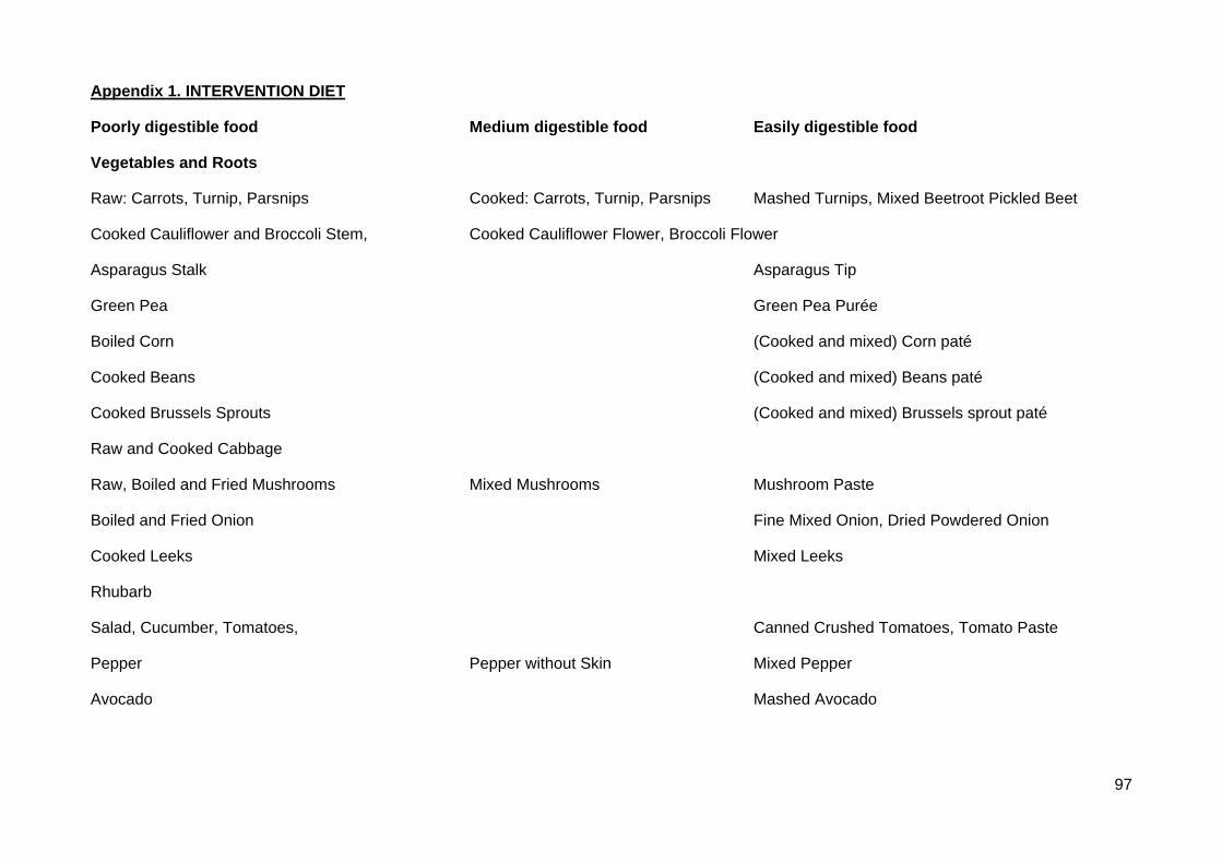
97
Appendix 1. INTERVENTION DIET
Poorly digestible food Medium digestible food Easily digestible food
Vegetables and Roots
Raw: Carrots, Turnip, Parsnips Cooked: Carrots, Turnip, Parsnips Mashed Turnips, Mixed Beetroot Pickled Beet
Cooked Cauliflower and Broccoli Stem, Cooked Cauliflower Flower, Broccoli Flower
Asparagus Stalk Asparagus Tip
Green Pea Green Pea Purée
Boiled Corn (Cooked and mixed) Corn paté
Cooked Beans (Cooked and mixed) Beans paté
Cooked Brussels Sprouts (Cooked and mixed) Brussels sprout paté
Raw and Cooked Cabbage
Raw, Boiled and Fried Mushrooms Mixed Mushrooms Mushroom Paste
Boiled and Fried Onion Fine Mixed Onion, Dried Powdered Onion
Cooked Leeks Mixed Leeks
Rhubarb
Salad, Cucumber, Tomatoes, Canned Crushed Tomatoes, Tomato Paste
Pepper Pepper without Skin Mixed Pepper
Avocado Mashed Avocado
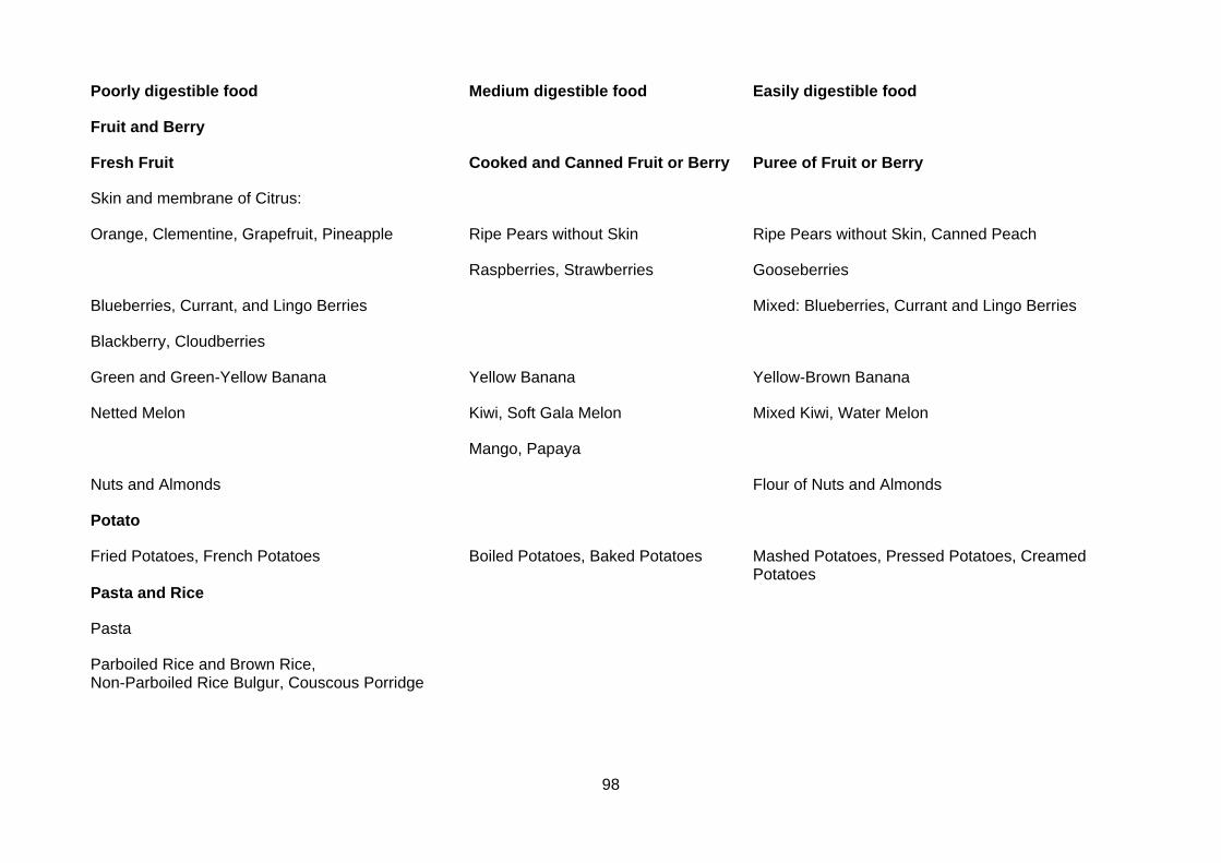
98
Poorly digestible food Medium digestible food Easily digestible food
Fruit and Berry
Fresh Fruit Cooked and Canned Fruit or Berry Puree of Fruit or Berry
Skin and membrane of Citrus:
Orange, Clementine, Grapefruit, Pineapple Ripe Pears without Skin Ripe Pears without Skin, Canned Peach
Raspberries, Strawberries Gooseberries
Blueberries, Currant, and Lingo Berries Mixed: Blueberries, Currant and Lingo Berries
Blackberry, Cloudberries
Green and Green-Yellow Banana Yellow Banana Yellow-Brown Banana
Netted Melon Kiwi, Soft Gala Melon Mixed Kiwi, Water Melon
Mango, Papaya
Nuts and Almonds Flour of Nuts and Almonds
Potato
Fried Potatoes, French Potatoes Boiled Potatoes, Baked Potatoes Mashed Potatoes, Pressed Potatoes, Creamed Potatoes Pasta and Rice
Pasta
Parboiled Rice and Brown Rice, Non-Parboiled Rice Bulgur, Couscous Porridge
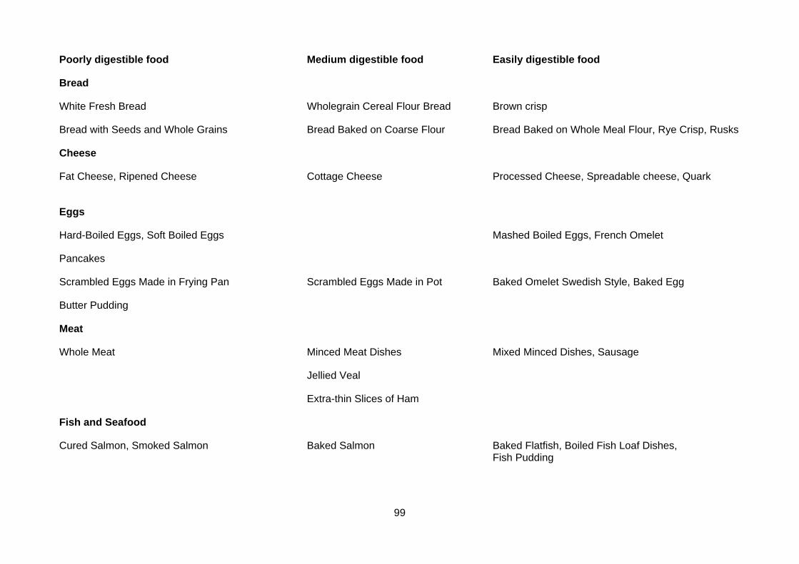
99
Poorly digestible food Medium digestible food Easily digestible food
Bread
White Fresh Bread Wholegrain Cereal Flour Bread Brown crisp
Bread with Seeds and Whole Grains Bread Baked on Coarse Flour Bread Baked on Whole Meal Flour, Rye Crisp, Rusks
Cheese
Fat Cheese, Ripened Cheese Cottage Cheese Processed Cheese, Spreadable cheese, Quark
Eggs
Hard-Boiled Eggs, Soft Boiled Eggs Mashed Boiled Eggs, French Omelet
Pancakes
Scrambled Eggs Made in Frying Pan Scrambled Eggs Made in Pot Baked Omelet Swedish Style, Baked Egg
Butter Pudding
Meat
Whole Meat Minced Meat Dishes Mixed Minced Dishes, Sausage
Jellied Veal
Extra-thin Slices of Ham
Fish and Seafood
Cured Salmon, Smoked Salmon Baked Salmon Baked Flatfish, Boiled Fish Loaf Dishes, Fish Pudding
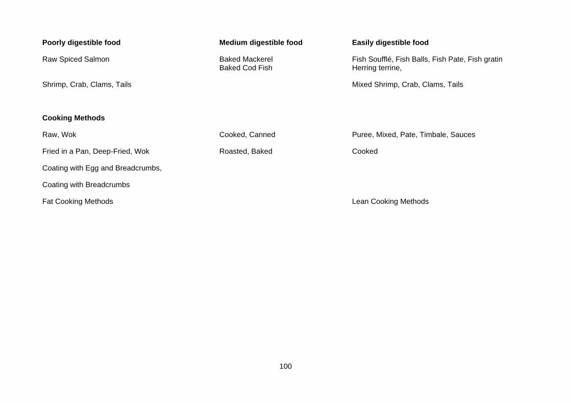
100
Poorly digestible food Medium digestible food Easily digestible food
Raw Spiced Salmon Baked Mackerel Fish Soufflé, Fish Balls, Fish Pate, Fish gratin Baked Cod Fish Herring terrine,
Shrimp, Crab, Clams, Tails Mixed Shrimp, Crab, Clams, Tails
Cooking Methods
Raw, Wok Cooked, Canned Puree, Mixed, Pate, Timbale, Sauces
Fried in a Pan, Deep-Fried, Wok Roasted, Baked Cooked
Coating with Egg and Breadcrumbs,
Coating with Breadcrumbs
Fat Cooking Methods Lean Cooking Methods
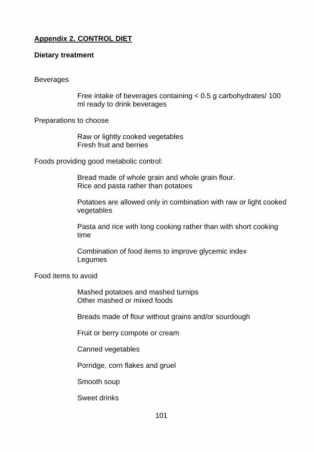
101
Appendix 2. CONTROL DIET
Dietary treatment
Beverages
Free intake of beverages containing < 0.5 g carbohydrates/ 100 ml ready to drink beverages
Preparations to choose
Raw or lightly cooked vegetables Fresh fruit and berries
Foods providing good metabolic control:
Bread made of whole grain and whole grain flour. Rice and pasta rather than potatoes
Potatoes are allowed only in combination with raw or light cooked vegetables
Pasta and rice with long cooking rather than with short cooking time
Combination of food items to improve glycemic index Legumes
Food items to avoid
Mashed potatoes and mashed turnips Other mashed or mixed foods
Breads made of flour without grains and/or sourdough
Fruit or berry compote or cream
Canned vegetables
Porridge, corn flakes and gruel
Smooth soup
Sweet drinks
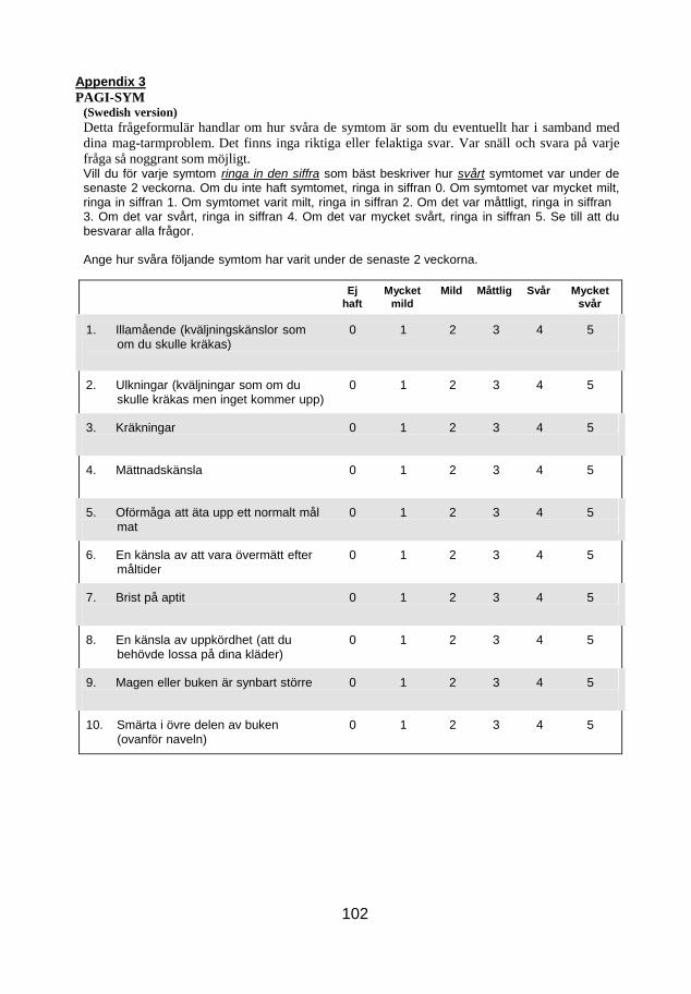
102
Appendix 3
PAGI-SYM (Swedish version)
Detta frågeformulär handlar om hur svåra de symtom är som du eventuellt har i samband med
dina mag-tarmproblem. Det finns inga riktiga eller felaktiga svar. Var snäll och svara på varje
fråga så noggrant som möjligt. Vill du för varje symtom ringa in den siffra som bäst beskriver hur svårt symtomet var under de senaste 2 veckorna. Om du inte haft symtomet, ringa in siffran 0. Om symtomet var mycket milt, ringa in siffran 1. Om symtomet varit milt, ringa in siffran 2. Om det var måttligt, ringa in siffran 3. Om det var svårt, ringa in siffran 4. Om det var mycket svårt, ringa in siffran 5. Se till att du besvarar alla frågor.
Ange hur svåra följande symtom har varit under de senaste 2 veckorna.
Ej Mycket Mild Måttlig Svår Mycket
haft mild svår
1. Illamående (kväljningskänslor som 0 1 2 3 4 5 om du skulle kräkas)
2. Ulkningar (kväljningar som om du 0 1 2 3 4 5 skulle kräkas men inget kommer upp)
3. Kräkningar 0 1 2 3 4 5
4. Mättnadskänsla 0 1 2 3 4 5
5. Oförmåga att äta upp ett normalt mål 0 1 2 3 4 5 mat
6. En känsla av att vara övermätt efter 0 1 2 3 4 5 måltider
7. Brist på aptit 0 1 2 3 4 5
8. En känsla av uppkördhet (att du 0 1 2 3 4 5 behövde lossa på dina kläder)
9. Magen eller buken är synbart större 0 1 2 3 4 5
10. Smärta i övre delen av buken 0 1 2 3 4 5 (ovanför naveln)
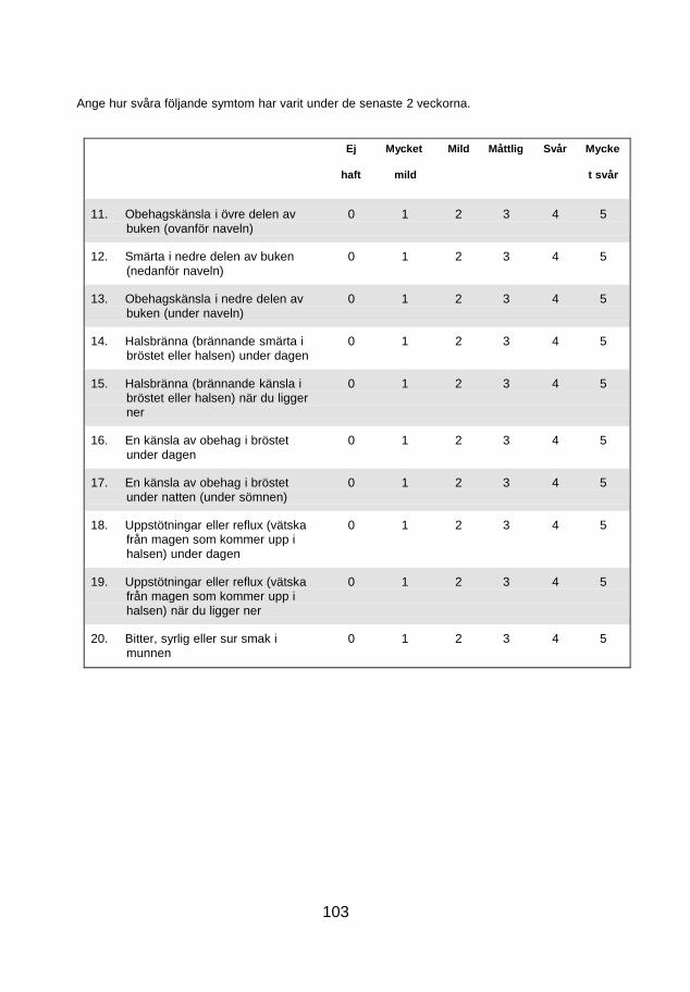
103
Ange hur svåra följande symtom har varit under de senaste 2 veckorna.
Ej Mycket Mild Måttlig Svår Mycke
haft mild t svår
11. Obehagskänsla i övre delen av 0 1 2 3 4 5 buken (ovanför naveln)
12. Smärta i nedre delen av buken 0 1 2 3 4 5 (nedanför naveln)
13. Obehagskänsla i nedre delen av 0 1 2 3 4 5 buken (under naveln)
14. Halsbränna (brännande smärta i 0 1 2 3 4 5 bröstet eller halsen) under dagen
15. Halsbränna (brännande känsla i 0 1 2 3 4 5 bröstet eller halsen) när du ligger ner
16. En känsla av obehag i bröstet 0 1 2 3 4 5 under dagen
17. En känsla av obehag i bröstet 0 1 2 3 4 5 under natten (under sömnen)
18. Uppstötningar eller reflux (vätska 0 1 2 3 4 5 från magen som kommer upp i halsen) under dagen
19. Uppstötningar eller reflux (vätska 0 1 2 3 4 5 från magen som kommer upp i halsen) när du ligger ner
20. Bitter, syrlig eller sur smak i 0 1 2 3 4 5 munnen
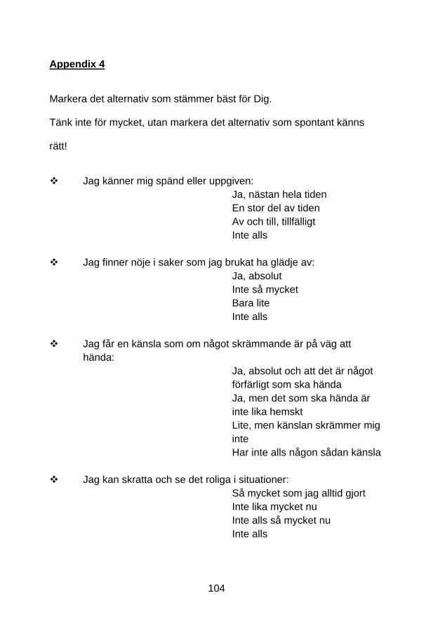
104
Appendix 4
Markera det alternativ som stämmer bäst för Dig.
Tänk inte för mycket, utan markera det alternativ som spontant känns
rätt!
Jag känner mig spänd eller uppgiven:
Ja, nästan hela tiden
En stor del av tiden
Av och till, tillfälligt
Inte alls
Jag finner nöje i saker som jag brukat ha glädje av:
Ja, absolut
Inte så mycket
Bara lite
Inte alls
Jag får en känsla som om något skrämmande är på väg att
hända:
Ja, absolut och att det är något
förfärligt som ska hända
Ja, men det som ska hända är
inte lika hemskt
Lite, men känslan skrämmer mig
inte
Har inte alls någon sådan känsla
Jag kan skratta och se det roliga i situationer:
Så mycket som jag alltid gjort
Inte lika mycket nu
Inte alls så mycket nu
Inte alls
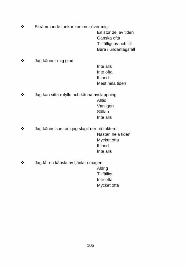
105
Skrämmande tankar kommer över mig:
En stor del av tiden
Ganska ofta
Tillfälligt av och till
Bara i undantagsfall
Jag känner mig glad:
Inte alls
Inte ofta
Ibland
Mest hela tiden
Jag kan sitta rofylld och känna avslappning:
Alltid
Vanligen
Sällan
Inte alls
Jag känns som om jag slagit ner på takten:
Nästan hela tiden
Mycket ofta
Ibland
Inte alls
Jag får en känsla av fjärilar i magen:
Aldrig
Tillfälligt
Inte ofta
Mycket ofta
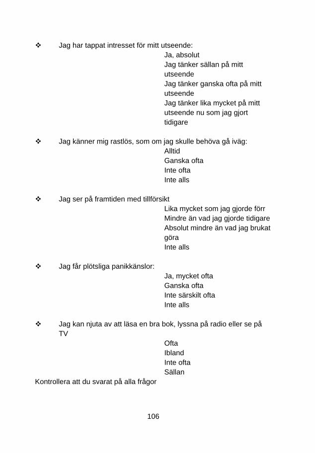
106
Jag har tappat intresset för mitt utseende:
Ja, absolut
Jag tänker sällan på mitt
utseende
Jag tänker ganska ofta på mitt
utseende
Jag tänker lika mycket på mitt
utseende nu som jag gjort
tidigare
Jag känner mig rastlös, som om jag skulle behöva gå iväg:
Alltid
Ganska ofta
Inte ofta
Inte alls
Jag ser på framtiden med tillförsikt
Lika mycket som jag gjorde förr
Mindre än vad jag gjorde tidigare
Absolut mindre än vad jag brukat
göra
Inte alls
Jag får plötsliga panikkänslor:
Ja, mycket ofta
Ganska ofta
Inte särskilt ofta
Inte alls
Jag kan njuta av att läsa en bra bok, lyssna på radio eller se på
TV
Ofta
Ibland
Inte ofta
Sällan
Kontrollera att du svarat på alla frågor

107
Appendix 5
HÄLSOENKÄT
INSTRUKTION: Detta formulär innehåller frågor om hur Du ser på Din hälsa.
Informationen skall hjälpa till att följa hur Du mår och fungerar i Ditt dagliga liv.
Besvara frågorna genom att sätta en ring runt den siffra Du tycker stammer bast in på Dig. Om Du ar osäker,ringa ändå in den siffra som känns riktigast.
1. lallmänhet, skulle Du vilja saga att Din hälsa ar:
(Satt en ring runt en siffra)
Utmärkt............................... ........................................................1
Mycket god ....................................................................................2
God ..................................................................................................3
Någorlunda...................................................................................4
Dålig .................................................... .......................................S
2. Jämfört med för ett år sedan, hur skulle Du vilja bedöma Ditt allmänna hälsotillstånd nu?
(Satt en ring runt en siffra)
Mycket bättre nu an för ett år sedan..............................................1
Något bättre nu an för ett år sedan ...............................................2
Ungefar detsamma.......................................................................3
Något sämre nu an för ett år sedan............................................... 4
Mycket sämre nu an för ett år sedan............................................5
Copyright C 199<4 MedicalOutcomes Trust. All rights reserved. (IQOLA SF-36 Standard Swedish Version 1.0)
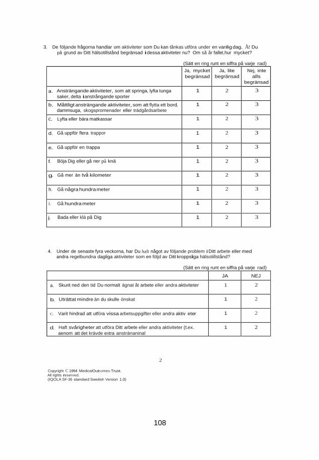
108
Ja, mycket
begränsad
Ja, lite
begränsad
Nej, inte
alls
begränsad
a. Ansträngande aktiviteter, som att springa, lyfta tunga
saker, delta ianstrångande sporter
1 2 3
b. Måttligt ansträngande aktiviteter, som att flytta ett bord, dammsuga, skogspromenader eller trädgårdsarbete
1 2 3
c. Lyfta eller bära matkassar 1 2 3
d. Gå uppför flera trappor 1 2 3
e. Gå uppför en trappa 1 2 3
f. Böja Dig eller gå ner på knä 1 2 3
g. Gå mer än två kilometer 1 2 3
h. Gå några hundra meter 1 2 3
i. Gå hundra meter 1 2 3
j. Bada eller klä på Dig 1 2 3
JA NEJ
a. Skurit ned den tid Du normalt ägnat åt arbete eller andra aktiviteter 1 2
b. Uträttat mindre än du skulle önskat 1 2
C. Varit hindrad att utföra vissa arbetsuppgifter eller andra aktiv eter 1 2
d. Haft svårigheter att utföra Ditt arbete eller andra aktiviteter (t.ex.
aenom att det krävde extra anstränaninal
1 2
3. De följande frågorna handlar om aktiviteter som Du kan tånkas utföra under en vanlig dag. Ar Du
på grund av Ditt hälsotillstånd begränsad idessa aktiviteter nu? Om så år fallet,hur mycket?
(Sätt en ring runt en siffra på varje rad)
4. Under de senaste fyra veckorna, har Du haft något av följande problem i Ditt arbete eller med
andra regelbundna dagliga aktiviteter som en följd av Ditt kroppsliga hälsotillstånd?
(Sätt en ring runt en siffra på varje rad)
2
Copyright C 1994 MedicalOutcomes Trust. All rights reserved. (IQOLA SF-36 standard Swedish Version 1.0)
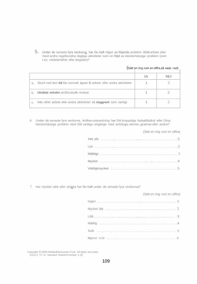
109
5. Under de senaste fyra veckorna. har Du haft något av följande problem iDitt arbete eller
med andra regelbundna dagliga aktiviteter som en följd av känslomässiga problem (som
t.ex. nedstämdhet eller ängslan)?
(Sätt en ring runt en siffra på varje rad)
JA NEJ
a. Skurit ned den tid Du normalt ägnat åt arbete eller andra aktiviteter 1 2
b. Uträttat mindre anDu skulle önskat 1 2
c. Inte utfört arbete eller andra aktiviteter så noggrant som vanligt 1 2
6. Under de senaste fyra veckorna, ivilken utstrackning har Ditt kroppsliga halsatillstånd eller Dina
känslomässiga problem stort Ditt vanliga umgänge med anhönga,vänner,gramar eller andra?
(Sätt en ring runt en siffra)
Inte alls ........................................................................................1
Lite ..............................................................................................2
Måttligt........................................................... ............................3
Mycket ................................................... ..... .............................4
Väldigtmycket .............................................................................5
7. Hur mycket värk eller smärta har Du haft under de senaste fyra veckorna?
(Sätt en ring runt en siffra)
Ingen ......................................................................................1
Mycket lätt ................................................................................ 2
Lått................................................. .......................................... ..3
Måttlig ..................................................................................... .4
Svår ........................................................... ...................... 5
Mycket svår ..................... ....................................................... ..6
Copyright C1994 Medci alOutcomes Trust. All rights reserved.
(IQOLA SF-36 Standard Swedish Verslon 1.0)
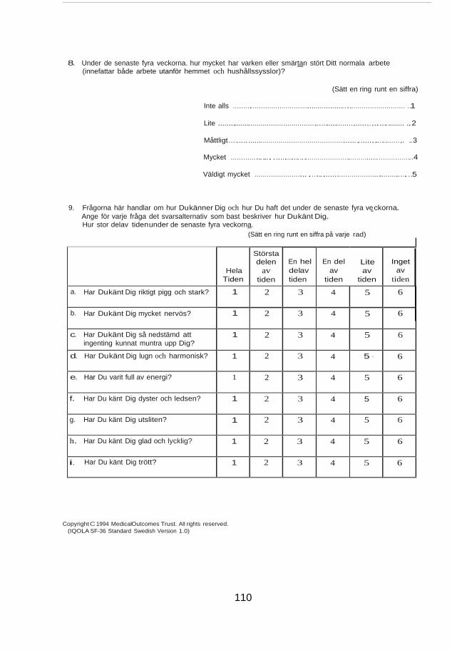
110
Hela
Tiden
Största delen
av tiden
En hel
delav
tiden
En del
av
tiden
Lite
av
tiden
Inget
av
tiden
a. Har Dukänt Dig riktigt pigg och stark? 1 2 3 4 5 6
b. Har Dukänt Dig mycket nervös? 1 2 3 4 5 6
c. Har Dukänt Dig så nedstämd att
ingenting kunnat muntra upp Dig? 1 2 3 4 5 6
d. Har Dukänt Dig lugn och harmonisk? 1 2 3 4 5 · 6
e. Har Du varit full av energi? 1 2 3 4 5 6
f. Har Du känt Dig dyster och ledsen? 1 2 3 4 5 6
g. Har Du känt Dig utsliten? 1 2 3 4 5 6
h. Har Du känt Dig glad och lycklig? 1 2 3 4 5 6
i. Har Du känt Dig trött? 1 2 3 4 5 6
8. Under de senaste fyra veckorna. hur mycket har varken eller smärtan stört Ditt normala arbete
(innefattar både arbete utanför hemmet och hushållssysslor)?
(Sätt en ring runt en siffra)
Inte alls ....................................................... ........................... ..1
Lite ............................................... .................. ..... . .............. ..2
Måttligt.............................................................. ..... ............. . ..3
Mycket ............ ...........................................................................4
Väldigt mycket ...................... ..... ......... ................... ..............5
9. Frågorna här handlar om hur Dukänner Dig och hur Du haft det under de senaste fyra ve ckorna.
Ange för varje fråga det svarsalternativ som bast beskriver hur Dukänt Dig. Hur stor delav tidenunder de senaste fyra veckorna.
(Sätt en ring runt en siffra på varje rad)
Copyright C 1994 MedicalOutcomes Trust. All rights reserved.
(IQOLA SF-36 Standard Swedish Version 1.0)
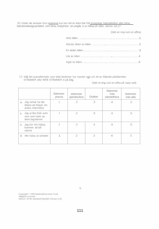
111
Stämmer
precis
stämmer
ganska bra
Osäker
Stämmer
Inte
särskilt bra
Stämmer
inte alls
a. Jag verkar ha lite
lättare att blisjuk än andra människor
1 2 3 4 5
b. Jag ar lika frisk som
vem som helst av dem Jag känner
1 2 3 4 5
c. Jag tror min hälsa kommer att bli
sämre
1 2 3 4 5
d. Min halsa ar utmärkt 1 2 3 4 5
1O. Under de senaste fyra veckorna,hur stor del av tiden har Ditt kroppsliga hälsotillstånd eller Dina känslomässiga problem stört Dina möjligheter att umgås (t ex hälsa på släkt, vänner, etc.)?
(Sätt en ring runt en siffra)
Hela tiden ....... ......................................... ...... ...........1
Slörsta delen av tiden ...................................................................2
En delav tiden..............................................................................3
Lite av tiden .......................................... .....................................4
Inget av liden..............................................................................5
11. Välj det svarsalternativ som bäst beskriver hur mycket vart och ett av följande påståenden
STÅMMER eller INTE STÅMMER in på Dig. (Sätt en ring runt en siffra på varje rad)
5
Copynght C 1994 MedicalOutcomes Trust.
Allrights reserved.
(IQOLA SF-36 standard Swedish Version 1.0)

112 112