Embed Size (px)
Citation preview

Directly Coded Summary StageCorpus Uteri
National Center for Chronic Disease Prevention and Health Promotion
Division of Cancer Prevention and Control, Cancer Surveillance Branch

Directly Coded Summary Staging is Back
Summary Staging (known also as SEER Staging) bases staging of solid tumors solely on whether or not the disease has spread.
Registrars need to be knowledgeable of the definitions of each stage to assign it correctly.
It is an efficient tool to categorize if and/or how far the cancer has spread from the original site.
It should be noted that in the SEER Summary Staging Schema, Kaposi Sarcoma, Lymphomas and Hematopoietic Diseases are addressed. The schemas are not the same methodology as the solid tumors but you need to be aware they are provided.

To Begin the Staging Process, Abstractors Should Always Review:
History and Physical Exam
Radiology Reports
Operative Reports
Pathology Reports
Medical Consults
Pertinent Correspondence

Determining how a Tumor Should be Staged requires the Registrar to:
Read the Physical Exam and Work Up
documents.
Read operative and pathology reports.
Review imaging reports for documentation of
any spread.
Become familiar with the anatomy of the
endometrium and the regional and distant
lymph node chains.
Refer to the online manuals regularly and
periodically check the site for updates and/or
changes.

Assigning the Correct Summary Stage Code
In-situ is coded as 0. Localized disease only is coded as 1. Regional disease by direct extension only is
coded as 2. Regional disease w/only regional lymph nodes
involved is coded as 3. Regional disease by both direct extension and
regional lymph node involvement is coded as 4.
Regional disease not otherwise specified is coded as 5.
Distant sites or distant lymph node involvement is coded as 7.
Unknown if there is extension or metastatic disease (unstaged, unspecified, death certificate only cases) is coded as 9.

Become Familiar With How Cancers May Spread
Lymphatic Spread is often evident in any of the following: aortic, iliac, parametrial, paracervical, and sacral lymph node chains.
Hematogenous Spread is most commonly found in bone, liver, lung or brain.

Corpus Uteri is Composed of 3 Anatomic Structures
Endometrium – (Mucosa)
Columnar Epithelium
o This has no blood vessels or lymphatics
Basement Membrane
Stroma (Lamina Propria)
o Areolar connective tissue contains blood vessels,
nerves and glands in some regions
Myometrium – 3 layers
Serosa (Tunica Serosa)

What does In-Situ Mean? In-situ is defined as malignancy without
invasion. Only occurs with epithelial or mucosal tissue
Must be microscopically diagnosed to visualize the basement membrane. In-situ of the endometrium may also be referred
to as non-invasive, pre-invasive, non-infiltrating, or used to be called FIGO Stage 0, FIGO no longer has a stage 0
If pathology states the tumor is in-situ with microinvasion it is no longer staged as in-situ but is considered to be at least a localized disease.
In-situ disease is coded as Summary Stage 0.

Staging In-situ Cancers Requires Knowledge
of a Specific Exception
In-situ is a non-invasive malignancy and is coded as Summary Stage ‘0’
UNLESS Primary Tumor was documented in the
pathology report as having only an in-situ behavior but there is an additional statement confirming malignancy has spread and is present in a local, regional node(s) or distant site.
Should that occur, the in-situ stage is not valid and the stage must be documented to reflect the regional or distant disease.

What does Localized Mean?
Localized corpus uteri cancer is a malignancy which is
Confined to the endometrium (stroma) FIGO stages in summary stage are not current and may not
be used Myometrium/serosa (or tunica serosa) of the
corpus invasion FIGO stages in summary stage are not current and may not
be used Localized, NOS or FIGO stage I with no further
information FIGO stages in summary stage are not current and may not
be used
Localized disease is coded as Summary Stage 1.

Staging of Regional Disease
Review records to confirm that tumor is more than localized.
Review all pertinent reports looking for specific regional disease references and exclusions of distant spread. Terms to watch for are seeding, implants and nodules – scrutinize diagnostic reports for regional disease spreading references to eliminate that spread is not distant.
Caution: A diagnosis of cancer with lymph node metastases means involvement by tumor – always confirm that the lymph nodes are regional.
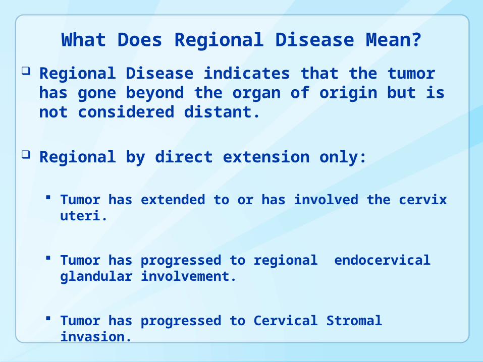
What Does Regional Disease Mean?
Regional Disease indicates that the tumor has gone beyond the organ of origin but is not considered distant.
Regional by direct extension only:
Tumor has extended to or has involved the cervix uteri.
Tumor has progressed to regional endocervical glandular involvement.
Tumor has progressed to Cervical Stromal invasion.
FIGO stages in summary stage are not current and may not be used.
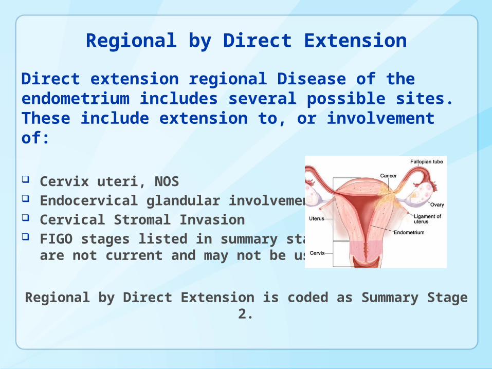
Regional by Direct Extension
Direct extension regional Disease of the endometrium includes several possible sites. These include extension to, or involvement of:
Cervix uteri, NOS Endocervical glandular involvement only Cervical Stromal Invasion FIGO stages listed in summary stage
are not current and may not be used
Regional by Direct Extension is coded as Summary Stage 2.

Regional by Direct Extension cont’d
Tumor has extended or metastasized to any of following sites: Fallopian tube(s) Broad, Round or Uterosacral Ligaments One or both ovaries Parametrium Pelvic Serosa# Pelvic tunica serosa# Ureter* Vulva*
Cancer cells in ascites@ Cancer cells in peritoneal washings@ FIGO stages in summary stage are not current & may not
be used*Considered distant in Historic Staging#Considered distant in SS 1977 @Not specifically categorized in Historic Staging or SS 1977Regional by Direct Extension is coded as Summary Stage
2.

Regional by Direct Extension cont’d
Tumor has extended or metastasized to any of the following sites:
Extension or metastasis*# Bladder, NOS excluding mucosa Bladder wall Bowel wall, NOS Rectum, NOS excluding mucosa Vagina* Pelvic wall(s)#
FIGO stages in summary stage are not current and may not be used
*Considered distant in Historic Staging#Considered distant in SS 1977
Regional by Direct Extension is coded as Summary Stage 2.

Regional Lymph Node Involvement OnlyTumor cells may have traveled through the lymphatic system to regional lymph nodes where they remain and begin to “grow.”
Aortic, NOS – Includes lateral aortic or lumbar, para-aortic and
periaortic#
Iliac – Includes common, external, and internal or hypogastic,
obturator
Paracervical#
Parametrial
Pelvic, NOS
Sacral, NOS – Includes lateral sacral or laterosacral; middle,
promontorial or Gerota’s node; presacral; and uterosacral#
FIGO stages in summary stage are not current and may not be
used
#Considered distant in SS 1977
Regional by Lymph Node Involvement only is coded as Summary Stage 3.

Regional Nodes for Corpus Uteri
Aortic, NOS#
Lateral or lumbar Para-aortic Periarotic
Iliac Common External Internal or hypogastric, NOS
o Obturator Paracervical# Parametrial Pelvic, NOS
#Considered distant in SS 1977
Regional to Lymph Nodes is coded as Summary Stage 3.
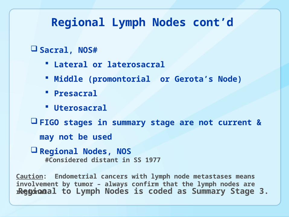
Regional Lymph Nodes cont’d
Sacral, NOS#
Lateral or laterosacral
Middle (promontorial or Gerota’s Node)
Presacral
Uterosacral
FIGO stages in summary stage are not current &
may not be used
Regional Nodes, NOS#Considered distant in SS 1977
Caution: Endometrial cancers with lymph node metastases means involvement by tumor – always confirm that the lymph nodes are regional.Regional to Lymph Nodes is coded as Summary Stage 3.

Regional Disease by Direct Extension and Lymph Nodes
Regional Extension into adjacent structures or organs and lymph nodes involvement are both present.
Regional disease by both direct extension and lymph nodes is coded
as Summary Stage 4.

.

Regional, NOS
It is unclear if the tissues involved are regional direct extension or lymph nodes
Physician statement says “Regional disease” with no additional documentation in the medical record.
Regional Disease with no further information is coded as Regional – NOS – Summary Stage 5

Read Carefully
Carcinoma of the corpus uteri with metastasis to regional lymph nodes.
This indicates that the involved lymph nodes are those that are the first to drain the primary and should be staged as regional to lymph nodes.
Don’t be misled by the term metastases – It doesn’t always mean distant disease.

Important Notes to Remember Adnexa is defined as tubes, ovaries and ligaments Frozen pelvis means tumor extends to the pelvic
sidewall(s). With no statement of involvement, code these cases as regional by direct extension. (With Historic Staging and SS 1977 these cases were coded as distant).
If the physician states adnexa palpated with no mention of lymph nodes, the Registrar should assume nodes are not involved.
If exploratory or definitive surgery was done with no mention of lymph nodes assume nodes are not involved.
Sounding of the corpus is no longer a prognostic factor as it was in the past.
Extension to bowel or bladder mucosa must be proven by biopsy. This is to rule out bullous edema.
Important to Know: This schema should be used for sarcomas of the myometrium. AJCC has separate staging in their corpus uteri chapter for sarcomas and carcinomas.
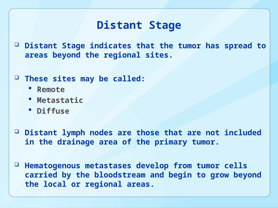
Distant Stage
Distant Stage indicates that the tumor has spread to areas beyond the regional sites.
These sites may be called: Remote Metastatic Diffuse
Distant lymph nodes are those that are not included in the drainage area of the primary tumor.
Hematogenous metastases develop from tumor cells carried by the bloodstream and begin to grow beyond the local or regional areas.
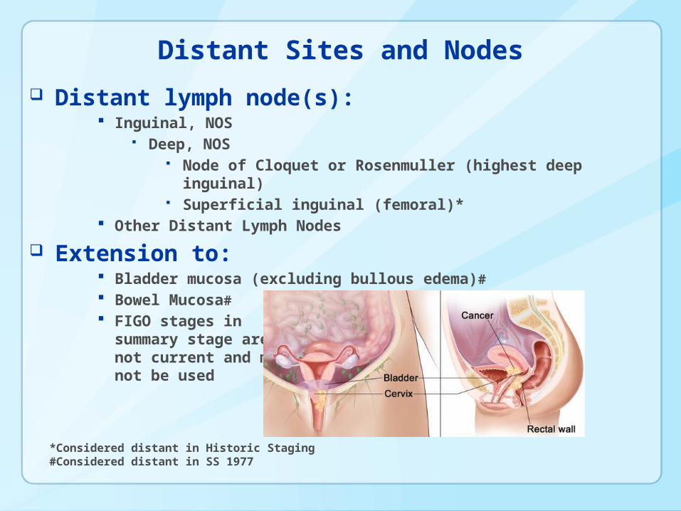
Distant Sites and Nodes
Distant lymph node(s): Inguinal, NOS
Deep, NOS Node of Cloquet or Rosenmuller (highest deep
inguinal) Superficial inguinal (femoral)*
Other Distant Lymph Nodes
Extension to: Bladder mucosa (excluding bullous edema)# Bowel Mucosa# FIGO stages in
summary stage arenot current and maynot be used
*Considered distant in Historic Staging#Considered distant in SS 1977

Distant Sites and Nodes cont’d
Further contiguous extension#: Abdominal serosa (peritoneum) Cul de sac (rectouterine pouch) Sigmoid colon Small intestine
Metastasis
FIGO stages in summary stage are not current and may not be used
#Considered regional in Historic Staging

Tips for the Abstractor If review of the patient’s records documents distant
metastases, the registrar can avoid reviewing records to identify local or regional disease.
Pathology reports that contain a statement of local, regional or metastatic spread cannot be staged as in-situ even if the pathology of the tumor states it.
If there are nodes involved, the stage must be at least regional.
If there are nodes involved but the chain is not named in the pathology report, assume the nodes are regional.
If the record does not contain enough information to assign a stage, it must be recorded as unstageable.

Exercise 1– How Would You Stage This Case?
Patient presented with abnormal vaginal bleeding. Physical examination was within normal limits – no abdominal masses or lymphadenopathy noted. Uterus and cervix did not reveal any abnormalities.
MRI was ordered and noted right fundal endometrium consistent with carcinoma. Further workup including CT of abdomen and pelvis did not reveal any additional abnormality.
Patient underwent a total abdominal hysterectomy with bilateral salpingo-oophorectomy with pathology noting moderately differentiated adenocarcinoma in the endometrium. No invasion of the myometrium, tubes or ovaries.
Summary Stage 1 Localized.

Exercise 2 – How Would You Stage This Case?
Patient presented with light spotting. No urinary frequency or incontinence. Normal findings with a speculum exam. Uterus was without tenderness. Rectovaginal exam did not find any rectal masses.
Ultrasound noted only a thickened endometrial stripe. Chest and abdominal/pelvic CT within normal limits.
Pathology from the total abdominal hysterectomy and bilateral salpingo oophorectomy revealed a well differentiated adenocarcinoma with invasion of the myometrium of 1.5 mm. No nodes present and therefore were unable to be assessed.
Summary Stage 1 localized, based on CT not showing regional involvement and no nodes resected.

Exercise 3 – How Would You Stage This Case?
Patient presented with complaints of urinary incontinence. Physician ordered an IVP with findings negative for kidney and ureters issues but a pelvic mass was identified which appeared to be compressing the bladder.
An endometrial biopsy was identified as adenocarcinoma. Hysterectomy was done and the carcinoma was found to be invading the myometrium. Six pelvic nodes were involved.
Summary Stage 3, based on the regional lymph node involvement.
Exercise 4 – How Would You Stage This Case?
Patient presented for her annual physical with a complaint of abdominal discomfort. Her physician noted a pelvic mass that was considered suspicious for malignancy.
She opted for and underwent a TAH-BSO which
revealed an endometrial adenocarcinoma. There was invasion of the vagina. Seven of 18 nodes were positive for malignancy.
Summary Stage 4, with direct extension to the vagina and lymph node involvement.

Excellent Resources for Summary Staging
http://seer.cancer.gov/manuals/2013/SPCSM_2013_maindoc.pdf
SEER Summary Stage 2000, SEER Training modules: http://training.seer.cancer.gov
SEER Coding Manuals – Historic – 1977.
http://training.seer.cancer.gov/modules_site_spec.html
http://training.seer.cancer.gov/endometrium/abstract-code-stage/extent/

Centers for Disease Control and PreventionChamblee Campus, Atlanta GA

Contact Information
For more information please contact Centers for Disease Control and Prevention
1600 Clifton Road NE, Atlanta, GA 30333Telephone: 1-800-CDC-INFO (232-4636)/TTY: 1-888-232-6348E-mail: [email protected] Web: http://www.cdc.gov
The findings and conclusions in this report are those of the authors and do not necessarily represent the official position of the Centers for Disease Control and Prevention.
National Center for Chronic Disease Prevention and Health Promotion
Division of Cancer Prevention and Control, Cancer Surveillance Branch