Embed Size (px)
DESCRIPTION
ECHOCARDIOGRAPHIC MONITORING ON ECMO. M.Mondino MD Dept.of Cardiac Anesthesia and CV-ICU Niguarda Hospital, Milan. Extra-Corporeal Membrane Oxygenation. - PowerPoint PPT Presentation
Citation preview
ECHOCARDIOGRAPHIC MONITORING ON ECMO
M.Mondino MD Dept.of Cardiac Anesthesia and CV-ICU
Niguarda Hospital, Milan
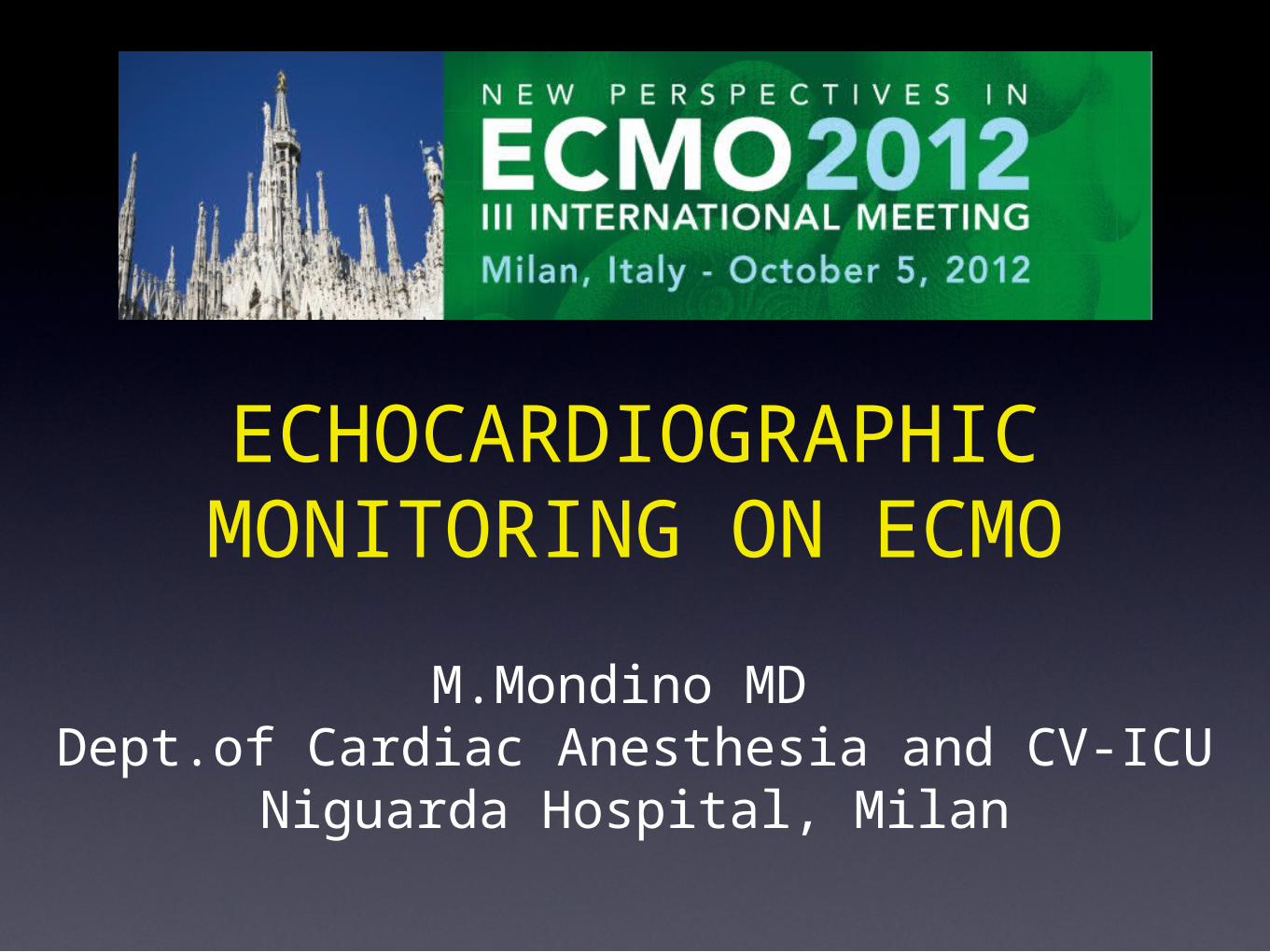
ECMO is a rescue therapy used to provide cardiac and/or respiratory support for critically ill patients in whom maximal conventional medical management has failed.
V-V ECMO: provides adequate oxygenation and carbon dioxide removal in isolated refractory respiratory failure.
V-A ECMO: when support is required for cardiac and/or respiratory failure.
Extra-Corporeal Membrane Oxygenation
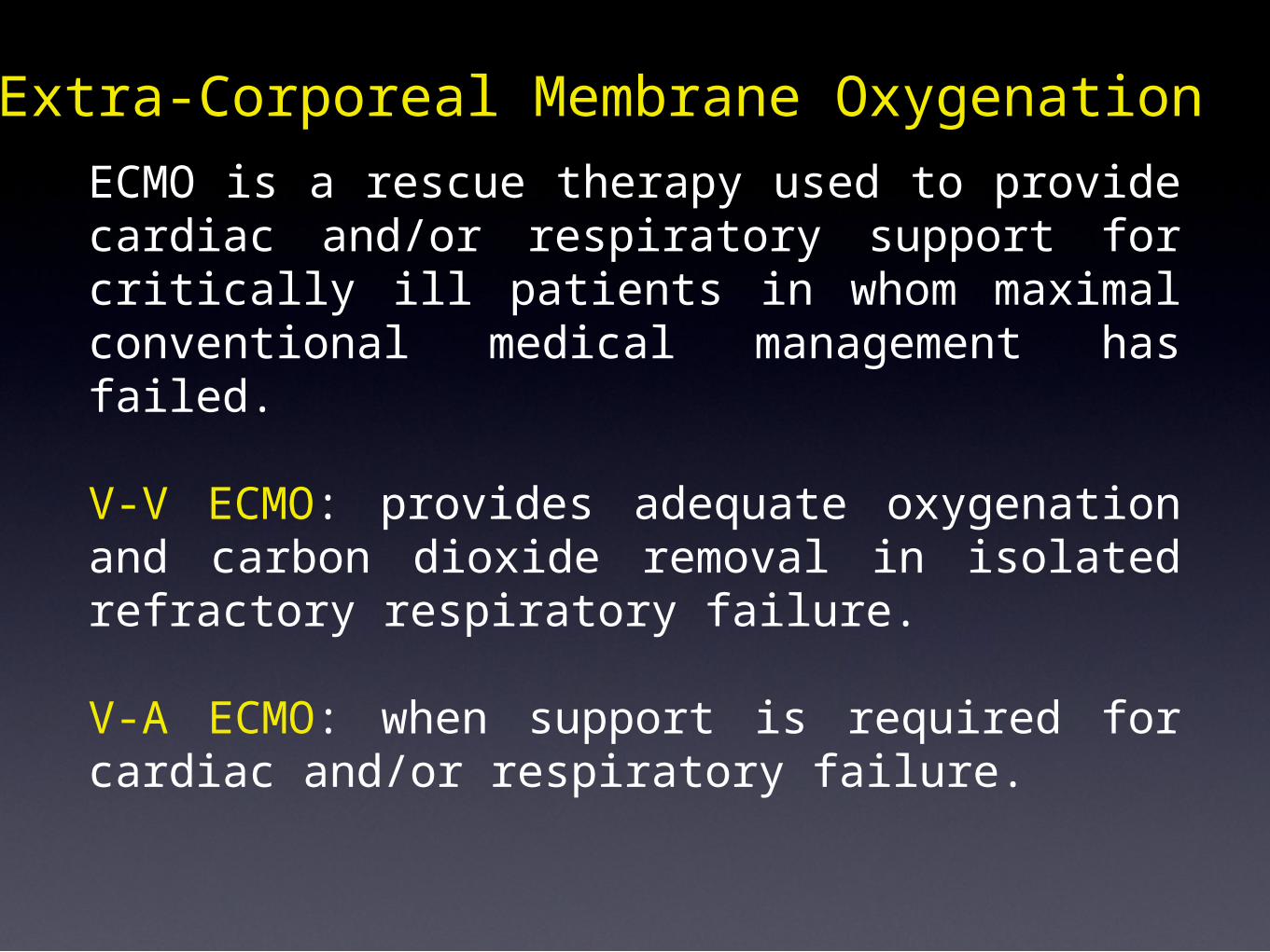
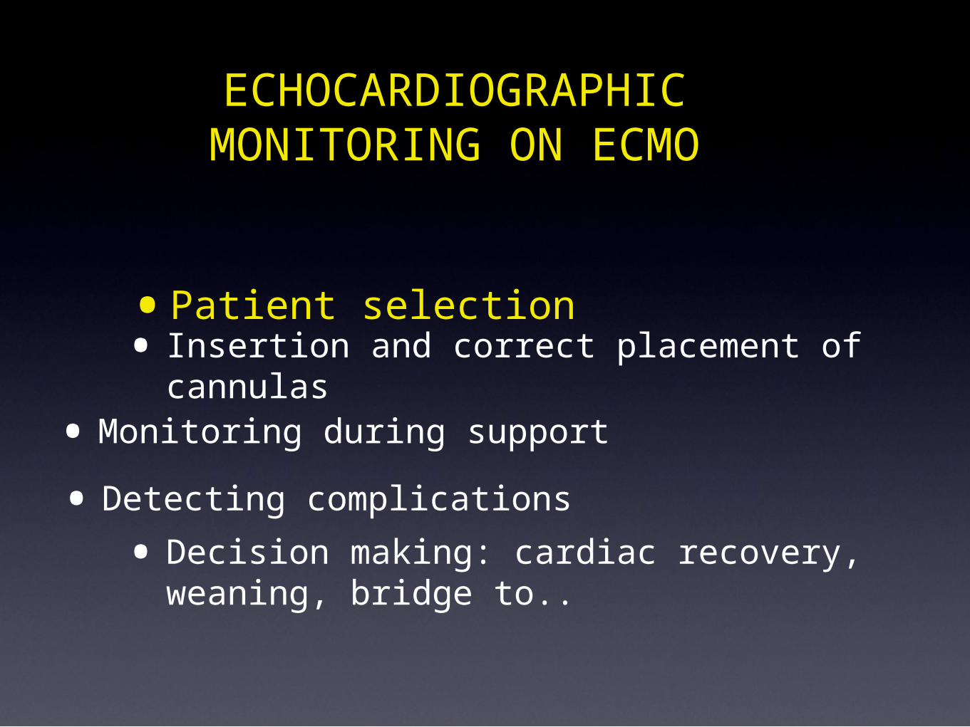
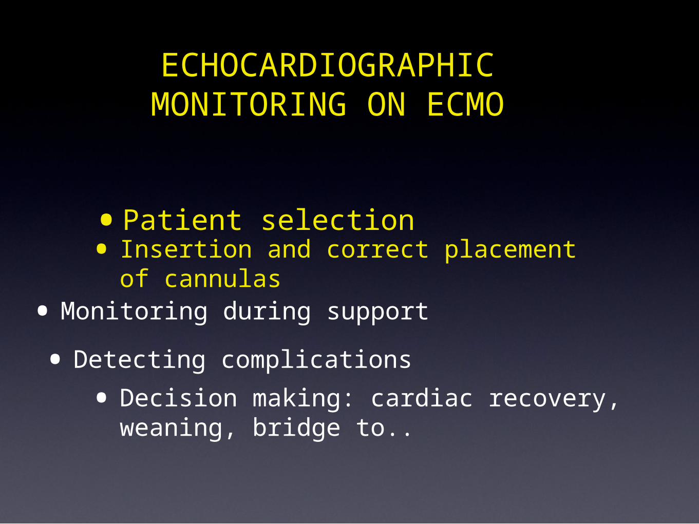
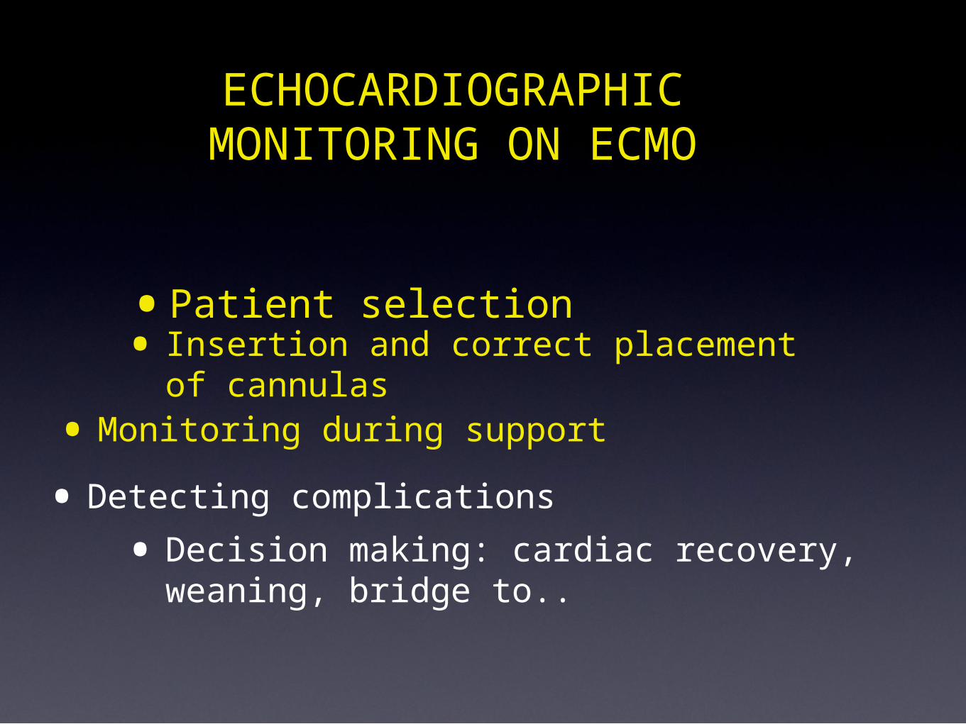
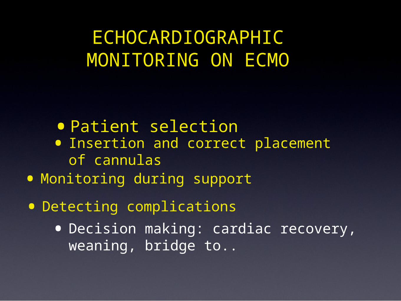
• Patient selection
ECHOCARDIOGRAPHIC MONITORING ON ECMO
• Insertion and correct placement of cannulas
• Monitoring during support
• Detecting complications• Decision making: cardiac recovery, weaning,
bridge to..
•Patient selection
ECHOCARDIOGRAPHIC MONITORING ON ECMO
• Insertion and correct placement of cannulas
• Monitoring during support
• Detecting complications• Decision making: cardiac recovery, weaning,
bridge to..
Pt selection- Reversible causes of hemodynamic instability
V-A ECMO controindications
Aortic dissection Aortic valve regurgitation
Severe Arterial vascular disease
Absoulte controindicationscentral vs peripheralsurgical vs percutaneous
5
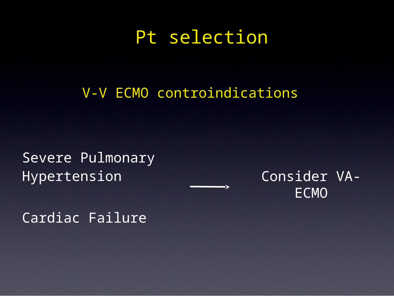
V-V ECMO controindications
Severe PulmonaryHypertension
Cardiac Failure
Pt selection
Consider VA-ECMO
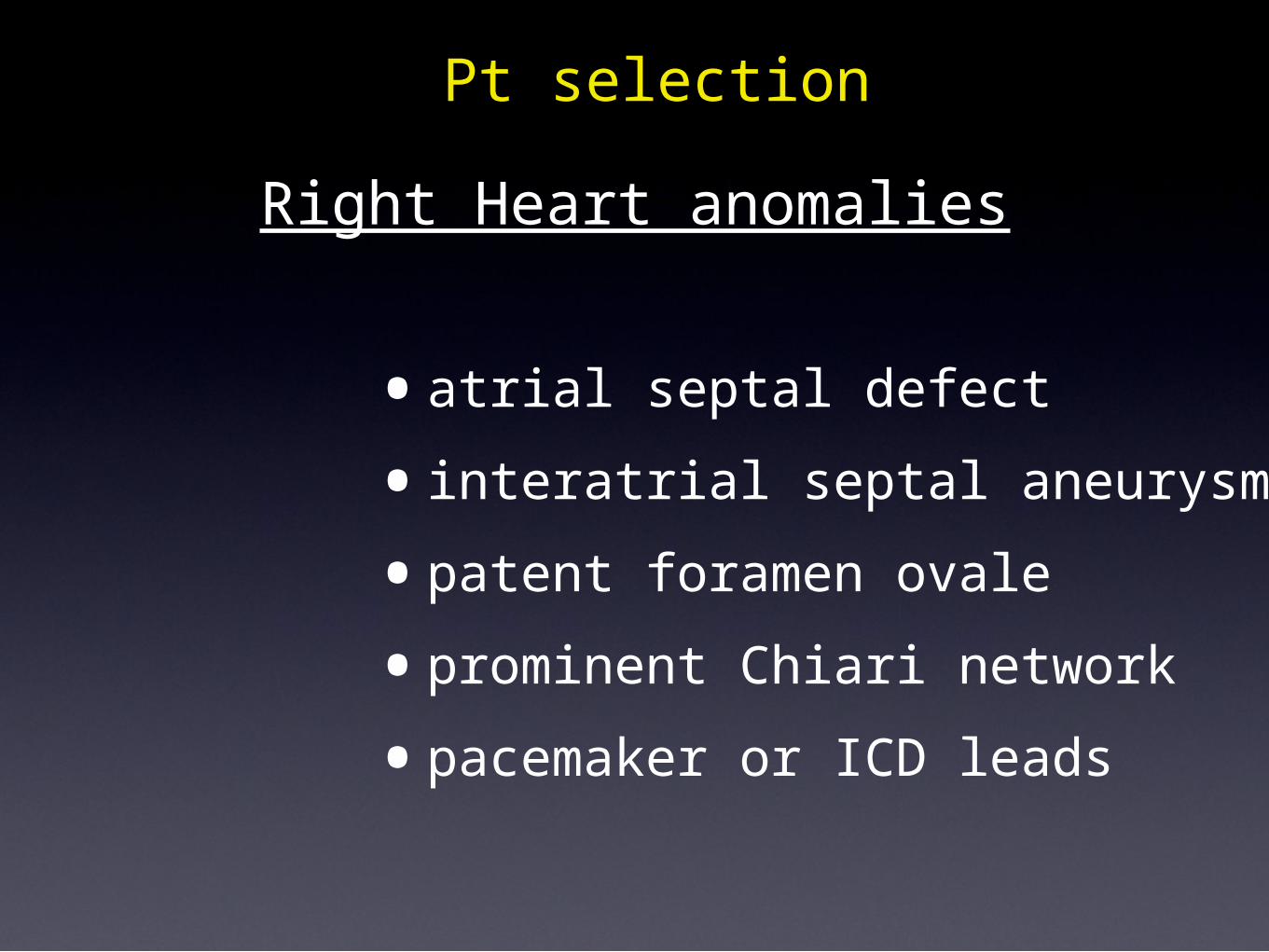
•atrial septal defect•interatrial septal aneurysm •patent foramen ovale•prominent Chiari network•pacemaker or ICD leads
Pt selectionRight Heart anomalies
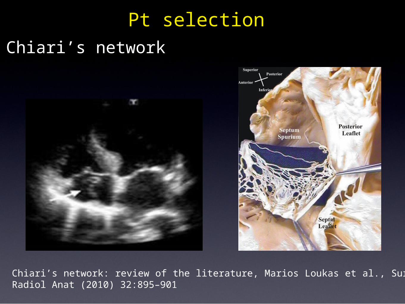
Chiari’s network: review of the literature, Marios Loukas et al., Surg Radiol Anat (2010) 32:895–901
Chiari’s networkPt selection
Pt selection
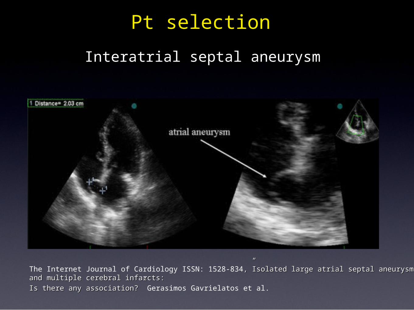
The Internet Journal of Cardiology ISSN: 1528-834,”Isolated large atrial septal aneurysm and multiple ,”Isolated large atrial septal aneurysm and multiple cerebral infarcts:cerebral infarcts:Is there any association?” Is there any association?” Gerasimos Gavrielatos et al.
Interatrial septal aneurysm
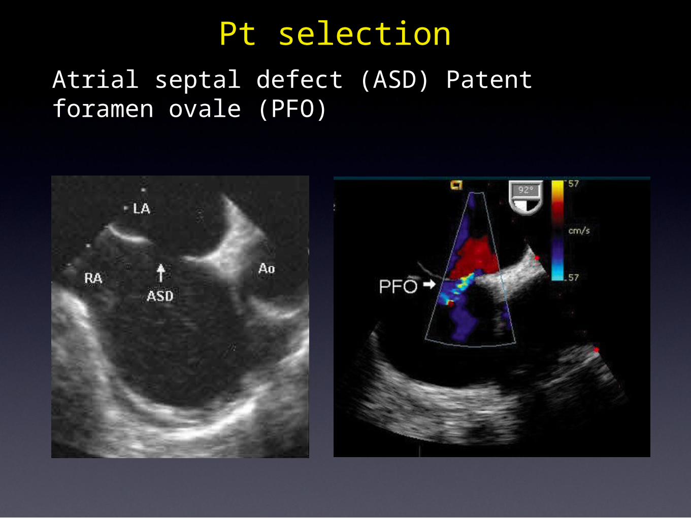
Pt selectionAtrial septal defect (ASD) Patent foramen ovale (PFO)
•Patient selection
ECHOCARDIOGRAPHIC MONITORING ON ECMO
• Insertion and correct placement of cannulas
• Monitoring during support
• Detecting complications• Decision making: cardiac recovery, weaning,
bridge to..
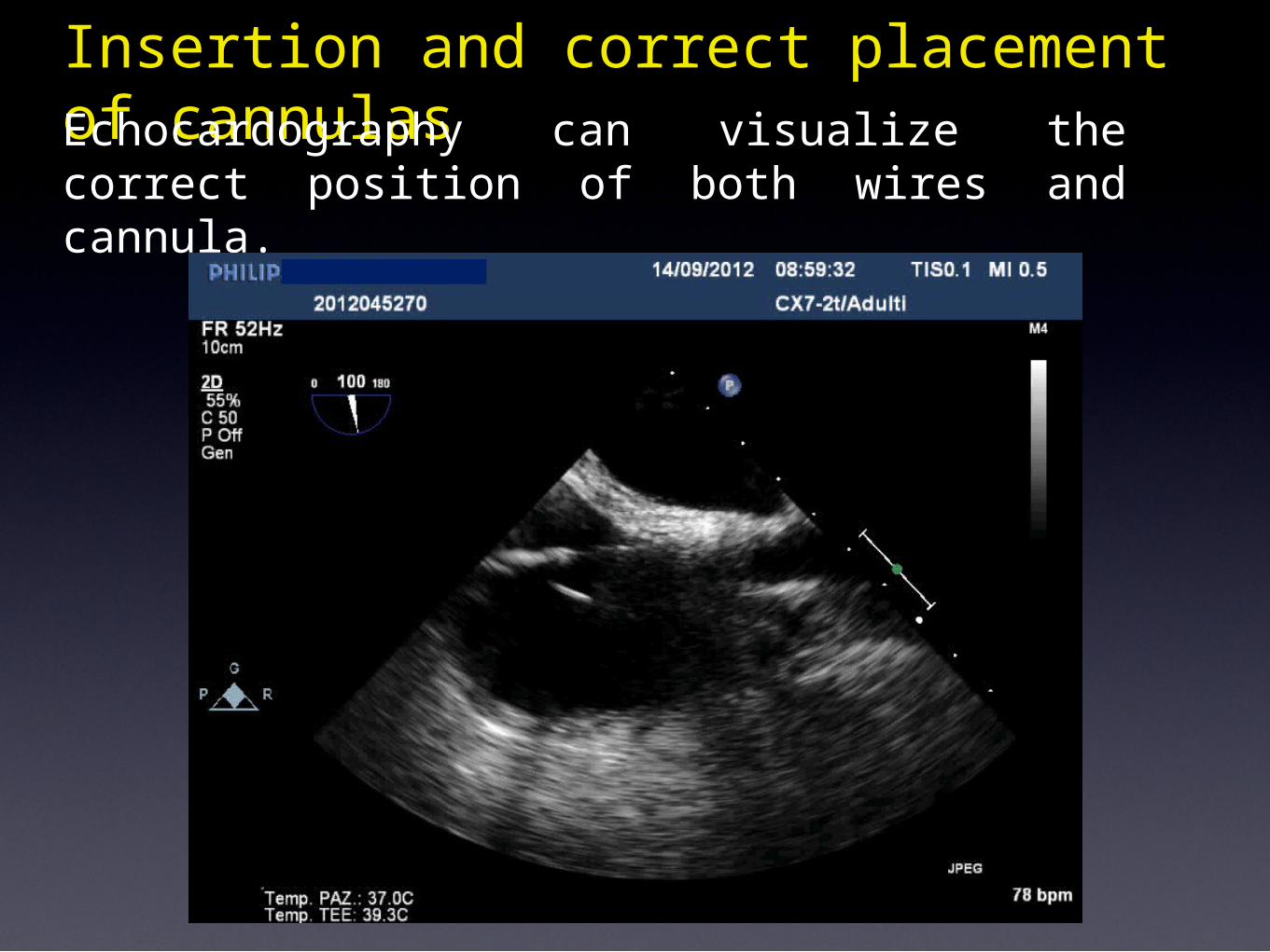
Insertion and correct placement of cannulas Echocardography can visualize the correct position of both wires and cannula.
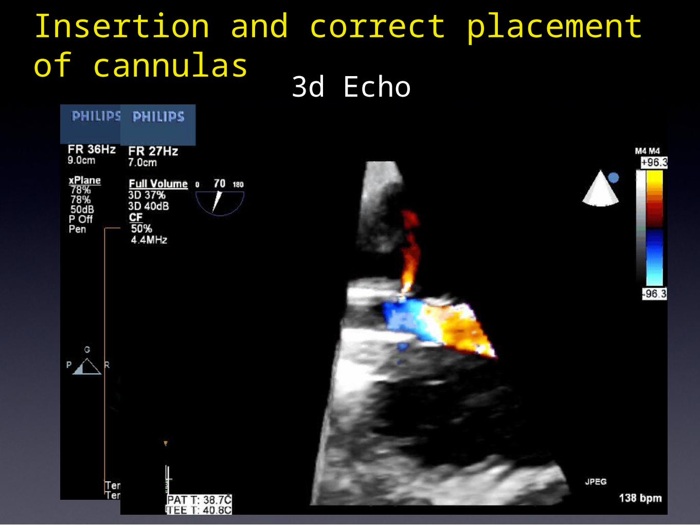
Insertion and correct placement of cannulas 3d Echo
Insertion and correct placement of cannulas
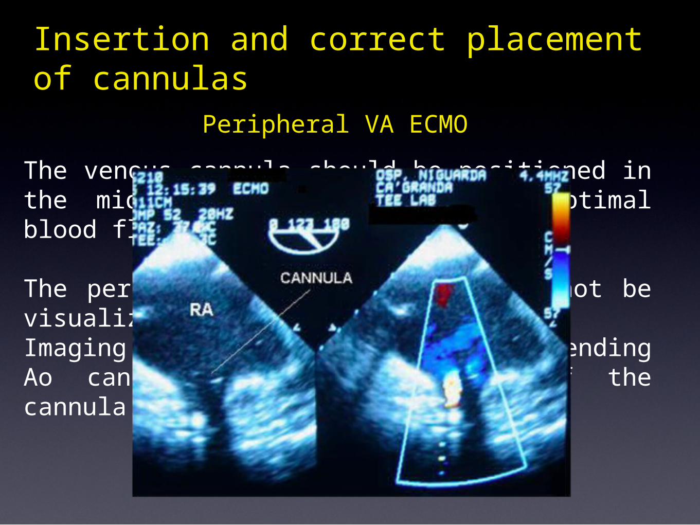
Peripheral VA ECMO
The venous cannula should be positioned in the mid right atrium to allow optimal blood flow into the circuit. The peripheral arterial cannula cannot be visualized. Imaging of the guide-wire in the ascending Ao can prevent malpositioning of the cannula.
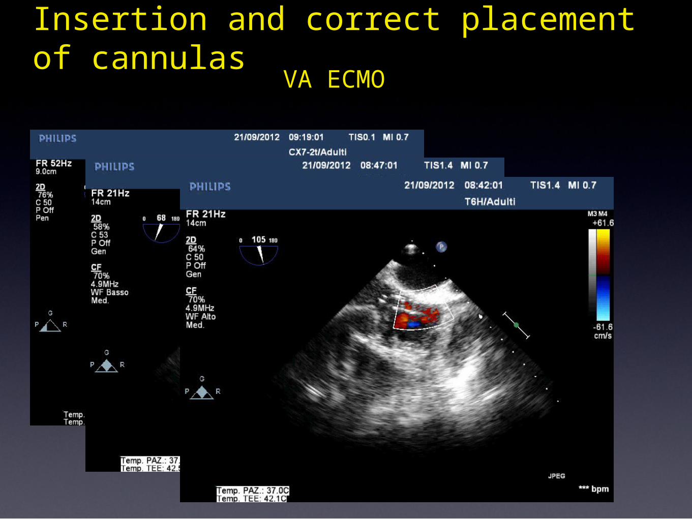
Insertion and correct placement of cannulas VA ECMO
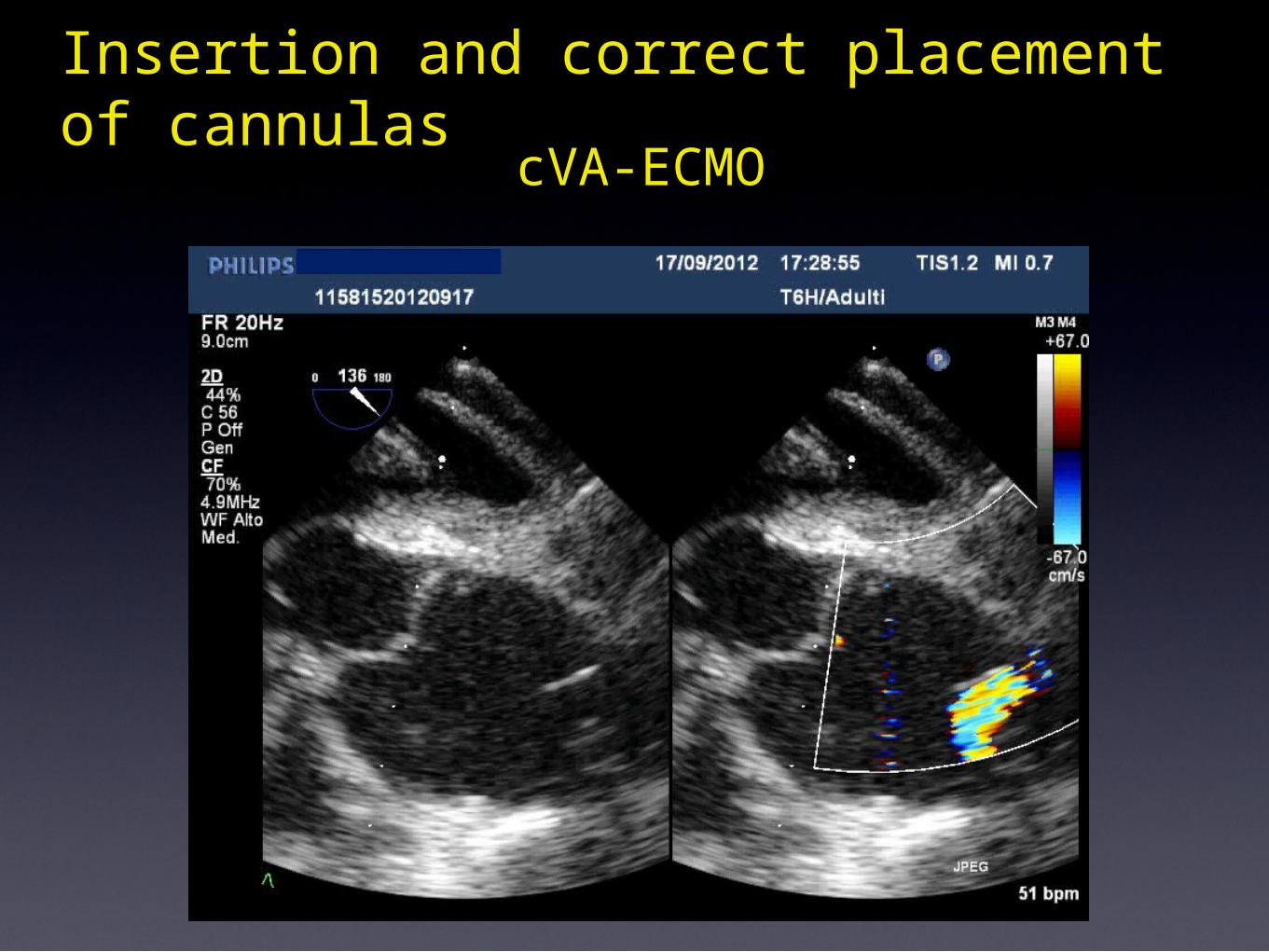
Insertion and correct placement of cannulas cVA-ECMO
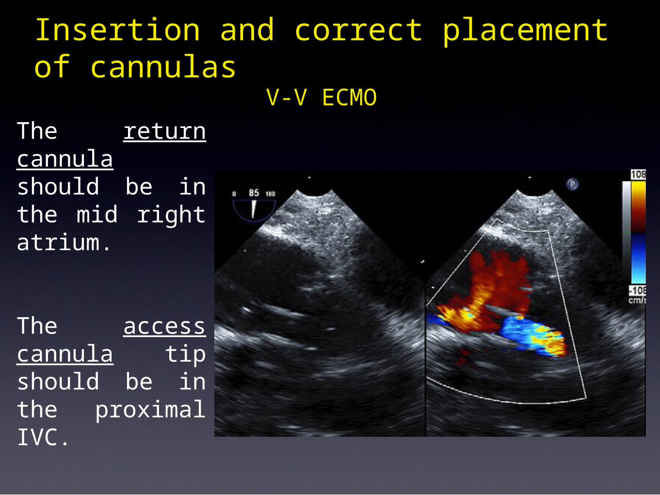
The return cannula should be in the mid right atrium.
The access cannula tip should be in the proximal IVC.
Insertion and correct placement of cannulas
V-V ECMO
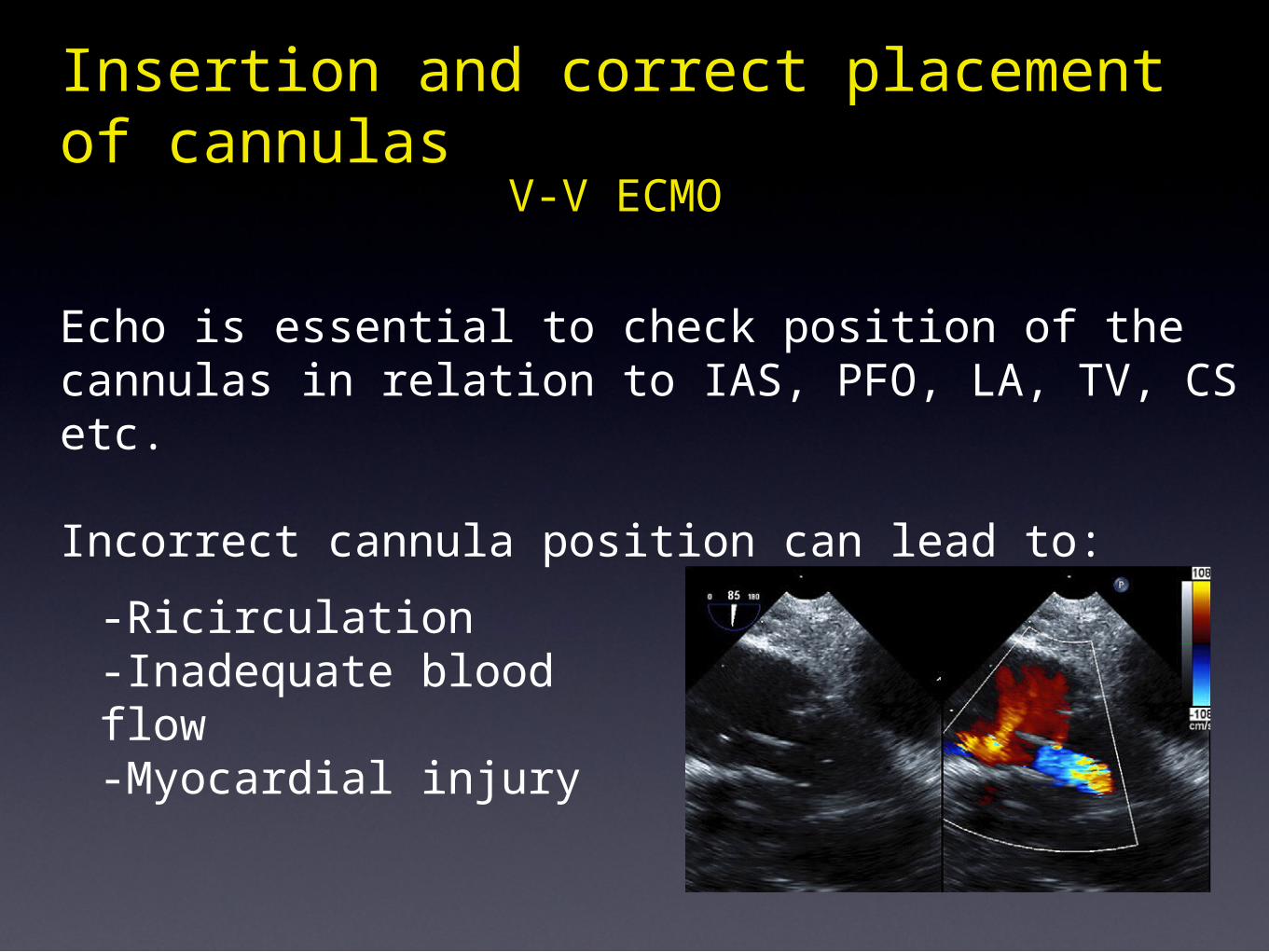
Echo is essential to check position of the cannulas in relation to IAS, PFO, LA, TV, CS etc.
Incorrect cannula position can lead to:
Insertion and correct placement of cannulas
V-V ECMO
-Ricirculation-Inadequate blood flow -Myocardial injury
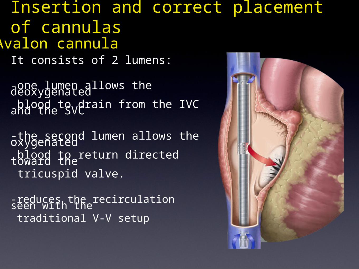
Avalon cannulaIt consists of 2 lumens:
-one lumen allows the deoxygenated blood to drain from the IVC and the SVC -the second lumen allows the oxygenated blood to return directed toward the tricuspid valve.
-reduces the recirculation seen with the traditional V-V setup
Insertion and correct placement of cannulas
•Patient selection
ECHOCARDIOGRAPHIC MONITORING ON ECMO
• Insertion and correct placement of cannulas
• Monitoring during support
• Detecting complications• Decision making: cardiac recovery, weaning,
bridge to..
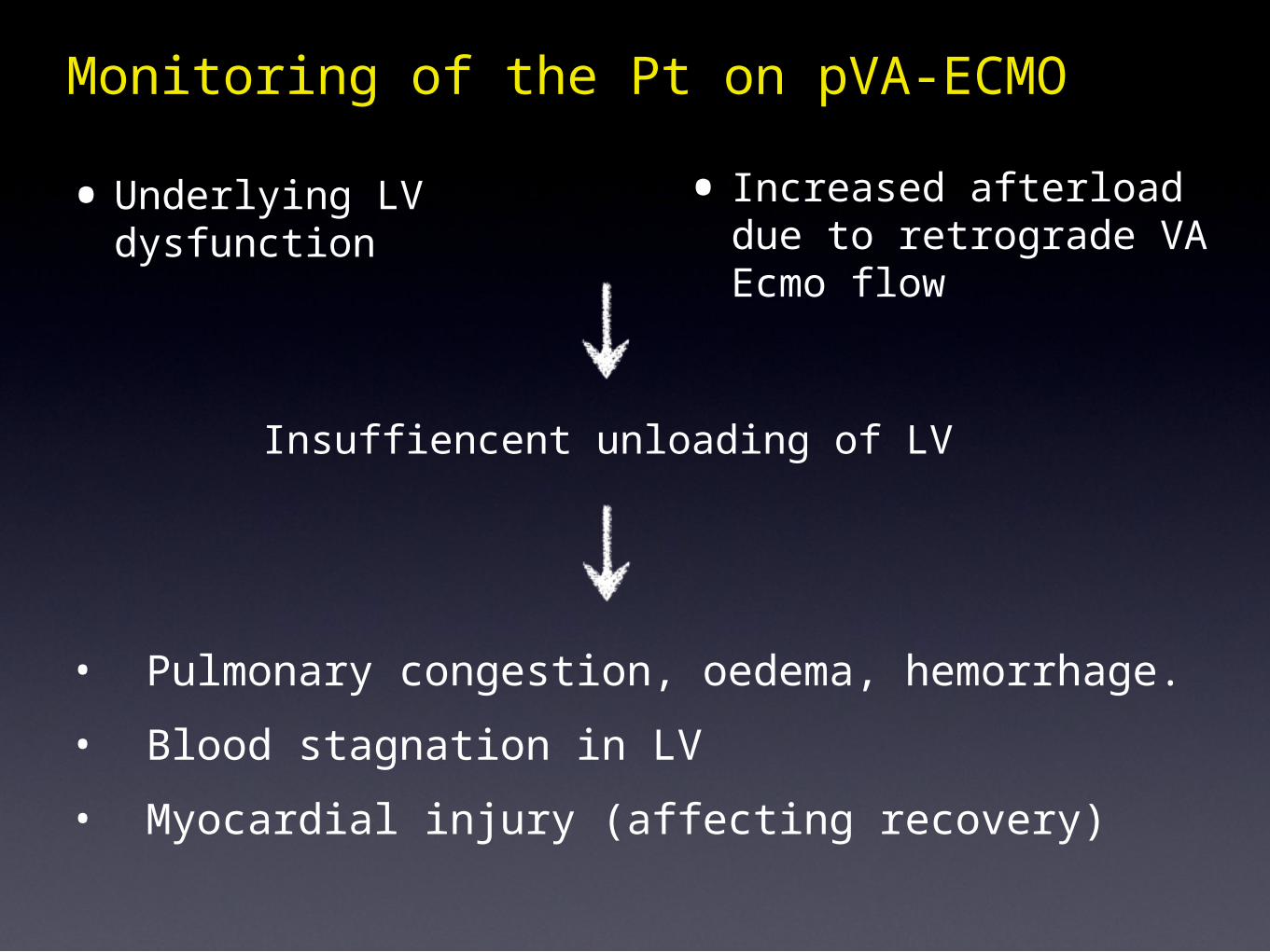
•Underlying LV dysfunction
Monitoring of the Pt on pVA-ECMO
Insuffiencent unloading of LV
• Increased afterload due to retrograde VA Ecmo flow
• Pulmonary congestion, oedema, hemorrhage.• Blood stagnation in LV• Myocardial injury (affecting recovery)
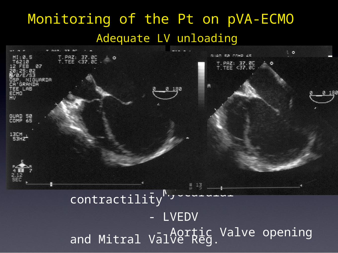
Adequate LV unloadingMonitoring of the Pt on pVA-ECMO
Echo Monitoring: - Myocardial contractility - LVEDV - Aortic Valve opening and Mitral Valve Reg.
AHF in DCMP, pVA ECMO (3 days) + Inotropes + IABP. WP=13, RAP= 8 mmHg
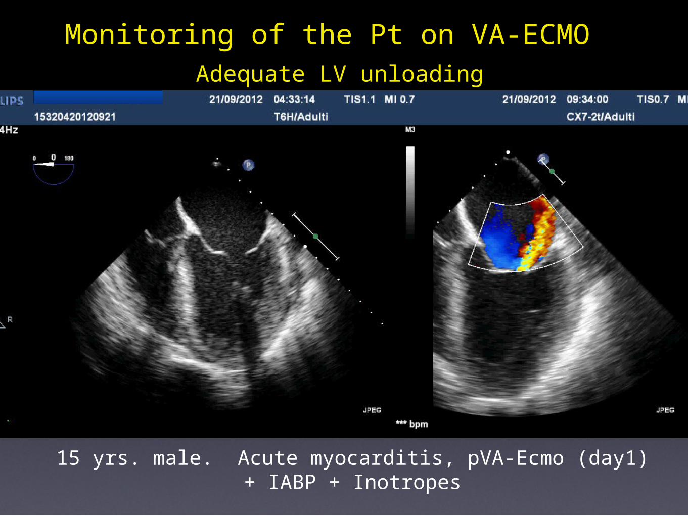
Adequate LV unloadingMonitoring of the Pt on VA-ECMO
15 yrs. male. Acute myocarditis, pVA-Ecmo (day1) + IABP + Inotropes
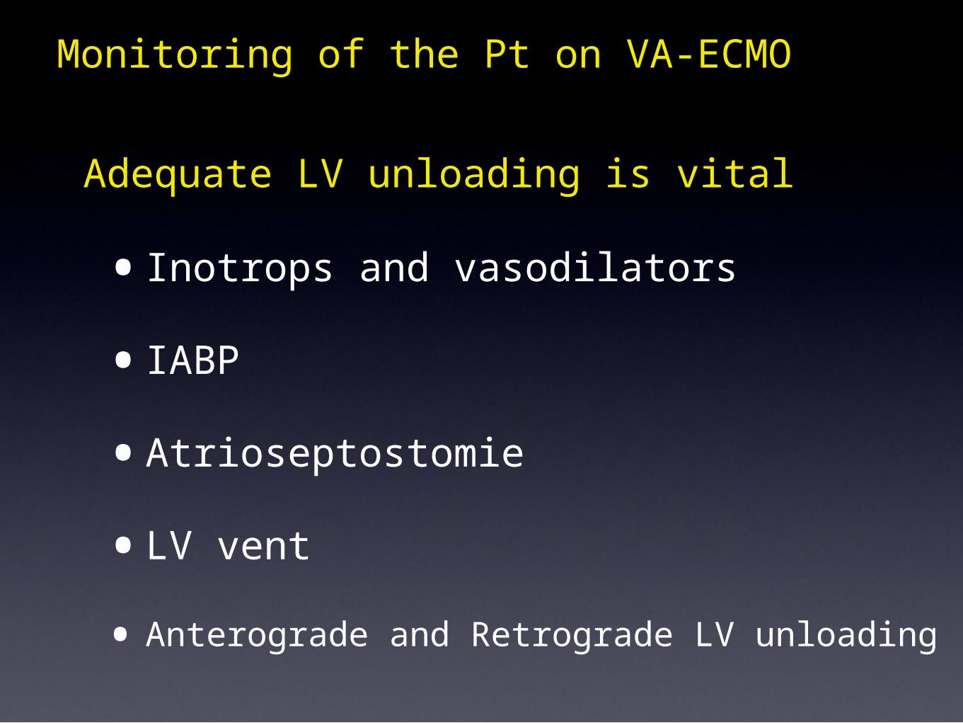
Adequate LV unloading is vital
Monitoring of the Pt on VA-ECMO
•Inotrops and vasodilators
•IABP
•Atrioseptostomie
•LV vent
• Anterograde and Retrograde LV unloading
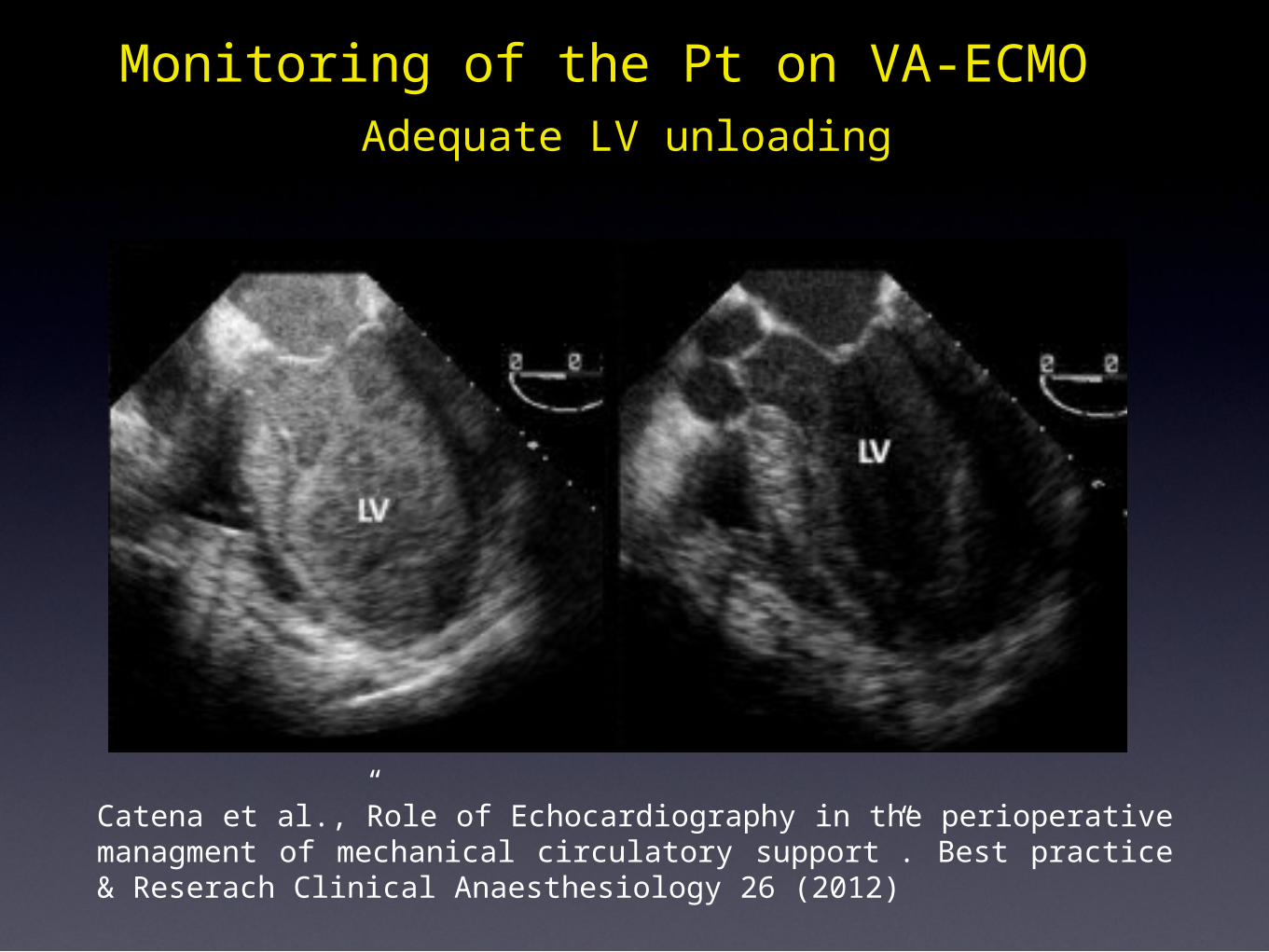
Catena et al.,”Role of Echocardiography in the perioperative managment of mechanical circulatory support”. Best practice & Reserach Clinical Anaesthesiology 26 (2012)
Adequate LV unloadingMonitoring of the Pt on VA-ECMO
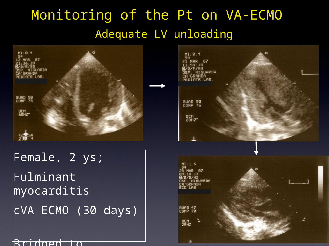
Adequate LV unloadingMonitoring of the Pt on VA-ECMO
Female, 2 ys; Fulminant myocarditiscVA ECMO (30 days) Bridged to recovery
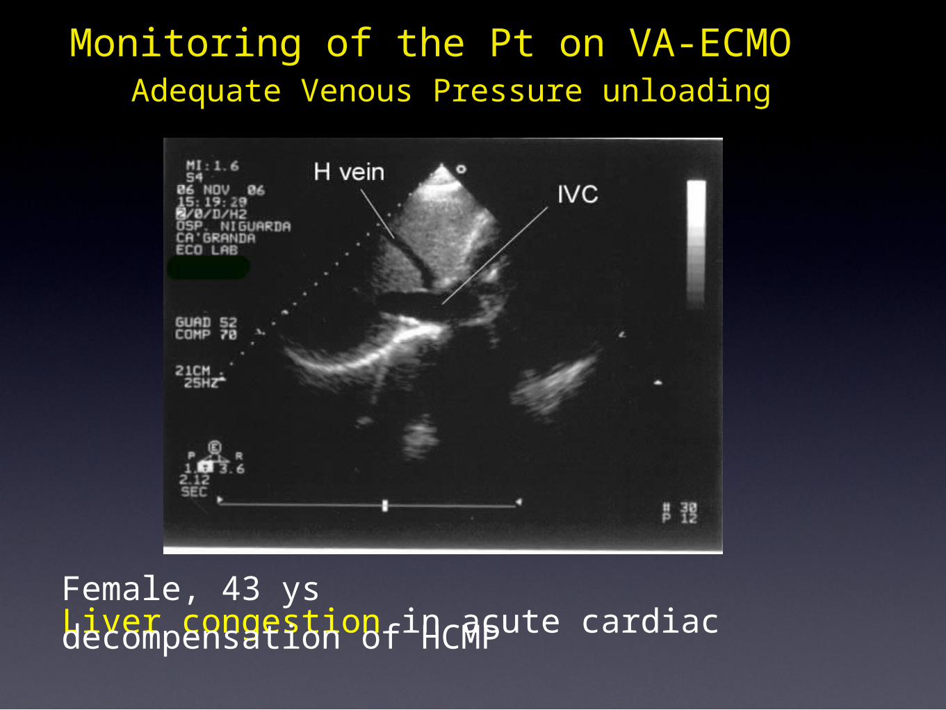
Adequate Venous Pressure unloadingMonitoring of the Pt on VA-ECMO
Female, 43 ysLiver congestion in acute cardiac decompensation of HCMP
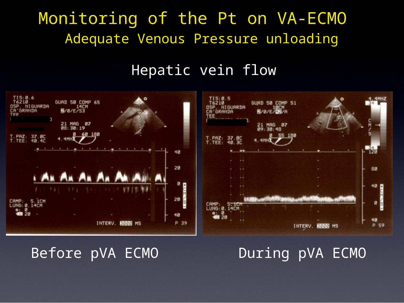
Adequate Venous Pressure unloadingMonitoring of the Pt on VA-ECMO
Hepatic vein flow
Before pVA ECMO During pVA ECMO
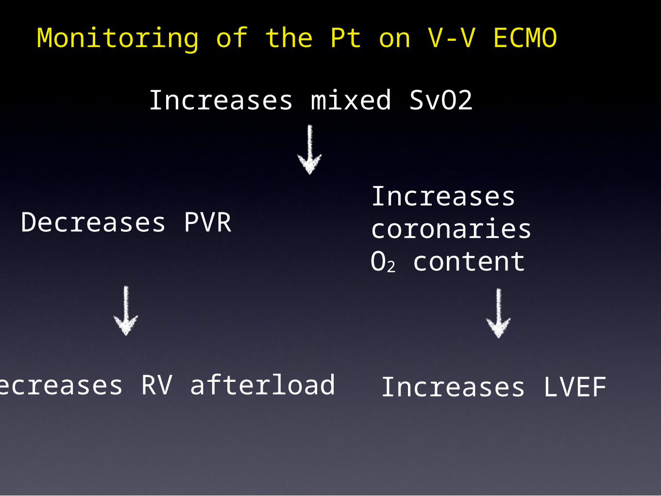
Monitoring of the Pt on V-V ECMO
Increases mixed SvO2
Decreases PVR
Decreases RV afterload
Increases coronaries O2 content
Increases LVEF
•Patient selection
ECHOCARDIOGRAPHIC MONITORING ON ECMO
• Insertion and correct placement of cannulas
• Monitoring during support
• Detecting complications• Decision making: cardiac recovery, weaning,
bridge to..
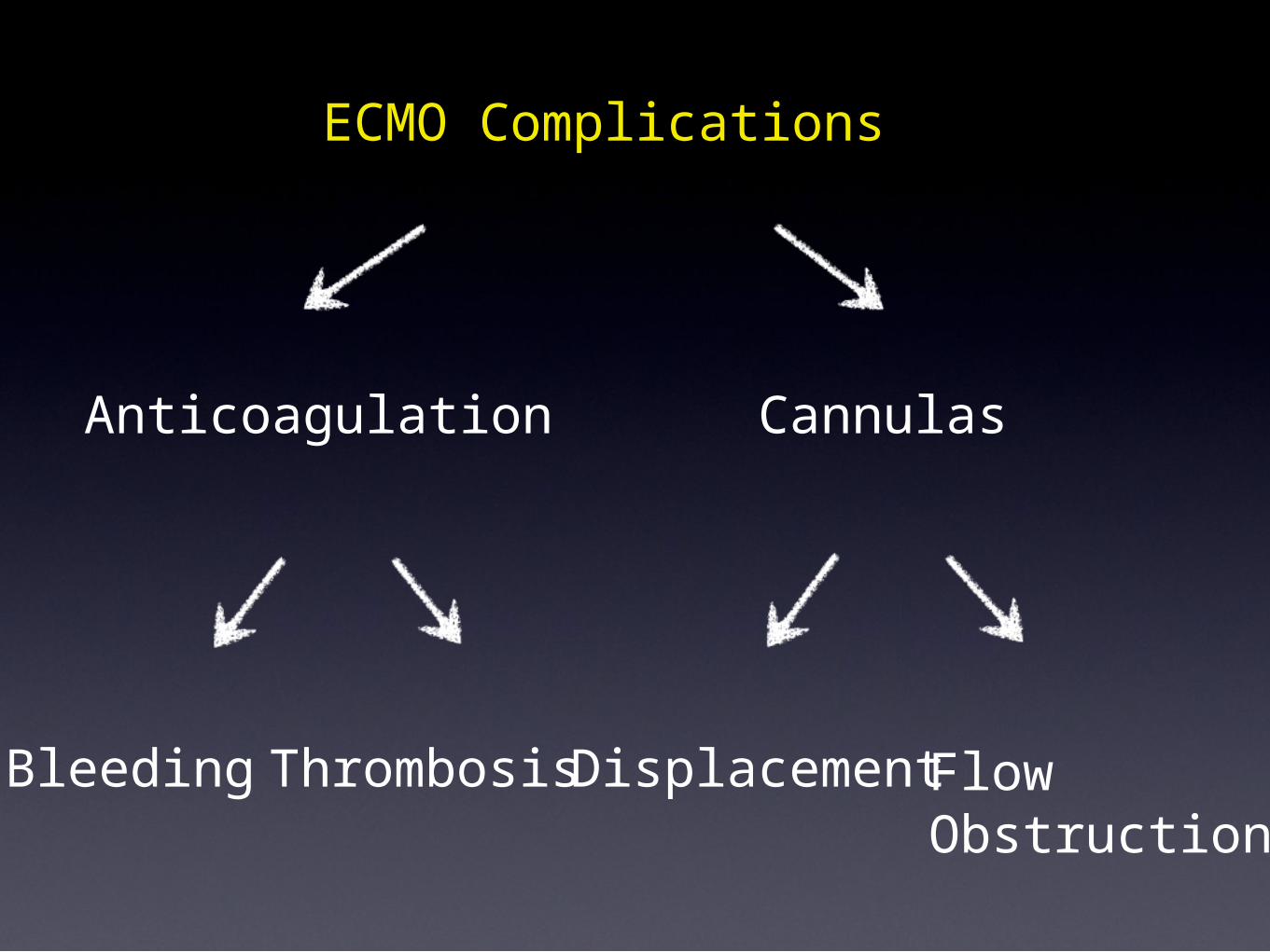
ECMO Complications
Anticoagulation
Bleeding
Cannulas
Thrombosis FlowObstruction
Displacement
32
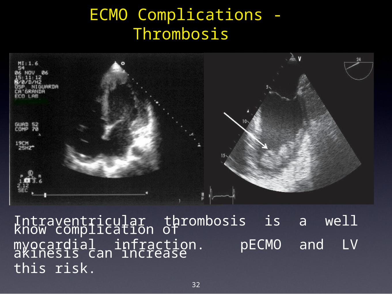
Intraventricular thrombosis is a well know complication ofmyocardial infraction. pECMO and LV akinesis can increasethis risk.
ECMO Complications - Thrombosis
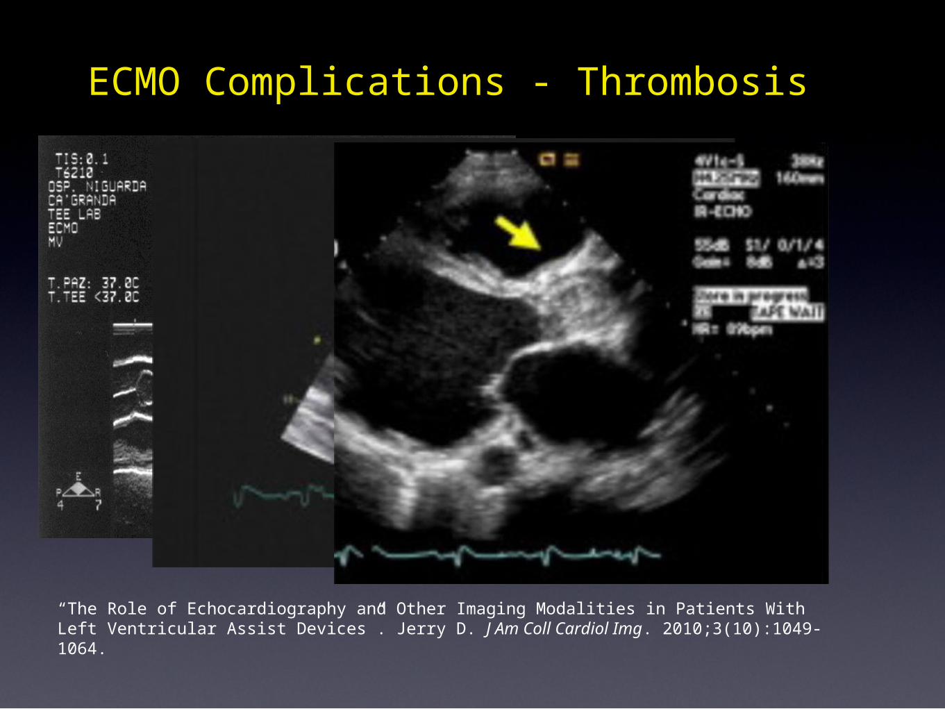
“The Role of Echocardiography and Other Imaging Modalities in Patients With Left Ventricular Assist Devices”. Jerry D. J Am Coll Cardiol Img. 2010;3(10):1049-1064.
ECMO Complications - Thrombosis
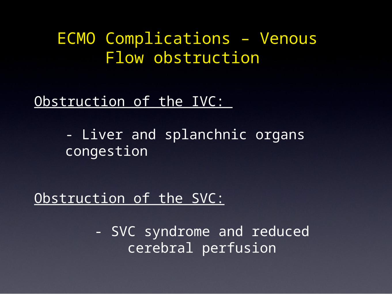
Obstruction of the IVC:
- Liver and splanchnic organs congestion
Obstruction of the SVC:
- SVC syndrome and reduced cerebral perfusion
ECMO Complications – Venous Flow obstruction
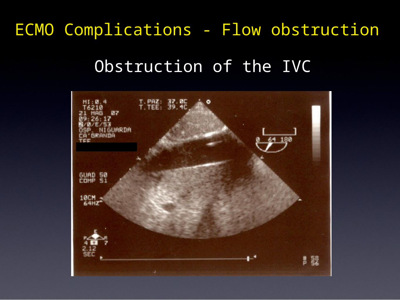
Obstruction of the IVC
ECMO Complications - Flow obstruction
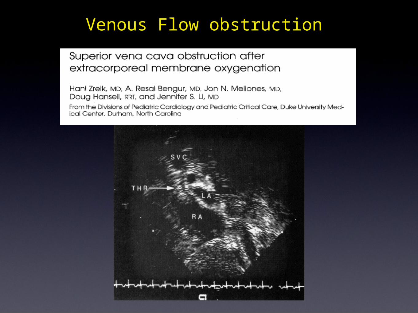
Venous Flow obstruction
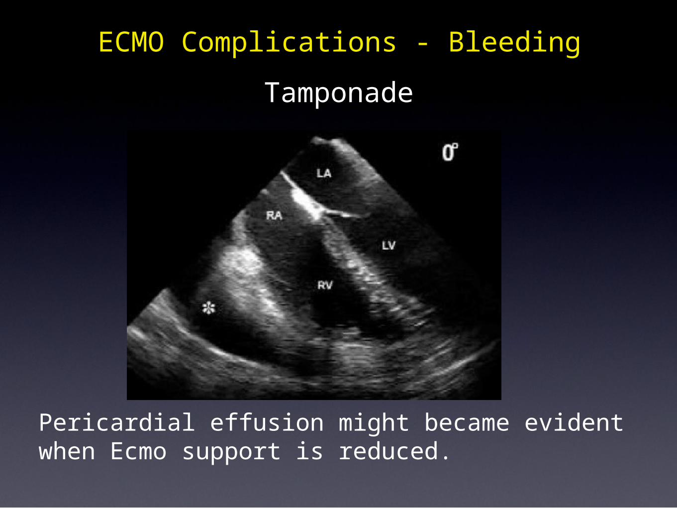
TamponadeECMO Complications - Bleeding
Pericardial effusion might became evident when Ecmo support is reduced.
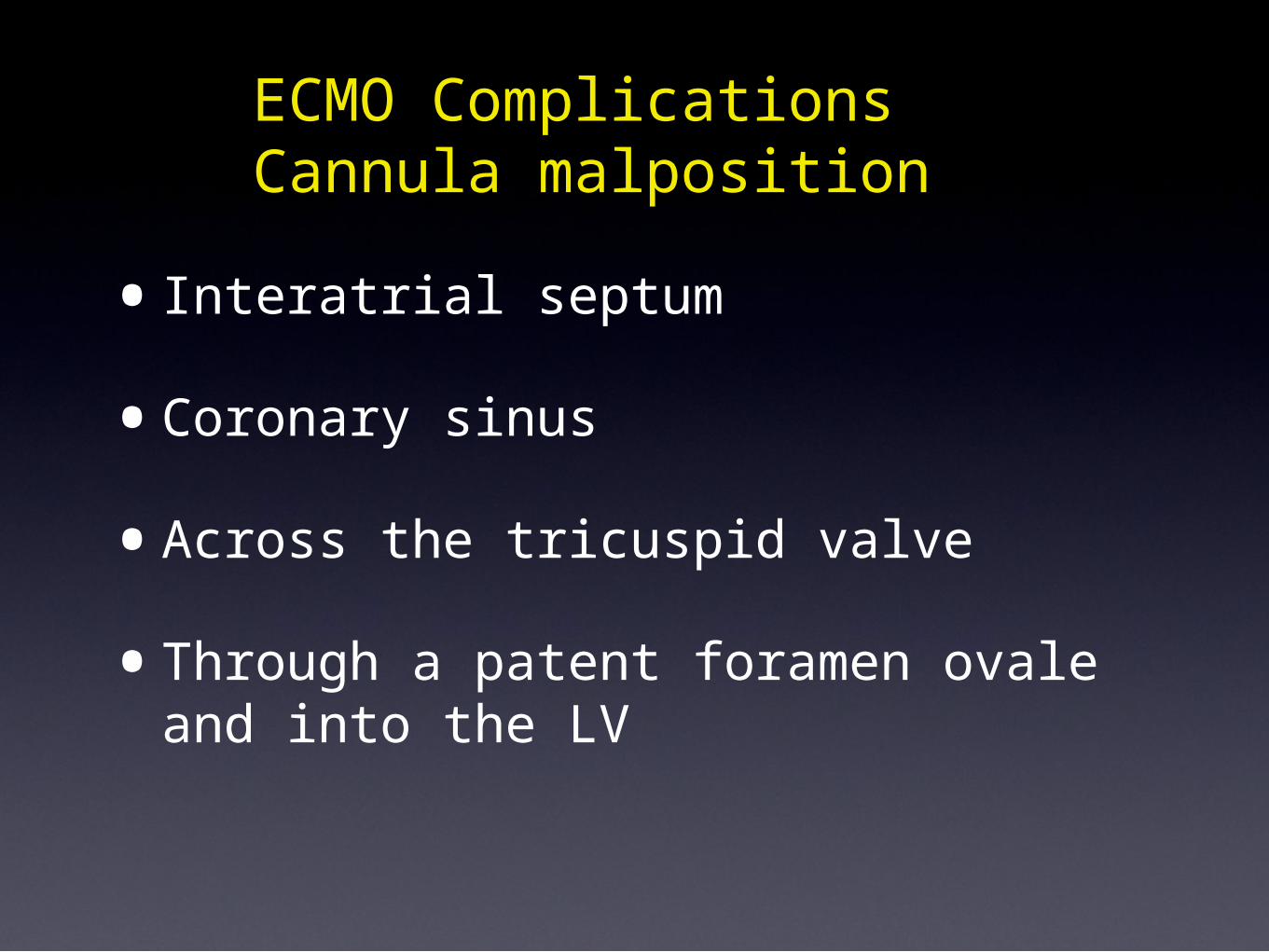
•Interatrial septum
•Coronary sinus
•Across the tricuspid valve
•Through a patent foramen ovale and into the LV
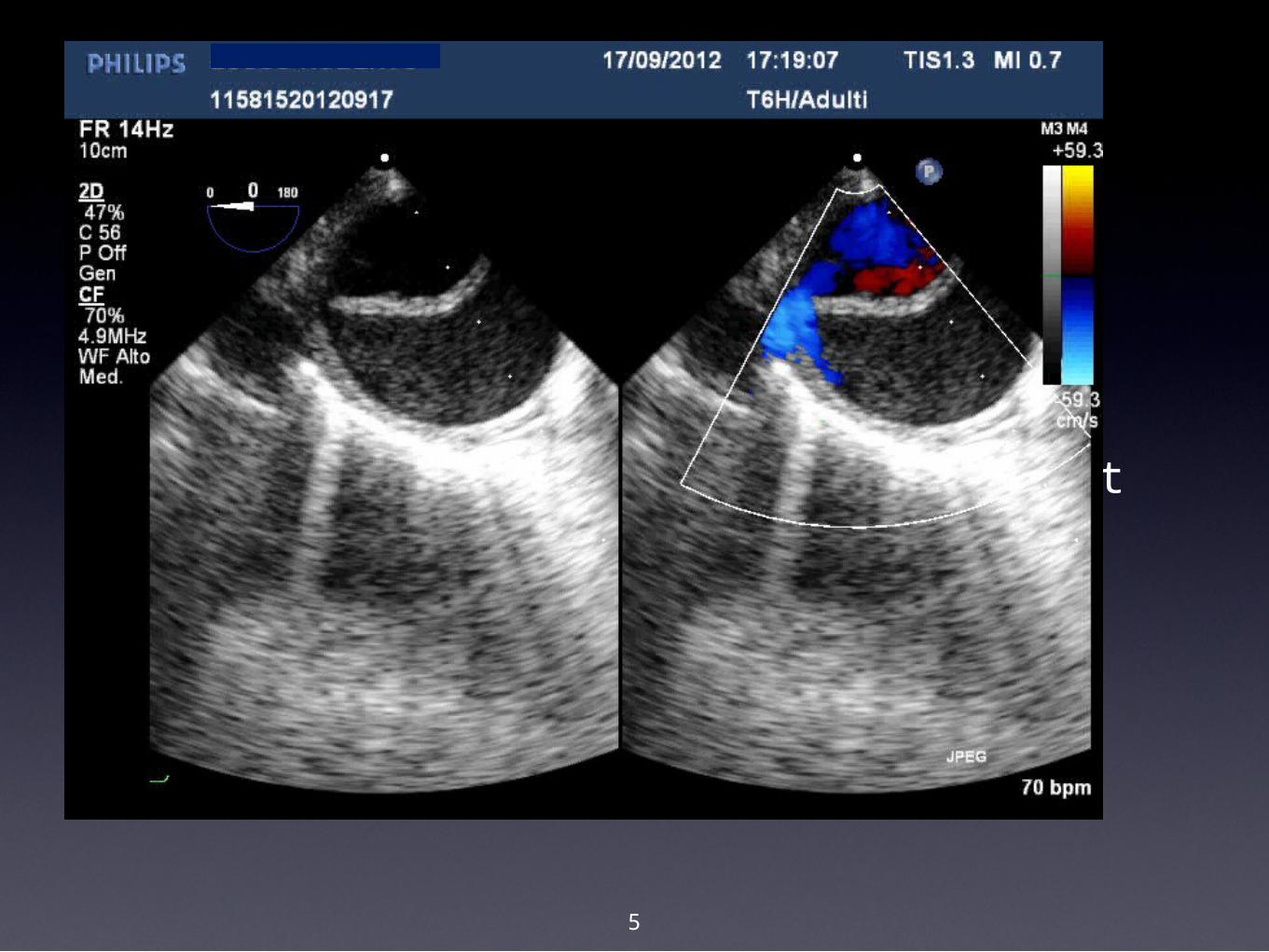
ECMO Complications Cannula malposition
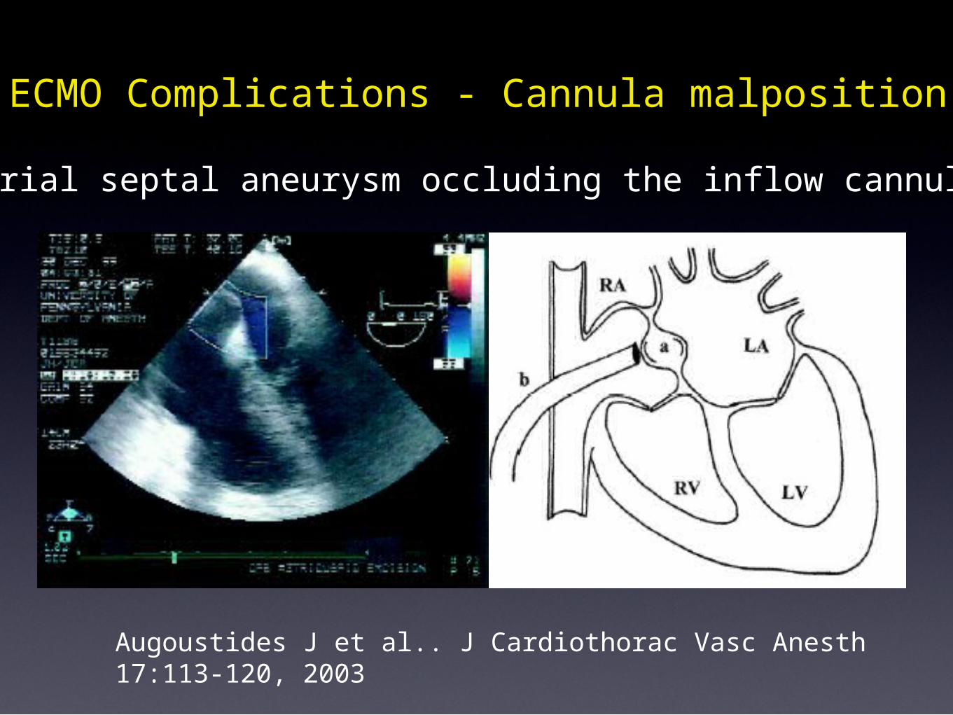
Augoustides J et al.. J Cardiothorac Vasc Anesth 17:113-120, 2003
Atrial septal aneurysm occluding the inflow cannula.
ECMO Complications - Cannula malposition
•Patient selection
ECHOCARDIOGRAPHIC MONITORING ON ECMO
• Insertion and correct placement of cannulas
• Monitoring during support
• Detecting complications• Decision making: cardiac recovery, weaning,
bridge to..
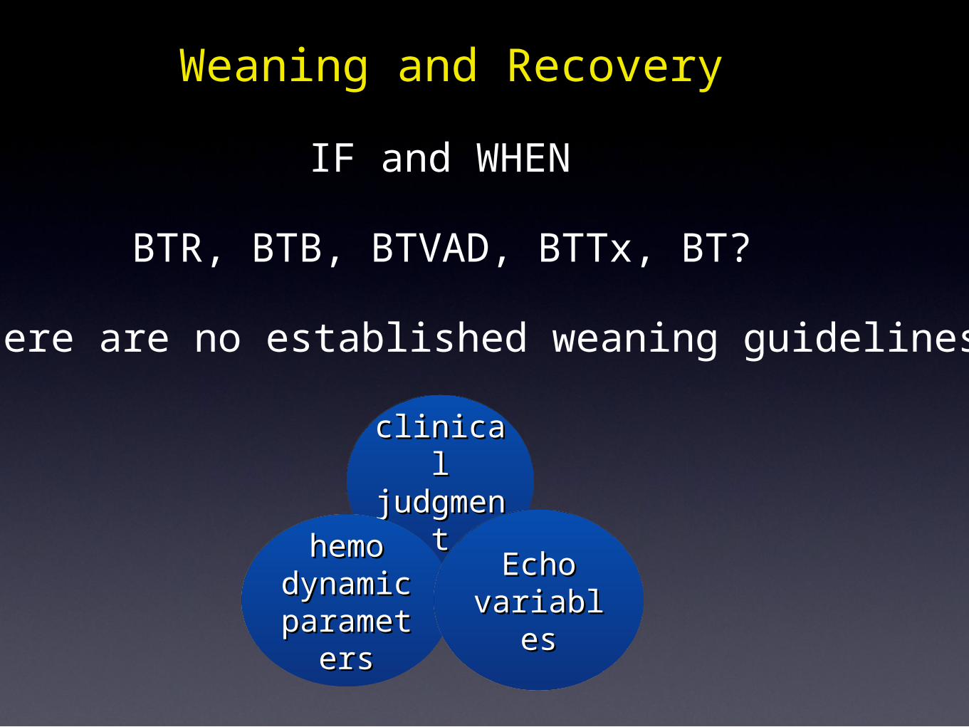
clinicalclinicaljudgmejudgme
ntnt
Weaning and Recovery
There are no established weaning guidelines
IF and WHEN
BTR, BTB, BTVAD, BTTx, BT?
hemohemodynamicdynamicparametparamet
ersers
EchoEchovariablesvariables
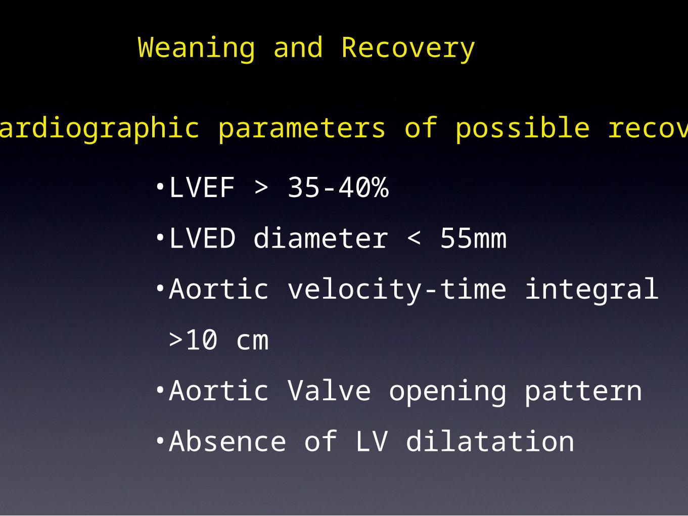
Echocardiographic parameters of possible recovery
•LVEF > 35-40%•LVED diameter < 55mm•Aortic velocity-time integral >10 cm•Aortic Valve opening pattern•Absence of LV dilatation
Weaning and Recovery
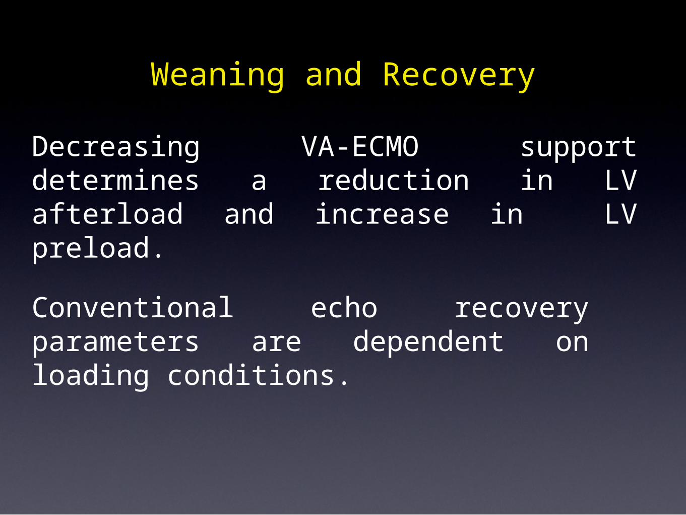
Decreasing VA-ECMO support determines a reduction in LV afterload and increase in LV preload.
Conventional echo recovery parameters are dependent on loading conditions.
Weaning and Recovery
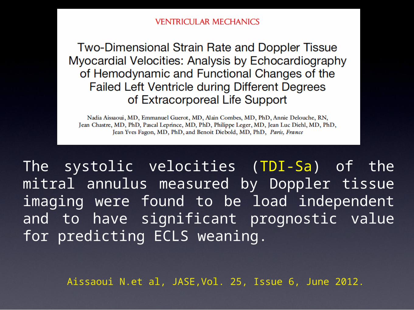
The systolic velocities (TDI-Sa) of the mitral annulus measured by Doppler tissue imaging were found to be load independent and to have significant prognostic value for predicting ECLS weaning.
Aissaoui N.et al, JASE,Vol. 25, Issue 6, June 2012.
Thank YouThank You
Niguarda Hospital 1950


![TCS - ECMO - [Bow]€¦ · The Paris International Congress on ECMO will therefore become the TCS-ECMO ... Ethics: end of life and ECMO FRIDAY 1 ... TCS for the right ventricle M](https://img.pdfslide.net/doc/110x75/5aef5ebd7f8b9a8b4c8c350f/tcs-ecmo-bow-the-paris-international-congress-on-ecmo-will-therefore-become.jpg)