Embed Size (px)
DESCRIPTION
This presentation, "Emergency Department Management of Radiation Casualties,” was prepared as a public service by the Health Physics Society for hospital staff training. - PowerPoint PPT Presentation
Citation preview
1
This presentation, "Emergency Department Management of Radiation Casualties,” was prepared as a public service by the Health Physics Society for hospital staff training.
The presentation includes talking points on the Notes pages, which can be viewed if you go to the File Menu and "Save As" a PowerPoint file to your computer.
The talking points are provided with each slide to assist the presenter in answering questions. It is not expected that all the information in the talking points will be presented during the training.
The presentation can be edited to fit the needs of the user. The authors request that appropriate attribution be given for this material and would like to know who is presenting it and to what groups. That information and comments may be sent to Jerrold T. Bushberg, PhD, UC Davis Health System, at [email protected] 3.0
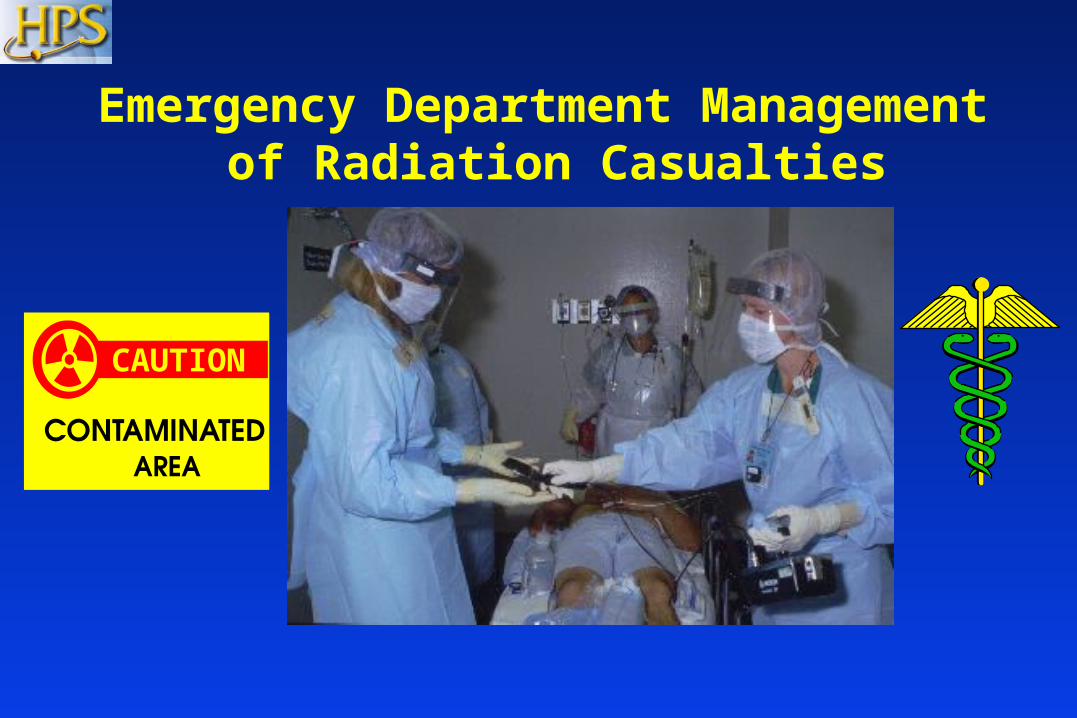
Emergency Department Management of Radiation Casualties
CAUTION
3
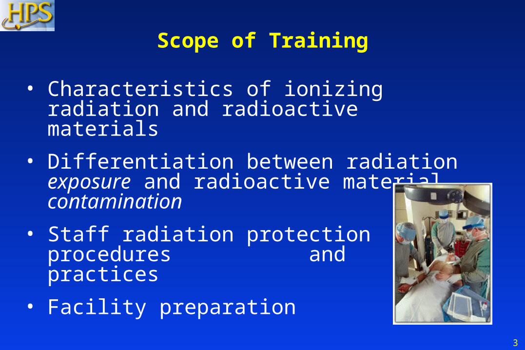
Scope of Training
• Characteristics of ionizing radiation and radioactive materials
• Differentiation between radiation exposure and radioactive material contamination
• Staff radiation protection procedures and practices
• Facility preparation
4
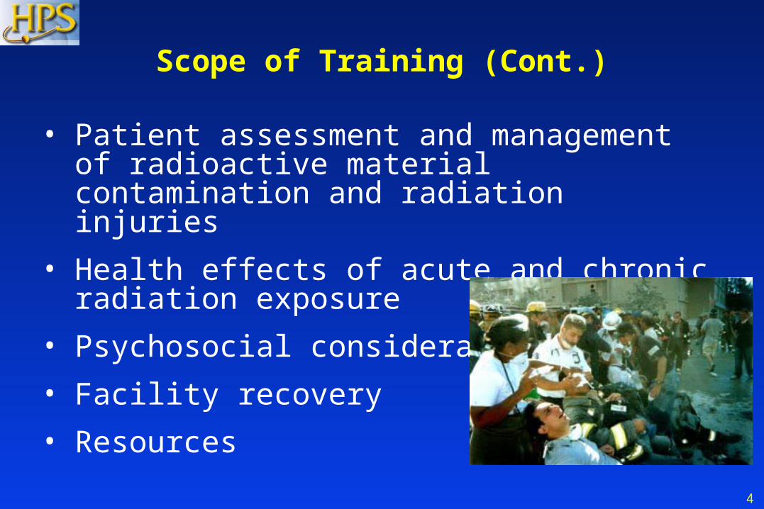
Scope of Training (Cont.)
• Patient assessment and management of radioactive material contamination and radiation injuries
• Health effects of acute and chronic radiation exposure
• Psychosocial considerations
• Facility recovery
• Resources

5
Ionizing Radiation
• Ionizing radiation is radiation capable of imparting its energy to the body and causing chemical changes.
• Ionizing radiation is emitted by:
- Radioactive material.
- Some devices such as x-ray machines.
6
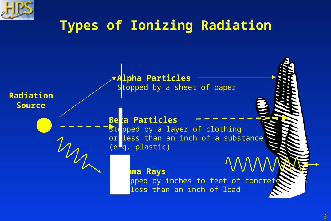
Types of Ionizing Radiation
Alpha ParticlesStopped by a sheet of paper
Beta ParticlesStopped by a layer of clothingor less than an inch of a substance (e.g. plastic)
Gamma RaysStopped by inches to feet of concreteor less than an inch of lead
RadiationSource
7
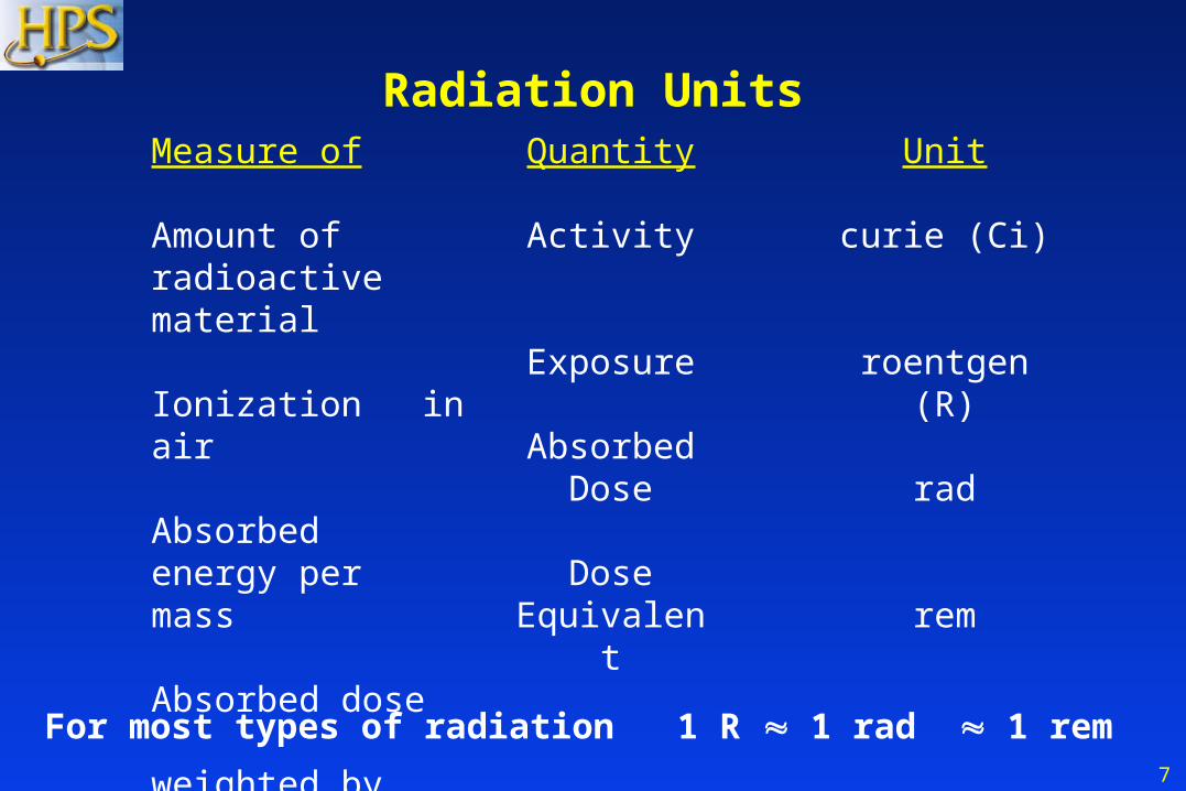
Measure of
Amount ofradioactive material Ionization in air
Absorbed energy per mass
Absorbed dose weighted by type of radiation
Radiation Units
For most types of radiation 1 R 1 rad 1 rem
Quantity
Activity
Exposure
Absorbed Dose
Dose Equivalent
Unit
curie (Ci)
roentgen (R)
rad
rem
8
Radiation Doses and Dose Limits
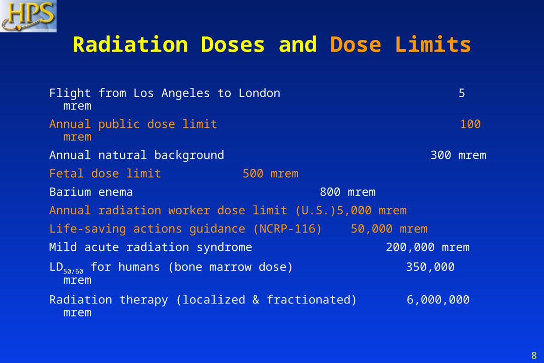
Flight from Los Angeles to London 5 mrem
Annual public dose limit 100 mrem
Annual natural background 300 mrem
Fetal dose limit 500 mrem
Barium enema 800 mrem
Annual radiation worker dose limit (U.S.) 5,000 mrem
Life-saving actions guidance (NCRP-116) 50,000 mrem
Mild acute radiation syndrome 200,000 mrem
LD50/60 for humans (bone marrow dose) 350,000 mrem
Radiation therapy (localized & fractionated) 6,000,000 mrem
9
Radioactive Material
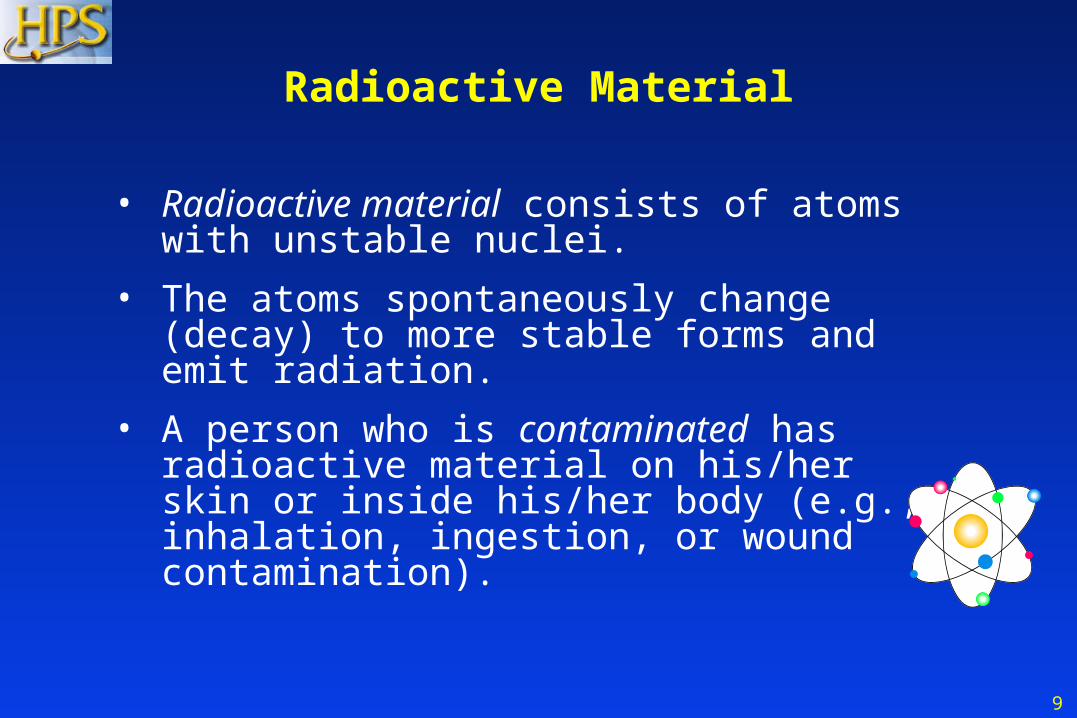
• Radioactive material consists of atoms with unstable nuclei.
• The atoms spontaneously change (decay) to more stable forms and emit radiation.
• A person who is contaminated has radioactive material on his/her skin or inside his/her body (e.g., inhalation, ingestion, or wound contamination).
10
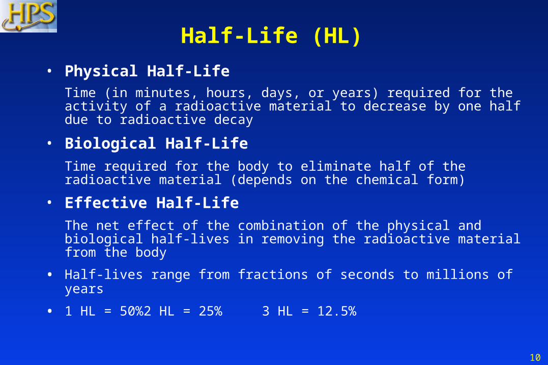
Half-Life (HL)
• Physical Half-LifeTime (in minutes, hours, days, or years) required for the activity of a radioactive material to decrease by one half due to radioactive decay
• Biological Half-Life
Time required for the body to eliminate half of the radioactive material (depends on the chemical form)
• Effective Half-Life
The net effect of the combination of the physical and biological half-lives in removing the radioactive material from the body
• Half-lives range from fractions of seconds to millions of years
• 1 HL = 50% 2 HL = 25% 3 HL = 12.5%
11
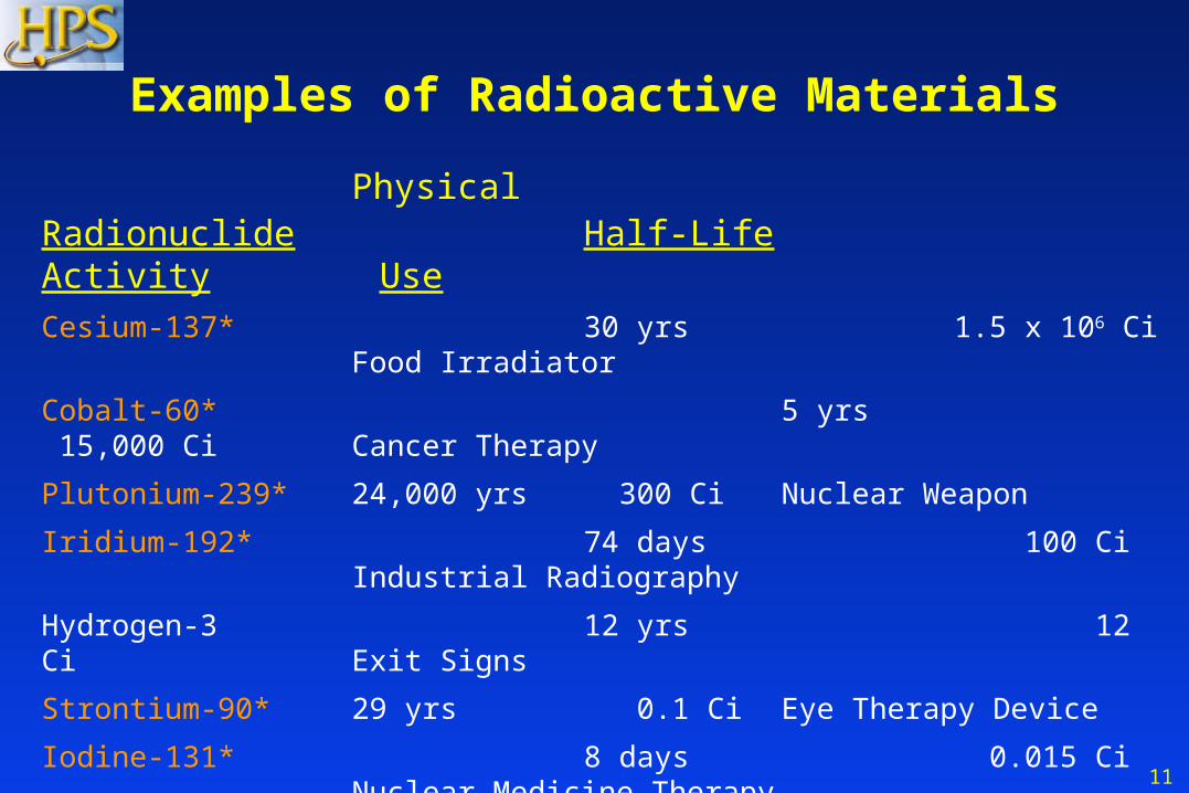
Physical Radionuclide Half-Life Activity Use
Cesium-137* 30 yrs 1.5 x 10⁶ Ci Food Irradiator
Cobalt-60* 5 yrs 15,000 Ci Cancer Therapy
Plutonium-239* 24,000 yrs 300 Ci Nuclear Weapon
Iridium-192* 74 days 100 Ci Industrial Radiography
Hydrogen-3 12 yrs 12 Ci Exit Signs
Strontium-90* 29 yrs 0.1 Ci Eye Therapy Device
Iodine-131* 8 days 0.015 Ci Nuclear Medicine Therapy
Technetium-99m 6 hrs 0.025 Ci Diagnostic Imaging
Americium-241* 432 yrs 0.000005 Ci Smoke Detectors
Radon-222 4 days 1 pCi/l Environmental Level * Potential use in radiological dispersion device
Examples of Radioactive Materials

12
Types of Radiation Hazards
• External Exposure -
Whole-body or partial-body (no radiation hazard to EMS staff)
• Contaminated -
– External radioactive material: on the skin or clothing
– Internal radioactive material: inhaled, swallowed, absorbed through skin or wounds
ExternalExposure
InternalContamination
ExternalContamination
13
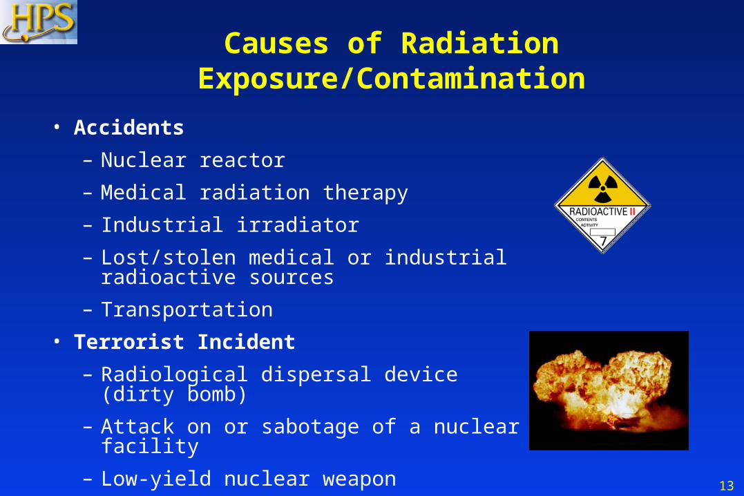
Causes of Radiation Exposure/Contamination
• Accidents
– Nuclear reactor
– Medical radiation therapy
– Industrial irradiator
– Lost/stolen medical or industrial radioactive sources
– Transportation
• Terrorist Incident
– Radiological dispersal device (dirty bomb)
– Attack on or sabotage of a nuclear facility
– Low-yield nuclear weapon

14
Scope of Incident
Incident Number of Deaths Most Deaths Due to
RadiationAccident
None/Few Radiation
RadioactiveDispersalDevice
Few/Moderate(Depends on
size of explosion andproximity of persons)
Blast Trauma
Low-YieldNuclear Weapon
Large(e.g., tens of thousands in an urban area even from
0.1 kT weapon) Radiation Exposure
Blast Trauma Thermal Burns
Fallout (Depends on Distance)
15
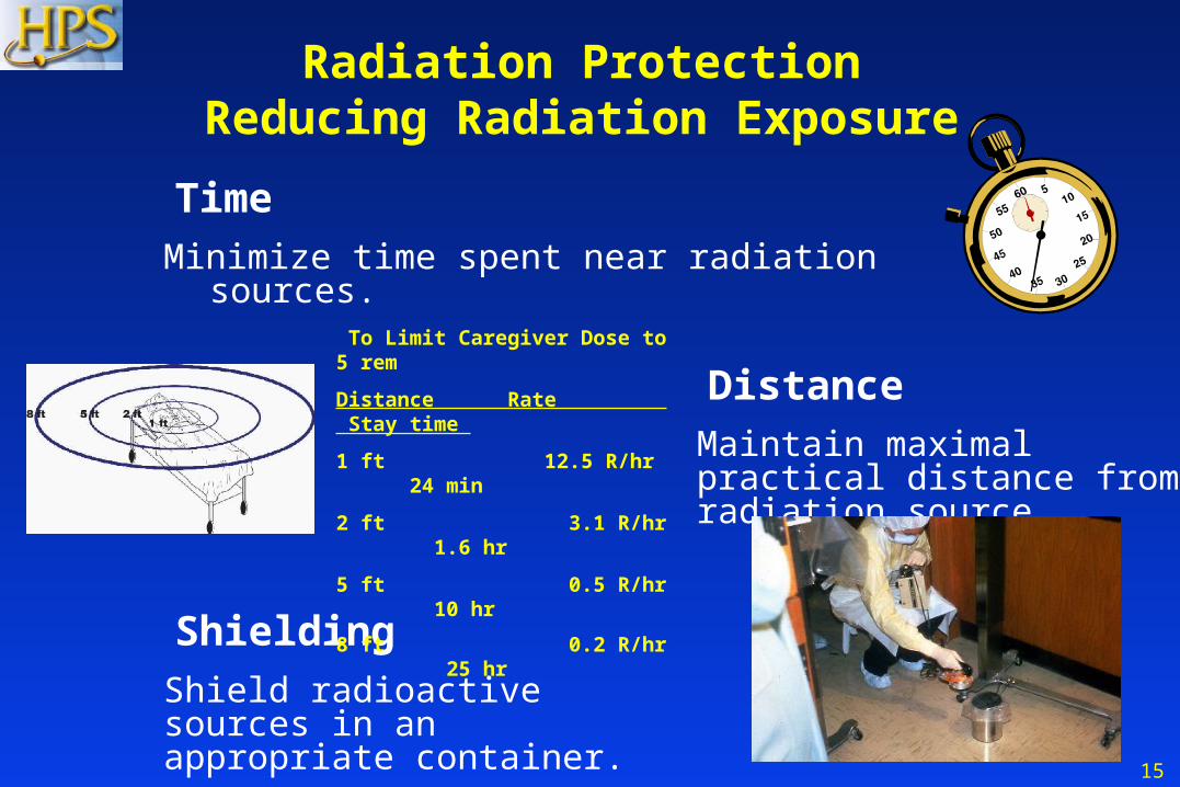
TimeMinimize time spent near radiation sources.
Radiation ProtectionReducing Radiation Exposure
DistanceMaintain maximal practical distance from radiation source.
ShieldingShield radioactive sources in an appropriate container.
To Limit Caregiver Dose to 5 rem
Distance Rate Stay time
1 ft 12.5 R/hr 24 min
2 ft 3.1 R/hr 1.6 hr
5 ft 0.5 R/hr 10 hr
8 ft 0.2 R/hr 25 hr
16
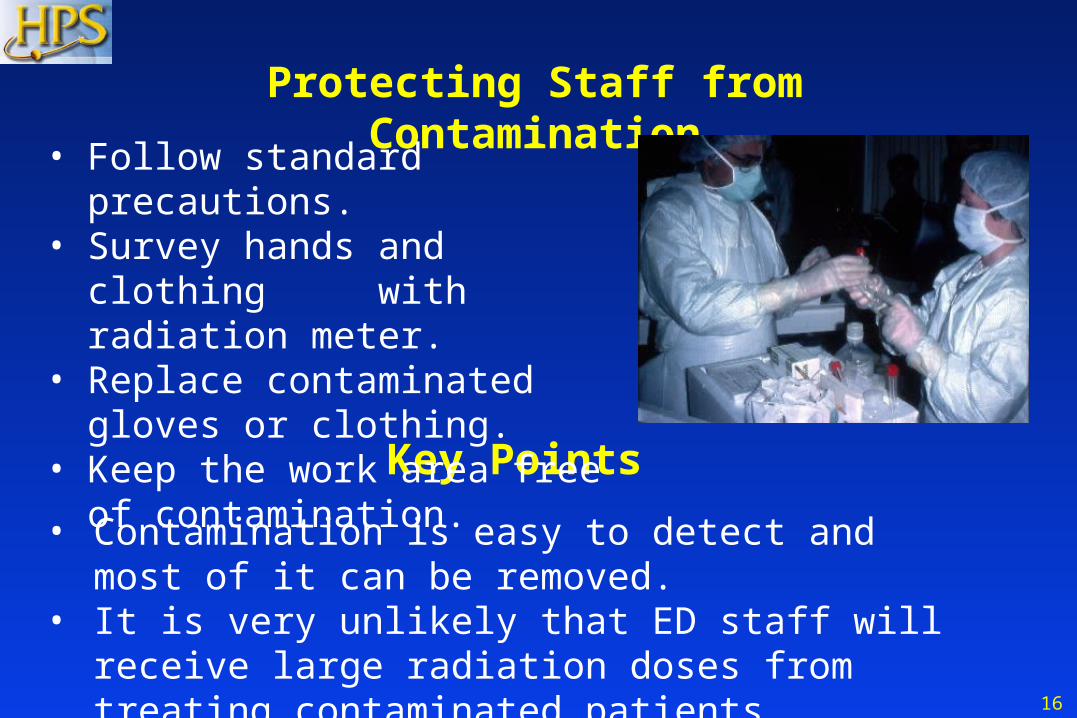
Key Points
• Contamination is easy to detect and most of it can be removed.
• It is very unlikely that ED staff will receive large radiation doses from treating contaminated patients.
Protecting Staff from Contamination
• Follow standard precautions.• Survey hands and clothing
with radiation meter.• Replace contaminated gloves or
clothing.• Keep the work area free of
contamination.
17
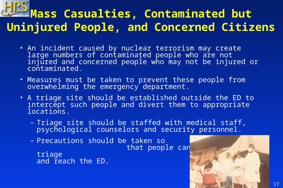
Mass Casualties, Contaminated butUninjured People, and Concerned Citizens
• An incident caused by nuclear terrorism may create large numbers of contaminated people who are not injured and concerned people who may not be injured or contaminated.
• Measures must be taken to prevent these people from overwhelming the emergency department.
• A triage site should be established outside the ED to intercept such people and divert them to appropriate locations.
– Triage site should be staffed with medical staff, psychological counselors and security personnel.
– Precautions should be taken so that people cannot avoid the triage center and reach the ED.
18
Decontamination Center
• Establish a decontamination center for people who are contaminated, but not significantly injured.– Center should provide showers for many people.
– Replacement clothing must be available.
– Provisions to transport or shelter people after decontamination may be necessary.
– Staff decontamination center with medical staff with a radiological background, health physicists or other staff trained in decontamination and use of radiation survey meters, and psychological counselors.
19
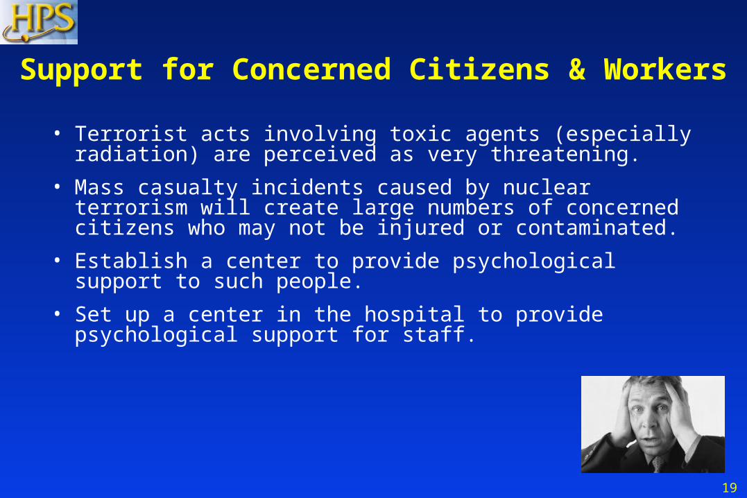
Support for Concerned Citizens & Workers
• Terrorist acts involving toxic agents (especially radiation) are perceived as very threatening.
• Mass casualty incidents caused by nuclear terrorism will create large numbers of concerned citizens who may not be injured or contaminated.
• Establish a center to provide psychological support to such people.
• Set up a center in the hospital to provide psychological support for staff.
20
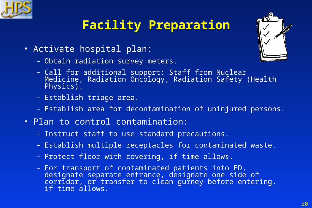
Facility Preparation
• Activate hospital plan:– Obtain radiation survey meters.
– Call for additional support: Staff from Nuclear Medicine, Radiation Oncology, Radiation Safety (Health Physics).
– Establish triage area.
– Establish area for decontamination of uninjured persons.
• Plan to control contamination:– Instruct staff to use standard precautions.
– Establish multiple receptacles for contaminated waste.
– Protect floor with covering, if time allows.
– For transport of contaminated patients into ED, designate separate entrance, designate one side of corridor, or transfer to clean gurney before entering, if time allows.
21
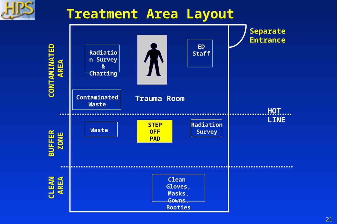
ContaminatedWaste
Waste
Treatment Area Layout
RadiationSurvey
HOTLINE
STEPOFFPAD
CO
NT
AM
INA
TE
D A
RE
AB
UF
FE
R Z
ON
EC
LE
AN
AR
EA
Radiation Survey
& Charting
ED Staff
Clean Gloves, Masks,Gowns, Booties
Separate Entrance
Trauma Room
22
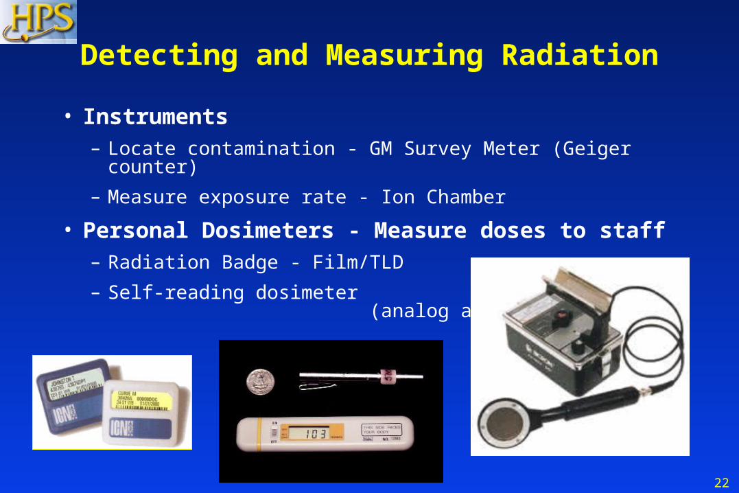
Detecting and Measuring Radiation
• Instruments– Locate contamination - GM Survey Meter (Geiger counter)
– Measure exposure rate - Ion Chamber
• Personal Dosimeters - Measure doses to staff– Radiation Badge - Film/TLD
– Self-reading dosimeter (analog and digital)
23
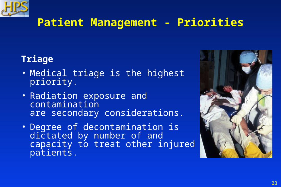
Patient Management - Priorities
Triage
• Medical triage is the highest priority.
• Radiation exposure and contamination are secondary considerations.
• Degree of decontamination is dictated by number of and capacity to treat other injured patients.

24
Patient Management - Triage
Triage based on:
• Injuries
• Signs and symptoms - nausea, vomiting, fatigue, diarrhea
• History - Where were you when the bomb exploded?
• Contamination survey
25
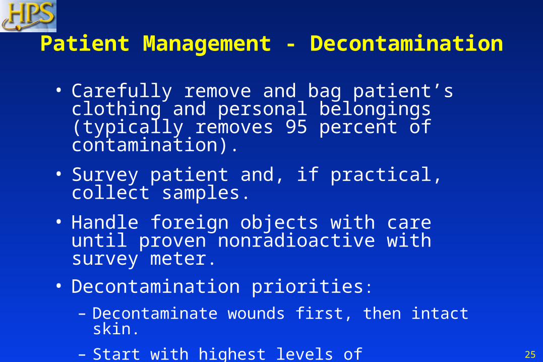
Patient Management - Decontamination
• Carefully remove and bag patient’s clothing and personal belongings (typically removes 95 percent of contamination).
• Survey patient and, if practical, collect samples.
• Handle foreign objects with care until proven nonradioactive with survey meter.
• Decontamination priorities:
– Decontaminate wounds first, then intact skin.
– Start with highest levels of contamination.
• Change gloves frequently to minimize spread of contamination.
26
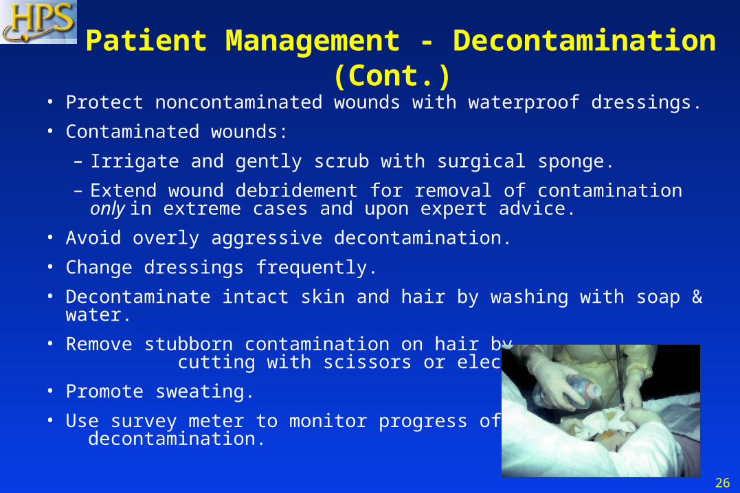
Patient Management - Decontamination (Cont.)
• Protect noncontaminated wounds with waterproof dressings.
• Contaminated wounds:
– Irrigate and gently scrub with surgical sponge.
– Extend wound debridement for removal of contamination only in extreme cases and upon expert advice.
• Avoid overly aggressive decontamination.
• Change dressings frequently.
• Decontaminate intact skin and hair by washing with soap & water.
• Remove stubborn contamination on hair by cutting with scissors or electric clippers.
• Promote sweating.
• Use survey meter to monitor progress of decontamination.
27
Patient Management - Decontamination (Cont.)
• Cease decontamination of skin and wounds:– When the area is less than twice background, or– When there is no significant reduction between decon efforts,
and– Before intact skin becomes abraded.
• Contaminated thermal burns– Gently rinse. Washing may increase severity of injury.– Additional contamination will be removed when dressings are
changed.
• Do not delay surgery or other necessary medical procedures or exams . . . residual contamination can be controlled.
28
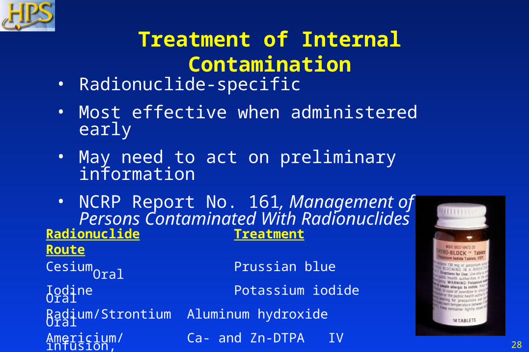
• Radionuclide-specific
• Most effective when administered early
• May need to act on preliminary information
• NCRP Report No. 161, Management of Persons Contaminated With Radionuclides
Treatment of Internal Contamination
Radionuclide Treatment RouteCesium Prussian blue OralIodine Potassium iodide OralRadium/Strontium Aluminum hydroxide OralAmericium/ Ca- and Zn-DTPA IV infusion,Plutonium/Cobalt/ nebulizerIridium
29
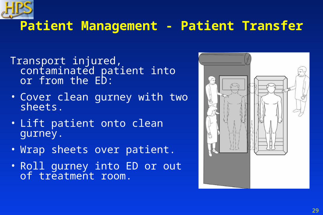
Patient Management - Patient Transfer
Transport injured, contaminated patient into or from the ED:
• Cover clean gurney with two sheets.
• Lift patient onto clean gurney.
• Wrap sheets over patient.
• Roll gurney into ED or out of treatment room.
30
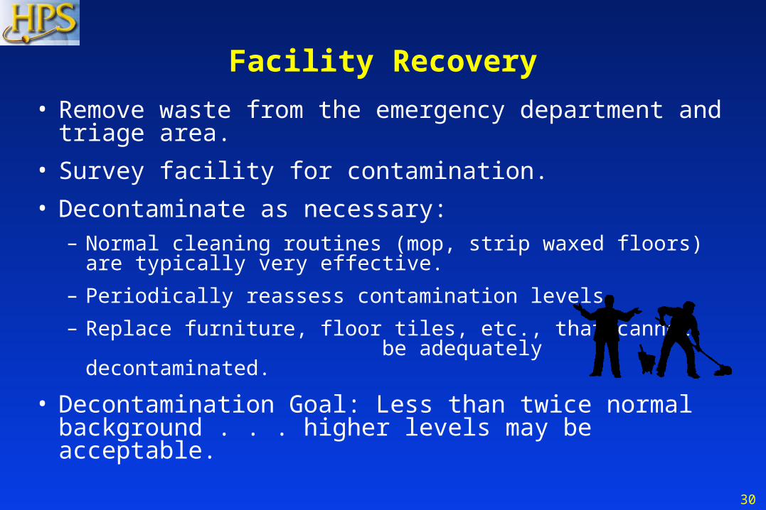
Facility Recovery
• Remove waste from the emergency department and triage area.
• Survey facility for contamination.
• Decontaminate as necessary:– Normal cleaning routines (mop, strip waxed floors) are typically
very effective.
– Periodically reassess contamination levels.
– Replace furniture, floor tiles, etc., that cannot be adequately decontaminated.
• Decontamination Goal: Less than twice normal background . . . higher levels may be acceptable.
31
• Occurs only in patients who have received very high radiation doses (greater than approximately 100 rem) to most of the body
• Dose ~15 rem – no symptoms, possible chromosomal aberrations
• Dose ~50 rem– no symptoms, minor decreases in white cells and platelets
Radiation Sickness Acute Radiation Syndrome
32
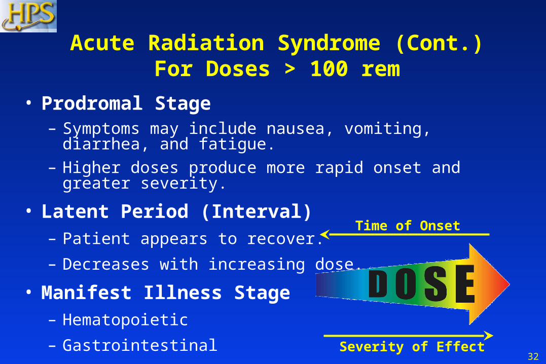
• Prodromal Stage
– Symptoms may include nausea, vomiting, diarrhea, and fatigue.
– Higher doses produce more rapid onset and greater severity.
• Latent Period (Interval)– Patient appears to recover.
– Decreases with increasing dose.
• Manifest Illness Stage– Hematopoietic
– Gastrointestinal
– CNS
Acute Radiation Syndrome (Cont.)For Doses > 100 rem
Time of Onset
Severity of Effect
33
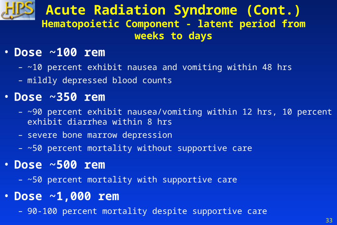
• Dose ~100 rem – ~10 percent exhibit nausea and vomiting within 48 hrs
– mildly depressed blood counts
• Dose ~350 rem– ~90 percent exhibit nausea/vomiting within 12 hrs, 10 percent exhibit diarrhea
within 8 hrs
– severe bone marrow depression
– ~50 percent mortality without supportive care
• Dose ~500 rem– ~50 percent mortality with supportive care
• Dose ~1,000 rem– 90-100 percent mortality despite supportive care
Acute Radiation Syndrome (Cont.)Hematopoietic Component - latent period from weeks to days
34
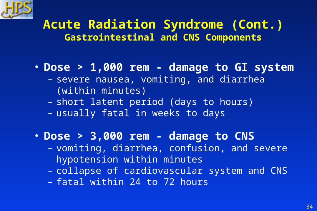
• Dose > 1,000 rem - damage to GI system– severe nausea, vomiting, and diarrhea (within minutes)– short latent period (days to hours)– usually fatal in weeks to days
• Dose > 3,000 rem - damage to CNS– vomiting, diarrhea, confusion, and severe hypotension
within minutes– collapse of cardiovascular system and CNS– fatal within 24 to 72 hours
Acute Radiation Syndrome (Cont.)Gastrointestinal and CNS Components
35
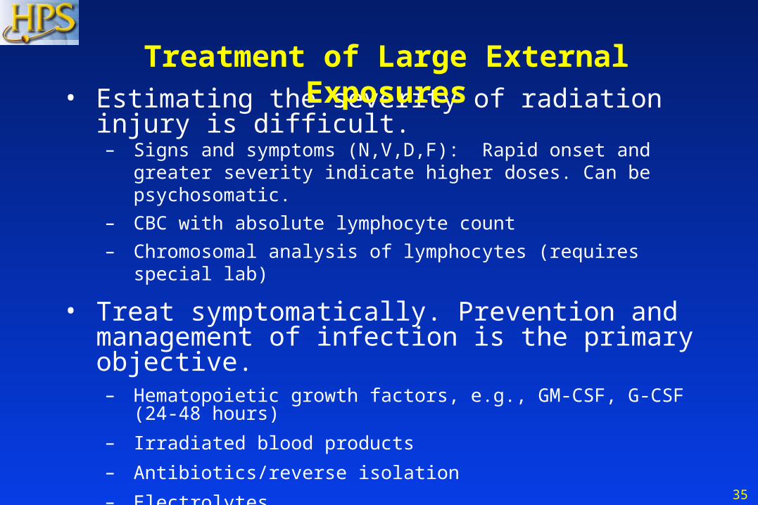
• Estimating the severity of radiation injury is difficult.– Signs and symptoms (N,V,D,F): Rapid onset and greater severity
indicate higher doses. Can be psychosomatic.
– CBC with absolute lymphocyte count
– Chromosomal analysis of lymphocytes (requires special lab)
• Treat symptomatically. Prevention and management of infection is the primary objective.– Hematopoietic growth factors, e.g., GM-CSF, G-CSF (24-48 hours)
– Irradiated blood products
– Antibiotics/reverse isolation
– Electrolytes
• Seek the guidance of experts.– Radiation Emergency Assistance Center/Training Site (REAC/TS)
– Medical Radiobiology Advisory Team (MRAT)
Treatment of Large External Exposures
36
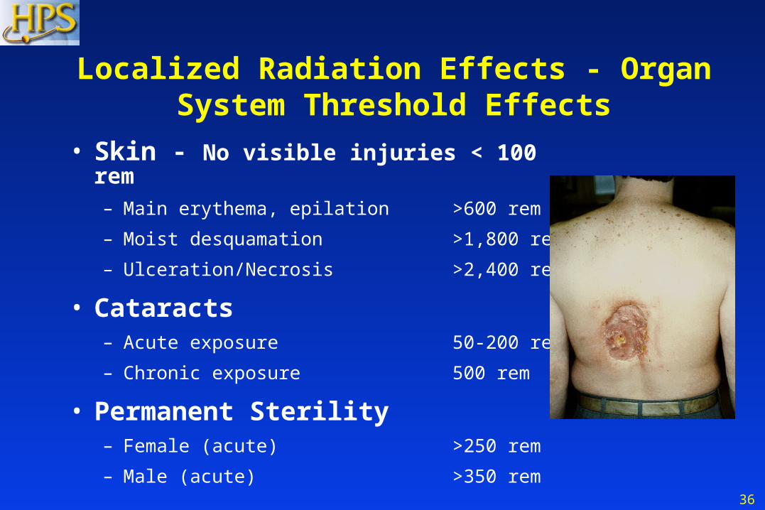
• Skin - No visible injuries < 100 rem– Main erythema, epilation >600 rem
– Moist desquamation >1,800 rem
– Ulceration/Necrosis >2,400 rem
• Cataracts– Acute exposure 50-200 rem
– Chronic exposure 500 rem
• Permanent Sterility– Female (acute) >250 rem
– Male (acute) >350 rem
Localized Radiation Effects - Organ System Threshold Effects

37
Special Considerations• High radiation dose and trauma interact
synergistically to increase mortality.
• Close wounds on patients with doses > 100 rem.
• Wound care, burn care, and surgery should be done in the first 48 hours or delayed for 2 to 3 months (> 100 rem).
24-48 Hours ~3 Months
EmergencySurgery
Hematopoietic RecoveryNo Surgery
After adequatehematopoietic recovery
SurgeryPermitted

38
Chronic Health Effects from Radiation
• Radiation is a weak carcinogen at low doses.
• There are no unique effects (type, latency, pathology).
• Natural incidence of cancer is ~40 percent; mortality ~25 percent.
• Risk of fatal cancer is estimated as ~5 percent per 100 rem.
• A dose of 5 rem increases the risk of fatal cancer by ~0.25 percent.
• A dose of 25 rem increases the risk of fatal cancer by ~1.25 percent.
39
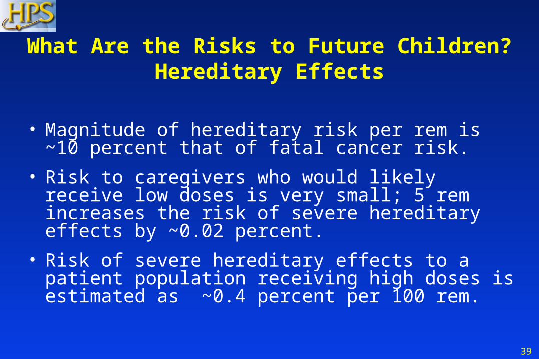
What Are the Risks to Future Children?Hereditary Effects
• Magnitude of hereditary risk per rem is ~10 percent that of fatal cancer risk.
• Risk to caregivers who would likely receive low doses is very small; 5 rem increases the risk of severe hereditary effects by ~0.02 percent.
• Risk of severe hereditary effects to a patient population receiving high doses is estimated as ~0.4 percent per 100 rem.
40
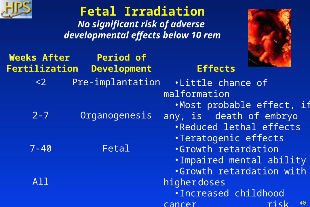
Fetal IrradiationNo significant risk of adverse
developmental effects below 10 rem
•Little chance of malformation •Most probable effect, if any, is
death of embryo •Reduced lethal effects •Teratogenic effects•Growth retardation•Impaired mental ability•Growth retardation with higher
doses•Increased childhood cancer
risk (~0.6 percent per 10 rem)
<2
2-7
7-40
All
Pre-implantation
Organogenesis
Fetal
Weeks After Fertilization
Period ofDevelopment
Effects
41
Key Points
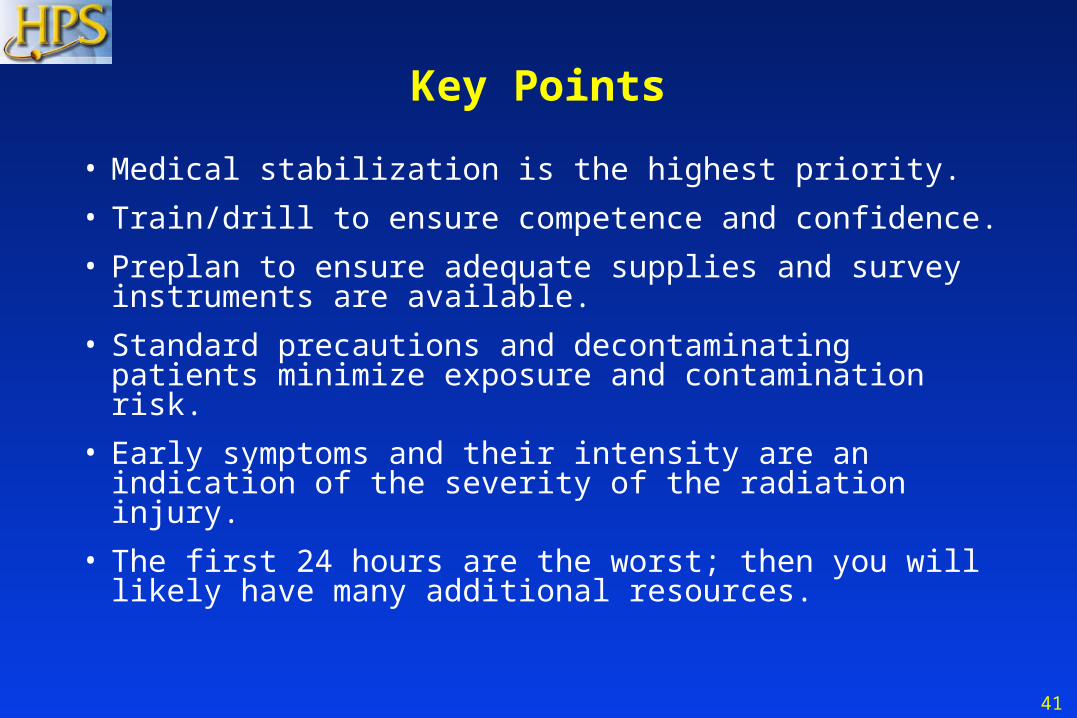
• Medical stabilization is the highest priority.
• Train/drill to ensure competence and confidence.
• Preplan to ensure adequate supplies and survey instruments are available.
• Standard precautions and decontaminating patients minimize exposure and contamination risk.
• Early symptoms and their intensity are an indication of the severity of the radiation injury.
• The first 24 hours are the worst; then you will likely have many additional resources.

42
Resources• Radiation Emergency Assistance Center/Training Site (REAC/TS),
865-576-1005, orise.orau.gov/reacts
• Medical Radiobiology Advisory Team (MRAT) Armed Forces Radiobiology Research Institute (AFRRI), 301-295-0530, afrri.usuhs.mil
• Web sites:
– remm.nlm.gov/ - Radiation Event Medical Management by Department of Health & Human Services
– emergency.cdc.gov/radiation/ - Response to Radiation Emergencies by the Centers for Disease Control and Prevention
– acr.org - “Disaster Preparedness for Radiology Professionals” by the American College of Radiology, (search for “disaster” on website)
43
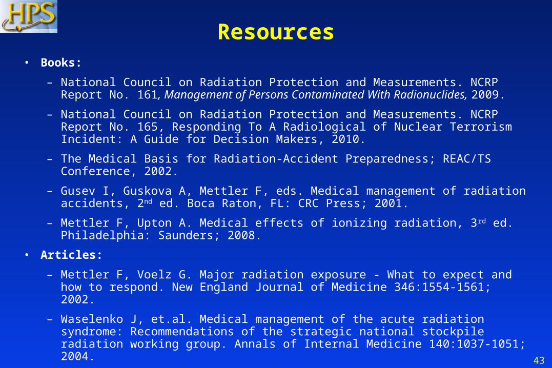
Resources• Books:
– National Council on Radiation Protection and Measurements. NCRP Report No. 161, Management of Persons Contaminated With Radionuclides, 2009.
– National Council on Radiation Protection and Measurements. NCRP Report No. 165, Responding To A Radiological of Nuclear Terrorism Incident: A Guide for Decision Makers, 2010.
– The Medical Basis for Radiation-Accident Preparedness; REAC/TS Conference, 2002.
– Gusev I, Guskova A, Mettler F, eds. Medical management of radiation accidents, 2nd ed. Boca Raton, FL: CRC Press; 2001.
– Mettler F, Upton A. Medical effects of ionizing radiation, 3rd ed. Philadelphia: Saunders; 2008.
• Articles:
– Mettler F, Voelz G. Major radiation exposure - What to expect and how to respond. New England Journal of Medicine 346:1554-1561; 2002.
– Waselenko J, et.al. Medical management of the acute radiation syndrome: Recommendations of the strategic national stockpile radiation working group. Annals of Internal Medicine 140:1037-1051; 2004.
44
Acknowledgments
Prepared by the Medical Response Subcommittee of the National Health Physics Society Homeland Security Committee. Subcommittee members when issued:
Jerrold T. Bushberg, PhD, ChairKenneth L. Miller, MSMarcia Hartman, MS
Robert Derlet, MDVictoria Ritter, RN, MBA
Edwin M. Leidholdt, Jr., PhD
ConsultantsFred A. Mettler, Jr., MD
Niel Wald, MDWilliam E. Dickerson, MD
Appreciation to Linda Kroger, MS, who assisted in this effort.
45
Health Physics Society* Version 3.0
Disclaimer: The information contained herein was current as of 10 June 2011, and is intended for educational purposes only. The authors and the Health Physics Society (HPS) do not assume any responsibility for the accuracy of the information presented herein. The authors and the HPS are not liable for any legal claims or damages that arise from acts or omissions that occur based on its use.
*The Health Physics Society is a non profit scientific professional organization whose mission is to promote the practice of radiation safety. Since its formation in 1956, the Society has grown to nearly 5,000 scientists, physicians, engineers, lawyers, and other professionals representing academia, industry, government, national laboratories, the department of defense, and other organizations. Society activities include encouraging research in radiation science, developing standards, and disseminating radiation safety information. Society members are involved in understanding, evaluating, and controlling the potential risks from radiation relative to the benefits. Official position statements are prepared and adopted in accordance with standard policies and procedures of the Society. The Society may be contacted at: 1313 Dolley Madison Blvd., Suite 402, McLean, VA 22101; phone: 703-790-1745; FAX: 703-790-2672; email: [email protected].