Embed Size (px)
Citation preview
99
Alexandria Journal of Veterinary Sciences 2014, 42: 99-104
www.alexjvs.com2047, -ISSN 1110
DOI: 10.5455/ajvs.153299
Enterobacteriaceae Associated with Farm Fish and Retailed Ones
1Mai F. Elsherief,, 2Mohamed M. Mousa, 2 Hosam Abd El-Galil, 1Engy F. El-Bahy.
1 Department of Food hygiene, animal health research institute, Agriculture research center, Giza, Egypt. 2 Department of Food hygiene, Faculty of Veterinary Medicine, Alexandria University, Egypt.
Key words ABSTRACT:
Enterobacteriaceae,
Tilapia niloticus,
Mugil cephalus.
The present study was carried out for detection and identification of
Enterobacteriaceae in retailed and farm fish as Tilapia nilotica and Mugil
cephalus and in Kafr El Shiekh Governorate. The obtained results were revealed
that 47 Enterobacteriaceae strains were isolated from 50 Tilapia niloticus fish
samples with a percentage of 94% (25 from retailed fish and 22 from farm fish).
On the other side 46 isolates of Enterobacteriaceae strains were isolated from 50
Mugil cephalus with a percentage of 92% (22 from retailed fish and 24 from farm
fish). The most dominants isolated strains were Citrobacter spp., Enteriobacter
spp., Klebsiella spp., Proteus spp., and Serratia spp. This together with the
highly pathogenic Enterobacteriaceae including Salmonella spp. and E. coli.
Corresponding Author: Mai Fouad Elsherief ([email protected])
1. INTRODUCTION
Fish had long been regarded as a desirable and
nutritional source of high quality protein and
generous supply of minerals and vitamins
constituting the major part of human diet. (Hastein
et al., 2006).
Bacterial diseases in fish are a serious threat to
aquaculture systems that cause severe damage and
mortality in Egypt (Noor El-Deen et al., 2010).
Enterobacteraceae in fish are considered as an
indicator to sewage pollution and has been reported
as opportunistic pathogen in fish (Rajasekaran,
2008). The pathogenic strains of Enterobacteraceae
may cause diarrhea in fish (Shender et al., 2009).
Enterobacteriaceae are widely distributed in nature
and found in feces of human, poultry and animals
(Wogu and Maduakol, 2010). Enterobacteriaceae
are a common water-borne bacterium, which may be
present in the tissues of apparently normal fish
(Newaj et al., 2008) .Whenever fish are exposed to
environmental stress, or injury, it causes serious
outbreaks of disease with mortalities. Environmental
stresses such as high temperature, poor water quality
and high organic content primarily contribute to the
onset and severity of Enterobacteriaceae infections
in fish (Zheng, et al. 2004 and Sekar et al., 2008).
Some human pathogens such as, Escherichia,
Klebsiella and Salmonella spp. have been found to
survive and multiply in the gut, mucus and tissues of
fish and that render fish acting as potential vector of
human disease over long periods (Onyango et al.,
2009).
The particular isolation of some most pathogenic
organisms such as Salmonella spp., E. coli and
potential pathogenic organisms as Klebsiella spp.,
Citrobacter spp. and Proteus spp., which when
isolated from fish and fish products gives an
indication about environmental fecal pollution of
fish (Wogu. and Maduakol, 2010).
2. MATERIALS AND METHODS
Collection and Preparation of collected Samples:
A total of 100 random samples of fresh water fish
represented by Tilapia nilotica, Mugil cephalus, 50
samples were collected from market and 50 samples
were collected from 5 different farms in Kafr El
Shiekh Governorate. The samples were placed
separately in clean sterile plastic bags and
transferred in an insulated icebox to the laboratory
under complete aseptic conditions without any
delay. All collected samples were subjected to
bacteriological examination according to the
methods adapted by (ICMSF,1996). All examined
Elsherief et. al. /Alexandria Journal of Veterinary Sciences 2014, 42: 99-104
011
fish samples were apparently normal. The scales and
fins were removed; the skin was sterilized by
alcohol and flamed. The muscles above the lateral
line was cut, from which 5 g were taken under
aseptic conditions to sterile homogenizer flask
containing 45 ml of sterile peptone water (0.1%).
the contents were homogenized at 14000 rpm for
2.5 minutes. The mixture was allowed to stand for
15 minutes at room temperature. 1ml of supernatant
was added separately to 9 ml of sterile peptone
water 0.1% and thoroughly mixed for preparation of
10thfold serial dilution. 0.1 ml from each of the
previously prepared serial dilution was transferred
and evenly distributed by a bent glass rod over the
surface of previously dried VRBG agar and then
overlaid by a thin layer of VRBG agar (ICMSF,
1996). The plates were incubated at 37o C for 48
hours, colonies that showed a purple color
surrounded by a purple zone were counted, and the
number per gram was calculated and recorded.A
loopfull from positive 2% Brilliant Green Bile broth
was streaked onto Sorbitol MacConkey agar at 37°C
for 24 hours. The positive purple colonies were
streaked onto Eosine Methylene Blue agar and
incubated at 37°C for 24 hours. Typical colonies
(greenish metallic with dark purple center) were
picked up and purified for identification according
to (Bailey and Scott, 1978). 1ml from supernatant
was inoculated into 10 ml of selinite F broth tubes
and incubated at 37°C for 24 hours then loopfull
from enrichment broth was streaked onto
Salmonella-Shigella agar (S.S.) and incubated at
37°C for 24 hours.
Identification of bacterial isolates:-
The isolated organisms were identified
biochemichally and serologically according to
(Cruickshank et al., 1975, Collins and lyne, 1984,
Kauffman, 1974 and Kok et al., 1996) by using
rapid diagnostic E.coli antisera sets (DENKA
SEIKEN Co., Japan) for diagnosis of the
Enteropathogenic types of E. coli and by using
antigens using Salmonella antisera (DENKA
SEIKEN Co., Japan) for Salmonella at Benha
University, Faculty of Veterinary Medicine,
Department of Food and Control .
3. RESULTS and DISCUSSION
The obtained results were revealed that
Enterobacteriaceae strains were isolated from 21
retailed Tilapia nilotica fish samples with
percentage of 84% and from 25 farm Tilapia nilotica
fish samples with percentage of 100%. On the other
side the strains were isolated from 25 retaild Mugil
cephalus fish samples with the percentage of 100%
and from 23 farms Mugil cephalus fish samples with
the percentage of 92%.
Fish from catch to consumption are proving to
contamination with several types of Micro-
organisms. The high perishability of commodity is
attributed to intrinsic factors, which favour rapid
microbial growth, namely, low, collagen and lipid
contents and comparatively high levels of soluble
nitrogen compounds in the muscle. The factors
which influence microbial contamination include
methods of catch, on- board handling fishing vessel
sanitation, processing and storage conditions (Sheen
et al., 2012)
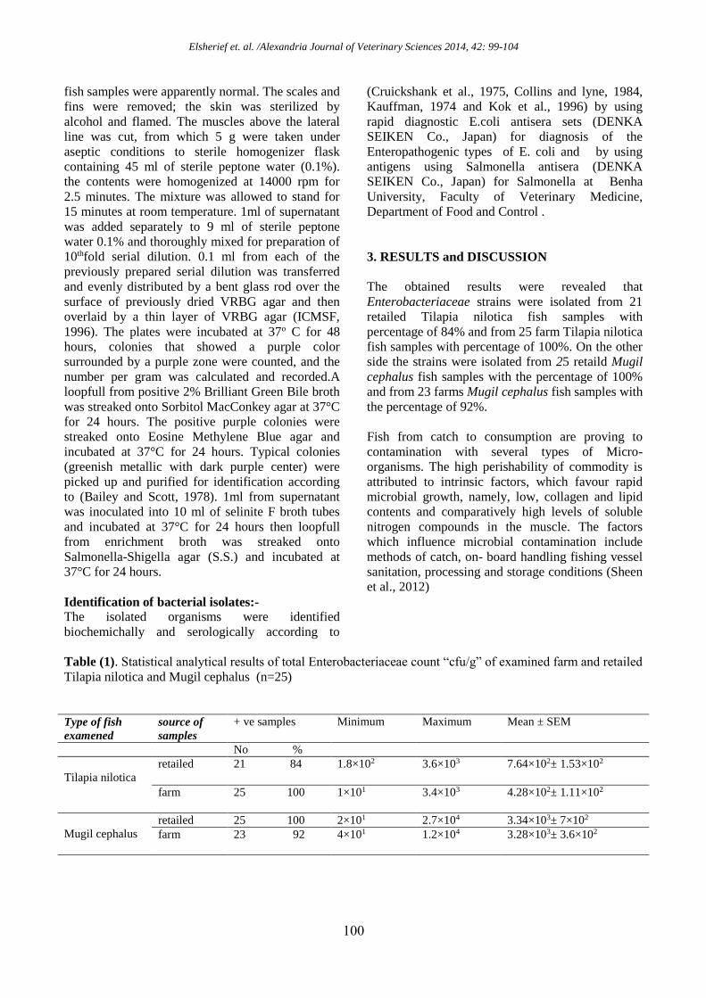
Table (1). Statistical analytical results of total Enterobacteriaceae count “cfu/g” of examined farm and retailed
Tilapia nilotica and Mugil cephalus (n=25)
Type of fish
examened
source of
samples
+ ve samples Minimum Maximum Mean ± SEM
No %
Tilapia nilotica
retailed 21 84 1.8×10² 3.6×10³ 7.64×10²± 1.53×10²
farm 25 100 1×101 3.4×10³ 4.28×10²± 1.11×10²
Mugil cephalus
retailed 25 100 2×101 2.7×10⁴ 3.34×10³± 7×10²
farm 23 92 4×101 1.2×10⁴ 3.28×10³± 3.6×10²
Elsherief et. al. /Alexandria Journal of Veterinary Sciences 2014, 42: 99-104
010
Table (2). Incidence percentage of Enterobacteriaceae isolated from farm and retailed Tilapia nilotica and
Mugil cephalus (n=25) Type of bacteria isolated Tilapia nilotica Mugil cephalus
Farm Retailed Farm Retailed
E.coli 2 8% 3 12% 2 8% 1 4%
Salmonella 4 16% 2 8% 1 4% 4 16%
Enterobacter cloacae 2 8% 2 8% 2 8% 2 8%
Enterobacter agglomerans 1 4% - - - - - -
Enterobacter aerogenes 2 8% 3 12% - - 1 4%
Enterobacter hafniae - - 1 4% 1 4% - -
Citrobacter diversus - - 1 4% 1 4% 2 8%
Citrobacter freundii 3 12% - - 1 4% 2 8%
Proteus mirabilis 2 8% 4 16% 1 4% 2 8%
Proteus vulgaris 1 4% 3 12% 3 12% 2 8%
Provedencia rettgeri - - 1 4% 2 8% - -
Pseudomonas species 1 4% - - 3 12% 2 8%
Serratia marcesens 2 8% - - 1 4% 1 4%
Serratia liquefaciens - - 2 8% 2 8% 1 4%
Klebsiella pneumonia - - 1 4% 3 12% 2 8%
Klebsiella ozaenae 2 8% 2 8% 1 4% - -
Table (3): Serological identification of isolated E.coli in examined farm and retailed Tilapia nilotica and
Mugil cephalus Tilapia nilotica Mugil cephalus
Retailed Farm Retailed Farm
Serodiagnosis Strain
characterization
Serodiagnosis Strain
characterization
Serodiagnosis Strain
characterization
Serodiagnosis Strain
characterization
O86
EPEC
O111 : H4
EHEC
O111 : H4
EHEC
O44 : H18
EPEC
O127 : H6 ETEC
O125 : H21
ETEC
O127 : H6
ETEC O114 : H21 EPEC
EPEC: Enteropathogenic E.coli., ETEC: Enterotoxigenic E.coli., EHEC: Enterohaemorrhagic E.coli.
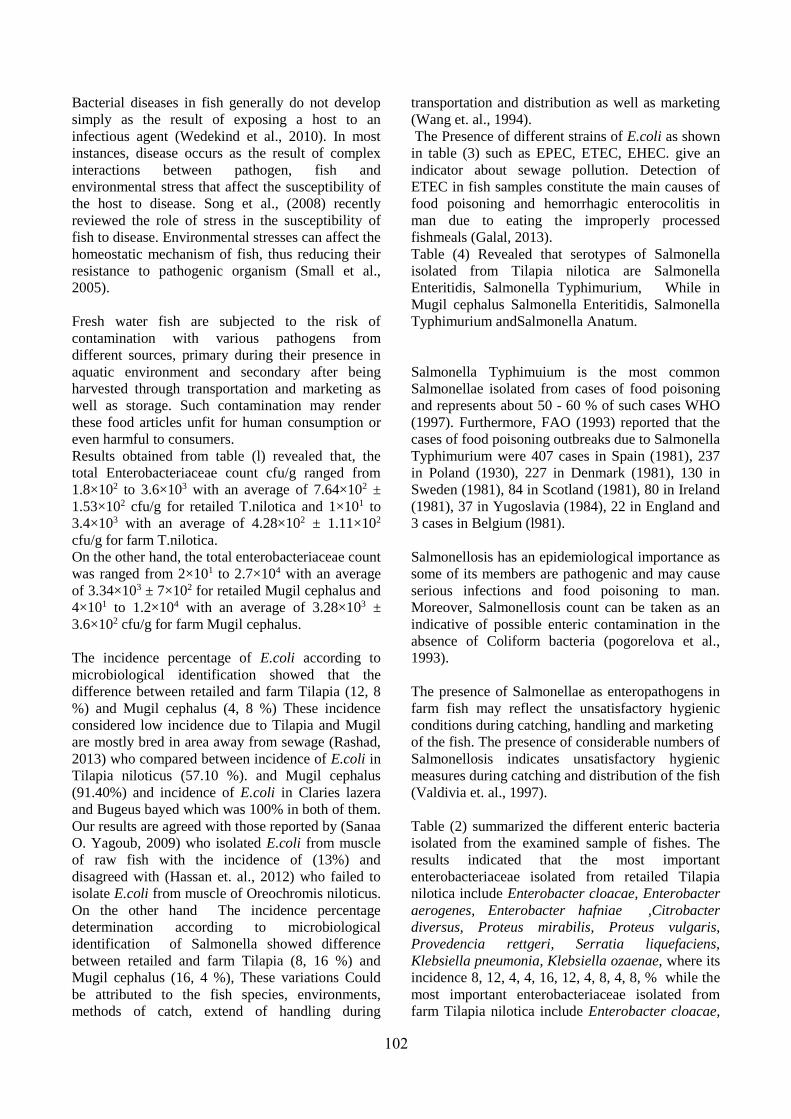
Table (4). Serological identification of isolated Salmonella species in examined farm and retailed Tilapia
nilotica and Mugil cephalus
Tilapia nilotica Mugil cephalus
Retailed Farm Retailed Farm
Identified
strains
Antigenic structure
Identified
strains
Antigenic structure Identified
strains
Antigenic structure Identified
strains
Antigenic
structure
O H O H O H O H
Salmonella
Enteritidis
1,9,12
g,m :
1,7
Salmonella
Typhimuriu
m
1,4,5,12
i : 1,2
Salmonella
Enteritidis
1,9,12 g,m :
1,7
Salmonella
Typhimuriu
m
1,4,5,12 i :
1,2
Salmonella
Enteritidis
1,9,12 g,m :
1,7
Salmonella
Enteritidis
1,9,12 g,m :
1,7
- - -
Salmonella
Typhimuriu
m
1,4,5,12 i : 1,2 Salmonella
Typhimuriu
m
1,4,5,12 i : 1,2 Salmonella
Typhimuriu
m
1,4,5,12 i : 1,2 - - -
Salmonella
Anatum
3,10,15,3
4
e,h : 1,6 - - -
011
Bacterial diseases in fish generally do not develop
simply as the result of exposing a host to an
infectious agent (Wedekind et al., 2010). In most
instances, disease occurs as the result of complex
interactions between pathogen, fish and
environmental stress that affect the susceptibility of
the host to disease. Song et al., (2008) recently
reviewed the role of stress in the susceptibility of
fish to disease. Environmental stresses can affect the
homeostatic mechanism of fish, thus reducing their
resistance to pathogenic organism (Small et al.,
2005).
Fresh water fish are subjected to the risk of
contamination with various pathogens from
different sources, primary during their presence in
aquatic environment and secondary after being
harvested through transportation and marketing as
well as storage. Such contamination may render
these food articles unfit for human consumption or
even harmful to consumers.
Results obtained from table (l) revealed that, the
total Enterobacteriaceae count cfu/g ranged from
1.8×10² to 3.6×10³ with an average of 7.64×10² ±
1.53×10² cfu/g for retailed T.nilotica and 1×101 to
3.4×10³ with an average of 4.28×10² ± 1.11×10²
cfu/g for farm T.nilotica.
On the other hand, the total enterobacteriaceae count
was ranged from 2×101 to 2.7×10⁴ with an average
of 3.34×10³ ± 7×10² for retailed Mugil cephalus and
4×101 to 1.2×10⁴ with an average of 3.28×10³ ±
3.6×10² cfu/g for farm Mugil cephalus.
The incidence percentage of E.coli according to
microbiological identification showed that the
difference between retailed and farm Tilapia (12, 8
%) and Mugil cephalus (4, 8 %) These incidence
considered low incidence due to Tilapia and Mugil
are mostly bred in area away from sewage (Rashad,
2013) who compared between incidence of E.coli in
Tilapia niloticus (57.10 %). and Mugil cephalus
(91.40%) and incidence of E.coli in Claries lazera
and Bugeus bayed which was 100% in both of them.
Our results are agreed with those reported by (Sanaa
O. Yagoub, 2009) who isolated E.coli from muscle
of raw fish with the incidence of (13%) and
disagreed with (Hassan et. al., 2012) who failed to
isolate E.coli from muscle of Oreochromis niloticus.
On the other hand The incidence percentage
determination according to microbiological
identification of Salmonella showed difference
between retailed and farm Tilapia (8, 16 %) and
Mugil cephalus (16, 4 %), These variations Could
be attributed to the fish species, environments,
methods of catch, extend of handling during
transportation and distribution as well as marketing
(Wang et. al., 1994).
The Presence of different strains of E.coli as shown
in table (3) such as EPEC, ETEC, EHEC. give an
indicator about sewage pollution. Detection of
ETEC in fish samples constitute the main causes of
food poisoning and hemorrhagic enterocolitis in
man due to eating the improperly processed
fishmeals (Galal, 2013).
Table (4) Revealed that serotypes of Salmonella
isolated from Tilapia nilotica are Salmonella
Enteritidis, Salmonella Typhimurium, While in
Mugil cephalus Salmonella Enteritidis, Salmonella
Typhimurium andSalmonella Anatum.
Salmonella Typhimuium is the most common
Salmonellae isolated from cases of food poisoning
and represents about 50 - 60 % of such cases WHO
(1997). Furthermore, FAO (1993) reported that the
cases of food poisoning outbreaks due to Salmonella
Typhimurium were 407 cases in Spain (1981), 237
in Poland (1930), 227 in Denmark (1981), 130 in
Sweden (1981), 84 in Scotland (1981), 80 in Ireland
(1981), 37 in Yugoslavia (1984), 22 in England and
3 cases in Belgium (l981).
Salmonellosis has an epidemiological importance as
some of its members are pathogenic and may cause
serious infections and food poisoning to man.
Moreover, Salmonellosis count can be taken as an
indicative of possible enteric contamination in the
absence of Coliform bacteria (pogorelova et al.,
1993).
The presence of Salmonellae as enteropathogens in
farm fish may reflect the unsatisfactory hygienic
conditions during catching, handling and marketing
of the fish. The presence of considerable numbers of
Salmonellosis indicates unsatisfactory hygienic
measures during catching and distribution of the fish
(Valdivia et. al., 1997).
Table (2) summarized the different enteric bacteria
isolated from the examined sample of fishes. The
results indicated that the most important
enterobacteriaceae isolated from retailed Tilapia
nilotica include Enterobacter cloacae, Enterobacter
aerogenes, Enterobacter hafniae ,Citrobacter
diversus, Proteus mirabilis, Proteus vulgaris,
Provedencia rettgeri, Serratia liquefaciens,
Klebsiella pneumonia, Klebsiella ozaenae, where its
incidence 8, 12, 4, 4, 16, 12, 4, 8, 4, 8, % while the
most important enterobacteriaceae isolated from
farm Tilapia nilotica include Enterobacter cloacae,
Elsherief et. al. /Alexandria Journal of Veterinary Sciences 2014, 42: 99-104
011
Enterobacter agglomerans, Enterobacter
aerogenes, Citrobacter freundii, Proteus mirabilis,
Proteus vulgaris, Pseudomonas species, Serratia
marcesens, Klebsiella ozaenae where its incidence
8, 4, 8, 12, 8, 4, 4, 8, 8 %. The results also cleared
that the most important enterobacteriaceae isolated
from retailed Mugil cephalus include Enterobacter
cloacae, Enterobacter aerogenes, Citrobacter
diversus, Citrobacter freundii, Proteus mirabilis,
Proteus vulgaris, Pseudomonas species, Serratia
marcesens, Serratia liquefaciens, Klebsiella
pneumonia where its incidence 8, 4, 8, 8, 8, 8, 8, 4,
4, 8 % while the most important enterobacteriaceae
isolated from farm Mugil cephalus include
Enterobacter cloacae, Enterobacter hafniae,
Citrobacter diversus, Citrobacter freundii, Proteus
mirabilis, Proteus vulgaris, Provedencia rettgeri,
Pseudomonas species, Serratia marcesens, Serratia
liquefaciens, Klebsiella pneumonia, Klebsiella
ozaenae where its incidence 8, 4, 4, 4, 4, 12, 8, 12,
4, 8, 12, 4 %. These results are closed to those
reported by (Yagoub, 2009) who isolated
enterobacteriaceae from muscles of raw fishes in
sold in fish market in Khartoum state; she isolated
Klebsiella spp., Citobacter spp., Proteus spp. and
Providencia spp. With incidence 10.9, 8.7, 15.2 and
4.3 %. While differs in incidence of Enterobacter
spp., Serritia spp., Pseudomonas spp. which failed
to be isolated .
Isolation of some potential pathogenic organisms
such as Klebsiella spp., Citrobacter spp., Proteus
spp. may indicates environmental pollution and
these supported the findings of (Yagoub and Ahmed
2004) who isolated pathogenic and potential
pathogenic organisms from tap water that originated
from Nile River. This also confirms the findings of
(Herrera et al., 2006) who isolated similar
organisms from fish and fish products.
The isolation of Pseudomonas spp from collected
fish samples is of highly importance because this
organisms plays a considerable role as potential
pathogenic bacteria for human and as an indicator of
food quality as spoilage organism. This is in accord
with previously mentioned by (Koutsoumanis and
Nychas, 2000) and (Jeyasekaran et al., 2006) who
identified pseudomonads as a good spoilage index.
This study revealed that the contamination of fish
with some enteropathogens may give an indication
about bad sanitary conditions under which fish were
exposed from catching till reach to the markets,
resulting in both public health hazards and
economic losses.
4. REFERENCES
Hassan, A.H.M., Noor- El Deen, A.E., Galal, H.M.,
Sohad, M. Dorgham, Bakry, M.A., Hakim, A.S.
2012. Further Characterization of
Enterobacteriaceae isolated from cultured
freshwater fish in Kafr El Shiek Governorate:
Clinical, biochemical and histopathological study
with emphasis on treatment trials. Glob. Vet. 9 (5):
617-629.
Bailey, W.R., Scott, E.G. 1978. Diagnostic
Microbiology. A textbook for the isolation and
identification of pathogenic microorganisms. 5th
Ed., Carroll B. Larson and Marjorie Gould
Publisher, Saint Louis, C.V. Mosby Co., Missouri,
USA.
Collins, C.H., Lyne, P.M. 1984. Microbiological
methods 5th microbiology laboratory manual,
British Liberary, Butter Wort Inc., London, UK.
Cruickshank, R., Duguid, J.P., Marmion, B.P.,
Swain, R.H.A. 1975.Medical Microbiology. 12th
ed., Edinburg, London and New York.
Food Agriculture organization "FAO" 1993.
Zoonotic diseases in the Near East Region.
Regional Office of the United Nation, Cairo.
Galal, H.M. Hakim, A.S.; and Sohad, M. Dorgham
2013. Phenotypic and virulence genes screening of
Escherichia coli strains isolated from different
sources in delta Egypt. Life Science Journal,
10(2).
Gonzalez-Rodriguez, M.N., Sanz, J.J, Santos, J.A,
Otero, A., Garcia-lopez, M.L. 2001.
Bacteriological Quality of aquaculture freshwater
fish portions in prepackaged trays stored at 3
degrees C. J. Food Prot. 64 (9): 1399 - 1404.
Håstein, T., Hjeltnes, B., Lillehaug, A., Utne Skåre,
J., Berntssen, M., Lundebye, A.K. 2006. Food
safety hazards that occur during the production
stage: challenges for fish farming and the fishing
industry. Rev. Sci. Technol. 25(2): 607-625.
Rashad, H. 2013. Enteropathogenes in some fresh
water fishes. M.Sc. Thesis, Dept. of food hygiene,
Fac. Vet. Med. Alex. Univ., Alexandria, Egypt.
Herrera, F.C., Santos, J.A., Otero, A., Garcia-Lopez,
M.L. 2006. Occurrence of Foodborne pathogenic
bacteria in retails prepackage portions of marine
fish in Spain. J. Appl. Microbiol. 100 (3): 527-36.
ICMSF (International Commission of
Microbiological Specification for Foods)1996.
microorganisms in foods. I-their significance and
methods of enumeration, 3rd Ed. Univ. of Toronto,
Canada.
Jeyasekaran, G., Ganesan, P., Anandaraj, R., Jeya
Shakila, R., Sukumar, D. 2006. Quantitative and
Elsherief et. al. /Alexandria Journal of Veterinary Sciences 2014, 42: 99-104
011
qualitative studies on the bacteriological quality of
Indian white shrimp (Penaeus indicus) stored in
dry ice. J. Food Microbiol. 23(6): 526-533.
Kauffman, G. 1974. Kauffmann white scheme. J.
Acta. Path. Microbiol. Sci., 61:385.
Kok, T. Worswich, D., Gowans, E.1996. Some
serological techniques for microbial and viral
infections. In Practical Medical Microbiology
(Collee, J.; Fraser, A.; Marmion, B. and Simmons,
A., eds.), 14th ed., Edinburgh, Churchill
livingstone, UK.
Koutsoumanis K., Nychas, G.J.2000. Application of
systemic experimental procedure to develop a
microbial model for rapid fish shelf life
predictions. Int. J. Food Microbiol. 60 (2-3): 171-
184.
Newaj, A., Mutani, A., Ramsubhag , A., Adesiyun,
A. 2008. Prevalence of bacterial pathogens and
their anti-microbial resistance in Tilapia and their
pond water in Trinidad. Zoonoses Public Health,
55(4): 206-213.
Noor El-Deen, A.E., Atta, N.S., Abd El Aziz, M.A.
2010. Oral vaccination of Nile Tilapia
(Orechromis niloticus) against motile Aeromonas
septicemia. Nat. Sci. 8(2): 21-25.
Onyango, M. D., Sarah Wandili, Rose Kakai, Eliud
N. W. 2009 Isolation of Salmonella and Shigella
from fish harvested from the Winam Gulf of Lake
Victoria, Kenya. J Infect Developing Countries,
3(2):99-104.
Pogorelova, N.P., Lartseva, L.v., Boiko, A.V.,
smirnova, I.E., zhigareva, T.M.; Zhuravleva, L.A.
Merkina, E.N. 1993. Microbiological evaluation
of water pollution in the Volga delta. Gigiena.
Sanitorya, 7: 35 - 38.
Rajasekaran, P. 2008. Enterobacteriaceae group of
organisms in sewage-fed fishes. Advanced
Biotech., 8:12-14.
Yagoub, S. O., Ahmed T.M. 2004. Pathogenic
Microorganisms in fresh water samples collected
from Khartoum central market. Sudan J. Vet. Sci.
Anim. Husbandry 43(1-2): 32-37.
Yagoub, S. O. 2009. Isolation of
Enterobacteriaceae and Pseudomonas spp. from
raw fish sold in fish market in Khartoum state, J.
Bacteriol. Res.1(7), pp. 085-088.
Sekar, V., T. Santiago, K. Vijayan, S. Alavandi , V.
Raj, J. Rajan, M. Sanjuktha and N. Kalaimani
2008. Involvement of Enterobacter cloacae in the
mortality of the fish, Mugil cephalus. Lett. Appl.
Microbiol., 46(6):667-672.
Sheen, S., Huang, L., Sommers, C. 2012. Survival
of Listeria monocytogenes, Escherichia coli
O157:H7 and Salmonella spp. on Catfish Fillets
Exposed to Microwave Heating in a Continuous
Mode. J. Food Sci. 77(8):E209-214.
Shender, L.A., T.R. Spraker 2009. Salmonellosis in
a free-ranging population of javelinas (Pecari
tajacu) in south central Arizona. J. Wild Dis.,
45(4): 941-51.
Small, B., Bilodeau, A 2005. Effects of cortisol and
stress on channel catfish (Ictalurus punctatus)
pathogen susceptibility and lysozyme activity
following exposure to Edwardsiella ictaluri. Gen.
Comp. Endocrinol., 142(1-2):256-62.
Song, J., Nakayama, K., Murakami, Y.,Jung, S., Oh,
M., Matsuoka, S., Kawakami, H., Kitamura, S.
2008. Does heavy oil pollution induce bacterial
diseases in Japanese flounder Paralichthys
olivaceus. Mar. Pollut. Bull., 57 (6- 12):889-894.
Valdivia Garvayo, M. de., Ruiz - Lope, M.D.,
Artacho, Martin - Lagos, R.,Lopez martinez, M.C.,
Muros-Guadix, P. 1997. Commerical or shelf - life
of fresh eviscerated and filleted rainbow trout
(Onchrhynchus mykiss). Alimentacion Equipos
Technol. 8: 97 - 102.
wang, S.J., chen, J.M., Fan, J.J. 1994. Quality
changes in fresh Tilapia and Milkfish during
refrigerated (4 degree C) and frozen (-15 degree
C) storage J. Food during Analysis, 2(4):311 - 316.
Wedekind, C., Gessner, M., Vazquez, F.,
Maerkiand, M., Steiner, D. 2010. Elevated
resource availability sufficient to turn
opportunistic into virulent fish pathogens. Ecol.
91(5): 1251-6.
Wogu, M.D., Maduakol M. 2010. Evaluation of
microbial spoilage of some aqua cultured fresh
fish in Benin City Nigeria. Ethiopian J. Environ.
Studies Manag. 3(3): 18-22.
World Health Organization (WHO). 1997.
Microbial aspects of food hygiene, technical
Report Series, No. 598, pP.2l - 23. WHO, Geneva,
Switzerland.
Zheng, D., K. Mai, S. Liu, C. Limin, Z. Liufu, W.
Xu, B. Tan and W. Zhang 2004. Effect of
temperature and salinity on virulence of
Edwardsiella tarda to Japanese flounder,
Paralichthys olivaceus (Temminck et Schlegel).
Aqua. Res., 35: 494–500.