Embed Size (px)
Citation preview
1
Essentials of Anatomy & Physiology, 4th Edition Martini / Bartholomew
PowerPoint® Lecture Outlines prepared by Alan Magid, Duke University
The Cardiovascular
System: The Heart 12
Copyright © 2007 Pearson Education, Inc., publishing as Benjamin Cummings
Slides 1 to 65
Heart’s Place in the Circulation
Heart Pumps Blood into Two Circuits in Sequence 1. Pulmonary circuit
• To and from the lungs
2. Systemic circuit • To and from the rest of the body
Copyright © 2007 Pearson Education, Inc., publishing as Benjamin Cummings
Heart’s Place in the Circulation
Three Kinds of Blood Vessels 1. Arteries
• Carry blood away from heart and carry it to the capillaries
2. Capillaries • Connect arteries and veins
• Exchange area between blood and cells
3. Veins • Receive blood from capillaries and carry it
back to the heart
Copyright © 2007 Pearson Education, Inc., publishing as Benjamin Cummings
Heart’s Place in the Circulation
Two Sets of Pumping Chambers in Heart 1. Right atrium
• Receives systemic blood
2. Right ventricle • Pumps blood to lungs (pulmonary)
3. Left atrium • Receives blood from lungs
4. Left ventricle • Pumps blood to organ systems (systemic)
Copyright © 2007 Pearson Education, Inc., publishing as Benjamin Cummings
Heart’s Place in the Circulation
Overview of the Cardiovascular System
Figure 12-1
The Anatomy of the Heart
Pericardial Cavity • Surrounds the heart
• Lined by pericardium • Two layers
1. Visceral pericardium (epicardium)
• Covers heart surface
2. Parietal pericardium
• Lines pericardial sac that surrounds heart
Copyright © 2007 Pearson Education, Inc., publishing as Benjamin Cummings
2
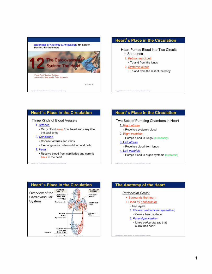
The Anatomy of the Heart
Figure 12-2
The Location of the Heart in the Thoracic Cavity
The Anatomy of the Heart
Surface Features of the Heart 1. Auricle —Outer portion of atrium
2.Coronary sulcus —Deep groove that marks boundary of atria and ventricles • Anterior & Posterior interventricular sulcus
• Mark boundary between left and right ventricles
• Sulci contain major cardiac blood vessels
• Filled with protective fat
Copyright © 2007 Pearson Education, Inc., publishing as Benjamin Cummings
The Anatomy of the Heart The Surface Anatomy of the Heart
Figure 12-3(a) 1 of 2
The Anatomy of the Heart The Surface Anatomy of the Heart
Figure 12-3(a) 2 of 2
The Anatomy of the Heart
Figure 12-3(b)
The Surface Anatomy of the Heart
The Anatomy of the Heart
The Heart Wall 1. Epicardium (visceral pericardium)
• Outermost layer • Serous membrane
2. Myocardium • Middle layer • Thick muscle layer
3. Endocardium • Inner lining of pumping chambers
Copyright © 2007 Pearson Education, Inc., publishing as Benjamin Cummings
3
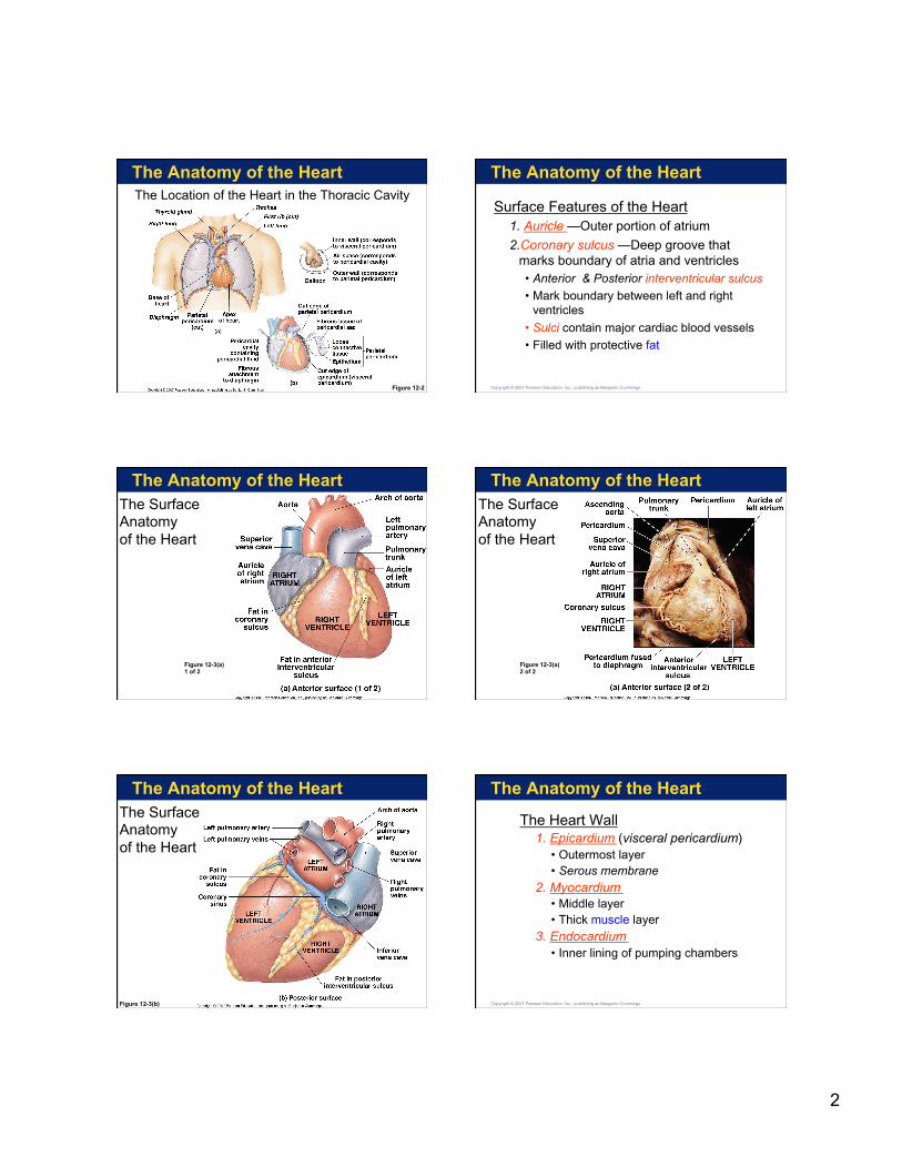
The Anatomy of the Heart
The Heart Wall and Cardiac Muscle Tissue
Figure 12-4
The Anatomy of the Heart
The Heart Wall and Cardiac Muscle Tissue
Figure 12-4(a)
The Anatomy of the Heart
Figure 12-4(b)
The Heart Wall and Cardiac Muscle Tissue
The Anatomy of the Heart The Heart Wall and Cardiac Muscle Tissue
Figure 12-4(c)
The Anatomy of the Heart
Figure 12-4(d)
The Heart Wall and Cardiac Muscle Tissue
The Anatomy of the Heart
Cardiac Muscle Cells • Shorter than skeletal muscle fibers
• Have single nucleus
• Have striations (sarcomere organization)
• Depend on aerobic metabolism
• Connected by intercalated discs • Make sure all cardiac muscle cells work
together so the heart beats as one unit
Copyright © 2007 Pearson Education, Inc., publishing as Benjamin Cummings
4
The Anatomy of the Heart
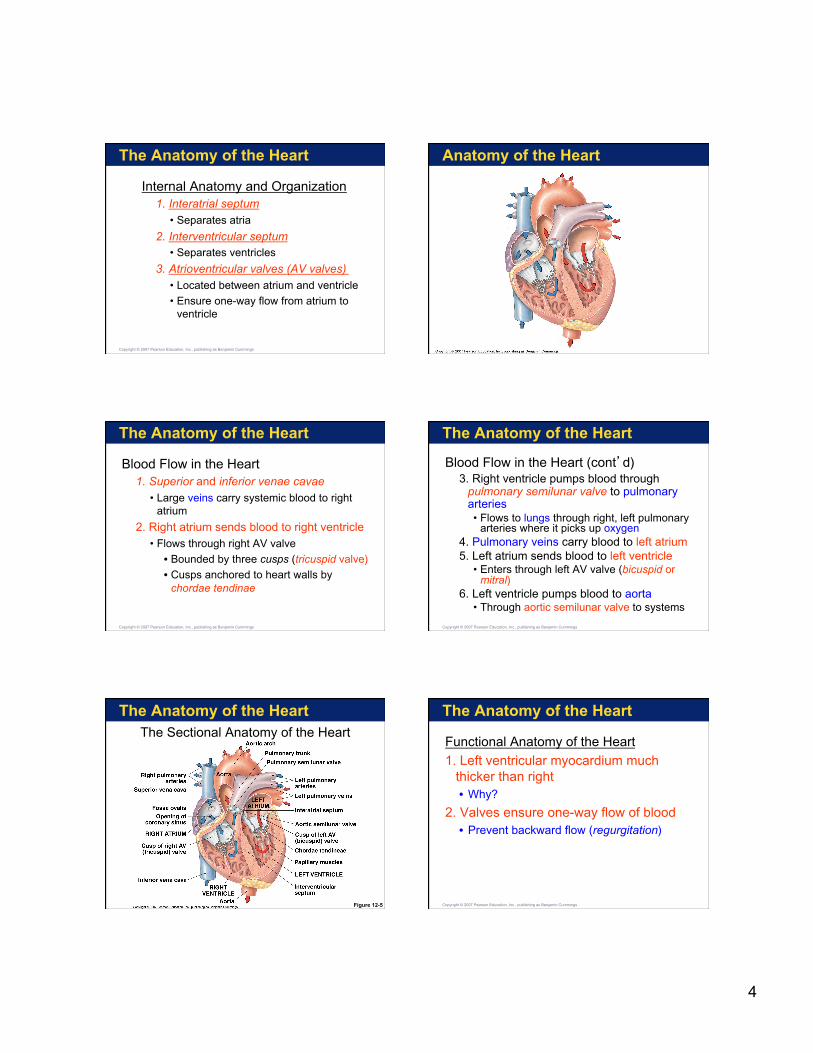
Internal Anatomy and Organization 1. Interatrial septum
• Separates atria
2. Interventricular septum • Separates ventricles
3. Atrioventricular valves (AV valves) • Located between atrium and ventricle
• Ensure one-way flow from atrium to ventricle
Copyright © 2007 Pearson Education, Inc., publishing as Benjamin Cummings
Anatomy of the Heart
The Anatomy of the Heart
Blood Flow in the Heart 1. Superior and inferior venae cavae
• Large veins carry systemic blood to right atrium
2. Right atrium sends blood to right ventricle • Flows through right AV valve
• Bounded by three cusps (tricuspid valve)
• Cusps anchored to heart walls by chordae tendinae
Copyright © 2007 Pearson Education, Inc., publishing as Benjamin Cummings
The Anatomy of the Heart
Blood Flow in the Heart (cont’d) 3. Right ventricle pumps blood through
pulmonary semilunar valve to pulmonary arteries • Flows to lungs through right, left pulmonary
arteries where it picks up oxygen 4. Pulmonary veins carry blood to left atrium 5. Left atrium sends blood to left ventricle
• Enters through left AV valve (bicuspid or mitral)
6. Left ventricle pumps blood to aorta • Through aortic semilunar valve to systems
Copyright © 2007 Pearson Education, Inc., publishing as Benjamin Cummings
The Anatomy of the Heart The Sectional Anatomy of the Heart
Figure 12-5
The Anatomy of the Heart
Functional Anatomy of the Heart
1. Left ventricular myocardium much thicker than right • Why?
2. Valves ensure one-way flow of blood • Prevent backward flow (regurgitation)
Copyright © 2007 Pearson Education, Inc., publishing as Benjamin Cummings
5
The Anatomy of the Heart
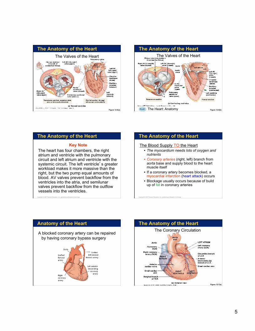
The Valves of the Heart
Figure 12-6(a)
The Anatomy of the Heart The Valves of the Heart
Figure 12-6(b) The Heart: Anatomy PLAY
The Anatomy of the Heart
Key Note The heart has four chambers, the right atrium and ventricle with the pulmonary circuit and left atrium and ventricle with the systemic circuit. The left ventricle’s greater workload makes it more massive than the right, but the two pump equal amounts of blood. AV valves prevent backflow from the ventricles into the atria, and semilunar valves prevent backflow from the outflow vessels into the ventricles.
Copyright © 2007 Pearson Education, Inc., publishing as Benjamin Cummings
The Anatomy of the Heart
The Blood Supply TO the Heart • The myocardium needs lots of oxygen and
nutrients • Coronary arteries (right, left) branch from
aorta base and supply blood to the heart muscle itself
• If a coronary artery becomes blocked, a myocardial infarction (heart attack) occurs
• Blockage usually occurs because of build up of fat in coronary arteries
Copyright © 2007 Pearson Education, Inc., publishing as Benjamin Cummings
Anatomy of the Heart
A blocked coronary artery can be repaired by having coronary bypass surgery
The Anatomy of the Heart The Coronary Circulation
Figure 12-7(a)
6
The Anatomy of the Heart The Coronary Circulation
Figure 12-7(b)
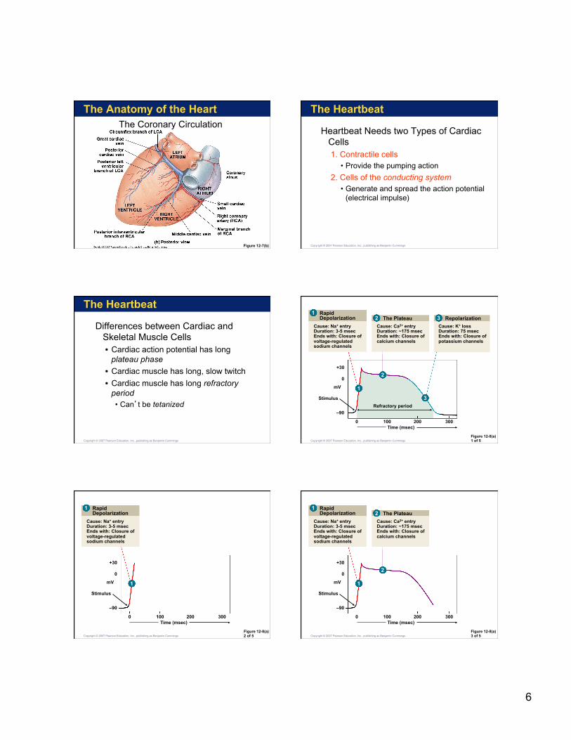
The Heartbeat
Heartbeat Needs two Types of Cardiac Cells 1. Contractile cells
• Provide the pumping action
2. Cells of the conducting system • Generate and spread the action potential
(electrical impulse)
Copyright © 2007 Pearson Education, Inc., publishing as Benjamin Cummings
The Heartbeat
Differences between Cardiac and Skeletal Muscle Cells • Cardiac action potential has long
plateau phase
• Cardiac muscle has long, slow twitch
• Cardiac muscle has long refractory period • Can’t be tetanized
Copyright © 2007 Pearson Education, Inc., publishing as Benjamin Cummings Figure 12-8(a) 1 of 5
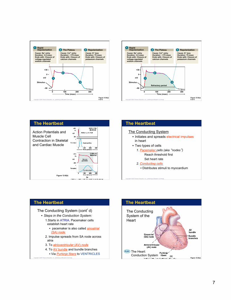
Rapid Depolarization
Cause: Na+ entry Duration: 3-5 msec Ends with: Closure of voltage-regulated sodium channels
The Plateau
Cause: Ca2+ entry Duration: ~175 msec Ends with: Closure of calcium channels
Repolarization
Cause: K+ loss Duration: 75 msec Ends with: Closure of potassium channels
+30
0
mV
Stimulus
–90
0 100 200 300 Time (msec)
Refractory period
1
1
2
2
3
3
Copyright © 2007 Pearson Education, Inc., publishing as Benjamin Cummings
Figure 12-8(a) 2 of 5
Rapid Depolarization
Cause: Na+ entry Duration: 3-5 msec Ends with: Closure of voltage-regulated sodium channels
+30
0
mV
Stimulus
–90
0 100 200 300 Time (msec)
1
1
Copyright © 2007 Pearson Education, Inc., publishing as Benjamin Cummings Figure 12-8(a) 3 of 5
Rapid Depolarization
Cause: Na+ entry Duration: 3-5 msec Ends with: Closure of voltage-regulated sodium channels
The Plateau
Cause: Ca2+ entry Duration: ~175 msec Ends with: Closure of calcium channels
+30
0
mV
Stimulus
–90
0 100 200 300 Time (msec)
1
1
2
2
Copyright © 2007 Pearson Education, Inc., publishing as Benjamin Cummings
7
Figure 12-8(a) 4 of 5
Rapid Depolarization
Cause: Na+ entry Duration: 3-5 msec Ends with: Closure of voltage-regulated sodium channels
The Plateau
Cause: Ca2+ entry Duration: ~175 msec Ends with: Closure of calcium channels
Repolarization
Cause: K+ loss Duration: 75 msec Ends with: Closure of potassium channels
+30
0
mV
Stimulus
–90
0 100 200 300 Time (msec)
1
1
2
2
3
3
Copyright © 2007 Pearson Education, Inc., publishing as Benjamin Cummings Figure 12-8(a) 5 of 5
Rapid Depolarization
Cause: Na+ entry Duration: 3-5 msec Ends with: Closure of voltage-regulated sodium channels
The Plateau
Cause: Ca2+ entry Duration: ~175 msec Ends with: Closure of calcium channels
Repolarization
Cause: K+ loss Duration: 75 msec Ends with: Closure of potassium channels
+30
0
mV
Stimulus
–90
0 100 200 300 Time (msec)
Refractory period
1
1
2
2
3
3
Copyright © 2007 Pearson Education, Inc., publishing as Benjamin Cummings
The Heartbeat
Action Potentials and Muscle Cell Contraction in Skeletal and Cardiac Muscle
Figure 12-8(b)
The Heartbeat
The Conducting System • Initiates and spreads electrical impulses
in heart
• Two types of cells 1. Pacemaker cells (aka “nodes”)
Reach threshold first
Set heart rate
2. Conducting cells
• Distributes stimuli to myocardium
Copyright © 2007 Pearson Education, Inc., publishing as Benjamin Cummings
The Heartbeat
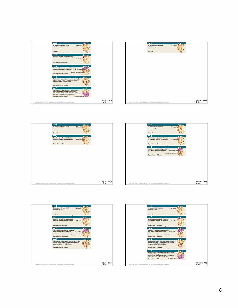
The Conducting System (cont’d) • Steps in the Conduction System:
1.Starts in ATRIA. Pacemaker cells establish heart rate
• pacemaker is also called sinoatrial (SA) node
2. Impulse spreads from SA node across atria
3. To atrioventricular (AV) node
4. To AV bundle and bundle branches
• Via Purkinje fibers to VENTRICLES Copyright © 2007 Pearson Education, Inc., publishing as Benjamin Cummings
The Heartbeat
Figure 12-9(a)
The Conducting System of the Heart
The Heart: Conduction System
PLAY
8
Figure 12-9(b) 1 of 6
SA node activity and atrial activation begin.
Stimulus spreads across the atrial surfaces and reaches the AV node.
There is a 100-msec delay at the AV node. Atrial contraction begins.
The impulse travels along the interventricular septum within the AV bundle and the bundle branches to the Purkinje fibers.
The impulse is distributed by Purkinje fibers and relayed throughout the ventricular myocardium. Atrial contraction is completed, and ventricular contraction begins.
Time = 0
SA node
AV node
Elapsed time = 50 msec
Elapsed time = 150 msec
AV bundle
Bundle branches
Elapsed time = 175 msec
Elapsed time = 225 msec Purkinje fibers
Copyright © 2007 Pearson Education, Inc., publishing as Benjamin Cummings Figure 12-9(b) 2 of 6
SA node activity and atrial activation begin.
Time = 0
SA node
Copyright © 2007 Pearson Education, Inc., publishing as Benjamin Cummings
Figure 12-9(b) 3 of 6
SA node activity and atrial activation begin.
Stimulus spreads across the atrial surfaces and reaches the AV node.
Time = 0
SA node
AV node
Elapsed time = 50 msec
Copyright © 2007 Pearson Education, Inc., publishing as Benjamin Cummings Figure 12-9(b) 4 of 6
SA node activity and atrial activation begin.
Stimulus spreads across the atrial surfaces and reaches the AV node.
There is a 100-msec delay at the AV node. Atrial contraction begins.
Time = 0
SA node
AV node
Elapsed time = 50 msec
Elapsed time = 150 msec
AV bundle
Bundle branches
Copyright © 2007 Pearson Education, Inc., publishing as Benjamin Cummings
Figure 12-9(b) 5 of 6
SA node activity and atrial activation begin.
Stimulus spreads across the atrial surfaces and reaches the AV node.
There is a 100-msec delay at the AV node. Atrial contraction begins.
The impulse travels along the interventricular septum within the AV bundle and the bundle branches to the Purkinje fibers.
Time = 0
SA node
AV node
Elapsed time = 50 msec
Elapsed time = 150 msec
AV bundle
Bundle branches
Elapsed time = 175 msec
Copyright © 2007 Pearson Education, Inc., publishing as Benjamin Cummings Figure 12-9(b) 6 of 6
SA node activity and atrial activation begin.
Stimulus spreads across the atrial surfaces and reaches the AV node.
There is a 100-msec delay at the AV node. Atrial contraction begins.
The impulse travels along the interventricular septum within the AV bundle and the bundle branches to the Purkinje fibers.
The impulse is distributed by Purkinje fibers and relayed throughout the ventricular myocardium. Atrial contraction is completed, and ventricular contraction begins.
Time = 0
SA node
AV node
Elapsed time = 50 msec
Elapsed time = 150 msec
AV bundle
Bundle branches
Elapsed time = 175 msec
Elapsed time = 225 msec Purkinje fibers
Copyright © 2007 Pearson Education, Inc., publishing as Benjamin Cummings
9
The Heartbeat
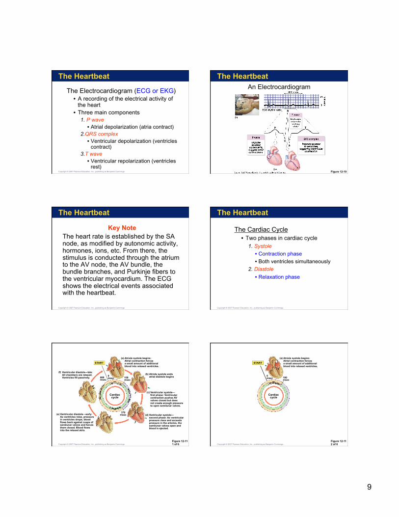
The Electrocardiogram (ECG or EKG) • A recording of the electrical activity of
the heart • Three main components
1. P wave • Atrial depolarization (atria contract)
2.QRS complex • Ventricular depolarization (ventricles
contract) 3.T wave • Ventricular repolarization (ventricles
rest) Copyright © 2007 Pearson Education, Inc., publishing as Benjamin Cummings
The Heartbeat
Figure 12-10
An Electrocardiogram
The Heartbeat
Key Note The heart rate is established by the SA node, as modified by autonomic activity, hormones, ions, etc. From there, the stimulus is conducted through the atrium to the AV node, the AV bundle, the bundle branches, and Purkinje fibers to the ventricular myocardium. The ECG shows the electrical events associated with the heartbeat.
Copyright © 2007 Pearson Education, Inc., publishing as Benjamin Cummings
The Heartbeat
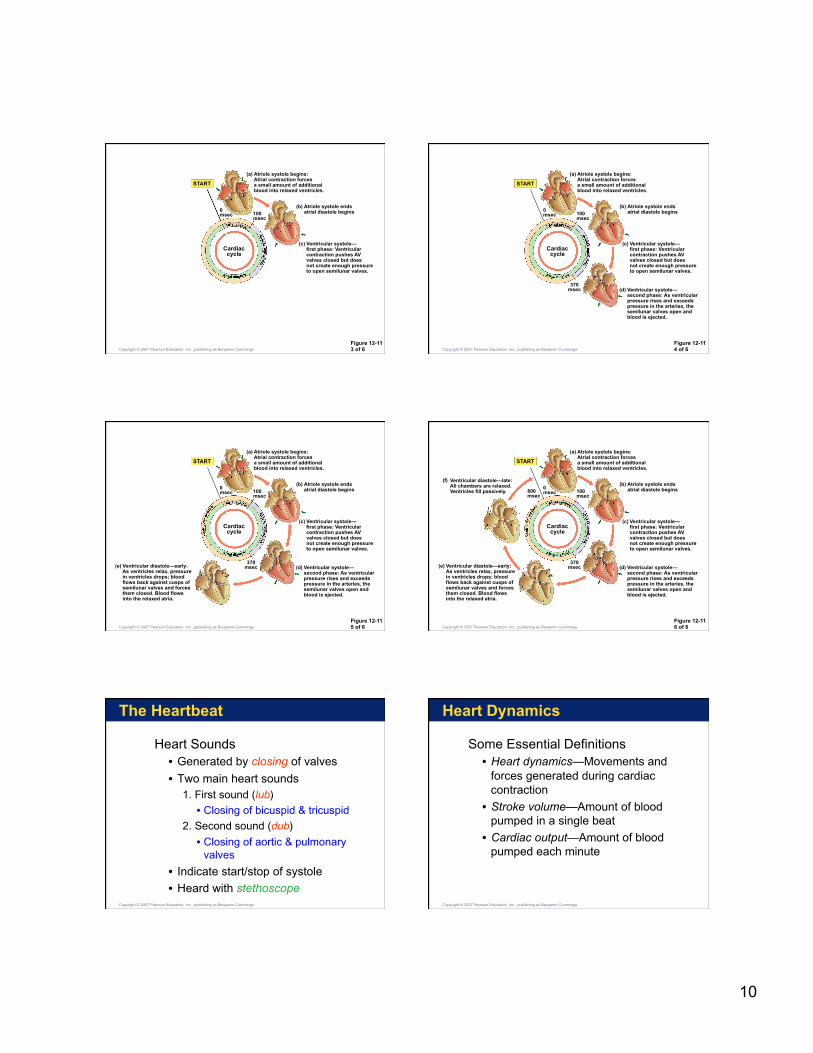
The Cardiac Cycle • Two phases in cardiac cycle
1. Systole
• Contraction phase
• Both ventricles simultaneously
2. Diastole
• Relaxation phase
Copyright © 2007 Pearson Education, Inc., publishing as Benjamin Cummings
Figure 12-11 1 of 6
START
(f) Ventricular diastole—late: All chambers are relaxed. Ventricles fill passively.
(e) Ventricular diastole—early: As ventricles relax, pressure in ventricles drops; blood flows back against cusps of semilunar valves and forces them closed. Blood flows into the relaxed atria.
(a) Atriole systole begins: Atrial contraction forces a small amount of additional blood into relaxed ventricles.
(b) Atriole systole ends atrial diastole begins
(c) Ventricular systole— first phase: Ventricular contraction pushes AV valves closed but does not create enough pressure to open semilunar valves.
(d) Ventricular systole— second phase: As ventricular pressure rises and exceeds pressure in the arteries, the semilunar valves open and blood is ejected.
370 msec
100 msec
0 msec 800
msec
Cardiac cycle
Copyright © 2007 Pearson Education, Inc., publishing as Benjamin Cummings Figure 12-11 2 of 6
START
(a) Atriole systole begins: Atrial contraction forces a small amount of additional blood into relaxed ventricles.
100 msec
0 msec
Cardiac cycle
Copyright © 2007 Pearson Education, Inc., publishing as Benjamin Cummings
10
Figure 12-11 3 of 6
START
(a) Atriole systole begins: Atrial contraction forces a small amount of additional blood into relaxed ventricles.
(b) Atriole systole ends atrial diastole begins
(c) Ventricular systole— first phase: Ventricular contraction pushes AV valves closed but does not create enough pressure to open semilunar valves.
100 msec
0 msec
Cardiac cycle
Copyright © 2007 Pearson Education, Inc., publishing as Benjamin Cummings Figure 12-11 4 of 6
START
(a) Atriole systole begins: Atrial contraction forces a small amount of additional blood into relaxed ventricles.
(b) Atriole systole ends atrial diastole begins
(c) Ventricular systole— first phase: Ventricular contraction pushes AV valves closed but does not create enough pressure to open semilunar valves.
(d) Ventricular systole— second phase: As ventricular pressure rises and exceeds pressure in the arteries, the semilunar valves open and blood is ejected.
370 msec
100 msec
0 msec
Cardiac cycle
Copyright © 2007 Pearson Education, Inc., publishing as Benjamin Cummings
Figure 12-11 5 of 6
START
(e) Ventricular diastole—early: As ventricles relax, pressure in ventricles drops; blood flows back against cusps of semilunar valves and forces them closed. Blood flows into the relaxed atria.
(a) Atriole systole begins: Atrial contraction forces a small amount of additional blood into relaxed ventricles.
(b) Atriole systole ends atrial diastole begins
(c) Ventricular systole— first phase: Ventricular contraction pushes AV valves closed but does not create enough pressure to open semilunar valves.
(d) Ventricular systole— second phase: As ventricular pressure rises and exceeds pressure in the arteries, the semilunar valves open and blood is ejected.
370 msec
100 msec
0 msec
Cardiac cycle
Copyright © 2007 Pearson Education, Inc., publishing as Benjamin Cummings Figure 12-11 6 of 6
START
(f) Ventricular diastole—late: All chambers are relaxed. Ventricles fill passively.
(e) Ventricular diastole—early: As ventricles relax, pressure in ventricles drops; blood flows back against cusps of semilunar valves and forces them closed. Blood flows into the relaxed atria.
(a) Atriole systole begins: Atrial contraction forces a small amount of additional blood into relaxed ventricles.
(b) Atriole systole ends atrial diastole begins
(c) Ventricular systole— first phase: Ventricular contraction pushes AV valves closed but does not create enough pressure to open semilunar valves.
(d) Ventricular systole— second phase: As ventricular pressure rises and exceeds pressure in the arteries, the semilunar valves open and blood is ejected.
370 msec
100 msec
0 msec 800
msec
Cardiac cycle
Copyright © 2007 Pearson Education, Inc., publishing as Benjamin Cummings
The Heartbeat
Heart Sounds • Generated by closing of valves
• Two main heart sounds 1. First sound (lub)
• Closing of bicuspid & tricuspid
2. Second sound (dub)
• Closing of aortic & pulmonary valves
• Indicate start/stop of systole
• Heard with stethoscope Copyright © 2007 Pearson Education, Inc., publishing as Benjamin Cummings
Heart Dynamics
Some Essential Definitions • Heart dynamics—Movements and
forces generated during cardiac contraction
• Stroke volume—Amount of blood pumped in a single beat
• Cardiac output—Amount of blood pumped each minute
Copyright © 2007 Pearson Education, Inc., publishing as Benjamin Cummings
11
Heart Dynamics
Factors Controlling Cardiac Output • Blood volume reflexes
• Autonomic innervation • Heart rate effects
• Stroke volume effects
• Hormones
Copyright © 2007 Pearson Education, Inc., publishing as Benjamin Cummings
Heart Dynamics
Blood Volume Reflexes • Stimulated by changes in venous return
• VR is amount of blood entering heart
• Atrial reflex • Speeds up heart rate
• Triggered by stretching wall of right atrium
• Frank-Starling principle • Increases ventricular output
• Triggered by stretching wall of ventricles
Copyright © 2007 Pearson Education, Inc., publishing as Benjamin Cummings
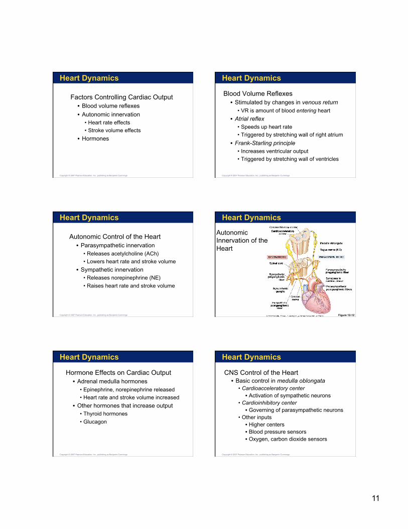
Heart Dynamics
Autonomic Control of the Heart • Parasympathetic innervation
• Releases acetylcholine (ACh)
• Lowers heart rate and stroke volume
• Sympathetic innervation • Releases norepinephrine (NE)
• Raises heart rate and stroke volume
Copyright © 2007 Pearson Education, Inc., publishing as Benjamin Cummings
Heart Dynamics
Autonomic Innervation of the Heart
Figure 12-12
Heart Dynamics
Hormone Effects on Cardiac Output • Adrenal medulla hormones
• Epinephrine, norepinephrine released
• Heart rate and stroke volume increased
• Other hormones that increase output • Thyroid hormones
• Glucagon
Copyright © 2007 Pearson Education, Inc., publishing as Benjamin Cummings
Heart Dynamics
CNS Control of the Heart • Basic control in medulla oblongata
• Cardioacceleratory center • Activation of sympathetic neurons
• Cardioinhibitory center • Governing of parasympathetic neurons
• Other inputs • Higher centers • Blood pressure sensors • Oxygen, carbon dioxide sensors
Copyright © 2007 Pearson Education, Inc., publishing as Benjamin Cummings
12
Heart Dynamics
Key Note
Cardiac output is the amount of blood pumped by the left ventricle each minute. It is adjusted moment-to-moment by the ANS, and by circulating hormones, changes in blood volume and in venous return. A healthy person can increase cardiac output by three-fold to five-fold.
Copyright © 2007 Pearson Education, Inc., publishing as Benjamin Cummings