Embed Size (px)
Citation preview
Available online at www.sciencedirect.com
Acta Biomaterialia 5 (2009) 1616–1625
www.elsevier.com/locate/actabiomat
Eugenol derivatives immobilized in auto-polymerizing formulationsas an approach to avoid inhibition interferences and improve
biofunctionality in dental and orthopedic cements
Luis Rojo a,*, Blanca Vazquez a, Sanjukta Deb b, Julio San Roman a
a Institute of Polymer Science and Technology, CSIC, and CIBER-BBN, C/Juan de la Cierva 3, 28006 Madrid, Spainb King’s College London Dental Institute at the Guy’s, King’s and St Thomas’ Hospitals, Department of Biomaterials, Biomimetics & Biophotonics,
Floor 17, Guy’s Tower, London Bridge, London SE1 9RT, UK
Received 1 October 2008; received in revised form 15 December 2008; accepted 16 January 2009Available online 31 January 2009
Abstract
Auto-polymerizing formulations based on poly(ethyl methacrylate) (PEMA) and eugenol derivatives are reported for dental andorthopedic applications. Spherical beads of PEMA were used as the pre-polymer powder and mixed with combinations of ethyl meth-acrylate and eugenyl methacrylate (EgMA) or ethoxyeugenyl methacrylate (EEgMA). A range of concentrations from 10 to 30 wt.% ofEgMA or EEgMA were used to impart bioactivity properties to the cements. Increasing concentrations of the eugenol derivativesdecreased the maximum polymerization temperatures from 69 to 37 �C without altering the working or setting time. At concentrationsof 10 and 15 wt.% of EgMA or EEgMA a noticeable increase in the compressive (8%), flexural (40%) and tensile (24%) strengths wererecorded in comparison to the control cements containing PEMA/ethyl methacrylate only. In addition to the improvement in mechanicalproperties the cements yielded a slightly crosslinked network due to the participation of the allylic group present in the eugenol deriv-atives, the presence of which has an intrinsically bactericidal effect against Escherichia coli and Streptococcus mutans strains, as reportedin a previous study, thus enhancing the properties of the cements.� 2009 Acta Materialia Inc. Published by Elsevier Ltd. All rights reserved.
Keywords: Poly(ethyl methacrylate); Auto-polymerizing formulations; Eugenol derivatives; Dental cements; Orthopedic cements
1. Introduction
Auto-polymerizing cements based on methacrylates arewidely used as synthetic biomaterials in dentistry [1], andcraniofacial and orthopedic surgery [2]. The most com-monly employed materials used clinically consist of poly(methyl methacrylate) (PMMA) microparticles (beads)with methyl methacrylate (MMA) as the polymerizablemonomer, which reacts via a redox reaction based on ben-zoyl peroxide (BPO) as initiator and a tertiary aromatic
1742-7061/$ - see front matter � 2009 Acta Materialia Inc. Published by Else
doi:10.1016/j.actbio.2009.01.029
* Corresponding author. Tel.: +34 915 622 900.E-mail addresses: [email protected], [email protected]
(L. Rojo).
amine (N,N-dimethyl-4-toluidine), as the activator underphysiological conditions [3,4].
This traditional composition has been employed overthe last 40 years without significant changes despite somedisadvantages derived from its components and way ofapplication, such as the high exotherm of the polymeriza-tion reaction which can evoke cellular necrosis in thesurrounding tissues or the prosthesis loosening in thelonger term. The toxicity of the aromatic tertiary amine(N,N-dimethyl-4-toluidine) [5] is of concern and otherchemically stable compounds such as phenolates and freeradicals produced during the curing reaction are likely toproduce adverse cytotoxic effects during several weeks postpolymerization, inducing a local inflammatory response inthe surrounding tissues [6–8].
vier Ltd. All rights reserved.
L. Rojo et al. / Acta Biomaterialia 5 (2009) 1616–1625 1617
Self-polymerizing systems based on poly(ethyl methac-rylate/n-butyl methacrylate) with better handling, lowermodulus and higher ductility in comparison to the conven-tional PMMA-based cements have been reported for use asorthopedic bone cements [9–11]. Other successful modifica-tions include the incorporation of crosslinking agents thathave resulted in improved mechanical properties due tothe formation of three-dimensional networks via crosslink-ing [12–15]. In general terms, it can be said that crosslink-ing points are able to restrain excessive movements,maintaining the properties of the network and enhancingits mechanical resistance without altering the handlingproperties or curing kinetics.
On the other hand, it is current practice to use zinc-oxide-eugenol auto-polymerizing cements in dentistry incombination with other widespread restorative materialsbased on dimethacrylates and other root canal sealers.One particular problem associated with the use of suchcombination is the incompatibility of both systems as aconsequence of the presence of free eugenol, which actsas a free radical scavenger inhibiting the cure of dentalcomposite resins [16]. However, the analgesic and anti-inflammatory activities [17], antimicrobial and anti-aggre-gating function [18], antipyretic activity [19], anti-anaphy-lactic properties preventing mast cell degranulation [20]and the capacity to prevent lipidic peroxidation [21] ofeugenol makes it an attractive component in the cementsfor both dental and orthopedic applications. Thus, com-bining the properties of the eugenol-based restorativematerials and auto-polymerizing acrylic resins in the samesystem is an interesting concept which has not beenexploited so far due to the inhibitory effects of the eugenolmolecule.
In previous works, our research group has synthesizedtwo eugenyl methacrylate derivatives, eugenyl methacrylate(EgMA) and ethoxyeugenyl methacrylate (EEgMA), whichwere successfully copolymerized with alkyl methacrylates[22,23]. Rheological analysis of these systems revealed apseudo-solid-like behaviour with relatively high values ofaverage molecular weights between crosslinks (�104 Da),confirming the proposed network structures of branchedsystems with a slightly crosslinked degree, in which a cer-tain amount of the eugenyl moieties remained pendantfrom the main chain [24]. It was also demonstrated by dif-ferent agar-test methods that these new materials possessedintrinsically bactericidal properties, showing clear halos ofinhibition, up to 7 mm, against different strains such asEscherichia coli and Streptococcus mutans, and conse-quently indicating activity against microorganisms com-monly found in both dental and osseous cavities [25]. Inaddition, the immobilization of eugenol is advantageousas it avoids the migration of this molecule to the surround-ing tissues and improves its hydrolytic stability.
Thus, the incorporation of eugenyl methacrylate deriva-tives (MEg) in auto-polymerizing formulations based onmethacrylates via copolymerization reaction yieldingslightly crosslinked polymer networks is an attractive alter-
native to impart biological properties and improvedmechanical resistance to the resultant cements withoutthe customary inhibitory effects of the inclusion of nativeeugenol. The objective of this study was to examine theeffect of EgMA and EEgMA in poly(ethyl methacrylate)/ethyl methacrylate-based self-curing cements. Furthermore4,40-bis(dimethylamino)diphenyl carbinol (BZN) wasemployed as an activator in the cement formulations dueto its established lower cytotoxicity [5].
2. Materials and methods
2.1. Materials
Eugenyl methacrylate (EgMA) and ethoxyeugenylmethacrylate (EEgMA) monomers were synthesized inour laboratories as reported previously [22]. Ethyl methac-rylate (EMA) and methyl methacrylate (MMA) stabilizedwith 100 ppm monomethylether of hydroquinone weresupplied by Acros (Spain) and used as received without fur-ther purification. Benzoyl peroxide (BPO) supplied byMerck (Germany) was used after fractional crystallizationfrom ethanol. 4,40-bis(dimethylamino)diphenyl carbinol(BZN) (Fluka, Spain), poly(vinyl alcohol) (PVA) powder(99%-hydrolysed, Mw = 90,000) (Aldrich, Spain) used assurfactant, poly(methyl methacrylate) (PMMA) beads(Degussa, Spain) and poly(ethyl methacrylate) (PEMA)beads (Aldrich, Spain), were used as received.
2.2. Synthesis of PEMA beads
PEMA beads were prepared using suspension polymer-ization [26]. An aqueous solution containing 3% (w/v) ofPVA was added as a suspension agent to a three neckedflask fitted with a nitrogen inlet, mechanical stirrer and acondenser, which was heated to 70 �C, stirred at 600 rpmand flushed with nitrogen for 10 min prior to the additionof 200 ml of EMA monomer in which the initiator, BPO(2% w/w with respect to monomer) was dissolved. Poly-merization was carried out at 70 �C over 3 h followed byanother 2 h at 80 �C to complete the reaction. The micro-particles obtained were filtered, thoroughly washed withwater and dried at 60 �C until constant weight. The chem-ical composition of microparticles was analysed by protonNMR spectroscopy. 1H NMR spectra were recorded at25 �C in CDCl3 using tetramethylsilane (TMS) as internalstandard on a Varian XLR-300 spectrometer. The mor-phology of the microparticles was analysed by scanningelectron microscopy (SEM) using a Philips XL 30 ESEMapparatus at an accelerating voltage of 15 keV. The sam-ples were sputter-coated with gold before examination.
The particle size distribution was determined by laserscattering using a Coulter LS320 (Beckman). Approxi-mately 10 mg of the sample was thoroughly dispersed in2 ml of water:ethanol 1:1 and an average of three readingswere recorded for each sample. Number and weight aver-age molecular weights were determined by size exclusion
1618 L. Rojo et al. / Acta Biomaterialia 5 (2009) 1616–1625
chromatography (SEC) (Water Alliance GPCV 2000 with arefractive index detector). Two columns of PL-gels 5 lmmixed bed were conditioned at 35 �C and used to elutethe samples of 1 mg ml–1 concentrations at 1 ml min–1
HPLC-grade chloroform flow rate. Calibration of SECwas carried out with monodisperse standard polystyrenesamples obtained from Polymer Laboratories.
2.3. Preparation of auto-polymerizing cements
PEMA beads as synthesized by the method describedabove were blended with 2 wt.% BPO and 8 wt.% of bar-ium sulphate as the radiopacifier. The powder was thenmixed with the liquid phase using a 2:1 ratio, containingmixtures of ethyl methacrylate and EgMA or EEgMA with2% by wt. of BZN as the activator. The details of the ratiosof the monomers used are elucidated in Table 1. Homolo-gous formulations were prepared based on PMMA/MMAfor mechanical properties studies. The mixture was stirredmanually at room temperature and when the reacting masswas a paste that could be easily handled, was introduced inappropriate Teflon moulds which were sealed and trans-ferred to an oven maintained at 37 �C for 1 h. No externalpressure was applied on the mould at any time during thecuring process.
2.4. Curing parameters
Three grams of the reacting mass (cement paste) wasintroduced in a cylindrical Teflon mould 10 mm in diame-ter and 15 mm high, equipped with a thermocouple con-nected to a high-sensitivity temperature recorder, andpositioned in the centre of the mould at a height of3.0 mm in the internal cavity. Further details on the goodcorrelation of this mould with that described in the ISO5833 [27] standard for acrylic resins have been reported ear-lier [28]. The measurements were done in triplicate accord-
Table 1Composition of the different acrylic formulations tested.
Formulationlabel
Liquid phase composition(% w/w)
Solid phase com(% w/w)
PEMA EMA 98%BZN 2%
PEMA/Eg10 EMA 88%BZN 2%
PEMA/EEg10 MEga 10%
PEMA 90%BaSO4 8%BPO 2%
PEMA/Eg15 EMA 83%BZN 2%
PEMA/EEg15 MEga 15%
PEMA/Eg30 EMA 68%BZN 2%
PEMA/EEg30 MEga 30%
a MEg = eugenyl derivative monomer (EgMA or EEgMA).
ing to ISO 5833 standards for acrylic resins by recordingthe polymerization temperature–time profiles at 23 �C.
2.5. Residual monomer content
The residual monomer content (RMC) was determinedby 1H NMR spectroscopy with a Varian XL300 spectrom-eter. Each type of the cements was dissolved in deuteratedchloroform (5% w/v) using tetramethylsilane as internalstandard and three samples were used for each group. Allthe specimens were kept for 7 days in air (25 �C and 60%RH), before the analysis. This criterion has been appliedaccording to the rules adopted with other international lab-oratories and assuming that the polymerization reactionreaches the maximum of conversion after 7 days [29].
2.6. Thermal properties of the cured cements
Glass transition temperatures (Tg) were measured bydifferential scanning calorimetry with a Perkin ElmerDSC7 interfaced to a thermal analysis data systemTAC7/DX. The dry samples were prepared in the formof thin films (15–20 mg) placed in aluminium pans andheated from 30 to 200 �C at a constant rate of 10 �C min–1.Tg was taken as the midpoint of heat capacity transition.Thermogravimetric diagrams were obtained in a thermo-gravimetric analyser using TGA Q500 (TA Instrument)apparatus, under dynamic nitrogen at a heating rate of5 �C min–1 in a range of 40–600 �C. Samples were testedin duplicate.
2.7. Mechanical properties
Tension, compression and bending tests were carried outat room temperature using an Instron universal testingmachine. A load cell of 5 kN and a crosshead speed of20 mm min–1 were used in the compression test according
position Sol:liq ratio Theoretical MEga content in thecement (% w/w)
0
3.3
2:1
5
10
L. Rojo et al. / Acta Biomaterialia 5 (2009) 1616–1625 1619
to the ISO 5833 [27] standard specification, whereas a loadcell of 0.5 kN and crosshead speed of 5 mm min–1 wereused in the tension test [30]. Three-point bending andfour-point bending tests were carried out according to stan-dards ISO 4049, for dental cements [31], and ISO 5833, foracrylic resins [27], respectively. The crosshead speeds forthe three and four-point bending tests were 0.75 and5 mm min–1, respectively. Specimens were tested after stor-age in air for 7 days and the corresponding strengths werecalculated according to the following equations:
rC ¼ 4F =pd2 ð1ÞrT ¼ F =bh ð2Þr3P ¼ 3Fl=2bh2 ð3Þr4P ¼ 3Fa=bh2 ð4Þ
where rC, rT, r3P and r4P are respectively the compressive,tensile, and bending strengths at three-point and four-pointbending tests, respectively; F is the maximum force appliedin Newtons; b, h, and d are the width, height and diameterof the specimen samples in millimetres, respectively; l is thedistance between the supports in millimetres and a is thedistance between the inner and the outer loading pointsin millimetres. Six specimens were used in each group.
Compression and four-point bending tests were performedon PEMA/EMA/Meg-based cements whereas tension andthree-point bending tests were done on homologous formula-tions based on PMMA/MMA/MEg. PMMA/MMA andPEMA/EMA-based cements were used in all the groups ascontrols. Six specimens were used in each group.
2.8. Fracture surface examination
Test specimens from four-point bending tests were col-lected just after the crack and stored in distilled water for24 h until their examination. The fracture surface analysisof the tested samples was sputter-coated with gold andexamined via scanning electron microscopy (SEM) usinga Philips XL 30 ESEM apparatus at an accelerating voltageof 20 keV.
2.9. Statistical analysis
Results of residual monomer and mechanical propertieswere analysed using one-way analysis of variance(ANOVA) at a level of significance p < 0.05. Values signif-icantly different with respect to the control formulations(PEMA/EMA or PMMA/MMA), are marked with anasterisk (*) in the corresponding results.
3. Results and discussion
3.1. Preparation of beads by suspension polymerization
PEMA beads were synthesized using the suspension freeradical polymerization technique in which droplets ofmonomer containing the free radical initiator were dis-
persed and stabilized in an aqueous medium containingpoly(vinyl alcohol) (PVA). The main function of PVAwas to control the viscosity of the medium, which in turnprovides the stabilization of the monomer droplets, thusfunctioning as a non-ionic surfactant agent which stabilizesthe droplets in the stirred medium, avoiding the agglomer-ation of particles during the polymerization reaction. Atthe end of the reaction the microparticles are gently washedwith water to remove all the components in the suspensionmedium. The monomer and PVA concentration, tempera-ture and stirring rate used in this study were based on aprevious study [26] as these parameters strongly influenceboth particle size and particle size distribution, which inturn affects the curing parameters of auto-polymerizingformulations [28]. The PEMA microparticles were obtain-ed with a yield around 60% and their 1H NMR spectrumshowed resonance signals characteristics of PEMA, at dH
4.0 (OCH2), 1.9 (CH2b rmr), 1.8 (CH2
b rrr), 1.45 (CH2b
mm), 1.25 (CH3), 1.05 (CH3a mr) and 0.88 (CH3
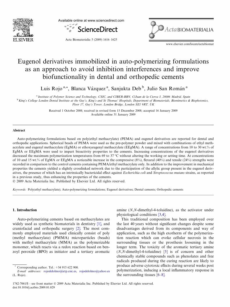
a rr).The examination of the microparticles by SEM (Fig. 1)showed a good homogenous and spherical morphologyand the absence of residuals on the microparticles surfaces.The particle size analysis revealed a unimodal distributionwith an average particle size diameter of 82 lm (Table 2).Number average molecular weight and the polidispersityvalues of the microparticles corresponded to a normal freeradical polymerization process and the PEMA beads pos-sessed average molecular weights high enough (see Table2) to be used in auto-polymerizing cements since theaccepted threshold for these materials correspond to105 Da [32]. Thus, the particle size distribution, morphol-ogy and molecular weights of the PEMA beads synthesizedwere comparable to if not superior than the commercialPEMA or PMMA beads obtained from Aldrich andDegussa respectively [33] (Fig. 1 and Table 2).
3.2. Auto-polymerization and curing parameters
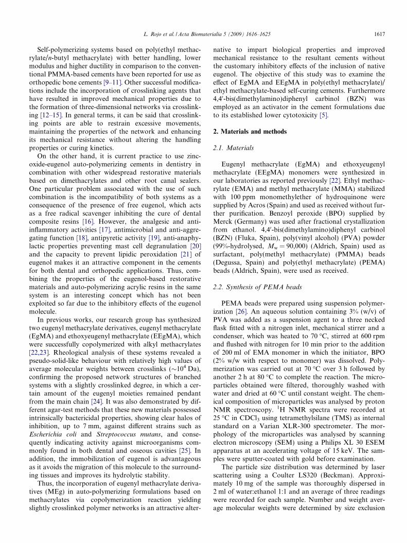
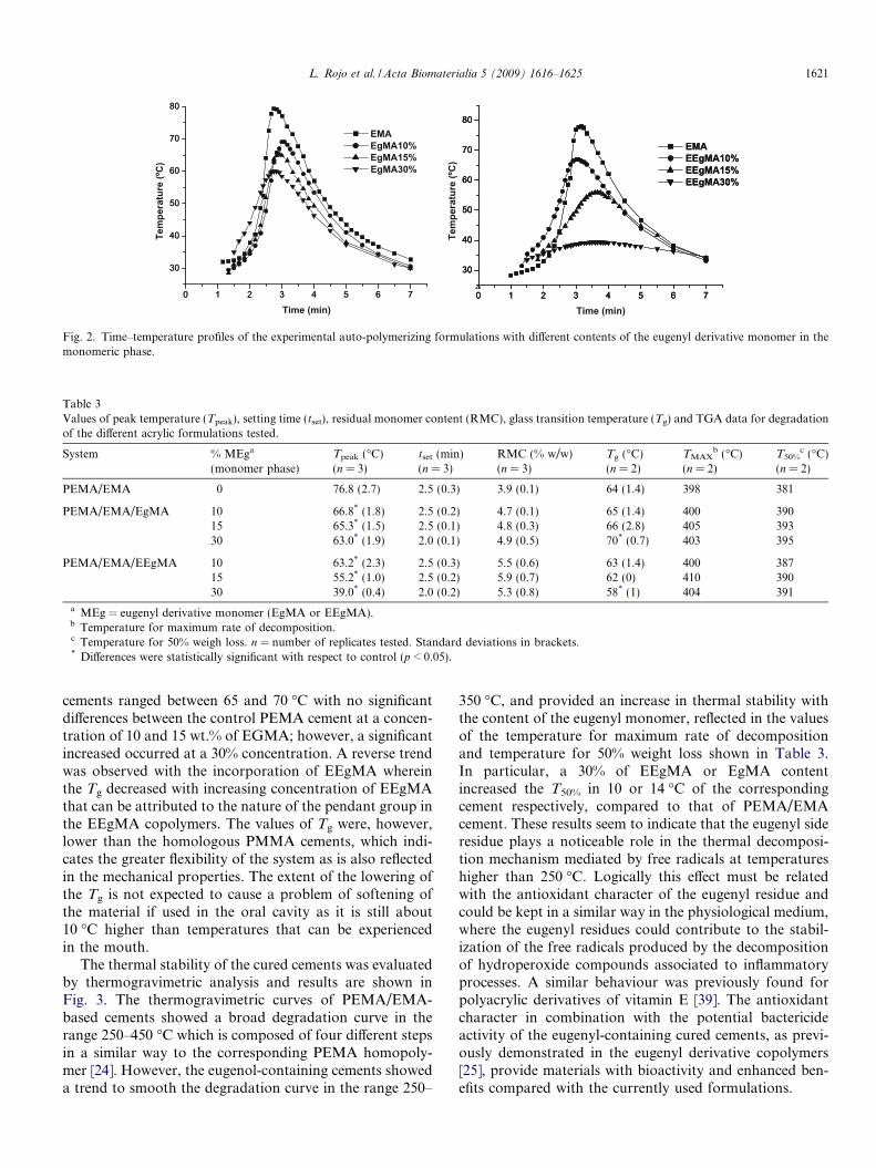
The novel cements were formulated with a redox initia-tor system, in which the traditional DMT was substitutedby the tertiary amine 4,40-bis(dimethylamino)diphenyl car-binol that has a lower toxicity, BZN. The results of previ-ous studies clearly indicated that BZN did not impair theefficiency of the polymerization reaction [5]. The bulk poly-merization of the monomers is mainly affected by diffusionat high conversions and as a consequence of the increase inthe polymerization rate providing the autoacceleration orTromsdorf–Norrish effect. The time–temperature curvesof the novel cements shown in Fig. 2 indicate a decreasein the peak temperature with increasing concentration ofEgMA or EEgMA being more pronounced in the case ofEEgMA as the comonomer. This may be attributed tothe higher molecular weight of these comonomers in theformulations, a result that is well documented in literature[34,35]. It should be noted that the formulations showedsetting times (tset) below 2.5 min which is acceptable fordental applications according to the standard UNE-EN

Fig. 1. Particle size distributions and ESEM photographs of synthesized PEMA microparticles (A), PEMA microparticles supplied by Aldrich (B) andPMMA microparticles supplied by Degussa (C).
Table 2Values of number average molecular weight (Mn), polydispersity index(PI) and average size of the different microparticles characterized in thiswork.
Material Mn (� 10–5 Da) PI Average size (lm)
PEMA (synthetic) 1.7 3.9 82PEMA (Aldrich) 3.4 2.6 46PMMA (Degussa) 0.7 2.5 40
1620 L. Rojo et al. / Acta Biomaterialia 5 (2009) 1616–1625
ISO 4049 [31], however, is too short for orthopedic applica-tions. Nevertheless, these cements can be further modifiedeither by using alternative activators of reduced toxicitysuch as those derived from naturally occurring long chainacids [36] or altering the particle size distribution of thePEMA pre-polymers. However, the salient observation isthe effect of both eugenyl derivative monomers on the set-ting process, which caused a significant lowering of thepeak temperature in comparison to the control PEMAcement. This reduction was more evident in the case ofEEgMA containing formulations which achieved peaktemperatures as low as 40 �C, that is, values very close tothe body temperature. A reduction in the polymerization
exotherm during the in situ curing of the cements withoutcompromising the mechanical properties is desirable as tis-sue necrosis is a well-documented problem and is expectedto minimize risk of thermal injury and thus enhance tissuecompatibility [37].
3.3. Residual monomer content in cured systems
Total residual monomer content (RMC) (considering allthe monomeric species) of all formulated cements waslower than 6% (see Table 3), indicating that the copolymer-ization reaction of ethyl methacrylate with an eugenylderivative monomer reached high conversions; however, aslight increase in RMC was obtained with respect to thatof control PEMA cements, but in each case, the valueswere within acceptable limits for use as commercial surgicalcements [29,38].
3.4. Thermal properties of the cured cements
The glass transition temperatures (Tg) of the cements areshown in Table 3. The Tg of the PEMA/EMA/EgMA
0 1 2 3 4 5 6 7
30
40
50
60
70
80
Tem
pera
ture
(ºC
)
Time (min)
EMAEEgMA10%EEgMA15%EEgMA30%
0 1 2 3 4 5 6 7
30
40
50
60
70
80
Tem
pera
ture
(ºC
)
Time (min)
EMAEgMA10%EgMA15% EgMA30%
0 1 2 3 4 5 6 7
30
40
50
60
70
80
EMAEEgMA10%EEgMA15%EEgMA30%
0 1 2 3 4 5 6 7
30
40
50
60
70
80
0 1 2 3 4 5 6 7
30
40
50
60
70
80
Fig. 2. Time–temperature profiles of the experimental auto-polymerizing formulations with different contents of the eugenyl derivative monomer in themonomeric phase.
Table 3Values of peak temperature (Tpeak), setting time (tset), residual monomer content (RMC), glass transition temperature (Tg) and TGA data for degradationof the different acrylic formulations tested.
System % MEga
(monomer phase)Tpeak (�C)(n = 3)
tset (min)(n = 3)
RMC (% w/w)(n = 3)
Tg (�C)(n = 2)
TMAXb (�C)
(n = 2)T50%
c (�C)(n = 2)
PEMA/EMA 0 76.8 (2.7) 2.5 (0.3) 3.9 (0.1) 64 (1.4) 398 381
PEMA/EMA/EgMA 10 66.8* (1.8) 2.5 (0.2) 4.7 (0.1) 65 (1.4) 400 39015 65.3* (1.5) 2.5 (0.1) 4.8 (0.3) 66 (2.8) 405 39330 63.0* (1.9) 2.0 (0.1) 4.9 (0.5) 70* (0.7) 403 395
PEMA/EMA/EEgMA 10 63.2* (2.3) 2.5 (0.3) 5.5 (0.6) 63 (1.4) 400 38715 55.2* (1.0) 2.5 (0.2) 5.9 (0.7) 62 (0) 410 39030 39.0* (0.4) 2.0 (0.2) 5.3 (0.8) 58* (1) 404 391
a MEg = eugenyl derivative monomer (EgMA or EEgMA).b Temperature for maximum rate of decomposition.c Temperature for 50% weigh loss. n = number of replicates tested. Standard deviations in brackets.* Differences were statistically significant with respect to control (p < 0.05).
L. Rojo et al. / Acta Biomaterialia 5 (2009) 1616–1625 1621
cements ranged between 65 and 70 �C with no significantdifferences between the control PEMA cement at a concen-tration of 10 and 15 wt.% of EGMA; however, a significantincreased occurred at a 30% concentration. A reverse trendwas observed with the incorporation of EEgMA whereinthe Tg decreased with increasing concentration of EEgMAthat can be attributed to the nature of the pendant group inthe EEgMA copolymers. The values of Tg were, however,lower than the homologous PMMA cements, which indi-cates the greater flexibility of the system as is also reflectedin the mechanical properties. The extent of the lowering ofthe Tg is not expected to cause a problem of softening ofthe material if used in the oral cavity as it is still about10 �C higher than temperatures that can be experiencedin the mouth.
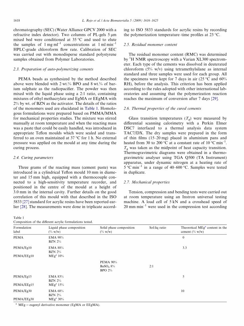
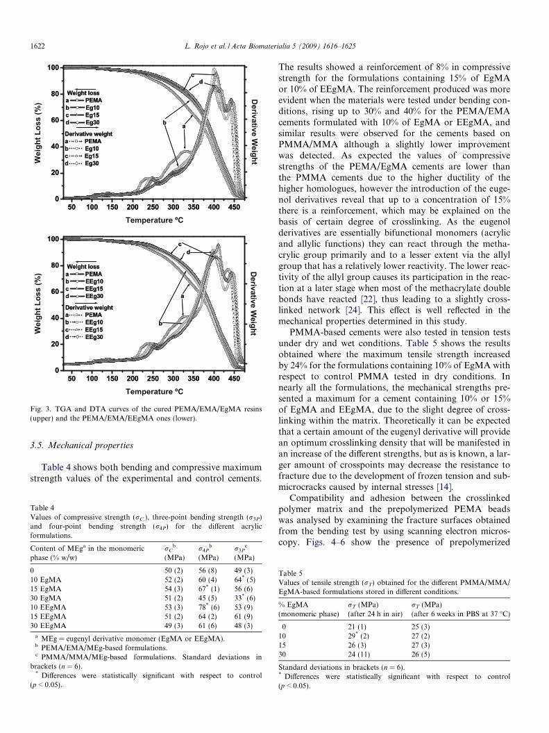
The thermal stability of the cured cements was evaluatedby thermogravimetric analysis and results are shown inFig. 3. The thermogravimetric curves of PEMA/EMA-based cements showed a broad degradation curve in therange 250–450 �C which is composed of four different stepsin a similar way to the corresponding PEMA homopoly-mer [24]. However, the eugenol-containing cements showeda trend to smooth the degradation curve in the range 250–
350 �C, and provided an increase in thermal stability withthe content of the eugenyl monomer, reflected in the valuesof the temperature for maximum rate of decompositionand temperature for 50% weight loss shown in Table 3.In particular, a 30% of EEgMA or EgMA contentincreased the T50% in 10 or 14 �C of the correspondingcement respectively, compared to that of PEMA/EMAcement. These results seem to indicate that the eugenyl sideresidue plays a noticeable role in the thermal decomposi-tion mechanism mediated by free radicals at temperatureshigher than 250 �C. Logically this effect must be relatedwith the antioxidant character of the eugenyl residue andcould be kept in a similar way in the physiological medium,where the eugenyl residues could contribute to the stabil-ization of the free radicals produced by the decompositionof hydroperoxide compounds associated to inflammatoryprocesses. A similar behaviour was previously found forpolyacrylic derivatives of vitamin E [39]. The antioxidantcharacter in combination with the potential bactericideactivity of the eugenyl-containing cured cements, as previ-ously demonstrated in the eugenyl derivative copolymers[25], provide materials with bioactivity and enhanced ben-efits compared with the currently used formulations.
50 100 150 200 250 300 350 400 4500
20
40
60
80
100d
c
b
a
Derivative weighta PEMAb EEg10c EEg15d EEg30
Weight lossa PEMAb EEg10c EEg15d EEg30
Temperature ºC
Wei
ght L
oss
(%) Derivative W
eight
50 100 150 200 250 300 350 400 4500
20
40
60
80
100
dc
b aDerivative weighta PEMAb Eg10c Eg15d Eg30
Weight lossa PEMAb Eg10c Eg15d Eg30
Temperature ºC
Wei
ght L
oss
(%)
Derivative W
eight
50 100 150 200 250 300 350 400 4500
20
40
60
80
100d
c
b
a
Derivative weighta PEMAb EEg10c EEg15d EEg30
Weight lossa PEMAb EEg10c EEg15d EEg30
50 100 150 200 250 300 350 400 4500
20
40
60
80
100
dc
b aDerivative weighta PEMAb Eg10c Eg15d Eg30
Weight lossa PEMAb Eg10c Eg15d Eg30
Fig. 3. TGA and DTA curves of the cured PEMA/EMA/EgMA resins(upper) and the PEMA/EMA/EEgMA ones (lower).
1622 L. Rojo et al. / Acta Biomaterialia 5 (2009) 1616–1625
3.5. Mechanical properties
Table 4 shows both bending and compressive maximumstrength values of the experimental and control cements.
Table 4Values of compressive strength (rC,), three-point bending strength (r3P)and four-point bending strength (r4P) for the different acrylicformulations.
Content of MEga in the monomericphase (% w/w)
rCb
(MPa)r4P
b
(MPa)r3P
c
(MPa)
0 50 (2) 56 (8) 49 (3)10 EgMA 52 (2) 60 (4) 64* (5)15 EgMA 54 (3) 67* (1) 56 (6)30 EgMA 51 (2) 45 (5) 33* (6)10 EEgMA 53 (3) 78* (6) 53 (9)15 EEgMA 51 (2) 64 (2) 61 (9)30 EEgMA 49 (3) 61 (6) 48 (3)
a MEg = eugenyl derivative monomer (EgMA or EEgMA).b PEMA/EMA/MEg-based formulations.c PMMA/MMA/MEg-based formulations. Standard deviations in
brackets (n = 6).* Differences were statistically significant with respect to control
(p < 0.05).
The results showed a reinforcement of 8% in compressivestrength for the formulations containing 15% of EgMAor 10% of EEgMA. The reinforcement produced was moreevident when the materials were tested under bending con-ditions, rising up to 30% and 40% for the PEMA/EMAcements formulated with 10% of EgMA or EEgMA, andsimilar results were observed for the cements based onPMMA/MMA although a slightly lower improvementwas detected. As expected the values of compressivestrengths of the PEMA/EgMA cements are lower thanthe PMMA cements due to the higher ductility of thehigher homologues, however the introduction of the euge-nol derivatives reveal that up to a concentration of 15%there is a reinforcement, which may be explained on thebasis of certain degree of crosslinking. As the eugenolderivatives are essentially bifunctional monomers (acrylicand allylic functions) they can react through the metha-crylic group primarily and to a lesser extent via the allylgroup that has a relatively lower reactivity. The lower reac-tivity of the allyl group causes its participation in the reac-tion at a later stage when most of the methacrylate doublebonds have reacted [22], thus leading to a slightly cross-linked network [24]. This effect is well reflected in themechanical properties determined in this study.
PMMA-based cements were also tested in tension testsunder dry and wet conditions. Table 5 shows the resultsobtained where the maximum tensile strength increasedby 24% for the formulations containing 10% of EgMA withrespect to control PMMA tested in dry conditions. Innearly all the formulations, the mechanical strengths pre-sented a maximum for a cement containing 10% or 15%of EgMA and EEgMA, due to the slight degree of cross-linking within the matrix. Theoretically it can be expectedthat a certain amount of the eugenyl derivative will providean optimum crosslinking density that will be manifested inan increase of the different strengths, but as is known, a lar-ger amount of crosspoints may decrease the resistance tofracture due to the development of frozen tension and sub-microcracks caused by internal stresses [14].
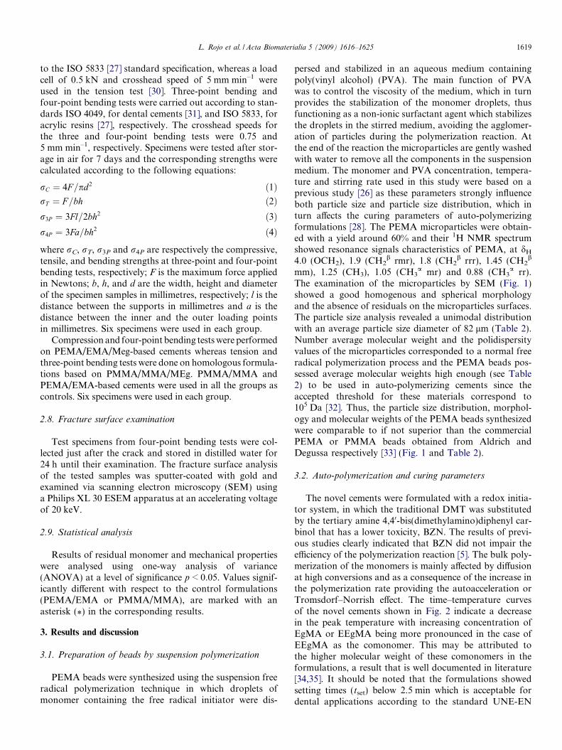
Compatibility and adhesion between the crosslinkedpolymer matrix and the prepolymerized PEMA beadswas analysed by examining the fracture surfaces obtainedfrom the bending test by using scanning electron micros-copy. Figs. 4–6 show the presence of prepolymerized
Table 5Values of tensile strength (rT) obtained for the different PMMA/MMA/EgMA-based formulations stored in different conditions.
% EgMA(monomeric phase)
rT (MPa)(after 24 h in air)
rT (MPa)(after 6 weeks in PBS at 37 �C)
0 21 (1) 25 (3)10 29* (2) 27 (2)15 26 (3) 27 (3)30 24 (11) 26 (5)
Standard deviations in brackets (n = 6).* Differences were statistically significant with respect to control(p < 0.05).
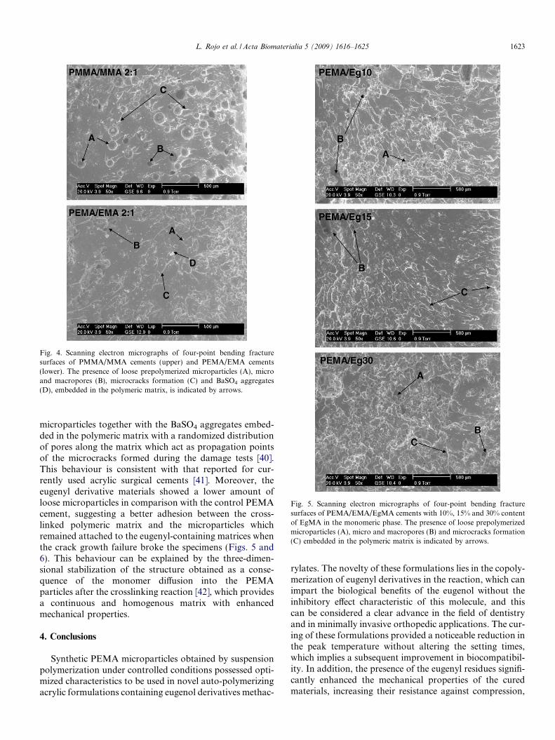
Fig. 4. Scanning electron micrographs of four-point bending fracturesurfaces of PMMA/MMA cements (upper) and PEMA/EMA cements(lower). The presence of loose prepolymerized microparticles (A), microand macropores (B), microcracks formation (C) and BaSO4 aggregates(D), embedded in the polymeric matrix, is indicated by arrows.
Fig. 5. Scanning electron micrographs of four-point bending fracturesurfaces of PEMA/EMA/EgMA cements with 10%, 15% and 30% contentof EgMA in the monomeric phase. The presence of loose prepolymerizedmicroparticles (A), micro and macropores (B) and microcracks formation(C) embedded in the polymeric matrix is indicated by arrows.
L. Rojo et al. / Acta Biomaterialia 5 (2009) 1616–1625 1623
microparticles together with the BaSO4 aggregates embed-ded in the polymeric matrix with a randomized distributionof pores along the matrix which act as propagation pointsof the microcracks formed during the damage tests [40].This behaviour is consistent with that reported for cur-rently used acrylic surgical cements [41]. Moreover, theeugenyl derivative materials showed a lower amount ofloose microparticles in comparison with the control PEMAcement, suggesting a better adhesion between the cross-linked polymeric matrix and the microparticles whichremained attached to the eugenyl-containing matrices whenthe crack growth failure broke the specimens (Figs. 5 and6). This behaviour can be explained by the three-dimen-sional stabilization of the structure obtained as a conse-quence of the monomer diffusion into the PEMAparticles after the crosslinking reaction [42], which providesa continuous and homogenous matrix with enhancedmechanical properties.
4. Conclusions
Synthetic PEMA microparticles obtained by suspensionpolymerization under controlled conditions possessed opti-mized characteristics to be used in novel auto-polymerizingacrylic formulations containing eugenol derivatives methac-
rylates. The novelty of these formulations lies in the copoly-merization of eugenyl derivatives in the reaction, which canimpart the biological benefits of the eugenol without theinhibitory effect characteristic of this molecule, and thiscan be considered a clear advance in the field of dentistryand in minimally invasive orthopedic applications. The cur-ing of these formulations provided a noticeable reduction inthe peak temperature without altering the setting times,which implies a subsequent improvement in biocompatibil-ity. In addition, the presence of the eugenyl residues signifi-cantly enhanced the mechanical properties of the curedmaterials, increasing their resistance against compression,
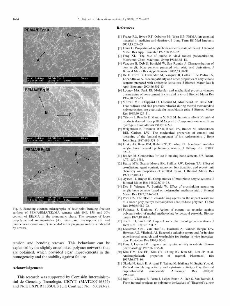
Fig. 6. Scanning electron micrographs of four-point bending fracturesurfaces of PEMA/EMA/EEgMA cements with 10%, 15% and 30%content of EEgMA in the monomeric phase. The presence of looseprepolymerized microparticles (A), micro and macropores (B) andmicrocracks formation (C) embedded in the polymeric matrix is indicatedby arrows.
1624 L. Rojo et al. / Acta Biomaterialia 5 (2009) 1616–1625
tension and bending stresses. This behaviour can beexplained by the slightly crosslinked polymer networks thatare obtained, which provided clear improvements in thehomogeneity and the stability against failure.
Acknowledgements
This research was supported by Comision Interministe-rial de Ciencia y Tecnologıa, CICYT, (MAT2007-63355)and NoE EXPERTISSUES (UE Contract No.: 500283-2).
References
[1] Frazer RQ, Byron RT, Osborne PB, West KP. PMMA: an essentialmaterial in medicine and dentistry. J Long Term Eff Med Implants2005;15:629–39.
[2] Lewis G. Properties of acrylic bone cements: state of the art. J BiomedMater Res Appl Biomater 1997;38:155–82.
[3] Feng XD. The role of amine in vinyl radical polymerisation.Macromol Chem Macromol Symp 1992;63:1–18.
[4] Vazquez B, Deb S, Bonfield W, San Roman J. Characterization ofnew acrylic bone cements prepared with oleic acid derivatives. JBiomed Mater Res Appl Biomater 2002;63:88–97.
[5] De la Torre B, Fernandez M, Vazquez B, Collıa F, de Pedro JA,Lopez-Bravo A. Biocompatibility and other properties of acrylic bonecements prepared with antiseptic activators. J Biomed Mater Res BAppl Biomater 2003;66:502–13.
[6] Looney MA, Park JB. Molecular and mechanical property changesduring aging of bone cement in vitro and in vivo. J Biomed Mater Res1986;20:555–63.
[7] Moreau MF, Chappard D, Lesourd M, Montheard JP, Basle MF.Free radicals and side products released during methyl methacrylatepolymerization are cytotoxic for osteoblastic cells. J Biomed MaterRes 1998;40:124–31.
[8] Cifkova I, Brynda E, Mandys V, Stol M. Irritation effects of residualproducts derived from p(HEMA) gels II. Compounds extracted fromhydrogels. Biomaterials 1988;9:372–5.
[9] Weightman B, Freeman MAR, Revell PA, Braden M, AlbrektssonBEJ, Carlson LVJ. The mechanical properties of cement andloosening of the femoral component of hip replacements. J BoneJoint Surg 1987;69B:558–64.
[10] Litsky AS, Rose RM, Rubin CT, Thrasher EL. A reduced modulusacrylic bone cement: preliminary results. J Orthop Res 1990;8:623–6.
[11] Braden M. Composites for use in making bone cements. US Patent.4,791,150. 1986.
[12] Beatty MW, Swartz Moore BK, Phillips RW, Roberts TA. Effect ofcrosslinking agent content, monomer functionality, and repeat unitchemistry on properties of unfilled resins. J Biomed Mater Res1993;27:403–13.
[13] Oysaed H, Ruyter IE. Creep studies of multiphase acrylic systems. JBiomed Mater Res 1989;23:719–33.
[14] Deb S, Vazquez V, Bonfield W. Effect of crosslinking agents onacrylic bone cements based on poly(methyl methacrylate). J BiomedMater Res 1997;37:465–73.
[15] Price CA. The effect of cross-linking agents on the impact resistanceof a linear poly(methyl methacrylate) denture-base polymer. J DentRes 1986;65:987–92.
[16] Fujisawa S, Kadoma Y. Action of eugenol as retarder againstpolymerisation of methyl methacrylate by benzoyl peroxide. Bioma-terials 1997;18:701–3.
[17] Sticht FD, Smith PM. Eugenol: some pharmacologic observations. JDent Res 1971;50:1531–5.
[18] Laekeman GM, Van Hoof L, Haemers A, Vanden Berghe DA,Herman AG, Vlietinck AJ. Eugenol a valuable compound for in vitroexperimental research and worthwhile for further in vivo investiga-tion. Phytother Res 1990;4:90–6.
[19] Feng J, Lipton JM. Eugenol: antipyretic activity in rabbits. Neuro-pharmacology 1987;26:1775–8.
[20] Kim HM, Lee EH, Kim CY, Chung JG, Kim SH, Lim JP, et al.Antianaphylactic properties of eugenol. Pharmacol Res1997;36:475–80.
[21] Okada N, Satoh K, Atsumi T, Tajima M, Ishihara M, Sugita Y, et al.Radical modulating activity and cytotoxic activity of synthesizedeugenol-related compounds. Anticancer Res 2000;20:2955–60.
[22] Rojo L, Vazquez B, Parra J, Lopez-Bravo A, Deb S, San Roman J.From natural products to polymeric derivatives of ‘‘Eugenol”: a new
L. Rojo et al. / Acta Biomaterialia 5 (2009) 1616–1625 1625
approach for preparation of dental composites and orthopedic bonecements. Biomacromolecules 2006;7:2751–61.
[23] Rojo L, Vazquez B, San Roman J, Deb S. Eugenol functionalizedpoly(acrylic acid) derivatives in the formation of glass-ionomercements. Dent Mater 2008;24:1709–16.
[24] Rojo L, Borzacchiello A, Parra J, Deb S, Vazquez B, San Roman J.The preparation of high conversion polymeric systems containingeugenol residues and their rheological characterization. J Mater SciMater Med 2008;19:1467–77.
[25] Rojo L, Barcenilla JM, Vazquez B, Gonzalez R, San Roman J.Intrinsically antibactericial materials based on polymeric derivatives ofeugenol for biomedical applications. Biomacromolecules 2008;9:2530–5.
[26] Abraham GA, Gallardo A, Motta A, Migliaresi C, San Roman J.Microheterogeneous polymer systems prepared by suspension poly-merization of methyl methacrylate in the presence of poly(e-capro-lactone). Macromol Mater Eng 2000;282:44–50.
[27] ISO Specification 5833. Standard specification for implants insurgery-acrylic resin cement; 2002.
[28] Pascual B, Vazquez B, Gurruchaga M, Go~ni I, Ginebra MP, Gil FJ,et al. New aspects of the effect of size and size distribution on thesetting parameters and mechanical properties of acrylic bone cements.Biomaterials 1996;17:509–16.
[29] Kuhn KD. Bone cements. Up-to-date comparison of physical andchemical properties of commercial materials. Berlin: Springer; 2000.
[30] International Standard ISO 527-1. Plastics-determination of tensileproperties; 1993.
[31] ISO Specification 4049. Standard specification for polymer basedfilling, restorative and luting materials; 2000.
[32] Kusy RP. Characterization of self-curing acrylic bone cements. JBiomed Mater Res 1978;12:271–305.
[33] Lautenschlager EP, Stupp SI, Keller JC. Structure and properties ofacrylic bone cements. In: Ducheyne P, Hastings G, editors. Func-
tional behaviour of orthopaedic biomaterials. Boca Raton, Flor-ida: CRC Press; 1987.
[34] Brauer GM, Steinberger DR, Stansbury JW. Dependence of curingtime, peak temperature and mechanical properties on the compositionof bone cement. J Biomed Mater Res 1986;20:839–52.
[35] Pascual B, Gurruchaga M, Goni I, Ginebra MP, Gil FJ, Planell JA,et al. Mechanical properties of a modified acrylic bone cement withethoxytriethyleneglycol monomethacrylate. J Mater Sci Mater Med1995;6:793–8.
[36] Vazquez B, San Roman J, Deb S, Bonfield W. Application of longchain amine activator in conventional acrylic bone cement. J BiomedMater Res Appl Biomater 1998;43:131–9.
[37] Jensen LN, Sturup J, Kramhøft M, Jense JS. Histological evaluationof cortical bone reaction to PMMA cementation. Acta Orthop Belg1991;57:254–9.
[38] Schoenfeld CM, Conard GJ, Lautenschlager EP. Monomer releasefrom methacrylate bone cements during simulated ‘‘in vivo” poly-merization. J Biomed Mater Res 1979;13:135–47.
[39] Ortiz C, Vazquez B, San Roman J. Hydrophilic acrylic biomaterialsderived from vitamin E with antioxidant properties. J Biomed MaterRes 1999;45:184–91.
[40] Topoleski LDT, Ducheyne P, Cuckler JM. Microstructural pathwayof fracture in poly(methyl methacrylate) bone cement. Biomaterials1993;14:1165–72.
[41] Sinnett-jones PE, Browne M, Ludwig W, Buffiere JY, Sinclair I.Microtomography assessment of failure in acrylic bone cements.Biomaterials 2005;26:6460–6.
[42] Shafranska O, Kokott A, Sulthaus D, Ziegler G. Effect of surfacemodification of polymer beads on the mechanical properties ofacrylic bone cements. J Biomater Sci Polymer Ed 2007;18:439–51.