Embed Size (px)
Citation preview

EVALUATION OF VARIOUS
PHARMACOLOGICAL PROPERTIES OF
THREE INDIAN MEDICINAL PLANTS
A THESIS
Submitted by
VINOTHAPOOSHAN G
In partial fulfillment for the award of the degree
Of
DOCTOR OF PHILOSOPHY
DEPARTMENT OF BIOTECHNOLOGY
KALASALINGAM UNIVERSITY
(Kalasalingam Academy of Research And Education)
ANAND NAGAR, KRISHNANKOIL - 626 126
OCTOBER 2013

ii
KALASALINGAM UNIVERSITY
KRISHNANKOIL - 626 126
BONAFIDE CERTIFICATE
Certified that this thesis titled “EVALUATION OF VARIOUS
PHARMACOLOGICAL PROPERTIES OF THREE INDIAN
MEDICINAL PLANTS” is the bonafide work of Mr. G. Vinothapooshan,
who carried out the research under my supervision. Certified further, that to
the best of my knowledge the work reported herein does not form part of any
other thesis or dissertation on the basis of which a degree or award was
conferred on an earlier occasion on this or any other scholar.
Place: Krishnankoil Dr. K. Sundar
Date: 18.10.13 SUPERVISOR

iii
ABSTRACT
Natural products isolated from higher plants and microorganisms have been a
source of novel and clinically active drugs. The success of discovering naturally
occurring therapeutic agents rests on bioassay-guided fractionation and
purification procedures. In the present study, immature plant leaves of Mimosa
pudica, Artabotrys hexapetalus and Adhatoda vasica were collected from
Courtallum and Thaniparai Hills in the state of Tamilnadu, India during early
winter season. The leaves of the above plants were shade-dried and made into
coarse powder which was passed through a 40-mesh sieve to get a uniform
particle size and then used for extraction. 500 g of the powder was subjected to
continuous hot extraction in Soxhlet apparatus with methanol, chloroform and
diethyl ether and the residual marc was collected.
The extracts were subjected to qualitative tests for identifying various plant
constituents. The acute toxicity study was performed for various extracts of M.
pudica, A. hexapetalus and A. Vasica according to the acute toxic classic method
as per Organization for Economic Co-operation and Development (OECD)
guidelines. Then the extracts were subjected to various pharmacological activities
such as immunomodulatory, hepatoprotective, anti-ulcer and wound healing
activities. The extracts were also subjected to antimicrobial and antioxidant
studies. The extracts were also separated using Thin Layer Chromatography

iv
(TLC) and High performance liquid chromatography (HPLC); the purified
compounds were analyzed by Fourier transform infrared spectroscopy (FTIR).
The extracts of M. pudica, A. hexapetalus and A. vasica administered orally on
albino rats showed significant increase in adhesion of neutrophils to nylon fibers
which correlates to the process of margination of cells in blood vessels. The
neutrophil adhesion was found to increase significantly with increasing in
concentration. The methanolic extracts of all the three plants exhibited a strong
immmunomodulatory activity, when compared to other extracts and untreated
control. The diethyl ether extracts of all three plants did not exhibit any influence
in modulating the immune response, with values being 43.29%, 34.95% and
63.31% for M. pudica, A. hexapetalus and A. vasica, respectively.
The delayed type hypersensitivity reaction has been widely used as one of the
parameters to measure cell-mediated immune response in a rat model. The
reaction was measured by the percent increase in the paw volume over the
control. A significant increase in paw volume was observed with diethyl ether
extracts of all the three plants, the values being 23.8%, 24.2% and 20.1%
respectively. These results were comparable to that of the positive control
(30.44%). The increase in DTH reaction in rats suggests the stimulatory effect of

v
the antigen on T-lymphocytes and accessory cell types required for the
expression of the reaction.
The hepatoprotective ability of the plant extracts was assessed by their ability to
protect the liver from carbon tetrachloride (CCl4) injury. Four marker enzymes
SGPT, SGOT, ALP and TBL were used for assessing hepatoprotective ability.
All three extracts of all the three test plants were found to be protective against
CCl4 injury. The animals were found to be markedly recovering from CCl4 effect
as noted from the activity of the enzymes. The enzyme activity was found to
decrease about one-half the injured liver and was almost equivalent to control.
The histopathological examination of liver sections of the control group showed
normal cellular architecture with distinct hepatic cells, sinusoidal spaces, and
central vein. The disarrangement of normal hepatic cells with necrosis and
vacuolization were observed in carbon tetrachloride intoxicated liver. The liver
sections of the albino rat treated with 200mg/kg body weight p.o. of methanolic,
chloroform and diethyl ether extracts of the selected plant, followed by carbon
tetrachloride intoxication, showed less vacuole formation and absence of
necrosis. The overall less visible changes observed were comparable with the
standard silymarin, supplementing the protective effect of ether extract of
selected plant and the standard hepatoprotective drug.

vi
All the three plant extracts exhibited anti-ulcer activity in all three models tested
(aspirin induced, alcohol induced and pylorus ligation), when treated with two
different concentrations viz. 100 mg/kg and 200 mg/kg, the methanolic extracts
of the plants exhibited a stronger anti-ulcer activity than other organic solvents.
The ulcer index was considerably reduced in albino rats treated with methanolic
extracts when compared to other solvents. The reduction in ulcer index was
found to be between 60-75% treated with methanol extracts of all the three
plants. The reduction in ulcer index was statistically significant and comparable
to that of the standard drug Ranitidine (20 mg/kg).
All three plant extracts exhibited significant wound healing activity, the activity
of different extracts varied from 65.46% to 93.87%. A lower wound healing
activity was exhibited by the diethyl ether extract whereas the activity was found
to be higher with methanolic extracts. The methanolic extract of M. pudica
exhibited a higher activity of 93.87% whereas the methanolic extracts of A.
hexapetalus and A. vasica exhibited 78.61% and 87.46% respectively.
The increasing in failure of existing chemotherapeutic agents and the rise in
antibiotic resistance of pathogenic microorganisms led to the search for newer
anti-microbial agents particularly from the plant kingdom. In the present study,
all the three extracts were found to exhibit a very strong and broad spectrum

vii
antibacterial activity. The effect was found to be pronounced against gram-
positive bacteria (Micrococcus luteus, Staphylococcus aureus and Bacilluccerus)
than against Gram-negative bacteria (Klebsiellapneumonieae, Salmonella
typhimurium and Salmonella paratyphimurium). Antimicrobial activity of the
plant extracts was analyzed by Gram- positive and Gram- negative organisms by
the well-diffusion assay using ciprofloxacin as standard.
As many plant metabolites exhibit potential anti-oxidant activity, the metabolites
of the plants under study were also assessed for anti-oxidant activity using DPPH
assay, FRAP assay and reducing power measurement methods. The TLC-
purified methanolic extracts of all the three plants exhibited potent anti-oxidant
activity as measured by potassium ferricyanide, FRAP and DPPH assays. The
activities of all three plants were comparable in all these assays.
In conclusion, the methanolic extracts exhibited remarkable ulcer-protective
properties when compared to other two extracts. Similarly methanolic extracts of
all the three extracts exhibited higher antimicrobial activity against Gram-
positive and Gram-negative pathogens and the methanolic extracts of all the three
plants showed better anti-oxidant activity. The methanolic extracts of M. pudica
was found to exhibit better wound healing activity compared to other extracts. In
contrast to this the ether extracts of selected plants (200 mg/kg) were found to

viii
possess significant hepatoprotective activity against carbon tetrachloride induced
hepatotoxification. Interestingly the methanolic extracts of M. pudica, A.
hexapetalus and A. vasica were found to have compounds mimopudine,
artobotrycinol and vasicine. Further investigations on the solvent extracts with
potential pharmacological activity could result in potential therapeutic agents
from these plants.

ix
ACKNOWLEDGEMENT
I am very much thankful to my mentor, Dr. K. Sundar, Professor and Head,
Department of Biotechnology, Kalasalingam University, Krishnankoil for
suggesting this problem, his expert guidance, constructive criticism, keen interest
and his enthusiastic support shown throughout my project work.
I express my sincere and respectful regards to “Kalvivallal” Thiru
T. Kalasalingam, Chairman, A.K. group of Institutions and ‘Ilayavallal’
Mr. K. Sridharan, Chancellor, Kalasalingam University for providing necessary
facilities in carrying out this work.
I am thankful to Dr. S. Saravanasankar, Vice Chancellor, Kalasalingam
University for permitting me to carry out this work at the Department of
Biotechnology.
I express my sincere thanks to Dr. H. Nellaiah, Dr. K. Palanichelvam, Dr. T.
Kathiresan and Dr. A. Muthukumaran for their critical comments and enthusiastic
support during the preparation of the thesis.
Dr. B. Balamurugan, Ms. V. Anbini, Ms. B. Vinobiah, Ms. G. Nadana Raja
Vadivu, Ms. V. ArunaJanani and Ms. J. Christina Rosy of the Department of
Biotechnology, Kalasalingam University are acknowledged for their helpful
suggestions during the course of this work.

x
The enthusiastic help and suggestions of Dr. A. Manohar, Associate Professor,
Department of Chemistry, Kalasalingam University in IR studies is gratefully
acknowledged.
I express my sincere thanks to Dr. S. Balamurali, Professor and Head,
Department of Computer Applications, Kalasalingam University for their critical
comments and enthusiastic support during the preparation of the thesis.
The kind support and suggestions given by Mr. R. Haribalaganesh, Mr. M.
Manikandan and Mr. V. Deepak, Research Scholars of the department during the
course of the study and in the final preparation of the thesis is greatly
appreciated.
My regards are due for Dr. P. Bharathidasan, Professor and Head, Dept. of
Pharmaceutics, Mr. N. R. Livingston Raja, Assistant Professor, Dept. of
Pharmacology, Arulmigu Kalasalingam college of Pharmacy, Krishnankoil for
their support and help in carrying out the Pharmacological work in their
institution.
I express my sincere thanks to Mr. R. Kalirajan, M.Pharm. Professor, Dept. of
Pharmaceutical Chemistry, J.S.S College of Pharmacy, Ooty, for his valuable
help and suggestions offered during the IR studies.
I take this opportunity to express my sincere thanks to all my batch mates, Mr. R.
Kasimani, Mrs. L. Muthulakshmi, Mr. S. RamkumarPandian, Mrs. L. Harini,

xi
Mr. B. Karthikeyan, Ms. M. Ajitha and Mr. C. Mariappan, Research Scholars of
the Department of Biotechnology, Kalasalingam University for making this
experience a memorable one.
I express my sincere thanks to Mr. P. Bharath and Mr. B. Ramar, Administrative
Staff of the Department of Biotechnology, Kalasalingam University for their help
during the course of the study.
The constant support of my family is recognized with gratitude.
Lastly, but not the least, the sacrifice made by the animals during the course of
my study will not be a waste as this study will be helpful in uplifting the
mankind.
Signature
(Vinothapooshan. G)

xii
TABLE OF CONTENTS CHAPTER
NO TITLE PAGE
NO ABSTRACT iii
LIST OF TABLES xvi
LIST OF FIGURES xvii
LIST OF SYMBOLS AND ABBREVIATIONS xix
1. INTRODUCTION 1.1 Natural Products 2
1.2 Sources of Natural Products 3
1.3 Plant Sources 6 2. REVIEW OF LITERATURE 2.1 Inflammation and Inflammatory Diseases 13
2.2 Inflammation and Cancer 14
2.3 Importance of plant extracts for treatment 16
2.4 Inflammation and Liver 17
2.5 Ulcer- inflammation 21
2.6 Inflammation-ROS-anti-oxidative system 24
2.7 Cancer- Immunomodulation 27
2.8 Plants as anti-microbial 28 3. MATERIALS AND METHODS 3.1 Collection of Plant Materials 29
3.2 Phytochemical Studies 29
3.2.1 Preparation of Plant Extracts 29
3.2.2 Qualitative Chemical Evaluation 29
3.3 Pharmacological study 31
3.3.1 Screening for Immunomodulatory activity 31
3.3.1.1 Neutrophil adhesion test in rats 31
3.3.1.2 Delayed type hypersensitivity (DTH) 33
3.3.2 Hepatoprotective Activity Screening 34

xiii
3.3.3 Anti-ulcer activity screening 37
3.3.4 Wound Healing Activity Screening 40
3.4 Isolation and Identification of Bioactive Compounds
42
3.5 Screening of anti-microbial Activity 45
3.5.1 Test microorganisms 46
3.6 Assay of anti-oxidant activity of the plant extracts
47
3.6.1 Quantitative assay for DPPH free-radical scavenging activity
47
3.6.2 Determination of Reducing Power 47
3.6.3 Determination of antioxidant activity by FRAP Assay
47
4. RESULTS
4.1 Analysis of Phytochemicals present in the plants
49
4.1.1 Extraction of phytochemicals 49
4.2 Chromatographic analysis of plant extracts 50
4.2.1 Isolation of active principles 50
4.2.1.1 Fractionation of compounds from Mimosa pudica using Column Chromatography
50
4.2.2 Isolation of compounds from Mimosa pudica using thin layer chromatography
52
4.3 Fractionation of compounds from Artabotrys hexapetalus using Column Chromatography
53
4.3.1 Isolation of compounds from Artabotrys hexapetalus using thin layer chromatography
55
4.4 Fractionation of compounds from Adhatoda vasica using Column Chromatography
56
4.5 Isolation of compounds from Adhatoda vasica using thin layer chromatography
58
4.6 Pharmacological Evaluation of Plant Extracts
59
4.6.1 Immunomodulatory activity 59

xiv
4.6.2 Delayed type hypersensitivity test 62
4.7 Hepatoprotective Activity Screening 63
4.7.1 Acute toxicity studies 63
4.7.2 Hepatoprotective activity 64
4.8 Histopathological section of liver 66
4.9 Anti-ulcer activity 68
4.9.1 Aspirin induced ulcer 69
4.9.2 Alcohol induced ulcer 69
4.9.3 Pylorus ligation induced ulcer 70
4.10 Wound healing Activity 75
4.10.1 M. pudica (MP) 75
4.10.2 A. hexapetalus (AH) 76
4.10.3 A. vasica (AV) 77
4.11 Wound Healing Activity of Fractions of Extracts
81
4.12 HPLC analysis 85
4.12.1 HPLC analysis of methanolic extract of Mimosa pudica
85
4.12.2 HPLC analysis of methanolic extract of Artabotrys hexapetalus
87
4.12.3 HPLC analysis of methanolic extract of Adhatoda vasica
88
4.13 FTIR Spectra of Compounds 89
4.13.1 FTIR Spectral Analysis 89
4.13.2 IR Spectral Studies 89
4.14 Microbiological Analysis 96
4.14.1 Anti-bacterial activity 96
4.15 Anti-oxidant activity of MP, AH and AV 99
4.15.1 Potassium ferricyanide assay 99
4.15.2 Fluorescence Recovery after Photobleaching(FRAP) Assay
100

xv
4.15.3 DPPH Assay 102 5. DISCUSSION 5.1 Phytochemical studies 104
5.2 Evaluation of Immunomodulatory Activity (neutrophil adhesion assay)
104
5.2.1 Delayed type hypersensitivity activity 106
5.3 Hepatoprotective activity 107
5.3.1 Histopathological section of liver 109
5.4 Anti-ulcer activity 110
5.5 Wound healing activity 112
5.6 Anti-bacterial activity 115
5.7 Separation of active principles using HPLC 116
5.8 Infrared Spectral Studies 116
5.8.1 IR studies on Artabotrycinol 116
5.8.2 IR Studies on Mimopudine 117
5.8.3 IR Studies on Vasicine 117
5.9 Anti-oxidant activity 118 6. CONCLUSION 120 7. REFERENCES 121 PUBLICATIONS CURRICULUM VITAE

xvi
S.NO LIST OF TABLES PAGE NO
1. List of cancer and the related chronic immunological conditions 15 2. Summary of major functions of liver 17 3. Formulation of Ointment 40 4. Extraction of phytochemicals from medicinal plants 49 5. Qualitative analysis of plant extracts 50 6. Methanolic extract of Mimosa pudica 50 7. Chloroform extract of Mimosa pudica 51 8. Diethyl ether extract of Mimosa pudica 51
9. Thin layer chromatographic analysis of methanol extract of Mimosa pudica 52
10. TLC analysis of chloroform extract of Mimosa pudica 52 11. TLC analysis of diethyl ether extract of Mimosa pudica 53 12.1 Methanol extract of Artabotrys hexapetalus 53 12.2 Chloroform extract of Artabotrys hexapetalus 54 12.3 Diethyl ether extract of Artabotrys hexapetalus 54 13.1 TLC analysis of methanolic extract of Artabotrys hexapetalus 55 13.2 TLC analysis of chloroform extract of Artabotrys hexapetalus 55 13.3 TLC analysis of diethyl ether extract of Artabotrys hexapetalus 56 14.1 Methanol extract of Adhatoda vasica 56 14.2 Chloroform extract of Adhatoda vasica 57 14.3 Diethyl ether extract of Adhatoda vasica 57 15.1 TLC analysis of methanolic extract of Adhatoda vasica 58 15.2 TLC analysis of chloroform extract of Adhatoda vasica 58 15.3 TLC analysis of diethyl ether extract of Adhatoda vasica 58
16. Effect of MP, AH and AV extracts on delayed type hypersensitivity footpad thickness 62
17. Effect of MP, AH and AV extracts on carbon tetrachloride induced hepatotoxicity 66
18. Effect of various plant extracts on aspirin and alcohol induced gastric ulcer in rats 71
19. Effect of plant extracts of MP, AH and AV against pylorus ligation induced gastric ulcer in rats 73
20. Effect of methanolic, chloroform and diethyl ether extract ointments of MP, AH and AV on excision wound model 78
21. Effect of methanolic extract ointments of Mimosa pudica fractions and their wound healing activity on excision wound model
82
22. Evaluation of anti-microbial activities of plant extracts 96

xvii
S.NO LIST OF FIGURES PAGE NO
1. Major factors that lead to the inflammation of liver which finally leads to cancer 19
2. Possible pathways which might trigger ROS and DNA damage which later leads to hepatocarcinoma 20
3. Involvement of H. pylori in the inflammation induced at gastric epithelial cell 21
4. Role of H. pylori in the induction of various factors leading to gastric cancer 22
5. Possible involvement of various factors that induce COX-2 as a factor that induces epithelial neoplasms which may end in cancer 23
6. Possible mutation sites for the ROS to exert its effects in leading to cancer 25
7. Both extrinsic and intrinsic pathways can induce ROS production where the anti-oxidant system can block the production of ROS 26
8. Use of immune modulators in the treatment of prostate cancer, where the T cells are activated by either threshold reduction or by enhancing the life cycle of T effector cells
27
9. Analysis of haemotological parameters in rats treated with the extracts of MP, AH and AV 60
10. Effect of Leaf Extracts of MP, AH and AV on Neutrophil counts in rats 61
11. Effect of Leaf Extracts of MP, AH and AV on Neutrophil Adhesion 61
12. Effect of MP, AH and AV extracts on delayed type hypersensitivity footpad thickness 63
13. Effect of M. pudica on Carbon tetrachloride induced hepatotoxicity in rats 64
14. Effect of A. hexapetalus on carbon tetrachloride induced hepatotoxicity in rats
65
15. Effect of A. vasica on carbon tetrachloride induced hepatotoxicity in rats 65
16. Resolution of CCl4 induced toxicity in liver of rats by extracts of various plants, Effect of Mimosa pudica on Carbon tetrachloride induced hepatotoxicity in rat’s enzymes
67
17. Effect of various extracts of A. hexapetalus against aspirin, alcohol and pylorus ligation induced gastric ulcer in rats 74
18. Effect of various extracts of MP, AH and AV against Anti-ulcer activity in rats 75
19. Effect of methanolic, chloroform and diethyl ether extract 79

xviii
ointments of M. pudica on excision wound model
20. Effect of methanolic, chloroform and diethyl ether extract ointments of Artabotrys hexapetalus on excision wound model 80
21. Effect of methanolic, chloroform and diethyl ether extract ointments of Adhatoda vasica on excision wound model 80
22. Effect of methanolic, chloroform and diethyl ether extract ointments of MP, AH and AV on excision wound model 81
23. Effect of methanolic extract ointments of Mimosa pudica fractions and their wound healing activity on excision wound model
84
24. Effect of methanolic extract ointments of Mimosa pudica fractions and their wound healing activity on excision wound model
85
25.1 Standard Mimopudine 86 25.2 HPLC results of methanolic extract of Mimosa pudica 86 26.1 Standard Artabotrycinol 87 26.2 HPLC results of methanolic extract of Artabotrys hexapetalus 87 27.1 Standard Vasicine 88 27.2 HPLC results of methanolic extract of Adhatoda vasica 88 28. FTIR Analysis of Mimopudine 90 29. FTIR analysis of methanolic extract of M. pudica 91 30. FTIR Analysis of Artabotrycinol 92 31. FTIR Analysis of Methanolic Extract of A. hexapetalus 93 32. FTIR Analysis of Vasicine 94 33. FTIR Analysis of Methanolic Extract of A. vasica 95 34. The antibacterial activity of Plant Extracts 98 35.1 Anti-oxidant activity (Potassium ferricyanide assay) 99 35.2 Pottasium ferricyanide assay fractions after TLC 100 36.1 Fluorescence Recovery After Photobleaching (FRAP) Assay 101 36.2 FRAP assay fractions after TLC 101 37.1 1,1-Diphenyl-2-Picryl Hydrazyl(DPPH)Assay 102 37.2 DPPH assay fractions after TLC 103

xix
LIST OF SYMBOLS AND ABBREVIATIONS
ALP - Alkaline Phosphatase
CCL4 - Carbon tetrachloride
CO2 - Carbon-dioxide
CuSO4 - Copper sulphate
dl - Deci liter
DLC - Differential leucocyte count
DMF - Dimethyl formaide
DTH - Delayed type hypersensitivity
ED - Effective dose
ES - Eosin stain
FeCl2 - Ferric chloride
FPT - Food pad thickness
g - gram
H2So4 - Sulphuric acid
Hb - Hemoglobin
HCL - Hydrochloric acid
HNO3 - Nitric acid
HPLC - High performance liquid chromatography
I.P - Intra peritoneal

xx
IAEC - Institutional Animal Ethics Committee
IC - Inhibition concentration
ie. - that is
IR - Infrared spectrophotometer
K - Potassium
KCL - Potassium chloride
Kg - Kilogram
L - Liter
LD - Lethal dose
M - Molarity
Meq/l - Milli-equlence per liter
Mg/dl - Milligram/deciliter
MIC - Minimum inhibition concentration
NAD - Nicotine adenine dinucleotide
NFTB - Nylon-fiber treated blood
NSAID’s - Non-steroidal anti-inflammatory drugs
OECD - Organisation for Economic Co-operation and
Development
P.O - Per oral
PBS - Phosphate buffer

xxi
pH - Hydronium ion concentration
ppm - Parts per million
PSI - Pressure per square inch
PUD - Peptic ulcer disease
RBC - Red blood cells
Rf - Relative front
rpm - Revolutions per minute
SEM - Standard error mean
SGOT - Serum glutamic oxaloacetic transaminase
SGPT - Serum glutamic pyruvic transaminase
SRBC - Sheep red blood cells
TB - Total Bilirubin
TLC - Total leucocyte count
TLC - Thin layer Chromatography
U/L - Unit/liter
UB - Untreated blood
W.B.C - White blood cells

1. Introduction
Interest in herbal remedies has been revived recently with a new zeal. Around the
world, research has been carried out to explore the hidden truths and to utilize the
healing property of herbs. Previously, information on the healing power of herbs
in traditional systems of medicine was considered as un-codified data. But the
recent scientific validation of herbs has changed the view of the scientists on the
miraculous effects of natural products. Production of drugs without proper quality
control measures would be harmful to both traditional systems of medicine and
human welfare. Hence, the World Health Organization (WHO), in 1991, brought
out guidelines for the assessment of herbal medicines with the objective of
defining basic criteria for the evaluation of quality, safety and efficacy of herbal
medicines. The assessment includes evaluating the effect of the crude raw drugs,
their preparation, and the finished product; these apart, stability and biological
activity studies also form part of the evaluation (Kamboj, 2000). Such studies help
the development process and also propagate these traditional systems of medicine.
Plants are the richest resource of drugs used in both the traditional and modern
medicinal systems; they are being used in folk medicines, as nutraceuticals,
pharmaceutical intermediates, food supplements and also provide chemical entities
for semi-synthetic drugs (Hammer et al., 1999). Plants and their products might
have been used as medicines right from the beginning of human civilizations. The
uses of plants for medicinal purpose have been practiced for centuries in the
Indian subcontinent. The “Aushadhisuktha” in the Rigveda, which is said to have
been written between 4500 - 1600 B.C., is the oldest document available on
medicinal plants (Shwetha et al., 2012). It briefly describes the morphological
characteristics of medicinal plants, their habitats and therapeutic classification, and
their uses in various ailments.
2
Medicinal plants are a source of great economic value all over the world. Nature
has conferred human a very rich and diverse kinds of plants present throughout the
world. In India, herbal medicine is still being used by huge population, where the
major portion of traditional therapy utilizes plant extracts and the active
constituents present in it (Akerele and Heywood, 1991). Plants have been used in
traditional medicines to treat a wide range of diseases in India (Kritikar and
Basu,1993). Approximately 3000 plant species in India are known to have
medicinal properties (Prakasha et al., 2010). The traditional Indian systems of
medicine viz., Ayurveda, Siddha and Unani, describes the use of plant products for
enhancing immunity and healing (Jain et al., 2006).
The Western Ghats (10°10′N 77°04′E), is one of the ‘Hotspots of Biodiversity’
identified in the world (Myers et al., 2000). About 5,000 species of an estimated
17,000 species of flowering plants of India are found in the Western Ghats and
almost all have at least one medicinal property (Nayar, 1996). A huge amount of
the plant types found here viz., 54 genera, 1720 species and 135 infra-specific taxa
are found to be endemic (Shetty and Kaveriappa, 1991).
1.1 Natural Products
By definition, the word ‘natural’ is an adjective referring to something that is
present in or produced by nature and not artificial or man-made. Today many
natural products are quite commonly understood to refer herbs, herbal
concoctions, dietary supplements, traditional or alternative medicines. But the use
of herbs as natural-product therapies is different from their use as a platform for
drug discovery process. The development of medicinal plants into therapeutic
drugs is a process that is time consuming and capital-intensive; the risks are also
high with low success rate. Despite all this, natural product drug discovery
programs are still in existence all over the world, mainly because of:

3
• The higher chemical diversity in natural products as compared to synthetics
and the largely unexplored potential of these products.
• The large number of terrestrial and marine species yet uninvestigated and the
back to nature syndrome.
• Modern technology and advancements made in this field in the last few years
that have made such programmes attractive.
• High-throughput screens and sensitive instrumentation for structure
elucidation that have greatly reduced the amount of time (and also the
amount of sample) required for the first stage of investigation (Lang et al.,
2001).
1.2 Sources of Natural Products
Natural products isolated from higher plants and microorganisms have been
providing novel and clinically active drugs. Screening of natural products has
resulted in a wide array of bioactive agents. For example, there are about 50
commercially available anticancer drugs (excluding endocrines) which have
been approved till date by the USFDA; and significantly, one-third of them are
based on natural products. The most recent addition is taxol, a natural product
derived from the Pacific yew tree, Taxus brevifolia, which is used for the
treatment of ovarian and breast cancers (Kharwar et al., 2011).
The sources of natural products vary from plants and animals to microorganisms
like bacteria, fungi and algae. Historical evidence indicates that certain
Neanderthal remains have been found to contain remnants of medicinal herbs.
One of the earliest collections about health sciences dates back to the 13th
century B.C. which is called as The Nei Ching. But the use of natural products
in medicines recorded dates back to 2600 B.C. which were in cuneiform in
Mesopotamia. Interestingly, these agents have still being used one or the other

4
way in the treatment of influenza, cough, inflammation and parasitic infestations
(Holt and Chandra, 2002).
There were several references for the use of the herbs in the medicines,
including ayurvedic hymns describing use of various herbs. Theophrastus, a
philosopher and natural scientist circa 300 B.C. wrote a History of Plants in
which he addressed the medicinal qualities of herbs and the ability to cultivate
them. The Greek botanist, Pedanius Dioscorides, circa 100 A.D. produced a
work entitled De Materia Medica, a very well-known European document, on
the use of herbs in medicine. Monks in monasteries in the Middle Ages copied
manuscripts about herbs and their uses. However, Arabs are the ones who
maintained most of the documentations of the Roman and Greeks knowledge of
medicinal plants and the natural products along with the information of their
knowledge of Chinese and Indian herbal medicine (Kroll, 2001). The first semi-
synthetic drug based on a natural product, aspirin was introduced by Bayer in
1899.
Peptic ulcer disease (PUD) was recognized through ages and civilizations. In
fact, peptic-ulcer has attracted most attention among gastro-intestinal diseases
by both the patients and clinicians (Naik and Dhiman, 1993). Dyspepsia in its
variable forms has been a companion to human ever since the advent of bad
cooking, over-indulgence and anxiety (Goodman and Gilman, 1991). The term
“peptic ulcer” is used to refer a group of ulcerative disorders of the upper
gastrointestinal tract which appear to have a common role to play in the
participation of acid-pepsin in their pathogenesis (Jain and Santani, 1994). There
are many causative agents for PUD including stress, hyperacidity, food habits,
NSAIDs and the mucosal barriers are to name a few.

5
Recent information suggests that the prevalence and changing patterns of the
disease are mainly due to a Gram-negative bacterium, Helicobacter pylori,
which colonize the gastric mucosa, particularly the antral region. About 60% of
patients with gastric ulcers were reported to have H. pylori infection (Jain and
Santani, 1994). Allopathic treatment of PUD has undergone a remarkable degree
of transformation. The therapeutic management includes antacids, anti-
cholinergic and anti-spasmodic drugs, H2-receptor antagonists such as
cimetidine, ranitidine, famotidine and proton-pump inhibitors viz., omeprazole,
lansprazole etc. Previously, since the discovery of the association of H. pylori
with PUD, many antibiotics have been used in combination including ampicillin,
tetracycline, clariothromycin and amoxicillin etc, for killing the bacteria and for
histological remedies. Apart from being highly expensive, these 3-4 drug
regimes produce many side-effects viz. constipation, osteomalacia,
encephalopathy, osteodystrophy and mild diarrhea and CNS depression in case
of non-systemic antacids (Romano and Cuomo, 2004).
Systemic antacids results in side-effects such as occasional risk of gastric
perforation by sodium bicarbonate, systemic alkalosis and edema due to sodium
retention. In case of anti-cholinergic drugs, dry mouth and blurred vision are the
main side effects. H2- receptor blockers mainly cause skin rashes, diarrhea,
muscle pain, hepatotoxicity, gynecomastia, sexual impotence, granulocytopenia
and reversible confusion (Fisher and Lecouteur, 2001). Proton-pump inhibitors
such as omeprazole and lansprezole cause hyper-gastrinaemia due to prolonged
achlorhydria. Other miscellaneous agents like bismuth salts, amylopectin sulfate,
gafarnate and sucralfate cause constipation. Anti-protozoal drugs like tinidazole
and metronidazole produce nausea and a metallic taste; these drugs have also
been found to be carcinogenic in rats (Laine et al., 2000).

6
As many conventional allopathic medicines for treating various ulcer conditions
with special reference to peptic ulcer are found to have toxic effects on chronic
administration, there is an urgent need for finding alternative herbal remedies for
PUD (Goodman and Gilman, 1991).
1.3 Plant Sources
Natural products, once served mankind as source of all drugs, were mostly
provided by higher plants. Even today, higher plant-derived natural products
represent about 50% of natural products available for clinical use. The WHO
estimates that 80% of people in developing countries rely on traditional
medicine for their primary healthcare and about 85% of traditional medicine
involves the use of plant extracts. This shows that about 3.5-4 billion people
depend upon the plants and their products as source of drugs. About 39% of
newly approved drugs were of natural origin including original natural products,
products derived semi-synthetically from natural products and synthetic
products based on natural product models (Jarvis, 2000).
The use of biodiversity as a source of medicine is an ancient and well proven
concept. At the start of the 21st century, an estimated 75% of the world’s
population continued to depend on traditional plant based medicines for primary
healthcare (Mann, 2002), and among the newly developed chemical entities for
the cancer treatment from 1940’s, over 70% were obtained from natural
products (Johnston, 1998). But the real exploration for the novel natural
products has not been seriously initiated since 1960s, where the modern and safe
equipments for the diving have been discovered (Kim et al., 1995) along with
safe unmanned submerged vehicles a decade later (Bhattaram et al., 2002).

7
All kinds of animals irrespective of their positions in the phylogenetic tree, their
dwelling place can be a good source of natural products. Unicellular organisms
like bacteria, yeasts and molds, which are considered as primitive life can
produce compounds or provide basic blue print for the production of new
compounds which might be potential therapeutic agents. The use of a natural
product as a therapeutic agent requires that the particular characteristic of the
compound should match with a disease. Developing natural products for therapy
needs to have knowledge of the therapeutic target and thorough understanding
of the pathophysiology of the disease, where the presence of a particular
character in the natural product may suggest the use in the particular condition.
Although the choice of the natural products for therapeutics is a trial and error
one, the search yielded many natural products at serendipity (Hogg, 1971).
The investigation of micro-organisms as sources of potential therapeutic
compounds has much shorter history than compounds from plant as a source of
human medicines. Secondary metabolites secreted by micro-organisms are the
natural substances which may not have any important role in the growth of the
organism which produces it. These metabolites might be secreted because of the
interactions between the various organisms present in the environment (Demain,
1983).
Although almost 20,000 microbial metabolites and approximately 100,000 plant
products have been described so far, secondary metabolites still appear to be an
inexhaustible source of lead structures for new antimicrobials, anti-virals, anti-
tumour drugs, agricultural and pharmacological agents. Later various secondary
metabolites like benzylpenicillin, erythromycin, strobilurin and cephalosporin
etc, were used as lead structures upon which numerous synthetic and semi-

8
synthetic compounds were derived with improved pharmacological properties
(Vicente et al., 2003).
Plant products have been used in different sectors like medical, industrial,
veterinary and diagnostic applications. Although several medicinal plant extracts
have been used for the treatment for centuries, only 1-10% of the estimated
250,000 to 500,000 species only have been exploited for the purpose (Borris,
1996). Plant products are relatively inexpensive source of biological products
which contains a wide spectrum of primary and secondary metabolites. Modern
medicine is increasingly expecting plant derivatives for the use of antimicrobial
and other drugs, since the traditions antibiotics are becoming ineffective.
Moreover the other reason for the growing interest on plant antimicrobials is the
extinction of rare plants (Lewis and Lewis, 1995). The scientific discipline,
Ethnobotany, utilizes the impressive array of facts gathered by indigenous
peoples about the plant and animal products they have used to maintain health
(Georges and Pandelai, 1949). Lastly, the emergence of new virus entities such
as human immunodeficiency virus (HIV) has spurred intensive investigations
into plant derivatives which may be effective, especially for use in developing
nations.
Various natural products have been already reported in the literature for the
treatment of leukemia, virus infection, thrombosis and coagulopathy, anemia,
malaria and bone marrow diseases. Extracts of Trichothecium roseum (fungus),
Cucumaria japonica (the sea cucumber), Amorpha fruitcosa (legume),
Magnolia officinalis (tree), etc. may be highly useful in treatment of Epstein-
Barr virus. Extracts from Mycena pura, Nidula candida and basidiomycetes, are
useful in the treatment of leukemia and compounds extracted from Streptomyces
platensis may be useful in the thrombocytopenia treatment (Miles et al., 1998).

9
Compounds obtained from the marine sponge, Aplysina archeri, have been
reported to inhibit the growth of the feline leukemia virus. A number of blood-
sucking invertebrates have small, low-molecular-weight proteins in their salivas
that interfere with the clotting of blood and therefore might be of value as
potential anticoagulants (Zhu et al., 1997). Streptomyces hygroscopicus var
ascomyceticus produces a macrolide that has been reported to have
immunosuppressive activity and may prove to be beneficial in preventing
transplant rejection in humans. It is quite possible that the plant compounds and
the other biological compounds offer a wide range of biological activity,
adequate structural diversity and difference in the mechanism of action.
Therefore a new, safer and more efficient drugs for the treatment of blood-based
disorders could well arise from this family (Sehgal, 2003).
There are several natural products which were claimed to possess the
immunosuppressive function, but often it is associated with cytotoxicity (Mann,
2002). Right from the first heart transplant which occurred in late 1960s,
modern medicine has travelled to a point where organ transplants have become
rather a routine procedure. The survival of the patients with transplants is due to
Cyclosporin A, a fungal metabolite discovered in 1970, which is being used for
immunosuppression since 1978 (Lechler et al., 2005). Apart from
immunosuppression, currently cyclosporine A is being investigated for the
treatment of Rheumatoid arthritis, Crohn’s disease and systemic lupus
erythematosus (Karampetsou et al., 2010).
Apart from cyclosporine A, a methyl analog of oligomycin F, which was
originally isolated from Streptomyces ostreogriseus, was reported to quite
efficiently suppress the activation of B-cell and T-cell in the presence of
mitogens at treatment concentrations equivalent to that of cyclosporine A.

10
Concanamycin F which was first isolated from the fungus Streptomyces
diastatochromogenes in 1992, has been reported to possess a wide spectrum of
biological activities, including antiviral and immunosuppressive activities
(Mann, 2001). The experimental immunosuppressant (+)-discodermolide,
isolated from the marine sponge Discodermia dissolute, exhibits relatively
nonspecific immunosuppression, causing the cell-cycle to be arrested during the
G2 and M phases. Current the compound is being investigated as a potential
neoplastic agent since it has been found to stabilize the microtubules and thwarts
the depolymerization effectively resulting in the cell cycle arrest in between
metaphase to anaphase transition (Goyal et al., 2010). The same mode of
activity is seen in taxol (Paclitaxel), epothilones, eleutherobin and sarcodictyins.
The cyclic peptide didemnins, first isolated from a marine tunicate,
Trididemnum solidum was found to exhibit immunosuppressive activity. It
involved the induction of cytotoxicity through inhibition of the cell cycle
progression through G1 phase but the mechanism was unknown (Janin, 2003).
The trichopolyns I to V produced by Trichoderma polysporum (fungus) are
lipopeptides which was reported to suppress the lymphocyte proliferation in a
murine allogeneic model (Mann, 2001). Triptolide a product from Tripterygium
winfordii (plant) exhibits immunosuppressive activity through the inhibition of
expression of IL-2 receptor and the subsequent signal transduction (Mann,
2002).
Anti-cancer drug discovery is one of the hottest fields of science where natural
product based anti-cancer drug remain as an active area of research throughout
the world. The tumor incidences, frequency and the type of tumor differ from
country to country (Shu, 1998). The most common positions in the body where
the frequency to develop cancer more is prostrate, breast, colon, rectum, breast,

11
cervix, uterus, liver, lung, stomach, esophagus kidney, urinary bladder, oral
cavity, blood and ovary (Bostwick and Brawer 1987). A variety of plant and
their derivatives based chemicals are used for the chemotherapeutic treatment of
the aforesaid cancers. They fall into drug classes like the lignans, taxanes, vinca
alkaloids, stilbenes, cephalotaxanes, flavones and camptothecins (Da Rocha et
al., 2001).
Although the occurrence of cancer is wide spread in the human body in different
organs with different functions, yet there remain basic similarities in the
pathogenesis of cancer. When more details about the molecular mechanism in
cancer get revealed, there is every chance of getting more targets for the
possible potential interventions in the growth and development of cancer. A
relatively new approach called cancer chemoprevention which either prevents or
delays or reverses the carcinogenesis (Mehta and Pezzuto, 2002).
Natural products, besides revealing new therapeutic approach had also played a
vital role in the understanding of various biochemical pathways. It also has
proved its volubility by acting as a tool in understanding biological chemistry,
molecular and cellular biology. Some more natural products which have been
used as potential drugs include staurosporine from Streptomyces, huperzine A
from moss and manoalide from marine sponge (Grabley and Sattler, 2003).
There is a steep increase in the costs of drug discovery and development
whereas there is also a decrease in the number of drugs which comes to the
market after all evaluations. Although there is huge amount of success with the
natural products in the drug discovery process, yet natural products have waxed
and waned in pharmaceutical industries. Since there is a large chemical diversity
in natural products, they are most likely to continue to exist and grow to become

12
even more valuable as sources of new drug leads. This is also because of the
novel molecular structures present in natural products that are much greater in
number and diversity than the other sources (Dahanukar et al., 2000).
There is a major concern today to improve the tools to develop new drugs and
pace by which new products are discovered and developed in the pharmaceutical
industries. This can be successfully achieved when the knowledge about the
procedures of drug-target elucidation followed by the optimization of the
procedures for the lead compound identification and optimization. Human
genome analysis will also help in developing innumerable potential targets
which may also need to be evaluated (Grabley and Sattler, 2003).
The objective of this study is to evaluate the pharmacologic potential of three
Indian medicinal plants viz. Mimosa pudica, Artabotrys hexapetalus and
Adhatoda vasica available in the Western Ghats, for their immunomodulatory,
hepatoprotective, anti-ulcer, wound healing, antimicrobial and anti-oxidant
activities. These three plantschosen are widely distributed, commonly used as a
part of herbal medicine and cultivated in gardens throughout India (Kritikar,
1993).

13
2. REVIEW OF LITERATURE 2.1 Inflammation and Inflammatory Diseases
Inflammation is considered as the most potent defense in the immune system
(Mogensen et al., 2009). It is a part of complex biological response by the vascular
tissues to harmful stimuli, such as pathogens, tissue injury or irritants (Eming et al.,
2007). A set of events that follows the wound or invasion of a pathogen, which may
result in a specific immune response for the clearance of the invasion or the invader
by the innate immune system is called the inflammatory response (Kindt and Kuby,
2007). It can be recognized based on symptoms like swelling, pain, heat and redness
in the affected tissue. It may occur around a skin infection like a boil or within a
tendon (tendinitis), a joint (arthritis) or a vital organ. Inflammation is mediated by
immune cells by releasing specific mediators which control local circulation and
cell activities. It can also occur when the host fights infection. It is a protective
attempt by the organism to remove the injurious stimuli and to initiate the healing
process.
Inflammation can be as either acute or chronic. The inflammatory response in the
former one is short-lived but in the latter the response stays relatively much longer.
Acute inflammation usually is highly helpful in isolating the damaged tissue and
healing the affected region. It is the initial response of the body to the harmful
stimuli and is achieved by the increased movement of plasma and leucocytes from
the blood into the injured tissues. This is followed by a cascade of biochemical
events that proceeds with the inflammatory response. On the other hand during
chronic inflammation, there will be prolonged secretion of various inflammatory
factors. Although chronic inflammation seems to be advantageous, the prolonged
effect has its own consequences of leading to various kinds of disorders such as hay
fever, atherosclerosis, rheumatoid arthritis and cancer. Balkwill and Mantovani
(2001) rightly mentioned that cancer is a fire lighted by the genetic mutations but
the inflammatory response may be the fuel for the flames of the cancer.

14
2.2 Inflammation and Cancer
In 1863, Virchow (Balkwill et al., 2001) in his hypothesis stated that some of the
irritants have potential for inducing cancer through inflammation. Later this was
found to be due to the irritants along with the tissue injury, which are for the wound
healing resulting in enhanced cell proliferation. As the wound gets healed these
inflammatory factors recedes from the site. But due to the chronic inflammation and
the presence of inflammatory factors along with various agents including DNA
damaging agents, there is a chance that some cells undergo mutations and continue
to proliferate in the nutrient rich microenvironment resulting in cancer (Coussens
and Werb, 2002). There are many factors which triggers the cancer via
inflammation which include autoimmune disorder (colon cancer–inflammatory
bowel disease), microbial factors (gastric cancer-Helicobacter pylori infections) and
miscellaneous factors (prostate cancer- prostitis) (Table: 1).
Peyton Reus reported that many factors including viral infections result in the sub
threshold neoplastic states (Rous, 1910). This part is referred to as “initiation” step
of cancer. During cancer the first step is always followed by secondary signals,
including irritants and chemicals like phorbol esters and chemicals produced at the
site of wound healing, organ resection etc. This step is referred to as “promotion”
step. This step is where the cells which have the mutations, in the presence of
various inflammatory factors continue to proliferate and at later stage results in a
tumor (Cossens and Werb, 2002).
The host leucocytes including macrophages, dendritic cells and lymphocytes are
present in the inflammatory microenvironment both in the supporting stroma and
the tumor (Lu et al., 2006). Tissue mast cells have also shown to play a major role
in inflammation. All these factors prepare a provisional extracellular matrix where
the endothelial and fibroblast cells grow and produce an environment where the

15
“promoted” cells grow. These conditions prevail during the wound healing of
injured tissues also. During tissue injury, platelet aggregation results in release of
thrombin which initiates the blood clotting preventing the loss of nutrients. Apart
from this, the platelet aggregation also induces various inflammatory processes by
secreting various proteins and α-granules to the affected site thus initiating
inflammation. During chronic inflammation, the process continuously goes on
resulting in possible mutations and suitable microenvironment for the cancer cells to
grow, thus resulting in tumor (Cossens and werb, 2002). Based on this Dovorak
(1986) called tumors as wounds that do not heal.
There are many inflammatory disorders. Some of them are not harmful to the body.
But some inflammatory bowel disorders like ulcerative colitis and Crohn’s disease
have strongest association with the tumor development in colon. Apart from these
schistosomiasis also plays a major role in colon carcinoma whereas the chronic
infection by H. pyroli is the leading cause for the development of stomach cancer.
The Gram-negative bacterium was proved to be the causative agent for gastric
cancer, where the mechanism is believed to be the DNA damage arising as a result
of chronic inflammation. Hepatatis C infection in liver also has strong influence
over the development of hepatocarcinoma. Here in this thesis, the effect of various
plant extracts on the possible inflammatory damage sites like liver (hepato
protection), stomach (ulcer) and external wounds (wound healing) with respect to
immune modulation was checked. Apart from this various extracts has been checked
for the potential anti-oxidative properties too.
Table 1: List of cancer and the related chronic immunological conditions
(Balkwill and Manowani, 2012)
Malignancy Inflammatory stimulus/condition
Bladder Schistosomiasis Cervical Papillomavirus

16
Ovarian Pelvic inflammatory disease/talc/tissue remodeling
Gastric H. pylori induced gastritis MALT lymphoma H. pylori Oesophageal Barrett’s metaplasia Colorectal Inflammatory bowel disease Hepatocellular Hepatitis virus (B and C) Bronchial Silica, asbestos, cigarette smoke Mesothelioma Asbestos Kaposi’s sarcoma Human herpesvirus type 8
2.3 Importance of plant extracts for treatment
Extracts from plants contains compounds which are used for curing various
disorders. Ayurveda is the Indian traditional medicine which utilizes the plant and
plant derived compounds for the treatment of various disorders including cancer.
Based on Ayurveda, cancer can be developed from both inflammation and non-
inflammatory disorders. But for development of tumors, inflammation plays a major
role (Garodia et al., 2007). But the use of plant extracts for the treatment is now
limited to use in a particular region. First one is the variations and the number of
herbs used for the preparation of the extracts. This might in turn result in its effect in
the levels of the alkaloids present in the extracts. Among all plants, active principle
has been defined only for certain plants and for only some the chemical structures
are known. Therefore it cannot be completely ascertained that the cure/side effect is
because of a particular compound or multiple compounds. The second reason
attributed is the lack of complete clinical studies. Therefore safety of the plant
extracts was the concern of the scientists (liver herbal products). Therefore more
studies have to be performed in order to find new plant sources and new compounds
for the treatment of various disorders.

17
2.4 Inflammation and Liver
Liver is an important organ in the human body. It controls the major part of the
human internal environment through various biochemical pathways. Some of the
major functions of the liver include protein and lipid metabolism, detoxification etc.
A summary of the functions of the liver is shown in the Table. 2. But the liver is
always subjected to huge amount of stress because of the pesticide contamination in
the food, alcoholism etc. which leads to increased oxidative stress in the liver.
Although liver has the capacity to regenerate itself, but if the contamination goes
beyond threshold limit, there will be change in the metabolism in the liver. Due to
this oxidative stress various potential disorders may raise in the liver including
inflammation.
Table 2: Summary of major functions of liver (table adopted from Treadway,
1998)
Carbohydrate Metabolism
Produces and stores glycogen (glycogenesis), produces glucose from liver glycogen and other molecules (gluconeogenesis) and releases it into the blood
Lipid Metabolism Oxidizes fatty acids to acetyl-CoA for energy production, synthesizes cholesterol, phospholipids, and bile salts, and excretes cholesterol in bile
Protein Metabolism Deamination of amino acids and produces urea, albumin, plasma transport proteins, and clotting factors Forms the intermediate product in the synthesis of active vitamin D hormone Stores iron as ferritin, and stores large amounts of vitamins A, D, and B12, and smaller amounts of other
Formation and Storage of Vitamins and Minerals
B-complex vitamins and vitamin K. Conjugates and excretes steroid hormones.
Detoxification of Blood
Biotransforms endogenous and exogenous compounds via Phase I and Phase II pathways of detoxif ication (glucuronidation, etc.)

18
Hepatic fibrosis is the response to the wounds formed in the liver due to chronic
hepatic injury (Curcumin inflammation). Hepatic injuries may arise due to hepatitis,
fatty liver, cirrhosis, biliary cirrhosis and alcoholic liver disease (Treadway, 1998).
This condition is characterized by the abnormal formation of extracellular matrix
(ECM) in the wounded site. Research showed that the hepatic stellate cells and
kupffer cells secrete various factors including PDGF-β during this condition. The
key event in HSC activation followed by hepatic fibrosis is the inflammation caused
due to oxidative stress. Carbon tetrachloride (CCl4) induced hepatic fibrosis has
been used as an experimental model system. The reponse to chronic administration
of CCl4 by the liver tissue in rats is similar to human cirrhosis (Tamayo, 1983).
CCl4 induces lipid peroxidation and production of free radicals in the liver (Basu,
2003) which leads to necrosis in the hepatocytic cells, inflammation and finally
mimics the conditions of hepatic fibrinogenesis (Curcumin inflammation).
19
Figure 1: Image showing the major factors that lead to the inflammation of liver
which finally leads to cancer (Image adopted from
http://www.in.gov/isdh/17438.htm).
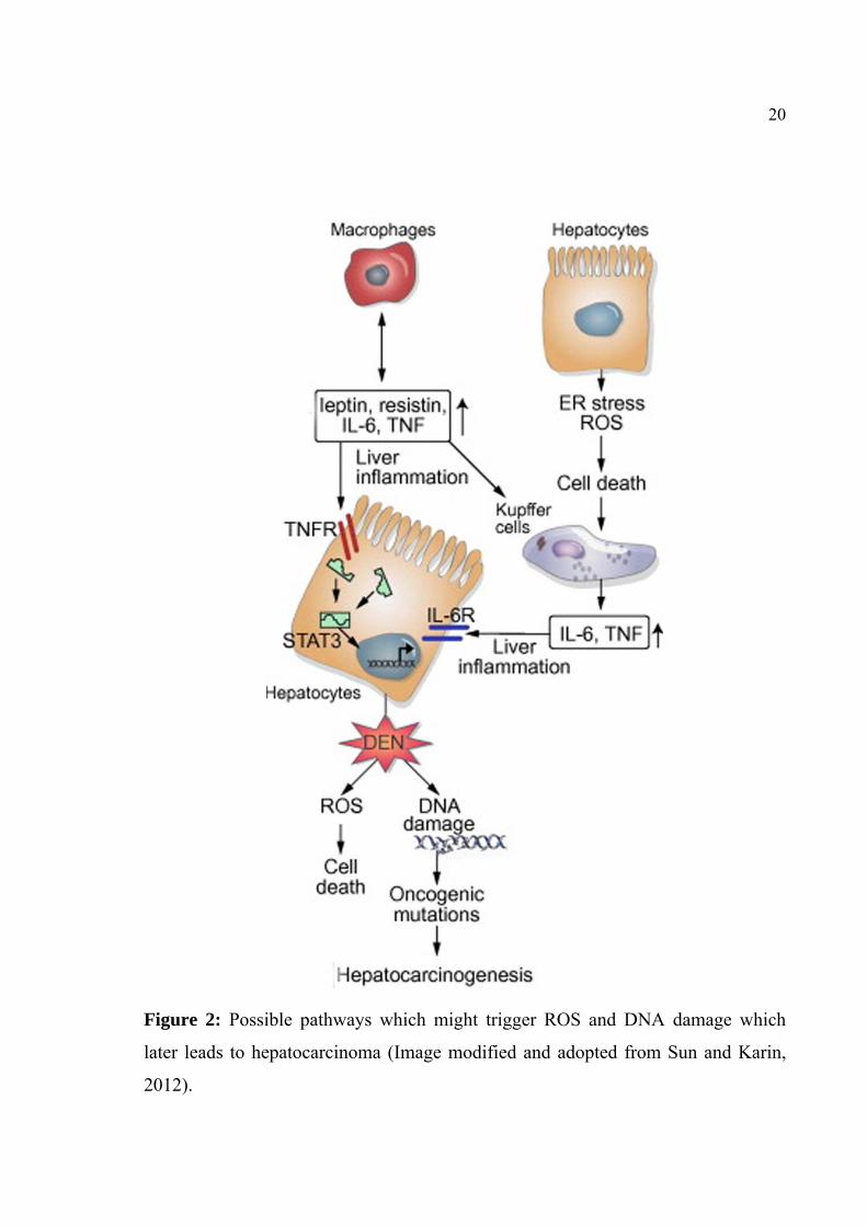
20
Figure 2: Possible pathways which might trigger ROS and DNA damage which
later leads to hepatocarcinoma (Image modified and adopted from Sun and Karin,
2012).

21
2.5 Ulcer- inflammation
Now it has been proven through various studies that there is a strong relationship
between H. pylori infection in stomach and adenocarcinoma. There is a hypothesis
porposed that H. pylori infection causes gastric inflammation which may lead to
atrophic gastritis and this finally may lead to gastric cancer. Moreover the
organisms infection is also related to the gastric and peptic ulcerations, as described
by many studies which have shown the role of H. pylori in idiopathic peptic ulcer.
But most of the infections which may lead to chronic gastric inflammation remain
clinically silent (Blazer et al., 1995).
Figure 3: Image showing the involvement of H. pylori in the inflammation induced
at gastric epithelial cell (Adapted from Smith et al., 2006).
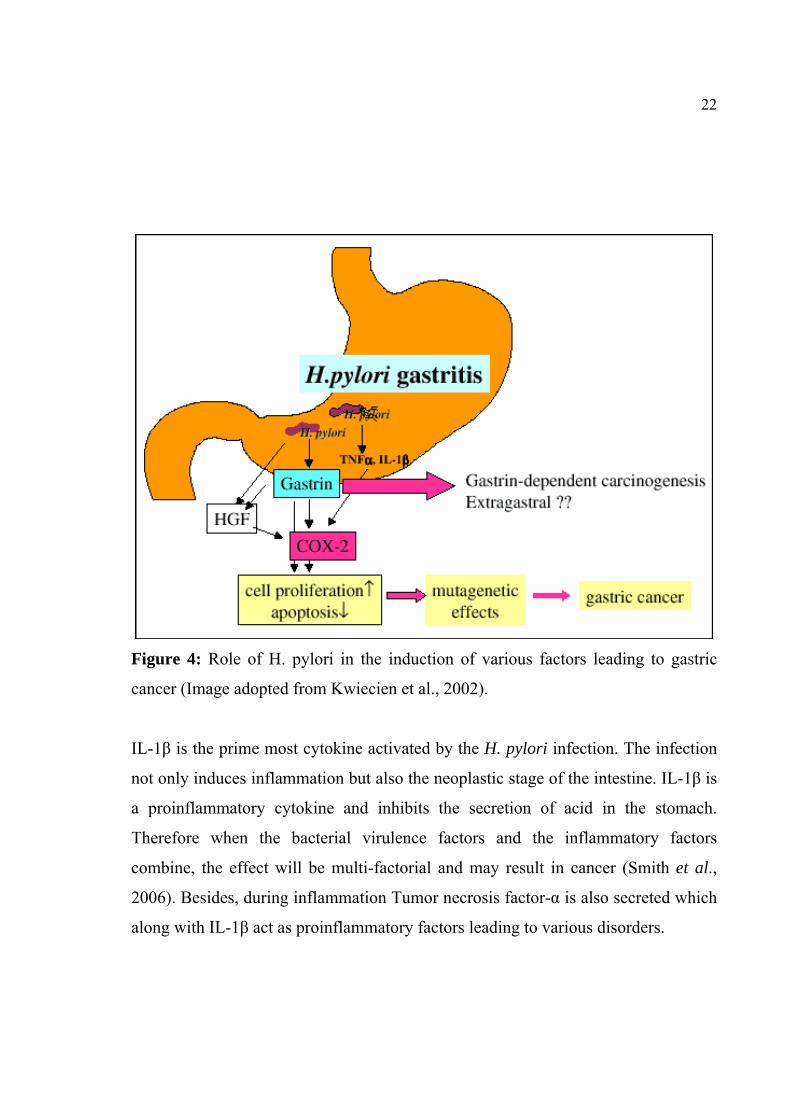
22
Figure 4: Role of H. pylori in the induction of various factors leading to gastric
cancer (Image adopted from Kwiecien et al., 2002).
IL-1β is the prime most cytokine activated by the H. pylori infection. The infection
not only induces inflammation but also the neoplastic stage of the intestine. IL-1β is
a proinflammatory cytokine and inhibits the secretion of acid in the stomach.
Therefore when the bacterial virulence factors and the inflammatory factors
combine, the effect will be multi-factorial and may result in cancer (Smith et al.,
2006). Besides, during inflammation Tumor necrosis factor-α is also secreted which
along with IL-1β act as proinflammatory factors leading to various disorders.
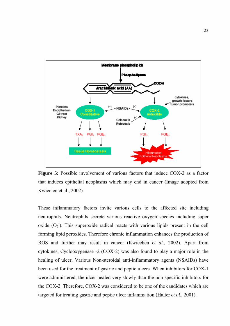
23
Figure 5: Possible involvement of various factors that induce COX-2 as a factor
that induces epithelial neoplasms which may end in cancer (Image adopted from
Kwiecien et al., 2002).
These inflammatory factors invite various cells to the affected site including
neutrophils. Neutrophils secrete various reactive oxygen species including super
oxide (O2-). This superoxide radical reacts with various lipids present in the cell
forming lipid peroxides. Therefore chronic inflammation enhances the production of
ROS and further may result in cancer (Kwiechen et al., 2002). Apart from
cytokines, Cyclooxygenase -2 (COX-2) was also found to play a major role in the
healing of ulcer. Various Non-steroidal anti-inflammatory agents (NSAIDs) have
been used for the treatment of gastric and peptic ulcers. When inhibitors for COX-1
were administered, the ulcer healed very slowly than the non-specific inhibitors for
the COX-2. Therefore, COX-2 was considered to be one of the candidates which are
targeted for treating gastric and peptic ulcer inflammation (Halter et al., 2001).

24
2.6 Inflammation – ROS – antioxidative system
Reactive Oxygen Species (ROS) is a name given to particular set of ions and
radicals which include super oxide (O2), peroxyl (RO2), hydroxyl (OH), alkoxyl
(RO) group. Apart from these there are certain other members like HOCl, Ozone
(O3), singlet oxygen (O2) and H2O2 which can be easily converted into radicals also
fall in to this category. Mutations induced by ROS can lead to cancer and can occur
in the following three ways.
1. Alterations in base pairs – ROS induced alternations in DNA may result in
mutations in proto-oncogenes and tumor suppressor genes which may lead to
cancer.
2. Affect the cytoplasmic signal pathways – enhanced H2O2 production may lead to
loss of the inhibitory segment in NF-κB which continuously result in
transcriptional activation.
3. Modulation of the stress related genes – H2O2 can activate c-jun and MAP kinase
(Wiseman and Halliwell, 1996).

25
Figure 6: Possible mutation sites for the ROS to exert its effects in leading to
cancer (Image modified and adopted from Nelson and Montgomery, 2003).
Oxidative stress is a major factor that triggers hepatic fibrosis, which in turn has
been shown to enhance the possibility of cancer (Curcumin inflammation liver).
Similarly H. pylori and other agents are shown to increase the ROS in the stomach
which in turn may result in cancer (Smith et al., 2006). Therefore, in both the cases
of liver and stomach, ROS plays a major role in inflammation and cancer. Therefore
inhibition of enhanced ROS production may be a possible strategy for the treatment
of the disorders in liver and stomach.

26
Figure 7: Both extrinsic (eg. radiations) and intrinsic pathways can induce ROS
production where the antioxidant system can block the production of ROS (Image
modified and adapted from Perera and Bardeesy, 2011).

27
2.7 Cancer – Immunomodulation
Many chemotherapy agents induce their effect through anti-proliferative and
cytotoxic effects. But the same agents had the capacity for immunosuppression as
the drug reduces the proliferation of immune cells which multiplies at a fast rate.
Some agents which exhibit the property of immunomodulation augmented the
treatment of cancer through the modulation of the immune system (Ehrke and Jane,
2003). Cyclophosphamide is a very good example for the immunomodulatory drug.
The drug has different role at higher concentrations but when the concentration is
lowered, administered alone or with some other agents exhibited anti-cancer and
immunomodulatory properties and cured cancer in mouse models. Apart from
cyclophosphamide, several other agents are shown to modulate the immune system,
nitrosoreus compounds such as adriamycin, arabinosylcitrosine etc. have been
shown to have the same potential (Ehrke et al., 1996).
Figure 8: Use of immune modulators in the treatment of prostate cancer, where the
T cells are activated by either threshold reduction or by enhancing the life cycle of T
effector cells (Image adopted from Kwek et al., 2012).

28
Immunomodulation is used as a method in the treatment various cancers including
prostate cancer where, by inhibiting the activity of an immune checkpoint protein,
Cytotoxic T Lymphocyte-associated antigen 4 (CTLA-4), a crucial impedance can
be removed. This is followed by activation of T cells by lowering the threshold or
by eliminating the inhibitory signals which attenuate the effector T cells (Kwek et
al., 2012).
2.8 Plants as anti-microbials
Apart from H. pylori in causing inflammatory effect, humans in day to day life have
been infected by various microorganisms, which range from infections which are
easily controlled by the host to serious infections resulting in severe morbidity and
mortality. This prompted many scientists to look for new anti-microbial agents
from various sources including plants. Since time immemorial, human have used
plants and their extracts to treat various infectious diseases. Some plants like
cranberry (Vaccinium macrocarpon) was used to treat urinary infections, garlic
(Allium sativum), lemon (Melissa officinalis) etc have been used as antimicrobial
agents. The compounds can be used either directly as phytomedicine for a particular
disease or used as a base compound from which new compounds can be derived
(Iwu et al., 1999).
The search for newer drugs from plant products continues every day since the
scientists predicted that the average effective of every antibiotic is limited, which
kindles the way to produce new antibiotics for the use of mankind.

29
3. MATERIALS AND METHODS
3.1 Collection of Plant Materials
The plant materials M. pudica, A. hexapetalus and A. vasica were collected from the
foot hills of Western Ghats in and around Courtallum and Thaniparari Hills, Tamil
Nadu, India during early winter season.
3.2 Phytochemical Studies
3.2.1 Preparation of Plant Extracts
The leaf extracts of all the three plants were prepared as described by Kokate,
(1991). The leaves of all three plants (M. pudica, A. hexapetalus and A. vasica) were
shade-dried and made into a coarse powder which was passed through a 40-mesh
sieve to get uniform particle size. A weighed quantity (500 g) of the powder was
then subjected to continuous hot extraction in Soxhlet apparatus individually with
methanol, chloroform and diethyl ether and the residual marc was collected. The
extract was filtered through a cotton plug, followed by Whatman filter paper (No.
1). The extract was evaporated under reduced pressure using a rotary evaporator
until all the solvent had been removed to obtain methanol, chloroform and diethyl
ether extracts.
3.2.2 Qualitative Chemical Evaluation
The chemical composition of the plant extracts was evaluated as described by
Harbone, (1984). The obtained extracts were tested for the presence of various plant
constituents such as alkaloids, flavanoids, tannins, saponins, glycosides, steroids,
steroidal terpenes, phenolic compounds, gums & muciages and carbohydrates.
Alkaloids: The extracts were dissolved in 1 ml of dilute H2SO4 and filtered using
Whatman No.1 filter paper and the filtrate was treated with Mayer’s, Dragendrof’s,
Hager’s and Wagner’s reagents separately. The appearance of cream, orange brown,

30
yellow and reddish brown precipitates in response to the above reagents respectively
indicates the presence of alkaloids.
Flavonoids: The extracts were mixed with 1-2 ml of alcohol and heated with 1-2
mgs of magnesium and then concentrated HCl was added under cooling. The
appearance of pink colour indicates the presence of flavonoids.
Test for Tannins: The extracts were dissolved in 10 ml of distilled water and
allowed to settle and filtered. To the filtrate 1-2 ml of 5% ferric chloride was added.
The appearance of deep green color indicates the presence of tannin. Another
portion of the filtrate was treated with 1-2 ml of iodine solution and a faint bluish
color confirmed the presence of tannin.
Saponins: About 1 ml of the test extract was dissolved in 20 ml of distilled water
and shaken in a graduated cylinder for 15 minutes. Formation of 1 cm layer of foam
indicates the presence of saponins.
Test for Glycosides: The extracts were dissolved in 10 ml of distilled water under
boiling conditions. This was filtered and 2 ml of the filtrate was hydrolyzed with a
few drops of concentrated HCl and the solution was rendered alkaline with 1-2
drops of ammonia solution. Five drops of this solution was added to 2 ml of
Bennedict’s qualitative reagent and boiled. A reddish–brown precipitate showed the
presence of glycosides.
Test for Steroids: The extractswere dissolved in 2 ml of chloroform. To this 2 ml
of concentrated sulphuric acid was carefully added to form a lower layer. A reddish
brown color at the interface indicated the presence of steroids.
31
Test for Steroidal Terpenes: The extractswere dissolved in 2 ml of chloroform and
1 ml of acetic anhydride. To this solution 2 drops of concentrated sulphuric acid
were added. A pink colour which changes to bluish green on standing indicated the
presence of steroidal terpenes.
Tannins and Phenols: The extractswere dissolved in 10 ml of water and ferric
chloride solution (5%) or gelatin solution (1%) or lead acetate solution (10%). The
appearance of blue colour with ferric chloride or precipitation with other reagents
indicates the presence of tannins and phenols.
Gums and Mucilages: About 10 ml of test extract was slowly added to 25 ml of
absolute alcohol under constant stirring. The precipitation indicates the presence of
gums and mucilages.
Carbohydrates: The extracts were dissolved in 2 ml of distilled water and then
filtered. The filtrate was treated with concentrated sulphuric acid then Molisch’s
reagent was added. The appearance of pink to violet color indicates the presence of
carbohydrates. The filtrate was boiled with Fehling’s or Benedict solutions. The
formation of brick red precipitate in Fehling’s and Benedict’s solution indicates the
presence of reducing sugars and non-reducing sugars respectively.
3.3 Pharmacological study
3.3.1 Screening for Immunomodulatory activity
3.3.1.1 Neutrophil adhesion test in rats
Adult male Wistar rats were weighing about 150-200gms were divided into
11groups of each 5 animals. The dosage of drugs was administered to the different
groups were as follows:
Group-1: Control (normal saline 10 ml/kg) - used common for all
32
Group -2: Cedrus deodara wood oil 100 mg/kg (Standard) - used common for all
Group -3: Methanolic extract of M. pudica 200 mg/kg & 400 mg/kg
Group -4: Chloroform extracts of M. pudica 200 mg/kg & 400 mg/kg
Group -5: Diethyl ether extracts of M. pudica 200 mg/kg & 400 mg/kg
Group -6: Methanolic extract of A. hexapetalus 200 mg/kg & 400 mg/kg
Group -7: Chloroform extract of A. hexapetalus 200 mg/kg & 400 mg/kg
Group -8: Diethyl ether extract of A. hexapetalus 200 mg/kg & 400 mg/kg
Group -9: Methanolic extract of A. vasica 200 mg/kg & 400 mg/kg
Group -10: Chloroform extract of A. vasica 200 mg/kg & 400 mg/kg
Group -11: Diethyl ether extract of A. vasica 200 mg/kg & 400 mg/kg
The neutrophil adhesion test was performed according to the methodology of
Wilkinson et al., (1978). The rats were divided into 11 groups, each group
consisting of 5 animals. First group was administrated with normal saline at
concentration of 10 ml/kg (negative control), the second group with C. deodara
wood oil by oral route (positive control) and the third to eleventh groups with
methanolic, chloroform and diethyl ether extracts to all three plants (M. pudica, A.
hexapetalus and A. vasica ) at a dose of 200mg/kg and 400mg /kg/day for 8 days.
On the 8th day blood samples were collected from the retro-orbital plexus in
heparinized vials and analyzed for total leukocyte count (TLC) using Erma PC-607
cell counter (Transasia Ltd., Mumbai, India). The differential leukocyte count
(DLC) was performed by fixing the blood smear and staining with leucofine and
neutrophils percentage in each sample were determined. After the initial counts,
blood samples were incubated with 80mg/ml of nylon fibers for 10 minutes at 370C.
The incubated blood samples were again analyzed for TLC, DLC and neutrophils
percent and neutrophil index was calculated. The neutrophil adhesion percent was
calculated from the following formula;

33
Neutrophil adhesion % = NIu—NIt NIu
Where,
NIu - Neutrophil index of untreated blood sample
NIt - Neutrophil index of the treated blood sample.
3.3.1.2 Delayed type hypersensitivity (DTH)
The hypersensitivity reaction to Sheep red blood cells (SRBC) was induced in mice
as per the method described by Ray et al., (1996). The sheep erythrocytes were
washed with pyrogen-free sterile normal saline and adjusted to a concentration of
1×10⁸ cells/ml and used for sensitization and challenge. The control group was
administered with an equal volume of PBS (pH 7.4) orally and positive control
group with standard Levamisole 50mg/kg. The negative control group was treated
with Normal saline (10ml/kg) and the test groups with methanolic, chloroform and
diethyl ether extracts ofthree plants (M. pudica, A. hexapetalus and A. vasica) at the
dose of 400mg /kg/day for 9 days. On 9th day, all the groups were challenged with
1×10⁸ SRBC cells, administered intradermally into the left footpad of each mouse,
and the increase in footpad thickness (FPT) was measured 24 h after the SRBC
challenge by volume differential meter.
Group -1: Negative control (normal saline 10ml/kg) - used common for all
Group -2: Positive control PBS (pH 7.4) +Levamisole 50mg/kg - used common for all
Group -3: Methanolic extract of M. pudica 400 mg/kg
Group -4: Chloroform extract of M. pudica 400 mg/kg
Group -5: Diethyl ether extract of M. pudica 400 mg/kg
Group -6: Methanolic extract of A. hexapetalus 400 mg/kg
Group -7: Chloroform extract of A. hexapetalus 400 mg/kg
Group -8: Diethyl ether extract of A. hexapetalus 400 mg/kg

34
Group -9: Methanolic extract of A. vasica 400 mg/kg
Group -10: Chloroform extract of A. vasica 400 mg/kg
Group -11: Diethyl ether extract of A. vasica 400 mg/kg
3.3.2 Hepatoprotective Activity Screening
Animals: Male albino rats weighing 150-200g maintained under standard
husbandary conditions (temp 23±2oC, relative humidity 55±10% and 12 hours light
dark cycle) were used for the screening. Animals were fed with standard laboratory
food and ad libitum during the entire period of study. All the experimental protocols
were conducted at the Arulmigu Kalasalingam College of Pharmacy, Krishnankoil,
Tamil Nadu, India and were approved by the Institutional Animal Ethics Committee
at Arulmigu Kalasalingam College of Pharmacy, Krishnankoil, India (Reg.
No.509/02/C/CPCSEA/2002).
Toxicity studies: The acute toxicity study was performed for various extracts of
three plants (M. pudica, A. hexapetalusand A. vasica) according to the acute toxic
classic method as per OECD guidelines (Ecobichon, 1997). The male albino rats
were used for acute toxicity study. The animals were kept fasting for overnight
providing only water, after which various extracts were administered orally at the
dose of 300mg/kg and observed for 14 days. If mortality was observed in two
animals out of three animals, then the dose administered was assigned as toxic dose.
If the mortality was observed in one animal, then the same dose was repeated to
confirm the toxic dose. If mortality was not observed, the procedure was repeated
for further higher doses such 400, 500 & 2000mg/kg body weight. The animals
were observed for toxic symptoms for 72 h.
Carbon tetrachloride induced hepatotoxicity: Male albinorats were divided into
12 groups of 6 animals in each group. Group I served as a control, which was
administrated normal saline (3 ml/kg, p.o.). Group II received CCl4 (0.5 ml/kg, i.p.),
35
Group III received CCl4 (0.5 ml/kg, i.p.) with Silymarin (100 mg/kg, p.o), Group IV,
V and VI received CCl4 (0.5 ml/kg, i.p.) with methanolic extract of three plants
namely, M. pudica, A. hexapetalusand A. vasica (200 mg/kg, p.o.), Group VII,VIII
and IX received CCl4 (0.5 ml/kg, i.p) with chloroform extract of three plants (M.
pudica, A. hexapetalus and A. vasica) (200 mg/kg, p.o.), Group X, XI and XII
received CCl4 (0.5 ml/kg, i.p) with diethyl ether extract of 3 plants namely M.
pudica, A. hexapetalusand A. vasica (200 mg/kg p.o.) separately for 7 days. After 7th
days of treatment and overnight fasting of rats blood samples were collected from
retro-orbital plexus region under mild ether anesthesia and the serum was separated
and to determine the various biochemical parameters (Rao and Mishra, 1997).
Group-I: Normal saline (3ml/kg, p.o.) - Solvent control
Group -II: CCl4 (0.5ml/kg, i.p.) - Hepatic control
Group -III: CCl4 (0.5ml/kg, i.p.) + Silymarin (100mg/kg p.o.)
Group-IV: CCl4 (0.5ml/kg, i.p.)+Methanolic extract of M.pudica (200mg/kg, p.o.)
Group-V: CCl4 (0.5ml/kg, i.p.)+Chloroform extract of M pudica (200mg/kg, p.o.)
Group-VI: CCl4 (0.5ml/kg, i.p.)+Diethyl ether extract of M pudica (200 mg/kg, p.o.)
Group-VII: CCl4 (0.5ml/kg, i.p.)+ Methanolic extract of A.Hexapetalus (200 mg/kg, p.o.)
Group-VIII: CCl4 (0.5ml/kg, i.p.)+Chloroform extract of A.Hexapetalus (200 mg/kg, p.o.)
Group-IX: CCl4 (0.5ml/kg, i.p.)+Diethylether extract of A Hexapetalus (200 mg/kg, p.o.)
Group-X: CCl4 (0.5ml/kg, i.p.) + Methanolic extract of A. vasica (200 mg/kg, p.o.)
Group-XI: CCl4 (0.5ml/kg, i.p.) + Chloroform extract of A. vasica (200 mg/kg, p.o.)
Group-XII: CCl4 (0.5ml/kg, i.p.)+Diethylether extract of A.vasica (200 mg/kg, p.o.)
Assessment of liver function: Blood was collected from all the groups by
puncturing the retro-orbital plexus and was allowed to clot at room temperature.
Serum was separated by centrifugation at 2500 rpm for 10 min for the estimation of
biochemical parameters and to determine the functional state of the liver. The
Serum Glutamic Oxaloacetic Transaminase (SGOT) and Serum Glutamic Pyruvic

36
Transaminase (SGPT) were estimated by a UV kinetic method based on the
reference method of International federation of Clinical Chemistry in which both
SGOT and SGPT were assayed based on enzyme-coupled system, where keto acid
formed by the aminotransferase reacts with NADH. The coenzyme is oxidized to
NAD and the decrease in absorbance at 340 nm is measured. For SGOT malated
dehydrogenase is used to reduce oxaloacetate to malate where as for SGPT the
pyruvate formed during the reaction is converted to lactate by lactate dehydrogenase
(Raitman and Frankel, 1957).
Alkaline phosphatase (ALP) was estimated as described by Comb and Bowers,
(1972). The total Bilirubin (TBL) was estimated by the method of Jendrassik and
Grof, (1938) which involves the reaction of bilirubin with diazotized sulphanilic
acid to form an azo compound and the colour was measured at 546 nm.
Histopathological studies: The abdomen of the animal was cut open and the liver
was removed. The liver was fixed in Boucin’s solution (mixture of 75 ml of
saturated picric acid, 25 ml of 40 % formaldehyde and 5 ml of glacial acetic acid)
for 12h, and then embedded in paraffin using conventional methods (Galighor and
Kozloff, 1976) and cut into 5μm thick sections and stained with haematoxylin-eosin
dye and finally mounted in di-phenyl xylene. The sections were then observed under
microscope for histopathological changes in liver architecture and their
photomicrographs were well documented.
Statistical analysis: The values of mean ± SEM were calculated for each of the
parameters. For determining the significant inter group difference in each
parameters were analysed separately. The analysis of variance one-way (Gennaro,
1995) was carried out and their individual comparisons of the each group mean
values were performed using Dunnet’s test (Dunnet, 1964).

37
3.3.3 Anti-ulcer activity screening
Animals: Male albino rats weighing 150-200 g were obtained from Madurai and
maintained under standard husbandry conditions (temp 23±2oC, relative humidity
55±10% and 12 hours light dark cycle) were used for the screening. The animals
were fed with standard laboratory diet ad libitum during the entire period of study.
The experimental protocol has been approved by institutional animal ethics
committee, Arulmigu Kalasalingam College of Pharmacy, Krishnankoil (Regd.
No.509/02/C/CPCSEA/2002.) India.
Toxicity studies: The acute toxicity study was performed using the extracts of
selected plant according to the acute toxic classic method as per OECD guidelines
(Ecobichon, 1997). The albino rats of both sexes were used for acute toxicity study.
The animals were kept fasting for overnight providing only water, after which the
various extracts were administered orally at the dose of 300 mg/kg and observed up
to 14 days. If mortality was observed in two animals out of three animals, then the
dose administered was assigned as toxic dose. If the mortality was observed in one
animal, then the same dose was repeated to confirm the toxic dose. If mortality was
not observed, the procedure was repeated for further higher doses such as 400, 500
and 2000 mg/kg body weight. The animals were observed for toxic symptoms for
72h.
Aspirin-induced gastric ulcer: In aspirin induced ulcer experiments, 11 groups of
male albino rats (100–200 g), with each group consisting of 5 animals were used.
The first group that served as a control administrated with normal saline (2 ml/kg),
the second group which served as positive control with ranitidine (20 mg/kg) orally
and the 3 to 11 groups were served as the test extracts (100mg/kg and 200 mg/kg)
groups for 8 days. The ulcer was produced by administration of aqueous suspension
of aspirin (a dose of 200 mg/kg orally) for eight days. After 8 days of treatment,

38
animals were allowed to fast for 24 h. On the 8thday the animals were sacrificed 4 h
later of the drug treatment and stomach was surgically opened to calculate the ulcer
index (Kunchandy et al., 1985).
Alcohol-induced gastric ulcer: The male rats were randomly divided into 11
groups each group consisting of 5 animals and fasted for 24 h with free access to
water. The animals were given methanolic, chloroform and diethyl ether extracts of
the 3plants (M. pudica, A. hexapetalus and A. vasica) at a dose of 100mg/kg and
200 mg/kg and Ranitidine (20 mg/kg) orally for seven days. One hour later, 1 ml of
80% ethanol was administered orally to each animal for 7th days. On the seventh day
animals were sacrificed by cervical dislocation, one hour after ethanol
administration, stomachs were surgically cut open along the greater curvature and
pinned on a soft board. The length of each gastric lesion was measured and the
lesion index was expressed as sum of the length of the entire lesion in mm
(Kunchandy et al., 1985).
Pylorus-ligation induced gastric ulcer: The male albino rats weighing 150-200 g
were selected for pyloric ligation ulcer model. The rats were divided into 11 groups,
each group consisting of 5 animals. First group administrated normal saline 2 ml/kg
(negative control), the second group with Ranitidine 20 mg/kg by oral route
(positive control) and the third to eleventh groups with methanolic, chloroform and
diethyl ether extracts ofthree plants(M. pudica, A. hexapetalus and A. vasica)
(100mg/kg and 200 mg/kg) by oral route for seven days. After one hour of the last
dosing, pylorus ligation was made under ether anesthesia. The animals were
returned to the observation chamber for 4h. After 4h, the animals were sacrificed by
decapitation, the abdomen of each animal was surgically opened and the stomach
was isolated after suturing the lower esophageal end. The gastric juice was collected
and the mucosal layer was washed with 1 ml distilled water. The ulcer scoring was

39
performed in the stomach of each animal. The total volume of gastric content was
also measured. The gastric contents were centrifuged at 1000 rpm for 10 min. One
ml of the supernatant liquid was pipetted out and diluted with 10 ml with distilled
water. The solution was titrated against 0.01 N NaOH using Topfer’s reagent as
indicator, to the endpoint when the solution turned to orange colour. The volume of
NaOH needed was taken as corresponding to the free acidity. The titration was
further continued till the solution regained pink colour (Shay et al., 1945).
The volume of NaOH required was noted and was taken as corresponding to
the total acidity. Acidity was expressed as:
Acidity = Volume of NaOH x Normality x 100 mEq/1
0.1
Statistical analysis: The values mean ± SEM are calculated for each parameter. For
determining the significant inter group differences, each parameter was analysed
separately and one-way analysis of variance (Gennaro, 1995) was carried out and
the individual comparisons of the group mean values were done using Dunnet’s test
(Dunnet, 1964).
Group-1: Normal saline (2 ml/kg, p.o.) - solvent control
Group -2: Ranitidine (20 mg/kg, p.o.) - standard
Group -3: Methanolic extract of M. pudica (100 mg/kg &200 mg/kg, p.o.)
Group -4: Chloroform extract of M. pudica (100 mg/kg &200 mg/kg, p.o.)
Group -5: Diethyl ether extract of M. pudica (100 mg/kg &200 mg/kg, p.o.)
Group -6: Methanolic extract of A. hexapetalus (100 mg/kg &200 mg/kg, p.o.)
Group -7: Chloroform extract of A. hexapetalus (100 mg/kg &200 mg/kg, p.o.)
Group -8: Diethyl ether extract of A. hexapetalus (100 mg/kg &200 mg/kg, p.o.)
Group -9: Methanolic extract of A. vasica (100 mg/kg &200 mg/kg, p.o.)
Group -10: Chloroform extract of A. vasica (100 mg/kg &200 mg/kg, p.o.)

40
Group -11: Diethyl ether extract of A. vasica (100 mg/kg &200 mg/kg, p.o.)
3.3.4 Wound Healing Activity Screening
Experimental Animals: Male albino rats (150-200 gm) were provided with a
standard diet (Pranav Agro, India) and water ad libitum and maintained under
standard laboratory conditions in the institutional animal house facility.
Table 3: Formulation of Ointment
Type: water miscible base
S. No. Ingredients Official Formula (gm) Working Formula (gm)
1. 2. 3.
Emulsifying wax White soft paraffin Liquid paraffin
30 50 20
3 5 2
Method of Preparation: The ingredients were mixed, heated gently with
continuous stirring until a homogenous mixture was formed. The above contents
were cooled at room temperature. The10% concentration of ointment was
prepared.In case of the plant extracts, 1 gm of suitable extract was mixed with 10
gms of ointment base (10%); then it was stirred well until a homogenous ointment
was obtained.
Types of ointment prepared: Eleven types of ointments were prepared as indicated
below:
a) Simple ointment base - Control
b) 0.2% w/w of Nitrofurazone ointment -Standard
c) Base + Methanolic Extract of M. pudica (10% w/w)
d) Base + Chloroform Extract of M. pudica (10% w/w)
e) Base + Diethyl Ether Extract of M. pudica (10% w/w)
f) Base + Methanolic Extract of A. hexapetalus (10% w/w)

41
a) Base + Chloroform Extract of A. hexapetalus (10% w/w)
b) Base + Diethyl Ether Extract of A. hexapetalus (10% w/w)
c) Base + Methanolic Extract of A. vasica (10% w/w)
d) Base + Chloroform Extract of A. vasica (10% w/w)
e) Base + Diethyl Ether Extract of A. vasica (10% w/w)
Excision Wound Model: Male Albino rats (150-200gms) were selected and divided
into 11 groups of 5 animals for each of these experiments. The animals were housed
in the experimental room which was maintained as per IAEC guide lines. The
experimental animals were anaesthetized using lignocaine 2% injections, over the
local selected region. The rats were depilated and an excision wound was created by
cutting away 500 mm square thickness of skin from the predetermine area, the
wound was left open then the drugs, reference standard (0.2 % w/w Nitrofurazone
ointment), control (simple ointment base B.P) and methanol, chloroform and diethyl
ether extracts of 3 plants ointment (M. pudica, A. hexapetalus and A. vasica) were
applied until the wound was healed. This model was used to monitor the wound
contraction and wound closer time. The wound contraction was calculated as
percentage reduction in wound area. The progressive change in wound area is
monitored by calculating the decreasing area (Muppayavarmath and Patil,
1999).RWH = Size of Wound in surface area (mm2) at Day 16 / Size of Wound in
surface area (mm2) at Day 1 X 100
% Reduction in Healing = 100 – RWH
Fractionation of Mimosa pudica Extract: Liquid–liquid extraction, also known as
solvent extraction and partitioning, is a method to separate compounds based on
their relative solubility in two different immiscible liquids, usually water and an
organic solvent. It is an extraction of a substance from one liquid phase into another
liquid phase. Methanolic extract of M pudica was subjected to solvent fractionation
between the following immiscible solvent mixtures in 1:1 ratio (purified water:

42
hexane, purified water: ethyl acetate, purified water: chloroform and purified water:
n-butanol).
Protocol: Two grams of methanolic extract of M. pudica was dissolved in 10
volumes of solvent purified water and equal volume of hexane was added in a first
separating funnel. This was shaken for 10 min, then allowed to settle or centrifuged
at a low speed for 15 minutes. The lower phase and the upper phase were collected
in different containers. Evaporate the two liquid phases separately. Repeat the
procedure using different solvent mixtures such as purified water: ethyl acetate,
purified water: chloroform and purified water: N-butanol. The combined aqueous
extracts and different organic extracts were used for evaluation of wound healing
activity (McCraken and Chaikin, 1974).
The ointments were prepared with different fractions of M. pudica as per the protocols described earlier.
a) Simple ointment base - Control
b) 0.2% w/w of Nitrofurazone ointment - Standard
c) Base + Aqueous fraction of M. pudica (10% w/w)
d) Base + Hexane fraction of M. pudica (10% w/w)
e) Base + Ethyl acetate fraction of M. pudica (10% w/w)
f) Base + Chloroform fraction of M. pudica (10% w/w)
g) Base + N-butanol fraction of M. pudica (10% w/w)
10% concentration of ointment was prepared.
3.4 Isolation and Identification of Bioactive Compounds
Column Chromatography: Column Chromatography was used for the separation
of extracted compounds. The method described by Wagner and Blatt (1996) was
used. The separation of the compounds was achieved by either increasing or
decreasing the polarity of the solvent system.

43
Sample Preparation: The methanol, chloroform and diethyl ether extracts (2 gm)
were dissolved in 1 ml of chloroform and this was loaded into the column for the
separation of metabolites.
Preparation of Column: The wet packing of column chromatography was adopted
for the preparation of the column. The column matrix, silica gel, was mixed with
petroleum ether and poured gently from the top of the column to a desired length.
Then the same solvent was run through the column for 2-3 times to prevent air
entrapment. The solvent was maintained up to 10cm above the column bed. The
sample mixture was applied on top of the column with the aid of a funnel. The
column was allowed to stand over-night undisturbed. On the next day, column was
eluted with different solvents viz. petroleum ether, benzene, chloroform, ethyl
acetate and methanol with gradual increasing in polarity. The flow rate of solvent
was adjusted between 16 and 20 drops per minute. 20 ml fractions were collected
and evaporated at room temperature. The extracted material was further analyzed
using Thin Layer Chromatography (TLC).
Thin Layer Chromatography: Glass plates coated with silica gel-G as stationery
phase were used to perform TLC. Plates were developed using iodine, and the spots
were identified and their Rf values determined by using the following formula;
Rf value = Distance traveled by solute / Distance traveled by solvent
The spots identified at the same Rf value were pooled and evaporated to dryness by
using vacuum drier and proceded for further studies (Beckett and Stenlake, 1986).
HPLC analysis: HPLC experiments were carried out using a Shimadzu HPLC - A
HT 2010, made in Japan. The stationary phase Phenomnex C18 column (100Å5µ,
150X4.6mm) and Princeton SPHER-100, C18 colum (100Å5µ, 150X4.6mm) were
used for the analysis of the isolated compounds. Commercially available

44
mimopudine, artabotricinol and vasicine were used as standards (Natural Remedies
Pvt. Ltd., Bangalore). The mimopudine andartabotricinol were detected at 254nm
and vasicine was detected at 300nm.
Chromatographic conditions for mimopudine standard
Stationary Phase : Phenomnex C18 100Å5µ, 150X4.6mm
Mobile Phase : Methanol: Water (pH 3.4) (60:40)
Detection Wave Length : 254nm
Flow Rate : 1.0 ml/min
Column Washing : Water: Methanol (50:50)
Injection Volume : 20µL/inj
Injection Type : Rheodyne injector
Chromatographic conditions for artabotrycinol
Stationary Phase : Phenomenex C18 100Å5µ, 150X4.6 mm
Mobile Phase : Methanol: Water (90:10)
Detection Wave Length : 254nm
Flow Rate : 1.0 ml/min
Column Washing : Water: Methanol (50:50)
Injection Volume : 20µL/inj
Inject ion Type : Rheodyne injector
Chromatographic conditions for vasicine
Stationary Phase : Princeton SPHER-100, C18 100Å5µ, 150X4.6mm
Mobile Phase : Acetonitrile–0.1M Phosphate buffer–Acetic acid
(15:85:1, v/v/v)
Detection Wave Length : 300nm
Flow Rate : 1.0 ml/min

45
Column Washing : Water: Methanol (50:50)
Injection Volume : 20µL/inj
Injection Type : Rheodyne injector
Instruments Used
Shimadzu, Detector UV 2487 Dual wave length, Pump 1515
Preparation of standard stock solution: Accurately weighed 5mg of standards viz.
mimopudine, artabotrycinol and vasicine were transferred in to separate tubes and
the contents were dissolved in a few ml of methanol and the volume was made up to
5 ml (1000µg/ml).
Preparation of extracts
1. Accurately weighed 10mg of methanolic extract of M. pudica, A. hexapetalus
and A. vasica weretransferred to separate tubes, the contents dissolved with
few ml of methanol and the volume was made up to 5 ml (1000µg/ml)
(Sharma et al.,1992; Srivastava et al., 1999).
2. 1 ml stock was made up to 10 ml with mobile phase.
3. The sample solution was injected and analysed.
FTIR Spectral Analysis: Methanolic extracts of three plants and their chemical
structure were analyzed by Fourier Transformer - Infrared Spectrophotometer
(Shimadzu – 8400) by KBr pellet method as described by Narmato, (1997).
3.5 Screening of anti-microbial Activity
Preparation of Mueller – Hinton Agar medium
Beef extract - 300 gm
Peptone - 17.5 gm

46
Starch - 1.5 gm
Agar - 17 gm
Cold distilled water - up to 1000ml
All the ingredients were weighed and suspended in 1000 ml of cold distilled water
and heated to boiling. The pH of the media was adjusted to 7.4 with 5 M sodium
hydroxide solution. Then 5 – 20 ml of this agar medium was transferred into each
boiling tube and plugged with non-absorbent cotton. The tube containing agar
medium was sterilized by pressure controlled heat sterilization technique using an
autoclave at 15 lbs at 121°C for 20 minutes. After sterilization the agar medium
was melted and cooled. A well was prepared in the plates with help of a cork-borer
(0.85 cm) and 100 µl of the test compound was introduced into the well. The plates
were incubated overnight at 370 C. The microbial growth was determined by
measuring the diameter of zone of inhibition. For each bacterial strain controls were
maintained where pure solvents were used instead of the extract. The result was
obtained by measuring the zone diameter (Mukerjee, 1996).
3.5.1 Test microorganisms
Known microbial strains were obtained from the National Chemical Laboratory
(NCL), Pune, India. They were three Gram-positive bacteria viz. Micrococcus
luteus, Staphylococcus aureus, Bacillus cerus and three Gram-negative bacteria viz.
Klebsiella pneumonieae, Salmonella typhimurium and Salmonella
paratyphimurium. The disc diffusion assay was used for testing the antibacterial
activity. The ciprofloxacin (100µg/ml) was used as a positive control along with the
extracts of three plants, M. pudica, A. hexapetalusand A. vasic (200 µg/ml)
incubated at 37° C for 24 hours (Adeniyi et al., 1996; Bauer et al., 1966).
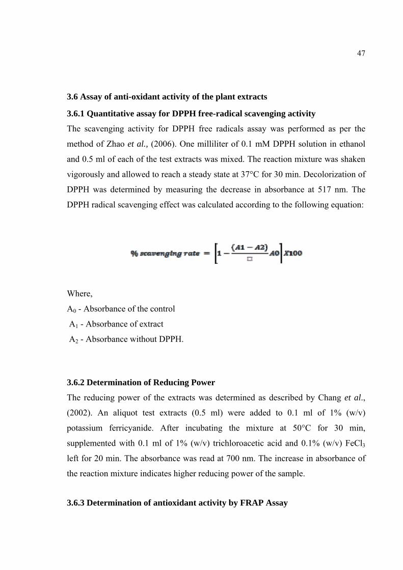
47
3.6 Assay of anti-oxidant activity of the plant extracts
3.6.1 Quantitative assay for DPPH free-radical scavenging activity
The scavenging activity for DPPH free radicals assay was performed as per the
method of Zhao et al., (2006). One milliliter of 0.1 mM DPPH solution in ethanol
and 0.5 ml of each of the test extracts was mixed. The reaction mixture was shaken
vigorously and allowed to reach a steady state at 37°C for 30 min. Decolorization of
DPPH was determined by measuring the decrease in absorbance at 517 nm. The
DPPH radical scavenging effect was calculated according to the following equation:
Where,
A0 - Absorbance of the control
A1 - Absorbance of extract
A2 - Absorbance without DPPH.
3.6.2 Determination of Reducing Power
The reducing power of the extracts was determined as described by Chang et al.,
(2002). An aliquot test extracts (0.5 ml) were added to 0.1 ml of 1% (w/v)
potassium ferricyanide. After incubating the mixture at 50°C for 30 min,
supplemented with 0.1 ml of 1% (w/v) trichloroacetic acid and 0.1% (w/v) FeCl3
left for 20 min. The absorbance was read at 700 nm. The increase in absorbance of
the reaction mixture indicates higher reducing power of the sample.
3.6.3 Determination of antioxidant activity by FRAP Assay

48
The FRAP assay was carried out according to Othman et al., (2007). The FRAP
reagent was prepared by mixing 300 mM acetate buffer (pH 3.6), 10 mM TPTZ and
20 mM ferric chloride in the ratio of 10:1:1. For FRAP assay, the reaction mixture
containing 2 ml of FRAP reagent, 0.5 ml of test extracts and 1 ml of distilled water
was incubated for 10 min. The absorbance was measured at 593 nm. The
antioxidant potential of the sample was compared with the activity of 0.5 ml stock
solution of 1 mg/ml FeSO4.

49
4. RESULTS
4.1 Analysis of Phytochemicals present in the plants
4.1.1 Extraction of phytochemicals
Mimosa pudica leaf extracts were prepared using three solvents viz. methanol,
chloroform and diethyl ether; the percentage yields of the extracts were found to be
6.4, 8 and 5.2 respectively. Similar results were obtained when extraction was
carried out with the other two plants, with A. hexapetalusthe yield was found to be
7, 6 and 5 with methanol, chloroform and diethyl ether respectively and extraction
of A. vasica powder with methanol, chloroform and diethyl ether yielded 7, 6.2 and
5.2 respectively (Table 4).
Table 4: Extraction of phytochemicals from medicinal plants
Weight of drug
Method of Extraction Solvent used Weight of
extract (gm) Yield (%)
Methanol 16 6.4
Chloroform 20 8
500gms Mimosa pudica powder
Soxhlet apparatus
Diethyl Ether 13 5.2
Methanol 15 7 Chloroform 13.5 6
Artabotrys hexapetalus powder (500gms)
Soxhlet apparatus
Diethyl Ether 12 5 Methanol 16 7
Chloroform 14 6.2
500gms Adhatoda vasica powder
Soxhlet apparatus
Diethyl Ether 12 5.2

50
Qualitative analysis of the extracts for the presence of phytochemicals
The plant extracts were analyzed for the presence of various phytochemicals using
standard analytical methods. The extracts of all three plants showed the presence of
alkaloids, flavanoids, tannins, steroids and phenolic compounds (Table 5).
Table 5: Qualitative analysis of plant extracts
Phytochemicals Extracts of Mimosa pudica
Extracts of Artabotrys hexapetalus
Extracts of Adhatoda vasica
CHLO DEE METH CHLO DEE METH CHLO DEE METHAlkaloid + ‐ + + ‐ + + ‐ + Flavanoid ‐ + + ‐ + + ‐ + + Tannins ‐ + + ‐ + + ‐ + + Saponins ‐ + + ‐ + + ‐ + + Glycosides ‐ + + ‐ + + ‐ + + Steroids + + + + + + + + + Steroidal terpenes ‐ + + ‐ + + ‐ + + Phenolic compounds
‐ ‐ + ‐ ‐ + ‐ ‐ +
Gums&mucilages ‐ ‐ + ‐ ‐ + ‐ ‐ + Carbohydrate ‐ ‐ + ‐ ‐ + ‐ ‐ +
“+” indicates the presence of phytochemicals and “-“indicates absence of
phytochemicals
4.2 Chromatographic analysis of plant extracts
Various constituents of all the extracts were separated by column chromatography
using silica gel as a matrix. They were separated using various solvents and a
different ratio. The solvents and the ratio used are presented in Table 6, 7 and 8.
4.2.1 Isolation of active principles
4.2.1.1 Fractionation of compounds from Mimosa pudica using Column Chromatography
Table 6: Methanolic extract of Mimosa pudica

51
S. No. Solvent Ratio Nature of Residue 1 Petroleum Ether: Benzene 20:80 Colorless Residue 2 Petroleum Ether: Benzene 50:50 Colorless Residue 3 Benzene 100 Light Green Residue 4 Benzene: Chloroform 70:30 Light Green Residue 5 Benzene: Chloroform 20:80 Green Residue 6 Chloroform 100 Green Residue
7 Chloroform: Ethyl Acetate 80:20 Greenish Yellow Residue
8 Chloroform: Ethyl Acetate 20:80 Greenish Yellow Residue
9 Ethyl Acetate 100 Light Brown Residue 10 Ethyl Acetate: Methanol 60:40 Light Brown Residue 11 Ethyl Acetate: Methanol 20:80 Dark Brown Residue 12 Methanol 100 Brownish Residue
When all the fractions were analyzed by TLC for the presence of compounds, the
fraction obtained using ethyl acetate and methanol at a ratio of 20:80, yielded
identifiable compound. Hence, this fraction was used for further analysis.
Table 7: Chloroform extract of Mimosa pudica
S. No. Solvent Ratio Nature of Residue 1 Petroleum Ether: Benzene 50:50 Colorless Residue 2 Petroleum Ether: Benzene 20:80 Colorless Residue 3 Benzene 100 Light Green Residue 4 Benzene: Chloroform 40:60 Light Green Residue 5 Benzene: Chloroform 20:80 Green Residue 6 Chloroform 100 Green Residue 7 Chloroform: Ethyl Acetate 70:30 Greenish Yellow Residue 8 Chloroform: Ethyl Acetate 10:90 Greenish Yellow Residue 9 Ethyl Acetate 100 Light Brown Residue 10 Ethyl Acetate: Methanol 70:30 Light Brown Residue 11 Ethyl Acetate: Methanol 20:80 Dark Brown Residue 12 Methanol 100 Brownish Residue
TLC analysis does not yield any significant band from these fractions.
Table 8: Diethyl ether extract of Mimosa pudica
S. No. Solvent Ratio Nature of Residue 1 Petroleum Ether: Benzene 40:60 Colorless Residue

52
2 Petroleum Ether: Benzene 10:90 Light Green Residue 3 Benzene 100 Light Green Residue 4 Benzene: Chloroform 30:70 Green Residue 5 Benzene: Chloroform 10:90 Green Residue 6 Chloroform 100 Green Residue 7 Chloroform: Ethyl Acetate 80:20 Greenish Yellow Residue 8 Chloroform: Ethyl Acetate 20:80 Greenish Yellow Residue 9 Ethyl Acetate 100 Light Brown Residue 10 Ethyl Acetate: Methanol 60:40 Light Brown Residue 11 Ethyl Acetate: Methanol 10:90 Dark Brown Residue 12 Methanol 100 Brownish Residue
TLC analysis does not yield any significant band from these fractions.
4.2.2 Isolation of compounds from Mimosa pudica using thin layer chromatography
The compounds present in the extracted fractions were resolved by TLC using ethyl
acetate and methanol at different ratios. The results are presented in Table 9, 10 and
11.
Table 9: Thin layer chromatographic analysis of methanol extract of Mimosa pudica
Detecting Reagent
Distance Run by Solvent
Number of spots
Distance Run by
solute (cm)
Rf Value Color of the spot
8 0.5333 Greenish Black 8.5 0.5666 Light Green 9.3 0.6200 Light Green 10.4 0.6933 Light Green
Iodine 15cm 5
12.8 0.8533 Light Brown
Note: Solvent system Ethyl Acetate: Methanol (20:80)
The spot at Rf value 0.62 matches with that of the standard marker Mimopudine
was collected and utilized for further HPLC and FTIR studies.
Table 10: TLC analysis of chloroform extract of Mimosa pudica
Detecting Reagent
Distance Run by Solvent
Number of spots
Distance Run by
solute (cm)
Rf Value Color spot

53
8.4 0.6461 Light Green 9 0.6923 Light Green
9.5 0.7307 Light Brown 10 0.7692 Dark Brown
Iodine 13cm 5
10.4 0.8 Dark Brown Note: Solvent system (Ethyl Acetate: Methanol (20:80)
None of the spots were matching with that of the standard marker Mimopudine.
Table 11: TLC analysis of diethyl ether extract of Mimosa pudica
Detecting Reagent
Distance Run by Solvent
Number of spots
Distance Run by solute
Value Color spot
7.5 0.5357 Light Brown 8 0.5714 Light Green
9.4 0.6714 Dark green
10.5 0.7500 Brownish Orange
Iodine 14cm 5
11.2 0.8000 Yellow Note: Solvent system (Ethyl Acetate: Methanol (10:90)
None of the spots were matching with that of the standard marker Mimopudine.
4.3 Fractionation of compounds from Artabotrys hexapetalus using Column Chromatography
Table 12.1: Methanolic extract of Artabotrys hexapetalus S. No. Solvent Ratio Nature of Residue
1 Petroleum Ether: Benzene 20:80 Colorless Residue 2 Petroleum Ether: Benzene 30:70 Colorless Residue 3 Benzene 100 Light Green Residue 4 Benzene: Chloroform 50:50 Green Residue 5 Benzene: Chloroform 20:80 Green Residue 6 Chloroform 100 Greenish Yellow Residue 7 Chloroform: Ethyl Acetate 70:30 Greenish Yellow Residue 8 Chloroform: Ethyl Acetate 20:80 Greenish Yellow Residue 9 Ethyl Acetate 100 Light Brown Residue
10 Ethyl Acetate: Methanol 50:50 Light Brown Residue 11 Ethyl Acetate: Methanol 10:90 Dark Brown Residue 12 Methanol 100 Dark Brown Residue

54
The fraction obtained from elution using Ethyl Acetate: Methanol (10:90) yielded a
partially pure marker compound. This fraction was subjected to TLC studies for
further purification.
Table 12.2: Chloroform extracts of Artabotrys hexapetalus
S. No. Solvent Ratio Nature of Residue 1 Petroleum Ether: Benzene 30:70 Colorless Residue 2 Petroleum Ether: Benzene 20:80 Colorless Residue 3 Benzene 100 Light Green Residue 4 Benzene: Chloroform 40:60 Light Green Residue 5 Benzene: Chloroform 10:90 Light Green Residue 6 Chloroform 100 Greenish Yellow Residue 7 Chloroform: Ethyl Acetate 60:40 Greenish Yellow Residue 8 Chloroform: Ethyl Acetate 10:90 Greenish Yellow Residue 9 Ethyl Acetate 100 Light Brown Residue
10 Ethyl Acetate: Methanol 70:30 Light Brown Residue 11 Ethyl Acetate: Methanol 20:80 Dark Brown Residue 12 Methanol 100 Brownish Residue
TLC analysis does not yield any significant band from these fractions.
Table 12.3: Diethyl ether extract of Artabotrys hexapetalus
S. No. Solvent Ratio Nature of Residue
1 Petroleum Ether: Benzene 60:40 Colorless Residue 2 Petroleum Ether: Benzene 20:80 Light Green Residue 3 Benzene 100 Light Green Residue 4 Benzene: Chloroform 50:50 Green Residue 5 Benzene: Chloroform 20:80 Green Residue 6 Chloroform 100 Green Residue 7 Chloroform: Ethyl Acetate 80:20 Greenish Yellow Residue 8 Chloroform: Ethyl Acetate 20:80 Greenish Yellow Residue 9 Ethyl Acetate 100 Light Brown Residue 10 Ethyl Acetate: Methanol 50:50 Light Brown Residue 11 Ethyl Acetate: Methanol 10:90 Dark Brown Residue

55
12 Methanol 100 Brownish Residue TLC analysis does not yield any significant band from these fractions.
4.3.1 Isolation of compounds from Artabotrys hexapetalus using thin layer chromatography
The compounds present in the extracted fractions were resolved by TLC using ethyl
acetate and methanol at different ratios. The results are presented in Table 13.1, 13.2
and 13.3.
Table 13.1: TLC analysis of methanolic extract of Artabotrys hexapetalus
Detecting Reagent
Distance Run by Solvent
Number of spots
Distance Run by
solute (cm)
Rf Value Color spot
9.4 0.9400 Greenish Black 8.6 0.8600 Greenish Black 8.2 0.8200 Green 8.0 0.8000 Green
Iodine 10cm 5
7.6 0.7600 Brown Note: Solvent system (Ethyl Acetate: Methanol (10:90)
The spot at Rf value 0.80 matches with that of the standard marker Artabotrycinol
was collected and utilized for further HPLC and FTIR analyses.
Table 13.2: TLC analysis of chloroform extract of Artabotrys hexapetalus
Detecting Reagent
Distance Run by Solvent
Number of spots
Distance Run by
solute (cm)
Rf Value Color spot
9.8 0.8909 Light Green 9.2 0.8363 Green 8.6 0.7818 Green 7.8 0.7090 Dark Brown
Iodine 11cm 5
7.2 0.6545 Dark Brown

56
Note: Solvent system (Ethyl Acetate: Methanol (20:80). The Rf value of the
compounds separated were not found to be similar to already known compound
Artabotrycinol.
Table 13.3: TLC analysis of Diethyl ether extract of Artabotrys hexapetalus
Detecting Reagent
Distance Run by Solvent
Number of spots
Distance Run by solute
(cm)
Rf Value Color spot
10.0 0.9523 Light Green 9.6 0.9142 Dark Green 9.2 0.8761 Dark Brown 8.4 0.8000 Brownish Orange
Iodine 10.5cm 5
7.2 0.6857 Yellow
Note: Solvent system (Ethyl Acetate: Methanol (10:90), none of the spots were
matching with that of the standard marker Artabotrycinol.
4.4 Fractionation of compounds from Adhatoda vasica using Column Chromatography
Table 14.1: Methanolic extract of Adhatoda vasica
S. No. Solvent Ratio Nature of Residue 1 Petroleum Ether: Benzene 30:70 Colorless Residue 2 Petroleum Ether: Benzene 40:60 Colorless Residue 3 Benzene 100 Light Green Residue 4 Benzene: Chloroform 60:40 Green Residue 5 Benzene: Chloroform 10:90 Green Residue 6 Chloroform 100 Greenish Yellow Residue 7 Chloroform: Ethyl Acetate 70:30 Greenish Yellow Residue 8 Chloroform: Ethyl Acetate 20:80 Light Brown Residue 9 Ethyl Acetate 100 Light Brown Residue
10 Ethyl Acetate: Methanol 50:50 Light Brown Residue 11 Ethyl Acetate: Methanol 10:90 Dark Brown Residue 12 Methanol 100 Dark BrownResidue
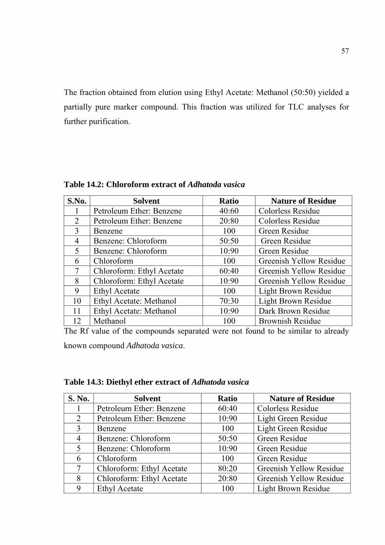
57
The fraction obtained from elution using Ethyl Acetate: Methanol (50:50) yielded a
partially pure marker compound. This fraction was utilized for TLC analyses for
further purification.
Table 14.2: Chloroform extract of Adhatoda vasica
S.No. Solvent Ratio Nature of Residue 1 Petroleum Ether: Benzene 40:60 Colorless Residue 2 Petroleum Ether: Benzene 20:80 Colorless Residue 3 Benzene 100 Green Residue 4 Benzene: Chloroform 50:50 Green Residue 5 Benzene: Chloroform 10:90 Green Residue 6 Chloroform 100 Greenish Yellow Residue 7 Chloroform: Ethyl Acetate 60:40 Greenish Yellow Residue 8 Chloroform: Ethyl Acetate 10:90 Greenish Yellow Residue 9 Ethyl Acetate 100 Light Brown Residue 10 Ethyl Acetate: Methanol 70:30 Light Brown Residue 11 Ethyl Acetate: Methanol 10:90 Dark Brown Residue 12 Methanol 100 Brownish Residue
The Rf value of the compounds separated were not found to be similar to already
known compound Adhatoda vasica.
Table 14.3: Diethyl ether extract of Adhatoda vasica
S. No. Solvent Ratio Nature of Residue 1 Petroleum Ether: Benzene 60:40 Colorless Residue 2 Petroleum Ether: Benzene 10:90 Light Green Residue 3 Benzene 100 Light Green Residue 4 Benzene: Chloroform 50:50 Green Residue 5 Benzene: Chloroform 10:90 Green Residue 6 Chloroform 100 Green Residue 7 Chloroform: Ethyl Acetate 80:20 Greenish Yellow Residue 8 Chloroform: Ethyl Acetate 20:80 Greenish Yellow Residue 9 Ethyl Acetate 100 Light Brown Residue

58
10 Ethyl Acetate: Methanol 50:50 Light Brown Residue 11 Ethyl Acetate: Methanol 10:90 Dark Brown Residue 12 Methanol 100 Brownish Residue
The Rf value of the compounds separated were not found to be similar to already
known compound Adhatoda vasica.
4.5 Isolation of compounds from Adhatoda vasica using thin layer chromatography
The compounds present in the extracted fractions were resolved by TLC using ethyl
acetate and methanol at different ratios. The results are presented in Table 15.1, 15.2
and 15.3.
Table 15.1: TLC analysis of methanolic extract of Adhatoda vasica
Detecting Reagent
Distance Run by Solvent
Number of spots
Distance Run by
solute (cm) Rf Value Color spot
10.5 0.8750 Greenish Black 9.4 0.7833 Greenish Black 9 0.7500 Light Green
8.2 0.6833 Green
Iodine 12cm 5
7.4 0.6166 Brown Note: Solvent system Ethyl Acetate: Methanol (10:90)
The spot at RF value 0.6833 matches with that of the standard marker Vasicine was
collected and utilized for further HPLC and FTIR analyses.
Table 15.2: TLC analysis of chloroform extract of Adhatoda vasica
Detecting Reagent L Number
of spots Distance Run by solute (cm)
Rf Value Color spot
8 0.8000 Light Green 8.2 0.8200 Green 7.5 0.7500 Light Brown 7.2 0.7200 Dark Brown
Iodine 10cm 5
6.8 0.6800 Dark Brown
Note: Solvent system (ethyl acetate: methanol (10:90))

59
None of the spots were matching with that of the standard marker Vasicine.
Table 15.3: TLC analysis of diethyl ether extract of Adhatoda vasica
Detecting Reagent
Distance Run by Solvent
Number of spots
Distance Run by solute
(cm)
Rf Value Color spot
10.2 0.9272 Light Green 9.6 0.8727 Light Green 9.2 0.8363 Dark Brown
8.4 0.7636 Brownish Orange
Iodine 11cm 5
8 0.7272 Yellow
Note: Solvent system: ethyl acetate: methanol (10:90)
None of the spots were matching with that of the standard marker Vasicine.
4.6. Pharmacological Evaluation of Plant Extracts
4.6.1 Immunomodulatory activity
Chloroform extracts of M. pudica, A. hexapetalus and A. vasica were found to be
modulating the innate immune response by increasing the neutrophil counts as
assayed by the neutrophil adhesion assay. The percentage of activity was found to
be 64.99, 54.12 and 84.30 for M. pudica, A. hexapetalus and A. vasica, respectively.
Methanolic extracts of all three plants were found to positively modulate the
immune response. Their values were found to be 78.73%, 71.80% and 94.72% for
M. pudica, A. hexapetalus and A. vasica, respectively. The diethyl ether extracts
were not found to positively influence the immune response, with values of 43.29%,
34.95% and 63.31% respectively (Figure 9).
Various extracts of Mimosa pudica, Artabotrys hexapetalus and Adhatoda vasica
administered at a dose of 200 and 400mg/kg/ per oral for 8 days significantly
inhibited the adhesion of neutrophils to nylon a fibre which stimulates the process of
margination of cells in the blood vessels. The methanol extracts of the plants to
produce maximum activity in the study. At the site of inflammation the chloroform

60
and ether extracts of selected plant to reduced the number of neutrophils and thus
decreased their phagocytosis action and the release of various enzymes and
mediators that make inflammation (Figure 10).
Percentages of neutrophil adhesion observed were on 8th day after treatment with
both the plant extracts. The percentage increase in neutrophil adhesion after
Mimosa pudica, Artabotrys hexapetalus and Adhatoda vasica
methanolextractsshowed a dose dependent activity. The methanol extract of Mimosa
pudica, Artabotrys hexapetalus and Adhatoda vasica at a dose of 400mg/kg showed
the highly significant activity in the study (Figure 11).
Figure 9: Analysis of haemotological parameters in rats treated with the extracts of
MP, AH and AV
UB - Untreated blood; NFTB – Nylon fibre treated blood; MP- M. pudica; AH- A. hexapetalus; AV- A. vasica
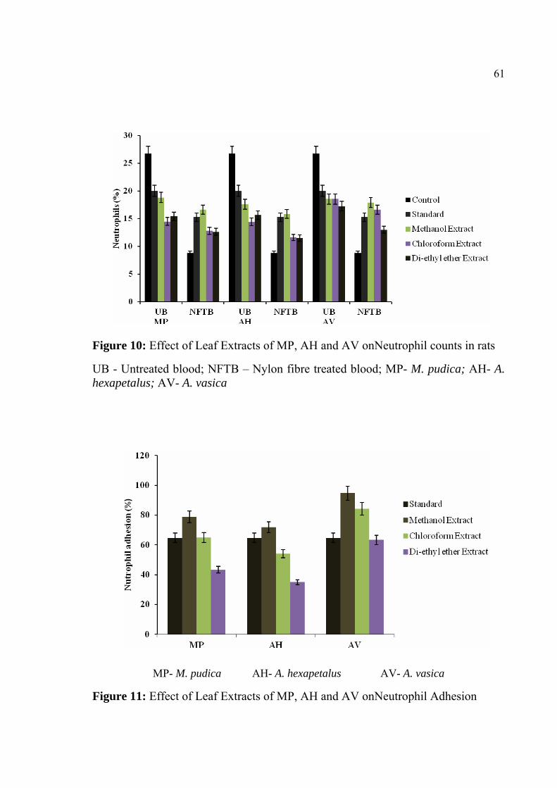
61
Figure 10: Effect of Leaf Extracts of MP, AH and AV onNeutrophil counts in rats
UB - Untreated blood; NFTB – Nylon fibre treated blood; MP- M. pudica; AH- A. hexapetalus; AV- A. vasica
MP- M. pudica AH- A. hexapetalus AV- A. vasica
Figure 11: Effect of Leaf Extracts of MP, AH and AV onNeutrophil Adhesion

62
4.6.2 Delayed type hypersensitivity test
Delayed type hypersensitivity reaction has been widely used as one of the
parameters to measure cell-mediated immune response of the animal (Shwetha et
al., 2012). The reaction was measured by the percent increase in the paw volume
over the control animal. A significant percent increase in paw volume was observed
with diethyl ether extracts of all the three plants, the values being 23.80%, 24.20%
and 20.10% for M. pudica, A. hexapetalus and A.vasica respectively. A significantly
present in paw volume was observed with methanol extracts of all the three plants,
the values being 15.20%, 15.80% and 11.56% for M. pudica, A. hexapetalus and A.
vasica respectively. A significantly present in paw volume was observed with
chloroform extracts of all the three plants, the values being 19.40%, 19.60% and
15.60% for M. pudica, A. hexapetalus and A. vasica respectively (Table 16). These
results were comparable to that of the positive control (30.44%) (Figure12).
Increase in DTH reaction in rats in response to thymus-dependent antigen suggests
that the stimulatory effect of NAEE on T-lymphocytes and accessory cell types is
required for the expression of the reaction (Luster et al., 1982).
Table 16: Effect of MP, AH and AV extracts on delayed type hypersensitivity footpad thickness
S. No. Group (Treatment) Dose (mg/Kg p.o)
Paw volume at 24 hrs (%)
1. Negative control (normal saline) 10ml/Kg 10.22±3.68 2. Positive control
(PBS + Levamisole 50mg/kg) 10ml/Kg 30.44 ± 5.24
3. MPME 400 mg/kg 15.20 ± 4.38* (50.06)
4. MPCE 400 mg/kg 19.40 ± 4.34* (36.44)
5. MPDEEE 400 mg/kg 23.80 ± 4.30* (21.81)
6. AHME 400 mg/kg 15.80 ± 3.38* (48.09)

63
7. AHCE 400 mg/kg 19.60 ± 3.26* (35.61)
8. AHDEEE 400 mg/kg 24.20 ± 3.12* (20.49)
9. AVME 400 mg/kg 11.56 ± 4.37* (62.02)
10. AVCE 400 mg/kg 15.60 ± 4.30* (48.75)
11. AVDEEE 400 mg/kg 20.10 ± 4.22* (33.96)
Results are expressed as mean ± SEM from five observations as compared to control group by students “t” test, n=5; *P<0.05; In brackets inhibition % is reported.
MP- M. pudica AH- A. hexapetalus AV- A. vasica
Figure 12: Effect of MP, AH and AV extracts on delayed type hypersensitivity
footpad thickness
4.7 Hepatoprotective Activity Screening
4.7.1 Acute toxicity studies
In acute toxicity study, it was found that the animals were safe up to a
maximumdose of 2000mg/kg body weight in rats, there were no changes in normal
behavioral pattern and no signs and symptoms of toxicity and mortality were

64
observed and hence the extract was considered to be safe and non-toxic for further
pharmacological screening.
4.7.2 Hepatoprotective activity
The hepatoprotective ability of the plant extracts was assessed by their ability to
protect the liver from CCl4 injury. Four marker enzymes SGPT, SGOT, ALP and
TBL were used for assessing hepatoprotective ability. All three extracts of plants
tested (M. pudica, A. hexapetalus and A. vasica) were found to be protective against
CCl4 injury. The animals were found to be markedly recovering from CCl4 effect as
noted from the activity of the marker enzymes (Figure 13, 14 and 15). The enzyme
activity was found to decrease about one-half the injured liver and was almost
equivalent to control (Farooq et al., 1997).
Figure 13: Effect of M. pudica on CCl4 induced hepatotoxicity in rats

65
Figure 14: Effect of A. hexapetalus on CCl4 induced hepatotoxicity in rats
Figure 15: Effect of A. vasica on CCl4 induced hepatotoxicity in rats

66
4.8 Histopathological section of liver
Histopathological liver sections of the Control group showed normal cellular
architecture with distinct hepatic cells, sinusoidal spaces, and central vein.
Disarrangement of normal hepatic cells with necrosis and vacuolization were
observed in Carbon tetrachloride intoxicated liver. The liver sections of the rat
treated with 200mg/kg bodyweight p.o. of methanolic, chloroform and diethyl ether
extracts of the selected plant, followed by Carbon tetrachloride intoxication, showed
less vacuole formation and absence of necrosis. Overall the less visible changes
observed were comparable with the standard silymarin (Mitra et al., 1998) (Table
17) (Figure 16).
Table 17: Effect of MP, AH and AV extracts on carbon tetrachloride induced hepatotoxicity
Group Dose SGPT(U/L) SGOT(U/L) ALP(U/L) TBL(mg/dl)
I-Control 3ml/kg p.o. 127.16±5.12 106.40±2.60 200.80±5.80 1.18±0.6
II-Ccl4 0.5ml/kg i.p. 285.10±35.60a
244.30±38.20a
428.60±47.60a 2.98±0.38a
III-Silymarin
100mg/kg p.o
124.10±11.60b 104.60±6.60b 194.60±7.20b 1.38±0.32
b
IV-MPME 200mg/kg p.o
132.20±15.16b 119.80±9.30b 205.60±49.80
b 1.52±0.34b
V-MPCE 200mg/kg p.o 156.62±8.14b 141.62±16.2
0b 220.80±32.60b
1.68±0.12b
VI-MPDEE 200mg/kg p.o 174.14±6.22b 172.24±1.60b 250.62±16.20
b 1.86±2.40b
VII-AHME 200mg/kg p.o
140.12±12.40b 125.12±6.40b 240.26±10.12
b 1.69±0.62b
VIII-AHCE 200mg/kg p.o 156.28±6.12b 139.81±8.12b 268.62±9.18b 1.92±0.84
b IX-AHDEEE
200mg/kg p.o 178.28±6.12b 162.74±6.40b 299.14±7.62b 2.34±0.14
b
X-AVME 200mg/kg p.o
130.62±14.12b 114.62±8.14b 203.12±44.62
b 1.49±0.62b

67
XI-AVCE 200mg/kg p.o
144.48±10.26b
132.62±14.40b
218.64±36.14b
1.62±0.24b
XII-AVDEEE
200mg/kg p.o 165.82±7.16b 158.46±2.62b 240.38±18.14
b 1.74±0.26b
Values are expressed as mean SEM; n=6 in each group, aP<0.01 Vs control group, bP<0.01 Vs CCl4 – treated group (ANOVA followed by Dunnet’s t- test).
a) b) C)
Control CCl4: central vein is damaged Standard
d) e) f)
MP-Methanolic extract: Central vein MP-Chloroform extract: Central vein and MP-Diethyl ether extract : and surrounding hepatocytes are seen. surrounding hepatocytes are seen without necrosis. There is no evidence of necrosis.
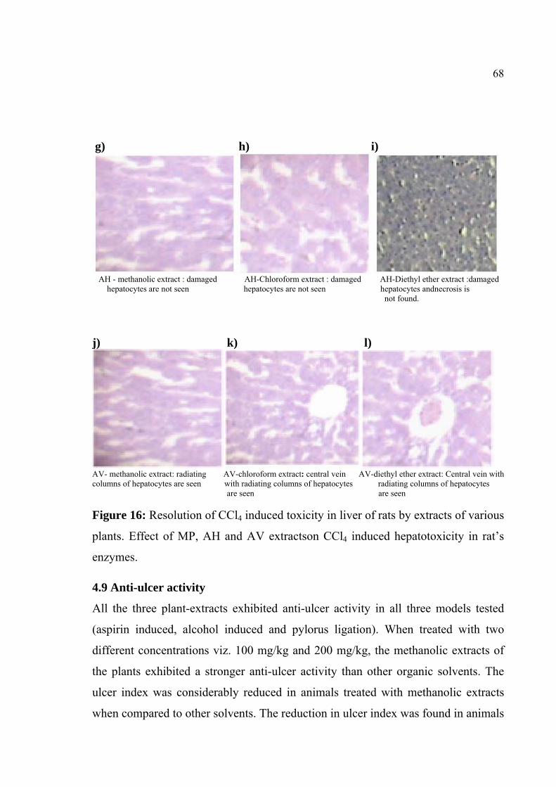
68
g) h) i)
AH - methanolic extract : damaged AH-Chloroform extract : damaged AH-Diethyl ether extract :damaged hepatocytes are not seen hepatocytes are not seen hepatocytes andnecrosis is not found.
j) k) l)
AV- methanolic extract: radiating AV-chloroform extract: central vein AV-diethyl ether extract: Central vein with columns of hepatocytes are seen with radiating columns of hepatocytes radiating columns of hepatocytes are seen are seen Figure 16: Resolution of CCl4 induced toxicity in liver of rats by extracts of various
plants. Effect of MP, AH and AV extractson CCl4 induced hepatotoxicity in rat’s
enzymes.
4.9 Anti-ulcer activity
All the three plant-extracts exhibited anti-ulcer activity in all three models tested
(aspirin induced, alcohol induced and pylorus ligation). When treated with two
different concentrations viz. 100 mg/kg and 200 mg/kg, the methanolic extracts of
the plants exhibited a stronger anti-ulcer activity than other organic solvents. The
ulcer index was considerably reduced in animals treated with methanolic extracts
when compared to other solvents. The reduction in ulcer index was found in animals

69
treated with methanol extracts of all three plants. The reduction in ulcer index was
statistically significant and comparable to that of the standard drug Ranitidine (20
mg/kg).
4.9.1 Aspirin induced ulcer
Aspirin-induced gastric ulceration in rats was another model used to study the effect
of extracts. The methanolic extract was found to possess remarkable ulcer-
protective properties at 100 and 200 mg/kg when compare to other two extracts. The
ulcer protection was found to be 70.46%, 57.84% and 46.46% at 200 mg/kg
respectively for methanolic, chloroform and diethyl ether extracts of M. pudica. The
ulcer protection was 66.15%, 54.76% and 47.07% (Figure 17c) at 200 mg/kg for
methanolic, chloroform and diethyl ether extracts of A. hexapetalus and 73.23%,
56.00% and 49.23% were produced at 200 mg/kg for methanolic, chloroform and
diethyl ether extracts of A. vasica; the standard drug, Ranitidine at 20 mg/kg gave
81.53% of ulcer protection (Table 18: Figure 18).
4.9.2Alcohol induced ulcer
The results obtained with the alcohol-inducedgastric ulceration model in rats were
comparable to that of the aspirin induced ulcer. Here also the methanolic extract was
found to exhibit good ulcer-protective properties at 100 and 200 mg/kg when
compare to other two extracts. The ulcer protection was found to be 69.53%,
58.76% and 47.38% at 200 mg/kg for methanolic, chloroform and diethyl ether
extracts of M. pudica; the protection was 67.07%, 56.61% and 48.30%(Figure 17d)
at 200 mg/kg for methanolic, chloroform and diethyl ether extracts of A.
hexapetalus and 74.46%, 57.84% and 49.84% were produced at 200 mg/kg for
methanolic, chloroform and diethyl ether extracts of A. vasica and the standard drug
(Ranitidine 20 mg/kg) gave 81.53% of ulcer protection. Pre-treatment of rats with
M. pudica extracts produced a dose dependent protection in the ethanol induced

70
ulceration model as compared to control group (Table 18). However the protection
was statistically significant reduced the severity of ulcer and caused a significant
reduction of ulcer index in this model. Ranitidine produced significant gastric ulcer
protection as compared to control group (Figure 18).
4.9.3 Pylorus ligation induced ulcer
The results obtained in the experimental model of Pylorus ligation induced gastric
ulceration in rats. The methanolic extract was found to possess remarkable ulcer-
protective properties at 100 and 200 mg/kg when compare to other two extracts. The
methanolic extract was found to possess remarkable ulcer-protective properties at
200 mg/kg when compare to other two extracts. The maximum effect of ulcer
protection (70.10%), (58.69%) and (46.73%) were produced at 200 mg/kg for
methanolic, chloroform and diethyl ether extracts of M. pudica, (67.39%), (56.52%)
and (48.36%) (Figure 17e)were produced at 200 mg/kg for methanolic, chloroform
and diethyl ether extracts of A. hexapetalus and (73.91%), (57.06%) and (50.00%)
were produced at 200 mg/kg for methanolic, chloroform and diethyl ether extracts
of A. vasica and the standard drug (Ranitidine 20 mg/kg) gave 80.70% of ulcer
protection (Table 19; Figure 18).
The methanolic, chloroform and diethyl ether extracts of the M. pudica in the doses
of 200 mg/kg produced a reduction in the ulcer index, gastric volume, free acidity,
total acidity and raised gastric pH significantly in comparison with control group.
Ranitidine reference drug produced significant reduction of gastric ulcer and total
acid output as compared to control group. The present study indicates that the
methanolic extract of selected M. pudica significantly reduces the total volume of
gastric juice, free and total acidity of gastric secretion and also has activity against
gastric ulcers in rats when compare to other two extracts. The control animals had

71
ulcers and haemorrhagic streaks. Where as in animals administered with the extracts
of M. pudica there was significant reduction in ulcer index.
Preliminary phytochemical screening revealed the presence of Alkaloids, Steroids,
polyphenolic constituents like flavonoids, Saponins, glycosides, tannins, gums and
mucilages. Acute toxicity studies of the various extracts of the M. pudica did not
exhibit any signs of toxicity up to 2 g/kg body weight. Since there was no mortality
of the animals found at high dose. Hence 100 and 200 mg/kg dose of the extract
selected for evaluation of anti-ulcer activity.
Table 18: Effect of various plant extracts on aspirin and alcohol induced gastric ulcer in rats
Aspirin Alcohol Treatment
Dose (mg/kg)
p.o. Ulcer Index
% of ulcer protection
Ulcer Index
% of ulcer protection
Control (Normal saline)
2ml/kg 6.5± 0.50 _ 6.5± 0.50 _
Standard (Ranitidine) 20mg/kg 1.20± 0.24 81.53 1.20± 0.24 81.53
100mg/kg 4.14±0.24 36.30 4.10±0.22 36.92 MPME 200mg/kg 1.92± 0.32 70.46∗∗∗ 1.98± 0.38 69.53∗∗∗ 100mg/kg 4.68±0.28 28.00 4.62±0.25 28.92 MPCE 200mg/kg 2.74± 0.36 57.84∗ 2.68± 0.33 58.76∗ 100mg/kg 4.86±0.29 25.23 4.82±0.26 25.84 MPDEEE 200mg/kg 3.48± 0.33 46.46 3.42± 0.29 47.38 100mg/kg 4.22±0.28 35.07 4.18±0.26 35.69 AHME 200mg/kg 2.20± 0.30 66.15∗∗ 2.14± 0.32 67.07∗∗ 100mg/kg 4.74±0.24 27.07 4.60±0.28 29.23 AHCE 200mg/kg 2.94± 0.34 54.76∗ 2.82± 0.30 56.61∗ 100mg/kg 5.14±0.22 20.92 5.06±0.22 22.15 AHDEEE 200mg/kg 3.44± 0.36 47.07 3.36± 0.32 48.30 100mg/kg 4.20±0.27 35.38 4.10±0.24 36.92 AVME 200mg/kg 1.74± 0.36 73.23∗∗∗ 1.66± 0.34 74.46∗∗∗

72
100mg/kg 4.60±0.24 29.23 4.44±0.27 31.69 AVCE 200mg/kg 2.86± 0.34 56.00∗ 2.74± 0.31 57.84∗ 100mg/kg 4.92±0.26 24.30 4.82±0.28 25.84 AVDEEE 200mg/kg 3.30± 0.35 49.23 3.26± 0.26 49.84
Results are mean ± S.E.M. (n=5); statistical comparison was performed by using ANOVA coupled with student’s t-test.*P<0.05, **P<0.01, ***P<0.001 were consider statistically significant when compared to control group.

73 Table 19: Effect of plant extracts of MP, AH and AV against pylorus ligation induced gastric ulcer in rats
Treatment Dose (mg/kg) p.o
Volume of gastric juice (ml/4h) PH Free Acidity
(mEq/L) Total Acidity
(mEq/L) Ulcer Index %Inhibition of ulcer
Control (Normal saline) 2ml/kg 4.02± 0.11 1.84± 0.14 26.84±
0.08 70.16± 0.30 3.68± 0.56 _
Standard (Ranitidine) 20mg/kg 1.94± 0.06 4.96± 0.18 10.42± 0.02 22.24± 0.18 0.71± 0.14 80.70
100mg/kg 3.66± 0.16 3.12± 0.14 21.18± 0.05 52.14± 0.38 2.34± 0.24 36.41 MPME 200mg/kg 2.40± 0.14 4.56± 0.18 11.76± 0.06 30.62± 0.26 1.10± 0.29 70.10*** 100mg/kg 3.79± 0.16 3.20± 0.14 22.96± 0.08 60.48± 0.24 2.62± 0.36 28.80 MPCE 200mg/kg 3.28± 0.21 3.88± 0.16 13.68± 0.02 35.45± 0.33 1.52± 0.44 58.69* 100mg/kg 3.86± 0.14 2.86± 0.14 25.54± 0.04 64.16± 0.19 2.76± 0.49 25.00 MPDEEE 200mg/kg 3.68± 0.12 3.24± 0.16 16.62± 0.06 39.52± 0.32 1.96± 0.56 46.73 100mg/kg 3.66± 0.12 3.12± 0.14 20.14± 0.02 54.62± 0.68 2.42± 0.24 34.23 AHME 200mg/kg 2.40± 0.14 4.48± 0.12 11.64± 0.03 33.62± 0.42 1.20± 0.28 67.39∗∗∗ 100mg/kg 3.78± 0.16 3.22± 0.18 18.46± 0.05 64.96± 0.62 2.66± 0.44 27.71 AHCE 200mg/kg 3.14± 0.12 3.82± 0.14 14.56± 0.03 39.76± 0.46 1.60± 0.48 56.52∗∗ 100mg/kg 3.98± 0.15 2.80± 0.18 23.86± 0.08 69.12± 0.12 2.88± 0.52 21.73 AHDEEE 200mg/kg 3.67± 0.16 3.20± 0.12 18.42± 0.02 43.42± 0.42 1.90± 0.56 48.36 100mg/kg 3.62± 0.14 3.14± 0.15 22.16± 0.03 56.34± 3.16 2.32± 0.20 36.95 AVME 200mg/kg 2.42± 0.18 4.52± 0.14 12.86± 0.04 32.46± 0.20 0.96± 0.22 73.91∗∗∗ 100mg/kg 3.74± 0.18 3.18± 0.16 20.84± 0.04 62.84± 0.42 2.50± 0.42 32.06 AVCE 200mg/kg 3.16± 0.16 3.86± 0.12 15.46± 0.02 38.54± 0.32 1.58± 0.46 57.06∗∗ 100mg/kg 3.92± 0.17 2.84± 0.16 24.84± 0.06 68.64± 0.39 2.84± 0.54 23.36 AVDEEE 200mg/kg 3.64± 0.14 3.18± 0.10 18.86± 0.02 42.58± 0.34 1.84± 0.52 50.00∗
Results are mean ± S.E.M.(n =5). Statistical comparison was performed by using ANOVA coupled with student’s t-test.* P<0.05, ** P<0.01, *** P<0.001 were consider statistically significant when compared to control group.
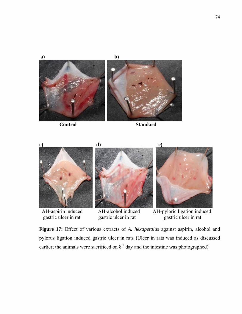
74
a) b)
Control Standard
c) d) e)
AH-aspirin induced AH-alcohol induced AH-pyloric ligation induced gastric ulcer in rat gastric ulcer in rat gastric ulcer in rat Figure 17: Effect of various extracts of A. hexapetalus against aspirin, alcohol and
pylorus ligation induced gastric ulcer in rats (Ulcer in rats was induced as discussed
earlier; the animals were sacrificed on 8th day and the intestine was photographed)

75
MP- M. pudica AH- A. hexapetalus AV- A. vasica
Figure 18: Effect of various extracts of MP, AH and AV against Anti-ulcer activity in
rats
4.10 Wound healing Activity
All three plant extracts exhibited wound healing activity. A lower wound healing
ability was observed with the diethyl extract whereas the activity was higher in
methanolic extracts. The methanolic extract of M. pudica exhibited a higher activity of
93.87% whereas the methanolic extracts of A. hexapetalus and A. vasica exhibited
78.61% and 87.46% respectively.
4.10.1 M. pudica (MP)
The wound healing activity was studied by using five groups; Group I negative control
simple ointment, In Group II positive control Nitrofurazone ointment (0.2% w/w),
Group III MPME, Group IV MPCE and Group V MPDEE. The size of the wound in

76
surface area, On the Day 1 (50.24) (50.36) (51.26) (50.54) (50.42). On the Day 4
(48.24) (28.26) (38.46) (38.36) (48.46). On the Day 8 (44.20) (12.56) (28.26) (30.26)
(40.32). On the Day 12 (40.46) (3.14) (12.56) (20.54) (36.16). On the Day 16 (35.46)
(0.758) (3.14) (18.76) (20.45). The mean percentage closure of excision wound model
on Day 16 (40.45) (98.44) (93.87) (80.72) (70.76) (Table 20; Figure 19 and 22).
4.10.2 A. hexapetalus(AH)
The wound healing activity was studied by using five groups; Group I negative control
base ointment, In Group II positive control Nitrofurazone ointment (0.2% w/w), Group
III AHME, Group IV AHCE and Group V AHDEE. The size of the wound was
determined by measuring the surface area. The area of wound on Day 1 was found to
be 48 to 50.24sq.mm. After 16 days, the size of the wound, in animals treated with A.
vasica extracts, were found to be reduced to 6.42 sq.mm, 9.80 sq.mm and 15.60 sq.mm
for methanol, chloroform and diethyl ether extracts (Table 20; Figure 20 and 22).
Contraction of the excision wound was observed from Day 4 and it progressed till Day
16. The percent wound contraction after 16 days was found to be 98.44, 78.61, 70.82
and 65.46 respectively in nitrofurazone, methanol, chloroform and diethyl ether extract
treated groups. Significant wound contraction was observed on 16th day for all treated
groups (p<0.001 for standard and methanolic extracts; p<0.01 for chloroform and
diethyl ether extracts), in comparison with the control group. Time for complete
epithelization was significantly short in drug and standard treated groups.
The epithelization of wound in case of rat treated with extracts was found to be quite
earlier than control. It is also comparable with the marketed preparation. It suggests
that the leaves extracts of Artabotrys hexapetalus promoted wound healing activity.
The excision wound model showed excellent wound healing property in methanolic
leaf extract which was well compared with standard drug.

77
4.10.3 A. vasica(AV)
The wound healing activity was studied by using five groups; Group I negative control
simple ointment, In Group II positive control Nitrofurazone ointment (0.2% w/w),
Group III AVME, Group IV AVCE and Group V AVDEE. The size of the wound in
surface area, On the Day 1 (50.24) (50.36) (51.16) (50.62) (49.84). On the Day 4
(48.24) (28.26) (38.14) (36.90) (37.10). On the Day 8 (44.20) (12.56) (27.84) (27.52)
(29.34). On the Day 12 (40.46) (3.14) (16.12) (18.42) (21.64). On the Day 16 (35.46)
(0.758) (6.42) (9.80) (15.60). The mean percentage closure of excision wound model
on Day 16 (40.45) (98.44) (87.46) (80.65) (68.70) (Table 20; Figure 21 and 22).
Contraction of the excision wound was promoted from day 1 to day 16. In excision
wounds, wound contraction was 98.44, 87.46, 80.65 and 68.70% respectively on 16th
day for nitrofurazone, methanolic, chloroform and diethyl ether extract treated groups.
Significant wound contraction was also observed on 16th day for all treated groups
(p<0.001 for standard and methanolic extracts; p<0.01for chloroform and diethyl ether
extract), in comparison with the control group. Time for complete epithelization was
significantly short in drug and standard treated groups.
The epithelization of wound in case of rat treated with extracts was found to be quite
earlier than control. It is also comparable with the marketed preparation. It suggests
that the leaves extracts of Adhatoda vasica promoted woundhealing activity. The
excision wound model showed excellent wound healing property in methanolic leaf
extract which was well compared with standard drug.

78
Table 20: Effect of methanolic, chloroform and diethyl ether extract ointments of MP, AH and AV on excision wound model
Size of wound surface area (mm 2) Group
Avg. wt of
animal Drug /extract Day
0 Day
1 Day
4 Day
8 Day 12
Day 16
% wound healing
I Control 50.24 50.24 48.24 44.20 40.46 35.46 40.45
II
Nitrofurazone ointment (0.2% w/w)
50.36 50.36 28.26 12.56 3.14 0.758 98.44
III
MPME (10%w/w) 51.26 51.26 38.46 28.26 12.56 3.14 93.87
**
IV MPCE(10%w/w) 50.54 50.54 28.36 30.26 20.54 18.76 80.72 **
V
150-200 gm
MPDEEE (10%w/w) 50.42 50.42 48.46 40.32 36.16 20.45 70.76**
VI AHME (10%w/w) 48.60 48.60 36.20 27.12 18.14 10.40 78.61**
VII AHCE (10%w/w) 49.20 49.20 35.22 28.84 21.20 14.36 70.82**
VIII AHDEEE (10%w/w) 50.42 50.42 38.96 32.14 24.20 17.42 65.46*
IX AVME (10%w/w) 51.16 51.16 38.14 27.84 16.12 6.42 87.46**
X AVCE (10%w/w) 50.62 50.62 36.90 27.52 18.42 9.80 80.65**
XI
150-200 gm
AVDEEE 49.84 49.84 37.10 29.34 21.64 15.60 68.70*
Values are mean ± SEM of 5 animals in each group. Numbers in Parenthesis indicate
percentage of wound contraction. * P<0.01, **P<0.001Vs respective control by
students t- test.

79
a) b)
Control 0 day Standard 0 day
c) d)
Control 16 day Standard 16 day
e) f) g)
Mimosa pudica Mimosa pudica Mimosa pudica (Diethyl ether Extract10%W/W) (Chloroform Extract10 %W/W) (Methanol Extract 10%W/W)
Figure 19: Effect of methanolic, chloroform and diethyl ether extract ointments of
M.pudica on excision wound model.

80
a) b) c)
Artabotrys hexapetalus Artabotrys hexapetalus Artabotrys hexapetalus (Diethyl ether Extract10%W/W) (Chloroform Extract10 %W/W) (Methanol Extract 10%W/W)
Figure 20: Effect of methanolic, chloroform and diethyl ether extract ointments of
Artabotrys hexapetalus on excision wound model.
a) b) c)
Adhatoda vasica Adhatoda vasica Adhatoda vasica (Diethyl ether Extract10%W/W) (Chloroform Extract10 %W/W) (Methanol Extract 10%W/W) Figure 21: Effect of methanolic, chloroform and diethyl ether extract ointments of
Adhatoda vasica on excision wound model.

81
MP- Mimosa pudica AH- Artabotrys hexapetalus AV- Adhatoda vasica
Figure 22: Effect of methanolic, chloroform and diethyl ether extract ointments of MP,
AH and AV on excision wound model.
4.11. Wound Healing Activity of Fractions of Extracts
Among the different fractions of M. pudica extract that were tested (aqueous, Hexane,
n-butanol, chloroform and ethyl acetate fractions) for wound healing activity the n-
butanol fraction exhibited a higher wound healing activity (71.70%); this was
comparable to that of the standard nitrofurazone ointment (0.2% w/w).
Contraction of the excision wound was promoted from day 1 of the treatment and
healing was observed on day 16. The epithelization of wound in case of rat treated with
fraction of extracts was found to be quite earlier than control. It is also comparable with
the commercial product available in the market. It suggests that the leaves extracts of
M. pudica promoted wound healing activity. The excision wound model showed

82
excellent wound healing property in methanol leaf extract which was well compared
with standard drug.
The results are given control (31.24%), standard (89.94%), hexane fraction (51.36%)
(Figure 23c), aqueous fraction-(67.56%) (Figure 23d), Ethyl acetate fraction-(61.81%)
(Figure 23e), chloroform fraction (57.52%) (Figure 23f) and n-butanol fraction
(71.70%) (Figure 23g). Overall n-butanol and aqueous fractions showed higher wound
healing activity compared to other fractions (Table 21; Figure 24).
Fractions and their wound healing activity:
1. Aqueous fraction
2. Hexane fraction
3. Chloroform fraction
4. N-butanol fraction
5. Ethyl acetate fraction
Table 21: Effect of methanolic extract ointments of Mimosa pudica fractions and their wound healing activityon excision wound model
Size of wound surface area (mm2) Group
Avg. wt of
animal
Drug /extract Day
0 Day
1 Day
4 Day
8 Day 12
Day 16
Percentage of wound healing
I Control 51.48 51.48 48.10 43.12 40.56 35.40 31.24
II
Nitrofurazone ointment
(0.2% w/w) 51.66 51.66 40.62 32.10 17.84 5.20 89.94
III
150-200 gm.
Aqueous fraction
(10%w/w) 50.80 50.80 45.40 38.64 30.60 16.48 67.56

83
IV
Hexane fraction
(10%w/w) 48.64 48.64 45.24 39.84 34.30 23.66 51.36
V
Chloroform fraction
(10%w/w) 49.10 49.10 44.20 37.64 28.94 20.86 57.52
VI N-butanol fraction
(10%w/w) 50.38 50.38 44.20 36.46 29.82 14.26 71.70
VII Ethyl acetate
fraction (10%w/w)
50.22 50.22 45.24 37.94 30.24 19.18 61.81
Values are mean ± SEM. Statistical comparison was performed by using ANOVA and
student’s t- test. Numbers in Parenthesis indicate percentage of wound contraction.
P<0.001 respective control.

84
a) b)
Control Standard c) d) e)
Hexane fraction Aqueous fraction Ethyl acetate fraction f) g)
Chloroform fraction n-Butanol fraction Figure 23: Effect of methanolic extract ointments of Mimosa pudica fractions and their
wound healing activityon excision wound model.

85
Figure 24: Effect of methanolic extract ointments of Mimosa pudica fractions and their
wound healing activity on excision wound model.
4.12 HPLC analysis
4.12.1 HPLC analysis of methanolic extract of Mimosa pudica
HPLC analysis of partially purified methanolic extract of Mimosa pudica presented a
distinct peak at a retention time of 9.141 which is similar to standard mimopudine
(retention time 9.439). Few more peaks with varying retention times were also
observed in this fraction.

86
AU
0.00
0.10
0.20
0.30
0.40
Minutes2.00 4.00 6.00 8.00 10.00 12.00 14.00 16.00 18.00 20.00
9.43
9
Figure 25.1: HPLC profile of Mimopudine standard.
AU
0.00
1.00
2.00
3.00
4.00
Minutes1.00 2.00 3.00 4.00 5.00 6.00 7.00 8.00 9.00 10.00 11.00 12.00 13.00 14.00 15.00 16.00 17.00
9.14
1
Figure 25.2: HPLC profile of methanolic extract of Mimosa pudica. A peak similar to
Mimopudine with a retention time of 9.14 was observed.

87
4.12.2 HPLC analysis of methanolic extract of Artabotrys hexapetalus
HPLC analysis of partially purified methanolic extract of A. hexapetalus gave a peak
with a retention time of 11.304 which is similar to the artabotrycinol standard
(retention time 10.788). Few more peaks with varying retention times were also
observed in this fraction.
0.0 2.5 5.0 7.5 10.0 12.5 min
-0.25
0.00
0.25
0.50
0.75
1.00
1.25
1.50
1.75
2.00
2.25
2.50
mAU(x10)254nm,4nm (1.00)
/8.539/5127
Artabotrycinol/10.788/532607
Figure 26.1: HPLC profile of standard Artabotrycinol
0.0 2.5 5.0 7.5 10.0 12.5 min
-0.5
0.0
0.5
1.0
1.5
2.0
2.5
3.0
3.5
4.0
mAU(x1,000)254nm4nm (1.00)
/3.428/15987
/3.853/21710
/4.134/10800
/4.501/6361
/4.770/5704
/5.223/4007
/5.653/22886942
Artaboteycinol/11.304/3571572
Figure 26.2: HPLC profile of methanolic extract of Artabotrys hexapetalus. A peak
similar to Artabotrycinol with a retention time of 11.304 was observed.

88
4.12.3 HPLC analysis of methanolic extract of Adhatoda vasica
HPLC analysis of partially purified methanolic extract of A. vasica gave a peak with a
retention time of 6.433 which is similar to the vasicine standard (retention time 6.22).
Few more peaks with varying retention times were also observed in this fraction.
0.0 1.0 2.0 3.0 4.0 5.0 6.0 7.0 8.0 9.0 min
-1.0
0.0
1.0
2.0
3.0
4.0
5.0
6.0
7.0
8.0
9.0
mAU(x10)302nm,4nm (1.00)
vasicine/6.266/1125785
Figure 27.1: HPLC profile of standard Vasicine
0.0 2.5 5.0 7.5 10.0 min
-1.5
-1.0
-0.5
0.0
0.5
1.0
1.5
2.0
2.5
mAU302nm4nm (1.00)
vasicine/6.443/8879
Figure 27.2: HPLC profile of methanolic extract of Adhatoda vasica. A peak similar to
Vasicine with a retention time of 6.433 was observed.

89
4.13 FTIR SPECTRA OF COMPOUNDS
4.13.1 FTIR Spectral Analysis
The structure of isolated compound was elucidated by Shimadzu – 8400 Series Fourier
Transformer - Infrared Spectrophotometer using KBr pallet method. IR results are
shown below:
4.13.2 IR Spectral Studies
FT-IR spectra of the methanolic extracts of Mimosa pudica. Artabotrys hexapetalus
and Adhatoda vasica confirm the existence of mimopudine, artabotrycinol and vasicine
respectively. Interestingly, the characteristic stretching frequencies of the functional
group (الO-H, الN-H, الC-H, الC-N, الC=O, الC=C), presentin the IR spectra of the methanolic
extracts match with the stretching frequencies obtained for the commercial samples. A
few characteristics peaks were shifted by 1-2cm-1 wave number, which may be due to
the presence of other organic moieties as impurities and solvent effects. The absorption
by the impurities is masked by the strong signals associated with mimopudine,
artabotrycinol and vasicine molecules. These trends confirm the presence of
mimopudine, artabotrycinol and vasicine of the plants (Figure 28-33).

90
Figure 28: FTIR analysis of standard Mimopudine: The absorption frequencies in the
IR spectrum of commercial mimopudine such as γc=0 (conjugated ketone), γN-H
(stretching), γN-H (bending) and γO-H (bending) are identified correctly.
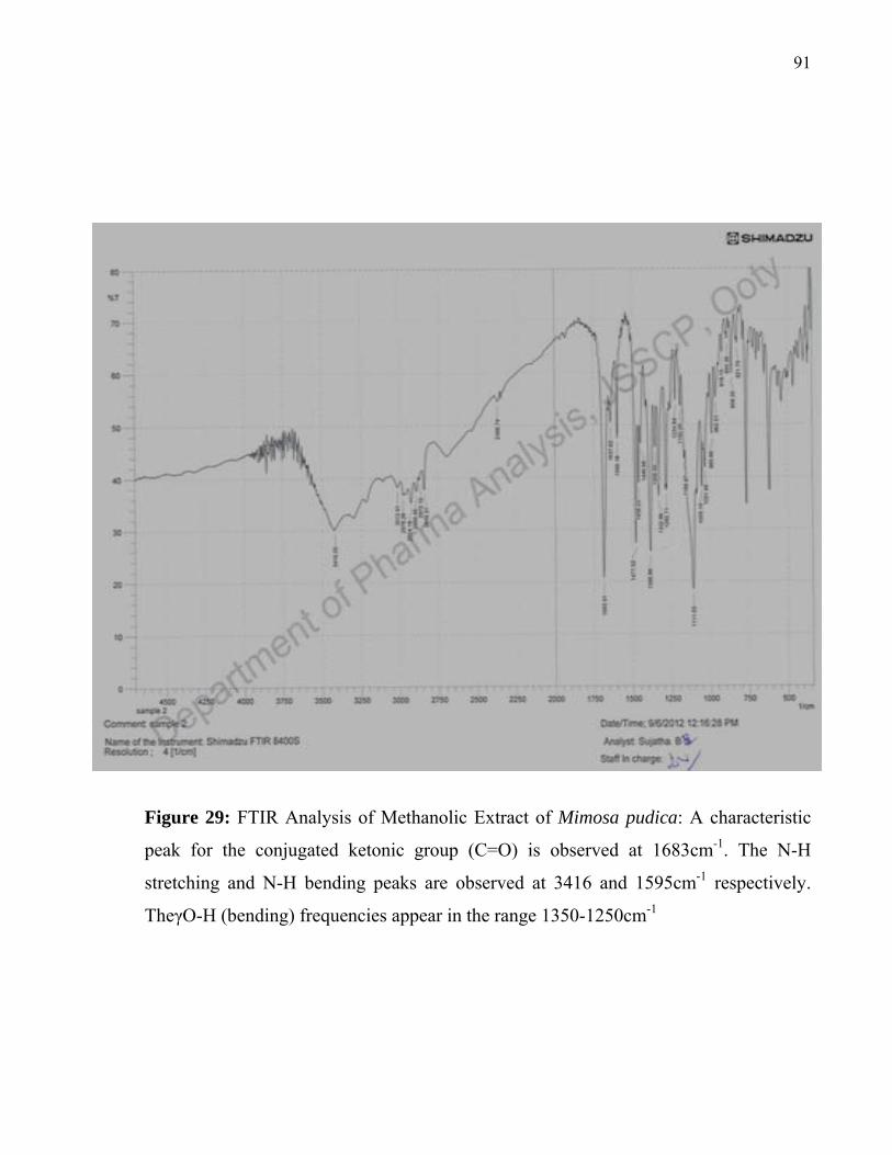
91
Figure 29: FTIR Analysis of Methanolic Extract of Mimosa pudica: A characteristic
peak for the conjugated ketonic group (C=O) is observed at 1683cm-1. The N-H
stretching and N-H bending peaks are observed at 3416 and 1595cm-1 respectively.
TheγO-H (bending) frequencies appear in the range 1350-1250cm-1

92
Figure 30: FTIR analysis of standard Artabotrycinol: The group frequencies for H-
bonded OH stretching, in-plane bending OH, alkenyl (-C=C-) stretching and C-H
stretching are identified correctly from the IR spectrum of commercial Artabotrycinol.

93
Figure 31: FTIR Analysis of Methanolic Extract of A. hexapetalus: A hydrogen
bonded broad band due to OH groups is observed in the range of 3570-3200cm-1.
Intrestingly γO-H frequency portions to -CH2OH group shows a strong band at
1290cm-1 due to in-plane bending vibrations. A characteristic alkenyl (-C=C-)
stretching frequency is observed at 1643cm-1.

94
Figure 32: FTIR analysis of standard Vasicine: The ring frequencies associated with
the aromatic system and group frequencies of imino group (C=N-) and hydroxyl group
are observed correctly from the IR spectrum of commercial Vasicine.
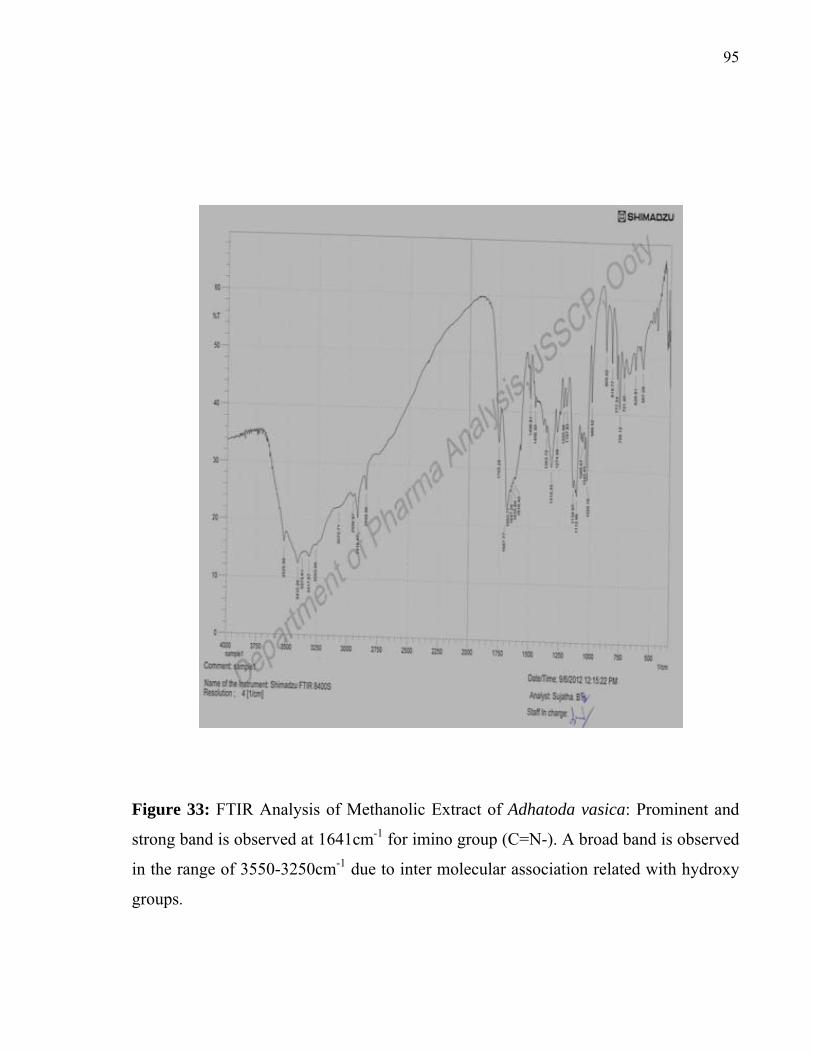
95
Figure 33: FTIR Analysis of Methanolic Extract of Adhatoda vasica: Prominent and
strong band is observed at 1641cm-1 for imino group (C=N-). A broad band is observed
in the range of 3550-3250cm-1 due to inter molecular association related with hydroxy
groups.
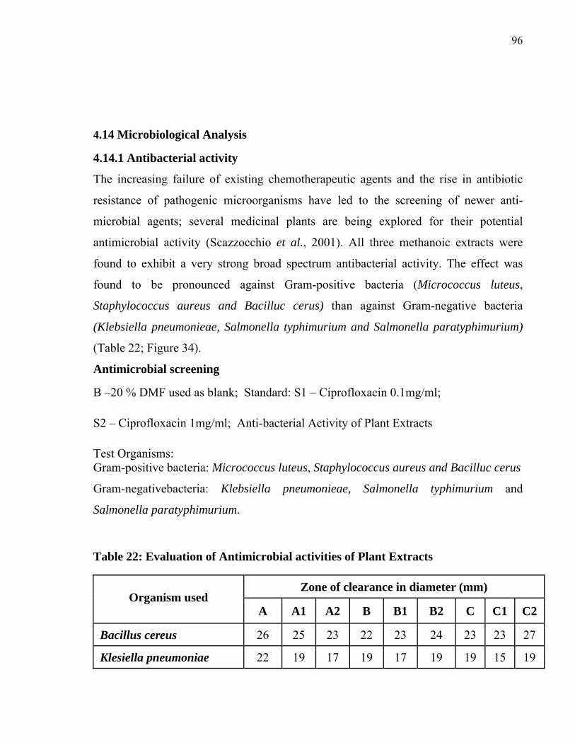
96
4.14 Microbiological Analysis
4.14.1 Antibacterial activity
The increasing failure of existing chemotherapeutic agents and the rise in antibiotic
resistance of pathogenic microorganisms have led to the screening of newer anti-
microbial agents; several medicinal plants are being explored for their potential
antimicrobial activity (Scazzocchio et al., 2001). All three methanoic extracts were
found to exhibit a very strong broad spectrum antibacterial activity. The effect was
found to be pronounced against Gram-positive bacteria (Micrococcus luteus,
Staphylococcus aureus and Bacilluc cerus) than against Gram-negative bacteria
(Klebsiella pneumonieae, Salmonella typhimurium and Salmonella paratyphimurium)
(Table 22; Figure 34).
Antimicrobial screening
B –20 % DMF used as blank; Standard: S1 – Ciprofloxacin 0.1mg/ml;
S2 – Ciprofloxacin 1mg/ml; Anti-bacterial Activity of Plant Extracts
Test Organisms: Gram-positive bacteria: Micrococcus luteus, Staphylococcus aureus and Bacilluc cerus
Gram-negativebacteria: Klebsiella pneumonieae, Salmonella typhimurium and
Salmonella paratyphimurium.
Table 22: Evaluation of Antimicrobial activities of Plant Extracts
Zone of clearance in diameter (mm) Organism used
A A1 A2 B B1 B2 C C1 C2
Bacillus cereus 26 25 23 22 23 24 23 23 27
Klesiella pneumoniae 22 19 17 19 17 19 19 15 19

97
(109)
Salmonella typhimurium B 19 18 18 16 15 17 14 17 17
Salmonella paratyphimurium 23 23 23 19 23 24 23 18 20
Micrococcus luteus 38 38 34 30 35 38 32 33 36
Staphylococcus aureus 28 25 21 24 23 25 24 22 25
Salmonella typhimurium (1251) 25 22 19 18 18 20 18 18 19
A, A1, A2= Methanolic extract of AH, MP, AV; B, B1, B2= Chloroform extract of
AH, MP, AV; C, C1, C2= Diethyl ether extract of AH, MP, AV
AH- Artabotrys hexapetalus MP- Mimosa pudica AV- Adhatoda vasica

98
Figure 34: Anti bacterial activity of plant extracts treated with Staphylococcus aureus a. A, A1, A2= Methanolic extract of AH, MP, AV; AH- Artabotrys hexapetalus; MP- Mimosa pudica; AV- Adhatoda vasica. b. B, B1, B2 = Chloroform extract of AH, MP, AV; AH- Artabotrys hexapetalus; MP- Mimosa pudica; AV- Adhatoda vasica. c. C, C1, C2= Diethyl ether extract; C, C1, C2= Diethyl ether extract
a. b.
Control CHO-Chloroform DEE-Diethyl ether MET-Methanol DMF-Dimethyl formamide
A, A1, A2= Methanolic extract of AH, MP, AV. AH- Artabotrys hexapetalus; MP- Mimosa pudica; AV- Adhatoda vasica
c. d.
B, B1, B2 = Chloroform Extract of AH, MP, AV. AH- Artabotrys hexapetalus; MP- Mimosa pudica; AV- Adhatoda vasica
C, C1, C2= Diethyl ether Extract; C, C1, C2= Diethyl ether Extract of AH, MP, AV. AH- Artabotrys hexapetalus; MP- Mimosa pudica; AV- Adhatoda vasica

99
of AH, MP, AV. d. Control CHO-Chloroform DEE-Diethyl ether MET-Methanol DMF-Dimethyl formamide. AH-Artabotrys hexapetalus; MP- Mimosa pudica; AV- Adhatoda vasica. 4.15 ANTI-OXIDANT ACTIVITY OF MP, AH AND AV
Crude extracts and TLC- purified methanolic extracts of all three plants exhibited a
potent anti-oxidant activity as measured by Potassium ferricyanide, FRAP and DPPH
assays. The activities of all three plants were comparable in all three assays.
4.15.1 Potassium ferricyanide assay
All the three plant extracts (crude and purified) were tested for potassium ferricyanide
reduction. Among the crude extracts of all the three plants, AV methanolic extracts
shown the higher reduction compare with other two plants methanolic extracts. Further
the TLC purified methanolic samples of all three palnts were tested again. In the
purified form sample A1 gives maximal reduction of potassium ferricyanide (Figure
35.1 and 35.2).

100
Figure 35.1: The reduction of potasium ferricyanide, sample A2 gives more reduction
compared with other crude samples. Sample A and A1 gives followed level of the
reduction respectively.
Figure 35.2: TLC purified samples were tested potasium ferricyanide reduction,
sample A1 gives the maximal reduction when compared with crude samples.
A –AH Methanolic extract, A1-MP Methanolic extract, A2-AV Methanolic extract, B-
AH Chloroform extract, B1-MP Chloroform extract, B2-AV Chloroform extract, C-AH
Diethyl ether extract, C1-MP Diethyl ether extract, C2-AV Diethyl ether extract
AH- Artabotrys hexapetalus MP- Mimosa pudica AV- Adhatoda vasica
4.15.2 Fluorescence Recovery after Photobleaching (FRAP) Assay
All the three plants extracts of crude and purified were tested for the ferric ion
reduction. In the crude extracts of all the three plants, AV methanolic extracts shown
the higher reduction compare with other two plants methanolic extracts. Further,

101
purified AV methanolic samples were tested again. In the purified form sample AV-A2
gives maximal reduction of potassium ferricyanide (Figure 36.1 and 36.2).
Figure 36.1: Crude extracts of all the three plants samples were tested forferric ion
reduction. The AV-A2 samples reduce the maximal level and indicate that higher anti-
oxidative property compared with other samples.
Figure 36.2: The purified AV-A2 samples were tested again forferric ion reduction,
The AV-A2 extracts shown the maximal reduction and anti-oxidant activity. A –AH
Methanolic extract,A1-MP Methanolic extract, A2-AV Methanolic extract, B-AH
Chloroform extract, B1-MP Chloroform extract, B2-AV Chloroform extract, C-AH
Diethyl ether extract, C1-MP Diethyl ether extract, C2-AV Diethyl ether extract.

102
AH- Artabotrys hexapetalus MP- Mimosa pudica AV- Adhatoda vasica
4.15.3 DPPH
The stock solution of plant extracts at a concentration of 100µl/ml of all the extracts
was assayed for antioxidant activity. The maximum inhibition for DPPH radical was
A2 (5.80) for followed by A1 (5.10), A (5.04), B1 (3.04), C (2.90), B (2.66), B2 (2.28),
C1 (2.19) and C2 (2.19). The reducing potential of 2.0, 1.1, 1.04, and 1.02 was
observed in sample B1, B, C, B2, C1 and C2, respectively. The A2-AV methanolic
extracts shows the maximal reduction in crude and purified state (Figure 37.1 and
37.2).
Figure 37.1: The observation of ferric ion reduction, sample A, A1 and A2 shows the
maximal level of absorbance indicates the anti-oxidant activity compare with other
extracts.

103
Figure 37.2: Purified A, A1 and A2 samples were shown the maximum absorbance
level indicates that the anti-oxidant activity compared with crude extracts.
A –AH Methanolic extract, A1-MP Methanolic extract, A2-AV Methanolic extract, B-
AH Chloroform extract, B1-MP Chloroform extract, B2-AV Chloroform extract, C-AH
Diethyl ether extract, C1-MP Diethyl ether extract, C2-AV Diethyl ether extract
AH- Artabotrys hexapetalus MP- Mimosa pudica AV- Adhatoda vasica

104
5. DISCUSSION
Natural products have been the source of most of the active ingredients that are used in
modern medicine (Harvey, 2008). More than 80% of the substances used to make drugs
are natural products or inspired by natural compounds (Sneader, 1996) and half of the
drugs approved since 1994 are based on natural products (Newman and Cragg, 2007;
Butler, 2008). Natural products and /or compounds derived from natural products
continue to play a major role in drug development process (Newman et al., 2003). The
natural products include compounds from plants, microbes and animals and synthetic
or semi-synthetic compounds based on natural drugs. They have been used to treat a
variety of disease conditions such as cancer, infections, diabetic and other metabolic
diseases.
In the present study, leaf extracts of three plants viz. M. pudia, A. hexapetalus and A.
vasica were evaluated for pharmacological activity in animal models.
5.1 Phytochemical studies
The preliminary phytochemical analysis of all three extracts (viz. methanol, chloroform
and diethyl ether) of M. pudica, A. hexapetalus and A. vasica indicate the presence of
alkaloids, flavanoids, tannins, saponins, glycosides, steroids, steroidal terpenes,
phenolic compounds, gums and muciages and carbohydrates.
5.2 Evaluation of Immunomodulatory Activity (neutrophil adhesion assay)
The immunomodulatory agents obtained from plant and animal origin, activate and
enhance the immune response of the host against the invading pathogens (Desai et al.,
1966). Inflammation is one of the conditions where in neutrophils, a part of innate
immunity, play a major role. The present study reveals the immunomodulatory activity

105
of methanolic extracts of the selected plant extracts. Many plant extracts are known to
exhibit anti-inflammatory activities. However, more studies have to be performed to
understand the underlying mechanism of these medicinal plants regard to their
therapeutic activities (Fulzele et al., 2003).
The methanol extracts of all three plants, when administered orally, exhibited
significant immune modulatory activity as measured by two assays: an increase in
adhesion of neutrophils to nylon fibers which correlates to the process of margination
of cells in blood vessels. The neutrophil adhesion was found to be significantly
increased in animals fed with the test extracts compared to untreated control.
Neutrophils circulate in the vasculature in a passive state and become more adhesive
upon stimulation at sites of inflammation; this would be followed by margination to the
vessel wall and subsequent transmigration and phagocytosis. The plant extracts
significantly increased the neutrophil chemotactic movement as indicated by the
increase in number of cells reaching the lower surface of the filter; therefore, the
extracts act as chemo attractants. These results are in comparison to that of a compound
isolated from R. communis leaf extract which significantly increased the neutrophil
chemotactic movement (Konig et al., 1987). Natural products derived antibiotics like
azithromycin are known to exhibit anti-inflammtory activities by suppressing the
abundance of neutrophils (Ivetic Tkalcevic et al., 2006). Another natural product
derived drug telithromycin reduces inflammation through reduction in cytokine
production (Lotter et al., 2006).
At the time of pathogens invasion, macrophages detect the invasion in the tissues and
recruit neutrophils to the affected site from the reserve pool in order to assist them in
eliminating the invaders (Hoffstein et al., 1981). Therefore, macrophages are supported
by neutrophils, which supply the effective antimicrobial neutrophil granules. This

106
would enhance the capacity of the macrophages which do not have these granules in
them (Silva et al., 1989).
5.2.1 Delayed type hypersensitivity activity
Delayed type hypersensitivity reaction is a type IV hypersensitivity reaction
characterized by large influxes of non-specific inflammatory cells. Generally these
cells are of TH1 subpopulation although rarely cytotoxic T-cells (TC) are also involved.
Activation of TDTHcells results in the secretion of various cytokines that includes
interleukin-2, interferon-γ, macrophage migration inhibition factor and tumor necrosis
factor-β (Askenase and Van Loveren, 1983). These cytokines recruit macrophages into
the area of inflammation and activate them. There are several evidences to postulate
that DTH is an important phenomenon to eliminate the parasites and bacteria which can
survive and grow intracellularly.
In the present study, it was observed that the diethyl ether extracts of all three plants
exhibited a strong T-cell immune response as measured by DTH. The interaction of
activated T-cells with the presented antigen is associated with the release of soluble
mediators like histamine which are products of arachidonic acid metabolism (Griswold
et al., 1987) and eventually interferon-gamma leading to DTH. Therefore, the
inhibitory action could be due to an influence of fraction on the biological mediators.
Prasad et al., (2006) reported the immunomodulatory activity of Momordica charantia
extracts using delayed type-hypersensitivity assay on rats. The authors observed a
significant increase in paw volume in animals administrated with the drug at a
concentration of 350 mg/kg/day. Similar observations were made by others in animals
administered with herbal drugs (Fulzel et al., 2003; Hafeez et al., 2003; Pradhan et al.,
2009). Dashputre and Naikwade (2010), while studying Abutilon indicum, attributed

107
these activities to the presence of flavonoids (quercetin), alkaloids, tannins, saponin,
glycosides and phenolic compounds.
Treatment of ether extract of selected plant enhanced DTH reaction, which is reflected
from the increased footpad thickness compared to control group suggesting heightened
infiltration of macrophages to the inflammatory site. This study may be supporting a
possible role of ether extract ofselected plantin assisting cell-mediated immune
response. T-sesquiterpene lactones, a group of compounds isolated from the Tridax,
have been reported to induce delayed type hypersensitivity (Picman, 1986). So it might
be possible that these sesquiterpene lactones may be present in the ether extract of the
plant extracts that was tested.
5.3 Hepatoprotective activity
Liver plays a major role in the detoxification and excretion of many endogenous and
exogenous compounds and any injury or impairment of its function may lead to several
complications. Management of liver diseases is still a major challenge to modern
medicine. Conventional drugs used in the treatment of liver diseases are often
inadequate. It is therefore necessary to search for alternative drugs for the treatment of
liver diseases that could replace the existing drugs.
Liver damage induced by CCl4 is a commonly used model for the screening of
hepatoprotective drugs. The hepatic cytochrome P-450 converts CCl4 into a reactive
halogenated free-radical which covalently binds with the membranes (both cell and
organelle) resulting in lipid peroxidation and tissue injury (Recknage et al., 1989). The
prophylactic administration of plant extracts offered significant protection against
CCl4-induced liver injury, as evident by the reduction in serum levels of liver enzymes,

108
SGPT, SGOT, ALP, and total bilirubin in rats. All three plant extracts (M. pudica, A.
hexapetalus and A. vasica) were found to be protective against CCl4 injury. The
animals were found to be markedly recovering from CCl4 effect as noted from the
activity of the marker enzymes. Incidentally, unlike immunomodulation, the ether
extracts of all three plants exhibited a higher hepato-protective activity compared to the
other extracts. This protective effect could possibly be due to the presence of tannins
and flavonoids (Brattin et al., 1985). The changes associated with CCl4 induced liver
damage of the present study appeared similar to the acute viral hepatitis (Venukaumar
and Latha, 2002).
Animals received CCl4 significantly lost their body weight and showed reduced food
consumption as compared to control group. Whereas animals of other groups treated
with CCl4 and test extracts / standard drug Silymarin showed a significant increase in
body weight and food consumption when compared to CCl4 administrated group
animals. These findings suggest that the extracts administered has significantly
neutralized the toxic effects of CCl4 resulting in the regeneration of hepatocytes
(Farooq et al., 1997).
Estimating the activities of serum marker enzymes, such as SGPT, SGOT, ALP can
make the assessment of liver function when liver cell plasma membrane is damaged, a
variety of enzyme normally located in the cytosol are released into the blood stream.
Estimation of these factors is now used as markers to determine the extent and type of
liver damage (Mitra et al., 1998). The tendency of these enzymes to return to near
normal level in extract administered group is a clear manifestation of anti-hepatotoxic
effects.
109
Reduction in the levels of SGPT and SGOT towards the normal value is an indication
of regeneration process. Reduction in ALP levels with concurrent depletion of raised
bilirubin levels suggests that the stability of the biliary function during injury with
CCl4. This hepato protective effect exhibited by the methanolic, chloroform and diethyl
ether extracts of selected plant at the dose level of 200mg/kg body weight was
comparable with the standard drug, Silymarin.
The methanolic extract of aerial parts of Plumbago zeylanica administered
prophylactically exhibited significant protection against CCl4-induced liver injury as
manifested by the reduction in toxin mediated increase in serum level of SGPT, SGOT,
ALP and total bilirubin in rats. The phytochemical analyses of the plant extracts
indicated the presence of carbohydrate, terpenes, steroids, tannins and flavonoid which
could play a role in protection against liver damage (Brattin et al., 1985). The ether
extracts of plants found to have significant hepatoprotective activity. This may
probably due to the higher content of the terpenes, tannins and flavonoids.
The potential usefulness of the plant extracts in clinical conditions associated with liver
damage is still need to be demonstrated. Further investigations on the isolation of the
active principle responsible for hepatoprotective activity are needed.
5.3.1 Histopathological section of liver
Histopathological liver sections of the Control group showed normal cellular
architecture with distinct hepatic cells, sinusoidal spaces and central vein.
Disarrangement of normal hepatic cells with necrosis and vacuolization were observed
in CCl4 intoxicated liver. The liver sections of the rat treated with 200mg/kg
bodyweight p.o. of methanolic, chloroform and diethyl ether extracts of the selected

110
plant, followed by CCl4 intoxication, showed less vacuole formation and absence of
necrosis. Overall the less visible changes observed were comparable with the standard
Silymarin (Mitra et al., 1998).
Histopathological liver sections of the animals also revealed that the normal liver
architecture was disturbed by hepatotoxin in CCl4 group, whereas in the liver sections
of rats treated with the methanolic, chloroform and diethyl ether extracts and
intoxicated with carbon tetrachloride, the normal cellular architecture was retained and
it was comparable with the standard Silymarin group, hence confirming the significant
hepato-protective effect of the selected plant extracts. The inhibitory effect of the plant
extractscan be attributed to chemical substances such as gallic acid and ethyle gallate that
are present in these plants (Krishna Mohan et al., 2007).
Based on the earlier observations the presence of phytoconstituents such as tannins,
flavonoid, saponin, alkaloid and glycosides which are present in the ether extracts of
plants tested could be contributing to the significant hepatoprotective activity. The
ether extracts of these plants exhibited a significant hepatoprotective effect against
carbon tetrachloride induced hepatotoxicity when compared to other two extracts.
5.4 Antiulcer activity
The proper cause of developing peptic ulcer is still unknown where ulcer may be
induced because of stress, alcoholism, long-time use of anti-inflammatory drugs and
many more (Barocelli, 1997). But, it is believed that gastric ulcers develop because of
the imbalance between the factors affecting and the maintenance of the mucosal
integrity by the host defence mechanisms (Szabo et al., 1987; Piper and stiel, 1986).
Prostaglandin (PG) induces formations of excess gastric acids which not only increases
the mucosal resistance but also decrease in the aggressive factors that induces the ulcer

111
(Aly and Scand, 1987). Therefore continuous aspirin intake inhibits PG synthesis
which results in the damage to the cells lining the mucosal layer (Rainsford, 1984). To
regain the balance, different therapeutic agents including plant extracts may be used.
There are various animal models available to study ulcer such as by employing asprin,
alcohol and pylorus ligation ulcer models. The main causative factor for gastric ulcer
pyloric ligation was found to be stress-induced escalation in the secretion and/stasis of
hydrochloric acid in the stomach. Here volume of HCl secreted is also a problem as it
might affect the unprotected lumen in the stomach (Raju, 2009). Pylorus ligation
induced ulcers are caused by the gastric mucosa autodigestion and subsequent
interruption of the gastric mucosal barrier (Wagnar, 1990). Ulceration in gastric
mucosa can also be induced by ethanol. When ethanol is metabolized, superoxide and
hydroperoxy radicals are released which cause the ulceration (Pihan et al., 1987; Jude
and paul, 2009). Ethanol also results in the gastric damage which is mainly due to the
stasis in the blood flow (Guth et al., 1984).
The anti-ulcer activity of the selected plant extracts were evaluated by employing
asprin, alcohol and pylorus ligation ulcer models. These models represent some of the
most common causes of gastric ulcer in humans. Many factors and mechanisms are
implicated in the ulcerogenesis and gastric mucosal damage induced by different
models employed. In the present study, methanolic extracts of the selected plants were
significantly effective in protecting gastric mucosa against aspirin, alcohol and pylorus
ligation-induced ulcers at all the dose levels examined.
The protection of ulceration by these plant extracts is evident by reduced values of
lesion index was compared to control group suggesting its potent cytoprotective effect.
Since the pyloric ligation is caused by the accumulation of gastric juice and meddling
of blood circulation, the antiulcer activity of selected plant extracts in pylorus ligation

112
model is evident from its significant reduction in gastric volume, total acidity, free
acidity, ulcer index and increase in pH of gastric juice. The animals treated with the
methanolic extracts not only inhibited the formation of pylorus ulcer but also reduced
the gastric volume, acid concentration and increased the pH values in the stomach. This
study suggests that methanolic extracts of selected plant can suppress gastric damage
induced by aggressive factors.
The antiulcer property of A. indicum in pylorus ligation model is evident from its
significant reduction in free acidity, total acidity, number of ulcers and ulcer index. In
the treated animals, formation of ulcer has been inhibited and also resulted in increase
in pH. Therefore it was suggested that A. indicum has the capacity to suppress the
gastric damage induced by the aggressive factors. The significant increase in the
antiulcer activity of A. indicum could be attributed to the presence of flavonoids
(quercetin), alkaloids, tannins, saponin, glycosides and phenolic compounds.
Flavonoids are among the cytoprotective materials for which anti-ulcerogenic efficacy
has been extensively confirmed. It was suggested that the compounds present in the
extract may accelerate mucous formation, bicarbonate secretion and also increase the
prostaglandin secretion. They also might counteract the effects of the reactive oxidants
present and causes ulcer in the gastrointestinal lumen (Sakat and Juvekar, 2009). So
the antiulcer activity of methanolic extract of selected plantmay be attributed to its
flavonoids content. The results of the present study suggest that the methanolic extract
of selected plant may be beneficial in the treatment of gastric lesions.
5.5 Wound healing activity
Wound healing is a complex multi-stage process with many different stages such as
contraction, epithelization, granulation and collagenation. It normally involves an
initial inflammatory phase followed by fibroblast proliferation, formation of collagen

113
fibres and shrinking, occurring concurrently but independent of one another. Several
plants are known to have wound healing potential. Flavonoids, glycosides and tannins
present in plant extracts are known to promote the wound healing process mainly by
their astringent and antimicrobial property (Nayak et al., 2007). Flavonoids are also
known to reduce lipid peroxidation not only by preventing or slowing onset of cell
necrosis, but also by improving vascularity. Lipid peroxidation is an important process
in several types of injuries like burns, infected wounds and skin ulcers. Hence all the
drugs which inhibit the lipid peroxidation are believed to enhance the strength of the
collagen fibres by preventing the cell damage or by increasing the circulation in the
tissue and by promoting the DNA synthesis.
Preliminary phytochemical analysis of the leaves of the three selected plant extracts
revealed the presence of alkaloids, glycosides, flavonoid, tannins and phenolic
compounds; the presence of these compounds may contribute to the wound healing
activity. The results of the present investigation indicate significant wound healing
activity by the methanolic, chloroform and diethyl ether extract ointment (10 % w/w)
ofM. Pudica when compared to the other extracts. Methanolic extract ointments (10 %
w/w) of the other two plants also showed a significant effect when compared to the
standard drug.
On the basis of the results obtained in the present investigation it is possible to
conclude that the methanolic, chloroform and diethyl ether extract ointment (10% w/w)
of M. pudica has significant wound healing activity. In both extract ointment, the
methanolic extract of mimosa pudica ointment (10% w/w) showed significant effect
when compare to standard drug (0.2% w/w of Nitrofurazone ointment) and other two
extract in excision wound model.

114
Minimization of the tissue damage, proper nutrition, providing necessary oxygen in the
wounded tissue and moist environment at the wound region are some of the basic
criteria needed for the wound healing which would reestablish the anatomical
continuity and the function in the wounded region. It can be found that there is no
significant increase in the contraction of the wound in the first four days when
compared with the control group. The results of the 8th day indicate that there is
significant increase in the percentage wound contraction in the group treated with
standard drug (nitrofurazone) and methanolic extract of Mimosa pudica ointment,
revealing that the extract has ability to induce cellular proliferation. Hydroxyproline is
an amino acid which is required for synthesis of protein collagen and it hydroxyproline
is a major component of the protein collagen. Hydroxyproline content has been used as
an indicator to determine the collagen synthesis.
Chemically, M. pudica is a rich source of steroidal and triterpenoidal saponin. The
constituents like steroids viz β -sitosterol and triterpenoides viz Lupeol seem to have
major role in pharmacological activities. The proliferative phase is characterized by
granulation tissue proliferation formed mainly by fibroblast and the angiogenesis
process. In the proliferative phase, angiogenesis is essential for the provision of oxygen
and metabolites to tissues. It is already reported that, β -sitosterol has therapeutic
angiogenic effect on damaged blood vessels (Choi et al., 2002). β - Sitosterol also
exhibited a M. pudica anti-inflammatory, anti-pyretic, anti-arthritic and anti-ulcer
activities (Patra et al., 2010). Lupeol shows activities like anti-protozoal, anti-
inflammatory and anti-microbial which are also supporting the wound healing process.
Lupeol is also used as utraceautical/chemopreventive agent (Gallo and Sarachine,
2009).

115
The results indicate that the plant extracts have significantly promoted collagen
synthesis as compared to that of control. Use of single model is inadequate and there is
no reference standard which can collectively represent the various components of
wound healing as drugs which, influence one phase may not necessarily influence
another (Sharma and Sikarwar, 2008).
5.6 Anti-bacterial activity
Natural products were considered as “silver bullets” in treating infectious diseases
(Baker et al., 2007). The tested plant extracts also exhibited anti-bacterial activity
against various clinical isolates. Methanolic extracts of all the three plants exhibited
broad-spectrum inhibitory activity against both Gram-positive and Gram-negative
clinical isolates. Recent reports suggest that tannins and propylgallate could be
inhibitory to food-borne, water-borne and off-flavor producing microorganisms (Neeraj
and Sharma, 2007). The tested plant extracts are found to have tannins, saponin and
terpenes in methanolic extracts which could be responsible for the significant anti-
bacterial activity.
The plant extracts showed significant antibacterial activity against almost all the
microorganisms that were tested. Particularly significant activity was found against
Micrococcus luteus. However, the ether extracts of selected plants exhibited lesser
antimicrobial activity. Whereas significant antimicrobial activity was observed in
methanolic extracts of selected plant. Amongst the test organisms used, Micrococcus
luteus was found to be most sensitive followed by Staphylococcus aureus and Bacilluc
cerus, Salmonella typhimurium, Klebsiella pneumonieae and Salmonella
paratyphimurium. The effect was found to be pronounced against Gram-positive
bacteria (Micrococcus luteus, Staphylococcus aureus and Bacilluc cerus) than against

116
Gram-negative bacteria (Klebsiella pneumonieae, Salmonella typhimurium and
Salmonella paratyphimurium).
These studies were in corroboration with that of C. pubescens, wherein the presence of
terpenes was found to be responsible for the antimicrobial activity (Toyota and
Asakawa, 1999). The inhibitory effect of the plant extractscan also be attributed to the
other chemical substances such as gallic acid and ethyle gallate that are present (Chung
et al., 1998). 5.7 Separation of active principles using HPLC
HPLC analysis of the partially purified methanolic extracts revealed the presence of
compound similar to mimopudine, atrabotrycinol and vasicine as determined by
retention time.
5.8 Infrared Spectral Studies
FT-IR spectra of the methanolic extracts are confirming the existence of artabotrycinol,
mimopudine and vasicine compounds in the respective extracts. IR spectra were
recorded by Shimadzu – 8400 Series Fourier Transformer - Infrared Spectrophotometer
using KBr pallet method.
5.8.1 IR studies on Artabotrycinol
The IR spectrum of methanolic extract of artabotrycinol shows the observed broad
band in the range of 3570-3200cm-1 is probably due to the dominant functions of
hydroxyl group present in the artabotrycinol structure. The observation shows the
hydroxyl group does not exist in isolation and a high degree of association may be
experienced as a result of extensive hydrogen bonding with other hydroxyl groups. A
characteristic absorption at 1290cm-1 is due to in-plane bending vibrations of γO-H

117
pertains to –CH2OH group. A strong band observed at 1643cm-1 is associated with the
possible existence of alkenyl -C=C- stretching in the structure. The γC-Hstretching
appears at 2962cm-1. The ring frequencies for the aromatics are also observed from IR
spectrum.
5.8.2 IR Studies on Mimopudine
The IR spectrum of methanolic extract of mimopudine shows an intense band at 1683
cm-1, which is due to the presence of conjugated ketonic group in the
structure. The absorption frequencies observed at 3416 and 1595cm-1 are due to the
characteristic N-H stretching and N-H bending associated with the aliphatic primary
amine group respectively. In plane bending γO-H vibrations are observed in the range
1350-1250 cm-1.
C=O
5.8.3 IR Studies on Vasicine
The IR spectrum of methonolic extracts of vasicine shows the vibrational modes at
1450 and 1504cm-1 are associated with the aromatic system present in the compound.
Prominent and strong band observed at 1641cm-1 is due to presence of double banded
nitrogen containing imino group. A broad band observed in the range 3550
- 3250cm-1 due to γO-H group indicates a probable intermolecular association in the
structure. The γC-H frequency appears at 2922 cm-1.
C=N
The characteristic absorptions of the functional groups present in purified extracts are
matching with the absorption frequencies obtained for the commercial samples.
However, the absorption frequency values are shifted by 1-2 cm-1 for a few
characteristic peaks. This trend may be due to the presence of other organic moieties as
impurities and solvent effect. The absorptions by the impurities are masked by the
strong signals associated with artabotrycinol and vasicine molecules. The overall trend

118
based on IR spectral studies confirms the presence of artabotrycinol, mimopudine and
vasicine organic compounds in the respective methanolic extracts.
5.9 Anti-oxidant activity
Antioxidants are known to play a pivotal role in conferring protection against several
diseases. Epidemiological studies have demonstrated that higher intake of antioxidants
results in reduced risk of heart disease and many other diseases. Because of this there
are lot of interests in natural antioxidants and to study their role in human health and
nutrition. Several medicinal plants, spices, vegetables, fruits and fungi have been
researched as sources of potentially safe natural antioxidants. Various compounds have
been isolated and many of these are polyphenols. Recently, various fungi, endophytes
and mushrooms have been reported to produce antioxidant activity. They are known to
produce several novel metabolites possessing antioxidant activity and are equally
potent as synthetic antioxidants and phytochemicals (Chandra and Arora, 2009).
Natural antioxidants that are present in herbs and spices are responsible for inhibiting
or preventing the deleterious consequences of oxidative stress. Spices and herbs
contain free radical scavengers like polyphenols, flavonoids and phenolic compounds.
In the present study, the free radical scavenger activity of methanolic extracts of
selected plants was evaluated. It was interesting to find that although methanolic
extract exhibited potent antioxidant activity, it was less effective in reducing power.
The ability of the compound to reduce (by addition of H+) is regarded as an indicator of
the potential antioxidant activity (Meir et al., 1995). Besides several other mechanisms
also has been proposed for the various antioxidants like binding to the transition metal
ion, decomposition of the peroxides formed, prevention of the H+ removal, prevention
of chain initiation and radical scavenging (Diplock et al., 1997). Hence we can suggest
that there is always no linear correlation between total antioxidant activity and reducing

119
power activity. Thus, although methanolic extract has low reducing power, it could
have high total antioxidant activity. The present study suggests that the methanolic
extract of selected plant might be a potential source of natural antioxidant. The
phytochemical analysis indicated the presence of alkaloids, glycosides, tannins and
flavonoids in the crude methanolic extract. Several of such compounds are known to
possess potent antioxidant activity (Lee et al., 2004).
Penicillium roquefortti produces various secondary metabolites like phenolic acid
derivatives, terpenoids, benzoic acid, rutin with antioxidant activity and also a wide
range of other biological activities such as antibacterial, antiviral, anti-mutagenic and
immunomodulatory activities (Huang et al., 2007). The results obtained from various
analyses indicate that the methanolic extracts of the plants exhibit a good antioxidant
activity. The activity was higher than many other already reported fungi, plants and
mushrooms (Bounatirou et al., 2007).
Phytochemical screening of extracts of all the three plants showed the presence of
alkaloids, flavanoids, tannins, steroids, Steroidal terpenes, Phenolic compounds,
Carbohydrate, Gums and mucilages. Methanolic extracts of all the three plants exhibits
a strong immuno-modulatory activity, where as the ether extracts exhibit a stronger
activity in delayed type hypersensitivity. Ether extracts of all three plants exhibited a
higher activity in hepato-protective as compare with other extracts. Methanolic extract
of M. pudica exhibited a higher level of wound healing activity than the others. In
solvent fractionation, n-butanol fraction showed well-mannered wound healing
activity. Methanolic extracts of all three plants exhibited comparably strong anti-ulcer
activity. The extracts of three plants were screened for antibacterial and anti-oxidant
activity, the methanolic extracts of all plants exhibit antibacterial and anti-oxidant
activity.

120
6. CONCLUSION
The extracts of leaves of three plants viz. Mimosa pudica, Artabotrys hexapetalus and
Adhatoda vasica were subjected to preliminary chemical characterization followed by
evaluation of pharmacological activity. Methanolic extracts of all the three plants
exhibited a strong immuno-modulatory activity as assayed by NAT assay whereas the
ether extracts exhibited a stronger activity in delayed type hypersensitivity assay. Ether
extracts of all three plants exhibited a higher hepato-protective activity than the other
extracts. Methanolic extracts of all the three plants exhibited comparably stronger anti-
ulcer activity. The methanolic extract of M. pudica exhibited a higher level of wound
healing activity than the others. In solvent fractionation, the n-butanol fraction showed
very good wound healing activity. The methanolic extracts of all the plants showed
significant antibacterial activity; similarly methanolic extracts of all the plants showed
significant anti-oxidant activity implying that they could be used as potential anti-
inflammatory agents.
The bioactive compounds were separated by TLC and HPLC and IR studies were
carried out using the purified compounds. The HPLC and IR analysis indicated that the
compounds isolated from M. pudica, A. hexapetalus and A. vasica were similar to
standard mimopudine, artabotrycinol and vasicine respectively.

121
REFERENCES
1. Adeniyi B.A, Odelola H.A and Oso B.A, (1996), “Antimicrobial potential of
Diospyros mesiliforus (Ebenaceae)” Afr. J. of Medical Sci, Vol. 25, pp. 221-224.
2. Akerle O.V and Heywood H.S, (1991), The conservation of medicinal plants,
Cambridge University Press, Cambridge.
3. Aly A and Scand J, (1987), “Prostaglandins in Clinical Treatment of Gastroduodenal
Mucosal Lesions: A Review” Gastroenterology, Vol. 22 (137), pp. 43–49.
4. Askenase P.W and Van Loveren M, (1983), “Delayed type hypersensitivity
inactivation of mast cells by antigen specific T-cells factors initiates cascade of
cellular interactions” Immunology Today, Vol. 4, pp. 259-264.
5. Baker D.D, Chu M, Oza U and Rajgarhia V, (2007), “The value of natural products
to future pharmaceutical discovery” Nat. Prod. Rep, Vol. 24, pp. 1225–1244.
6. Balkwill F and Mantovani A, (2001), “Inflammation and cancer: back to Virchow?”
The Lancet, Vol. 357 (9255), pp. 539-545.
7. Balkwill F.R and Mantovani A, (2012), “Cancer-related inflammation: common
themes and therapeutic opportunities” Semi Cancer Biol, Vol. 22(1), pp. 33-40.
8. Barocelli E, Chiavarini M, Ballabeni V, Barlocco D, Vianello P, Dal Piaz V and
Impicciatore M, (1997), “Study of the antisecretory and antiulcer mechanism of new
indenopyridazinone derivative in rats” Pharmacol Res, Vol. 35 (5), pp. 487- 492.
9. Basu S, (2003), “Carbon tetrachloride-induced lipid peroxidation: eicosanoid
formation and their regulation by antioxidant nutrients”Toxicology, Vol. 189 (1), pp.
113-127.
10. Bauer A.W, Kirby W.M and Sherris J.C, (1966), “Antibiotic susceptibility testing by
a standardized single disk method” Am J Clin Pathol, Vol. 45, pp. 493-496.

122
11. Beckett H.H and Stenlake J.B, (1986), Practical Pharmaceutical Chemistry, 3rded,
Vol-II, pp. 74-95.
12. Bhattaram V.A, Graefe U, Kohlert C, Veit M and Derendorf H,
(2002),“Pharmacokinetics and bioavailability of herbal medicinal products”
Phytomedicine, Vol. 9 (3), pp. 1-33.
13. Blaser M.J, Chyou P.H and Nomura A, (1995), “Age at establishment of
Helicobacter pylori infection and gastric carcinoma, gastric ulcer, and duodenal
ulcer risk”Cancer research, Vol. 55 (3), pp. 562-565.
14. Borris R.P, (1996), “Natural products research: perspectives from a major
pharmaceutical company”J.Ethanopharmacol, Vol. 51(1-3), pp. 29-38.
15. Bostwick D.G and Brawer M.K, (1987), “Prostatic Intra-Epithelial Neoplasia and
Early Invasion in Prostate Cancer” Cancer, Vol. 59, pp. 788–794.
16. Bounatirou S, Smiti S, Miguel M.G, Falerio L, Rejeb M.N and Neffati M, (2007),
“Chemical composition, antioxidant and antibacterial activities of the essential oils
isolated from Tunisian Thymus capitatus Hoff. et Link” Food Chemistry, Vol. 105,
pp. 146-155.
17. Brattin W.J, Glend F.A and Recknagel R.O, (1985), “Pathological mechanism in
carbon tetrachloride hepatotoxicity” J Free Radical Boil Med, Vol. 1, pp. 27-28.
18. Butler M.S, (2008), Nat. Chem. Bio, Vol. 2, pp. 674-681.
19. Chandra P and Arora D.S, (2009), “Antioxidant activity of fungi isolated from soil
of different areas of Punjab India” Journal of Applied and Natural Science, Vol. 1,
pp. 123-128.
20. Chang L.W, Yen W.J, Huang S.C and Duh P.D, (2002), “Antioxidant activity of
sesame coat” Food Chemistry, Vol. 78, pp. 347-354.

123
21. Choi S, Kim K.W, Choi J.S, Han S.T, Part YI, Lee S.K, Kim J.S and Chung M.H,
(2002), “Angiogenic activity of β-sitosterol in the ischaemia/reperfusion-damaged
brain of Mongolian Gerbil” Planta Med, Vol. 68 (4), pp. 330–335.
22. Chung K.T, Wong T.Y, Wei C.I, Huang Y.W and Lin Y, (1998), “Tannins and
human health: a review” Crit. Rev. Food Sci. Nutr, Vol. 38 (6), pp. 421-464.
23. Comb R.B and Bowers G.N, (1972), “Study of optimum buffer conditions for
measuring alkaline phosphatase activity in human serum” Clin Chem, Vol. 18 (2),
pp.97-104.
24. Cossens L.M and Werb Z, (2002), “Inflammation and cancer” Nature, Vol. 420
(6917), pp. 860-867.
25. Da Rocha A.B, Lopes R.M and Schwartsmann G, (2001), “Natural products in
anticancer therapy” Current Opinion in Pharmacology, Vol. 1(4), pp. 364-369.
26. Dahanukar S.A, Kulkarni R.A and Rege N.N, (2000), “Pharmacology of medicinal
plants and natural products” Ind. J.of Pharmacol, Vol. 32, pp. 81-118.
27. Dashputre N.L and Naikwade N.S, (2010), “Immunomodulatory Activity of
Abutilon Indicum linn on Albino Mice”Int J Pharma Sci Res, Vol. 1 (3), pp. 178-
184.
28. Demain A.L, (1983), “New applications of microbial products” Science, Vol. 219,
pp. 709-714.
29. Desai P.D, Ganguly A.K, Govindachari T.R, Joshi B.S, Kamat V.N, Manmade A.H,
Mohamed P.A, Nagle S.K, Nayak R.H, Saksena A.K, Sathe S.S and Vishwanathan
N, (1966), “Chemical investigation of some Indian Plants.Part II” Indian J. Chem,
Vol. 4, pp. 457-459.
30. Diplock A, (1997), “Will the good fairies please prove to us that vitamin E lessens
human degenerative diease” Free Rad. Res, Vol. 27 (5), pp. 511-532.

124
31. Dunnet C.W, (1964), “New tables for multiple comparison with a control”
Biometrics, Vol. 20, pp. 482-491.
32. DvorakH.F, (1986), “Tumors: wounds that do not heal: similarities between tumor
stroma generation and wound healing”The New England journal of medicine, Vol.
315 (26), pp. 1650-1659.
33. Ecobichon D.J, (1997), The basis of toxicology testing, 2nd ed, Newyork, CRC press,
pp 43-46.
34. EhrkeM and Jane, (2003), “Immunomodulation in cancer therapeutics” International
immunopharmacology, Vol. 3 (8), pp. 1105-1119.
35. Ehrke M.J, Verstovsek S, Zaleskis G, Ho R.L, Uihazy P, Maccubbin D.L and
Mihich E, (1996),“Specific anti-EL4-lymphoma immunity in mice cured 2 years
earlier with doxorubicin and interleukin-2” Cancer Immunol Immunother,Vol.
42(4), pp. 221-230.
36. EmingS.A, KriegT and DavidsonJ.M, (2007), “Inflammation in wound repair:
molecular and cellular mechanisms”Journal of Investigative Dermatology, Vol. 127
(3), pp. 514-525.
37. Farooq S, Ahmed I and Pathak G.K, (1997), “Protective role of koflet (an Ayurvedic
preparation) against cellular toxicity caused by Carbon tetrachloride and flyash” J.
Ethnopharmacol, Vol. 53, pp. 109-116.
38. Fisher A.A and Le Couteur D.G, (2001), “Nephrotoxicity and hepatotoxicity of
histamine H2 receptor antagonists” Drug safety, Vol. 24(1), pp. 39-57.
39. Fulzele S.V, Satturwar P.M, Joshi S.B and Dorle A.K, (2003), “Study of the
immunomodulatory activity of Haridradl ghrita in rats” Ind. J Pharmacol, Vol. 35,
pp. 51-54.
40. Galighor A.E and Kozloff E.N, (1976), Essentials of practical microtechnique, 2nd
ed, Lea and Febiger, NewYork.

125
41. Gallo M.B and Sarachine M.J, (2009), “Biological activities of lupeol” Int J Biomed
and Pharmaceu Sci, Vol. 3, pp. 46-66.
42. Garodia P, Ichikawa H, Malani N, Sethi G and Aggarwal B.B, (2007),“From ancient
medicine to modern medicine: ayurvedic concepts of health and their role in
inflammation and cancer” J Soc Integr Oncol,Vol. 5 (1), pp. 25-37.
43. Gennaro A.R, (1995), The science and Practice of Pharmacy, 9th ed, Vol-1, Mac
publishing Company, Easton PA.
44. Georges M and Pandelai K.M, (1949), “Investigations on plant antibiotics. IV.
Further search for antibiotic substances in Indian medicinal plants” Indian J. Med.
Res, Vol. 37, pp. 169-181.
45. Goodman and Gilman, (1991), The pharmacological basis of therapeutics 6th edn,
McGraw Hill, London,pp. 145-147.
46. Goyal S.S, Patel R.M, Sukhramani P.S and Kamothi K.A, (2010), “A novel target
for cancer therapy” IJPSR, Vol. 1(4), pp. 1-21.
47. Grabley S and Sattler I, (2003), Natural products for lead identification: Nature is a
valuable resource for providing tools. In A. Hillisch and R. Hingenfeld (Eds.),
Modern Methods of Drug Discovery, Birkhäuser Verlag, Switzerland, pp 87-107.
48. Griswold D.E, Marshall P.J, Webb E.F, Godfrey R, Newton J, DiMartino M.J, Sarau
H.M, Gleason J.G, Poste G and Hanna N, (1987), “A structurally novel anti-
inflammatory agent that inhibits lipoxygenase and cyclooxygenase mediated
metabolism of arachidonic acid” Biochem Pharmacol, Vol. 36 (20), pp. 3463–3470.
49. Guth P.H, Paulsen G and Nagata H, (1984), “Histologic and microcirculatory
changes in alcohol-induced gastric lesions in the rat: effect of prostaglandin
cytoprotection” Gastroenterol, Vol. 87 (5), pp. 1083-1090.

126
50. Hafeez B, Haque R, Parvez S, Pandey S, Sayeed I and Raisuddin S, (2003),
“Immunomodulatory effects of fenugreek (Trigonella foenum graecum L.) extract in
mice” Int Immunopharmacol, Vol. 3 (2), pp. 257-265.
51. HalterF, Tarnawski A.S, Schmassmann A and Peskar B.M, (2001),
“Cyclooxygenase 2—implications on maintenance of gastric mucosal integrity and
ulcer healing: controversial issues and perspectives”Gut, Vol. 49 (3), pp. 443-453.
52. Hammer K.A, Carson C.F and Riley T.V, (1999), “Antimicrobial activity of
essential oils and other plant extracts” Journal of Applied Microbiology, Vol. 86, pp.
985-990.
53. Harvey A.L, (2008), “Natural products in drug discovery” Drug discovery today,
Vol. 13 (19), pp. 894-901.
54. Hoffstein S.T, Weissmann G and Pearlstein E, (1981), “Fibonectin is a component
of the surface coat of human neutrophils” J. Cell Sci, Vol. 50 (1), pp. 315–327.
55. Hogg J.A, (1971), Upjohn Co, Kalamazoo, June 01, Mich. 49001, Drug Discovery
Chapter 2:14-32.
56. Holt G.A and Chandra A, (2002), “Herbs in the modern healthcare environment-An
overview of uses, legalities, and the role of the healthcare professional” Clin. Res.
Regulatory Affair (USA), Vol. 19, pp. 83-107.
57. Horbone J.B, (1984), Phytochemical methods, a guide to modern techniques of plant
analysis, II ed, Chapman and Hall, New York, pp. 85.
58. http://www.in.gov/isdh/17438.htm
59. Huang W.Y, Cai Y.Z, Hyde K.D, Corke H and Sun M, (2007), “Endophytic fungi
from Nerium oleander L (Apocynaceae): main constituents and antioxidant activity”
World Journal of Microbiology & Biotechnology, Vol. 23 (9), pp. 1253-1263.
60. Ivetic Tkalcevic V, Bosnjak B, Hrvacic B, Bosnar M, Marjanovic N, Ferencic and
Erakovic V, (2006), “Anti-inflammatory activity of azithromycin attenuates the

127
effects of lipopolysaccharide administration in mice” European journal of
pharmacology, Vol. 539 (1), pp. 131-138.
61. Iwu M.W, Angela R, Duncan and Chris O. Okunji, (1999), New antimicrobials of
plant origin. Perspectives on new crops and new uses, ASHS Press, Alexandria, VA,
457-462.
62. Jain S.M and Santani D.D, (1994), “Peptic ulcer disease and status of current drug
therapy” Indian Drugs, Vol. 31(9), pp. 395-400.
63. Jain V, Prasad V and Pandey R.S, (2006),“Wound healing activity of Desmodium
gangeticum in Different Wound Model” Journal of Plant Sciences, Vol. 1(3), pp.
247-253.
64. Janin Y.L, (2003), “Peptides with anticancer use or potential” Amino acids, Vol.
25(1), pp. 1-40.
65. Jarvis B.B, (2000), “Chapter One The role of natural products in evolution” Recent
Advances in Phytochemistry, Vol. 34, pp. 1-24.
66. Jendrassik L and Grof P, (1938), “Quantitative determination of total and direct
bilirubin in serum and plasma” Biochem, Vol. 297, pp. 81-89.
67. Johnston B.A, (1998), “Major diversity loss: 1 in 8 plants in global study
threatened” Herbal Gram, Vol. 43, pp. 54.
68. Jude E.O and Paul A, (2009), “Antiulcer and Anticonvulsant Activity of Croton
Zambesicus” J. Pharm. Sci, Vol. 22, pp. 384-390.
69. Kamboj V.P, (2000), “Herbal medicine” Current science, Vol. 78(1), pp. 35-51.
70. Karampetsou M.P, Liossis S.N and Sfikakis P.P, (2010), “TNF-α antagonists
beyond approved indications: stories of success and prospects for the future”Qjm,
Vol. 103(12), pp. 917-928.

128
71. Kharwar R.N, Mishra A, Gond S.K, Stierle A and Stierle D, (2011), “Anticancer
compounds derived from fungal endophytes: their importance and future challenges”
Natural Product Reports, Vol. 28(7), pp. 1208-1228.
72. Kim Y, Chung I, Ahn J, Lee H, Chen G, Manji H.K, Potter W.Z and Pickar D,
(1995), “Pharmacologic profile of natural products used to treat psychotic illnesses”
Psychopharmacol. Bull, Vol. 31 (1), pp. 139-145.
73. Kindt T.J and Kuby J, (2007), Kuby immunology, Macmillan.
74. Kokate C.K, (1991), Practical Pharmacognosy, III (Eds. Dinesh K Furia , N.)
Prakasan, Pune, pp. 122.
75. Konig P, Hordvik N.L and Kreutz C, (1987), “The preventive effect and duration of
action of nedocromil sodium and cromolyn sodium on exercise-induced asthma
(EIA) in adults” J. Allergy. Clin. Immunol, Vol. 79, pp. 64-68.
76. Krishna Mohan G, Pallavi E, Ravi Kumar B, Ramesh M and Venkatesh S, (2007),
“Hepatoprotective activity of Ficus carica Linn. leaf extract against carbon
tetrachloride-induced hepatotoxicity in rats” DARU, Vol. 15 (3), pp. 162-166.
77. Kritikar K.R and Basu B.D, (1993), Indian Medicinal Plants,Dehradun, India, Shiva
Publishers, Vol- 2.
78. Kroll D.J, (2001), “Concerns and needs for research in herbal supplement
pharmacotherapy and safety” J. of Herbal Pharmcother, Vol. 1 (2), pp. 3-23.
79. Kunchandy J, Khanna S and Kulkarni S.K, (1985), “Effect of alpha2 agonists
clonidine, guanfacine and B-HT 920 on gastric acid secretion and ulcers in rats”
Arch. Int. Pharmacodyn, Vol. 275 (1), pp. 123-138.
80. Kwek S.S, Cha E and Fong L, (2012), “Unmasking the immune recognition of
prostate cancer with CTLA4 blockade” Nature Reviews Cancer,Vol. 12 (4), pp. 289-
297.

129
81. Kwiecien S, Brzozowski T and Konturek S.J, (2002), “Effects of reactive oxygen
species action on gastric mucosa in various models of mucosal injury”Journal of
physiology and pharmacology, Vol. 53 (1), pp. 39-50.
82. Laine, Ahnen, Mcclain, Solcia and Walsh, (2000), “Potential gastrointestinal effects
of long-term acid suppression with proton pump inhibitors” Alimentary
Pharmacology & Therapeutics, Vol. 14, pp. 651–668.
83. Lang F, Keller K, Ihrig M, Oudtshoorn-Eckard J, Moller H, Srinivasan S and Yu H,
(2001), “Biopharmaceutical characterisation of herbal medicinal products”
Pharmazeutische Industrie, Vol. 63(10), pp. 1005-1010.
84. Lechler R.I, Sykes M, Thomson A.W and Turka L.A, (2005), “Organ
transplantation-how much of the promise has been realized?” Nature medicine, Vol.
11(6), pp. 605-613.
85. Lee J, Koo N and Min D.B, (2004), “Reactive oxygen species, aging and
antioxidative neutraceuticals” Comp Rev Food Sci Food Safety, Vol. 3, pp. 21-33.
86. Lewis W.H and Lewis M.P, (1995), “Medicinal plants as sources of new
therapeutics” Ann. Mo. Bot. Gard, Vol. 82, pp. 16-24.
87. Lotter K, Hocherl K, Bucher M and Kees F, (2006), “In vivo efficacy of
telithromycin on cytokine and nitric oxide formation in lipopolysaccharide-induced
acute systemic inflammation in mice” Journal of antimicrobial chemotherapy, Vol.
58 (3), pp. 615-621.
88. LuH, OuyangW and Huang C, (2006), “Inflammation, a key event in cancer
development”Molecular Cancer Research, Vol. 4 (4), pp. 221-233.
89. Luster M.L, Dean J.H and Boorman G.A, (1982), “Cell mediated immunity and its
application in Toxicology” Environmental Health Perspectives, Vol. 43, pp. 31-36.
90. Mann J, (2001), “Natural products as immunosuppressive agents” Natural product
reports, Vol. 18(4), pp. 417-430.

130
91. Mann J, (2002), “Natural products in cancer chemotherapy: Past, present and future”
Nat.Rev.Cancer, Vol. 2(2), pp. 143-148.
92. McCraken J.R and Chaikin M, (1974), “Polarity fractionation of solvent extracted
Wool Grease” Journal of the Textile Institute, Vol. 65(5), pp. 261.
93. Mehta R.G and Pezzuto J.M, (2002), “Discovery of cancer preventive agents from
natural products: from plants to prevention” Curr. Onol.Rep, Vol. 4(6), pp. 478-486.
94. Meir S, Kanner J, Akiri B and Hadas S.P, (1995), “Determination and involvement
of aqueous reducing compounds in oxidative defense systems of various senescing
leaves” J. Agric. Food Chem, Vol. 43, pp. 1813-1821.
95. Miles D.H, Nguyen C.L and Miles D.H, (1998),“Utilization of natural products for
treatment of blood diseases” Current medicinal chemistry, Vol. 5, pp. 421-440.
96. Mitra S.K, Venkataranganna M.V, Sundaram R and Gopumadhavan S, (1998),
“Protective effects of HD-03, a herbal formulation, against various hepatotoxic
agents in Rats” Ethnopharmacol J, Vol. 63 (3), pp. 181-186.
97. Mogensen T.H, (2009), “Pathogen recognition and inflammatory signaling in innate
immune defenses”Clinical microbiology reviews, Vol. 22 (2), pp. 240-273.
98. Mukerjee K.L, (1996), Medical Laboratory Technology MC Granohill, New Delhi,
1st ed, 674-675.
99. Muppayavarmath S.S and Patil P.A, (1999), “The influence of tricyclic
antidepressants on resutured incision and deep space wound healing” Indian J.
pharmacology, Vol. 31, pp. 290-293.
100. Myers N, Mittermeier R.A, Mittermeier C.G and Fonseca G.A, (2000), “Biodiversity
hotspots for conservation priorities” Kent J, Vol. 403, pp. 853–858.
101. Naik S.R and Dhiman R.K, (1993), “Peptic ulcer” Q.Med. Review, Vol. 44(1), pp.
1-15.

131
102. Narmato K, (1997), Infrared and Raman spectra of Inorganic and Coordination
compounds, Wiley Inter Science, IIIrd ed, New York. Pp. 296-346.
103. Nayak B.S, Sandiford S and Maxwell A, (2007), “Evaluation of the wound-healing
activity of ethanolic extract of Morinda citrifolia L” Evid Based Complement
Alternat Med, Vol. 6 (3), pp. 351–356.
104. Nayar M.P, (1996), Hot spots of Endemic plants of Inia, Nepal and Bhutan. Tropical
Botanical Garden and Research Institute, Palode, Thiruvanathapuram.
105. Neeraj D and Sharma S, (2007), “Food spoilage, food infections and intoxications
caused by microorganisms and methods for their detection” Food and Industrial
Microbiology, Vol. 25, pp. 1-42.
106. Nelson P.S and Montgomery B, (2003), “Unconventional therapy for prostate
cancer: good, bad or questionable?”Nature Reviews Cancer, Vol. 3 (11), pp. 845-
858.
107. Newman D.J and Cragg G.M, (2007), “Natural products as sources of new drugs
over the last 25 years” J. Nat. Prod, Vol. 70 (3), pp. 461–477.
108. Newman D.J, Cragg G.M and Snader K.M, (2003), “Natural Products as Sources of
New Drugs over the Period 1981-2002” J. Nat. Prod, Vol. 66, pp. 1022-1037.
109. Othman A, Ismail A, Ghani N.A and Adenan I, (2007), “Antioxidant capacity and
phenolic content of cocoa beans” Food Chemistry, Vol. 100, pp. 1523-1530.
110. Patra A.S, Jha P.N, Murthy, Manikier and Sharone A, (2010), “Isolation and
charactzation of stigmast-5-en-3β-ol (β-sitosterol) from the leaves of Hygrophila
spinosa T. Anders” Int. J. Pharm. Sci. Res, Vol. 1 (2), pp. 95-100.
111. Perera R.M and Bardeesy N, (2011), “Cancer: when antioxidants are bad”Nature,
Vol. 475 (7354), pp. 43-44.
112. Picman A.K, (1986), “Biological activity of sesquiterpene lactones” Biochem. Syst.
Ecol, Vol. 14, pp. 255-281.

132
113. Pihan G, Regillo C and Szabo S, (1987), “Free radicals and lipid peroxidation in
ethanol aspirin induced gastric mucosa injury” Dig. Dis. Sci, Vol. 32 (12), pp. 1395-
1401.
114. Piper D.W and Stiel D.D, (1986), “Pathogenesis of chronic peptic ulcer, current
thinking and clinical Implications” Medical Progress, Vol. 2, pp. 7-10.
115. Pradhan D, Kakkar N, Bal A, Singh S.K and Joshi K, (2009), “Sub-typing of renal
cell tumours, contribution of ancillary techniques” Diagnostic Pathology, Vol. 4, pp.
21.
116. Prakasha H.M, Krishnappa M, Krishnamurthy Y.L and Poornima S.V, (2010),“Folk
medicine of NR PuraTaluk in Chikamaglur district of Karnataka” Indian Journal of
Traditional Knowledge, Vol. 9(1), pp. 55-60.
117. Prasad V, Jain V and Dorle A.K, (2006), “Evaluation of Momordica charantia ghrita
for immunomodulatory activity” J. Plant Sci, Vol. 1, pp. 80-85.
118. Rainsford, K.D, (1984), Adv. Inflamm. Res, Vol. 6, pp. 51–64.
119. Raitman S and Frankel S, (1957), “A colorimetric method for the determination of
serum glutaminic oxaloacetic and glutamic pyruvic transaminase” Am J. Clin.
Pathol, Vol. 28, pp. 56-63.
120. Raju D, (2009), “Evaluation of Anti-ulcer activity of methanolic extract of
Terminalia chebula fruits in experimental rats” J. Pharm. Sci. & Res, Vol. 1 (3), pp.
101-107.
121. Rao K.S and Mishra S.H, (1997), “Screening of anti-inflammatory and
hepatoprotective aactivities of alantolactone isolated from the roots of Inula
racemosa” Indian Drugs, Vol. 34 (10), pp. 571-575.
122. Ray A, Banerjee B.D and Sen P, (1996), “Modulation of humoral and cell mediated
immune responses by Azadirachta indica in mice” Ind J Exp Biol, Vol. 34, pp. 698–
701.

133
123. Recknage R.O, Glender E.A and Walter R.L, (1989), “Mechanism of Carbon
tetrachloride toxicity” Pharmal ther, Vol. 43, pp. 139-154.
124. Romano M and Cuomo A, (2004), “Eradication of Helicobacter pylori: a clinical
update” Medscape General Medicine, Vol. 6(1), pp. 19-25.
125. Rous P, (1910), “A transmissible avian neoplasm.(Sarcoma of the common fowl)”
The journal of experimental medicine,Vol. 12 (5), pp. 696-705.
126. Sakat S.S and Juvekar R.A, (2009), “Antiulcer Activity of Methanol Extract of
Erythrina indica Lam. Leaves in Experimental Animals” Pharmacognosy Research,
Vol. 1 (6), pp. 396-401.
127. Scazzocchio F, Cometa M.F, Tomassini L and Palmery M, (2001), “Antibacterial
activity of Hydrastis Canadensis extract and it’s major isolated alkaloids” Planta.
Med, Vol. 67, pp. 561-563.
128. Sehgal S.N, (2003), “Sirolimus: its discovery, biological properties, and mechanism
of action” In Transplantation proceedings, Vol. 35(3), pp. 7-14.
129. Sharma A, Lal K and Handa S.S, (1992), “Herbal Drug Standardization: HPLC
Determination of Vasicine in Polyherbal Formulations” Int. J. Pharmacognosy, Vol.
30, pp. 205–208.
130. Sharma S and Sikarwar M.S, (2008), “Wound healing activity of ethanolic extract
of leaves of Eclipta alba” Pharmacognosy Magazine, Vol. 4 (13), pp. 108-111.
131. Shay H, Komarov S.A, Fels S.S, Meranze D, Gruenstein M and Siplet H, (1945),
“A simple method for uniform production of gastric ulceration in the rat”
Gastroenterology, Vol. 5, pp. 43-61.
132. Shetty B.V and Kaveriappa K.M, (1991), The Western Ghats – Need for
Preservation in and Kodagu, Mangalore University, Decennial volume,
Mangalagangothri, Karnataka. 258-272.

134
133. Shu Y.Z, (1998), “Recent natural products based drug development: a
pharmaceutical industry perspective” Journal of natural products, Vol. 61(8), pp.
1053-1071.
134. Shwetha R. Ballal, Rama Bhat P, Sumalatha and Sadananda Acharya, (2012),
“Studies on Immunostimulatory Effect of Pajanelia longifolia (willd) Schumann on
Albino Rats” International Journal of Research in Pharmaceutical and Biomedical
Sciences, Vol. 3(4), pp. 1642-1651.
135. Silva M.T, Silva M.N and Appelberg R, (1989), “Neutrophil-macrophage
cooperation in the host defence against mycobacterial infections” Microb. Pathog,
Vol. 6, pp. 369-380.
136. Smith M.G, Hold G.L, Tahara E and El-Omar E.M, (2006), “Cellular and molecular
aspects of gastric cancer”World Journal of Gastroenterology,Vol. 12 (19), pp. 2979.
137. Sneader W, (1996), Drug Prototypes and Their Exploitation, Wiley, UK.
138. Srivastava S, Verma R.K, Gupta M.M and Kumar S, (1999), “Reversed Phase HPLC
Determination of 1,3-benzodioxanes in P. mullesua” J. Chromatogr. & Rel.
Technol, Vol. 841, pp. 123–126.
139. Sun B and Karin M, (2012), “Obesity, inflammation, and liver cancer”Journal of
hepatology, Vol. 56 (3), pp. 704-713.
140. Szabo S, Pihan G and Regillo C, (1987), “Free radicals and lipid peroxidation in
ethanol aspirin induced gastric mucosa injury” Dig. Dis. Sci, Vol. 32 (12), pp. 1395-
1401.
141. Tamayo R.P, (1983), “Is Cirrhosis of the Liver Experimentally Produced by CC14 an
Adequate Model of Human Cirrhosis?”Hepatology, Vol. 3 (1), pp. 112-120.
142. Toyota M and Asakawa Y, (1999), “Sesquiterpenoids and cyclic bis (bibenzyls)
from the Pakistani Liverwort Plagiochasma appendiculatum” J Hattori Botanical
Laboratory, Vol. 86, pp. 161–167.

135
143. Treadway S, (1998), “An Ayurvedic herbal approach to a healthy liver”Clin Nutr
Insights, Vol. 6 (16), pp. 1-3.
144. Venkumar M.R and Latha M.S, (2002), “Hepatoprotective effect of the methanoic
effect of the orchids in Carbontetrachloride treated male rats” J.Pharmacol, Vol. 34,
pp. 2.
145. Vicente M.F, Basilio A, Cabello A and Pelaez F, (2003), “Microbial natural products
as a source of antifungals” Clinical microbiology and infection, Vol. 9(1), pp. 15-32.
146. Wagnar G and Blatt S, (1996), Plant drug analysis, a thin layer chromatography
atlas 2nd ed, Springer Verlog, pp. 352-355.
147. Wagner H, (1990), “Search for plant derived natural products with
immunostimulatory activity” Pure and Appl. Chem, Vol. 62 (7), pp. 1217-1222.
148. Wilkinson P.C, Vane J.K and Ferreria S.H, (1978), Handbook of experimental
pharmacology, Berlin, Springer Verlog, pp.109.
149. Wiseman H and Halliwell B, (1996), “Damage to DNA by reactive oxygen and
nitrogen species: role in inflammatory disease and progression to cancer”Biochem.
J,Vol. 313, pp. 17-29.
150. Zhao R, Xiang Z.J, Ye T.X, Yaun J.Y and Guo X. Z, (2006), “Antioxidant activities
of Salvia miltiorrhiza and Panax notoginseng” Food Chemistry, Vol. 99, pp. 767-
774.
151. Zhu K, Sauer J.R, Bowman A.S andDillwith J.W, (1997), “Identification and
characterization of anticoagulant activities in the saliva of the lone star tick,
Amblyomma americanum (L)” The Journal of parasitology, pp. 38-43.

136
PUBLICATIONS
LIST OF PUBLICATIONS IN JOURNALS:
1. Vinothapooshan G and Sundar K, (2011), “Immunomodulatory Activity of Various
extracts of Adhatoda vasica Linn. In experimental rats” African Journal of
Pharmacy and Pharmacology, Vol.5 (3), pp. 306-310.
2. Vinothapooshan G and Sundar K, (2010), “Wound Healing effect of various
extracts of Mimosa pudica” Pharmacologyonline,Vol. 1, pp. 307-315.
3. Vinothapooshan G and Sundar K, (2010), “Hepatoprotective activity of Adhatoda
vasica leaves against carbontetrachloride induced toxicity” Pharmacologyonline,
Vol. 2, pp. 551-558.
4. Vinothapooshan G and Sundar K, (2010), “Wound Healing effect of various
extracts of Adhatoda vasica” International Journal of Pharma and Bio
Sciences,Vol. 1(4), pp. 530-536.
5. Vinothapooshan G and Sundar K, (2010), “Anti-ulcer activity of Mimosa pudica
leaves against gastric ulcer in rats” Research journal of Pharmaceutical, Biological
and chemical Sciences, Vol. 1(4),pp. 606-614.

137
Curriculum Vitae
Mr. G. Vinothapooshan is currently working as an Assistant professor at Arulmigu
Kalasalingam College of Pharmacy, Krishnankoil, India. Mr. Vinothapooshan obtained
his Bachelor of Pharmacy degree from Arulmigu Kalasalingam College of
Pharmacyaffiliated to Tamilnadu Dr. M.G.R Medical University, Chennai and Master
of Pharmacy in Pharmaceutics from J.S.S. College of Pharmacy, Ooty affiliated to
Tamilnadu Dr. M.G.R Medical University, Chennai. He has authored 15 peer-reviewed
research articles and presented more than 20 research papers in National and
International conferences. His research interests include exploration of natural products
for various pharmacological activities.