Embed Size (px)
Citation preview

Extraction of DNA by Magnetic Ionic Liquids: Tunable Solvents forRapid and Selective DNA AnalysisKevin D. Clark, Omprakash Nacham, Honglian Yu, Tianhao Li, Melissa M. Yamsek, Donald R. Ronning,and Jared L. Anderson*
Department of Chemistry and Biochemistry, The University of Toledo, 2801 West Bancroft Street, MS 602, Toledo, Ohio 43606,United States
*S Supporting Information
ABSTRACT: DNA extraction represents a significant bottle-neck in nucleic acid analysis. In this study, hydrophobicmagnetic ionic liquids (MILs) were synthesized and employedas solvents for the rapid and efficient extraction of DNA fromaqueous solution. The DNA-enriched microdroplets weremanipulated by application of a magnetic field. The threeMILs examined in this study exhibited unique DNA extractioncapabilities when applied toward a variety of DNA samples andmatrices. High extraction efficiencies were obtained for smallersingle-stranded and double-stranded DNA using the benzyl-trioctylammonium bromotrichloroferrate(III) ([(C8)3BnN
+]-[FeCl3Br
−]) MIL, while the dicationic 1,12-di(3-hexadecylbenzimidazolium)dodecane bis[(trifluoromethyl)-sulfonyl]imide bromotrichloroferrate(III) ([(C16BnIM)2C12
2+][NTf2−, FeCl3Br
−]) MIL produced higher extraction efficienciesfor larger DNA molecules. The MIL-based method was also employed for the extraction of DNA from a complex matrixcontaining albumin, revealing a competitive extraction behavior for the trihexyl(tetradecyl)phosphonium tetrachloroferrate(III)([P6,6,6,14
+][FeCl4−]) MIL in contrast to the [(C8)3BnN
+][FeCl3Br−] MIL, which resulted in significantly less coextraction of
albumin. The MIL-DNA method was employed for the extraction of plasmid DNA from bacterial cell lysate. DNA of sufficientquality and quantity for polymerase chain reaction (PCR) amplification was recovered from the MIL extraction phase,demonstrating the feasibility of MIL-based DNA sample preparation prior to downstream analysis.
Nucleic acids are biopolymers that have powerful andfundamental implications on the biochemical processes
of every organism. Their applications in the life sciences haveincluded the identification of DNA biomarkers in blood,1 DNA-based therapeutics,2 the study of ancient populations,3,4
bioprospecting,5 analysis of DNA from biopsies,6 and under-standing gene-disease relationships.7 Research in these areas isfueled by the wealth of information made available throughpolymerase chain reaction (PCR), hybridization assays, and thevarious DNA sequencing methods. Unfortunately, the reliabilityof experimental results obtained from these techniques islimited by the complexity of isolating highly pure DNA from acellular matrix or complex environmental samples. Proteins,small organic molecules, polysaccharides, and phospholipids areinterfering agents that often challenge downstream applica-tions.8−10 Sensitive methods such as mass spectrometry or PCRthat are preferred or necessary when only minute quantities ofDNA are available are particularly affected by interferingcompounds.11−13 While numerous methodologies have beenemployed for the purification and preconcentration of DNA,14
nucleic acid extraction remains a formidable bottleneck in manylaboratories.
Traditionally, liquid−liquid extraction (LLE) with phenol-chloroform was used for the purification of DNA frombiological samples.15 Several adaptations to this methodinvolving the addition of detergents to assist in the removalof proteins and polysaccharides have been made.8,11 However,the dependence of these protocols on organic solvents andoften time-consuming centrifugation steps has resulted in thedevelopment of more environmentally benign techniques thatare capable of high sample throughput. In this realm, solidphase extraction (SPE) has been employed for the isolation ofDNA prior to downstream analysis.16−18 SPE technologies areprimarily reliant upon the affinity of DNA toward a sorbentphase, commonly silica-based, in the presence of high ionicstrength and/or chaotropic salts.19 DNA retained on the SPEmaterial is washed to remove interfering proteins, salts, andother cellular components and subsequently eluted with lowionic strength buffer. Several commercially available DNAextraction kits utilize SPE for DNA preconcentration andpurification. While solvent consumption and analysis times are
Received: June 30, 2014Accepted: January 11, 2015Published: January 11, 2015
Article
pubs.acs.org/ac
© 2015 American Chemical Society 1552 DOI: 10.1021/ac504260tAnal. Chem. 2015, 87, 1552−1559

reduced in these approaches, they suffer from high cost persample, particularly when the method involves the use ofmagnetic beads. Additionally, the recovery and purity of DNAis highly variable from kit to kit.20 SPE approaches have alsobeen incorporated into microfluidic domains for thepurification of DNA.21 Although reagent consumption isfurther reduced, these devices may require specialized equip-ment for fluid manipulation.22
Recently, ionic liquids (ILs) have been explored as solventsin nucleic acid applications. ILs are organic molten salts thatpossess melting points at or below 100 °C. Owing to the broadrange of potential cation and anion combinations, ILs may betailored to interact with a variety of important biomole-cules.23,24 Careful engineering of the IL structure has given riseto innovative DNA extraction systems,25,26 ion conductiveDNA films,27 and DNA preservation media.28 Wang and co-workers described a LLE method in which DNA was extractedfrom aqueous solution using the 1-butyl-3-methylimidazoliumhexafluorophosphate ([BMIM+][PF6
−]) IL.25 They suggestedthat the extraction was driven by electrostatic interactionsbetween the IL cation and the negatively charged phosphatebackbone of DNA. In a more recent study, our groupinvestigated the extraction performance of several ILs usingan in situ dispersive liquid−liquid microextraction (DLLME)technique.26 Several important structural features of the cationwere found to promote hydrophobic and hydrogen bondinginteractions with DNA. For example, the 1-(1,2-dihydrox-ypropyl)-3-hexadecylimidazolium bromide ([C16POHIM+]-[Br−]) IL exhibited high extraction efficiencies for duplexDNA using very small volumes of the IL extraction solvent.Although structural engineering of the ILs offered highextraction efficiencies, manipulation of the resulting DNA-enriched IL microdroplet proved to be a challenge.Magnetic extraction phases have been employed in nucleic
acid analysis as mobile substrates for the rapid extraction ofDNA. In magnet-based approaches, the DNA-enrichedextraction medium is readily isolated and controlled byapplication of an external magnetic field. Functionalizedmagnetic beads are commonly used in forensics and drugdiscovery applications to increase sample throughput byeliminating the need for tedious centrifugation steps.29,30
Although magnet-based extractions are capable of recoveringhighly pure nucleic acids, variable extraction efficiencies rangingfrom as low as 40% to 70% have been reported when usingDNA IQ paramagnetic beads.29 The development of magneticIL solvents for analytical extractions has the potential toprofoundly impact nucleic acid analysis by combining thetunability of the IL with the magnetic nature of the solvent.Compared to existing methodologies, there are several benefitsto a magnetic IL-based DNA extraction approach. The ability totailor the IL structure to achieve favorable electrostaticinteractions with the phosphate backbone of DNA can provideenhanced extraction efficiency. Additionally, recovery of theDNA-enriched extraction phase by the application of amagnetic field has the potential to significantly reduce thetime required for sample preparation. The ability to magneti-cally manipulate the IL can also be exploited in downstreamanalysis, such as injection into microfluidic devices.Magnetic ionic liquids (MILs) are a special subclass of ILs
that retain the unique, tunable physicochemical properties oftraditional ILs while also exhibiting a strong susceptibility toexternal magnetic fields. Several magnetoactive ILs have beenpreviously reported in the literature containing high-spin
transition metals such as iron(III), gadolinium(III), anddysprosium(III).31−33 Recently, Deng and co-workers em-ployed the trihexyl(tetradecyl)phosphonium tetrachloroferrate-(III) MIL for the extraction of phenolic compounds fromaqueous solution.34 However, the use of MILs as solvents in thepreconcentration and purification of biomolecules has neverbeen explored. This is likely due to the challenge of designing aMIL extraction medium that exhibits both magnetic suscept-ibility and sufficient hydrophobic character to achieve phaseseparation in an aqueous sample environment upon exposureto an applied magnetic field.This study constitutes the first report involving the extraction
of DNA using hydrophobic MILs. In total, three hydrophobicMILs, namely 1,12-bis[N-(N′-hexadecylbenzimidazolium)-d o d e c a n e b i s [ ( t r i fl u o r ome t h y l ) s u l f o n y l ] im i d ebromotrichloferrate(III) ([(C16BnIM)2C12
2+][NTf2− ,
FeCl3Br−]), benzyltrioctylammonium bromotrichloroferrate-
(III) ([(C8)3BnN+][FeCl3Br
−]), and trihexyl(tetradecyl)-phosphonium tetrachloroferrate(III) ([P6,6,6,14
+][FeCl4−]),
were employed for the direct extraction of DNA from anaqueous solution. Isolation of the extraction phase was achievedby applying an external magnetic field, thereby circumventingtime-consuming centrifugation steps. The optimized MIL-based extraction procedures are capable of performing rapidand highly efficient extraction of double-stranded and single-stranded DNA from a matrix containing metal ions and protein.Plasmid DNA (pDNA) extracted from a bacterial cell lysateusing the MIL-based method was shown to be a high qualitytemplate for PCR.
■ EXPERIMENTAL SECTIONReagents. Benzimidazole, trioctylamine, 1,12-dibromodo-
decane, and guanidine hydrochloride (GuHCl) were purchasedfrom Acros Organics (NJ, USA). Trihexyl(tetradecyl)-phosphonium chloride was purchased from Strem Chemicals(Newburyport, MA, USA). Deuterated chloroform wasobtained through Cambridge Isotope Laboratories (Andover,MA, USA). Iron(III) chloride hexahydrate (FeCl3•6H2O), 1-bromohexadecane, benzyl bromide, sodium dodecyl sulfate(SDS), albumin from chicken egg white, and DNA sodium saltfrom salmon testes (stDNA, approximately 20 kbp) werepurchased from Sigma-Aldrich (St. Louis, MO, USA). Sodiumchloride, sodium hydroxide, potassium chloride, calciumchloride dihydrate, magnesium chloride hexahydrate, potassiumacetate, silica gel sorbent (230−400 mesh), and tris-(hydroxymethyl)aminomethane hydrochloride (Tris-HCl)were purchased from Fisher Scientific (Fair Lawn, NJ, USA).Synthetic oligonucleotides including duplex (20 bp, molecularweight = 12 232 Da), single-stranded DNA oligonucleotides(33 mer, molecular weight = 10 075 Da), and primers werepurchased from IDT (Coralville, IA, USA). The pET-32plasmid was obtained from EMD Millipore (Billerica, MA,USA). NEB 5-alpha Competent Escherichia coli cells andPhusion High-Fidelity DNA Polymerase were obtained fromNew England Biolabs (Ipswich, MA, USA). Agarose andtris(hydroxymethyl)aminomethane (Tris) were obtained fromP212121 (Ypsilanti, MI, USA). A 1 Kb Plus DNA Ladder(250−25,000 bp) was obtained from Gold Biotechnology, Inc.(St. Louis, MO, USA) with SYBR Safe DNA gel stain andbromophenol blue being supplied by Life Technologies(Carlsbad, CA, USA) and Santa Cruz Biotech (Dallas, TX,USA), respectively. QIAquick Gel Extraction and QIAampDNA Mini Kits were purchased from QIAgen (Valencia, CA,
Analytical Chemistry Article
DOI: 10.1021/ac504260tAnal. Chem. 2015, 87, 1552−1559
1553

USA). Deionized water (18.2 MΩ cm) obtained from a Milli-Qwater purification system was used for the preparation of allsolutions (Millipore, Bedford, MA, USA).Synthesis and Characterization of Hydrophobic
Magnetic Ionic Liquids. Chemical structures of the threeMILs investigated in this study are shown in Figure 1. The
[P6,6,6,14+][FeCl4
−] MIL was prepared using a previouslyreported procedure.32 The synthesis of two hydrophobicMILs, namely, [(C16BnIM)2C12
2+][NTf2−, FeCl3Br
−] (1) and[(C8)3BnN
+][FeCl3Br−] (2), was carried out as described in
our recent work35 and is shown in Figure S1. A detailedsynthetic procedure is available in the Supporting Information.1H NMR, 13C NMR, ESI-MS, and UV−vis were used tocharacterize the three MILs, as shown in Figures S2−S9. Toillustrate the hydrophobic and paramagnetic behavior exhibitedby the MILs, two videos are provided in the SupportingInformation that show microdroplets of the benzyltrioctylam-monium bromotrichloroferrate(III) MIL being magneticallymanipulated in an aqueous sample.Instrumentation. High performance liquid chromatogra-
phy with UV detection was performed on a LC-20A liquidchromatograph (Shimadzu, Japan) consisting of two LC-20ATpumps, a SPD-20 UV/vis detector, and a DGU-20A3 degasser.Chromatographic separations were performed on a 35 mm ×4.6 mm i.d. × 2.5 μm TSKgel DEAE-NPR anion exchangecolumn with a 5 mm × 4.6 mm i.d. × 5 μm TSKgel DEAE-NPR guard column from Tosoh Bioscience (King of Prussia,PA). The column was equilibrated with a mobile phasecomposition of 50:50 (A) 20 mM Tris-HCl (pH 8) and (B) 1M NaCl/20 mM Tris-HCl (pH 8). For stDNA analysis,gradient elution was performed beginning with 50% mobilephase B and increased to 100% B over 10 min. In theseparation of ssDNA as well as DNA and albumin, the columnwas first equilibrated with 20 mM Tris-HCl followed bygradient elution from 0% to 50% B over 10 min and then 50%to 100% B over 5 min. A flow rate of 1 mL min−1 was used forall HPLC separations. DNA and albumin were detected at 260and 280 nm, respectively.All extractions were performed in 4 mL screw cap vials.
Isolation of the magnetic ionic liquid extraction phase was
achieved using a cylinder magnet (B = 0.9 T) or rod magnet (B= 0.66 T) obtained from K&J Magnetics (Pipersville, PA). ATechne FTgene2D thermal cycler (Burlington, NJ, USA) wasused for all PCR experiments. Agarose gel electrophoresis wasperformed in a Neo/Sci (Rochester, NY) electrophoresischamber with a dual output power supply. Gels were visualizedat 468 nm on a Pearl Blue Transilluminator (Pearl Biotech, SanFrancisco, CA).
MIL-Based Single Droplet Extraction. The procedure forthe MIL-based static single droplet extraction (SDE) methodwas performed as shown in Figure S10. Briefly, a 20 μL dropletof MIL was suspended from a magnetic rod (B = 0.66 T) andlowered into a 4.17 nM solution of stDNA buffered by 20 mMTris-HCl (pH 8). After 5−120 min, the MIL droplet wasremoved from the sample and a portion of the aqueous phasesubjected to HPLC analysis to determine the concentration ofDNA remaining after extraction.
MIL-Based Dispersive Droplet Extraction. The generalMIL-based dispersive droplet extraction (DDE) approachemployed in this study is depicted in Figure S11. A 4.17 nMsolution of stDNA was prepared in 20 mM Tris-HCl (pH 8).An optimized volume of MIL (typically 20 μL) was added tothe aqueous DNA solution and manually shaken for 5−60 s,resulting in a dispersion of the hydrophobic MIL in theaqueous phase. In the case of the [(C16BnIM)2C12
2+][NTf2−,
FeCl3Br−] MIL, it was gently heated prior to extraction. The
vial was then placed in a 0.9 T magnetic field to facilitate therapid isolation of MIL followed by HPLC analysis of a 20 μLaliquot of the aqueous phase.
Extraction of Synthetic Oligonucleotides and DuplexDNA. Solutions of synthetic oligonucleotides and duplex DNAwere prepared such that the mass of DNA in aqueous solutionwas consistent with the experiments involving stDNA (100 μgof stDNA in 2 mL of Tris-HCl). For extractions of ssDNA, a 33base oligonucleotide with sequence 5′-CAC CAT GAC AGTGGT CCC GGA GAA TTT CGT CCC-3′ was dissolved in 20mM Tris-HCl (pH 8) resulting in a final concentration of 1499nM. In the case of synthetic dsDNA, an aqueous solutioncontaining 1224 nM of 20 bp duplex (sequence: 5′-ATG CCTACA GTT ACT GAC TT-3′ and its complementary strand)was prepared in 20 mM Tris-HCl (pH 8). Solutions containingsingle-stranded oligonucleotides or duplex DNA were subjectedto MIL-based DDE with a 20 μL portion of the aqueous phasebeing analyzed by HPLC.
Extraction of DNA from a Complex Matrix. Samplematrices containing either metal ions or protein (albumin) wereprepared from stock solutions. A sample solution containing388 mM NaCl, 153 mM KCl, 38.1 mM CaCl2·2H2O, 28.3 mMMgCl2·6H2O, and 4.17 nM stDNA was extracted in triplicateusing MIL-based DDE for all three MILs. For experimentsinvolving protein as a matrix component, the samples wereprepared at an albumin concentration of 3.4 μM and stDNAconcentration of 4.17 nM with the pH varied from 3.5 to 8.
PCR and DNA Sequence Analysis. For DNA sequenceanalysis, a modified pET-32 plasmid containing an 879 bp geneencoding human 5′-methylthioadenosine phosphorylase(MTAP) was extracted using the [(C8)3BnN
+][FeCl3Br−]
MIL in the DDE approach. The pDNA-enriched MILmicrodroplet was removed from solution using a 0.66 T rodmagnet and stored at room temperature for 24 h. Recovery ofthe pDNA was achieved by dispersion of the MIL microdropletin 200 μL of 20 mM Tris-HCl (pH 8) for 2 min. A 2 μL aliquotof the aqueous phase was subjected to PCR using primers for
Figure 1. Structures of the three hydrophobic MILs examined in thisstudy: (1) [(C16BnIM)2C12
2+][NTf2−, FeCl3Br
−], (2) [(C8)3BnN+]-
[FeCl3Br−], and (3) [P6,6,6,14
+][FeCl4−].
Analytical Chemistry Article
DOI: 10.1021/ac504260tAnal. Chem. 2015, 87, 1552−1559
1554
the MTAP gene. The PCR products were separated by agarosegel electrophoresis, and the band containing the MTAP genewas extracted from the gel using a QIAquick Gel Extraction Kit.An external DNA sequencing service (Eurofins Genomics,Huntsville, AL) performed sequence analysis of the MTAPgene amplified from the pDNA recovered from the MILextraction phase.Amplification of the MTAP gene was performed using the
primers 5′-TGC TGT TCC AGG GAC CT-3′ (molecularweight = 5,177.4 Da) and 5′-GAA TTC GGA TCC GGACGC-3′ (molecular weight = 5,524.6 Da). A 2 μL aliquot ofaqueous solution containing pDNA recovered from the MILextraction phase was added to a PCR tube with 34.5 μL of DIH2O and 10 μL of 5X Phusion HF buffer. Primers and dNTPswere added to achieve a final concentration of 0.2 μM and 200μM, respectively. Finally, 1 unit of Phusion High Fidelity DNApolymerase was added to the reaction mixture. The totalreaction volume was 50 μL. The following temperatureprogram was used for amplification of MTAP: 5 min initialdenaturation at 95 °C and 30 cycles comprised of a 30 sdenaturation step at 95 °C, a 45 s hold at 54 °C for annealing,and a 45 s elongation step at 72 °C.Recovery of DNA from the MIL Extraction Phase.
Following MIL-based DDE of a 4.17 nM solution of stDNA,the DNA-enriched MIL microdroplet was first transferred intoa microcentrifuge tube containing 1 mL of 3 M potassiumacetate (pH 4.8) and vortexed for 2 min, ensuring ahomogeneous solution. A silica sorbent column was con-structed by measuring 750 mg of silica particles into a Pasteurpipet with the exit end blocked by a glass wool frit. The columnwas conditioned with 2 mL of 6 M GuHCl, and the sample wassubsequently loaded at approximately 1 mL min−1. The sorbentwas flushed with 1 mL of isopropanol and the first fractioncollected. Next, 750 μL of ethanol was added, and the turbidsolution was centrifuged at 16,200g for 15 min. The pellet waswashed with 80% ethanol for 1 min. The sample wascentrifuged once more at 16,200g for 10 min, and thesupernatant was decanted. The pellet was dried under an airstream and reconstituted in 100 μL of Tris-HCl (pH 8), and a20 μL aliquot was removed for HPLC analysis.As an alternative, a rapid approach to DNA recovery was
employed. After MIL-based DDE, the DNA-enriched MILmicrodroplet was collected from aqueous solution using a 0.66T rod magnet and immersed in 200 μL of Tris-HCl (pH 8) for2 min. The microdroplet was then removed from solution andthe aliquot subjected to PCR amplification.Extraction of DNA from Bacterial Cell Lysate. The
conditions used to culture NEB 5-alpha Competent E. coli cellscontaining pDNA are described in the Supporting Information.A 10 mL aliquot of an overnight E. coli cell culture wascentrifuged at 16,200g for 5 min and resuspended in 300 μL of20 mM Tris buffer containing 10 mM EDTA (pH 8).Lysozyme (200 μg) was added to the solution, which wasthen incubated for 5 min at room temperature, followed by theaddition of 600 μL of 0.2 N NaOH, 1% SDS (w/v). Aftergentle mixing of the solution, 400 μL of 3 M potassium acetate(pH 4.8) was added. The contents were thoroughly mixed andcentrifuged at 16,200g for 10 min. A 400 μL aliquot of thesupernatant was transferred to a clean vial, and the solution wasextracted using the MIL-based DDE approach. The pDNA wasthen recovered using either the aforementioned silica-based orthe rapid immersion procedure prior to PCR amplification.
■ RESULTS AND DISCUSSIONStructural Design of Hydrophobic MILs for DNA
Extraction. The selection of MILs as solvents for the rapidextraction of nucleic acids from aqueous solutions requirescompounds that are highly hydrophobic while also possessingsufficient magnetic susceptibility. A recent study of the[BMIM+][FeCl4
−] MIL in aqueous solution (less than 20%(v/v) MIL) showed that phase separation did not occur uponapplication of a 1 T magnetic field.36 Consequently, relativelyhydrophilic MILs have limited utility in aqueous extractionsystems due to the high phase ratio required to avoid completemiscibility of the MIL.Common strategies for imparting hydrophobicity to ILs
involve selection of a noncoordinating, hydrophobic anion and/or functionalization of the IL cation. The incorporation ofanions such as [NTf2
−] generally not only reduces the solubilityof ILs in water but also precludes the use of paramagneticanions, such as [FeCl4
−]. In an effort to develop sufficientlyhydrophobic MILs that still possess paramagnetic behavior, adicationic platform with [NTf2
−]/[FeCl3Br−] heteroanions was
chosen. As shown in Figure 1, the [(C16BnIM)2C122+][NTf2
−,FeCl3Br
−] MIL takes advantage of this approach and iscomprised of both hydrophobic and paramagnetic anions.Although a greater magnetic moment can be achieved byemploying two [FeCl3Br
−] anions in a dicationic MIL,increased water-miscibility is also observed.37 The cationicportions of the [(C16BnIM)2C12
2+][NTf2−, FeCl3Br
−] and[(C8)3BnN
+][FeCl3Br−] MILs are functionalized with long
alkyl chains and benzyl moieties, which significantly increasestheir overall hydrophobicity.
Optimization of DNA Extraction Mode. The amount ofDNA extracted by the hydrophobic MIL extraction phases wasevaluated indirectly by subjecting an aliquot of the post-extraction aqueous phase to HPLC analysis. An externalcalibration curve for both dsDNA and ssDNA was establishedand used to calculate the DNA concentration in aqueoussolution. Values of extraction efficiency (E) were obtainedusing the relationship between the DNA concentration in theaqueous phase following extraction (Caq) and the concentrationof DNA in the standard solution (Cst), as shown in eq 1.
= − ×⎡⎣⎢
⎤⎦⎥E
C
C1 100
aq
st (1)
Time-consuming centrifugation steps in extraction andpurification protocols represent a major bottleneck in nucleicacid sample preparation. In the development of MIL-basedDNA extraction methods, considerable attention was given tothe compromise between extraction time and efficiency.Identical volumes of MIL were used to extract DNA from anaqueous solution using both SDE and DDE modes. An obviousadvantage of DDE over SDE is the dynamic mixing of the MILextraction solvent with the aqueous medium, which allows forrapid distribution of DNA between the two phases. This isillustrated in Table 1 where the extraction efficiency of stDNAis shown for the [(C16BnIM)2C12
2+][NTf2−, FeCl3Br
−] MILusing both SDE and DDE modes. The relatively low extractionefficiencies observed for the SDE technique, particularly atshort extraction times, are likely due to less available MILsurface area for interaction with DNA when compared to DDE.The precision of each extraction mode ranged from 1.6 to 8.7%and 0.4 to 3.4% for SDE and DDE, respectively, using triplicateextractions. While the SDE mode required 2 h to achieve an
Analytical Chemistry Article
DOI: 10.1021/ac504260tAnal. Chem. 2015, 87, 1552−1559
1555
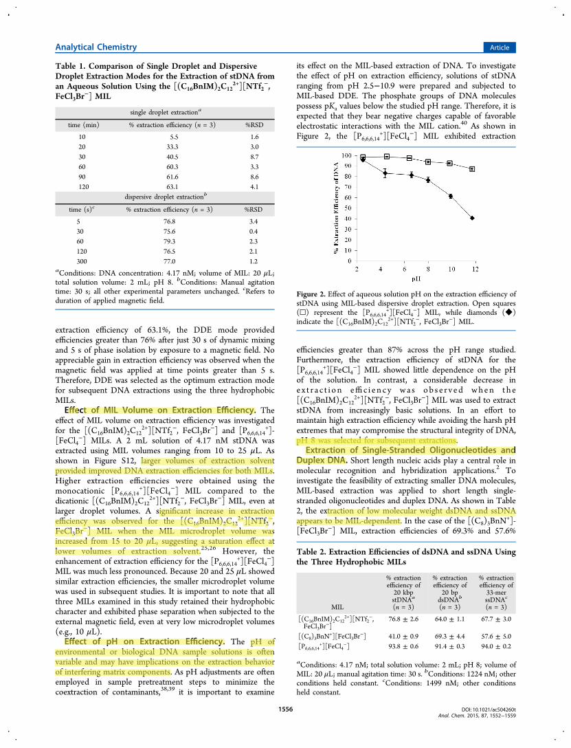
extraction efficiency of 63.1%, the DDE mode providedefficiencies greater than 76% after just 30 s of dynamic mixingand 5 s of phase isolation by exposure to a magnetic field. Noappreciable gain in extraction efficiency was observed when themagnetic field was applied at time points greater than 5 s.Therefore, DDE was selected as the optimum extraction modefor subsequent DNA extractions using the three hydrophobicMILs.Effect of MIL Volume on Extraction Efficiency. The
effect of MIL volume on extraction efficiency was investigatedfor the [(C16BnIM)2C12
2+][NTf2−, FeCl3Br
−] and [P6,6,6,14+]-
[FeCl4−] MILs. A 2 mL solution of 4.17 nM stDNA was
extracted using MIL volumes ranging from 10 to 25 μL. Asshown in Figure S12, larger volumes of extraction solventprovided improved DNA extraction efficiencies for both MILs.Higher extraction efficiencies were obtained using themonocationic [P6,6,6,14
+][FeCl4−] MIL compared to the
dicationic [(C16BnIM)2C122+][NTf2
−, FeCl3Br−] MIL, even at
larger droplet volumes. A significant increase in extractionefficiency was observed for the [(C16BnIM)2C12
2+][NTf2−,
FeCl3Br−] MIL when the MIL microdroplet volume was
increased from 15 to 20 μL, suggesting a saturation effect atlower volumes of extraction solvent.25,26 However, theenhancement of extraction efficiency for the [P6,6,6,14
+][FeCl4−]
MIL was much less pronounced. Because 20 and 25 μL showedsimilar extraction efficiencies, the smaller microdroplet volumewas used in subsequent studies. It is important to note that allthree MILs examined in this study retained their hydrophobiccharacter and exhibited phase separation when subjected to theexternal magnetic field, even at very low microdroplet volumes(e.g., 10 μL).Effect of pH on Extraction Efficiency. The pH of
environmental or biological DNA sample solutions is oftenvariable and may have implications on the extraction behaviorof interfering matrix components. As pH adjustments are oftenemployed in sample pretreatment steps to minimize thecoextraction of contaminants,38,39 it is important to examine
its effect on the MIL-based extraction of DNA. To investigatethe effect of pH on extraction efficiency, solutions of stDNAranging from pH 2.5−10.9 were prepared and subjected toMIL-based DDE. The phosphate groups of DNA moleculespossess pKa values below the studied pH range. Therefore, it isexpected that they bear negative charges capable of favorableelectrostatic interactions with the MIL cation.40 As shown inFigure 2, the [P6,6,6,14
+][FeCl4−] MIL exhibited extraction
efficiencies greater than 87% across the pH range studied.Furthermore, the extraction efficiency of stDNA for the[P6,6,6,14
+][FeCl4−] MIL showed little dependence on the pH
of the solution. In contrast, a considerable decrease ine x t r a c t i o n effi c i e n c y wa s ob s e r v e d when t h e[(C16BnIM)2C12
2+][NTf2−, FeCl3Br
−] MIL was used to extractstDNA from increasingly basic solutions. In an effort tomaintain high extraction efficiency while avoiding the harsh pHextremes that may compromise the structural integrity of DNA,pH 8 was selected for subsequent extractions.
Extraction of Single-Stranded Oligonucleotides andDuplex DNA. Short length nucleic acids play a central role inmolecular recognition and hybridization applications.2 Toinvestigate the feasibility of extracting smaller DNA molecules,MIL-based extraction was applied to short length single-stranded oligonucleotides and duplex DNA. As shown in Table2, the extraction of low molecular weight dsDNA and ssDNAappears to be MIL-dependent. In the case of the [(C8)3BnN
+]-[FeCl3Br
−] MIL, extraction efficiencies of 69.3% and 57.6%
Table 1. Comparison of Single Droplet and DispersiveDroplet Extraction Modes for the Extraction of stDNA froman Aqueous Solution Using the [(C16BnIM)2C12
2+][NTf2−,
FeCl3Br−] MIL
single droplet extractiona
time (min) % extraction efficiency (n = 3) %RSD
10 5.5 1.620 33.3 3.030 40.5 8.760 60.3 3.390 61.6 8.6120 63.1 4.1
dispersive droplet extractionb
time (s)c % extraction efficiency (n = 3) %RSD
5 76.8 3.430 75.6 0.460 79.3 2.3120 76.5 2.1300 77.0 1.2
aConditions: DNA concentration: 4.17 nM; volume of MIL: 20 μL;total solution volume: 2 mL; pH 8. bConditions: Manual agitationtime: 30 s; all other experimental parameters unchanged. cRefers toduration of applied magnetic field.
Figure 2. Effect of aqueous solution pH on the extraction efficiency ofstDNA using MIL-based dispersive droplet extraction. Open squares(□) represent the [P6,6,6,14
+][FeCl4−] MIL, while diamonds (◆)
indicate the [(C16BnIM)2C122+][NTf2
−, FeCl3Br−] MIL.
Table 2. Extraction Efficiencies of dsDNA and ssDNA Usingthe Three Hydrophobic MILs
MIL
% extractionefficiency of20 kbpstDNAa
(n = 3)
% extractionefficiency of
20 bpdsDNAb
(n = 3)
% extractionefficiency of33-merssDNAc
(n = 3)
[(C16BnIM)2C122+][NTf2
−,FeCl3Br
−]76.8 ± 2.6 64.0 ± 1.1 67.7 ± 3.0
[(C8)3BnN+][FeCl3Br
−] 41.0 ± 0.9 69.3 ± 4.4 57.6 ± 5.0[P6,6,6,14
+][FeCl4−] 93.8 ± 0.6 91.4 ± 0.3 94.0 ± 0.2
aConditions: 4.17 nM; total solution volume: 2 mL; pH 8; volume ofMIL: 20 μL; manual agitation time: 30 s. bConditions: 1224 nM; otherconditions held constant. cConditions: 1499 nM; other conditionsheld constant.
Analytical Chemistry Article
DOI: 10.1021/ac504260tAnal. Chem. 2015, 87, 1552−1559
1556

were observed for 20 bp DNA and 33-mer ssDNA, respectively.However, the same MIL produced an extraction efficiency ofonly 41.0% for stDNA indicating that it appears topreferentially extract smaller oligonucleotides. In contrast, thedicationic MIL exhibited higher extraction efficiency values forstDNA than the 20 bp dsDNA, while the [P6,6,6,14
+][FeCl4−]
MIL provided extraction efficiencies exceeding 91% for stDNA,20 bp dsDNA, and ssDNA. These preliminary data indicate thatit may be possible to design MILs that are selective forparticular sizes of oligonucleotides or duplex DNA.Extraction of DNA from a Complex Matrix. Compo-
nents of biological samples, such as metal ions and proteins, areknown to diminish the sensitivity and reproducibility of nucleicacid analysis.9 In some cases, the viability of downstreamexperiments may be compromised if the sample is notsufficiently purified from contaminants.10,11 Thus, it isimportant to determine the effect of biologically relevantimpurities on MIL-based DNA extraction. To study this, acomplex matrix was simulated through the addition of metalions or proteins (albumin) to an aqueous solution of DNA.The extraction performance of the [(C16BnIM)2C12
2+]-[NTf2
−, FeCl3Br−], [(C8)3BnN
+][FeCl3Br−], and [P6,6,6,14
+]-[FeCl4
−] MILs was evaluated for 20 kbp stDNA in the presenceof NaCl, KCl, CaCl2·2H2O, and MgCl2·6H2O. Figure S13shows that the extraction efficiency for the dicationic[(C16BnIM)2C12
2+][NTf2−, FeCl3Br
−] MIL was somewhatdiminished by the addition of the mono- and divalent metalions, in contrast to what was observed for monocationicimidazolium-based ILs.25 A very small to negligible variation inextraction efficiencies was observed for the [(C8)3BnN
+]-[FeCl3Br
−] and [P6,6,6,14+][FeCl4
−] MILs.The effect of protein on the extraction efficiency of DNA was
studied by preparing aqueous 20 kbp stDNA solutionscontaining albumin as a model protein. The extractionefficiencies of both stDNA and albumin were monitored overa pH range from 3.5 to 8. As shown in Figure 3, each of thethree studied MILs exhibited unique extraction behavior in thepresence of stDNA and albumin. Figure 3A shows that highextraction efficiencies for both stDNA and albumin wereobtained using the dicationic [(C16BnIM)2C12
2+][NTf2−,
FeCl3Br−] MIL at pH 8. Interestingly, a comparison of Figure
2 and Figure 3A reveals that the extraction efficiencies ofstDNA in the absence of albumin were similar to thoseobserved after albumin had been spiked into the aqueoussolution. However, Figure 2 and Figure 3B show that theextraction efficiency of stDNA for the [P6,6,6,14
+][FeCl4−] MIL
was decreased by 46% in the presence of albumin at pH 8. Asshown in Figure 3C, the [(C8)3BnN
+][FeCl3Br−] MIL
provided relatively lower extraction efficiencies of stDNAacross the pH range studied.With an isoelectric point of 4.6, albumin possesses an overall
negative charge at higher pH and may compete with DNA byalso engaging in electrostatic interactions with the MILcation.41 To examine this effect, the pH of the sample solutionwas lowered which resulted in a corresponding decrease in theamount of extracted albumin for the [P6,6,6,14
+][FeCl4−] and
[(C16BnIM)2C122+][NTf2
−, FeCl3Br−] MILs. Furthermore,
lowering of the sample pH significantly enhanced the extractionefficiency of stDNA for the [P6,6,6,14
+][FeCl4−] MIL. Although
these results seem to suggest that electrostatic interactionsbetween the MIL and albumin are diminished at low pH,coextraction of albumin was still observed for all three MILsinvestigated. This may be due to interactions between the
hydrophobic amino acid side chains of albumin and the longalkyl groups of the MIL cations that promote the extraction ofprotein, regardless of solution pH.41 As shown in Figure 3C,the coextraction of albumin was less pronounced whenemploying the [(C8)3BnN
+][FeCl3Br−] MIL. Although it
extracted less stDNA compared to the other two MILs, the[(C8)3BnN
+][FeCl3Br−] MIL exhibited an albumin extraction
efficiency of just 5.0% at pH 4.4, while the [(C16BnIM)2C122+]-
[NTf2−, FeCl3Br
−] and [P6,6,6,14+][FeCl4
−] MILs producedextraction efficiencies nearing 40% at the same pH. Thesefindings suggest that DNA extracted by the [(C8)3BnN
+]-[FeCl3Br
−] MIL microdroplet may have less protein con-tamination than DNA extracted by the other two MILs underthe same conditions. Though not fully understood, theextraction behavior of the three MILs investigated in thisstudy suggests that it may be possible to design MIL-basedsolvents capable of enhancing the selectivity toward DNA inthe presence of proteins.
Recovery of DNA from the MIL Extraction Phase. Therecovery of high quality DNA following an extraction step isimportant for accurate downstream analysis, especially in PCR
Figure 3. Effect of hydrophobic MIL type, pH, and albumin on theextraction efficiency of 20 kbp stDNA: (A) [(C16BnIM)2C12
2+][NTf2−,
FeCl3Br−], (B) [P6,6,6,14
+][FeCl4−], and (C) [(C8)3BnN
+][FeCl3Br−].
Diamonds (◊) represent extraction efficiency values of stDNA andcircles (○) denote extraction efficiencies of protein.
Analytical Chemistry Article
DOI: 10.1021/ac504260tAnal. Chem. 2015, 87, 1552−1559
1557

and DNA sequencing experiments.3,4 To ensure that DNAextraction performed by the MIL solvent did not alter anyportion of the DNA sequence, pDNA extracted by the[(C8)3BnN
+][FeCl3Br−] MIL was subjected to sequence
analysis. The MTAP gene sequence obtained from pDNAextracted by the MIL and the sequence of a pDNA standard areshown in Figures S14 and S15, respectively. The pDNAextracted by the MIL was shown to contain a MTAP geneidentical to the standard, indicating that the pDNA was notaltered during the MIL extraction step or that the amount ofany alterations to the integrity of the biomolecule aresufficiently low to be detected.To assess the total quantity of DNA recovered after MIL-
based DNA extraction, a 4.17 nM solution of stDNA wasextracted using the [(C8)3BnN
+][FeCl3Br−] MIL. After
dissolution of the stDNA-enriched MIL microdroplet in 3 Mpotassium acetate (pH 4.8), the sample was loaded onto silicasorbent. The sorbent was flushed with 1 mL of isopropanol,and the first fraction was collected, which contained stDNA andexcess salt. The stDNA was precipitated with cold ethanol, andthe excess salt was removed by washing the pellet with 80%ethanol. In this approach, HPLC analysis determined therecovery of stDNA from the MIL microdroplet to be 57 ± 6%.The yield of the MIL-based DDE method was 23.5 μg ofstDNA. Comparatively, a QIAamp DNA Mini Kit was capableof recovering 84 ± 5% of the stDNA from a 4.17 nM solutionwith a yield of 84.4 μg.Extraction of DNA from Bacterial Cell Lysate. To test
the applicability of the MIL-based DNA extraction method,pDNA in an E. coli cell lysate was extracted using the[(C8)3BnN
+][FeCl3Br−] MIL and subjected to PCR. This MIL
was chosen to minimize protein coextraction (vide supra). Thefollowing two methods were employed for the isolation ofDNA from the MIL extraction phase: an approach targetinggreater quantities of high purity DNA and a rapid approach forrecovering a sufficient quantity of high quality template DNAfor PCR. In order to assess whether each recovery procedurewas capable of isolating PCR-amplifiable DNA from E. coli,pDNA was extracted from a bacterial cell lysate using the[(C8)3BnN
+][FeCl3Br−] MIL and subjected to both the silica-
based and the rapid immersion method. As shown in Figure 4,the silica-based method provided a more intense PCR productband (Lane 3) than did the rapid immersion approach (Lane2). Nonetheless, immersion of the pDNA-enriched MILmicrodroplet in Tris-HCl for just 2 min was capable oftransferring sufficient pDNA for PCR amplification and visualdetection of the MTAP gene on an agarose gel. This methodhas great potential for high throughput nucleic acid analysessuch as the rapid screening of an environmental sample formicroorganisms or identification of DNA biomarkers invirtually any sample.1,5
■ CONCLUSIONSAs the demand for high-throughput nucleic acid analysiscontinues to grow, so does the need for developing DNAextraction methods capable of addressing the time-consumingbarriers encountered during traditional extraction procedures.In this study, hydrophobic MILs were employed for the firsttime as solvents for the extraction of DNA from aqueoussolution. The MIL-based method allows for rapid, highlyefficient extractions providing a DNA-enriched microdropletthat is easily manipulated in aqueous solution by application ofa magnetic field. Higher extraction efficiencies were obtained
for shorter oligonucleotides and DNA duplexes with the[(C8)3BnN
+][FeCl3Br−] MIL, while the dicationic
[(C16BnIM)2C122+][NTf2
−, FeCl3Br−] MIL afforded higher
extraction efficiencies for the much longer stDNA. MIL-basedextraction of stDNA from a complex matrix containing albuminfurther highlighted the unique extraction profiles for the MILs,revealing competitive extraction behavior for the [P6,6,6,14
+]-[FeCl4
−] MIL and less pronounced coextraction for the[(C8)3BnN
+][FeCl3Br−] MIL. These results provide a basis
for the structural customization of MILs to achieve enhancedselectivity toward a variety of DNA samples. Key to the broadapplicability of this method is the recovery of DNA from theMIL extraction phase which was determined to be 57 ± 6%.Furthermore, sequence analysis demonstrated that the DNArecovered from the MIL extraction phase was intact and thesequence unmodified. Plasmid DNA from a bacterial cell lysatewas extracted using MIL-based DDE and shown to providesufficient pDNA quantity and quality for PCR. These materialsmay serve as interesting solvent systems in many applications. Aparticularly intriguing application is in microfluidic deviceswhere their paramagnetic properties can be exploited forprecise control of sample movement.
■ ASSOCIATED CONTENT*S Supporting InformationDetailed synthetic procedure, Figures S1−S15, and two videos.This material is available free of charge via the Internet athttp://pubs.acs.org.
■ AUTHOR INFORMATIONCorresponding Author*Phone: 4195301508. Fax: 4195304033. E-mail: [email protected].
NotesThe authors declare no competing financial interest.
Figure 4. Agarose gel electrophoresis of the MTAP gene after PCRamplification from pDNA recovered from the [(C8)3BnN
+][FeCl3Br−]
MIL extraction phase. Lane 1 shows a 250−25,000 bp DNA ladder,Lane 2 represents PCR products from pDNA recovered by rapidimmersion of the DNA-enriched microdroplet in Tris-HCl, and Lane 3shows the PCR products obtained from pDNA recovered bysemiexhaustive DNA recovery.
Analytical Chemistry Article
DOI: 10.1021/ac504260tAnal. Chem. 2015, 87, 1552−1559
1558

■ ACKNOWLEDGMENTS
The authors acknowledge funding from Agilent Technologiesand the Chemical Measurement and Imaging Program at theNational Science Foundation (Grant number CHE-1413199).K.D.C. thanks the University of Toledo College of GraduateStudies for a graduate fellowship. J.L.A. and M.M.Y. thank theOffice of Undergraduate Research at The University of Toledofor funding of a summer research grant.
■ REFERENCES(1) Gormally, E.; Caboux, E.; Vineis, P.; Hainaut, P. Mutat. Res. Rev.Mutat. Res. 2007, 635, 105−117.(2) Juliano, R. L.; Ming, X.; Nakagawa, O. Acc. Chem. Res. 2012, 45,1067−1076.(3) Hofreiter, M.; Serre, D.; Poinar, H. N.; Kuch, M.; Paabo, S. Nat.Rev. Genet. 2001, 2, 353−359.(4) Willerslev, E.; Cooper, A. Proc. R. Soc. B 2005, 272, 3−16.(5) Bull, A. T.; Ward, A. C.; Goodfellow, M. Microbiol. Mol. Biol. Rev.2000, 64, 573−606.(6) Lehmann, U.; Kreipe, H. Methods 2001, 25, 409−418.(7) Jarraud, S.; Mougel, C.; Thioulouse, J.; Lina, G.; Meugnier, H.;Forey, F.; Nesme, X.; Etienne, J.; Vandenesch, F. Infect. Immun. 2002,70, 631−641.(8) Monteiro, L.; Bonnemaison, D.; Vekris, A.; Petry, K. G.; Bonnet,J.; Vidal, R.; Cabrita, J.; Megraud, F. J. Clin. Microbiol. 1997, 35, 995−998.(9) Rogacs, A.; Marshall, L. A.; Santiago, J. G. J. Chromatogr. A 2014,1335, 105−120.(10) Alaeddini, R. Forensic Sci. Int.: Genet. 2012, 6, 297−305.(11) Rossen, L.; Nørskov, P.; Holmstrøm, K.; Rasmussen, O. F. Int. J.Food Microbiol. 1992, 17, 37−45.(12) Porebski, S.; Bailey, L. G.; Baum, B. Plant Mol. Biol. Rep. 1997,15, 8−15.(13) Tretyakova, N.; Goggin, M.; Sangaraju, D.; Janis, G. Chem. Res.Toxicol. 2012, 25, 2007−2035.(14) Demeke, T.; Jenkins, G. R. Anal. Bioanal. Chem. 2010, 396,1977−1990.(15) Patel, R.; Kvach, J. T.; Mounts, P. J. Gen. Microbiol. 1986, 132,541−551.(16) Wen, J.; Guillo, C.; Ferrance, J. P.; Landers, J. P. Anal. Chem.2006, 78, 1673−1681.(17) Bernardo, G. D.; Gaudio, S. D.; Galderisi, U.; Cascino, A.;Cipollaro, M. Biotechnol. Prog. 2007, 23, 297−301.(18) Tian, H.; Huhmer, A. F. R.; Landers, J. P. Anal. Biochem. 2000,283, 175−191.(19) Poeckh, T.; Lopez, S.; Fuller, A. O.; Solomon, M. J.; Larson, R.G. Anal. Biochem. 2008, 373, 253−262.(20) Dauphin, L. A.; Stephens, K. W.; Eufinger, S. C.; Bowen, M. D.J. Appl. Microbiol. 2010, 108, 163−172.(21) Price, C. W.; Leslie, D. C.; Landers, J. P. Lab Chip 2009, 9,2484−2494.(22) Cho, Y.-K.; Lee, J.-G.; Park, J.-M.; Lee, B.-S.; Lee, Y.; Ko, C. LabChip 2007, 7, 565−573.(23) Fujita, K.; MacFarlane, D. R.; Forsyth, M. Chem. Commun. 2005,4804−4806.(24) Chandran, A.; Ghoshdastidar, D.; Senapati, S. J. Am. Chem. Soc.2012, 134, 20330−20339.(25) Wang, J.-H.; Cheng, D.-H.; Chen, X.-W.; Du, Z.; Fang, Z.-L.Anal. Chem. 2007, 79, 620−625.(26) Li, T.; Joshi, M. D.; Ronning, D. R.; Anderson, J. L. J.Chromatogr. A 2013, 1272, 8−14.(27) Leones, R.; Rodrigues, L. C.; Pawlicka, A.; Esperanca, J. M. S. S.;Silva, M. M. Electrochem. Commun. 2012, 22, 189−192.(28) Vijayaraghavan, R.; Izgorodin, A.; Ganesh, V.; Surianarayanan,M.; MacFarlane, D. R. Angew. Chem., Int. Ed. 2010, 49, 1631−1633.(29) Fregeau, C. J.; De Moors, A. Forensic Sci. Int.: Genet. 2012, 6,511−522.
(30) Bruno, J. G.; Kiel, J. L. BioTechniques 2002, 32 (178−180),182−173.(31) Hayashi, S.; Hamaguchi, H. O. Chem. Lett. 2004, 33, 1590−1591.(32) Del Sesto, R. E.; McCleskey, T. M.; Burrell, A. K.; Baker, G. A.;Thompson, J. D.; Scott, B. L.; Wilkes, J. S.; Williams, P. Chem.Commun. 2008, 447−449.(33) Mallick, B.; Balke, B.; Felser, C.; Mudring, A.-V. Angew. Chem.,Int. Ed. 2008, 47, 7635−7638.(34) Deng, N.; Li, M.; Zhao, L.; Lu, C.; de Rooy, S. L.; Warner, I. M.J. Hazard. Mater. 2011, 192, 1350−1357.(35) Nacham, O.; Clark, K. D.; Yu, H.; Anderson, J. L. Chem. Mater.2015, DOI: 10.1021/cm504202v.(36) Lee, S. H.; Ha, S. H.; Ha, S.-S.; Jin, H.-B.; You, C.-Y.; Koo, Y.-M.J. Appl. Phys. 2007, 101, 09J102−109J102−103.(37) Brown, P.; Butts, C. P.; Eastoe, J.; Padron Hernandez, E.;Machado, F. L. d. A.; de Oliveira, R. J. Chem. Commun. 2013, 49,2765−2767.(38) Purohit, H. J.; Kapley, A.; Moharikar, A. A.; Narde, G. J.Microbiol. Methods 2003, 52, 315−323.(39) Bai, J.; Baldwin, E.; Liao, H.-L.; Zhao, W.; Kostenyuk, I.; Burns,J.; Irey, M. J. Agric. Food Chem. 2013, 61, 9339−9346.(40) Wang, H.; Wang, J.; Zhang, S. Phys. Chem. Chem. Phys. 2011, 13,3906−3910.(41) Pei, Y.; Wang, J.; Wu, K.; Xuan, X.; Lu, X. Sep. Purif. Technol.2009, 64, 288−295.
Analytical Chemistry Article
DOI: 10.1021/ac504260tAnal. Chem. 2015, 87, 1552−1559
1559