Embed Size (px)
Citation preview

Fluid and Electrolytes: Balance and Disturbance
Dr. Ahmad Aqel, 2018
The University of Jordan
School of NursingBrunner & Suddarth’s Textbook of Medical-Surgical Nursing 13th ed. Chapter 13. (p 237)
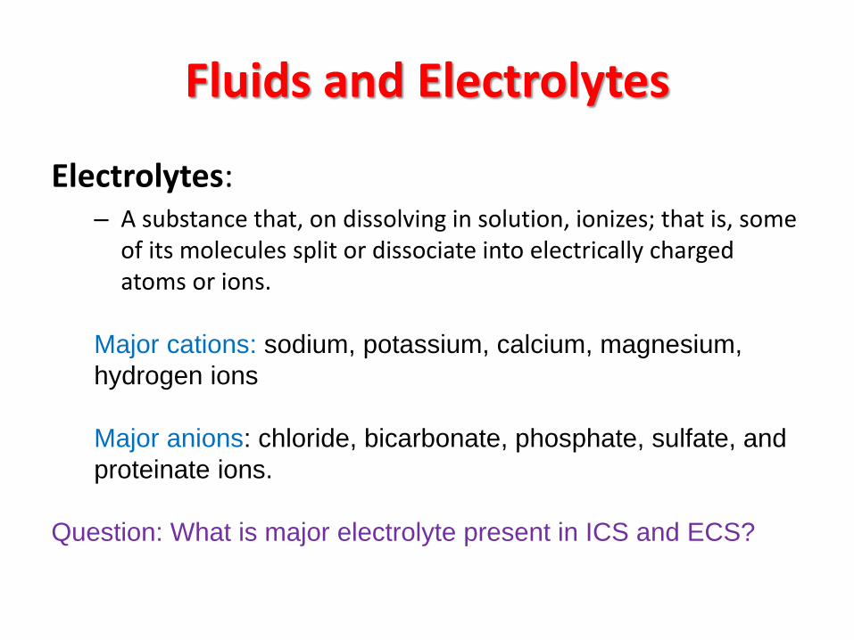
Fluids and Electrolytes
Electrolytes: – A substance that, on dissolving in solution, ionizes; that is, some
of its molecules split or dissociate into electrically charged atoms or ions.
Major cations: sodium, potassium, calcium, magnesium,
hydrogen ions
Major anions: chloride, bicarbonate, phosphate, sulfate, and
proteinate ions.
Question: What is major electrolyte present in ICS and ECS?
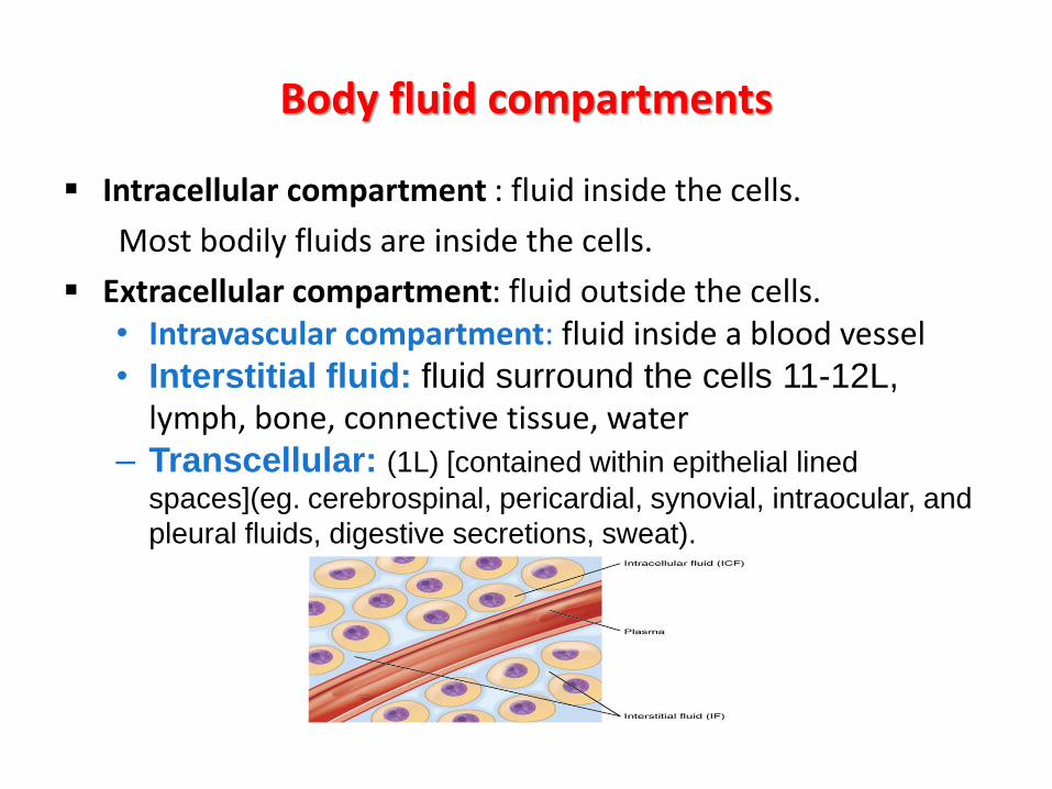
Body fluid compartments
Intracellular compartment : fluid inside the cells.
Most bodily fluids are inside the cells.
Extracellular compartment: fluid outside the cells. • Intravascular compartment: fluid inside a blood vessel • Interstitial fluid: fluid surround the cells 11-12L,
lymph, bone, connective tissue, water– Transcellular: (1L) [contained within epithelial lined
spaces](eg. cerebrospinal, pericardial, synovial, intraocular, and
pleural fluids, digestive secretions, sweat).
Third-spacing:
Is the accumulation and sequestration of trapped extracellular fluid in an actual or potential body space as a result of disease or injury.
The trapped fluid represents a volume loss and is unavailable for normal physiological processes.
Fluids trapped in body spaces such as the pericardial, pleural, peritoneal, or joint cavities; the bowel; or the abdomen, or within soft tissues after trauma or burns
Edema
• Edema is an excess accumulation of fluid in the interstitial space; it occurs as a result of alterations in oncotic pressure, hydrostatic pressure, capillary permeability, and lymphatic obstruction.
1. Localized edema occurs as a result of traumatic injury from accidents or surgery, local inflammatory processes, or burns.
2. Generalized edema also called anasarca, is an excessive accumulation of fluid in the interstitial space throughout the body and occurs as a result of conditions such as cardiac, renal, or liver failure
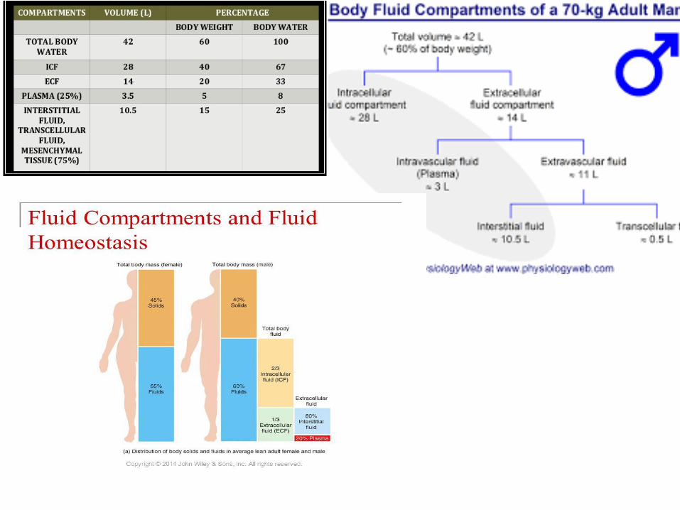
Body fluid • Total body fluid: about 60% of body weight in the adult, 55% in
the older adult, and 80% in the infant.
• Movement of body fluid through capillary walls depends on
– Diffusion: solutes move from area of higher concentration to one of lower concentration
– Osmosis: area of low solute concentration to area of high solute concentration
– Filtration: movement of water, solutes occurs from area of high hydrostatic pressure to area of low hydrostatic pressure
– Hydrostatic pressure: the pressure exerted on walls of vessels
Osmolality
• Osmolality is the concentration of fluid that affects the movement of water between fluid compartments by osmosis.
– Serum osmolality primarily reflects the concentration of sodium (blood urea nitrogen (BUN) and glucose).
– Urine osmolality is determined by urea, creatinine, and uric acid.
– serum osmolality is 280 to 300 mOsm/kg, and normal urine osmolality is 200 to 800 mOsm/kg
– Urine specific gravity measures the kidneys’ ability to excrete or conserve water. (1.010 to 1.025).
Dr. Ahmad Aqel 2016 9
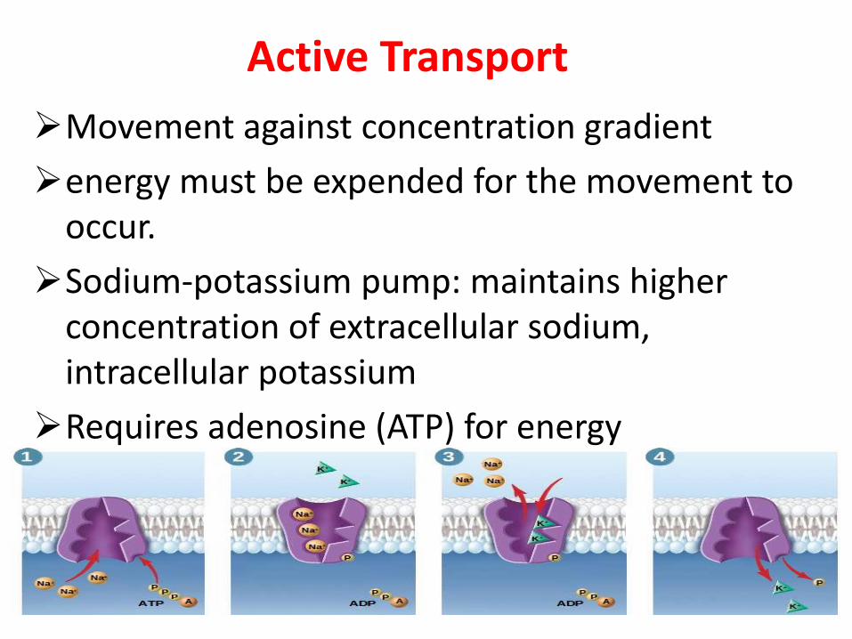
Active Transport
Movement against concentration gradient
energy must be expended for the movement to occur.
Sodium-potassium pump: maintains higher concentration of extracellular sodium, intracellular potassium
Requires adenosine (ATP) for energy
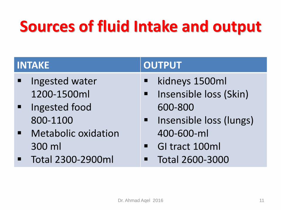
Sources of fluid Intake and output
Dr. Ahmad Aqel 2016 11
INTAKE OUTPUT
Ingested water 1200-1500ml
Ingested food 800-1100
Metabolic oxidation 300 ml
Total 2300-2900ml
kidneys 1500ml Insensible loss (Skin)
600-800 Insensible loss (lungs)
400-600-ml GI tract 100ml Total 2600-3000
Regulation of Fluid
(Kidney)
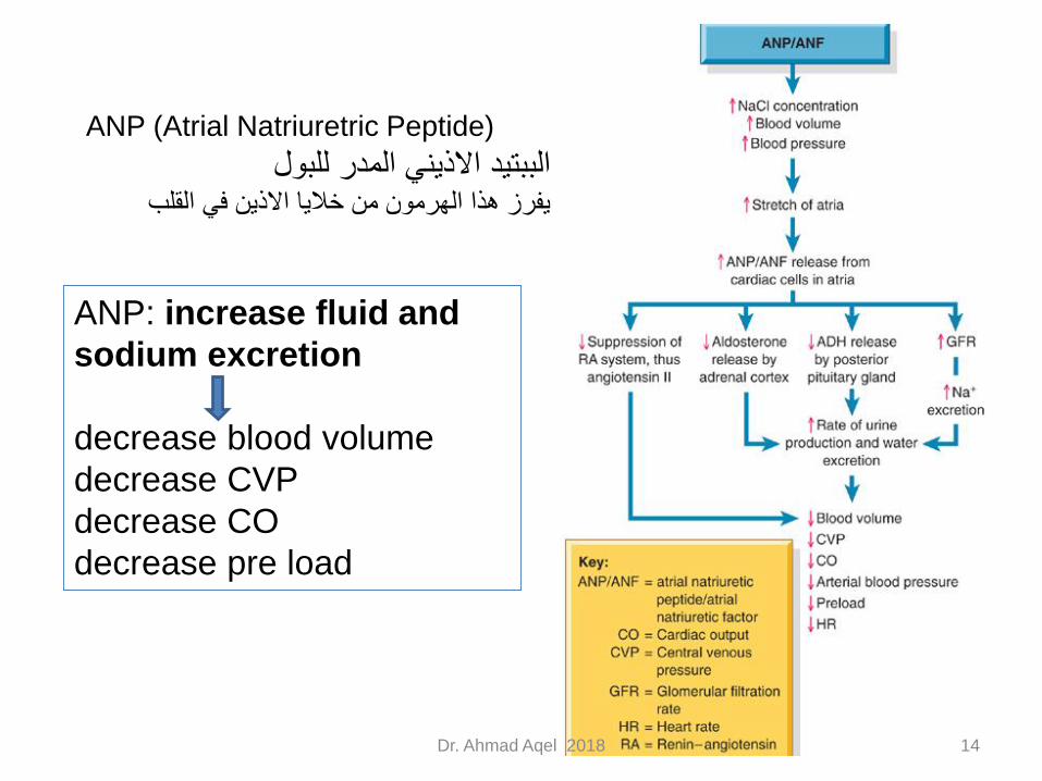
ANP (Atrial Natriuretric Peptide)
الببتيد االذيني المدر للبوليفرز هذا الهرمون من خاليا االذين في القلب
ANP: increase fluid and
sodium excretion
decrease blood volume
decrease CVP
decrease CO
decrease pre load
Dr. Ahmad Aqel 2018 14
Fluid Volume Deficit
Dehydration occurs when the fluid intake of the body is not sufficient to meet the fluid needs of the body.
The goal of treatment
• to restore fluid volume,
• replace electrolytes
• eliminate the cause of the fluid volume deficit.
Dr. Ahmad Aqel 2018 15
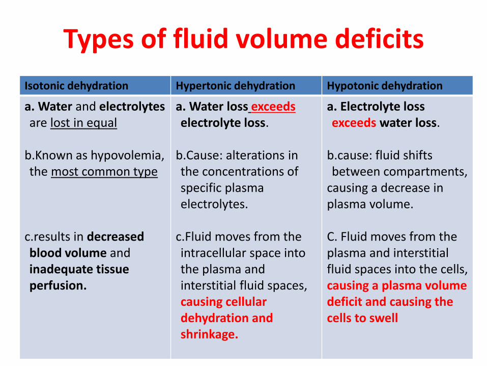
Types of fluid volume deficits
Dr. Ahmad Aqel 2018 16
Isotonic dehydration Hypertonic dehydration Hypotonic dehydration
a. Water and electrolytesare lost in equal
b.Known as hypovolemia, the most common type
c.results in decreased blood volume and inadequate tissue perfusion.
a. Water loss exceedselectrolyte loss.
b.Cause: alterations in the concentrations of specific plasma electrolytes.
c.Fluid moves from the intracellular space into the plasma and interstitial fluid spaces, causing cellular dehydration and shrinkage.
a. Electrolyte loss exceeds water loss.
b.cause: fluid shifts between compartments,
causing a decrease in plasma volume.
C. Fluid moves from the plasma and interstitial fluid spaces into the cells, causing a plasma volume deficit and causing the cells to swell
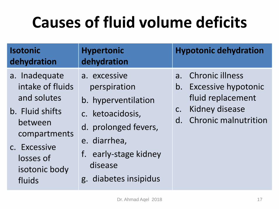
Causes of fluid volume deficits
Dr. Ahmad Aqel 2018 17
Isotonic dehydration
Hypertonic dehydration
Hypotonic dehydration
a. Inadequate intake of fluids and solutes
b. Fluid shifts between compartments
c. Excessive losses of isotonic body fluids
a. excessive perspiration
b. hyperventilation
c. ketoacidosis,
d. prolonged fevers,
e. diarrhea,
f. early-stage kidney disease
g. diabetes insipidus
a. Chronic illness b. Excessive hypotonic
fluid replacementc. Kidney disease d. Chronic malnutrition
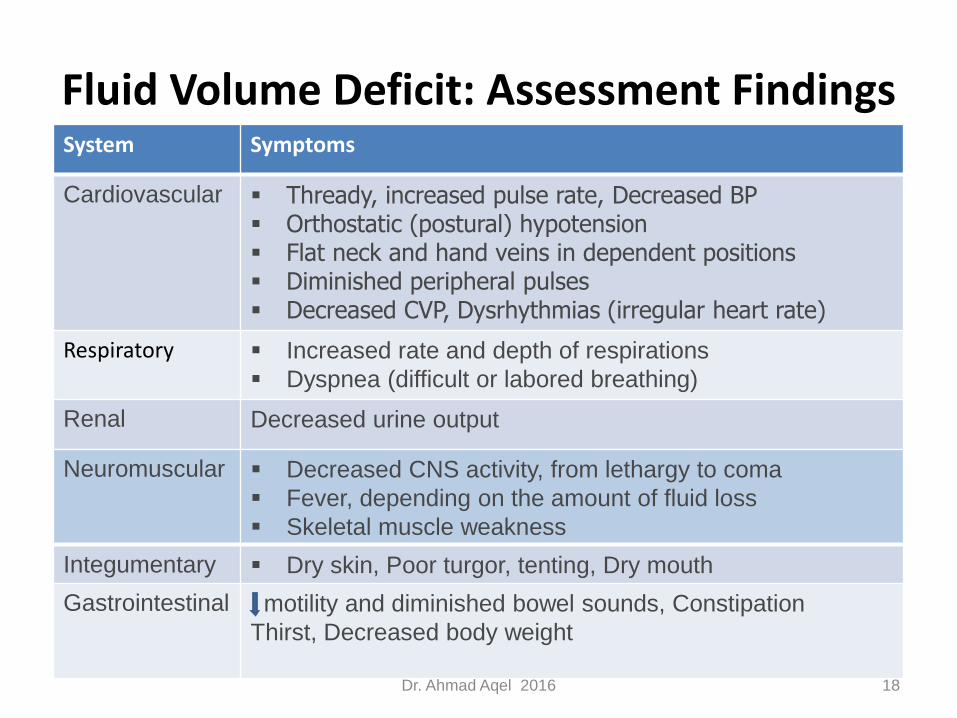
Fluid Volume Deficit: Assessment Findings
Dr. Ahmad Aqel 2016 18
System Symptoms
Cardiovascular Thready, increased pulse rate, Decreased BP Orthostatic (postural) hypotension Flat neck and hand veins in dependent positions Diminished peripheral pulses Decreased CVP, Dysrhythmias (irregular heart rate)
Respiratory Increased rate and depth of respirations
Dyspnea (difficult or labored breathing)
Renal Decreased urine output
Neuromuscular Decreased CNS activity, from lethargy to coma
Fever, depending on the amount of fluid loss
Skeletal muscle weakness
Integumentary Dry skin, Poor turgor, tenting, Dry mouth
Gastrointestinal motility and diminished bowel sounds, Constipation
Thirst, Decreased body weight
Dr. Ahmad Aqel 2016 19
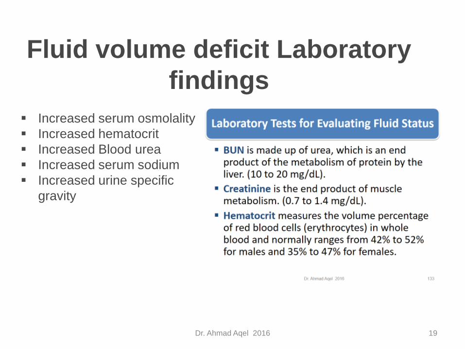
Fluid volume deficit Laboratory
findings
Increased serum osmolality
Increased hematocrit
Increased Blood urea
Increased serum sodium
Increased urine specific
gravity
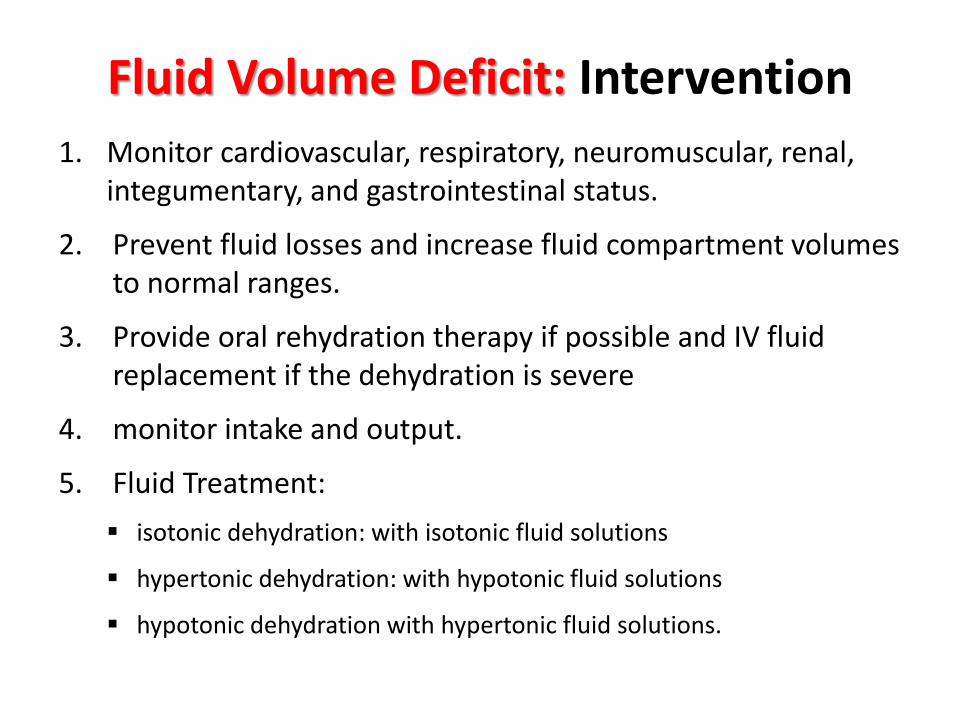
Fluid Volume Deficit: Intervention
1. Monitor cardiovascular, respiratory, neuromuscular, renal, integumentary, and gastrointestinal status.
2. Prevent fluid losses and increase fluid compartment volumes to normal ranges.
3. Provide oral rehydration therapy if possible and IV fluid replacement if the dehydration is severe
4. monitor intake and output.
5. Fluid Treatment:
isotonic dehydration: with isotonic fluid solutions
hypertonic dehydration: with hypotonic fluid solutions
hypotonic dehydration with hypertonic fluid solutions.
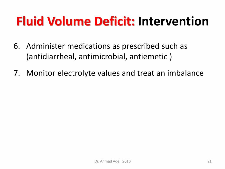
Fluid Volume Deficit: Intervention
6. Administer medications as prescribed such as (antidiarrheal, antimicrobial, antiemetic )
7. Monitor electrolyte values and treat an imbalance
Dr. Ahmad Aqel 2016 21
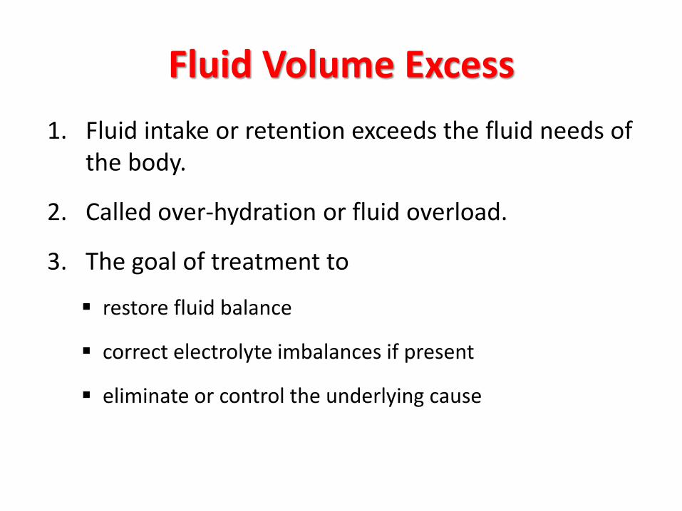
Fluid Volume Excess
1. Fluid intake or retention exceeds the fluid needs of the body.
2. Called over-hydration or fluid overload.
3. The goal of treatment to
restore fluid balance
correct electrolyte imbalances if present
eliminate or control the underlying cause
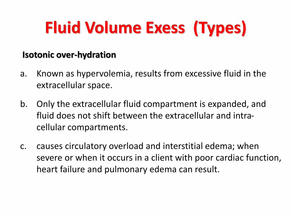
Fluid Volume Exess (Types)
Isotonic over-hydration
a. Known as hypervolemia, results from excessive fluid in the extracellular space.
b. Only the extracellular fluid compartment is expanded, and fluid does not shift between the extracellular and intra-cellular compartments.
c. causes circulatory overload and interstitial edema; when severe or when it occurs in a client with poor cardiac function, heart failure and pulmonary edema can result.
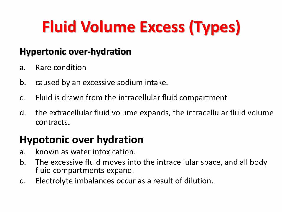
Fluid Volume Excess (Types)
Hypertonic over-hydration
a. Rare condition
b. caused by an excessive sodium intake.
c. Fluid is drawn from the intracellular fluid compartment
d. the extracellular fluid volume expands, the intracellular fluid volume contracts.
Hypotonic over hydration a. known as water intoxication. b. The excessive fluid moves into the intracellular space, and all body
fluid compartments expand. c. Electrolyte imbalances occur as a result of dilution.
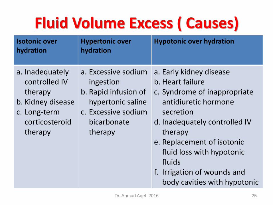
Fluid Volume Excess ( Causes)
Dr. Ahmad Aqel 2016 25
Isotonic over hydration
Hypertonic over hydration
Hypotonic over hydration
a. Inadequately controlled IV therapy
b. Kidney disease c. Long-term
corticosteroid therapy
a. Excessive sodium ingestion
b. Rapid infusion of hypertonic saline
c. Excessive sodium bicarbonate therapy
a. Early kidney disease b. Heart failure c. Syndrome of inappropriate
antidiuretic hormone secretion
d. Inadequately controlled IV therapy
e. Replacement of isotonic fluid loss with hypotonic fluids
f. Irrigation of wounds and body cavities with hypotonic
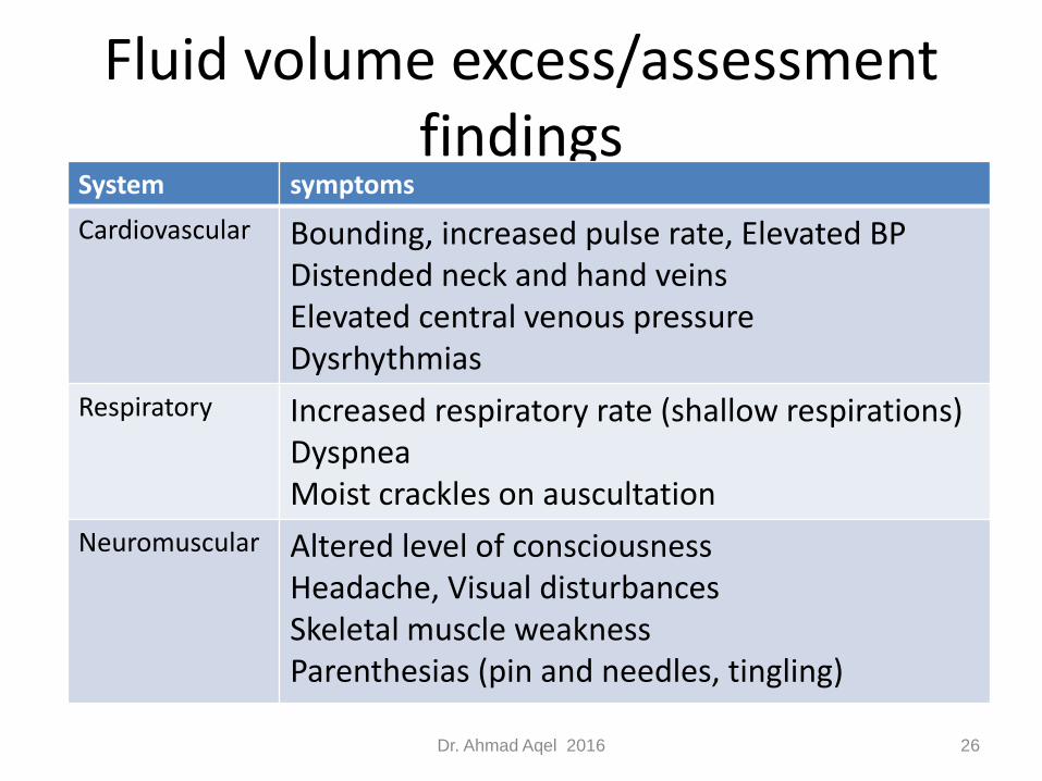
Fluid volume excess/assessment findings
Dr. Ahmad Aqel 2016 26
System symptoms
Cardiovascular Bounding, increased pulse rate, Elevated BPDistended neck and hand veinsElevated central venous pressureDysrhythmias
Respiratory Increased respiratory rate (shallow respirations)DyspneaMoist crackles on auscultation
Neuromuscular Altered level of consciousnessHeadache, Visual disturbancesSkeletal muscle weaknessParenthesias (pin and needles, tingling)
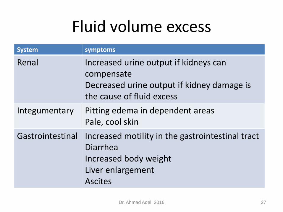
Fluid volume excess
Dr. Ahmad Aqel 2016 27
System symptoms
Renal Increased urine output if kidneys can compensateDecreased urine output if kidney damage is the cause of fluid excess
Integumentary Pitting edema in dependent areasPale, cool skin
Gastrointestinal Increased motility in the gastrointestinal tractDiarrheaIncreased body weightLiver enlargementAscites
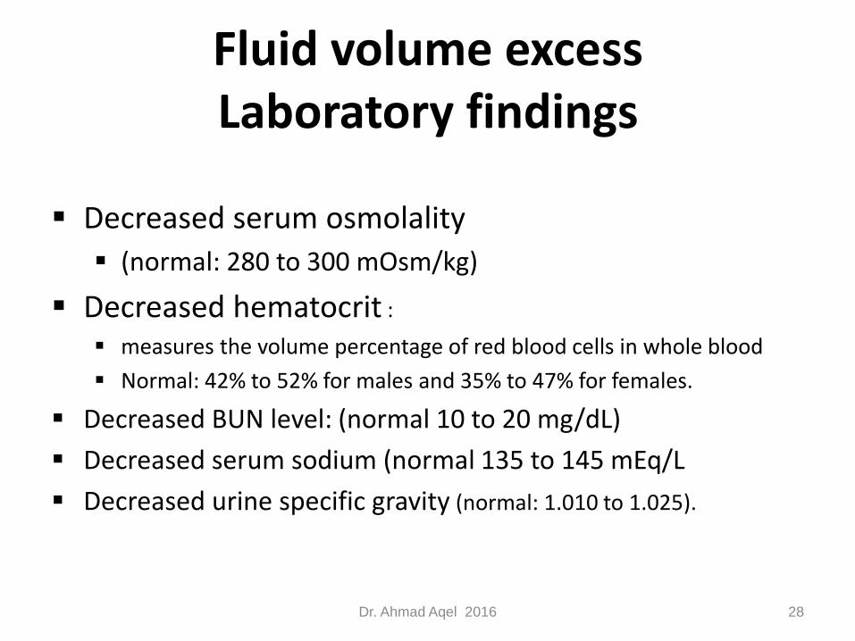
Fluid volume excessLaboratory findings
Decreased serum osmolality
(normal: 280 to 300 mOsm/kg)
Decreased hematocrit :
measures the volume percentage of red blood cells in whole blood
Normal: 42% to 52% for males and 35% to 47% for females.
Decreased BUN level: (normal 10 to 20 mg/dL)
Decreased serum sodium (normal 135 to 145 mEq/L
Decreased urine specific gravity (normal: 1.010 to 1.025).
Dr. Ahmad Aqel 2016 28
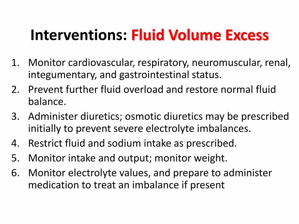
Interventions: Fluid Volume Excess
1. Monitor cardiovascular, respiratory, neuromuscular, renal, integumentary, and gastrointestinal status.
2. Prevent further fluid overload and restore normal fluid balance.
3. Administer diuretics; osmotic diuretics may be prescribed initially to prevent severe electrolyte imbalances.
4. Restrict fluid and sodium intake as prescribed.
5. Monitor intake and output; monitor weight.
6. Monitor electrolyte values, and prepare to administer medication to treat an imbalance if present
Hypokalemia
Hypokalemia is a serum potassium level lower than 3.5 mEq/L (3.5 mmol/L) (Box 8-2).
life-threatening because every body system is affected.
Causes:
1. Actual total body potassium loss. Excessive use of medications such as diuretics or corticosteroids
Increased secretion of aldosterone, such as in Cushing’s syndrome
Vomiting, diarrhea
Wound drainage, particularly gastrointestinal
Prolonged nasogastric suction
Excessive diaphoresis
Kidney disease impairing reabsorption of potassium
Hypokalemia
Causes …. continued
2. Inadequate potassium intake: Fasting; NPO status
3. Movement of K from extracellular to intracellular
In case of a. Alkalosis b. Hyperinsulinism
4. Dilution of serum potassium a. Water intoxication b. Iv therapy with potassium-deficient solution
Assessment (Tables 8-2 and 8-3)
Hypokalemia (Intervention) 1. Monitor cardiovascular, respiratory, neuromuscular,
gastrointestinal, and renal status, and place the client on a cardiac monitor.
2. Monitor electrolyte values.
3. Administer potassium supplements orally or intravenously, as prescribed.
4. Oral potassium supplements: Oral potassium supplements may cause nausea and vomiting and they
should not be taken on an empty stomach; if the client complains of abdominal pain, distention, nausea, vomiting, diarrhea, or gastrointestinal bleeding, the supplement may need to be discontinued.
Liquid potassium chloride has an unpleasant taste and should be taken with juice or another liquid.
Hypokalemia (Intervention) 5. Intravenously administered potassium (Box 8-3)
6. Institute safety measures for the client experiencing muscle weakness.
7. If the client is taking a potassium-losing diuretic, it may be discontinued; a potassium-retaining diuretic may be prescribed.
8. Instruct the client about foods that are high in potassium content (see Box 8-2)
Hypokalemia
Potassium is never
administered by IV push,
intramuscular, or
subcutaneous routes.
IV potassium is always
diluted and administered
using an infusion device!
Hyperkalemia
• Hyperkalemia is a serum potassium level that exceeds 5.0 mEq/L (5.0 mmol/L) (see Box 8-2).
• Pseudohyperkalemia: a condition that can occur due to methods of blood specimen collection and cell lysis; if an increased serum value is obtained in the absence of clinical symptoms, the specimen should be redrawn and evaluated.
Hyperkalemia ( Causes)1. Excessive potassium intake
a. Over ingestion of potassium-containing foods or medications, such as potassium chloride or salt substitutes
b. Rapid infusion of potassium-containing IV solutions
2. Decreased potassium excretiona. Potassium-retaining diuretics b. Kidney disease c. Adrenal insufficiency, such as in Addison’s disease
3. Movement of potassium from the intracellular fluid to the extracellular fluid
a. Tissue damage b. Acidosis c. Hyperuricemia d. Hypercatabolis
Hyperkalemia ( Causes)
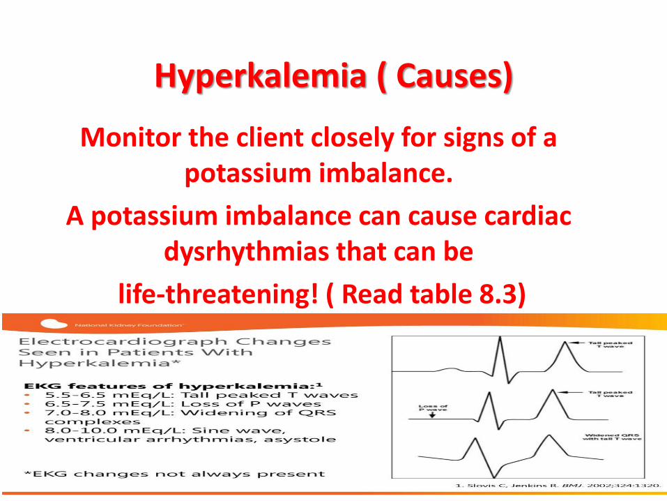
Monitor the client closely for signs of a potassium imbalance.
A potassium imbalance can cause cardiac dysrhythmias that can be
life-threatening! ( Read table 8.3)
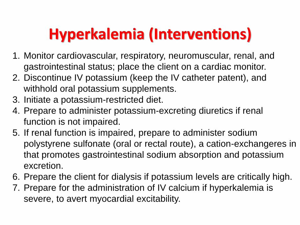
Hyperkalemia (Interventions)1. Monitor cardiovascular, respiratory, neuromuscular, renal, and
gastrointestinal status; place the client on a cardiac monitor.
2. Discontinue IV potassium (keep the IV catheter patent), and
withhold oral potassium supplements.
3. Initiate a potassium-restricted diet.
4. Prepare to administer potassium-excreting diuretics if renal
function is not impaired.
5. If renal function is impaired, prepare to administer sodium
polystyrene sulfonate (oral or rectal route), a cation-exchangeres in
that promotes gastrointestinal sodium absorption and potassium
excretion.
6. Prepare the client for dialysis if potassium levels are critically high.
7. Prepare for the administration of IV calcium if hyperkalemia is
severe, to avert myocardial excitability.
Hyperkalemia (Interventions)
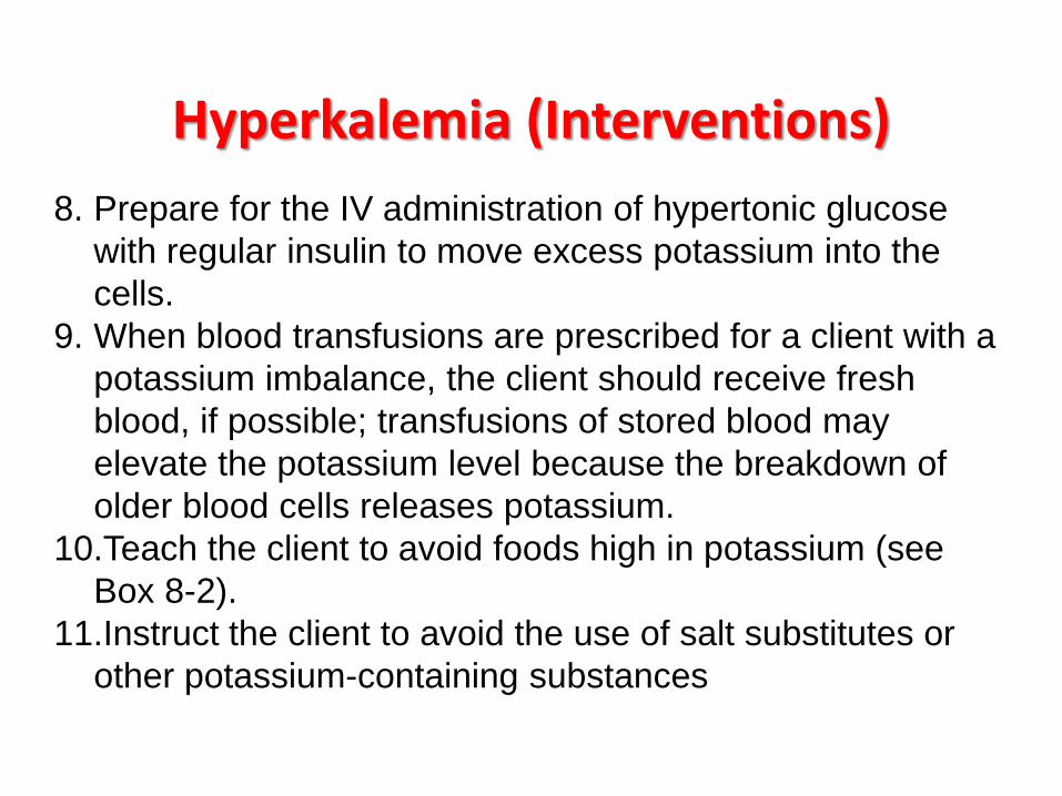
8. Prepare for the IV administration of hypertonic glucose
with regular insulin to move excess potassium into the
cells.
9. When blood transfusions are prescribed for a client with a
potassium imbalance, the client should receive fresh
blood, if possible; transfusions of stored blood may
elevate the potassium level because the breakdown of
older blood cells releases potassium.
10.Teach the client to avoid foods high in potassium (see
Box 8-2).
11.Instruct the client to avoid the use of salt substitutes or
other potassium-containing substances
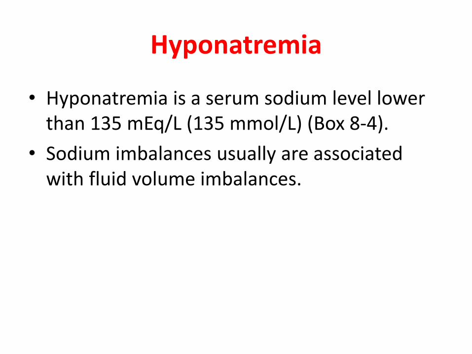
Hyponatremia
• Hyponatremia is a serum sodium level lower than 135 mEq/L (135 mmol/L) (Box 8-4).
• Sodium imbalances usually are associated with fluid volume imbalances.
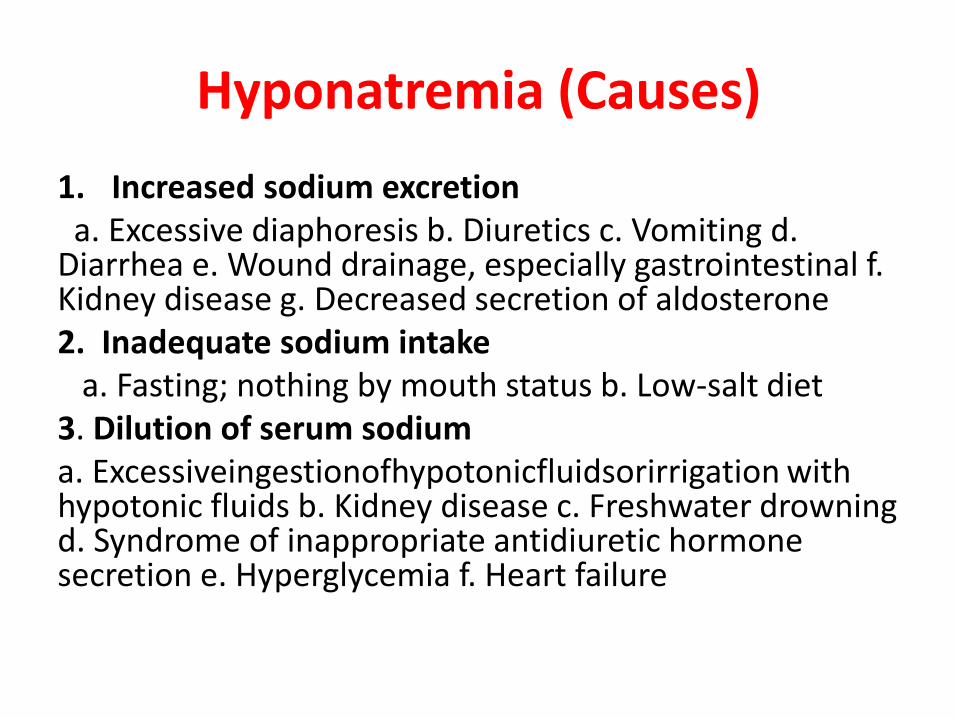
Hyponatremia (Causes)
1. Increased sodium excretion a. Excessive diaphoresis b. Diuretics c. Vomiting d.
Diarrhea e. Wound drainage, especially gastrointestinal f. Kidney disease g. Decreased secretion of aldosterone 2. Inadequate sodium intake
a. Fasting; nothing by mouth status b. Low-salt diet 3. Dilution of serum sodium a. Excessiveingestionofhypotonicfluidsorirrigation with hypotonic fluids b. Kidney disease c. Freshwater drowning d. Syndrome of inappropriate antidiuretic hormone secretion e. Hyperglycemia f. Heart failure
Hyponatremia (Intervention)Assessment (Table 8-4)
1. Monitor cardiovascular, respiratory, neuromuscular, cerebral, renal, and gastrointestinal status.
2. If hyponatremia is accompanied by a fluid volume deficit (hypovolemia), IV sodium chloride infusions are administered to restore sodium content and fluid volume.
3. If hyponatremia is accompanied by fluid volume excess(hypervolemia),osmoticdiureticsmaybe prescribed to promote the excretion of water rather than sodium.
4. If caused by inappropriate or excessive secretion of antidiuretic hormone, medications that antagonize antidiuretic hormone may be administered.
Hyponatremia (Intervention)Assessment (Table 8-4)
5. Instruct the client to increase oral sodium intake as prescribed and inform the client about the foods to include in the diet (see Box 8-4).
6. If the client is taking lithium, monitor the lithium level, because hypornatremia can cause diminished lithium excretion, resulting in toxicity.
Common Food Sources
frankfurters, lunch meat Butter, cheese
Canned food Ketchup, mustard Milk
Processed food Snack foods Soy sauce Table
salt
Hypernatremia (Description)
• Description: Hypernatremia is a serum sodium level that exceeds145 mEq/L(145 mmol/L)(seeBox8-4).
1. Decreased sodium excretion a. Corticosteroids b. Cushing’s syndrome c. Kidney disease d. Hyperaldosteronism
2. Increased sodium intake: Excessive oral sodium ingestion or excessive administration of sodium-containing IV fluids
3. Decreased water intake: Fasting; nothing by mouth status
4. Increased water loss: Increased rate of metabolism, fever, hyperventilation, infection, excessive diaphoresis, watery diarrhea, diabetes insipidus
Hypernatremia (causes)
Interventions
1. Monitor cardiovascular, respiratory, neuromuscular, cerebral, renal, and integumentary status.
2. If the cause is fluid loss, prepare to administer IV infusions.
3. If the cause is inadequate renal excretion of sodium, prepare to administer diuretics that promote sodium loss. 4. Restrict sodium and fluid intake as prescribed (see Box 8-4)
Hypernatremia
Assessment (see Table 8-4)
Hypocalcemia
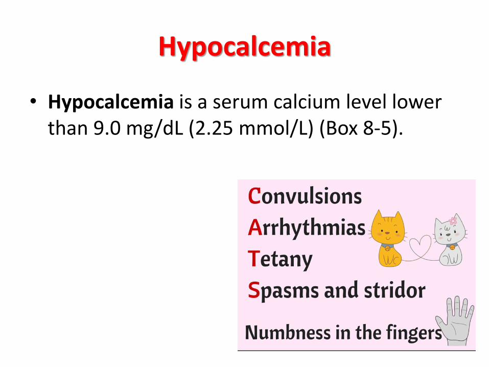
• Hypocalcemia is a serum calcium level lower than 9.0 mg/dL (2.25 mmol/L) (Box 8-5).
Hypocalcemia (Causes)
1. Inhibition of calcium absorption from the gastrointestinal tract
a. Inadequate oral intake of calcium b. Lactose intolerance c. Malabsorption syndromes such as celiac sprue or Crohn’s disease d. Inadequate intake of vitamin D e. End-stage kidney disease
2. Increased calcium excretion a. Kidney disease, polyuric phase b. Diarrhea c. Steatorrhea d. Wound drainage, especially gastrointestinal
Hypocalcemia (Causes)
3. Conditions that decrease the ionized fraction of calcium a. Hyperproteinemia
b. Alkalosis
c. Medications such as calcium chelators or binders
d. Acute pancreatitis
e. Hyperphosphatemia
f. Immobility
g. Removal or destruction of the parathyroid glands
Hypocalcemia
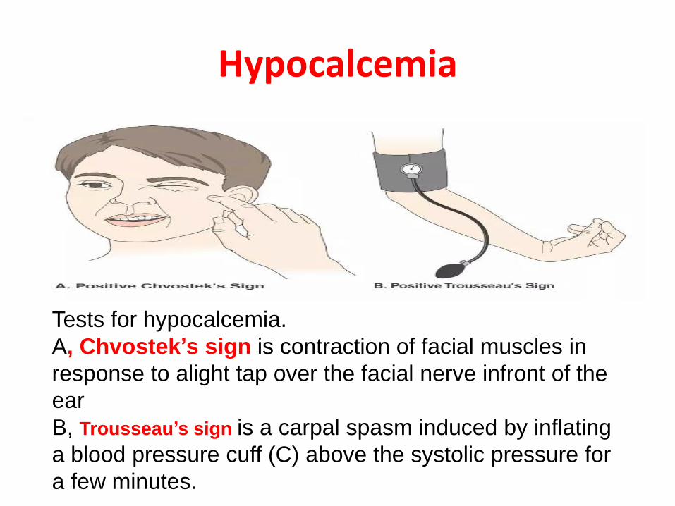
Tests for hypocalcemia.
A, Chvostek’s sign is contraction of facial muscles in
response to alight tap over the facial nerve infront of the
ear
B, Trousseau’s sign is a carpal spasm induced by inflating
a blood pressure cuff (C) above the systolic pressure for
a few minutes.
Hypocalcemia (Intervention )
1. Monitor cardiovascular, respiratory, neuromuscular, and gastrointestinal status; place the client on a cardiac monitor.
2. Administer calcium supplements orally or calcium intravenously.
3. When administering calcium intravenously, warm the injection solution to body temperature before administration and administer slowly; monitor for electrocardiographic changes, observe for infiltration, and monitor for hypercalcemia.
4. Administer medications that increase calcium absorptiona. Aluminum hydroxide reduces phosphorus levels, causing the
counter effect of increasing calcium levels. b. Vitamin D aids in the absorption of calcium from the intestinal
tract.
Hypocalcaemia (Intervention )
5. Provide a quiet environment to reduce environmental stimuli. 6. Initiate seizure precautions. 7. Move the client carefully, and monitor for signs of a pathological fracture. 8. Keep 10% calcium gluconate available for treatment of acute calcium deficit Monitor cardiovascular, respiratory, neuromuscular, and gastrointestinal status; place the client on a cardiac monitor. Instruct the client to consume foods high in calcium (see Box 8-5).
Hypercalcemia (Description)
Hypercalcemia is a serum calcium level that exceeds10.5 mg/dL(2.75 mmol/L)(seeBox8-5).
Hypocalcemia (Causes)1. Increased calcium absorption
a. Excessive oral intake of calcium b. Excessive oral intake of vitamin D
2. Decreased calcium excretion a. Kidney disease b. Use of thiazide diuretics
3. Increased bone resorption of calcium a. Hyperparathyroidism b. Hyperthyroidism c. Malignancy (bone destruction from metastatic tumors) d. d. Immobility e. Use of glucocorticoids
4. Hemoconcentration a. Dehydration b. Use of lithium c. Adrenal insufficiency
Hypercalcemia (Interventions)
Interventions
1. Monitor cardiovascular, respiratory, neuromuscular, renal, and gastrointestinal status; place the client on a cardiac monitor.
2. Discontinue IV infusions of solutions containing calcium and oral medications containing calcium or vitamin D.
3. Thiazide diuretics may be discontinued and replaced with diuretics that enhance the excretion of calcium.
4. Administer medications as prescribed that inhibit calcium resorption from the bone, such as phosphorus, calcitonin, bisphosphonates, and prostaglandin synthesis inhibitors (acetylsalicylic acid, nonsteroidal antiinflammatorymedications).
Assessment (see Tables 8-3 and 8-5)
Hypercalcemia (Interventions)Interventions
5. Prepare the client with severe hypercalcemia for dialysis if medications fail to reduce the serum calcium level.
6. Move the client carefully and monitor for signs of a pathological fracture.
7. Monitor for flank or abdominal pain, and strain the urine to check for the presence of urinary stones.
8. Instruct the client to avoid foods high in calcium (see Box 8-5)
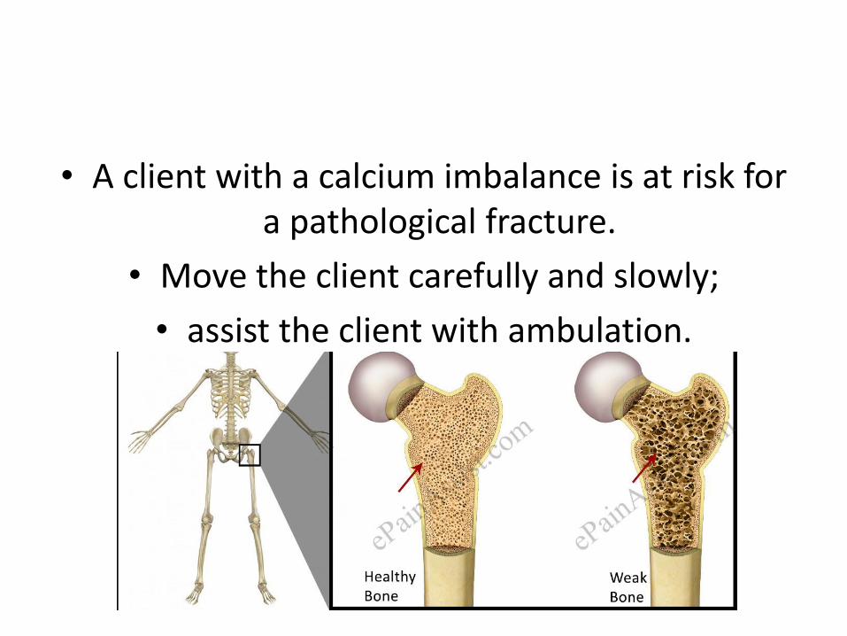
• A client with a calcium imbalance is at risk for a pathological fracture.
• Move the client carefully and slowly;
• assist the client with ambulation.